Note: Descriptions are shown in the official language in which they were submitted.
CA 02863685 2014-08-01
WO 2013/114204 PCT/IB2013/000332
A METHOD OF USING ADENOSINE AND DIPYRIDAMOLE
FOR PHARMACOLOGIC STRESS TESTING, WITH SPECIFIC COMPOSITIONS,
UNIT DOSAGE FORMS AND KITS
BACKGROUND
[0001] Various compositions and methods for functional assessment of
myocardial perfusion
have been described but are not optimal.
SUMMARY
[0002] Described herein are exemplary methods, dosages, compositions,
concentrations, unit
dosage forms (UDFs) and kits that allow adenosine and dipyridamole to be used
in
combination so as to meet the elements defined in the following paragraphs.
[0003] In an exemplary method of performing cardiac diagnosis, the method
further
comprises administering to a patient in need thereof a fixed dose of
dipyridamole and a fixed
dose of adenosine, wherein the fixed doses of dipyridamole and adenosine are
independent of
patient weight, and both dipyridamole and adenosine are parenterally injected
as a slow
bolus.
[0004] In an exemplary method of performing cardiac diagnosis, the method
described above
is used to perform hemodynamic or cardio-electric measurements under stress
conditions
using transthoracic doppler-echocardiography or transesophageal doppler
echography and/or
electrocardiography techniques.
[0005] In an exemplary embodiment, the method described above comprises
administering
to a patient in need thereof a fixed dose of dipyridamole and a fixed dose of
adenosine and
also administering to said patient an imaging agent in the form of a
radionuclide, a
radiopharmaceutical or a contrast agent using either nuclear techniques, such
as SPECT or
PET-scanning, two and three dimensional ultrasound techniques, such as
Echography, and
real-time myocardial contrast echocardiography (MCE), nuclear magnetic
resonance (NMR)
imaging or CT-scan imaging techniques.
[0006] In an exemplary method of reducing radiation and total study time to a
patient
undergoing cardiac diagnosis using a nuclear technique, such as single photon
emission
-1-
CA 02863685 2014-08-01
WO 2013/114204 PCT/IB2013/000332
computed tomography (SPECT), especially new SPECT technologies performed with
ultrafast gamma-cameras, as described below, the fixed doses of dipyridamole
and adenosine
are administered as a bolus over a total period of about 30 seconds, the
imaging agent is then
administered over a period of less than about 10 seconds, and imaging of the
patient is
performed for about 4 minutes to about 5 minutes after the administration of
the imaging
agent.
[0007] In an exemplary method of reducing radiation exposure and total study
time to a
patient undergoing cardiac diagnosis using a nuclear technique such as single
photon
emission computed tomography (SPECT), dipyridamole and adenosine are
administered as a
bolus over a total period of about 30 seconds, the imaging agent is then
administered over a
period of less than about 10 seconds, imaging of the patient is performed for
about 3 minutes
after the administration of the imaging agent; a second bolus administration
of dipyridamole
and adenosine at a reduced dosage is made about 3 minutes to about 4 minutes
after the
initial administration, and imaging of the patient is performed for about 2
minutes after the
second administration of dipyridamole and adenosine.
[0008] In an exemplary method of reducing radiation exposure and the duration
of the full
stress-rest or rest-stress protocol to a patient undergoing cardiac diagnosis
using a nuclear
technique such as single photon emission computed tomography (SPECT),
especially new
SPECT technologies performed with ultrafast gamma-cameras as described below,
the
method comprises administering parenterally to a patient in need thereof a
fixed dose of
dipyridamole and a fixed dose of adenosine injected as a slow bolus and
administering to
said patient a single reduced, but effective, amount of a radiopharmaceutical
called boronic
acid teboroxime-Technetium 99 (BATO-Tc99m) or a derivative thereof, acting as
a
myocardial radio-labeled imaging agent, wherein the amount of the imaging
agent results in
reduced radiation exposure for the patient.
[0009] In an exemplary method of reducing radiation exposure and total study
time to a
patient undergoing cardiac diagnosis using a nuclear technique such as single
photon
emission computed tomography (SPECT) ), especially new SPECT technologies
performed
with ultrafast gamma-cameras as described below, the method comprises
administering to a
-2-
CA 02863685 2014-08-01
WO 2013/114204 PCT/IB2013/000332
patient in need thereof a single amount of BATO allowing for the
administration of no more
than about 5 to about 15 millicuries (mCi) of Tc99m while the patient is under
rest
conditions, acquiring images of the patient for a period of about 2.5 minutes
to about 4
minutes after administration of the imaging agent, administering dipyridamole
and adenosine
as a slow bolus (stress phase) after about 2.5 minutes to about 4 minutes, and
acquiring
images of the patient for an additional period of up to about 2 to about 4
minutes, preferably
about 2.5 minutes to about 3.5 minutes, after administration of dipyridamole
and adenosine.
[0010] An exemplary composition, in dry or fluid form, comprises adenosine and
dipyridamole in a unit dosage form, with the unit dosage form being suitable
for bolus
administration for effecting coronary vasodilation for cardiac diagnosis in a
patient in need
thereof, where the doses of the dipyridamole and the adenosine are fixed and
can be
administered independent of the patient's weight, and when administered to a
patient
undergoing cardiac diagnosis using a nuclear technique, allow for a reduction
in the amount
of the radiopharmaceutical administered to the patient.
[0011] In an exemplary method of manufacturing a dry or fluid composition in a
unit dosage
form, the method comprises combining a fixed dose of adenosine and a fixed
dose of
dipyridamole in dry or fluid form, into the unit dosage form, such that the
unit dosage form is
suitable for bolus administration for effecting coronary vasodilation for
cardiac diagnosis in a
patient in need thereof, and wherein the fixed doses of adenosine and
dipyridamole allow for
the adenosine and dipyridamole to be administered independent of the patient's
weight, and
when administered to a patient undergoing cardiac diagnosis using a nuclear
technique, allow
for a reduction in the amount of the radiopharmaccutical agent administered to
the patient.
[0012] An exemplary composition, in dry or fluid form, comprises two unit
dosage forms,
with a first unit dosage form comprising a fixed dose of adenosine and a
second unit dosage
form comprising a fixed dose of dipyridamole, each of said unit dosage forms
being suitable
for bolus administration for effecting coronary vasodilation for cardiac
diagnosis in a patient
in need thereof, and wherein the fixed doses of adenosine and dipyridamole
allow for the
adenosine and dipyridamole to be administered independent of the patient's
weight, and
-3-
CA 02863685 2014-08-01
WO 2013/114204 PCT/IB2013/000332
when administered to a patient undergoing cardiac diagnosis using a nuclear
technique, allow
for a reduction in the amount of the radiopharmaceutical administered to the
patient.
[0013] In an exemplary method of manufacturing a dry or fluid composition, the
method
comprises combining a first unit dosage form comprising a fixed dose of
adenosine and a
second unit dosage form comprising a fixed dose of dipyridamole, each of the
unit dosage
fauns being suitable for bolus administration for effecting coronary
vasodilation for cardiac
diagnosis in a patient in need thereof, and wherein the fixed doses of
adenosine and
dipyridamole allow for the adenosine and dipyridamole to be administered
independent of
the patient's weight, and when administered to a patient undergoing cardiac
diagnosis using a
nuclear technique, allow for a reduction in the amount of the
radiopharmaceutical
administered to the patient.
[0014] An exemplary kit comprises at least one unit dosage form of
dipyridamole and at least
one unit dosage form of adenosine, wherein each of the unit dosage forms is
suitable for
bolus administration for effecting coronary vasodilation for cardiac diagnosis
in a patient in
need thereof and wherein the fixed dose of adenosine and the fixed dose of
dipyridamole
allow for the adenosine and dipyridamole to be administered independent of the
patient's
weight, and when administered to a patient undergoing cardiac diagnosis, allow
for a
reduction in the amount of imaging agent administered to the patient, and
wherein the kit
further comprises at least one of a connector, a diluent, an extension set and
a venous line.
[0015] An exemplary kit can also comprise all the units and elements described
in the kit
above with in addition a unit dosage form of BATO, or a BATO derivative,
allowing for the
administration of about 5 mCi to about 15 mCi of Tc99m. The unit dosage form
of BATO,
or a BATO derivative, and the units of adenosine or dipyridamole or both can
be also
copackaged.
BRIEF DESCRIPTION OF THE TABLES
[0016] Table 1 describes in the same patient serving as an example, the effect
of different
combination dose-ratios on mean and peak blood velocities (related to blood
flow) measured
in resting basal and stress conditions ( in the left descending coronary
artery), as well as
tolerance: measurements were made before infusion of adenosine 140 p g/kg/min,
during a 2
-4-
CA 02863685 2014-08-01
WO 2013/114204
PCT/IB2013/000332
minute infusion of standard adenosine, before injection of the combination
bolus (after a 5
mm interval at baseline) and after injection of the combination
administered as a 30 second
IV bolus.
100171 Table 2 describes the mean blood velocities (related to blood flow) in
the left
descending coronary artery in different series of 3 patients each tested at
the same dose ratio
of 4 mg adenosine:12 mg dipyridamole in resting basal and stress conditions.
Measurements
were made before infusion of adenosine 140 pg/kgfrnin, during infusion of
standard
adenosine for about 2 minutes, before injection of the combination bolus
(after a 5 min
interval at baseline) and after injection of the combination given as an IV 30
second bolus.
100181 Table 3 describes the effects of dose ratios of 2.4 mg adenosine: 12 mg
dipyridamole
and of 2 ing adenosine: 12 mg dipyridainole versus standard adenosine in a
series of 12
patients .
BRIEF DESCRIPTION OF THE DRAWINGS
100191 Fig. 1 illustrates the steps involved in study protocol 1, where
administration of the
stressor is immediately followed by the imaging agent and continuous imaging
over a period
of about 5 minutes allows for obtaining information under both stress and
resting conditions.
100201 Fig. 2 illustrates the steps involved in study protocol 2, where two
doses of the
stressor are administered.
100211 Fig. 3 illustrates the steps involved in study protocol 3, where a
single injection of a
complex of BATO, or a BATO derivative, with a radioisotope is injected under
rest
conditions, followed after about 2.5 to about 3 minutes by the injection of
the stressor and
allows for obtaining information under both rest and stress conditions..
100221 Fig. 4 illustrates changes in coronary conductance over time after
administration of
regadenoson, binodenoson, CGS-21680 (apadenoson-like product), and adenosine.
-5-
RECTIFIED SHEET (RULE 91)
CA 02863685 2014-08-01
WO 2013/114204 PCT/IB2013/000332
DETAILED DESCRIPTION
5.1 Overview: Herein are described methods of use, doses, dose ratios, unit
dosage forms,
kits and compositions.
[0023] Functional assessment of myocardial perfusion, also called "coronary
reserve
assessment," can lead to the detection of myocardial ischemic defects or
coronary blood flow
insufficiencies, and is important in guiding therapeutic decisions in the care
of patients with
coronary artery disease. Ischemia is a condition in which blood flow (and thus
oxygen) is
restricted to a part of the body, often with resultant damage or dysfunction
of tissue.
Myocardial ischemia, also called cardiac ischemia, occurs when blood flow to
the heart
muscle is decreased by a partial or complete blockage of an artery that
carries blood to the
heart. This reduces the ability of the heart to pump efficiently. The decrease
in blood flow
reduces the supply of oxygen to the heart. A sudden, severe blockage of a
coronary artery
may lead to a heart attack (myocardial infarction). Myocardial ischemia may
also cause
serious abnormal heart rhythms. While ischemia is well-correlated to prognosis
and to the
risks of disease progression, the number of critical stenoses (the abnormal
narrowing in a
blood vessel) and the degree of narrowing, as detected by coronary
angiography, does not
appear to correlate with the patient's symptoms, the motion of the muscular
walls of the
heart, the performance of the heart, the blood flow through the coronary
arteries, the patient's
prognosis, or with the results of coronary artery bypass surgery. As a
consequence, more and
more functional tests are now performed during cardiac check-ups (1,2)
[0024] Most current tests for exploring myocardial ischemia status are non-
invasive nuclear
perfusion imaging methodologies using single photon emission computed
tomography
(SPECT), projecting a three-dimensional image, with thallium and technetium as
the most
currently used isotopes. A class of compounds known as BATOs (boronic acid
adducts of
technetium) has been developed for use in myocardial imaging. Boronic acid
teboroxime,
also referred to as "BATO", had been approved by the FDA, but has not reached
the market
and is no longer available for clinical use because current gamma camera
detectors are not
sensitive enough to detect its signal. However, the arrival of new ultrafast
gamma-cameras
allow for improved detectability. The development of new semiconductors for
gamma
-6-
CA 02863685 2014-08-01
WO 2013/114204 PCT/IB2013/000332
photon detection, and in particular the use of cadmium telluride zinc (CZT),
is an important
breakthrough. It has given rise to the emergence of stationary gamma ray multi-
detectors
made of CZT modules and to the creation of cameras that reduce SPECT-MP
imaging
acquisition time. Other ultrafast camera systems are expected to become
commercially
available (e.g. the avalanche photodiode system using a silicon semiconductor
device).
Positron emission tomography (PET-scan) using rubidium 82 was recently
approved by the
FDA for this indication, and is gaining recognition for providing improved
images while
requiring less exposure to radiation. Nuclear magnetic resonance imaging (MRI)
is another
technique that may be used to explore myocardial perfusion. Among ultrasound
techniques,
semi-invasive transeosophageal Doppler Echography is useful to study the
motion of
ventricular walls, and non-invasive transthoracic Doppler echocardiography is
an easy and
non-invasive technique for measurement of coronary flow reserve. Coronary flow
reserve is
the maximum increase in blood flow through the coronary arteries above the
normal resting
volume. Coronary reserve is the difference between the supply, or flow of
blood, in a
normal, autoregulated state, and the supply available with maximal
vasodilation. The term
-coronary reserve dysfunction" relates to a number of conditions in which the
coronary
reserve is decreased relative to normal values. Another technology, myocardial
contrast
echography (MCE) uses agents detectable by ultrasound to study myocardium
perfusion and
heart function in real time with a single test. Yet another imaging
technology, X-ray
computed tomography (CT) scanning, has been used in studying myocardial
perfusion
possibly coupled with coronary angiograms. High speed CT scanners, such as
ultrafast CT
scanners, are capable of taking multiple images of the heart within the time
of a single
heartbeat. Recently, this technology has been further improved using dual-
energy imaging
leading to the development of ultra high speed CT scanners.
[0025] These functional tests typically require that the patient's heart be
"stressed," either
through controlled exercise or by pharmacologic means or even both. With
technologies
such as PET-scanning, MRI or CT scanning, pharmacological agents are the most
convenient
option to induce stress. With the SPECT technique the two options are possible
but there are
numerous reasons for choosing pharmacologic stress testing, rather than
exercise-induced
stress testing. Around thirty percent of patients cannot exercise adequately
(due to, for
-7-
CA 02863685 2014-08-01
WO 2013/114204 PCT/IB2013/000332
example, peripheral artery disease, left bundle block, aortic aneurysm,
obesity or the use of a
pace maker) or need special imaging studies to answer specific questions,
including whether
those with stable angina should be stented, to determine ventricular reserve,
to assess
responsiveness in assessing patients for valvular surgery, testing of the
effectiveness of a
therapeutic program in relieving ischemic distress, estimation of the risk of
coronary events
and cardiac mortality. However, the use of exercise in combination with a
stressing agent is
becoming more commonly practiced, in particular during SPECT studies.
[0026] Adenosine and dipyridamole are two coronary vasodilators used for
pharmacologic
stress testing (3' 4). Both of these compounds produce near-maximal coronary
dilation
through the activity of adenosine. By dilating normal vessels to a greater
extent than
diseased vessels, these compounds establish a shunt or "myocardial steal" that
produces
different degrees of increase in flow in healthy versus diseased arteries in
patients with
coronary artery disease, optimizing the imaging of cardiac muscle areas in
need of oxygen
supply. Adenosine acts directly by stimulating adenosine purinergic P1
receptors on the
arterial wall. These receptors are subdivided into Al, A2a, A2b and A3
receptors.
Dipyridamole is believed to work indirectly by blocking the reuptake of
adenosine at the
cellular level, leading to an increase in endogenous adenosine plasma
concentration.
Dipyridamole produces similar near-maximal coronary hyperemia (blood flow) to
that
produced by exogenous adenosine, but it acts less quickly.
[0027] Adenosine phosphate derivatives(5, 6), and more particularly adenosine
triphosphate
(ATP), have equivalent vasodilating effects as adenosine and can be used for
the same
purpose. However, since ATP exerts its effect mainly through adenosine, and
stimulates
both P1 and P2 purino-receptors, ATP has more risks of adverse effects
compared to
adenosine alone, which acts exclusively on Pl receptors. Therefore, the latter
is the preferred
molecule and the most commonly used compound.
[0028] In current practice, to ensure near-maximal coronary vasodilation and
to provide
sufficient time for haemodynamic measurements or the acquisition of cardiac
images,
adenosine must be infused for at least 2 minutes (transthoracic echodoppler
study) to 6
minutes (typical SPECT study) at the dosage of 140 g/kg/min, while
dipyridamole must be
-8-
CA 02863685 2014-08-01
WO 2013/114204 PCT/IB2013/000332
infused for 4 minutes at the same dosage. Thus, the total recommended dose is
0.84 mg/kg
for adenosine and 0.56 mg/kg for dipyridamole. However, in the case of
dipyridamole, the
total dose can be augmented up to 0.80 mg/kg over 4 minutes and even up to
0.95 mg/kg
over 6 minutes if its effect is not considered sufficient (4). The average
weight of both black
and white males between the ages of 35 and 65 is about 80 kg, while the
average weight of
white females between the ages of 35 and 70 is about 65-70 kg and the average
weight of
black females between the ages of 35 and 70 is about 75-80 kg.
(http://www.halls.md/chart/
height-weight.htm) A person with an average body mass of about 70 kg would
therefore
receive a total recommended dose of about 58 mg of adenosine or about 39 mg of
dipyridamole.
[0029] Adenosine is easier to use than dipyridamole due to its immediate
effect, while the
effect induced by dipyridamole is delayed. For example, when adenosine is used
with
thallium or technetium scintigraphy, the imaging agent is injected during
infusion, about 2 or
about 3 minutes after starting infusion, but not at the end of the infusion,
because near-
maximal vasodilatation is rapidly achieved (after 60 seconds on average).
However, when
the agent is dipyridamole, the imaging agent is injected about 2 to about 4
minutes following
completion of the infusion (typically at about 7 minutes) when the peak
pharmacologic
effects is reached.
[0030] On the side of tolerance, adenosine is responsible for more side
effects but due to its
very short half-life (less than 10 seconds); the cessation of these side
effects is easily
obtained by immediate cessation of infusion. In contrast, dipyridamole has a
longer half-life
with a peak of activity lasting up to 20-30 minutes, which makes adverse
reactions last
longer, and their management more difficult, this including extra-monitoring
time and the
frequent use of intravenous aminophylline (7). For all these reasons,
adenosine is commonly
preferred to dipyridamole, at least in North America. (8)
[0031] Although infused during only a few minutes, compounds that stimulate
adenosine
receptors are accompanied by numerous uncomfortable side effect which are dose-
dependent. The most frequently reported adenosine adverse reactions are
flushing (44%),
chest pain or chest discomfort (40%), dyspnoea (28%), headache (18%), throat
or neck or
-9-
CA 02863685 2014-08-01
WO 2013/114204 PCT/IB2013/000332
jaw discomfort (15%), and gastrointestinal discomfort (13%). Other side
effects are below
10% in incidence (1).
[0032] Adverse effects mostly involve A2a and Al adenosine receptors and only
marginally
involve A2b and A3 receptors. For example, flushing, dyspnoea (carotid body
stimulation),
and hypotension are attributable to A2a receptors, while chest-jaw-neck
discomfort or pain,
bradycardia and atrio-ventricular blocks are connected with Al receptors
stimulation.
Bronchoconstriction is attributable to A2b receptors, but its incidence is
very low (7/10,000)
(13).
[0033] Because dipyridamole is understood to act by increasing endogenous
adenosine, use
of both adenosine and dipyridamole at full intravenous dosage is
contraindicated. Similarly,
oral intake of dipyridamole prior to an adenosine pharmacologic stress testing
is generally
avoided.
[0034] U.S. Patent Application No. 11/772,684 (now U.S. Patent No. 7,811,549)
and the
patent family related to PCT/EP 2007/005923 describe the sequential and
concurrent infusion
for at least one minute and for a maximum of 6 minutes of adenosine with
dipyridamole,
where both compounds are used at lower doses than normally applied, for the
diagnosis of
reversible myocardial ischemic defects. This combination showed that it could
maintain
optimal coronary vasodilation with a continuous IV infusion while reducing
side effects. The
recommended dose of adenosine taught in this patent family is 70 ,t.g/kg/min
(50% of the
standard dose) and the recommended dose of dipyridamole is 10 lag/kg/min (5%
of the
standard dose), where these compounds are concurrently administered
intravenously during 2
to 4 minutes with an electric syringe pump. Another mode of administration can
be the bolus
administration of dipyridamole 40 jug/kg immediately followed by infusion of
adenosine 70
,t.g/kg/min for the same period of time (2 to 4 minutes) with an electric
pump. The duration
of action, i.e. near-maximal coronary hyperemia, from the administration of
this combination
of adenosine and dipyridamole, is very similar to that of the standard dose of
adenosine with
identical time-to-peak and time to return to baseline. More generally, the
patent family
discloses a method of inducing coronary vasodilation for use in cardiac
diagnosis, the method
comprising parenterally administering dipyridamole to said patient; and
concurrently or
-10-
CA 02863685 2014-08-01
WO 2013/114204 PCT/IB2013/000332
sequentially thereafter parenterally administering adenosine to said patient,
wherein
dipyridamole is administered intravenously at a total dose of 23-4014/kg, and
adenosine is
administered intravenously at a dosage ratio of about 35 i.tg/kg/min to about
100 ,t,g/kg/min.
[0035] In an effort to reduce side effects at maximally effective agonist
doses, adenosinergic
agents were, or are being, developed that are selective for the A2a receptor
subtype. See, e.g.,
U.S. Pat. Nos. 6,531,457; 6,448,235; 6,322,771; and 5,877,180. Specific
compounds either
recently approved by FDA or still in development include regadenoson,
binodenoson and
apadenoson (14-19). However, despite increased receptor selectivity, these
adenosinergic
agents exhibit prolonged and sometimes unpleasant side effects related to
their activity on the
A2a receptor and/or undue side effects related to their incomplete selectivity
or to
sympathetic stimulation. The overall reduction of side effects remains modest
and
sometimes, as is the case for regadenoson, shows an increase in frequency and
severity of
some adverse events. Regadenoson treatment, for example, results in an
increase in dyspnea,
headache and gastrointestinal disorders as compared to treatment with the
reference drug
Adenoscan0 (adenosine). These compounds also have a longer duration of action
than
adenosine (e.g., 10 6 minutes for binodenoson) due to a tighter affinity to
the A2a receptor.
Accordingly, the A2a-related side effects, e.g., flushing, headache, and
dyspnoea, are longer
lasting. Thus, although more specific than adenosine, these agents may be more
likely to
trigger prolonged side effects requiring administration of pharmacologic
antidotes, than
adenosine itself, whose side effects rapidly dissipate once administration is
stopped.
Additionally, these products (e.g., regadenoson) may induce direct sympathetic
stimulation,
in particular an increase in heart rate that is greater than that observed
with adenosine.
Therefore, the potential risk of ventricular arrhythmia in severe coronary
patients should not
be underestimated and could pose safety problems in the future.
[0036] Nonetheless, a true advantage of A2 agonists is their convenience of
use: they can be
administered as an IV bolus and at a fixed dose. This simplifies the technique
of
administration and reduces the risk of dose error.
[0037] Most important is the fact that none of the stressing agents recited
above (adenosine
alone, dipyridamole alone, the combination of adensosine and dipyridamole, and
A2
-11-
CA 02863685 2014-08-01
WO 2013/114204
PCT/IB2013/000332
agonists) impacts the level of radiation exposure received by patients during
Myocardial
Perfusion Imaging (MP1) studies. Currently, the total duration for a MP1-SPECT
study
including stress and rest protocols, as a rule, ranges from about 4 to about
24 hours and
results in radiation exposure to the patient of about 20 to about 30
millisieverts (mSv) and
sometimes more. This is considered to be a high dose compared to 8 mSv
exposure for a
scanner, 2 mSv natural exposure/year for each person, and the total cumulated
dose of 100-
150 mSv /year, which is considered to be a level at which people are at risk
of developing
cancer. Due to their kinetic profiles, radiophamiaceuticals commonly used in
clinical
practice, thallium-201 (T1-201), Tc99m-sestannibi, and Tc99m-tetrofosmin have
the
disadvantages of requiring time-consuming protocols and of exposing patients
to high
radiation levels. In this regard, the ideal radio-labeled imaging agent is
Tc99m-BATO and
other boronic acid adducts of technetium. This is because their kinetic
profiles in the heart
level are extremely rapid (less than about 4 minutes in total) allowing, in
theory, for very
short study protocols and low radiation exposure, Unfortunately existing
stressors, either
because of their mode of administration or long duration effect, do not fit
well with Tc99m-
BATO's kinetic and ultrafast camera systems. Thus a full stress/rest MP1 study
with current
gamma cameras and marketed stressing agents requires, at the minimum, two
isotope
injections with an interval of several hours between them. Thus, there is a
need to reduce the
duration of protocols for the studies and the radiation exposure. Many
countries other than
the United States, especially European countries, consider radiation exposure
as an important
issue and a key limiting factor for extensive use of myocardial perfusion
scintigraphy in
clinical practice. In the United States, higher levels of radiation exposure
has been accepted,
but this is changing.
100381 Thus, there is still a continuing need in the art for a stressing agent
offering the
advantages of both adenosine and A2 agonists without their drawbacks. Such an
agent
would have the following features: convenience of use (no dose adjustment, no
electric pump
needed), rapid onset of action, maximal coronary dilation maintained for at
least about 30 to
about 60 seconds but no more than about 90 seconds for optimal efficacy/safety
ratio (short
duration of action), quick return to baseline (<60 seconds), reduction of side
effects, and
-12-
RECTIFIED SHEET (RULE 91)
CA 02863685 2014-08-01
WO 2013/114204 PCT/IB2013/000332
being adaptable for use in to new techniques allowing for short study
protocols and low
radiation exposure. The following exemplary embodiments provide such an agent.
[0039] The methods described herein comprise the parenteral bolus injection of
adenosine
and dipyridamole, at fixed doses which are not based on the patient's weight.
A bolus
injection involves the administration of a bolus dose of the compound where
all of the drug is
injected manually in one shot into the vein or artery over a period of about
several seconds (a
rapid bolus) up to a maximum of about one minute. The term "slow bolus" as
used herein
refers to the administration of a bolus dose where the compound is
administered manually
over a period of at least about 20 seconds up to one minute or less. A bolus
dose differs from
an IV infusion in that in a bolus dose the administration occurs over a short
period of time,
generally less than about one minute, while an IV infusion takes place over a
longer period of
time, generally from several minutes to several hours using either an electric
syringe or the
drip technique. The bolus dosage form has a much higher concentration of the
compounds
than in the IV infusion, where the compounds are diluted with a carrier, such
as an aqueous
solution of potassium chloride.
[0040] The term "about" means approximately or in the region of The values
encompassed
by the number are related to the significant digits in the number. When used
in the context of
the quantity of a compound, such as "about 3 mg," the weight of the compound
could range
from 2.5 mg to 3.5 mg, while when used in "about 3.0 mg," the weight of the
compound
could range from 2.95 mg to 3.05 mg. When used in the context of the
concentration of a
compound, such as "about 6 mg/ml," the concentration of the compound could
range from
5.5 mg/ml to 6.5 mg/ml. When used in the context of the quantity of time, such
as "about 45
seconds," the time could range from 42.5 seconds to 47.5 seconds. When the
time period is
under 10 seconds, the range is 2 second.
[0041] The use of the terms "from" and "between" in the description of values
of ranges
include the upper and lower limits.
[0042] The terms ultrafast gamma-cameras or ultrafast (MP1) SPECT techniques
or ultrafast
SPECT cameras as herein means the use of gamma-cameras that have faster
imaging
acquisition time compared to standard SPECT techniques.
-13-
CA 02863685 2014-08-01
WO 2013/114204 PCT/IB2013/000332
[0043] The term "BATO derivatives" means the following compounds:
(a) a boron compound of the formula B-R', wherein R1 is hydroxy, alkyl,
alkenyl, cycloalkyl,
cycloalkenyl, alkoxy, carboxyalkyl, carboxyalkenyl, hydroxyalkyl,
hydroxyalkenyl,
alkoxyalkyl, alkoxyalkenyl, haloalkyl, haloalkenyl, aryl, arylalkyl or (R2R3N)-
alkyl and
R2 and R3 are each independently hydrogen, alkyl, or arylalkyl, or R2 and R3
when taken
together with the nitrogen atom to which they are attached form a 5 or 6-
membered
nitrogen containing heterocycle.
(b) a boron compound of the formula ,or a pharmaceutically acceptable
salt thereof, wherein R4 is hydroxy, alkyl, alkenyl, cycloalkyl, cycloalkenyl,
alkoxy,
carboxyalkyl, carboxyalkenyl, hydroxyalkyl, hydroxyalkenyl, alkoxyalkyl,
alkoxy-
alkenyl, haloalkyl, haloalkenyl, aryl, arylalkyl, or R6R7N-alkyl and R6 and R7
are each
independently hydrogen, alkyl, or arylalkyl, or R4 and R5 when taken together
with
nitrogen atom to which they are attached form a 5 or 6-membered nitrogen
containing
heterocycle, and R5 is hydrogen, alkyl or aryl;
[0044] In an embodiment, the parenteral bolus administration is performed in a
sequential
mode where dipyridamole is administered first as a parenteral bolus injection
administered
over a period of about 1 to about 5 seconds, immediately followed by a fixed
dose of
adenosine given as a slow parenteral bolus injection over a period of about 20
seconds to
about 45 seconds, preferably about 30 seconds. The efficacy of this mode of
administration
is not as reliable as the concurrent mode, probably because optimal
dipyridamole's effect
(inhibition of adenosine capture) on circulating cells requires the drug to
act on those cells
which are directly in contact with adenosine during the bolus injection and
not on the cells
that precede adenosine penetration into the vessel. In other words, a
simultaneous contact of
the two active substances with circulating cells seems to be an important
factor for optimal
efficacy.
[0045] With either sequential or concurrent administration, the total duration
of the bolus
injection should not be shorter than about 20 seconds or longer than about 45
seconds.
Preferred duration of injection is about 30 seconds.
-14-
CA 02863685 2014-08-01
WO 2013/114204 PCT/IB2013/000332
[0046] Parenteral injection of the imaging agent can be performed before or
after the
administration of adenosine and dipyridamole, depending upon study protocol to
be
performed, as described in various embodiments herein. Injection of the
imaging agent itself
is generally recommended as about a 10 second bolus.
[0047] This mode of administration, using a bolus and a fixed dose of
dipyridamole and a
fixed dose of adenosine, wherein the doses of dipyridamole and adenosine are
independent of
the patient's weight, has not been described before for determining coronary
reserve or in
MPI studies using either adenosine or dipyridamole as stressing agents, since
several minutes
of infusion are required for each of these products when they are used alone.
[0048] Quite surprisingly, it has been found that it is possible to
administer, concurrently or
sequentially using a bolus to a patient in need thereof a fixed dose of
adenosine and
dipyridamole where the doses of dipyridamole and adenosine can be administered
independent of the patient's weight for the assessment of coronary reserve and
in Myocardial
Perfusion Imaging (MPI) assessments. As opposed to the usual mode, which
requires a
continuous infusion during several minutes to maintain efficacy and adjustment
of the dose
to the patient's weight, it has been discovered that: i) efficacy (maximal
coronary hyperemia)
with the bolus mode mainly depends on a dose-threshold (about 2 mg adenosine
with a
minimal dose of 9 mg dipyridamole) rather than on a dose adjusted to the
patient's weight,
since no dose response is seen beyond this threshold: and ii) at specific dose-
ratios beyond
this threshold, and regardless of patient weight, the optimal effect can be
prolonged by
increasing dipyridamole doses, for sufficient time to allow measurements, or
imaging
assessments without compromising adenosine's capacity to rapidly return to
baseline. The
fixed dose of adenosine can be from about 1 mg to about 4 mg, preferably about
2 mg to
about 3 mg, more preferably about 2.0 mg to about 2.5 mg, most preferably
about 2.1 mg to
about 2.4 mg, and the fixed dose of dipyridamole can be from about 9 mg to
about 14 mg,
preferably about 12 mg to about 14 mg, more preferably about 12 mg to about
12.6 mg.
Optimal efficacy- duration of action; safety-tolerance ratios are about 2 mg
to about 2.4 mg
adenosine combined with about 12 mg to about 12.6 mg dipyridamole. It should
be noted
that these dosages, especially for adenosine, are extremely low compared to
the total dose of
adenosine administered with the reference drug Adenocan (50 to 90 mg of
-15-
CA 02863685 2014-08-01
WO 2013/114204
PCT/IB2013/000332
adenosine/patient depending on the weight). As a result, and as shown in the
examples, at
specific dose-ratios and following the appropriate protocols described herein,
this
combination provides efficacy equivalent to that of the reference drug
Adenoscant) for a
short, but sufficient, length of time to perform measurements or acquire
images with a greater
reduction of undue risks (e.g. radiation exposure) than those described in
U.S. Patent
Application 11172,684. This also results in improved safety, improved
convenience of use
and a reduction in the risk of dosing errors.
[00491 Doses containing between about 3 mg - about 7 mg adenosine with between
about 9
mg - about 14 mg dipyridamole can be efficacious but do not appear to he well
tolerated.
Such doses could be employed if used in conjunction with sedation, which may
no be
convenient. Doses of adenosine above about 7 mg with about 9 mg to about 14 mg
dipyridarnolc have a high incidence of 2nci degree atrioventricular blocks and
thus are not
recommended.
100501 The methods described in this specification can be used to detect a
reduced coronary
reserve, the presence of any reversible myocardial perfusion defect or
myocardial
dysfunction using radionuclide imaging techniques (e.g., SPEC, PET-scan) or
using
ultrasound techniques (TTDE, echocardiography, real time MCE) or MRI and CT-
scan
imaging techniques.
100511 The methods described herein allow for the drastic reduction in the
duration of the
SPECT-MPI stress/rest protocols from about several hours to about a few
minutes and the
reduction by a factor of about 10 of patients' exposure to radiation provided
that: i) ultrafast
SPECT cameras are used, and ii) the stressor is the combination of adenosine
and
dipyridamole described herein coupled to radionuclides or radiophan-
naceuticals that can be
rapidly captured by the myocardium and washed-out from it such as Tc99m-BATO
and other
buronic acid adducts of technetium. Additionally, it allows for the use of
software capable or
quantifying Te99m-BATO's clearance from the heart when used with the
adenosine/dipyridamole bolus combination. This makes it possible to quantify
almost
immediately the severity of myocardial perfusion defects (if any), This
information cannot
be obtained so rapidly and by a quantitative method using methods currently
available.
-16-
RECTIFIED SHEET (RULE 91)
CA 02863685 2014-08-01
WO 2013/114204 PCT/IB2013/000332
[0052] Under these conditions a single injection of BATO-Tc99m, or a complex
of a BATO
derivative with Tc99m, is sufficient to achieve a full stress/rest or
rest/stress study protocol
with a radioactivity dose of at least about 5 mCi (but not less than about 5
mCi to preserve
image sensitivity), up to about 15 mCi but no more than this dose to maintain
a low radiation
exposure. Currently all other existing methods, using either ultrafast or
standard cameras,
require at least two radiotracer injections to achieve a full stress,/rest or
rest/stress study
protocol with much higher doses injected at rest than under stress conditions.
Doses of less
than about 5 mCi are not applicable to the method of use described here (which
is applicable
with a single radiotracer injection).
[0053] A hypothetical, but "a posteriori" plausible, understanding of the
behavior of
adenosine in the blood stream help explain why fixed doses of adenosine and
dipyridamole
given as a bolus can be administered independent of the patient's weight.
[0054] When adenosine is administered by infusion for several minutes (6
minutes
recommended at the dose of 140 lug/kg/min), it is circulated throughout the
body, going into
the lean mass as well as the fat mass. In this type of administration, the
body weight (BW)
(lean mass + fat mass) of the patient has a strong impact on the dose/adverse
events ratio and
adjustment of the dose to the weight of the patient is both justified and
required. However,
when adenosine is administered as a bolus, it is rapidly cleared from the
blood stream and
most of the effect is limited to the first pass through the lung and the
heart, which occurs over
a maximum of about 90 seconds in the uses evaluated to date. There is no
second pass
through the lung and the heart. When administered as a bolus, adenosine is
found in a circuit
restricted to the veins of the arm, the lung, the heart and the proximal
segments of the main
arteries close to the heart. In this case, body weight is a poor indicator of
the dose/adverse
events ratio when red cell volume (between the site of injection and the
coronary arteries)
becomes the determining factor. Since adenosine is primarily captured by red
cells and
secondarily by endothelial cells (especially those lining the pulmonary
capillaries), red cell
volume (RCV) and incidentally functional capillary surface area (FCSA) in the
lung are the
natural factors that modulate adenosine circulating level and the best
variables to predict the
side effects of a fixed dose of adenosine given as a bolus in individuals. Of
these two, FCSA
can be ignored since it is difficult to assess and plays a minor role. RCV is
not well related
-17-
CA 02863685 2014-08-01
WO 2013/114204 PCT/IB2013/000332
to bodyweight. It has a linear relationship with body surface area (BSA) in
women and a
slight curvilinear relationship in men (Hurley, Red cell and plasma volumes, J
Nucl _Med
1974, 16, 46-52) and there are several equations relating RCV and BSA. [Two of
these
equations are: 1100 x BSA (males), 840 x 1.85 (females) and 1550 x BSA-890
(males) and
1167 x BSA-479 (females) ¨International Committee for standardization in
haematology, J
Nucl Med, 1980, 21,, 793-800.] These equations show that BSA reflects RCV.
However,
the circuit described above is limited representing a small part of total BSA
so much so that it
is nearly impossible to detect dose-effect and dose-adverse event differences,
using for
example BSA, when adenosine is given as a bolus. Only a threshold is
detectable,
independently of patients' weight and BSA with no further discrimination above
this
threshold using these criteria.
[0055] Studies to date have found that adenosine doses of about 2 mg to about
3 mg with
dipyridamole doses of 9 mg seem correspond to the dose efficacy threshold.
Dipyridamole
doses of about 12 to about 12.6 mg ensure a duration of the optimal stressing
effect of at least
about 30 seconds to about 60 seconds on average (not including the non-optimal
coronary
hyperemia of the landing phase) which is sufficient to perform measurements
and acquire
images under stress conditions.
[0056] Given that adenosine side effects are dose-dependent, that dipyridamole
alone used as
a 12 mg or 14 mg bolus as no coronary effect and no side effect as mentioned
in the
examples, that adenosine doses of about 1 to about 4 mg are extremely low
dosages
compared to those of Adenoscan, the methods of the present invention reduce
the deleterious
side effects observed in current practice when adenosine is administered as a
single agent at
its currently-recommended dosage (e.g., in pharmacological stress testing).
Moreover
exemplary methods when applied with the appropriate radiotracer and an
ultrafast gamma
camera, further reduce the radiation exposure incidence and rate of possible
serious adverse
events related to the use of the adenosine - dipyridamole combination given as
a parenteral
infusion for more than about one minute. The present simplifies the method of
administering
adenosine. Adenosine triphosphate can be substituted for adenosine, at
approximately the
same dosage.
-18-
CA 02863685 2014-08-01
WO 2013/114204 PCT/IB2013/000332
[0057] The agents described herein can be administered by intravenous or intra-
arterial
routes to a human.
[0058] Convenient unit dosage forms are vials or prefilled syringes containing
about 1 mg to
about 4 mg adenosine mixed with about 9 mg to about 14 mg dipyridamole.
Convenient unit
dosage forms are vials or prefilled syringes containing about 2 mg to about
2.4 mg adenosine
mixed with about 12 mg to about 12.6 mg dipyridamole. The amounts of adenosine
and
dipyridamole can be present in about 0.1 mg amounts within the ranges
described above.
The pharmaceutical compositions can be dry requiring saline to be added to
solubilize them
prior to injection. They can be in liquid form comprising carriers and
excipients suitable for
intravenous or intra-arterial administration, as are well known in the art.
Among such
excipients are those used in currently approved adenosine compositions, such
as saline and
mannitol, or those currently approved with dipyridamole, such as tartaric
acid, hydrochloric
acid and polyethylene glycol (macrogol 60). Hydrochloric acid or other proton
donors and
sodium hydroxide can be added if necessary for ph adjustment or used instead
of tartaric
acid.
[0059] Unit dosage forms containing the two products mixed together in liquid
form have an
acid pH of about 2 to about 4.
[0060] However in this case, the unit dosage form containing the two products
does not
require introduction of saline prior to injection, although this is permitted.
The mixing of the
adenosine-dipyridamole solution with about 2 milliliters to about 3 or 4
milliliters of liquid
(including blood) present in the venous line, or/and in the connector, prior
to injection, is
generally sufficient to buffer the solution. Moreover, in our experience, the
slow 30 second
injection of the two products at a pH of about 3, directly into the vein
through a catheter is
immediately buffered by blood and never induced any local side effect or any
pain to
patients.
[0061] In some embodiments, adenosine and dipyridamole are provided in
separate unit
dosage forms (UDF) and are mixed prior to administration to the patient. These
UDFs can
be vials or/and prefilled syringes with connection systems permitting the
sterile introduction
of products from one UDF into the other.
-19-
CA 02863685 2014-08-01
WO 2013/114204 PCT/IB2013/000332
[0062] In other embodiments, adenosine and dipyridamole are in separate unit
dosage forms
and injected sequentially to the patient in less than about 45 seconds (i.e.,
total time for both
injections) as described further above.
[0063] The concentration of adenosine is about 0.5 mg/ml to about 4 mg/ml.
Preferably, the
concentration of adenosine is about 0.5 mg/ml to about 1.2 mg/ml. The
concentration of
dipyridamole is about 3 mg/ml to about 7 mg/m1 more preferably about 3 mg/ml,
about 3.1
mg/ml, about 4 mg/ml, about 4.1 mg/ml, or about 4.2 mg/ml. Most preferred
adenosine
concentrations are about 0.6 mg/m1 and about 0.8 mg/ml. Most preferred
dipyridamole
concentrations are about 3 mg/ml, about 3.1 mg/m1 and about 4 mg/ml. The
concentrations
of adenosine and dipyridamole can be in about 0.1 mg/m1 incremental amounts
within the
ranges described above.
[0064] The kits comprise at least one unit dosage form of adenosine and at
least one unit
dosage form of dipyridamole or one unit dosage form containing both products
(vial,
prefilled syringe) and possibly also a separate unit dosage form of saline
acting as a diluent.
Adapted connectors and extension set/venous lines are usefully included in the
kits.
[0065] In some embodiments the kits also contain a separate monodose of BATO,
or a
BATO derivative, for the administration of a minimal dose of about 5
millicuries and a
maximal dose of about 15 millicuries of Tc-99m.
5.2 Methods
[0066] The methods described above can be used to detect the presence of a
reversible
myocardial perfusion defect and/or assess the severity of myocardial
dysfunction during
electrocardiography, echography, echodoppler and/or myocardial perfusion
imaging
performed by any one of several techniques including the use of a radionuclide
agent,
regardless of the isotope utilized, such as single photon emission computed
tomography
(SPECT), positron emission tomography (PET), but also nuclear magnetic
resonance (NMR)
imaging also called MRI, real time perfusion contrast echocardiography,
digital subtraction
angiography (DSA), and CT-scan, in particular, ultrafast x-ray computed
tomography.
[0067] The methods described above can be used with radionuclide angiography
(first pass
and equilibrium studies utilizing, for example, technetium 99m labeled red
blood cells) , with
-20-
CA 02863685 2014-08-01
WO 2013/114204
PCT/1B2013/000332
any isotope useful for the study of myocardial perfusion, such as thallium-
201, technetium
sestamibi, technetium tetrofosmine, technetium BATO or a derivative thereof,
rubidium 82,
nitrogen-13 etc, as well as with any contrast agent used for the same purpose
in particular
microbubble-based contrast agents (such as perflubutane polymer microspheres,
perfluorocarbon, F6 sulphur hexafluoride, and the like).
i. Typically an ultrafast SPECT-MPI sequence with Tc99m-BATO, or a BATO
derivative, and the stressing agent, such as the adenosine-dipyridamole bolus
combination (ADBC) described in this application requires the patient to stay
under
the camera during the entire procedure. Exemplary embodiments of three
different
full (stress/rest) study protocols are provided below. Each of these protocols
can be
performed in about 6 to about 7 minutes and include a single low dose
injection of
radiopharmaceutical for the assessment of both stress and rest images. These
protocols are not feasible with the use of other stressors and imaging agents.
In
developing these protocols, the following factors were considered: The
duration of
action of a stressing agent has a direct impact on the sensitivity of MP1
studies to
detect myocardial ischcmic regions (reversible defects) and on the liver
background
(its propensity to cause interference with heart images).
Sustained coronary vasodilatation accelerates myocardial imaging agent wash-
out.
This considerably lowers MP1 sensitivity since the duration of the contrast
between
normal and ischemic myocardium is then shortened (a long duration "stress
effect"
reduces the contrast between healthy and unhealthy regions compared to a short
duration stress effect). Having the shortest duration of action, therefore the
lowest
imaging agent wash-out effect, ADBC is correlated with the highest myocardial
sensitivity and thus ADBC is presumed to be the best diagnostic performer.
It is also presumed to reduce the capture of Tc99m-BATO, or a BATO derivative,
by
the liver after about 6 min resulting in reduced liver interference (as
opposed to a
stressor with a prolonged vasodilatation effect that would increase liver's
uptake of
BA'1.0).
iv. Since the capture of BATO by the liver is reduced with ADBC, no serious
liver
interference is expected before about 7 to about 8 minutes with this stressor.
This
allows for continuous stress-imaging acquisition sequences of about 4 minutes
to
about 5 minutes without the risk of interference and thus does not require the
use of
Tc99m-BATO at high dosages (about 5 mCi to about 15 mCi, but no more).
v. Low doses of BATO-technetium (such as 6 mCi, corresponding to a 1.5 rnSv
radiation exposure) require longer imaging acquisition times (about 3 to about
4
minutes) compared to a Tc99m-BATO __ technetium dose of about 9 mCi
-21-
RECTIFIED SHEET (RULE 91)
CA 02863685 2014-08-01
WO 2013/114204
PCT/1B2013/000332
(corresponding to a 3 mSv radiation exposure) or of about 12 mCi ( about 4
inSv)
with acquisition times of about 2 to about 3 minutes.
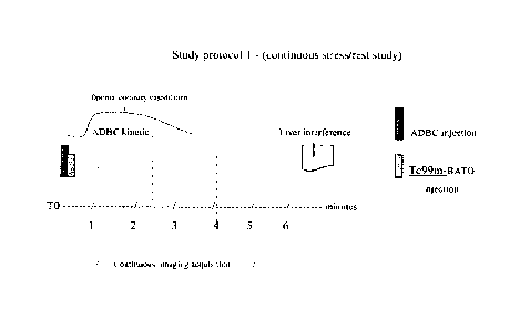
[0068] Study protocol 1 (continuous stress/rest study)
The steps of this protocol are listed below. The total procedure time for the
three phases in
protocol 1 is about 5 - about 6 minutes.
1. Injection of ADBC (stressor) typically in about 30 seconds,
2. Injection of Tc99m-BATO, or a F3A1 0 derivative, immediately afterwards
in less
than about 10 seconds,
3. Continuous acquisition of stress images starting about 40 to about 60
seconds
after Tc99m-BATO's injection for about 4 to about 5 minutes. Software analysis
of Tc99m-BATO' myocardial clearance and of imaging contrast changes is
performed at the same time, providing the same information as a full stress-
rest
protocol.
A graphical depiction of the time sequence of the steps of this protocol are
shown in Fig. I.
100691 Study protocol 2 (continuous stress/rest study)
The steps of this protocol are listed below. The total procedure time for the
four phases in
protocol 2 is about 6 to about 7 minutes.
I. Injection of ADBC (stressor) in about 30 seconds,
2. Injection of Tc99m-BATO, or a BATO derivative, immediately afterwards in
less
than about 10 seconds,
3. Continuous acquisition of stress images starting about 40 to about 60
seconds after
Tc99m-BATO's injection and continuing for about 3 minutes.
4. Second injection from about minute 3 through about minute 4 (shown at about
minute 4) of ADBC at a lower dose followed by about a two-minute acquisition
of "redistribution images" (redistribution phase with coronary blood flow
going
back to baseline = same information as rest images).
A graphical depiction of the time sequence of the steps of this protocol are
shown in Fig. 2.
This protocol allows for a modification of protocol I that, if necessary, can
be implemented
by modifying protocol 1 during the execution of protocol I.
-22-
RECTIFIED SHEET (RULE 91)
CA 02863685 2014-08-01
WO 2013/114204
PCT/IB2013/000332
100701 Study protocol 3 (continuous rest/stress study)
The steps of this protocol are listed below. The total procedure time for the
four phases in
protocol 3 is about 7 minutes.
I. Injection of Te99m-13A10, or a BATO derivative, under rest
conditions,
2. Continuous acquisition of rest images starting about 40 to about 60
seconds after
Tc99m-BATO's injection and for about 2 to about 3.5-4 minutes,
3. Injection of ADBC at from about 2.5 minutes to about 4 minutes (shown at
about
3 minutes) without any additional injection of Tc99m-BATO and continuous
acquisition of stress images for about 2 to about 3 minutes
4. In this protocol, ischemic regions appear in positive instead of in
negative (defect)
as opposed to usual protocols
A graphical depiction of the time sequence of the steps of this protocol are
shown in Fig. 3.
This study mode, which is totally new, is optimal with ADBC because of its
ultra-rapid
kinetic that enhances contrasts between healthy and non-healthy zones after
the uptake of
Tc99m-BATO by the myocardium. Again the use of other stressing agents for this
rest/stress
protocol would result in longer study times and lower imaging sensitivity.
10071] These very short stress/rest protocols are ideally performed with a
stressor that is well
adapted to the kinetics of Tc99m-BATO, or a BATO derivative. At about 6
minutes after the
injection of Tc99m-BATO, the liver captures Tc99m-BATO at a very high
concentration.
Liver interference usually jeopardizes the acquisition of clean myocardial
rest images. This
results in the need to perfon-n a separate rest phase study that requires an
additional
radiopharmaceutical injection. Therefore, the amount of time available for
image acquisition
under rest conditions following the stress phase must be very short, estimated
between about
3 and about 7 minutes after Tc99m-BATO's administration. Among all existing
stressors
ADBC is the sole product that can perfectly fit with Tc991-n-BATO's kinetics,
since maximal
coronary hyperaemia with ADBC occurs after 30 seconds (post-injection),
remains for about
40 to 60 seconds and returns to baseline in about one minute or less.
-23-
RECTIFIED SHEET (RULE 91)
CA 02863685 2014-08-01
WO 2013/114204
PCT/1B2013/000332
100721 It is also worth noting that the protocol designs described above
cannot be achieved
with other stressors either currently commercialized or under development with
the exception
of ADBC. Time course of changes in coronary conductance caused by the
stressors
regadenoson, binodenoson, CGS-21680, apadenoson, and adenosine are shown in
Fig. 4.
These protocols cannot provide the same benefit in terms of reduction of
radiation exposure
study duration and imaging sensitivity, with Persantine (dipyridamole) due to
its prolonged
vasodilating effect and prolonged return to baseline.
100731 The same remark applies to Lexiscan (regadenoson) as this product
induces
coronary hyperaemia after 30 seconds. Hyperaemia is maintained for 2 to 3
minutes and is
followed by a slow return to baseline, as shown in Fig. 4, meaning that a
sustained
vasodilating effect persists until about 7 to about 10 minutes post-injection.
This requires
waiting for a minimum of about 12 to about 15 minute before the acquisition of
rest images
and requires a second isotope injection. This is too long of a waiting period,
with the risk of
liver interference (excretion of Tc99m-BATO is enterohepatic, with peak
hepatic uptake at
about 6 min following injection and there is persistent retention of imaging
agent activity
within the liver in comparison to technetium) and most importantly of
accelerated myocardial
imaging agent wash-out that may result in reduced myocardial counts and
degraded image
sensitivity. Tc99m-BATO's clearance from the myocardium is related to CBF and
the
shortest clearance is the best. This suggests that a rapid return to baseline
(as occurs with the
use of ADBC) is an important image sensitivity factor, whereas a prolonged
return to base
line (Lexiscanal) leads to decreased sensitivity. Sensitivity is the
diagnostic determining
factor. There is an inverse relationship between affinity and duration of
action of A2A
agonists. Source: Gao Z et al. J Pharmacol Exp Ther 2001; 298:209-218
[0074] Other A2a agonists under development, such as binodenoson and
apadenoson, cannot
be used because their duration of action (including their return to baseline)
is too long
= [0075] Adenoscan (adenosine) (the reference product) cannot bc used
without modification
of its recommended method of use (6 minutes infusion). Adenoscane would also
have the
disadvantage of requiring the use of an electric syringe and a dose adjustment
to patient's
weight.
-24-
RECTIFIED SHEET (RULE 91)
CA 02863685 2014-08-01
WO 2013/114204
PCT/1B2013/000332
100761 This shows that ADBC is the ideal stressor for Tc99m-BATO, that the use
of Tc99m-
BATO with ADBC is the ideal "radio-labeled imaging agent -pharmacological
agent
combination" for MPI-SPECT ultrafast cameras, and the combination of these
three
components can be used together to provide methods that can improve imaging
processes
and reducing exposure to radiation by reducing the amount of imaging agent
that needs to be
used,
5.4 Concentrations and unit dosage forms
10077J In a range of embodiments of the compositions herein provided, the
concentration of
adenosine is about 0.5 mg/ml to about 4 mg/ml, preferably from about 0.5 mg/ml
to about 1.2
mg/ml. The concentrations of adenosine may be in about 0.1 mg/m1 increments
within this
range.
100781 In certain embodiments, the concentration of dipyridarnole is about 3
mg/m1 to about
7 mg/ml, preferably between about 3 mg/ml to about 4.2 mg/ml. The
concentrations of
dipyridamole may be in about 0.1 mg/m1 increments within this range.
100791 In various embodiments of compositions comprising adenosine in
combination with
dipyridarnole, the composition has a pH of about 4.0, about 3.9, about 3.8,
about 3.7, about
3.6, about 3.5, about 3.4, about 3.3, about 3.2, about 3.1 or about 3Ø In
certain
embodiments, the pH is between about 2.0 and about 3Ø The term "about" means
approximately or in the region of. When used in the context of the pH of the
solution, such
as 'about 3.1," the pH of the solution could range from 3.05 to 3.15.
100801 In some embodiments, the unit dosage form contains about 1 ml to about
10 ml of the
pharmaceutical composition formulated as a sterile fluid, typically a sterile,
nonpyrogenic,
solution suitable for parenteral administration. In some embodiments, the unit
dosage form
contains about 2 ml, about 3 nil, about 4 ml, about 5 ml, about 6 ml, or about
7 ml. The
preferred UDF contains the active substances in about 3 ml or about 4 ml of
solution.
100811 In other embodiments, the unit dosage forms of adenosine and
dipyridamole are
separate with each of them containing, independently of each other, about 1
ml, about 2 ml,
about 3 ml, about 4 ml, about 5 ml, about 6 ml, or about 7 ml.
-25-
RECTIFIED SHEET (RULE 91)
CA 02863685 2014-08-01
WO 2013/114204 PCT/IB2013/000332
[0082] In an embodiment, the unit dosage forms of adenosine and dipyridamole
are separate,
with one unit dose containing about 1 mg to about 4 mg of adenosine,
preferably about 2 mg
to about 3 mg, more preferably about 2.0 mg to about 2.5 mg, most preferably
about 2.1 mg
to about 2.4 mg and another unit dose containing about 9 mg to about 14 mg of
dipyridamole, preferably about 12 mg to about 14 mg, more preferably about 12
mg to about
12.6 mg. In another embodiment, the unit dosage forms of adenosine and
dipyridamole are
separate, with one unit dose containing about 1 mg to about 4 mg of adenosine
and another
unit dose containing about 9 mg to about 14 mg of dipyridamole.
5.5 Stressing agent Kits
[0083] Also provided are kits. In exemplary kits, the two agents can be
copackaged. For
example, in embodiments, the package can usefully contain at least one unit
dosage form of
dipyridamole copackaged with at least one unit dosage form of adenosine, of an
adenosine
phosphate, as described above.
[0084] In these embodiments, the at least one adenosine and the at least one
dipyridamole
unit dosage forms are either mixed prior to administration to the patient or
injected
sequentially. The unit dose of dipyridamole can be packaged in a vial or a pre-
packed
syringe with or without an injection port and the adenosine unit dose
similarly packaged as a
vial or a prefilled syringe with or without an injection port, such as a
septum, the final
objective being to permit sterile introduction of dipyridamole into the
adenosine dose or the
contrary.
[0085] In some kit embodiments, the kit comprises one unit dosage form of the
adenosine:dipyridamole composition and possibly also one unit dosage form with
saline.
[0086] Adapted connectors, diluent (e.g., saline) and extension set/venous
lines are usefully
included in the kits.
5.6 Compositions
[0087] In some embodiments, the composition is dry, and suitable for
reconstitution prior to
injection by addition of a sterile fluid (e.g. saline) into which both
dipyridamole and
adenosine are readily solubilized. The composition comprises adenosine and
dipyridamole in
-26-
CA 02863685 2014-08-01
WO 2013/114204 PCT/IB2013/000332
amounts suitable to permit reconstitution in the enclosing vessel to the
adenosine and
dipyridamole doses above-described.
[0088] The pharmaceutical compositions can also be present in separate unit
dosage forms,
where the amount of adenosine is between about 1 mg to about 4 mg, preferably
about 2 mg
to about 3 mg, more preferably about 2.0 mg to about 2.5 mg, most preferably
about 2.1 mg
to about 2.4 mg and the amount of dipyridamole is about 9 mg to about 14 mg,
preferably
about 12 mg to about 14 mg, more preferably about 12 mg to about 12.6 mg.
These separate
dosage forms can be mixed together to form a single composition prior to
injection.
5.7 Radiopharmaceuticals (imaging agents) and BATO kits
[0089] Technetium alone, Thallium alone, are isotopes which are also termed
radionuclides.
This refers to their capacity to be used as radioactive markers. When isotopes
are combined
to a carrier that targets a specific organ (sestamibi, tetrofosmine, BATO) the
term
radiopharmaceutical applies. The term radiopharmaceutical or imaging agent is
used herein
regardless of this distinction.
[0090] Most commonly used radiopharmaceuticals in clinical practice are
Thallium-201 (T1-
201), Tc99m-sestamibi and Tc99m-tetrofosmin. Upon injection they are primarily
distributed throughout the myocardium in proportion to coronary blood flow
(these imaging
agents have a high first pass myocardial extraction of over 65%). They are
also captured by
other tissues but only in a second stage. To facilitate the procedure the
initial imaging agent
injection is often coupled to the acquisition of images under stress
conditions. The
acquisition of images at rest then follows. However, this requires the imaging
agent, after its
initial uptake, to be homogcnously redistributed throughout the myocardium,
which is not
always the case. When it is not the case a delayed session under rest
conditions, with a
second radiopharmaccutical injection, is needed.
[0091] Thallium has a long half-life (73 hrs) and must be given at very low
doses to limit
radiation exposure over time. This reduces image quality. However, it is well
redistributed
into the myocardium allowing for the acquisition of images at rest (following
those acquired
under stress conditions), but not before 4 to 24 hrs after the initial
injection, since T1-201
-27-
CA 02863685 2014-08-01
WO 2013/114204
PCT/1B2013/000332
redistribution is a very slow phenomenon. Even then, a mini dose of thallium
is often
administered again.
100921 Tc99m-sestamibi and Tc99m-tetrosfortnin both have a very similar
profile. They can
be given at higher doses than Thallium and produce better image quality.
However, their
disadvantage is the lack of redistribution that requires a second
radiopharmaceutical injection
under rest conditions. This second injection can be done with either thallium
or for example
To99m, but in the latter case at increased dosages.
[00931 These radiophannaccuticals have the disadvantages of requiring time-
consuming
protocols and expose patients to high radiation levels.
[00941 Tc99m-BATO (boronic acid teboroxime) is close to the ideal myocardial
perfusion
imaging agent and potentially the best of all radiophannaceuticals. Its
extraction fraction
(capture by the myocardium) is higher than those of T1-201 or Tc99m. This goes
with its
excellent correlation to coronary blood flow. Tc99m-BATO's myocardial uptake
occurs
after only one minute. The imaging agent is redistributed no more than one
minute after its
capture by myocytes and washed-out from the myocardium after 3 minutes. This
rapid
uptake and washout, that includes a redistribution phase, allows, in theory,
for extremely
short stress/rest protocols.
100951 in one embodiment of the present invention, a unit dosage form
comprising an
amount of boronic acid teboroxime (BATO) or a derivative allowing for the
administration
of about 5 millicuries to about 15 millicuries of Tc-99m is provided.
100961 In another embodiment, a unit dosage form comprising an amount of
boronic acid
teboroxime or a derivative allowing for the administration of about 5
millicuries to about 10
millicuries of Tc-99in is provided.
100971 In another embodiment, a unit dosage form comprising an amount of
boronic acid
teboroxime or a derivative allowing for the administration about 6 millicuries
to about 12
millicuries of Tc-99m is provided.
-28-
RECTIFIED SHEET (RULE 91)
CA 02863685 2014-08-01
WO 2013/114204 PCT/IB2013/000332
[0098] In another embodiment, a unit dosage form comprising an amount of
boronic acid
teboroxime or a derivative allowing for the administration of about 10
millicuries to about 15
millicuries of Tc-99m is provided.
[0099] In a specific embodiment, the unit dosage form of BATO or of a BATO
derivative is
copackaged with the stressing agent or the two products are in the same kit.
[00100] The administration of adenosine at 140 ug/kg/min for a period of 6
minutes is the
reference product used in cardiac imaging for inducing near maximal coronary
vasodilation.
However, the use of the product is accompanied by numerous uncomfortable side
effects. In
addition to the side effects present, its mode of use requires adjusting the
dose to the patient's
weights. The method of use also requires the use of an electric infusion pump,
which is not
convenient. New A2 agonists are more convenient to use (fixed bolus dose) but
remain
impaired by the persistence of many side effects and a long duration of
action. The
combined product described in the present application is as efficient as the
reference product
(adenosine), surprisingly as convenient to use as A2 agonists but additionally
has a better
safety tolerance profile due to its low dosage and a short duration of action,
which is critical
with regard to safety. This allows for very short SPECT-MPI stress/rest
protocols and
reduction of radiation exposure to approximately 1.5-4 mSv/study using
ultrafast gamma
cameras.
EXAMPLES
[00101] As set forth in these examples, the effects of administering adenosine
and
dipyridamole intravenously as a bolus and a combined pharmacological stressor,
were
evaluated versus standard adenosine using transthoracic doppler
echocardiography (TTDE)
and flow velocity measurements as reflecting blood flow measurements. The
value of
absolute coronary blood flow (CBF) may be substituted by the value of CBF
velocity (peak
and mean velocities). This is possible because there is little change in the
diameter of
epicardial coronary arteries after adenosine administration, the effect of the
drug
(vasodilatation and coronary steal) being predominant in the intramural
microcirculation of
the myocardium. Given that blood flow = velocity flow x cross section, if
cross section is
constant, velocity variations reflect blood flow variations.
-29-
CA 02863685 2014-08-01
WO 2013/114204 PCT/IB2013/000332
[00102] TTDE is a non invasive approach conducted under the same conditions as
for
thallium or technetium scintigraphy (intravenous injection) and another way to
study
Coronary Flow Reserve (CFR). Thus, it provides more realistic data than those
obtained
through invasive catheterization laboratory settings. TTDE usefulness to
assess CFR has
been reported in various clinical studies including more than 1000 patients
and has been
validated as reproducible and comparable to intracoronary doppler and positron
emission
tomography (ref 24-27)
[00103] The protocol was designed as follows:
= Left anterior descending coronary artery (LAD) velocity measurements (PV
and MV)
at rest
= Standard adenosine 140 iLig/kg/min infusion during 2 minutes.
= LAD velocity measurements during and after 2 minutes of adenosine 140
p.g/kg/min
infusion.
= Stabilization to baseline during 5 minutes.
= Left anterior descending coronary artery (LAD) velocity measurement at
rest
= Bolus injection (30 seconds) of adenosine: dipyridamole at various
dosages
= LAD velocity measurement after adenosine:dipyridamole bolus injection
= After each product injection measurements of Time to Peak (TTP), Duration
of
Optimal effect (DOE), Time to Return to Baseline (RBL),
= Coronary Reserve (CR) calculation
= Assessment of tolerance using specific scales for symptoms, continuous
EKG, Blood
pressure monitoring
[00104] In a dose ranging study still underway, 18 patients were first
assessed at
adenosine:dipyridamole dose ratios with higher or equivalent dosages of
adenosine than
dipyridamole such as for example 7 mg to 9 mg of adenosine combined with 6 mg
to 9 mg
dipyridamole. Results showed that efficacy and in some cases duration of the
optimal effect
could be reached at cost of an increase of adverse events with occurrence of
unacceptable
safety issues (3 patients/18 patients with second degree atrioventricular
blocks).
[00105] The effects of various dosages and ratios of adenosine and
dipyridamole
administered to patients using several inverted dose-ratios given as about a
30 seconds bolus
were evaluated in 25 distinct patients. Different doses ranging from 1 mg to 5
mg of
-30-
CA 02863685 2014-08-01
WO 2013/114204 PCT/IB2013/000332
adenosine combined with 12 and 14 mg dipyridamole were repeatedly evaluated.
Four tables
are shown as examples to illustrate this approach.
[00106] Different adenosine:dipyridamole combinations versus standard
adenosine
140i,t,g/kg/min (same as Adenoscan ) given as a 2 minute infusion, in the same
patient:
Table 1 shows 5 consecutive studies conducted in the same coronary patient
(BW=70 kg,
Height=175 cm, BMI=23) each at a one week interval comparing different
adenosine-
dipyridamole bolus dose-ratios versus standard adenosine (Stad). There was no
significant
difference between all the doses tested and Stad in terms of efficacy ¨ The
efficacy obtained
with adenosine 1 mg is slightly lower than that of Stad with a DOE limited to
40 seconds.
Best efficacy, duration of optimal effect and coronary reserve results were
obtained with the
2 and 2.4 mg adenosine doses, that also induced the highest peak velocities.
The 3 mg, 4 mg
and 5 mg adenosine doses did not improve (ns) the performance of the
combination versus
Stad, but clearly increased side effects (scale going from 0 to 10 in
severity). It should be
noted that side effects with the combination did not last more than 10 seconds
which is much
shorter than those induced by Stad. No safety issue was observed.
-31-
CA 02863685 2014-08-01
WO 2013/114204 PCT/IB2013/000332
[00107] Effects of the adenosine 4 mg:dipyridamole 12 mg combination
Table 1: Series of different combinations in the same patient
'Table I
Adenosine Ade . dipy Ade : dipy Ade: dipy Ade :4:11i.py
Ade :dipy
i.140nz:1nlin) 1 in g:17'. lir 2 nag.-.1 2 ing 3 m: 12 t.-n 4
ni7:1 2 mg :5 raz 12 my.
PV 23-98 '74-87
.
24-105 24-105
24-105 26-110
$7-108 31-99
26-96 32-103
19-78 MV 18-68
13-80 17-78
20-91
26-75 22-72
19-74 25-73
.CR 4.11 3.73
tba3al al 4.44 4.59
4.56 4.55
2.88 377
.3.80 3.12
TTP 40-50 30 30 30 30 30
DOE 60-90 40 70 80 75 70
RBL 40-50 25 25 30 35 30
DY5pEtoea 2-1- to 3+ Dyspnota Dyspskon 2+ ar.sptsftoa 3-- Enecia 4
Dyitnicea
Hrt5h.+ (lest
dIsconifort
P V= Peak Velocity (cm/sec) ¨MV=Mean Velocity (cm/sec) : first data is at
rest, second data is under
stress conditions - CR =Coronary reserve ratio
TTP= Time to peak (second)-DOE = Duration of optimal effect (second) -RBL=
Return to base
line(second) - Toler. = Tolerance
[00108] Table 2 shows the same 4 mg adenosine and 12 mg dipyridamole dose
ratio used
in three different coronary patients. The 4:12 dose ratio is efficient in all
of them and the
DOE sufficient. There was no statistical difference between the combination
and Stad in
terms of efficacy. The only adverse events were dyspnoea and flush. However
side effects
were not decreased with the combination compared to Stad. No safety issue was
observed.
-32-
CA 02863685 2014-08-01
WO 2013/114204 PCT/IB2013/000332
Table 2. Adenosine 4 mg: dipyridamole 12 mg
St. Adeno4ne Patient 1 Patient 2 Patient 3
140p atficginlin Ade :dipy Ade .dipy Me :dipy
4 inz ;12 4 inv :12 4 ma 12
Irkp mg fig
-
.11ViV 23-50 20 =7,-7
s
14-33
11-32
.16-75
CR 2.85
(based on 2.36 2.91
MAT) 2,88
3.12
TIP 30 35 30
DOE 55 80 70
RBL 20 2.3 30
Tolerance DYVII0e2 5+ Dyninoea 3+
Dyspila : 2+ DIN-Tnoea 3+
Dvsprickea : 3+ Dypnoen
[00109] Tables 3 and 4 show a series of coronary patients who received either
2 mg
adenosine: 12 mg dipyridamole (n=7) or 2.4 mg adenosine : 12 mg dipyridamole
(n=10).
Efficacy was similar to that of Stad with the 2 mg adenosine combination or
even improved
while tolerance was better and the number of side effects fewer. The 2.4 mg
adenosine dose
provided similar results to the 2 mg adenosine dose in terms of efficacy as
well as in terms of
tolerance. Body weight was not correlated to the results. No safety issue was
recorded.
-33-
CA 02863685 2014-08-01
WO 2013/114204
PCT/IB2013/000332
[00110] Effects of 2 mg and 2.4 mg adenosine with 12 mg dipyridamole
Table 3
Dose ratio Adenosine 2 mg;dipyridamole 12 mg
Pt N 1 2 3 4 5 6 7 Mean
value
Sex MMF F MMM
BW (Kilogram) 70 98 65 69 80 64 -- 57
BMI 23 30 25 23.5 23.4 29.5 24.4
TTP
30 30 20 60 40 50 30 37
DOE 80 30 50 65 35 45 40 49
RTBL 30 20 30 30 25 35 15 27
Efficacy vs Stad = > > < > > < ns
Side effects = < < < = < < <
severity
Table 4
Dose ratio Adenosine 2.4 mg: dipyridamole 12 mg
Pt N 8 9 10 11 12 13 14 15 16 17 Mean
value
Sex MMMMMMMF MF
BW (Kilog) 73 84 90 72 74 70 89 84 80 74
BMI 26 28 29 23.5 23.4 26.3 29.4 32.6 27.7 27
TTP 30 35 35 50 45 45 35 30 40 40 38.5
DOE 50 90 45 35 40 40 45 55 45 50 49.5
RTBL 30 40 60 15 15 20 25 25 30 30 29
Efficacy vs Stad = = = = > = = > = = ns
Side effects = > = = = > < < = > ns
severity
Mean velocity (cm/sec): first data is at rest, second data is under stress
conditions.
CR = Coronary reserve -TTP= Time to peak (second)- DOE = Duration of optimal
effect
('second)-RTBL = return to base line (second) - BW= Body Weight (kilogram)
BMI=Body Mass Index -[ overweight= 25-29.9; obesity>30; normal weight=18.5-
24.91
-34-
CA 02863685 2014-08-01
WO 2013/114204 PCT/IB2013/000332
[00111] Additionally a series of 3 patient with 2.1 mg adenosine combined to
12.6 mg
dipyridamole and a series of 3 patients with 2.4 mg adenosine combined to 12.4
mg
dipyridamole showed very similar results to those described above.
[00112] Summary - The following results were also observed:
- Doses as low as 1 mg adenosine (n=3 patients) combined with at least 12
mg
dipyridamole can provide as much efficacy as standard adenosine for about 30
to
about 40 seconds, in some but not all patients (2 out of three), independently
of the
patient's weight. This dose is just below and very close to the efficacy
threshold.
- Dose ratios of 2 mg adenosine/12 mg dipyridamole and above (e.g. 2.4 mg
or 3 mg
adenosine:12 mg dipyridamole) show consistent results which are similar to
those of
standard adenosine in terms of efficacy, equally efficient and comparable for
the
duration of the optimal effect (always > 30 seconds) independently of the
patient's
weight (Tables 1, 3 and 4).
- Dose ratios of 3 mg or 4 mg or 5 mg adenosine/12 mg dipyridamole (n=3
patients
/combination) do not further improve efficacy and are not as well tolerated as
the 2
mg to 2.4 mg adenosine /12 mg dipyridamole dose-ratios. Tolerance is dose-
dependent (Table 2).
- While patients' tolerance of the 2.4 mg adenosine/12 mg dipyridamole dose-
ratio and
above is comparable to that of Stad given as a 2 minute infusion, the side
effects of
the combinations are at least two or three times shorter in duration (they do
not last
more than 10 seconds) than those associated with the administration of Stad.
- Doses of 2.4 mg, (n=10 patients) 3 mg (n=3 patients) and 4 mg (n=3
patients) of
adenosine alone administered as a 30 second bolus, are less efficient than
Stad. These
doses of adenosine alone induce no more than 50 to 60% of the optimal effect
induced by Stad and these effects last for only 10 to 15 seconds.
- Dipyridamole, when injected as a bolus alone (n= 5 patients) at the dose
of 12 mg,
does not induce any significant coronary effect or symptom.
- Dipyridamole mainly serves to modulate the duration of the optimal
adenosinergic
effect, regardless of the dose of adenosine, provided that dipyridamole is
-35-
CA 02863685 2014-08-01
WO 2013/114204 PCT/IB2013/000332
administered at the minimal dose of 9 mg and not above 14 mg. The effect of
the
dipyridamole dose on mean optimal effect duration is shown below. 14 mg
ensures a
60 to 100 seconds duration effect but has the disadvantage to also prolong
side effects
when they occur.
Average
Dipyridamole Optimal Effect
Dose (mg) Duration (sec)
9 30-40
10 40-50
12 40-90
14 60-100 **
** While the dose of 14 mg dipyridamole ensures an average
optimal effects for about 60 to about 100 seconds, this dose
has the disadvantage of prolonging side effects when they
occur.
[00113] Useful dose ratios are summarized in the next paragraph. Ex: 2 mg
adenosine/12
mg dipyridamole - 2.1 mg adenosine/12 mg dipyridamole, 2.4 mg adenosine/12 mg
dipyridamole etc. Preferred dose ratios are those corresponding to UDFs of 3
and 4 ml.
[00114] The table below summarizes all useful and possible unit dosage forms
Unit dosage form Adenosine concentration Dipyridamole concentration
(mg/ml) (mg/ml)
2m1 1, or 1.1 or 1.2 6, or 6.1, or 6.2, or 6.3
3m1 0.7 or 0.8 4 or 4.1 or 4.2
4m1 0.5 or 0.6 3 or 3.1
[00115] Regardless of the dose injected, base line is reached at about 30
seconds on
average after the end of the optimal effect. At 90 seconds after injection no
more effect of
any sort is observed.
[00116] One must understand that beyond the efficacy threshold, corresponding
to the
minimal number of adenosine receptors recruited to induce coronary hyperemia,
efficacy is
-36-
not improved by the recruitment of additional receptors. However adverse
events are then
increased. This is why side effects are significantly higher at the dose of 3
mg and 4 mg
adenosine/12 mg dipyridamole and above compared to lower doses, while efficacy
remains
unchanged at these doses compared to lower dose-ratios (such as 2 mg
adenosine/12 mg
dipyridamole and 2.4 mg adenosine/12 mg dipyridamole).
1001171 The same tolerance profile has been observed with regadenoson which is
also
given as a bolus independently of the patient's weight: 300 g is the efficacy
threshold in
many but not all patients, 400 g is efficient in all patients, 500 ng
provides the same
efficacy as 400 ittg but tolerance is worst. 400 g is the marketed dose.
[00118] While various specific embodiments have been illustrated and
described, it will be
appreciated that various changes can be made without departing from the spirit
and scope of
the invention(s).
REFERENCES
1. Bateman TM, Prvulovich E - Assessment of prognosis in chronic coronary
artery disease.
Heart 2004; 90; v10
2. Katritsis DG & Jionnidis JPA - Percutaneous coronary intervention versus
conservative
therapy in Nonacute coronary artery disease. Circulation 2005, 111, 2906-12.
3. Adenoscan prescribing information (Fujisawa)
4. Injectable Persantin prescribing information (Boehringer Ingelheim)
-37-
CA 2863685 2020-01-22
CA 02863685 2014-08-01
WO 2013/114204 PCT/IB2013/000332
5. Komukai K, Hashimoto K, Shibata T, Iwano K, Muto M, Mogi J, Imai K, Hone T,
Mochizuki S. Effect of continuous ATP injection on human hemodynamics. Circ J.
2002
Oct; 66(10):926-9.
6. Ferreira J, Gil VM, Ventosa A, Calqueiro J, Seabra-Gomes R. Effectiveness
and safety of
coronary vasodilation with adenosine triphosphate with thallium-201 for the
diagnosis of
coronary disease. Rev Port Cardiol. 1995 Mar; 14(3):215-24, 188
7. Johnston DL, Daley JR, Hodge DO, Hopfenspirger MR, Gibbons RJ. Hemodynamic
responses and adverse effects associated with adenosine and dipyridamole
pharmacologic
stress testing: a comparison in 2000 patients. Mayo Clin Proc 1995; 70:331-336
8. Taillefer R, Amyot R, Turpin S, Lambert R, Pilon C, Jarry M. Comparison
between
dipyridamole and adenosine as pharmacologic coronary vasodilators in detection
of
coronary artery disease with thallium 201 imaging. J Nucl Cardiol. 1996 May-
Jun;
3(3):204-11
9. Lagerqvist Bo, Christer Sylven, Elvar Theodorsen, Lennart Kaijser, Gunnar
Helmius and
Anders Waldenstrom ¨ Adenosine induced chest pain - a comparison between
intracoronary bolus injection and steady state infusion. Cardiovascular Res,
1992;
26:810-814
10. Crea F, Pupita G, Galassi AR, el-Tamimi H, Kaski JC, Davies G, Maseri A. -
Role of
adenosine in pathogenesis of anginal pain. Circulation. 1990 Jan; 81(1):164-72
11. Uematsu T, Kozawa 0, Matsuno H, Niwa M, Yoshikoshi H, Oh-uchi M, Kohno K,
Nagashima S, Kanamaru M. - Pharmacokinetics and tolerability of intravenous
infusion
of adenosine (SUNY4001) in healthy volunteers. Br.J.Clin.Pharmacol. 2000. Aug;
50(2):177-81.
12. Ross AM, Gibbons RJ, Stone GVV, Kloner RA, Alexander RVV A randomized,
double
blinded, placebo-controlled, multicenter trial of adenosine as an adjunct to
reperfusion in
-38-
CA 02863685 2014-08-01
WO 2013/114204 PCT/IB2013/000332
the treatment of acute myocardial infarction (Amistad-II) ¨ J Am Coll Cardiol,
2005, 45,
1775-80
13. Cerqueira MD, Verani MS, Schwaiger M, Heo J, Iskandrian AS. Safety profile
of
adenosine stress perfusion imaging: results from the Adenoscan Multicenter
Trial
Registry. J Am Coll Cardiol. 1994 Feb; 23(2):384-9
14. Cerqueira MD. The future of pharmacologic stress: selective A2A adenosine
receptor
agonists. Am J Cardiol. 2004 Jul 22; 94(2A):33D-40D; discussion 40D-42D.
15. Iskandrian AE, Bateman TM, Belardinelli L, Blackburn B, Cerqueira MD,
Hendel RC,
Lieu H, Mahmarian JJ, Olmsted A, Underwood SR, Vitola J, Wang W; ADVANCE MPI
Investigators. Adenosine versus regadenoson comparative evaluation in
myocardial
perfusion imaging: results of the ADVANCE phase 3 multicenter international
trial. J
Nucl Cardiol. 2007 Sep-Oct; 14(5):645-58.
16. Udelson JE, Heller GV, Wackers FJ, Chai A, Hinchman D, Coleman PS,
Dilsizian V,
DiCarli M, Hachamovitch R, Johnson JR, Barrett RJ, Gibbons RJ., - Randomized,
Controlled Dose-Ranging Study of the selective A2A receptor agonist
Binodenoson for
pharmacological stress as an adjunct to myocardial perfusion imaging.
Circulation 2004;
109:457-64
17. Bristol-Myers Squibb Medical Imaging and Adenosine Therapeutics ¨ Press
release
March 24, 2004
18. University of Virginia- Induction of pharmacological stress with adenosine
receptor
agonists. U.S. Pat. No. 6,322,771 and U.S. Patent Application Publication No.
2010/0003193 Al
19. Glover DK, Ruiz M, Takehana K, Petruzella FD, Riou LM, Rigger JM,
Macdonald TL,
Watson DD, Linden J, Beller GA. Pharmacological stress myocardial perfusion
imaging
with the potent and selective A(2A) adenosine receptor agonists ATL193 and
ATL146e
-39-
CA 02863685 2014-08-01
WO 2013/114204 PCT/IB2013/000332
administered by either intravenous infusion or bolus injection. Circulation.
2001 Sep 4;
104(10):1181-7
20. Masaaki Takeuchi et al. - Direct demonstration by Transthoracic Doppler
Echocardiography of adenosine¨induced coronary steel in the collateral-
dependent
vessel. Am J Cardiol 2005; 95: 1363-66
21. Di Mario C, MD, PhD; Jeffrey W. Moses, MD; Todd J. Anderson, MD, MRCP;
Raoul
Bonan, MD; Toshiya Muramatsu, MD; Abnash Chander Jain, MD; Jose Suarez de
Lezo,
MD; Seung Yun Cho, MD; Morton Kern, MD; Ian T. Meredith, MBBS, PhD; David
Cohen, MD, MSc; Issam Moussa, MD; Antonio Colombo, MD; on behalf of the
DESTINI Study Group (Doppler Endpoint STenting INternational Investigation) -
Randomized comparison of elective stent implantation and coronary balloon
angioplasty
guided by online quantitative angiography and intracoronary Doppler. DESTINI
Study
group. Circulation 2000, 103:2938-44
22. Serruys, Patrick W., MD, PhD; Carlo di Mario, MD, PhD; Jan Pick, MD, PhD;
Erwin
Schroeder, MD; Christian Vrints, MD; Peter Probst, MD, PhD; Bernard de Bruyne,
MD;
Claude Hanet, MD; Eckart Fleck, MD; Michael Haude, MD; Edoardo Verna, MD;
Vasilis Voudris, MD; Herbert Geschwind, MD; Halan Emanuelsson, MD; V.
Milhlberger, MD; Giambattista Danzi, MD; Hans 0. Peels, MD; Andrew J. Ford,
Jr; Eric
Boersma, MSc;; for the DEBATE Study Groupl - Prognostic value of intracoronary
flow velocity and diameter stenosis in assessing the stenosis and the short
and long-term
outcomes of coronary balloon angioplasty. The DEBATE Study. Circulation 1997,
96:3369-77
23. Serruys, Patrick W., MD; Bernard de Bruyne, MD; Stephane Cartier, MD; Jose
Eduardo
Sousa, MD; Jan Pick, MD; Toshiya Muramatsu, MD; Chris Vrints, MD; Peter
Probst,
MD; Ricardo Seabra-Gomes, MD; Ian Simpson, MD; Vasilis Voudris, MD; Olivier
Gurne, MD; Nico Pijls, MD; Jorge Belardi, MD; Gerrit-Anne van Es, PhD; Eric
Boersma, PhD; Marie-Angele Morel, MS; Ben van Hout, PhD; on behalf of the
Doppler
-40-
CA 02863685 2014-08-01
WO 2013/114204 PCT/IB2013/000332
Endpoints Balloon Angioplasty Trial Europe (DEBATE) II Study Group -
Randomized
comparison of primary stenting and provisional balloon angioplasty guided by
flow
velocity measurement. Doppler Endpoints Balloon Angioplasty Trial Europe
(DEBATE
II) Study group. Circulation 2000, 102:2930-37
24. Pizzuto F., MD, Paolo Voci, MD, PhD, Enrica Mariano, MD, Paolo Emilio
Puddu, MD,
FESC, FACC, Gennaro Sardella, MD and Antonio Nigri, MD - Assessment of flow
velocity Reserve by transthoracic doppler echocardiography and venous
adenosine
infusion before and after left anterior descending coronary artery stenting. J
Am Coll
Cardiol; 38,(1), 2001, 155-161.
25. Takeshi Hozumi, Kiyoshi Yoshida, Takashi Akasaka, Yoshio Asami, Yumiko
Ogata,
Tsutomu Takagi, Shuichiro Kaji, Takahiro Kawamoto, Yoshiaki Ueda, and
Shigefumi
Morioka Noninvasive assessment of coronary flow velocity and coronary flow
velocity
reserve in the left anterior descending coronary artery by Doppler
echocardiography:
Comparison with invasive technique. J.Am.Coll.Cardiol.,November 1, 1998;
32(5):
1251-1259.
26. Saraste M, Koskenvuo JW, Knuuti J, Toikka JO, Laine H, Niemi P, Sakuma H,
Hartiala
JJ. - Coronary flow reserve: measurement with transthoracic Doppler
echocardiography
is reproducible and comparable with positron emission tomography. Clin Physiol
2001,21:114-22
27. Daimon M., MD, Hiroyuki Watanabe, MD, Hiroyuki Yamagishi, MD, Takashi
Muro,
MD, Kaname Akioka, MD, Kumiko Hirata, MD, Kazuhide Takeuchi, MD and Junichi
Yoshikawa, MD, FACC Physiologic assessment of coronary artery stenosis by
coronary
flow reserve measurements with transthoracic Doppler echocardiography:
comparison
with exercise thallium-201 single photon emission computed tomography. J Am
Coll
Cardiol 2001, 37:1310-15
-41-