Note: Descriptions are shown in the official language in which they were submitted.
CDIM BINDING PROTEINS AND USES THEREOF
[0001] SEQUENCE LISTING
[0002] The instant application contains a Sequence Listing which has been
submitted in
ASCII format via EFS-Web. Said ASCII copy, created on February 7, 2013, is
named 0155-
005W0 l_SL.txt and is 225,619 bytes in size.
[0003] 1. FIELD
[0004] The present disclosure relates to Cell Death Inducing Molecule
(hereinafter
"CDIM") binding proteins and pharmaceutical compositions thereof.
Particularly, the disclosure
provides CDIM binding proteins that are useful in the selective depleting and
killing of B cells,
including neoplastic B cells as well as other neoplastic cells that express
CDIM or CDIM-like
antigens. The disclosure also provides polynucleotides encoding the disclosed
CDIM binding
proteins, and expression systems for producing the same. Further encompassed
in the present
disclosure are methods of treating patients with B cell proliferative and B
cell mediated diseases
by administering the CDIM binding proteins. The disclosure further
contemplates diagnostic
assays for identifying patient populations that can be treated with the CDIM
binding proteins.
100051 II. BACKGROUND
[0006] The major responsibility for carrying out the functions of the
immune system is
born by white blood cells called lymphocytes. Lymphocytes can be categorized
into two major
classes, i.e., T cells and B cells. T cells (i.e., T-lymphocytes) originate
from stem cells in the
bone marrow, develop in the thymus gland and secrete lymphokines. B cells
(i.e., B-
lymphocytes) originate from stem cells in the bone marrow and are the source
of antibodies. In
fact, B cells generate five different types of antibodies including IgM, IgG,
IgA, IgD and IgE.
These antibodies can neutralize substances that can trigger an immune
response, i.e., antigens, by
attaching to specific sites on the antigens in order to block them. IgM is the
largest antibody and
the primary antibody against A and B antigens on red blood cells.
Structurally, IgM forms
polymers where multiple immunoglobulins are covalently linked together with
disulfide bonds,
primarily
1
CA 2863714 2019-04-30
CA 02863714 2014-08-01
WO 2013/120012
PCT/US2013/025430
as a pentamer but also as a hexamer. IgM has a molecular mass of approximately
900kDa in its pentameric form. Because each monomer has two antigen binding
sites, a pentameric IgM has ten (10) binding sites.
[0007] Numerous diseases are associated with altered or dysfunctional B
cells including, but not limited to, autoimmune diseases and cancer. The
proliferation and differentiation of B cells is regulated by receptors
localized on
the cell surface. The engagement of these receptors induces the activation of
intracellular signaling proteins that transmit the receptor signals to
specific
targets inside the cell that control the cellular responses. Many signaling
proteins are the products of oncogenes and many oncogenes are associated with
tumorgenesis. The molecular mechanisms of signaling pathways that control the
proliferation and differentiation of B cells are still being studied (Jumaa
etal.
(2005) Annu. Rev. Immunol. 23:415-445).
[0008] An example of a disease involving neoplastic B lymphocytes is
acute lymphoplastic leukemia (ALL). Some progress in combating this disease is
due to intensification of chemotherapy, as well as better supportive care for
both, pediatric and adult ALL. While the risk of relapse is lower in the
pediatric
population, both pediatric and adult patients face dire outcomes if the
disease
recurs. Less than one third of children and few adults with relapsed ALL
survive
this disease despite the use of aggressive regimens and stem cell
transplantation.
Novel therapies are therefore needed that reach beyond conventional
chemotherapy. For ALL, there is preclinical and early clinical data with a
variety
of monoclonal antibodies including rituximab, epratuzumab and gemtuzumab,
suggesting that the use of monoclonal antibodies alone or in combination with
standard chemotherapy is a viable treatment option.
[0009] U.S. Patent No. 5,593,676 describes ways of inducing cell death in
neoplastic B cells by using reagents that bind a specific B cell epitope
called cell
death inducing molecule (CDIM). Herein, the B cell specific oligosaccharide
epitope CDIM is used as a neoplastic B cell marker. IgM antibodies specific
for
this marker are administered to a host in vivo to induce death in neoplastic B
cells. The same concept can be applied in ex vivo clinical situations to
selectively
remove B cells. A human monoclonal antibody [i.e., MAb 216), which recognizes
2
the B cell epitope CDIM is cytotoxic to neoplastic and normal B cells and
binds all CD19+ and
CD20+ B lymphocytes in human peripheral blood and spleen. Furthermore, MAb 216
does not
distinguish B cells by the isotype expressed, binding IgG+ and 1gM+ cells with
equal intensity.
MAb 216 also binds all B cells regardless of their CD5 expression. Hence, MAb
216, is useful in
diagnosis and therapy. See, also Bhat et al. (2000), Scand. J Immunol., 51:134-
140.
[00010] However, there remains a need in the art to identify antibodies
that are specific for
B cells to selectively kill and/or remove them from the host with reduced off-
target binding
and/or tissue damaging side effects. Cancer therapy still has a tremendous
need for such
therapeutic antibodies. The present application addresses this need.
[00010a] SUMMARY
100010b1 In one aspect, the present invention provides an isolated antigen
binding protein
that specifically binds to CDIM, comprising: (a) a heavy chain comprising a
complementarity
determining region 1 (CDRH1) having a sequence shown in SEQ ID NO:76, a CDRH2
having a
sequence shown in SEQ ID NO:77, and CDRH3 having a sequence shown in SEQ ID
NO: 78,
and (b) a light chain comprising a complementarity determining region 1
(CDRL1) having a
sequence shown in SEQ ID NO: 101, a CDRL2 having a sequence shown in SEQ ID
NO:103,
and a CDRL3 having a sequence shown in SEQ ID NO: 105.
[00010c] In another aspect, the present invention provides an isolated
antigen binding
protein that binds to CDIM, comprising a heavy chain variable region having
the sequence of
SEQ ID NO:1, and a light chain variable region having the sequence of SEQ ID
NO:24.
[0010d] In another aspect, the present invention provides a mixture of
antigen binding
proteins according to the invention, wherein said mixture is a mixture
comprising pentamers and
hexamers.
[00010e] In another aspect, the present invention provides a pharmaceutical
composition
for preventing, treating or diagnosing a disease caused by abnormal B-cell
proliferation,
comprising at least one isolated binding protein according to the invention,
and a
pharmaceutically acceptable carrier, diluent or adjuvant, wherein the B-cell
expresses CDIM,
and in another aspect, a kit comprising same.
10001011 In another aspect, the present invention provides a polynucleotide
encoding the
3
Date Recue/Date Received 2020-12-30
antigen binding protein of the invention.
[00010g] In another aspect, the present invention provides a recombinant
expression vector
which comprises the polynucleotide of the invention operably linked to
sequences effective for
its expression.
[00010h] In another aspect, the present invention provides a recombinant
host cell
comprising the expression vector of the invention.
[000101] In another aspect, the present invention provides a method to
prepare an antigen
binding protein which comprises culturing the host cell of the invention and
recovering said
protein.
[00011] III. BRIEF DESCRIPTION OF THE FIGURES
[00012] The present disclosure is best understood when read in conjunction
with the
accompanying figures, which serve to illustrate the embodiments. It is
understood, however, that
the disclosure is not limited to the specific embodiments disclosed in the
figures.
[00013] FIGS. 1A-D depict amino acid sequences of heavy chain variable
regions (SEQ
ID NOS:1-22) that are representative of the CDIM binding proteins disclosed
herein. The three
heavy chain complementarity determining regions (CDRH1, CDRH2, and CDRH3) and
framework regions of the heavy chain variable region (FR1, FR2, and FR3), and
hi (joining
region) are shown.
[00014] FIG. 1E depicts amino acid sequences of light chain variable
regions (SEQ ID
NOS:23 and 24) that are representative of the CDIM binding proteins disclosed
herein. The three
light chain complementarity determining regions (CDRL1, CDRL2, and CDRL3) and
framework
regions of the light chain variable region (FR1, FR2, and FR3), and JL,
(joining region) are
shown.
[00015] FIG. 1F depicts amino acid sequences of a heavy chain constant
region (Igp.)
(SEQ ID NO:25), and two light chain constant regions (10, and Igx,
respectively) (SEQ ID
NOS:26 and 27) utilized in representative examples disclosed herein.
3a
CA 2863714 2020-02-20
CA 02863714 2014-08-01
WO 2013/120012
PCT/US2013/025430
[00016] FIGS. 2A-2V depict the complete amino acid sequences of the 44
anti-CDIM antibodies disclosed herein, designated IGM1 through IGM44. The 44
disclosed antibodies are formed by combining each of the 22 disclosed heavy
chains (SEQ ID NOS:28-49) with each of the two disclosed light chains (SEQ ID
NOS:50 and 51).
[00017] FIG. 3 depicts the CDR3 sequences of the representative H1
through H22 CDIM binding proteins disclosed herein. The arginine residues of
the various sequences are underlined.
100018] FIGS. 4A-K depict exemplary polynucleotide sequences (SEQ ID
NOS:52-73) encoding the 22 heavy chains of the antigen binding proteins
disclosed herein.
[00019] FIG. 4L depicts exemplary polynucleotide sequences (SEQ ID
NOS:74 and 75) encoding the two light chains, lambda and kappa, of the antigen
binding proteins disclosed herein.
[00020] FIG. 5 depict native SDS gels of crude cell extracts from CHO cells
expressing H1 through H7 (panel A), and H9 through H21 (panel B),
respectively.
The band at 1,048 kD represents IgM pentamers, while the band at 1,236 kD
represents IgM hexamers.
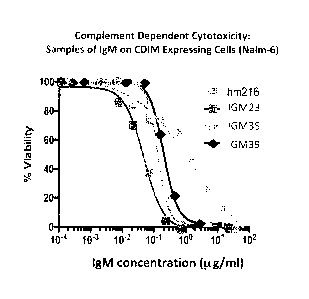
[00021] FIG. 6 illustrates the binding of CDIM binding proteins to CDIM
expressed on a human B cell line and subsequent cytotoxicity results for the
disclosed antibodies. Cell cultures were harvested and analyzed by flow
cytometry using (1) mean fluorescence intensity to quantitate binding and (2)
propidium iodine uptake to distinguish live from dead cells. As shown in FIG.
6A, all antibodies tested bind to the CDIM expressing human B cell line, NALM-
6
across a broad dose range. FIG. 6B shows the cytotoxicity results following
binding of the antibodies to the CDIM epitope.
[00022] FIG 7 shows cytotoxicity results following binding of the
antibodies to the CDIM epitope.
[00023] FIG. 8, panels A-E depict ELISA based binding data that is
representative of the CDIM binding proteins to antigens other than CDIM.
Results using the antigens single stranded DNA (ssDNA), double stranded DNA
4
CA 02863714 2014-08-01
WO 2013/120012
PCT/US2013/025430
(dsDNA), lipid A, cardiolipin, and maleonaldehyde LDL (MDA-LDL) are shown in
panels A-E, respectively. As shown, MAb 216 binds to all of the antigens
across a
broad dose range in comparison with all the disclosed antibodies which
demonstrate markedly reduced binding or total lack of binding to these select
antigens.
[00024] FIG. 9, panels A-F depict ELISA based binding data that is
representative of the CDIM binding proteins to antigens other than CDIM.
Results using the antigens single stranded DNA (ssDNA), double stranded DNA
(dsDNA), lipopolysaccharide, cardiolipin, chondoitrin and heparan, are shown
in
panels A-F, respectively. As shown, MAb 216 binds to all of the antigens
across a
broad dose range in comparison with all the disclosed antibodies which
demonstrate markedly reduced binding or total lack of binding to these select
antigens.
[00025] IV. DETAILED DESCRIPTION OF THE EMBODIMENTS
[00026] The section headings used herein are for organizational purposes
only and are not to be construed as limiting the subject matter described.
[00027] Unless otherwise defined herein, scientific and technical terms
used in connection with the present application shall have the meanings that
are
commonly understood by those of ordinary skill in the art. Further, unless
otherwise required by context, singular terms shall include pluralities and
plural
terms shall include the singular.
[00028] Generally, nomenclatures used in connection with, and techniques
of, cell and tissue culture, molecular biology, immunology, microbiology,
genetics
and protein and nucleic acid chemistry and hybridization described herein are
those well known and commonly used in the art. The methods and techniques of
the present application are generally performed according to conventional
methods well known in the art and as described in various general and more
specific references that are cited and discussed throughout the present
specification unless otherwise indicated. See, e.g., Sambrook et al.,
Molecular
Cloning: A Laboratory Manual, 3rd ed., Cold Spring Harbor Laboratory Press,
Cold Spring Harbor, N.Y. (2001), Ausubel et al., Current Protocols in
Molecular
Biology, Greene Publishing Associates (1992), and Harlow and Lane Antibodies:
A Laboratory
Manual Cold Spring Harbor Laboratory Press, Cold Spring Harbor, N.Y. (1990).
Enzymatic
reactions and purification techniques are performed according to
manufacturer's specifications,
as commonly accomplished in the art or as described herein. The terminology
used in connection
with, and the laboratory procedures and techniques of, analytical chemistry,
synthetic organic
chemistry, and medicinal and pharmaceutical chemistry described herein are
those well known
and commonly used in the art. Standard techniques can be used for chemical
syntheses, chemical
analyses, pharmaceutical preparation, formulation, and delivery, and treatment
of patients.
1000291 It should be understood that this invention is not limited to the
particular
methodology, protocols, and reagents, etc., described herein and as such may
vary. The
terminology used herein is for the purpose of describing particular
embodiments only, and is not
intended to limit the scope of the disclosed, which is defined solely by the
claims.
[00030] Other than in the operating examples, or where otherwise indicated,
all numbers
expressing quantities of ingredients or reaction
conditions used herein should be understood as modified in all instances by
the term "about."
The term ''about" when used in connection with percentages may mean +/-1%.
[00031] General Overview
[00032] The present disclosure provides materials and methods related to
treating or
diagnosing proliferative diseases involving cells expressing the CDIM antigen.
In particular, the
disclosure provides CDIM binding proteins with improved ex vivo and in vivo
performance that
are useful in the selective killing and/or depleting of neoplastic B cells,
specifically in patients
who are afflicted with a condition characterized by B cell proliferative and B
cell mediated
diseases. In addition, the CDIM binding proteins are useful for treating solid
tumors that express
the CDIM antigen. The disclosed CDIM binding proteins may be used alone, or in
combination
with small molecules chemotherapeutics. As a result of a unique pore inducing
effect of the
disclosed CDIM binding proteins,
6
CA 2863714 2019-04-30
CA 02863714 2014-08-01
WO 2013/120012
PCT/US2013/025430
Le., membrane wounding, the targeted cells become more accessible to
chemotherapeutic molecules. Therefore, the disclosed binding proteins are
particularly suitable to treat cells otherwise resistant to small molecule
compounds in combination with the same.
[00033] Definitions
[00034] The following terms used herein shall have the meaning as
indicated below.
[00035] The term "antigen" refers to any substance capable of inducing a
specific immune response and of reacting with a specific antibody.
[00036] The "antigen binding protein" or "CDIM binding protein," as used
herein is a scaffold protein having an antibody like binding activity or an
antibody, i.e., an anti-CDIM antibody.
[00037] The term "CDIM" ("Cell Death Inducing Molecule"), as used herein,
refers to a poly n-acetyl lactosamine glycoform attached to cell surface
molecules. The CDIM epitope is found on nearly all peripheral B lymphocytes
and splenic B lymphocytes and on certain cultured B cell lymphoma lines. The
epitope is also found on primary B cell lymphomas of various histopathologic
classifications, and on the cells of some solid tumors.
[00038] In more specific terms, the CDIM epitope is a linear B cell
lactosamine antigen (Le., a poly-N-acetyl lactosamine type 2 determinant, with
or
without a terminal sialic acid) that has a three-dimensional structural
conformation and is sensitive to the enzyme endo-beta-galactosidase. The
epitope has no branching or substitutions and it can be attached to a
glycolipid
or a glycoprotein. On glycoproteins, the epitope could branch off a mannose
frame work (e.g., enzyme MGAT4), or could be a long chain branching off a
"large
I" structure, but is normally at least about four hexose moieties in a
straight
chain (i.e., type 2) after the branch Gal [31-4 GlcNac p1-3 Gal (31-4 Glc 131;
at least
about six hexoses for good affinity; and least about twelve hexoses in the
longest
form. The chain is made by enzymes (e.g., B3GNT1, B4GALT1), which add
alternate sugars to the eptitope. Notably, the glycosylated epitope CDIM is
7
CA 02863714 2014-08-01
WO 2013/120012
PCT/US2013/025430
present on multiple proteins ranging from molecular weights of about 20KD to
greater than about 200KD proteins.
[00039] The CDIM epitope has been further elucidated in that the
glycoform of the antigen is capped with sialic acid, making it a more mature
type
of glycosylation.
[00040] The term "epitope" generally refers to part of an antigen (i.e.,
the
antigenic determinant of a molecule), which is recognized by the immune
system. An epitope can be composed of sugars, lipids, and/or amino acids or
mixtures thereof. The epitope is recognized by immune cells such as specific T
cells, B cells, and/or antibodies produced by B cells. When immune cells
recognize and are activated by specific epitopes, they mount an immune
response. Alternatively, when antibodies recognize and bind specific epitopes,
the cells carrying the epitopes may be depleted, killed, deactivated, wounded,
removed, and/or altered.
[00041] The term "scaffold protein", or "antigen binding protein," as used
herein, means a polypeptide or protein with exposed surface areas in which
amino acid insertions, substitutions or deletions are highly tolerable.
Examples
of scaffold proteins that can be used in accordance with the present invention
are protein A from Staphylococcus aureus, the bilin binding protein from
Pieris
brassicae or other lipocalins, ankyrin repeat proteins, and human fibronectin
(reviewed in Binz and Pliickthun (2005) Curr. Opin. Biotechnol. 16:459-69).
Engineering of a scaffold protein can be regarded as grafting or integrating
an
affinity function onto or into the structural framework of a stably folded
protein.
Affinity function means a protein binding affinity according to the present
invention. A scaffold can be structurally separable from the amino acid
sequences conferring binding specificity. In general, proteins appearing
suitable
for the development of such artificial affinity reagents may be obtained by
rational, or most commonly, combinatorial protein engineering techniques such
as panning against CDIM, either purified protein or protein displayed on the
cell
surface, for binding agents in an artificial scaffold library displayed in
vitro, skills
which are known in the art (Skerra, A. (2000)]. Mol. Recog. 13:167-187; Binz
and
Pliickthun, supra). In addition, a scaffold protein having an antibody like
binding
8
CA 02863714 2014-08-01
WO 2013/120012
PCT/US2013/025430
activity can be derived from an acceptor polypeptide containing the scaffold
domain, which can be grafted with binding domains of a donor polypeptide to
confer the binding specificity of the donor polypeptide onto the scaffold
domain
containing the acceptor polypeptide. Said inserted binding domains may be, for
example, the complementarity determining region (CDR) of an antibody, in
particular an anti-CDIM antibody. Insertion can be accomplished by various
methods known to those skilled in the art including, for example, polypeptide
synthesis, nucleic acid synthesis of an encoding amino acid as well by various
forms of recombinant methods well known to those skilled in the art.
Importantly, the term "heavy chain" or "light chain" is to be understood
broadly
to be a scaffold protein, embedding one or several of the disclosed CDRs,
rather
than limited to the traditional meaning of the term in the context of antibody
technology.
[00042] Moreover, the term "antibody" or "CDIM-binding antibody," as
used herein, means a monoclonal antibody, a polyclonal antibody, a recombinant
antibody, a humanized antibody (Jones etal. (1986) Nature 321:522-525;
Riechmann etal. (1988) Nature 332:323-329; and Presta (1992) Curr. Op. Struct.
Biol. 2:593-596), a chimeric antibody (Morrison etal. (1984) Proc. NatL Acad.
ScL
U.S.A. 81:6851-6855), a multispecific antibody (e.g., a bispecific antibody)
formed
from at least two antibodies, or an antibody fragment thereof. The term
"antibody
fragment" comprises any portion of the afore-mentioned antibodies, preferably
their antigen binding or variable regions. Examples of antibody fragments
include
Fab fragments, Fab' fragments, F(ab')2 fragments, Fv fragments, diabodies
(Hollinger etal. (1993) Proc. Natl. Acad. ScL U.S.A. 90:6444-6448), single
chain
antibody molecules (Plackthun in: The Pharmacology of Monoclonal Antibodies
113,
Rosenburg and Moore, EDS, Springer Verlag, N.Y. (1994), 269-315) and other
fragments as long as they exhibit the desired capability of binding to CDIM.
[00043] In addition, the term "antibody" or "CLAM binding antibody," as
used
herein, may include antibody-like molecules that contain engineered sub-
domains
of antibodies or naturally occurring antibody variants. These antibody-like
molecules may be single-domain antibodies such as VH-only or VL-only domains
derived either from natural sources such as camelids (Muyldermans etal. (2001)
Reviews in Molecular Biotechnology 74:277-302) or through in vitro display of
9
CA 02863714 2014-08-01
WO 2013/120012
PCT/US2013/025430
libraries from humans, camelids or other species (Holt etal. 2003 Trends
Biotechnol. 21:484-90).
[00044] In accordance with the present invention, the "FAT fragment" is the
minimum antibody fragment that contains a complete antigen-recognition and -
binding site. This region consists of a dimer of one heavy- and one light-
chain
variable domain in tight, non-covalent association. It is in this
configuration that
the three CDRs of each variable domain (heavy chain CDRH1, CDRH2, and
CDRH3; light chain CDRL1, CDRL2, and CDRL3) interact to define an antigen-
binding site on the surface of the VH-VL dimer. Collectively, the six CDR's
confer
antigen-binding specificity to the antibody. However, even a single variable
domain (or half of an FAT comprising only three CDR's specific for an antigen)
has
the ability to recognize and bind the antigen. The "Fab fragment" also
contains
the constant domain of the light chain and the first constant domain (CH1) of
the
heavy chain. The "Fab fragment" differs from the "Fab' fragment" by the
addition
of a few residues at the carboxy terminus of the heavy chain CH1 domain
including one or more cysteines from the antibody hinge region. The "F(ab')2
fragment" originally is produced as a pair of "Fab' fragments" which have
hinge
cysteines between them. Methods of preparing such antibody fragments, such as
papain or pepsin digestion, are known to those skilled in the art.
[00045] In some embodiment of the present invention, the anti-CDIM
antibody is of the IgA-, IgD-, IgE, IgG- or IgM-type, preferably of the IgG-
or IgM-type
including, but not limited to, the IgG1-, IgG2-, IgG3-, IgG4-, IgM1- and IgM2-
type. In
most embodiments, the antibody is of the IgM type. The light chain may be
either a
lambda-1, lambda-2, or a kappa. A J chain may be included or omitted.
[00046] IgG has several subtypes, including, but not limited to, IgG1,
lgG2,
lgG3, and lgG4. IgA subtypes include IgA1 and lgA2. In humans, the IgA isotype
contain four heavy chains and four light chains; the IgG and IgE isotypes
contain
two heavy chains and two light chains; and the IgM isotype contains ten or
twelve heavy chains and ten or twelve light chains (pentameric or hexameric).
In naturally occurring IgM molecules, the J chain stabilizes the pentameric
configuration.
[00047] The heavy chain C region typically comprises one or more domains
that may be responsible for effector function. The number of heavy chain
CA 02863714 2014-08-01
WO 2013/120012
PCT/US2013/025430
constant region domains will depend on the isotype. In one embodiment, the
CDIM binding proteins are of the IgM subtype. In full-length light and heavy
chains, the variable and constant regions may be joined by a "I" region of
about
twelve or more amino acids, with the heavy chain also including a "D" region
of
about ten more amino acids. (See, e.g., Fundamental Immunology, 2nd ed., Ch. 7
(Paul, W., ed.) (1989) New York: Raven Press).
100048] The CDIM Binding Proteins
100049] A first aspect of the present disclosure relates to an isolated
binding protein that binds to the CDIM epitope on human peripheral B
lymphocytes, splenic B lymphocytes, neoplastic B lymphocytes, and some solid
tumors.
100050] In one embodiment, the antigen binding protein comprises a heavy
chain comprising a at least one of a CDRH1, CDRH2, and CDRH3 having a
sequence shown in any of SEQ ID NOS:1-22, and/or a light chain comprising at
least one of a CDRL1, CDRL2, and CDRL3 shown in SEQ ID NOS:23 or 24. In one
embodiment, the antigen binding protein comprises a heavy chain comprising at
least a CDRH3 shown in SEQ ID NOS:1-22, and a light chain. In yet another
embodiment, the antigen binding protein comprises each a CDRH1 , CDRH2, and
CDRH3 shown in SEQ ID NOS:1-22, and a light chain. In other embodiments, the
antigen binding protein additionally comprises a CDRL1, a CDRL2, and a CDRL3
of SEQ ID NOS:23 or 24, embedded into the light chain. In some embodiments,
the antigen binding protein additionally has a FR1 shown in SEQ ID NOS:1-22,
embedded in the heavy chain.
100051] In yet another embodiment, the antigen binding protein comprises
a heavy chain variable region shown in any of SEQ ID NOS:1-22. Additionally,
the
disclosure includes an embodiment where the antigen binding protein comprises
a light chain variable region that has the sequence shown in SEQ ID NO:23 or
24.
Further, the disclosure contemplates an antigen binding protein comprising a
heavy chain variable region shown in any of SEQ ID NOS:1-22, and a light chain
variable region shown in SEQ ID NO:23 or 24. FIGS. 1A-D illustrate the 22
exemplary unique heavy chain variable regions of the CDIM binding proteins
disclosed herein. FIG. 1E depicts two light chain variable regions (SEQ ID
11
CA 02863714 2014-08-01
WO 2013/120012
PCT/US2013/025430
NOS:23 and 24). FIG. 1F shows a constant region for the heavy chain (Ip) (SEQ
ID NO:25), as well as constant regions for the light chains (IgX and Igic)
(SEQ ID
NOS:26 and 27). SEQ ID NO: 108 represents MAb 216 (Bhat eta!, 2000, supra), a
CDIM binding antibody, which was used as experimental reference antibody in
assessing potency and specificity. See, Examples, infra.
[00052] Each of the heavy chain variable regions may be attached to a
heavy chain constant region to form a full heavy chain, and each light chain
variable region may be attached to a light chain constant region to form a
full
light chain, respectively. The amino acid sequences of the exemplary full
heavy
chains disclosed herein have a sequence shown in SEQ ID NOS:28-49. The amino
acid sequences of the exemplary light chains disclosed herein have an amino
acid
sequence shown in SEQ ID NOS:50 and 51. As explained, supra, two heavy chain
and two light chain sequences may form a full antibody tetramer. Disclosed
herein are, inter alia, exemplary CDIM binding antibody tetramers, designated
IGM1, IGM2, IGM3, IGM4, IGM5, IGM6, IGM7, IGM8, IGM9, IGM10, IGM11, IGM12,
IGM13, IGM14, IGM15, IGM16, IGM17, IGM18, IGM19, IGM20, IGM21, IGM22,
IGM23, IGM24, IGM25, IGM26, IGM27, IGM28, IGM29, IGM30, IGM31, IGM32,
IGM33, IGM34, IGM35, IGM36, IGM37, IGM38, IGM39, IGM40, IGM41, IGM42,
IGM43, and IGM44 (collectively also referred to herein as "IGM1-IGM44"). As
shown in FIGS. 2A-2V, these 44 disclosed CDIM binding proteins are comprised
of the heavy chains of SEQ ID NOS:28-49, each combined with either of the
light
chains of SEQ ID NOS:50-51. TABLES 3, infra, show the correlation between the
various polypeptide and polynucleotide SEQ ID NOS and the IGM1-IGM44
antigen binding proteins.
[00053] In one embodiment, the isolated antigen binding protein binds to
CDIM, and comprises a heavy chain CDR3 sequence X1X2X3AX4GX5SX6X7, wherein:
X1 is an G, A, or an R;
X2 is an R, a G, or an A;
X3 is an M, an T, or a R;
X4 is an R, a W, or a Y;
X5 is an A, an S or a G;
X6 is an I, a V, or a Y; and
12
X7 is an N, or no amino acid;
and wherein there is one, and not more than one, Arginine within positions 1
through 3 (relative
to heavy chain variable region, positions 98 through 100, position 97 being
the invariable
Arginine preceding the CDR3 region.
[00054] In another embodiment, the isolated antigen binding protein binds
to CDIM, and
comprises a heavy chain CDR3 sequence XIX2X3AX4GX5SX6X7, wherein:
Xi is an G, A, or an R;
X2 is an R, a G, or an A;
X3 is an M, an T, or a R;
X4 is an R, or a W;
X5 is an A, or an S;
X6 is an I, or a V; and
X7 is an N. or no amino acid;
and wherein there is one, and not more than one, Arginine within positions 1
through 3 (relative
to heavy chain variable region, positions 98 through 100, position 97 being
the invariable
Arginine preceding the CDR3 region.
[00055] In accordance with the present invention, it is to be understood,
that the amino
acid sequence of the binding protein of the invention is not limited to the
twenty conventional
amino acids (See, Immunology - A Synthesis (2nd Edition, E.S. Golub and D.R.
Gren, Eds.,
Sinauer Associates, Sunderland, Mass. (1991)). For example, the amino acids
may include
stereoisomers (e.g., D-amino acids) of the twenty conventional amino acids,
unnatural amino
acids such as a-,a-disubstituted amino acids, N-alkyl amino acids, lactic
acid, and other
unconventional amino acids. Examples of unconventional amino acids, which may
also be
suitable components for the binding protein of the invention, include: 4-
hydroxyprolinc, y-
carboxyglutamate, a-N,N,N-trimethyllysine, a-N-acetyllysine, 0-phosphoserine,
N-acetylserine,
N-formylmethionine, 3-methylhistidine, 5-hydroxylysine, a-N-methylarginine,
and other similar
amino acids and imino acids, e.g., 4-hydroxyproline.
13
CA 2863714 2019-04-30
[000561 Furthermore, in accordance with the present invention, minor
variations in the
amino acid sequences shown in SEQ ID NOS:1-51 are contemplated as being
encompassed by
the present invention, providing that the variations in the amino acid
sequence maintain at least
75%, more preferably at least 80%, 90%, 95%, and most preferably 99% of the
sequences shown
in SEQ ID NOS:1-51. The variations may occur within the framework regions
(i.e., outside the
CDRs), within the CDRs, or within the framework regions and the CDRs.
Preferred variations in
the amino acid sequences shown in SEQ ID NOS:1-51. i.e., deletions, insertions
and/or
replacements of at least one amino acid, occur near boundaries of functional
domains. Structural
and functional domains can be identified by comparison of the nucleotide
and/or amino acid
sequence data to public or proprietary sequence databases. Computerized
comparison methods
can be used to identify sequence motifs or predicted protein conformation
domains that occur in
other binding proteins of known structure and/or function. Methods to identify
protein sequences
that fold into a known three-dimensional structure are known. See, e.g., Bowie
et al. (1991)
Science 253:164; Proteins, Structures and Molecular Principles (Creighton,
Ed., W. H. Freeman
and Company, New York (1984)); Introduction to Protein Structure (C. Branden
and J. Tooze,
eds., Garland Publishing, New York, N.Y. (1991)); and Thornton et al. 1991
Nature 354: 105.
Thus, those of skill in the art can recognize sequence motifs and structural
conformations that
may be used to define structural and functional domains in accordance with the
invention.
[000571 Especially preferred variations in the amino acid sequences shown
in SEQ ID
NOS:1-51, are those that lead to a reduced susceptibility to proteolysis or
oxidation, alter
glycosylation patterns or alter binding affinities or confer or modify other
physicochemical or
functional properties of the binding protein. In particular, conservative
amino acid replacements
are contemplated. Conservative replacements are those that take place within a
family of amino
acids that are related in their side chains. Preferred amino acid families are
the following: acidic
family = aspaitate, glutamate; basic family = lysine, arginine, histidine; non-
polar family =
alanine, valine, leucine, isoleucine, proline, phenylalanine, methionine,
tryptophan; and
uncharged polar family = glycine,
14
CA 2863714 2019-04-30
CA 02863714 2014-08-01
WO 2013/120012
PCT/US2013/025430
asparagine, glutamine, cysteine, serine, threonine, tyrosine. More preferred
families are: aliphatic-hydroxy family = serine and threonine; amide-
containing
family = asparagine and glutamine; aliphatic family = alanine, valine, leucine
and
isoleucine; and aromatic family = phenylalanine, tryptophan, and tyrosine. For
example, it is reasonable to expect that an isolated replacement of a leucine
with
an isoleucine or valine, an aspartate with a glutamate, a threonine with a
serine,
or a similar replacement of an amino acid with a structurally related amino
acid
will not have a major effect on the binding or properties of the resulting
binding
protein, especially if the replacement does not involve an amino acid within a
framework site. Whether an amino acid change results in a functional binding
protein, i.e., in an antigen binding protein that binds to CDIM can be readily
determined by assaying in ELISA or FACS.
[00058] In some embodiments, the CDIM binding protein is a "scaffold
protein" having an antibody like binding activity, where one or several CDRs
of
SEQ ID NOS:1-24 are embedded in a scaffold as defined, supra. In some
embodiments at least CDRH3 and CDRL3 are embedded in the scaffold. In some
embodiments all six CDRs are embedded in the scaffold. Whether the scaffold
protein has CDIM binding activity can be readily determined by assaying in
ELISA or FACS competition for binding with MAb 216, which is a naturally
occurring CDIM binding antibody, or in vitro or in vivo functional assays.
[00059] Furthermore, according to the present invention, it is appreciated
that the CDIM binding antibody of the invention is a fully human or humanized
antibody. Human antibodies avoid certain of the problems associated with
xenogeneic antibodies, for example antibodies that possess murine or rat
variable and/or constant regions. The presence of xenogeneic-derived proteins
such murine or rat derived proteins can lead to the generation of an immune
response against the antibody by a patient, subsequently leading to the rapid
clearance of the antibodies, loss of therapeutic utility through
neutralization of
the antibody and/or severe, even life-threatening, allergic reactions.
[00060] The antigen binding proteins described herein may be antibodies
or may be derived from antibodies. In certain embodiments, the polypeptide
structure of the antigen binding proteins is based on antibodies, including,
but
CA 02863714 2014-08-01
WO 2013/120012
PCT/US2013/025430
not limited to, monoclonal antibodies, bispecific antibodies, minibodies,
domain
antibodies, synthetic antibodies (sometimes referred to herein as "antibody
mimetics"), chimeric antibodies, humanized antibodies, human antibodies,
antibody fusions (sometimes referred to herein as "antibody conjugates"), and
fragments thereof. The antigen binding proteins provided herein have been
shown to bind CDIM epitopes on all B cells, including neoplastic B cells and
some
solid tumor cells. As demonstrated in the examples, the ability of injured B
cells
to repair themselves and survive is reduced or inhibited. As a consequence,
the
disclosed antigen binding proteins are capable of depleting and killing B
cells,
including tumor cells. The antigen binding proteins that are disclosed herein
have a variety of utilities. Some of the antigen binding proteins, are, for
example,
useful in specific binding assays, affinity purification of CDIM expressing
cells,
and in screening assays to identify CDIM expressing cells including solid
tumor
cells, cells of B cell origin. In addition, the disclosed antigen binding
proteins
may be used for the diagnosis and/or treatment of disease, such as B cell
proliferative disorders and autoimmune diseases. To that end, the disclosed
antigen binding proteins may be used alone, or in combination with small
molecules chemotherapeutics.
100061] In one embodiment, the antigen binding protein is a polyvalent
CDIM binding protein [i.e., CDIM binding proteins with two or more binding
sites
for the CDIM epitope). As such, the binding proteins function as receptors
with a
specific affinity and avidity for the CDIM epitope, generally at least about
10-6 M,
and more preferably at least about 10-7 M. The polyvalent nature of the
receptor
allows the simultaneous binding of at least two CDIM epitopes on the cell
membrane surface. Antibodies can be used from any of the immunoglobulin
families, such as IgA, IgD, IgE, IgG, and IgM; it is not a requirement that
the
antibody be associated with various cytotoxic processes associated with
particularly Fc-initiated processes. In one embodiment, the antibody will be
IgM,
since the pentameric or hexameric structure of this molecule allows cross-
linking unhindered by steric interference. In some embodiments, the antibody
composition is a mixture of IgM pentamers and IgM hexamers, including at least
20% hexamers, or at least 30% hexamers, at least 40% hexamers, at least 50%
hexamers, or at least 60% hexamers, or at least 70% hexamers, or at least 80%
16
CA 02863714 2014-08-01
WO 2013/120012
PCT/US2013/025430
hexamers. Alternatively, fragments of antibodies may be used or synthetic
alternatives thereof that act like antibodies. For example, small synthetic
molecules can be devised which will allow for specific binding and cross-
linking
of the CDIM epitope.
[00062] In one embodiment, the antibody has the J chain, in another
embodiment the antibody lacks the J chain.
[00063] In one aspect, the antigen binding protein is a recombinant
antibody constructed based on the VH4-34 germ line sequence. The VH4-34
gene (variable heavy region) is one of the 53 identified human functional
antibody germline genes. The VH4-34 gene is present in all haplotypes, and no
sequence variation was found in germline DNA that was isolated from unrelated
individuals. Anti-B cell VH4-34 antibodies are cytotoxic to B cells (Bhat
etal.
(1997) Clin. Exp. Immuna 108:151 and Bhat etal. (2001) Crit. Rev. Oncol.
Hematol. 39:59). As alluded to above, the plasma membrane defects or pores
induced by the antibodies are larger than those formed by other well-known
pore-forming proteins. By permeabilizing the cells, the disclosed CDIM binding
proteins effect significant depletion of the targeted cells (see, also Patent
Publication Number 20100322849). B cells that have been permeabilized are
more susceptible to the action of additional cytotoxic agents, including, but
not
limited to, radioactive isotopes, cytotoxic antibodies, immunoconjugates,
ligand
conjugates, immunosuppressants, cell growth regulator and/or inhibitors,
toxins, and/or mixtures thereof. The compromised cell membrane allows entry
of cytotoxic agents such as chemotherapeutic agents, thus increasing the
efficacy
of the chemotherapeutic agents, even in cells that are resistant or
impermeable
to such agents. Any B cell or cancer cell that expresses the CDIM epitope or
CDIM-like epitope, respectively, can be treated with the CDIM binding proteins
and is subject to depletion and killing via the disclosed antigen binding
proteins.
[00064] The CDIM binding proteins of the present disclosure recognize the
CDIM epitope on human peripheral B lymphocytes, splenic B lymphocytes and
on neoplastic B lymphocytes, and some solid tumors. Many IgM antibodies are
polyreactive, i.e., they can bind to a variety of different and structurally
unrelated
self and non-self foreign antigens. However, the antigen binding proteins
17
CA 02863714 2014-08-01
WO 2013/120012
PCT/US2013/025430
disclosed herein were found to have less polyreactivity than some naturally
occurring CDIM antibodies. As such, the disclosed antigen binding proteins are
subject to less off-target binding, making them better therapeutic and
diagnostic
candidates for in vivo applications. Their reduced polyreactivity is
illustrated in
FIGS. SA-SE, which shows examples of the disclosed antigen binding proteins
that have reduced or lost their binding affinity for multiple non-CDIM
antigens,
specifically ssDNA, dsDNA, lipid A, cardiolipin and MDA-LDL. This suggests
that
the disclosed CDIM binding proteins are safer for therapeutic applications
because the dose required to bind the target cells will be lower since there
is no
"antigen sink" for the antibody (binding to antigens other than CDIM). The
affinity of a polyreactive antibody for different antigens varies by as much
as
1000-fold and is generally lower than that of a monoreactive antibody for its
antigen. Many of the polyreactive antibodies are usually germline or near
germline although some have a small number of substitutions. Polyreactive
antibodies may be cleared from the circulation faster than monoreactive
antibodies. The rapid clearance of the polyreactive antibodies may be due to
the
binding of these antibodies to endogenous host antigens [see, also Zhou etal.
(2007)]. Autoimmun. 29(4):219-228). Many of the natural antibodies are
polyreactive antibodies, which have broad antibacterial activity. This partly
explains the antibacterial activity in the sera of newborns in the absence of
known antigenic stimulation (see, also Zhou etal. (2007), supra). However, for
therapeutic purposes, it is generally desirable to employ antibodies that are
mono-specific and are not cleared too rapidly so as to accomplish binding and
killing to B cells and cancer cells that express the CDIM epitope.
100065] In specific therapeutic applications, for example, in treating an
autoimmune disease it is desirable that the CDIM binding proteins bind B cells
and kill them selectively as B cells contribute to multiple autoimmune
diseases
by a variety of mechanisms (Browning, J.L. (2006) Nature (Reviews) 5:564-576).
The rapid depletion of the B cells reduces the activity of the immune system
which in turn reduces many associated side-effects such as inflammation and
tissue damage. In cancer treatment it is desirable to kill selective cell
populations, such as neoplastic B cells or cancer cells in order to stop hyper-
proliferation of these cells and the spread of cancer to other organs. Herein,
the
18
CA 02863714 2014-08-01
WO 2013/120012
PCT/US2013/025430
combination therapy with other agents and cancer drugs can be beneficial in
directing the killing of specific cells. Thus, the CDIM binding proteins find
therapeutic application in both autoimmune disease and cancer treatment.
[00066] As discussed above, binding of the disclosed antigen binding
proteins to its linear lactosamine ligand leads to disruption of the plasma
membrane and formation of large membrane pores resulting in cell lysis. The
combination of vincristine, for example, and the disclosed antigen binding
proteins results in an enhanced degree of cytotoxicity to B cells when
compared
to the additive effect of each single agent alone. Hence, the CDIM binding
proteins can be administered to patients alone and in combination with other
agent and/or cancer drugs to assess tumor targeting and efficacy. Furthermore,
the CDIM binding proteins can be administered to patients alone and in
combination with other agents and/or cancer drugs to treat and/or diagnose
various diseases including cancer and autoimmune diseases. Examples of other
agents that could be used in combination with CDIM binding proteins are shown
in TABLE 1 below:
TABLE 1
COMPOUND ACTION EFFECT
Etoposide (VP-16) Topoisomerase II Additive Effect
Inhibitor
Paclitaxel (Taxol) Freezes Microtubules Possible/Undermined
Effect
Ara-C (Cytarabine) Analog Additive Effect
Vincristine, Nocodazole, Depolymerize Synergistic Effect
Colchisine Microtubules
Daunorubicin Anthracyclines Additive Effect
[00067] In some embodiments, an antigen binding protein of the invention
is coupled to a labeling group. Such a binding protein is particularly
suitable for
diagnostic applications. As used herein, the term "labeling group" refers to a
detectable marker, e.g., a radiolabeled amino acid or biotinyl moiety that can
be
detected by conjugated avidin (e.g., streptavidin bound to a fluorescent
marker
or enzymatic activity that can be detected by optical or colorimetric
methods).
Various methods for labeling polypeptides and glycoproteins, such as
antibodies,
are known in the art and may be used in performing the present invention.
19
CA 02863714 2014-08-01
WO 2013/120012
PCT/US2013/025430
Examples of suitable labeling groups include, but are not limited to, the
following: radioisotopes or radionuclides (e.g., 3H; 14c; 15N, 35S, 90y, 99Tc,
"In;
1251, 131I) fluorescent groups (e.g., FITC, rhodamine, lanthanide phosphors),
enzymatic groups (e.g., horseradish peroxidase,13-galactosidase, luciferase,
alkaline phosphatase), chemiluminescent groups, biotinyl groups, or
predetermined polypeptide epitopes recognized by a secondary reporter (e.g.,
leucine zipper pair sequences, binding sites for secondary antibodies, metal
binding domains, epitope tags). In certain respects, it may be desirable that
the
labeling groups are attached by spacer arms of various lengths to reduce
potential steric hindrance.
[00068] Alternatively, an antigen binding protein disclosed herein may be
coupled to an effector group in another preferred embodiment of the invention.
Such a binding protein is especially suitable for therapeutic applications. As
used herein, the term "effector group" refers to a cytotoxic group such as a
radioisotope or radionuclide, a toxin, a therapeutic group or other effector
group
known in the art. Examples for suitable effector groups are radioisotopes or
radionuclides (e.g., 3H; 14C, 151\1, 35S, 90y, 99Tc, 1111n, 1251, 1311),
calicheamicin,
dolastatin analogs such as auristatins, and chemotherapeutic agents such as
geldanamycin and maytansine derivates, including DM1. In certain respects, it
may be desirable that the effector groups are attached by spacer arms of
various
lengths to reduce potential steric hindrance.
[00069] Polynucleotides Encoding CDIM Binding Proteins And
Expression Systems
[00070] Another aspect of the present invention relates to an isolated
nucleic acid molecule encoding a binding protein of the invention. Within the
context of the present invention, the term "isolated nucleic acid molecule''
means
polynucleotide of genomic, cDNA or synthetic origin or some combination
thereof, which by virtue of its origin, the "isolated nucleic acid molecule"
(1) is
not associated with all or a portion of a polynucleotide in which the
"isolated
polynucleotide" is found in nature, (2) is operably linked to a polynucleotide
which it is not linked to in nature, or (3) does not occur in nature as part
of a
larger sequence. Further, the term "nucleic acid molecule", as referred to
herein,
CA 02863714 2014-08-01
WO 2013/120012
PCT/US2013/025430
means a polymeric form of nucleotides of at least 10 bases in length, either
ribonucleotides or deoxynucleotides or a modified form of either type of
nucleotide, such as nucleotides with modified or substituted sugar groups and
the like. The term also includes single and double stranded forms of DNA.
[00071] Exemplary complete nucleic acid sequences encoding the heavy
chain sequences of IGM1-IGM44 (SEQ ID NOS:52-73) are provided in FIGS. 3A-K.
Exemplary complete nucleic acid sequences encoding the light chain sequences
of IGM1-IGM44 (SEQ ID NOS:74 and 75) are provided in FIG. 3L. Of course, due
to the degeneracy of the genetic code, other nucleic acids encoding the CDIM
binding proteins described herein can be contemplated.
[00072] In a one embodiment of the present invention, a nucleic acid
molecule of the invention is operably linked to a control sequence. The term
"control sequence", as used herein, refers to polynucleotide sequences that
are
necessary to effect the expression and processing of coding sequences to which
they are ligated. The nature of such control sequences differs depending upon
the host organism. In prokaryotes, such control sequences generally include
promoters, ribosomal binding sites, and transcription termination sequences.
In
eukaryotes, generally, such control sequences include promoters and
transcription termination sequences. In accordance with the present invention,
the term "control sequence" is intended to include, at a minimum, all
components whose presence is essential for expression and processing, and can
also include additional components whose presence is advantageous, for
example, leader sequences and fusion partner sequences. Furthermore, the term
"operably linked", as used herein, refers to positions of components so
described
which are in a relationship permitting them to function in their intended
manner. Moreover, according to the present invention, an expression control
sequence operably linked to a coding sequence is ligated in such a way that
expression of the coding sequence is achieved under conditions compatible with
the expression control sequence.
[00073] A further aspect of the present invention is a vector comprising a
nucleic acid molecule that encodes a binding protein of the invention. The
nucleic acid molecule can be operably linked to a control sequence.
21
Furthermore, the vector may additionally contain a replication origin or a
selection marker gene.
Examples of vectors that may be used in accordance with the present invention
are, e.g.,
plasmids, cosmids, phages, viruses, etc.
[00074] Another aspect of the present invention relates to a host cell
transformed with a
nucleic acid molecule or vector of the invention. Transformation could be done
by any known
method for introducing polynucleotides into a host cell, including for example
packaging the
polynucleotide in a virus (or into a viral vector) and transducing a host cell
with the virus (or
vector) or by transfection procedures known in the art, as exemplified by U.S.
Patent Nos.
4,399,216, 4,912,040, 4,740,461, and 4,959,455. Particularly, methods for
introducing
heterologous polynucleotides into mammalian cells are well known in the art
and include
dextran-mediated transfection, calcium phosphate precipitation, polybrene
mediated transfection,
protoplast fusion, electroporation, encapsulation of the polynucleotide(s) in
liposomes, and direct
microinjection of the DNA into nuclei. Examples of host cells that may be used
according to the
present invention are hybridomas eukaryotic cells such as mammalian cells,
e.g., hamster, rabbit,
rat, pig, mouse or other animal cells; plant cells and fungal cells, e.g.,
corn, tobacco,
Saccharomyces cerevisiae, Pichia pastoris; prokaryotic cells such as E. coil;
and other cells used
in the art for the production of antibodies. Especially mammalian cell lines
available as hosts for
expression are well known in the art and include many immortalized cell lines
available from the
American Type Culture Collection (ATCC), including but not limited to Chinese
hamster ovary
(CHO) cells, HeLa cells, baby hamster kidney (BHK) cells, monkey kidney cells
(COS), human
hepatocellular carcinoma cells (e.g., Hep G2), and a number of others.
1000751 Pharmaceutical Compositions of CDIM Binding Proteins and Methods of
Treatment and Diagnosis
[00076] A further aspect of the present disclosure are pharmaceutical
compositions and of
the CDIM binding proteins. The binding proteins are formulated as
pharmaceuticals to be used in
the methods of the disclosure. Any composition or compound that can stimulate
a biological
response associated
22
CA 2863714 2019-04-30
CA 02863714 2014-08-01
WO 2013/120012
PCT/US2013/025430
with the binding of the CDIM binding proteins to the CDIM epitope of B
lymphocytes can be used as a pharmaceutical in the disclosure. General details
on techniques for formulation and administration are well described in the
scientific literature (see, "Remington's Pharmaceutical Sciences", Maack
Publishing Co, Easton Pa.). CDIM binding protein pharmaceutical formulations
can be prepared according to any method known in the art for the manufacture
of pharmaceuticals. The CDIM binding proteins can be formulated for
administration in any conventionally acceptable way including via intravenous
injection, intramuscular, intraperitoneal, orally, topically or through other
routes. Illustrative examples are set forth below.
[00077] Pharmaceutical formulations for oral administration can be
formulated using pharmaceutically acceptable carriers well known in the art in
dosages suitable for oral administration. Such carriers enable the
pharmaceutical formulations to be formulated in unit dosage forms as tablets,
pills, powder, capsules, liquids, lozenges, gels, syrups, slurries,
suspensions, and
the like, suitable for ingestion by the patient. Pharmaceutical preparations
for
oral use can be obtained through combination of the CDIM binding proteins with
a solid excipient, optionally grinding a resulting mixture, and processing the
mixture of granules, after adding suitable additional compounds, if desired,
to
obtain tablets or pills. Suitable solid excipients are carbohydrate or protein
fillers which include, but are not limited to, sugars, including lactose,
sucrose,
mannitol, or sorbitol; starch from corn, wheat, rice, potato, or other plants;
cellulose such as methyl cellulose, hydroxypropylmethyl-cellulose, or sodium
carboxymethylcellulose; and gums including arabic and tragacanth; as well as
proteins such as gelatin and collagen. If desired, disintegrating or
solubilizing
agents may be added, such as the cross-linked polyvinyl pyrrolidone, agar,
alginic acid, or a salt thereof, such as sodium alginate. Pharmaceutical
preparations of the present disclosure that can also be used orally are, for
example, push-fit capsules made of gelatin, as well as soft, sealed capsules
made
of gelatin and a coating such as glycerol or sorbitol. Push-fit capsules can
contain
the CDIM binding proteins mixed with a filler or binders such as lactose or
starches, lubricants such as talc or magnesium stearate, and, optionally,
stabilizers. In soft capsules, the CDIM binding proteins may be dissolved or
23
CA 02863714 2014-08-01
WO 2013/120012
PCT/US2013/025430
suspended in suitable liquids, such as fatty oils, liquid paraffin, or liquid
polyethylene glycol with or without stabilizers.
[00078] Aqueous suspensions of the disclosure contain the CDIM binding
proteins in admixture with excipients suitable for the manufacture of aqueous
suspensions. Such excipients include a suspending agent, such as sodium
carboxymethylcellulose, methylcellulose, hydroxypropylmethylcellulose, sodium
alginate, polyvinylpyrrolidone, gum tragacanth and gum acacia, and dispersing
or wetting agents such as a naturally occurring phosphatide (e.g., lecithin),
a
condensation product of an alkylene oxide with a fatty acid (e.g.,
polyoxyethylene
stearate), a condensation product of ethylene oxide with a long chain
aliphatic
alcohol (e.g., heptadecaethylene oxyethanol), a condensation product of
ethylene
oxide with a partial ester derived from a fatty acid and a hexitol (e.g.,
polyoxyethylene sorbitol mono-oleate), or a condensation product of ethylene
oxide with a partial ester derived from fatty acid and a hexitol anhydride
(e.g.,
polyoxyethylene sorbitan monooleate). The aqueous suspension can also
contain one or more preservatives such as ethyl or n-propyl p-hydroxybenzoate,
one or more coloring agents, one or more flavoring agents and one or more
sweetening agents, such as sucrose, aspartame or saccharin. Formulations can
be adjusted for osmolarity.
[00079] Oil suspensions can be formulated by suspending CDIM binding
proteins in a vegetable oil, such as arachis oil, olive oil, sesame oil or
coconut oil,
or in a mineral oil such as liquid paraffin. The oil suspensions can contain a
thickening agent, such as beeswax, hard paraffin or cetyl alcohol. Sweetening
agents can be added to provide a palatable oral preparation. These
formulations
can be preserved by the addition of an antioxidant such as ascorbic acid.
[00080] Dispersible powders and granules of the disclosure suitable for
preparation of an aqueous suspension by the addition of water can be
formulated from the CDIM binding proteins in admixture with a dispersing,
suspending and/or wetting agent, and one or more preservatives. Suitable
dispersing or wetting agents and suspending agents are exemplified by those
disclosed above. Additional excipients, for example sweetening, flavoring and
coloring agents, can also be present.
24
CA 02863714 2014-08-01
WO 2013/120012
PCT/US2013/025430
[00081] The CDIM binding protein pharmaceutical formulations can also
be in the form of oil-in-water emulsions. The oily phase can be a vegetable
oil,
such as olive oil or arachis oil, a mineral oil, such as liquid paraffin, or a
mixture
of these. Suitable emulsifying agents include naturally-occurring gums, such
as
gum acacia and gum tragacanth, naturally occurring phosphatides, such as
soybean lecithin, esters or partial esters derived from fatty acids and
hexitol
anhydrides, such as sorbitan mono-oleate, and condensation products of these
partial esters with ethylene oxide, such as polyoxyethylene sorbitan mono-
oleate. The emulsion can also contain sweetening and flavoring agents. Syrups
and elixirs can be formulated with sweetening agents, such as glycerol,
sorbitol
or sucrose. Such formulations can also contain a demulcent, a preservative, a
flavoring or a coloring agent.
[00082] When the CDIM binding proteins are delivered by intravenous
injection, the pharmaceutical formulations can be in the form of a sterile
injectable preparation, such as a sterile injectable aqueous or oleaginous
suspension. This suspension can be formulated according to the known art using
those suitable dispersing or wetting agents and suspending agents, which have
been mentioned above. The sterile injectable preparation can also be a sterile
injectable solution or suspension in a nontoxic parenterally-acceptable
diluent or
solvent. Among the acceptable vehicles and solvents that can be employed are
water and Ringer's solution, an isotonic sodium chloride. In addition, sterile
fixed oils can conventionally be employed as a solvent or suspending medium.
For this purpose any bland fixed oil can be employed including synthetic mono-
or diglycerides. In addition, fatty acids such as oleic acid can likewise be
used in
the preparation of injectables.
[00083] The methods of the present disclosure treat human and non-
human patients who suffer from lymphoid cancer or leukemia (e.g., B-cell acute
lymphoblastic leukemia or ALL), any form of autoimmune disease involving B
cells (e.g, rheumatoid arthritis, systemic lupus erythematosus or SLE), any
form
of B cell hyper-proliferation such as lymphomas and myelomas (e.g., non-
Hodgkin's lymphomas), certain forms of solid tumors that express the CDIM
antigen, and/or related conditions. The amount of CDIM binding protein that is
CA 02863714 2014-08-01
WO 2013/120012
PCT/US2013/025430
adequate to accomplish this is considered the therapeutically effective dose.
Alternatively, the amount of CDIM binding protein in combination with another
agent or another drug that is adequate to accomplish this is also considered a
therapeutically effective dose. Other agents are, for example, cytotoxic
agents
including, but not limited to, a radioactive isotope, a cytotoxic antibody, an
immunoconjugate, a ligand conjugate, an immunosuppressant, a cell growth
regulator and/or inhibitor, a toxin, or mixtures thereof. A chemotherapeutic
agent or compound (see, also TABLE 1) is often an agent that interferes with
the
polymerization or depolymerization of microtubules such as a taxane, vinca
alkaloid or colchicine, or mixtures thereof. The vinca alkaloid includes
vinblastine, vincristine, vindesine, or vinorelbine, or mixtures thereof. The
taxane includes, but is not limited to, paclitaxel, docetaxel, or mixtures
thereof.
The cytotoxic antibody that can be administered in combination with the
disclosed antigen binding proteins usually has specific binding for a cell
surface
receptor on a B cell, including CD11a, CD19, CD20, CD21, CD22, CD25, CD34,
CD37, C1J38, CD40, CD45, CD52, CD80, CD 86, IL-4R, IL-6R, 1L-8R, IL-13, IL-
13R,
a-4/43-1 integrin (VLA4), BLYS receptor, cell surface idiotypic Ig, tumor
necrosis
factor (TNF), or mixtures thereof. As such, the cytotoxic antibody can be
efalizumab (RAPTIVA), rituximab (RITUXAN), daclizumab (ZENAPAX),
epratuzumab, basiliximab (SIMULECT), anti-CD52 (CAMPATH), natalizumab,
infliximab (REMICADE), and the like. The immunosuppressant includes, but is
not limited to, a glucocorticoid, a calcineurin inhibitor, an
antiproliferativefantimetabolic agent, or an immunosuppressive antibody. In
one embodiment, the agents are etoposide (VP-16), paclitaxel (taxol), ara-C
(cytarabine), vincristine, nocodazole, colchisine, daunorubicin, cytochalasin,
jasplakinolide, and the like.
100084] In one embodiment of the present invention, at least one binding
protein disclosed herein contained in the pharmaceutical composition is
coupled
to an effector, such as calicheamicin, Auristatin-PE, a radioisotope or a
toxic
chemotherapeutic agent such as geldanamycin and maytansine. In particular,
these binding protein conjugates are useful in targeting cells, e.g., cancer
cells,
expressing CDIM for elimination.
100085] Moreover, linking the binding proteins disclosed herein to
26
CA 02863714 2014-08-01
WO 2013/120012
PCT/US2013/025430
radioisotopes provides advantages to tumor treatments. Unlike chemotherapy
and other forms of cancer treatment, radioimmunotherapy or the administration
of a radioisotope-binding protein combination directly targets the cancer
cells
with minimal damage to surrounding normal, healthy tissue. With this "magic
bullet", the patient can be treated with much smaller quantities of
radioisotopes
than other forms of treatment available today. Certain radioisotopes include
yttrium90 (90Y), indium" (llijn); 1311, 99mTc, radiosilver-111, radiogold-199,
and
Bismuth213. The linkage of radioisotopes to binding proteins may e.g., be
performed with conventional bifunctional chelates. Since silver and gold can
exist in a monovalent state, for radiosilver-111 and radiogold-199 can utilize
sulphur-based linkers may be used (Hazra etal. (1994) Cell Biophys. 24-25:1-
7).
Linkage of silver radioisotopes may involve reducing the immunoglobulin with
ascorbic acid. Furthermore, tiuxetan is an MX-DTPA linker chelator attached to
ibritumomab to form ibritumomab tiuxetan (Zevalin) (Witzig, T.E. (2001) Cancer
Chemother. Pharmacol. 48 Suppl 1:91-5). Ibritumomab tiuxetan can react with
radioisotypes such as indium" (min) or 9 Y to form "In-ibritumomab tiuxetan
and 90Y-ibritumomab tiuxetan, respectively.
[00086] Furthermore, a binding protein disclosed herein, particularly when
used to treat cancer, may be conjugated with toxic chemotherapeutic drugs such
as calicheamicin (Hamann etal. (2002) Bioconjug. Chem. 13:40-46, geldanamycin
(Mandler etal., (2002)1. Natl. Cancer Inst., 92:1549-1951) and maytansine, for
example, the maytansinoid drug, DM1 (Liu etal. (1996) Proc. Natl. Acad. Sci.
U.S.A. 93:8618-8623). Different linkers that release the drugs under acidic or
reducing conditions or upon exposure to specific proteases may be employed
with this technology. According to the present invention, a binding protein
disclosed herein may be conjugated as described in the art.
[00087] Auristatin-PE, e.g., is an antimicrotubule agent that is a
structural
modification of the marine, shell-less mollusk peptide constituent dolastatin
10.
Auristatin-PE has both anti-tumor activity and anti-tumor vascular activity
(Otani etal. (2000) Jpn. J. Cancer Res. 91:837-44). For example, auristatin-PE
inhibits cell growth and induces cell cycle arrest and apoptosis in pancreatic
cancer cell lines (Li etal. (1999) Int. J. MoL Med. 3:647-653). Accordingly,
to
specifically target the anti-tumor activity and anti-tumor vascular activities
of
27
CA 02863714 2014-08-01
WO 2013/120012
PCT/US2013/025430
auristatin-PE to particular tumors, auristatin-PE may be conjugated to the
binding protein disclosed herein.
[00088] The dosage schedule and amounts effective for this use, Le., the
"dosing regimen," will depend upon a variety of factors, including the stage
of the
disease or condition, the severity of the disease or condition, the severity
of the
adverse side effects, the general state of the patient's health, the patient's
physical status, age and the like. In calculating the dosage regimen for a
patient,
the mode of administration is also taken into consideration. The dosage
regimen
must also take into consideration the pharmacokinetics, Le., the rate of
absorption, bioavailability, metabolism, clearance, and the like (see, for
example,
Liedtke etal. (2012) Haematologica 97(1):30-37).
[00089] The state of the art allows the clinician to determine the dosage
regimen for each individual patient. CDIM binding proteins can be administered
alone or in combination with other compounds. If administered in combination
with other compounds, better patient responses and more durable outcomes
would be expected. The combined compounds may act synergistically, or
additively.
[00090] As an illustrative example, the guidelines provided below for CDIM
binding proteins can be used as guidance to determine the dosage regimen, Le.,
dose schedule and dosage levels, of any CDIM binding protein administered
when practicing the methods disclosed herein. The clinical efficacy of CDIM
binding proteins may be enhanced by the co-administration of a second
compound such as vincristine or similar agent. Likewise, the efficacy of a
small
molecule chemotherapeutic may be enhanced by the co-administration with the
CDIM antigen binding protein. CDIM binding proteins are effective in a dose
range of about between 0.25mg/kg to 100mg/kg. Single or multiple
administrations of CDIM binding protein formulations may be administered
depending on the dosage and frequency as required and tolerated by the patient
who suffers from lymphoid cancer or leukemia (e.g., B-cell acute lymphoblastic
leukemia or ALL), any form of autoimmune disease involving B cells (e.g.,
rheumatoid arthritis, systemic lupus erythematosus or SLE), or any form of B
cell
hyperproliferation such as lymphomas and myelomas (e.g., non-Hodgkin's
28
CA 02863714 2014-08-01
WO 2013/120012
PCT/US2013/025430
lymphomas) and/or related conditions. The formulations should provide a
sufficient quantity of CDIM binding protein to effectively ameliorate the
condition. For example, any one of the 44 antigen binding proteins disclosed
herein may be administered to a patient through monotherapy (i.e., with no
other medications) or in combination therapy with, for example, vincristine or
other agents, see, supra). The antigen binding proteins having specific
binding
for the CDIM epitope on a B cell can be administered at a dose of from about
2.5
to about 3000mg/m2, or more preferably, from about 25 to 1000mg/m2, or in
particular, about 75, 150, 300 or 600mg/m2. In additional aspects, the
antibody
is administered at a dose of from about 0.25mg/kg to about 100mg/kg, and more
preferably, at about 1.25, 2.5, 5, 10, or 20mg/kg. The anti-CDIM antibody is
typically administered on a weekly basis, and in some embodiments, more
frequently than once per week, as often as once per day. Additional cytotoxic
antibodies can be administered in an amount of 10-375mg/m2 per week for four
weeks, or 0.4-20mg/kg per week for 2 to 10 weeks in form of a combination
therapy. In one embodiment, CDIM binding proteins are currently administered
to a patient daily as monotherapy in an amount from about 0.25mg/kg to about
100mg/kg. In another embodiment, CDIM binding proteins are administered to
a patient daily in combination therapy with a second agent selected from the
group consisting of vinblastine, vincristine, vindesine, vinorelbine, or
mixtures
thereof in an amount from about from about 0.15mg/kg to about 50mg/kg.
[00091] Notably, the dosages of selective CDIM binding proteins
administered to a patient may vary depending on age, degree of illness, drug
tolerance, and concomitant medications and conditions. The CDIM binding
proteins may be administered to the patient in combination with another drug
in
order to potentiate the effect of the CDIM binding proteins and in order to
reduce
adverse side effects. Using a second drug, the activity of co-administration
of
CDIM binding proteins may be enhanced by between 10% and 90% and the
combination therapy will continue until the combination treatment is no longer
deemed beneficial or necessary. The CDIM binding proteins may be
administered to a patient simultaneously or within specific time frames of one
another. Different CDIM binding proteins can be administered to the patient
simultaneously in separate pills or tablets or in the form of a combination
pill.
29
CA 02863714 2014-08-01
WO 2013/120012
PCT/US2013/025430
[00092] Disorders and Diseases
100093] The CDIM binding proteins of the present disclosure can be used to
treat patients who suffer from lymphoid cancer or leukemia, any form of
autoimmune disease involving B, or any form of B cell hyperproliferation such
as
acute or chronic leukemia, lymphomas and myelomas, and/or related disorders.
Any condition that is characterized by a hyperproliferation of B cells
including
lymphoid cancer, viral infection, immunodeficiency, or autoimmune disease can
be treated with the CDIM binding proteins. Similarly, any tumor cell or cancer
cell that expresses the CDIM epitope or a CDIM-like antigen can be treated
with
the CDIM binding proteins.
[00094] The disclosure provides improved CDIM binding proteins for
selective B cell killing and depleting in disorders related to autoimmunity
including, but not limited to, multiple sclerosis, rheumatoid arthritis,
systemic
lupus erythematosus (SLE), Myasthenia gravis, Pemphigus vulgaris, Grave's
disease and autoimmune thrombocytopaenia. Autoreactive B cells secrete
autoantibodies directed against self-proteins. B lymphocytes not only produce
autoantibodies but also play an important regulatory role independent of their
function as antibody-producing cells. This is relevant with respect to
autoimmunity, since autoreactive B cells have the ability to activate
pathogenic T
cells to produce pro-inflammatory cytokines. Myasthenia gravis, Pemphigus
vulgaris, Grave's disease and autoimmune thrombocytopaenia are good
examples of conditions in which pathogenic antibodies drive the clinical
phenotype (see, Browning, J.F, supra). In addition, autoimmune disorders lead
to
overactive and increased numbers of B cells that should be removed in order to
prevent massive inflammation and tissue damage. Thus, the depletion of B
lymphocytes is useful in the treatment of such autoimmune diseases. Since the
treatment of many rheumatic autoimmune diseases such as rheumatoid arthritis
relies primarily on the use of cytotoxic immunosuppressants and
corticosteroids
patients often suffer additional severe side effects. In addition, patient
relapse
rates remain high. There is a need for safer and more effective drugs such as
the
CDIM binding proteins of the present disclosure.
CA 02863714 2014-08-01
WO 2013/120012
PCT/US2013/025430
[00095] A lymphoid cancer is any acute or chronic leukemia or lymphoma
of B cell origin, including, but not limited to, acute lymphocytic leukemia
(ALL),
non-Hodgkins lymphoma (NHL), Burkitt's lymphoma, B progenitor ALL, adult
ALL, chronic lymphocytic leukemia (CLL), and Waldenstrom's
macroglobulinaemia. The CDIM binding proteins bind to the epitope CDIM,
which is found on cancerous B cells.
[00096] In order to allow prediction of patients who will respond to
treatment with the disclosed CDIM antigen binding proteins, in vitro or in
vivo
analysis may be performed. In vivo imaging may be performed prior to
treatment by administering CDIM antigen binding proteins as a conjugate which
allows visualization of the CDIM antigen on the tissue of interest. A
particular
level of reactivity may be established that would allow prediction of patient
response to therapy. Alternatively, this type of analysis may help in
establishing
dosing parameters based on tumor load. In vitro analysis may be performed on
samples of patient's lymphoid cells (peripheral blood, bone marrow or other)
prior to treatment. Cells will be stained with CDIM antigen binding proteins
using standard flow cytometric analysis. A cut-off will be established that
allows
prediction of positive outcome following therapy. For example, a minimal mean
fluorescence intensity may be established which predicts positive outcome.
[00097] V. EXAMPLES
[00098] The following specific examples are intended to illustrate the
disclosure and should not be construed as limiting the scope of the claims.
[00099] Example 1: Generation and Sequence Determination of
Exemplary CDIM Binding Proteins
[000100] Plasmid DNA construction. All the H series mu chain constructs
variable heavy regions (designated H1-H22) were synthesized by Genscript with
Xba1-Kpn1 sites on the 5' and 3' ends respectively. All the H series Mu chain
constructs were assembled by isolating each of heavy chain variable regions as
Xba1-Kpn1 fragments and joining them in a three-part ligation with a Kpn1-
BamH1 fragment spanning the constant mu region together with the expression
vector AB11 that had been digested with Xba1 and BamH1. The ligated plasmids
were transformed into competent bacteria. The resulting cloned plasmids were
31
CA 02863714 2014-08-01
WO 2013/120012
PCT/US2013/025430
confirmed by direct sequencing. Plasmid midi-preps were generated using
Qiagen, to provide adequate amounts of DNA for transfection into CHO-S cells.
10001011 The light chain plasmid were constructed in a similar fashion. The
lambda insert was cloned in a three part ligation; the variable lambda region
was
designed as a Xbal-EcoRI fragment, which was mixed with the EcoRI-BamHI
lambda constant region fragment and ligated into the AB2 vector between Xbal
and BamHI sites. The L2 kappa insert was isolated as a XbaI-BamHI fragment
and ligated into the AB2-Kappa vector.
10001021 Transfection of CHO-S cells with the Hl-H22/L2 plasmid DNA.
DNA corresponding to heavy and light chains, was prepared for co-transfection
(equal amounts of L2 and of H1-22) using the PEI technique. CHO-S cells (1E8
of
log phase growth) used for transfection were grown in RPMI1640 media.
DNA:PEI was mixed 1:2 and this DNA:PEI mixture was added to CHO-S cells in
CD OptiCHO supplemented with 0.5x Pen/Strep, Glutamax and HT. After
overnight incubation in shaker flask, the media was exchanged into CD OptiCHO
with 0.5x P/S, Glu. On day 7 post-transfection, the cell culture supernatant
(100
ml) was harvested by centrifugation of the cells. For characterization of
these
IgM examples, 15 ml of cell culture supernatant was concentrated 10x by
Centricon.
10001031 The amino acid sequences of the 22 distinct heavy chain variable
regions are depicted in FIGS. 1A-D. The amino acid sequences of the 2 distinct
light chain variable regions are depicted in FIG. 1E. The amino acid sequences
of
the constant regions of the heavy and light chains are shown in FIG. 1F. It is
understood that either kappa or lambda constant regions can be utilized. The
amino acid sequences that are representative of the identified and isolated
CDIM
binding proteins, IGM1-IGM44, shown in FIGS 2A-2V. The CDR3 sequences of
H1 through H22 are depicted in FIGURE 3.
10001041 Examples of polynucleotide sequences that could be used to
encode the 44 disclosed CDIM binding proteins are depicted in FIGS. 4A-L. It
is
understood that due to degeneracy in the genetic code, other sequences could
be
utilized to encode the exact amino acid sequence. The sequences of the various
complementarity determining regions (CDRHs) and framework (FR) regions of
32
CA 02863714 2014-08-01
WO 2013/120012
PCT/US2013/025430
the heavy chain variable region of the antibodies were determined include
framework 1 (FR1), complementarity determining region 1 (CDRH1), framework
2 (FR2), complementarity determining region 2 (CDRH2), framework 3 (FR3)
and complementarity determining region 3 (CDRH3).
[000105] Example 2: Making of CDIM Binding Proteins
10001061 This example describes how some of the disclosed binding
proteins were made. Sequences consisted of heavy and light chain variable
region variants (both kappa and lambda constant regions were encoded).
Specific combinations of heavy and light chain variants were transfected into
CHO cells, i.e., DG44 CHO cells and transient transfections were made.
Expression levels of 10-100 ug/m1 were obtained from the initial
transfections.
IgM antibodies were purified by affinity chromatography, evaluated by gel
electrophoresis and tested for bioactivity by cell binding, cytotoxicity
assays and
ELISA testing for binding to multiple biomolecules. Results obtained from the
functional assays were used to guide lead candidate selection. Once selected,
the
lead candidates were stably transfected into CHO cells and sub-cloned into 20
plates (approximately 2000 wells) and screened by ELISA for highest level of
secretion. The top 50 clones were expanded to T75 flasks followed expansion of
the best 24 clones being transferred into shaker flasks. IgM from these 24
clones
were purified and quantified and the top six clones (selected by IgM antibody
secretion levels) were grown under further selection in methotrexate (50 nM,
100 nM, 200 nM, 400 nM) to facilitate outgrowth of high secreting cell lines.
10001071 Example 3: Evaluation of IgM Expression using Non-Reduced
SDS-PAGE
10001081 Non-reducing gel electrophoresis is a separation method typically
used
in proteomics and resolves proteins based on molecular mass and oligomeric
structures. Under non-reducing conditions, protein disulfide bonds are left
intact and
so the IgM oligomer can be resolved as pentameric or hexameric structures.
10001091 To confirm IgM antibody expression by transient transfection of
CHO-
S cell supernatant, we performed non-reducing SDS polyacrylamide gel
electrophoresis as described in Vorauer-Uhl, et al, J lmmunol. Methods 359
(2010):
21-27. This example describes how transiently transfected IgM mAbs were
investigated for purity and multimeric structure. Protein sample of interest
is
33
CA 02863714 2014-08-01
WO 2013/120012
PCT/US2013/025430
incubated with NuPage SDS sample buffer for 5 minutes at room temperature.
After
incubation, the molecular weight standards and test samples are loaded onto
the gel
and run at 100V constant voltage for approximately 2 hours until dye front
reaches
the bottom of the gel. After electrophoresis, the gel is removed from XCell
Mini-Cell
apparattus, fixed and stained with Colloidal Blue dye.
10001101 To demonstrate pentamer and hexamer formation, non-reducing
SDS PAGE of concentrated supernatant of CHO cells, transfected with the
various
CDIM binding protein samples (L1-L7, and L9-L21, respectively), was performed
using SDS PAGE, Life Technologies' (Carlsbad, CA) Native Page Novex 3-12%
Bis-Tris gels.
10001111 Fig. 5 shows the SDS gel stained according to the Colloidal Blue
Staining protocol (Life Technologies, Carlsbad, CA), with a prominent band at
1,048 kD, which represents the IgM pentamers, and a prominent band at 1,236
kD, which represents the IgM hexamers. As can be seen in the figure, pentamer
formation for the IgM examples (solid arrow) is more dominant compared with
hexamer formation. Similarly, the isolated human IgM 216 is predominately of
pentameric form (dashed arrow), however, it appears as a lower molecular
weight due to the smaller lambda light chains included in this IgM format.
10001121 Example 4: Binding of CDIM Binding Proteins to CDIM Antigen
10001131 This example describes how the binding properties of the
disclosed antibodies were investigated. The antibodies were prepared by
affinity chromatography. In FIG. 6A, the human pro-B cell line, NALM-6, which
expresses the CDIM antigen on the cell surface was stained with the series of
recombinant IgM antibodies. The antibodies were used to determine the dose
response beginning at 20 [tg/tube diluted stepwise by 2, to a final
concentration
at 0.039 rig. Cells were stained in a 100 ul volume in 3% FBS/PBS on ice for 1
hour followed by a subsequent staining with DyLight-488 conjugated anti-
human IgM (Jackson Immuno), 1 ,t,g/m1 for 20 minutes at 4 C.
10001141 Analysis of CDIM binding was performed on FACScan flow
cytometer using CellQuest analysis software. Results are shown as raw data
34
CA 02863714 2014-08-01
WO 2013/120012
PCT/US2013/025430
points with Mean Fluorescence Intensity (MFI) on Y-axis and concentration of
primary antibody ( g/100 !LAI) on X-axis.
10001151 The data were re-analyzed using GraphPad analysis program to fit
the results using non-linear regression curve fit. EC50 (n/100 ,L1) for each
antibody was (a) 10.6 for IGM1, (b) 2.2 for IGM23, (c) 2.2 for IGM34, and (d)
1.7
for IGM36.
10001161 Example 5: Cytotoxicity Resulting from Cellular Binding of
CDIM Binding Proteins
10001171 This example describes how the cytotoxicity of the CDIM binding
proteins was investigated. The human pro-B cell line NALM-6 was stained
identically as described in FIG. 6A above. In FIG. 6B, cell killing was
evaluated
by measuring cell viability after 1 hour of staining. By quantifying the
proportion of cells that did not uptake propidium iodine, the percentage of
viable
cells was calculated. Results are shown as raw data with % viability on Y-axis
and concentration (pg/100 pl) on X-axis.
10001181 Identical results were graphed using GraphPad analysis program
and data fit using non-linear regression curve fitting. The ECso was
calculated for
each antibody using the same approach described above. ECso (m/100 0) for
each antibody was (a) 3.0 for IGM1, (b) 0.6 for IGM 23, (c) 0.6 for IGM34, and
(d)
0.3 for IGM36.
10001191 Results from FIGS. 6A and 6B show that the disclosed antibodies
bind human B cells across a broad dose range and that these antibodies are
cytotoxic for B cells resulting in cellular death.
10001201 Example 6: Cytotoxicity Resulting from Human Complement
Dependent Cytotoxicity Assay of CDIM Binding Proteins
10001211 "Complement-dependent cytotoxicity" or "CDC" refers to the lysis
of a target cell in the presence of complement. Activation of the classical
complement pathway is initiated by the binding of the first component of the
complement system (Clq) to antibodies, which are bound to their cognate
antigen. To assess complement activation, a CDC assay, as described in Hinton
et
al, J. Immunol. 2006 Jan 1; 176(1):346-56, may be performed. Briefly, this
example describes how the cytotoxicity of the CDIM binding proteins was
CA 02863714 2014-08-01
WO 2013/120012
PCT/US2013/025430
investigated. Human pro-B NALM-6 cells (50,000) were plated in a 96-well flat
bottom microtiter plate. Human complement is added at the optimal tested
dilution with or without a serial dilution of supernatant containing expressed
antibody of interest. After 24 hours, a colorimetric substrate CCK8 was added
for 3 hours and then the optical density (OD) was measured at 450nm
wavelength on a Microtiter Plate Reader. Quantification of the increase in OD
is
directly proportional to viable proliferating NALM-6 cells. The OD of
untreated
Nalm6 without supernatant is then used to calculate 100%, with supernatant OD
values normalized accordingly to obtain viability percentages. In Figure 8,
the
results are shown as percent viability on plotted on the Y-axis versus
antibody
containing concentration of supernatant (ug/ml) on X-axis. These results were
analyzed using the GraphPad program and data were modeled using four
parameter, non-linear regression curve fitting. The IC50 was calculated for
each
antibody using the same approach as described above.
10001221 Example 7: Binding of CDIM Binding Proteins to non-CDIM
Antigens
10001231 This example describes the binding specificity of the CDIM binding
proteins for antigens other than CDIM. The antibodies were purified by
affinity
chromatography. Antibodies were tested across a broad dose range against a
panel of antigens available in commercially available ELISA kits. ELISA plates
pre-coated with the antigens, ssDNA (Inova Diagnostics, Inc, San Diego, CA),
clsDNA (Inova Diagnostics, Inc., San Diego, CA.), cardiolipin (Inova
Diagnostics,
Inc., San Diego, CA.) MDA-LDL (malondialdehyde modified low density
lipoprotein) (Rocky Mountain Diagnostics, Colorado Springs, CO.) were
purchased from various vendors. Lipid A (Avanti Polar Lipids, Alabaster, AL)
was purchased as an antigen, dissolved in ethanol and diluted in 100mM Na
carbonate buffer at pH 9.5 prior to coating onto ELISA plates. Anti-CDIM
antibodies were used at concentrations indicated and ELISA was performed
according to manufacturers recommendations. Briefly, plates were blocked with
BSA-milk powder for 1 hour, followed by washing. The disclosed antibodies
were added at the concentrations indicated. The antibodies were allowed to
bind for 1.5 hours at room temperature followed by a wash step. The HRP-
conjugated anti-human IgM antibody was added, followed by substrate addition.
36
CA 02863714 2014-08-01
WO 2013/120012 PCT/US2013/025430
Results were quantified using absorbance at 450nm (Bio-Tek Synergy HT). As
shown in FIGS. 8 and 9, MAb 216 binds to all the antigens tested while each of
the disclosed antibodies show either decreased binding (i.e., ssDNA) or
complete
elimination of specificity for the antigen (i.e., cardiolipin). These results
demonstrate that the disclosed antibodies have more restricted antigen binding
specificities than MAb 216.
10001241 TABLE 2 summarizes the potency and specificity characteristics of
the various CDIM binding proteins disclosed herein.
TABLE 2
Summary of Name and Characteristics of the Various Samples
ELISA Cytotoxicity Cardio- Chondroitin Heparan
Example (Ftg/ml) IC50 (ng/ml) LPS ss DNA ds DNA lipin
Sulfate Sulfate
hm216 71 1762 ++++ ++++ ++++ +++ ++++ ++++
H1 66 47 + + + + + +
H2 61 49 - - - - + +
H3 27 1762 - - - - + ++
H4 44 66 - - - - +
H5 82 190 - - - - + +
H7 88 60 - + - - + +
H8 50 150 - + ++ ++ ++ ++
H9 201 29 + + - + + ++
H12 223 46 - + - + +
H13 74 128 - + - +++ ++ +++
H14 72 184 + + ++ + ++
H16 79 30 - - - +
H17 46 194 + - + ++ + +
H18 185 33 + + - + + ++
H21 136 42 - - - + +
10001251 TABLE 2 summarizes the data shown in FIG. 6 and FIG. 7 for the
CDIM binding proteins comprising variable heavy chains H1, H2, H3, H4, H5, H6,
H7, H9, H10, H11, H12, H13, H14, H15, H16, H17, H18, H19, H20, and H21,
respectively. The ELISA value refers to the concentration of IgM determined in
the conditioned media following transient transfection of IgM heavy and light
chains. The cytotoxicity IC50 is the half-maximal concentration of IgM
antibody
37
CA 02863714 2014-08-01
WO 2013/120012
PCT/US2013/025430
that results in 50% cell death upon incubation of antibodies, with human
complement and Nalm-6 cells (see, also Fig. 6). The IC50 value represents the
potency of the various CDIM binding proteins disclosed herein.
10001261 The additional binding to other antigens (LPS, ssDNA, dsDNA, etc.)
is the binding observed in ELISA format (see, also, FIGS. 9A-9F). The relative
reactivity of IgM samples to various antigens is based on the maximal reaction
(0D450 nm) when the IgM is at 10 [tg/ml. The values shown represent the values
of non-specificity of the various CDIM binding proteins. The scoring of
relative
reactivity is as follows: 01)450 is 0 to 0.3, the score is given as (-)minus.
The (+),
(++), (+++) and (++++) scores are given for maximal ELISA values of 0.3 to 1,
1 to
2, 2 to 3 and above 3, respectively.
10001271 Various modifications and variations of the present disclosure
will
be apparent to those skilled in the art without departing from the scope and
spirit of the disclosure. Although the disclosure has been described in
connection with specific embodiments, it should be understood that the claims
should not be unduly limited to such specific embodiments. Indeed, various
modifications of the described modes for carrying out the disclosure, which
are
understood by those skilled in the art are intended to be within the scope of
the
claims.
38
CA 02863714 2014-08-01
WO 2013/120012
PCT/US2013/025430
TABLE 3A: CDIM BINDING PROTEIN: POLYPEPTIDES
Name of Heavy Chain Light Chain Heavy Chain Light Chain
Complete Complete
Antibody Variable Variable Constant
Constant Heavy Chain Light Chain
Region Region Region Region
Polypeptide Polypeptide
Polypeptide Polypeptide Polypeptide
Polypeptide Sequence ID Sequence ID
Sequence ID Sequence ID Sequence ID Sequence ID
Number Number
Number Number Number Number (SEQ ID (SEQ ID
(SEQ ID NO) (SEQ ID NO) (SEQ ID NO) (SEQ ID NO) NO)
NO)
1GM1 1 23 25 26 (lambda) 28 50
IGM2 2 23 25 26 (lambda) 29 50
IGM3 3 23 25 26 (lambda) 30 50
IGM4 4 23 25 26 (lambda) 31 50
IGM5 5 23 25 26 (lambda) 32 50
IGM6 6 23 25 26 (lambda) 33 50
IGM7 7 23 25 26 (lambda) 34 50
IGM8 8 23 25 26 (lambda) 35 50
IGM9 9 23 25 26 (lambda) 36 50
IGMI 0 10 23 25 26 (lambda) 37 50
IGM11 11 23 25 26 (lambda) 38 50
IGM12 12 23 25 26 (lambda) 39 50
IGM13 13 23 25 26 (lambda) 40 50
IGM14 14 23 25 26 (lambda) 41 50
IGM15 15 23 25 26 (lambda) 42 50
IGM16 16 23 25 26 (lambda) 43 50
IGM17 17 23 25 26 (lambda) 44 50
IGM18 18 23 25 26 (lambda) 45 50
IGM19 19 23 25 26 (lambda) 46 50
IGM20 20 23 25 26 (lambda) 47 50
IGM21 21 23 25 26 (lambda) 48 50
IGM22 22 23 25 26 (lambda) 49 50
IGM23 1 24 25 27 (kappa) 28 51
IGM24 2 24 25 27 (kappa) 29 51
IGM25 3 24 25 27 (kappa) 30 51
IGM26 4 24 25 27 (kappa) 31 51
IGM27 5 24 25 27 (kappa) 32 51
IGM28 6 24 25 27 (kappa) 33 51
IGM29 7 24 25 27 (kappa) 34 51
IGM30 8 24 25 27 (kappa) 35 51
IGM31 9 24 25 27 (kappa) 36 51
IGM32 10 24 25 27 (kappa) 37 51
1GM33 11 24 25 27 (kappa) 38 51
IGM34 12 24 25 27 (kappa) 39 51
IGM35 13 24 25 27 (kappa) 40 51
IGM36 14 24 25 27 (kappa) 41 51
IGM37 15 24 25 27 (kappa) 42 51
IGM38 16 24 25 27 (kappa) 43 51
IGM39 17 24 25 27 (kappa) 44 51
IGM40 18 24 25 27 (kappa) 45 51
IGM41 19 24 25 27 (kappa) 46 51
IGM42 20 24 25 27 (kappa) 47 51
IGM43 21 24 25 27 (kappa) 48 51
IGM44 22 24 25 27 (kappa) 49 51
TABLE 3B: HEAVY CHAIN CDR SEQUENCES
39
CA 02863714 2014-08-01
WO 2013/120012 PCT/US2013/025430
Antibody/ Heavy Heavy Heavy Chain CDR2 Heavy Heavy
Chain Heavy
Heavy Chain Chain Chain Sequence Chain CDR3 Sequence Chain
Designation CDR1 CDR1 CDR2 CDR3
Sequence SEQ ID SEQ ID SEQ ID
NO)\ NO NO
1GM1 (HI) FSGYYWS 76 EINHSGSTNYNPSLKS 77
GRMAWGASVN 78
IGM2 (H2) FSGYYWS 76 EINHSGSTNYNPSLKS 77 GRRAWGASVN 79
IGM3 (H3) FSGYYWS 76 EINHSGSTNYNPSLKS 77 GRMARGASVN 80
IGM4 (H4) FSGYYWS 76 EINHSGSTNYNPSLKS 77
GRRARGASVN 81
IGM5 (H5) FSGYYWS 76 EINHSGSTNYNPSLKS 77 RGMAWGASVN 82
IGM6 (H6) FSGYYWS 76 EINHSGSTNYNPSLKS 77 RRMAWGASVN 83
IGM7 (H7) FSGYYWS 76 EINHSGSTNYNPSLKS 77 RGMARGASVN 84
IGM8 (H8) FSGYYWS 76 EINHSGSTNYNPSLKS 77
RGRARGASVN 85
IGM9 (H9) FSGYYWS 76 EINHSGSTNYNPSLKS 77 RRGARGASVN 86
IGM10 (H10) FSGYYWS 76 EINHSGSTNYNPSLKS 77 AGRAwGASvN 87
IGM11 (H11) FSGYYWS 76 EINHSGSTNYNPSLKS 77 RGRAWGASVN 88
IGM12 (II12) FSGYYWS 76 EINHSGSTNYNPSLKS 77 ARTAWGSSI 89
IGM13 (1113) FSGYYWS 76 EINHSGSTNYNPSLKS 77 ARRAWGSSI 90
IGM14 (114) FSGYYWS 76 EINHSGSTNYNPSLKS 77 ARTARGSSI 91
IGM15 (H15) FSGYYWS 76 EINHSGSTNYNPSLKS 77 ARRARGSSI 92
KiM16 (H16) FSGYYWS 76 EINHSGSTNYNPSLKS 77 RATAWGSSI 93
IGM17 (H17) FSGYYWS 76 EINHSGSTNYNPSLKS 77 RRTAWGSSI 94
IGM18 (H18) FSGYYWS 76 EINHSGSTNYNPSLKS 77 RATARGSSI 94
IGM19 (H19) FSGYYWS 76 EINHSGSTNYNPSLKS 77 RARARGSSI 95
IGM20 (H20) FSGYYWS 76 EINHSGSTNYNPSLKS 77 RRTARGSSI 97
IGM21 (H21) FSGYYWS 76 EINHSGSTNYNPSLKS 77 GARAWGS S I
98
IGM22 (H22) FSGYYWS 76 EINHSGSTNYNPSLKS 77 RARAWGSSI 99
IGIVI23 (H1) FSGYYWS 76 EINHSGSTNYNPSLKS 77 GRMAWGASVN 78
IGM24 (H2) FSGYYWS 76 EINHSGSTNYNPSLKS 77 GRRAWGASVN 79
1GM25 (H3) FSGYYWS 76 EINHSGSTNYNPSLKS 77 GRMARGASVN 80
1GM26 (H4) FSGYYWS 76 EINHSGSTNYNPSLKS 77 GRRARGASVN 81
IGM27 (H5) FSGYYWS 76 EINHSGSTNYNPSLKS 77 RGMAWGASVN 82
IGM28 (116) FSGYYWS 76 EINHSGSTNYNPSLKS 77 RRMAWGASVN 83
IGM29 (117) FSGYYWS 76 EINHSGSTNYNPSLKS 77 RGMARGASVN 84
IGM30 (48) FSGYYWS 76 EINHSGSTNYNPSLKS 77 RGRARGASVN 85
IGM31 (149) FSGYYWS 76 EINHSGSTNYNPSLKS 77 RRGARGASVN 86
IGM32 (H10) FSGYYWS 76 EINHSGSTNYNPSLKS 77 AGRAWGASVN 87
IGM33 (H11) FSGYYWS 76 EINHSGSTNYNPSLKS 77 RGRAWGASVN 88
IGM34 (H12) FSGYYWS 76 EINHSGSTNYNPSLKS 77 ARTAWGSSI 89
IGM35 (H13) FSGYYWS 76 EINHSGSTNYNPSLKS 77 ARRAWGSSI 90
IGM36 (H15) FSGYYWS 76 EINHSGSTNYNPSLKS 77 ARTARGSSI 91
IGM37 (H15) FSGYYWS 76 EINHSGSTNYNPSLKS 77 ARRARGSST 92
IGM38 (H16) FSGYYWS 76 EINHSGSTNYNPSLKS 77 RATAWGSSI 93
IGM39 (H17) FSGYYWS 76 EINHSGSTNYNPSLKS 77 RRTAWGSSI 94
IGM40 (H18) FSGYYWS 76 EINHSGSTNYNPSLKS 77 RATARGSSI 94
IGM41 (1119) FSGYYWS 76 EINHSGSTNYNPSLKS 77 RARARGSSI 95
1GM42 (H20) FSGYYWS 76 EINHSGSTNYNPSLKS 77 RRTARGSSI 97
IGM43 (1121) FSGYYWS 76 EINHSGSTNYNPSLKS 77 GARAWGSSI 98
IGM44 (H22) FSGYYWS 76 EINHSGSTNYNPSLKS 77 RARAWGSSI 99
CA 02863714 2014-08-01
WO 2013/120012 PCT/US2013/025430
TABLE 3C: LIGHT CHAIN CDR SEQUENCES
Light Chain Light Chain CDR1 Light Light Light Light
Chain Light
SEQ IG NO. Sequence Chain Chain Chain CDR3 Chain
CDR1 CDR2 CDR2 Sequence CDR3
SEQ ID Sequence SEQ ID SEQ ID
NO NO NO
23 TGTSSDVGGYNYVS 100 GVSNRFS 102 SSYTSSSTL 104
(Used with
lambda light
chain constant
region)
24 RASQSISSYLN 101 AASSLQS 103 QQSYSTP 105
(Used with
kappa light chain
region)
41
CA 02863714 2014-08-01
WO 2013/120012 PCMJS2013/025430
TABLE 4: POLYNUCLEOTIDES
Name of Antibody Heavy Chain Variable Region Light Chain Variable Region
Polynucleotide Sequence ID Polynucleotide Sequence ID
Number (SEQ ID NO) Number (SEQ ID NO)
IGM1 52 74
IGM2 53 74
IGM3 54 74
IGM4 55 74
IGM5 56 74
IGM6 57 74
IGM7 58 74
IGM8 59 74
IGM9 60 74
IGM10 61 74
IGM11 62 74
IGM12 63 74
IGM13 64 74
IGM14 65 74
IGM15 66 74
IGM16 67 74
IGM17 68 74
IGM18 69 74
IGM19 70 74
IGM20 71 74
IGM21 72 74
IGM22 73 74
IGM23 52 75
IGM24 53 75
IGM25 54 75
IGM26 55 75
IGM27 56 75
IGM28 57 75
IGM29 58 75
IGM30 59 75
IGM31 60 75
IGM32 61 75
IGM33 62 75
IGM34 63 75
IGM35 64 75
IGM36 65 75
IGM37 66 75
IGM38 67 75
IGM39 68 75
IGM40 69 75
IGM41 70 75
IGM42 71 75
IGM43 72 75
IGM44 73 75
42
CA 02863714 2014-.08-01
SEQUENCE LISTING IN ELECTRONIC FORM
In accordance with section 111(1) of the Patent Rules, this
description contains a sequence listing in electronic form
in ASCII text format (file: 95514-3 seq 01-08-14 vi.txt).
A copy of the sequence listing in electronic form is
available from the Canadian Intellectual Property Office.
42a