Note: Descriptions are shown in the official language in which they were submitted.
CA 02863899 2014-08-06
WO 2013/119434
PCT/US2013/023756
SUTURE DISTAL LOCKING FOR INTRAMEDULLARY NAIL
Background
[0001] Intramedullary nails are inserted into medullary canal of long bones to
fix fractures
thereof and may include locking holes extending laterally through distal and
proximal portions
thereof to fix the nail to the bone. During insertion, an aiming arm may be
attached to a
proximal end of an intramedullary nail to aid in locating the positions of the
proximal and distal
locking holes which are inside the bone and not visible to the surgeon.
However, the natural
curvature of a medullary canal may cause an intramedullary nail to bend as it
is inserted moving
the distal locking holes out of alignment with the corresponding holes of an
aiming arm.
Summary of the Invention
[0002] The present invention is directed to a system for locking an
intramedullary nail to a
bone, comprising a first plate sized and shaped to be inserted through a
channel of an
intramedullary nail and dimensioned to prevent its passing through a locking
hole of the
intramedullary nail and a second plate sized and shaped to be positioned along
a portion of an
exterior of a hole drilled in the bone and dimensioned to prevent its passing
through the hole
drilled in the bone along with a connector couplable to the first and second
plates and slidable
through the channel of the intramedullary nail to extend through the locking
hole from an interior
of the channel to an exterior of a bone in which the intramedullary nail has
been inserted.
Brief Description of the Drawings
[0003] Fig. 1 shows a side view of a system according to an exemplary
embodiment of the
present invention;
Fig. 2 shows a longitudinal cross-sectional view of a portion of an
intramedullary nail
1
CA 02863899 2014-08-06
WO 2013/119434
PCT/US2013/023756
inserted into a bone according to the exemplary embodiment of the system of
Fig. 1;
Fig. 3 shows a longitudinal cross-sectional view of a drill inserted through
the
intramedullary nail according to the system of Fig, I;
Fig. 4 shows a longitudinal cross-sectional view of a drill guide and drill
inserted
through the intramedullary nail according to the system of Fig. 1;
Fig. 5 shows a perspective view of a proximal end of the drill guide of Fig.
4;
Fig. 6 shows a perspective view of a proximal end of the intramedullary nail
according
to the system of Fig. 1;
Fig. 7 shows a longitudinal cross-sectional view of the drill drilling a hole
through the
bone according to the system of Fig. 1;
Fig. 8 shows a longitudinal cross-sectional view of a first plate positioned
within a
channel of the intramedullary nail according to the system of Fig. 1;
Fig. 9 shows a longitudinal cross-sectional view of first and second plates
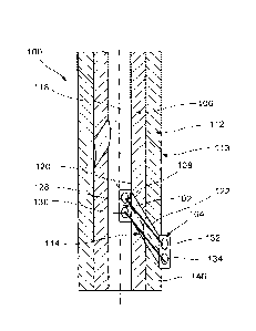
fixing the
intramedullary nail to the bone according to the system of Fig. 1; and
Fig. 10 shows a longitudinal cross-sectional view of a second set of plate
fixing the
intramedullary nail to the bone according to the system of Fig. 1.
Detailed Description
[0004] The present invention may be further understood with reference to the
following
description and the appended drawings, wherein like elements are referred to
with the same
reference numerals. The present invention relates to a system for treating
bone fractures and in
CA 02863899 2014-08-06
WO 2013/119434
PCT/US2013/023756
particular, a system for providing locking of intramedullary nails. Exemplary
embodiments of
the present invention describe a pair of plates and a connector which may be
passed through a
locking hole to fix an intrarnedullary nail to a bone. It should be noted that
the terms "proximal"
and "distal," as used herein, are intended to refer to a direction toward
(proximal) and away from
(distal) a user of the device.
[0005] As shown in Figs. 1 - 10, a system 100 according to an exemplary
embodiment of the
present invention comprises a first plate 102 and a second plate 104 for
fixing an intramedullary
nail 106 to a bone 112 via a connector 114 passing through a distal locking
hole 108 of the nail
106. As described in more detail below, the first plate 102 is positioned
outside the bone while
the second plate 104 is within the intramedullary nail with the two plates
102, 104 affixed on
opposite sides of the distal locking hole 108 and affixed to one another via
the connector 114,
which passes through the distal locking hole 108 and a corresponding hole 122
in the bone 112,
to fix the intramedullary nail 106 at a desired position within the bone 112.
The system 100
further comprises a flexible drill 116 for threading the connector 114 and the
first plate 102
through a channel 118 of the nail 106 to the distal locking hole 108 and for
drilling a hole 122
into the bone 112 aligned with the distal locking hole 108. The system 100 may
also comprise a
drill guide 124 for guiding the flexible drill 116 through the laterally
extending distal locking
hole 108.
[0006] As shown in Fig. 2, the intramedullary nail 106 is sized and shaped to
be inserted into
a medullary canal of a bone 112 to fix a fracture 113 thereof. The channel 118
extends through
the intramedullary nail 106 along a longitudinal axis L thereof and the distal
locking hole 108
extends laterally through the wall 110 of a distal portion thereof from an
interior 120 of the
channel 118 to an exterior 126 of the nail 106. A central axis C of the distal
locking hole 108
may extend through the wall 110 at an acute angle with respect to a
longitudinal axis L of the
intramedullary nail 106 such that the locking hole 108 extends from the
interior 120 to the
exterior 126 of the wall 110 toward a distal end of the intramcdullary nail
106. Although the
exemplary embodiment describes a single distal locking hole 108, it will be
understood by those
3
of skill in the art that the intramedullary nail 106 may include a plurality
of distal locking holes
108. Where the intramedullary nail 106 includes a plurality of distal locking
holes 108, each of
the locking holes 108 may extend through the wall 110 at varying positions
about and along the
nail 106. It will also be understood by those of skill in the art that
although the exemplary
embodiment describes a system 100 for distal locking of the intramedullary
nail, the system of
the present invention may also be used to lock the intramedullary nail to bone
via proximal
locking holes.
[0007] As shown in Figs. 7 - 10, the first and second plates 102, 104 are
sized and shaped so
that, in a first orientation they may be inserted through the channel 118 of
the intramedullary nail
106 but, in a second orientation (when oriented transverse to a locking hole
108), the plates 102,
104 contact edges of the locking holes 108 and are prevented from passing
therethrough. The
first and second plates 102, 104 include first openings 128, 132,
respectively, and second
openings 130, 134, respectively, extending therethrough similarly to a button.
The first and
second openings 128, 130, 132, 134 of the first and second plates 102, 104 are
sized and shaped
to permit the connector 114 to be threaded therethrough. The connector 114 may
be any
connector suitable for flexibly connecting the first and second plates 102.
104 together. It will be
appreciated by those skilled in the art that the thread-like connector 114 may
be any element
suitable to flexibly connect the first and second plates 102, 104 to one
another including, for
example, a suture, a shortening joining element such as the joining element
described in U.S.
Patent Application Publication No. 2008/281355, and a flexible wire such as a
cerclage wire. As
will be described in greater detail below, the first and second plates 102,
104 are positioned on
opposite sides of the distal locking hole 108 and attached to one another via
the connector 114 to
fix the intramedullary nail 106 to the bone 112.
[0008] As shown in Fig. 1, the drill 116 extends longitudinally from a
proximal end 136 to a
distal end 138 and is flexible along a length thereof. The proximal end 136
includes an eyelet
140 extending laterally therethrough such that the connector 114 may be
threaded therethrough,
4
CA 2863899 2019-05-17
CA 02863899 2014-08-06
WO 2013/119434
PCT/US2013/023756
similarly to a thread and needle. The connector 114 is threaded through the
eyelet 140 to attach
the first plate 102 thereto by threading the connector 114 through the first
and second openings
128, 130 thereof to form a closed loop with the connector 114 by tying and/or
knotting ends
thereof. The distal end 138 includes a drill tip 142 configured to facilitate
drilling of the hole
122 through the bone 112 as would be understood by those skilled in the art.
The drill 116 may
be formed of any of a variety of flexible materials suitable for drilling
through bone such as, for
example, nitinol, gum metal and PEEK.
1110091 The drill guide 124, as shown in Figs. 3 - 6, includes a shaft 154
extending from a
proximal end 156 to a distal end 158, the shaft 154 being sized and shaped for
insertion through
the channel 118 of the intramedullary nail 106. The proximal end 156 includes
an end member
160 couplable with a proximal end 107 of the intramedullary nail 106. The
drill guide 124
includes a lumen 148 extending through the end member 160 and at least a
portion of a length of
the shaft 154 along with a slot 150 extending laterally therethro ugh in
communication with a
distal end of the lumen 148. The lumen 148 is sized and shaped to permit the
drill 116 and the
first plate 102 to be slid therethrough.
[00101 The end member 160 and the proximal end 107 of the intramedullary nail
106 are
couplable to one another such that the slot 150 is aligned with the distal
locking hole 108. For
example the end member 160, as shown in Fig. 6, includes a protrusion 162
extending radially
outward from a portion thereof to engage a corresponding recess 164, as shown
in Fig. 7, along
an inner surface 166 of the proximal end 107 of the intramedullary nail 106.
Thus, when the
protrusion 162 and the recess 164 engage one another, the slot 154 is aligned
with the distal
locking hole 108. It will be understood by those of skill in the art that the
end member 160 and
the intramedullary nail 106 may include more than one protrusion 162 and
recess 164,
respectively. A length of the shaft 154 may be selected such that when the end
member 160 is
coupled with the proximal end 107 of the intramedullary nail 106, the slot 150
is aligned with the
distal locking hole 108. The lumen 148 includes a ramped surface 152
substantially opposing
the slot 150 such that when the drill 116 is slid therethrough, the distal end
138 of the drill 116
CA 02863899 2014-08-06
WO 2013/119434
PCT/US2013/023756
contacts the ramped surface 152 and is guided laterally out of the slot 150.
Thus, the end
member 160 is coupled to the intramedullary nail 106 so that the shaft 154 is
inserted into the
channel 118 and positioned therein such that the slot 150 is in alignment with
the distal locking
hole 108 to guide the drill 116 therethrough.
[0011] According to an exemplary surgical technique using the system 100, the
intramedullary
nail 106 is inserted along the medullary canal of the bone 112 to fix a
fracture thereof, as shown
in Fig. 2. The connector 114 may be threaded through the eyelet 140 of the
drill 116, inserted
through first and second openings 128, 130 of the first plate 102 and tied to
form a closed loop,
as shown in Fig. 1. It will be apparent to those skilled in the art, however,
that other ways of
connecting the first plate 102 to the drill 140 are also possible. For
example, the connector 114
may be pre-arranged or pre-connected to the eyelet 140. The shaft 154 of the
drill guide 124 is
inserted into the channel 118 and the end member 160 coupled to the proximal
end 107 of the
intramedullary nail 106 such that the slot 150 is aligned with the distal
locking hole 108. The
drill 116, with the connector 114 and first plate 102 assembled therewith, are
then inserted
through the lumen 148 of the drill guide 124, as shown in Figs. 3 and 4. The
drill 116 is slid
along the lumen 148 until the distal end 138 comes into contact with the
ramped surface 152 and
is deflected laterally out of the distal locking hole 108 with which it is
aligned. As the distal end
138 passes through the locking hole 108, the drill is rotated so that the
drill tip 142 drills a hole
122 into the bone 112 in alignment with the distal locking hole 108. The drill
116 is passed to
the exterior 146 of the bone 112, as shown in Fig. 7, until the first plate
108 contacts the interior
120 of the wall 110 surrounding the distal locking hole 108, as shown in Fig.
8.
[0012] The drill guide 124 is then removed from the intramedullary nail 106
and the drill 116
is detached from the first plate 102 by cutting the connector 114 to form two
loose ends 142,
144, which are then used to attach the second plate 104 to the exterior 146 of
the bone 112. In
particular, the first loose end 142 is threaded through the first opening 132
of the second plate
104 and the second loose end 144 is threaded through the second opening 134 of
the second plate
104. The two loose ends 142, 144 are then tied/knotted, applying a tension to
the connector 114
6
CA 02863899 2014-08-06
WO 2013/119434
PCT/US2013/023756
and affixing the second plate 104 to the exterior 146 of the bone 112
surrounding the hole 122.
Thus, as shown in Fig. 9, the first and second plates 102, 104 are attached to
one another via the
connector 114 from the interior 120 of the channel 118 to the exterior 146 of
the bone 112 such
that the intramedullary nail 106 is fixed to the bone 112. The above described
steps may be
repeated using additional plates 102', 104' and connectors 114' to fix the
intramedullary nail 106
to the bone 112 via any additional locking holes 108' of the intrarnedullary
nail 106, as shown in
Fig, 10.
[0013] It
will be apparent to those skilled in the art that various modifications and
variations
can be made in the structure and the methodology of the present invention,
without departing
from the spirit or scope of the invention. Thus, it is intended that the
present invention cover the
modifications and variations of this invention provided that they come within
the scope of the
appended claims and their equivalents.
7
