Note: Descriptions are shown in the official language in which they were submitted.
CA 02864291 2014-08-12
WO 2013/159798 PCT/EP2012/002780
- 1 -
EXTRACTING LENTICULES FOR REFRACTIVE CORRECTION
TECHNICAL FIELD
The present disclosure relates generally to corneal surgical devices, and more
particularly to extracting lenticules for refractive correction.
BACKGROUND
Refractive surgery uses lasers to reshape the cornea to correct refractive
defects of the eye. According to some techniques, a flap of the eye is lifted
to
expose a portion of the cornea that is reshaped by ablation using an excimer
laser.
The flap is then replaced. According to other techniques, a femtosecond laser
makes incisions in the cornea to create a lenticule. The lenticule is removed
to
reshape the cornea.
BRIEF SUMMARY
In certain embodiments, a device for refractive correction comprises a laser
device and a control computer. The laser device is configured to create a
lenticule in
an eye using pulsed laser radiation having ultrashort pulses. The laser device
includes one or more controllable components configured to control a focus of
the
pulsed laser radiation. The control computer is configured to instruct the one
or
more controllable components to create a channel with the pulsed laser
radiation to
facilitate separation of the lenticule from the eye, create a posterior
incision with the
pulsed laser radiation to form a posterior side of the lenticule, and create
an anterior
incision with the pulsed laser radiation to form an anterior side of the
lenticule.
In certain embodiments, a method for refractive correction includes
controlling
a focus of pulsed laser radiation having ultrashort pulses. A channel is
created with
the pulsed laser radiation to facilitate separation of the lenticule from the
eye. A
posterior incision is created with the pulsed laser radiation to form a
posterior side of
the lenticule. An anterior incision is created with the pulsed laser radiation
to form an
anterior side of the lenticule.
In certain embodiments, a tangible computer-readable medium stores
computer code for refractive correction that when executed by a computer is
configured to control a focus of pulsed laser radiation having ultrashort
pulses. The
- 2 -
computer code is also configured to create a channel with the pulsed laser
radiation
to facilitate separation of the lenticule from the eye, create a posterior
incision with
the pulsed laser radiation to form a posterior side of the lenticule, and
create an
anterior incision with the pulsed laser radiation to form an anterior side of
the
lenticule.
In further exemplary embodiments, a device for refractive correction, the
device comprising: a laser device having a plurality of ultrashort pulses, the
laser
device comprising one or more controllable components configured to control a
focus of the pulsed laser radiation; and a control computer configured to
instruct the
1.0 one or more controllable components to: move the one or more
controllable
components to control an application and the focus of the pulsed laser
radiation
from the laser device such that a straight posterior channel in an eye having
a
length between 1 to 5 millimeters is produced; move the one or more
controllable
components to control the application and the focus of the pulsed laser
radiation
from the laser device such that a posterior incision to form a posterior side
of a
lenticule in the eye, the posterior channel substantially tangential to the
posterior
incision to facilitate separation of the posterior side of the lenticule from
the eye is
produced; move the one or more controllable components to control the
application
and the focus of the pulsed laser radiation from the laser device such that a
straight
anterior channel having a length between 1 to 5 millimeters is produced; move
the
one or more controllable components to control the application and the focus
of the
pulsed laser radiation from the laser device such that an anterior incision to
form an
anterior side of the lenticule is produced , the anterior incision and the
posterior
incision yielding a refractive profile for refractive correction, the anterior
channel
substantially tangential to the anterior incision to facilitate separation of
the anterior
side of the lenticule from the eye, the anterior channel disconnected from the
posterior side of the lenticule, the posterior channel disconnected from the
anterior
side of the lenticule; and move the one or more controllable components to
control
the application and the focus of the pulsed laser radiation from the laser
device
such that one of the anterior channel or the posterior channel with the same
width at
CA 2864291 2018-02-28
- 2a -
each end and produce the other of the anterior channel or the posterior
channel
with different widths at each end is produced.
In another exemplary embodiment one or more tangible computer-readable
non-transitory media storing statements and instructions that when executed by
a
.. computer cause the computer to: move one or more controllable components to
control an application and a focus of a pulsed laser radiation from a laser
device
such that a lenticule in an eye is produced, the pulsed laser radiation having
a
plurality of ultrashort pulses; move the one or more controllable components
to
control the application and focus of the pulsed laser radiation from the laser
device
such that a straight posterior channel having a length between 1 to 5
millimeters is
produced; move the one or more controllable components to control the
application
and focus of the pulsed laser radiation from the laser device such that a
posterior
incision to form a posterior side of the lenticule is produced, the posterior
channel
substantially tangential to the posterior incision to facilitate separation of
the
posterior side of the lenticule from the eye; move the one or more
controllable
components to control the application and focus of the pulsed laser radiation
from
the laser device such that a straight anterior channel having a length between
1 to 5
millimeters is produced; move the one or more controllable components to
control
the application and focus of the pulsed laser radiation from the laser device
such
that an anterior incision to form an anterior side of a lenticule is produced,
the
anterior incision and the posterior incision yielding a refractive profile for
refractive
correction, the anterior channel substantially tangential to the anterior
incision to
facilitate separation of the anterior side of the lenticule from the eye, the
anterior
channel disconnected from the posterior side of the lenticule, the posterior
channel
disconnected from the anterior side of the lenticule; and move the one or more
controllable components to control the application and focus of the pulsed
laser
radiation from the laser device such that one of the anterior channel or the
posterior
channel with the same width at each end and produce the other of the anterior
channel or the posterior channel with different widths at each end is
produced.
CA 2864291 2018-02-28
- 2b -
BRIEF DESCRIPTION OF THE DRAWINGS
Exemplary embodiments of the present disclosure will now be described by
way of example in greater detail with reference to the attached figures, in
which:
FIGURE 1 illustrates an example of a device configured to perform refractive
correction according to certain embodiments;
FIGURE 2 illustrates a top view of an example of creating a lenticule
according to certain embodiments;
FIGURE 3 illustrates a cross-section of an example of creating a lenticule
according to certain embodiments; and
1.0 FIGURE 4 illustrates an example of a method for creating a lenticule
according to certain embodiments.
DESCRIPTION OF EXAMPLE EMBODIMENTS
Referring now to the description and drawings, example embodiments of the
disclosed apparatuses, systems, and methods are shown in detail. The
description
and drawings are not intended to be exhaustive or otherwise limit or restrict
the
claims to the specific embodiments shown in the drawings and disclosed in the
description. Although the drawings represent possible embodiments, the
drawings
are not necessarily to scale and certain features may be simplified,
exaggerated,
removed, or partially sectioned to better illustrate the embodiments. In
addition,
certain drawings may be in schematic form.
FIGURE 1 illustrates an example of a device 10 configured to create a
lenticule according to certain embodiments. In the embodiments, the device 10
includes a laser device and a control computer. The laser device can create a
lenticule in the cornea (such as the stroma) of an eye using pulsed laser
radiation
with ultrashort pulses (such as pico-, femto-, or aftosecond pulses). The
lenticule
CA 2864291 2018-02-28
CA 02864291 2014-08-12
WO 2013/159798 PCT/EP2012/002780
- 3 -
may be shaped according to a refractive correction profile such that when the
lenticule is removed the refractive correction is applied.
The laser device may include controllable components that focus the pulsed
laser radiation. The control computer instructs the controllable components to
focus
the pulsed laser radiation at the cornea to create a channel (such as an
anterior or
posterior channel) to facilitate separation of the lenticule. The pulsed laser
radiation
may create an anterior incision to form an anterior side of the lenticule and
a
posterior incision to form a posterior side of the lenticule. In certain
embodiments,
the pulsed laser radiation may create a removal incision through which the
lenticule
may be manually or automatically removed.
In the illustrated example of FIGURE 1, the device 10 performs surgery on an
eye 22. The device 10 includes a laser device 15, a patient adapter 20, a
control
computer 30, and a memory 32 coupled as shown. The laser device 15 may include
a laser source 12, a scanner 16, one or more optical elements 17, and/or a
focusing
objective 18 coupled as shown. The patient adapter 20 may include a contact
element 24 (which has an abutment face 26 disposed outwardly from a sample)
and
a sleeve 28 coupled as shown. The memory 32 stores a control program 34. The
sample may be an eye 22.
The laser source 12 generates a laser beam 14 with ultrashort pulses. In this
document, an "ultrashort" pulse of light refers to a light pulse that has a
duration that
is less than a nanosecond, such as on the order of a picosecond, femtosecond,
or
attosecond. The focal point of the laser beam 14 may create a laser-induced
optical
breakdown (LIOB) in tissues such as the cornea. The laser beam 14 may be
precisely focused to allow for precise incisions in the corneal cell layers,
which may
reduce or avoid unnecessary destruction of other tissue.
Examples of laser source 12 include femtosecond, picosecond, and
attosecond lasers. The laser beam 14 may have any suitable wavelength, such as
a
wavelength in the range of 300 to 1500 nanometers (nm), for example, a
wavelength
in the range of 300 to 650, 650 to 1050, 1050 to 1250, or 1100 to 1500 nm. The
laser beam 14 may also have a relatively small focus volume, e.g., 5
micrometers
(pm) or less in diameter. In certain embodiments, the laser source 12 and/or
delivery
channel may be in a vacuum or near vacuum.
CA 02864291 2014-08-12
WO 2013/159798 PCT/EP2012/002780
- 4 -
The scanner 16, optical elements 17, and focusing objective 18 are in the
beam path. The scanner 16 transversely and longitudinally controls the focal
point of
the laser beam 14. "Transverse" refers to a direction at right angles to the
direction
of propagation of the laser beam 14, and "longitudinal" refers to the
direction of beam
propagation. The transverse plane may be designated as the x-y plane, and the
longitudinal direction may be designated as the z-direction. In certain
embodiments,
the abutment face 26 of the patient interface 20 is on an x-y plane.
The scanner 16 may transversely direct the laser beam 14 in any suitable
manner. For example, the scanner 16 may include a pair of galvanometrically
io actuated scanner mirrors that can be tilted about mutually
perpendicular axes. As
another example, the scanner 16 may include an electro-optical crystal that
can
electro-optically steer the laser beam 14. The scanner 16 may longitudinally
direct
the laser beam 14 in any suitable manner. For example, the scanner 16 may
include
a longitudinally adjustable lens, a lens of variable refractive power, or a
deformable
15 mirror that can control the z-position of the beam focus. The focus
control
components of the scanner 16 may be arranged in any suitable manner along the
beam path, e.g., in the same or different modular units.
One (or more) optical elements 17 direct the laser beam 14 towards the
focusing objective 18. An optical element 17 may be any suitable optical
element
20 that can reflect, refract, and/or diffract the laser beam 14. For
example, an optical
element 17 may be an immovable deviating mirror. The focusing objective 18
focuses the laser beam 14 onto the patient adapter 20, and may be separably
coupled to the patient adapter 20. The focusing objective 18 may be any
suitable
optical element, such as an f-theta objective.
25 Patient adapter 20 interfaces with the cornea of the eye 22. In the
example,
the patient adapter 20 has a sleeve 28 coupled to a contact element 24. The
sleeve
28 couples to the focusing objective 18. The contact element 24 may be
translucent
or transparent to the laser radiation and has an abutment face 26 that
interfaces with
the cornea and may level a portion of the cornea. In certain embodiments, the
30 abutment face 26 is planar and forms a planar area on the cornea.
The abutment
face 26 may be on an x-y plane, so the planar area is also on an x-y plane. In
other
embodiments, the abutment face 26 need not be planar, e.g., may be convex or
concave.
CA 02864291 2014-08-12
WO 2013/159798 PCT/EP2012/002780
- 5 -
The control computer 30 controls controllable components, e.g., the laser
source 12 and scanner 16, in accordance with the control program 34. The
control
program 34 contains computer code that instructs the controllable components
to
focus the pulsed laser radiation at a region of the cornea to photodisrupt at
least a
portion of the region.
In certain examples of operation, the scanner 16 may direct the laser beam 14
to form incisions of any suitable geometry. Examples of types of incisions
include
bed incisions and lateral incisions. A bed incision is two-dimensional
incision that is
typically on an x-y plane. The scanner 16 may form a bed incision by focusing
the
io laser beam 14 at a constant z-value under the abutment face 26 and
moving the
focus in a pattern in an x-y plane. A lateral incision is an incision that
extends from
under the corneal surface (such as from a bed incision) to the surface. The
scanner
16 may form a lateral incision by changing the z-value of the focus of the
laser beam
14 and optionally changing the x and/or y values.
15 Any suitable portion of the cornea may be photodisrupted. One or
more of
any of the corneal layers may be selected for photodisruption. In addition, a
portion
of a cell layer may be photodisrupted in the z-direction, but part of the cell
layer may
remain on the cornea. Moreover, a particular area (or "target zone") in the x-
y plane
may be selected for photodisruption. For example, a target zone that forms a
bed
20 incision may be photodisrupted.
The device 10 may photodisrupt a corneal layer in any suitable manner. In
certain embodiments, the control computer 30 may instruct the laser device to
focus
the laser beam 14 at a constant z-value under the abutment face 26 and move in
a
pattern in the x-y plane that substantially covers the target zone. Any
suitable
25 pattern may be used. For example, according to a zigzag pattern, the
scan path has
a constant y-value and moves in the +x direction. When the scan path reaches a
point of the border of the target zone, the path moves to a next y value that
is a
predetermined distance from the previous y-value and then moves in the ¨x
direction
until it reaches another point of the border. The scan path continues until
the entire
30 target zone is scanned. As another example, according to a spiral
pattern, the scan
path starts at or near the center of the target zone and moves in a spiral
pattern until
the path reaches the border of the target zone, or vice-versa.
CA 02864291 2014-08-12
WO 2013/159798 PCT/EP2012/002780
- 6 -
As the laser beam 14 travels along the scan path, the laser beam pulses
create microdisruptions. In certain situations, a scan path pattern may yield
a non-
uniform distribution of microdisruptions over the target zone. In these cases,
the
laser beam 14 may be modified to make the distribution more uniform. For
example,
certain pulses may be blocked or the pulse energy may be decreased to reduce
number of or the effect of the pulses in a particular region.
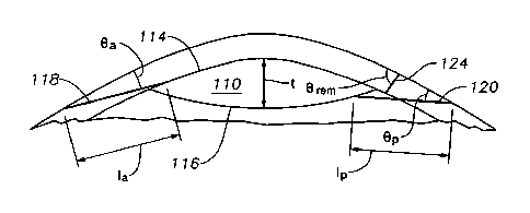
FIGURES 2 and 3 illustrate an example of creating a lenticule 110 according
to certain embodiments. FIGURE 2 illustrates a top view of creating the
lenticule
110, and FIGURE 3 illustrates a cross-section of creating the lenticule 110.
The lenticule 110 may have any suitable shape. In certain embodiments, the
lenticule 110 may have a flattened, disc shape with any suitable perimeter,
e.g., a
circular, elliptical, free form, or irregular. The lenticule 110 may have any
suitable
size. For example, the lenticule 110 may have any suitable diameter d (or
radius r),
such as a diameter d in the range of 1 to 10 mm, such as approximately 6.5 mm.
The lenticule 110 may have any suitable thickness t, such as a value in the
range of
10 to 200 micrometers (pm), such as approximately 50 pm.
The device 10 may create the lenticule 110 in any suitable manner. In certain
embodiments, the control computer 30 may instruct the laser device to create
an
anterior incision 114 and a posterior incision 116, which are types of bed
incisions,
using laser radiation. The anterior incision 114 forms the anterior side of
the
lenticule 110, and the posterior incision 116 forms the posterior side of the
lenticule
110. In certain embodiments, the anterior incision 114 and/or posterior
incision 116
yields a refractive profile for refractive correction such that a refractive
correction is
applied after removal of the lenticule 110.
The anterior 114 and posterior 116 incisions may be created in any suitable
order and in any suitable manner. In certain embodiments, a channel, which may
be
a type of lateral incision, may facilitate removal of the lenticule 110. For
example, an
anterior channel 118 may be used to separate the anterior side of the
lenticule 110
from the surrounding tissue, and/or a posterior channel 120 may be used to
separate
the posterior side of the lenticule 110 from the surrounding tissue. In the
embodiments, the channel may be used to insert (e.g., manually or
automatically) an
instrument into an incision to separate a surface of the lenticule 110 from
the rest of
the cornea in order to remove the lenticule 110.
CA 02864291 2014-08-12
WO 2013/159798 PCT/EP2012/002780
- 7 -
The channels and incisions may be created in any suitable order. For
example, a channel may be created before or after the corresponding incision.
As
another example, an anterior channel and/or anterior incision may be created
before
or after a posterior channel and/or posterior incision.
A channel may have any suitable size and shape. In certain embodiments, a
channel with a center line ak where i identifies the channel, may have any
suitable
length Ii, width w,, angle (Di of center line a, with respect to radius r, and
angle 8, of
center line a, with respect to the anterior surface of the eye. In FIGURE 2,
the
anterior channel 118 has a narrower width wa towards the entrance of the
channel
and a wider width w." towards the center of lenticule 110. The posterior
channel 120
has the same width wp from end to end. The widths may have any suitable value,
such as a value in the range 0.5 to 4, 1 to 3, or 1.5 to 2.0 mm. In other
examples,
the posterior channel may be shaped like channel 118 or may have any other
suitable shape, and the anterior channel may be shaped like channel 120 or may
have any other suitable shape. The posterior and anterior channels may have
the
same shape or may have different shapes. The center line a3 of the anterior
channel
118 is at an angle (1)a with respect to radius r. The center line ap of the
posterior
channel 120 is at an angle Op (not labeled) of 0 with respect to radius r.
The angles
(Di may have any suitable value, such as a value in the range 0 to 5, 5 to 10,
10 to
.. 15, or 15 to 20 degrees.
The channels and incisions may be created in any suitable order. For
example, a channel may be created before or after the corresponding incision.
As
another example, an anterior channel and/or anterior incision may be created
before
or after a posterior channel and/or posterior incision.
In FIGURE 3, the anterior channel 118 has length I., and the posterior
channel 120 has length 1p. The lengths may have any suitable value, such as a
value
in the range 1 to 5 mm. The center line a3 of anterior channel 118 has an
angle ea
with respect to the surface of the eye, and the center line ap of posterior
channel 120
has an angle 8p. The angles 8, may have any suitable value, such as a value
where
the channel is substantially tangential or nearly tangential (within 5
degrees) to the
corresponding incision to allow for an instrument that is inserted into the
channel to
enter the incision and separate a surface of the lenticule from the rest of
the cornea.
For example, angles 8; may have a value in the range 0 to 10, 10 to 20, or 20
to 30
CA 02864291 2014-08-12
WO 2013/159798 PCT/EP2012/002780
- 8 -
degrees, which may allow the channels to be tangential or nearly tangential to
a
surface of the lenticule. In certain embodiments, the angles 8; may have
different
values at the entrance of the eye (e.g., approximately 90 degrees) and then
change
to values that allow the channels to be tangential or nearly tangential to a
surface of
the lenticule.
The lenticule 110 may be removed in any suitable manner. In certain
embodiments, the lenticule 110 may be extracted through an anterior incision
or
posterior incision. In other embodiments, the control computer 30 may instruct
the
laser device to form a removal incision through which the lenticule 110 may be
manually or automatically extracted. The removal incision 124 may have any
suitable size or shape. In certain embodiments, the removal incision 124 may
have
any suitable length 'rem and angle eõm with respect to the surface of the eye.
For
example, length 'rem may have a value that allows for the lenticule 110 to be
extracted through it, such as a value that is approximately the size of
diameter d, but
perhaps up to 2 mm larger or smaller. Angle Orem may have a value in the range
of
80 to 110 degrees.
FIGURE 4 illustrates an example of a method for creating a lenticule in a
cornea of an eye according to certain embodiments. The method may be performed
by the system 10 of FIGURE 1.
The method starts at step 210, where a posterior channel 120 is created. The
posterior channel 120 may be used to separate the posterior side of the
lenticule 110
from the rest of the eye. A posterior incision 116 is created at step 212. The
posterior incision 116 forms a posterior surface of the lenticule 110. An
anterior
channel 118 is created at step 214. The anterior channel 118 may be used to
separate the anterior side of the lenticule 110 from the rest of the eye. An
anterior
incision 114 is created at step 216. The anterior incision 114 forms an
anterior
surface of the lenticule 110.
A removal incision 124 is created at step 218. The removal incision 124
allows for the removal of the lenticule 110. The lenticule 110 is removed
through the
removal incision 124 at step 220. The lenticule 110 may be manually or
automatically removed. In other embodiments, the lenticule 110 may be removed
through the anterior 118 or posterior 120 channel.
CA 02864291 2014-08-12
WO 2013/159798 PCT/EP2012/002780
- 9 -
A component (such as the control computer 30) of the systems and
apparatuses disclosed herein may include an interface, logic, memory, and/or
other
suitable element, any of which may include hardware and/or software. An
interface
can receive input, send output, process the input and/or output, and/or
perform other
suitable operations. Logic can perform the operations of a component, for
example,
execute instructions to generate output from input. Logic may be encoded in
memory
and may perform operations when executed by a computer. Logic may be a
processor, such as one or more computers, one or more microprocessors, one or
more applications, and/or other logic. A memory can store information and may
comprise one or more tangible, computer-readable, and/or computer-executable
storage medium. Examples of memory include computer memory (for example,
Random Access Memory (RAM) or Read Only Memory (ROM)), mass storage media
(for example, a hard disk), removable storage media (for example, a Compact
Disk
(CD) or a Digital Video or Versatile Disk (DVD)), database and/or network
storage
(for example, a server), and/or other computer-readable media.
In particular embodiments, operations of the embodiments may be performed
by one or more computer readable media encoded with a computer program,
software, computer executable instructions, and/or instructions capable of
being
executed by a computer. In particular embodiments, the operations may be
performed by one or more computer readable media storing, embodied with,
and/or
encoded with a computer program and/or having a stored and/or an encoded
computer program.
Although this disclosure has been described in terms of certain embodiments,
modifications (such as changes, substitutions, additions, omissions, and/or
other
modifications) of the embodiments will be apparent to those skilled in the
art.
Accordingly, modifications may be made to the embodiments without departing
from
the scope of the invention. For example, modifications may be made to the
systems
and apparatuses disclosed herein. The components of the systems and
apparatuses
may be integrated or separated, and the operations of the systems and
apparatuses
may be performed by more, fewer, or other components. As another example,
modifications may be made to the methods disclosed herein. The methods may
include more, fewer, or other steps, and the steps may be performed in any
suitable
order.
CA 02864291 2014-08-12
WO 2013/159798 PCT/EP2012/002780
- 10 -
Other modifications are possible without departing from the scope of the
invention.
For example, the description illustrates embodiments in particular
practical applications, yet other applications will be apparent to those
skilled in the
art. In addition, future developments will occur in the arts discussed herein,
and the
disclosed systems, apparatuses, and methods will be utilized with such future
developments.
The scope of the invention should not be determined with reference to the
description.
In accordance with patent statutes, the description explains and
illustrates the principles and modes of operation of the invention using
exemplary
embodiments. The description enables others skilled in the art to utilize the
systems,
apparatuses, and methods in various embodiments and with various
modifications,
but should not be used to determine the scope of the invention.
The scope of the invention should be determined with reference to the claims
and the full scope of equivalents to which the claims are entitled. All claims
terms
should be given their broadest reasonable constructions and their ordinary
meanings
as understood by those skilled in the art, unless an explicit indication to
the contrary
is made herein. For example, use of the singular articles such as "a," "the,"
etc.
should be read to recite one or more of the indicated elements, unless a claim
recites an explicit limitation to the contrary. As another example, "each"
refers to
each member of a set or each member of a subset of a set, where a set may
include
zero, one, or more than one element. In sum, the invention is capable of
modification, and the scope of the invention should be determined, not with
reference to the description, but with reference to the claims and their full
scope of
equivalents.