Note: Descriptions are shown in the official language in which they were submitted.
CA 02865182 2014-08-21
WO 2013/124-162
PCT/EP2013/053632
1
MEANS AND METHODS OF MEASURING PARATHYROID HORMONE
IN PATIENTS SUFFERING FROM OXIDATIVE STRESS
FIELD OF THE INVENTION
[001] The invention relates to means and methods of measuring parathyroid
hormone in samples of body fluid.
BACKGROUND OF THE INVENTION
[002] The parathyroid hormone (PTH) is formed in the parathyroid gland
(Giandulae parathyroideae) and secreted into the blood circulation. In the
intact form it
consists of a single polypeptide chain having 84 amino acids and a molecular
weight of
ca. 9500 Dalton (see SWISS-PROT: P01270, PTHY-HUMAN). Together with vitamin-D
and calcitonin it brings about the mobilization of calcium and phosphate out
of the bone
skeleton and increases the uptake of calcium in the intestines and the
excretion of
phosphate via the kidneys. The concentration of biologically active PTH
peptides in
plasma or serum is thus an important diagnostic parameter for determining
presence
and degree of hyper- or hypo-parathyroidism; for a quantification of
osteablast and/or
osteoclast activity; a treatment with vitamin-D and vitamin-D metabolites; an
estimation
of the presence of aluminium or a possible oestrogen deficiency in post-
menopausal
dialysis patients; for determining the steroid or cyclosporin dosage after
kidney
transplantations or a treatment or prevention of pathological bone marrow
changes,
uraemic conditions and chronic kidney failure.
[003] Secondary hyper-parathyroidism further occurs frequently in chronic
kidney disease as an adaptive response to deteriorating renal function. This
is because
circulating 1,25-dihydroxy vitamin D starts to decrease very early in stage 2
of chronic
kidney disease and continues to fall as the glomerular filtration rate (GFR)
decreases
further, and the renal 1 a-hydroxylase is inhibited by hyperphosphataemia,
hyper-
uricaernia, metabolic acidosis as well as 25-hydroxyvitamin D deficiency. As
GFR
decreases below 60 mlimin/1-73.m2 phosphate is retained which stimulates
secretion
of PTH. Hypocalcaemia develops as the GFR decreases below 50 mUmin/1=73.m2,
further stimulating a release of PTH. With disease progression, intact PTH (aa
1-84)
half-life increases and C-terminal fragments of the hormone accumulate in
serum. A
relative state of end-organ resistance to the hormone exists but chronic
elevation of it
CA 02865182 2014-08-21
WO 2013/12-1462
PCT/EP2013/053632
has major consequences resulting in bone loss, particularly of cortical bone,
fractures,
vascular calcification, cardiovascular disease, and hence an increased
cardiovascular
mortality (cf Fraser WD, Hyperparathyroidism. Lancet. 2009; 374:145f). A
reliable
method of determining the concentration of biologically active PTH peptides in
serum is
therefore key for detecting patients with hyperparathyroidism as well as for
subsequent
monitoring of therapeutic interventions.
[004] The first generation of immunoassays for measuring PTH in serum were
based on radiolabeled bovine PTH peptides and polyclonal antisera against
parathyroid hormone (Berson SA et al, Proc Natl Acad Sci U S A. 1963; 49:613-
617).
As the biologic activity is located in the amino-terminal portion of the PTH
peptide and
the PTH peptide following its secretion into circulation degraded within
minutes in
active and inactive fragments, the radioimmunoassay were also detecting
inactive
degradation products. The first generation of PTH assays therefore produced no
reliable clinical measurements since the sera of patients with a renal failure
contain
high concentrations of inactive PTH fragments.
[005] The second generation of immunoassays uses two antibodies, one
binding in the amino-terminal portion of the PTH peptide with the biologic
activity and
the other in its C-terminal portion. The characterising with synthetic
fragments showed
however that these immunoassays also determined an inactive large PTH (aa 7-
84)
fragment (John MR et al. (1999), J. Clin. Endocrinol. Metab., 84. 4287-4290;
Gao P et
at. 2000, Poster M455, ASBMR 22nd Annual Meeting; Roth HJ et at. (2000),
Poster
P1288; 11th International Congress of Endocrinology, Sydney). This co-
determination
of the inactive large PTH fragment (7-84) was made responsible for the
discrepancy
between measured PTH concentrations and clinical findings as the large PTH
fragment
is likely competing with intact PTH peptides for the binding site of the PTH
receptor.
[006] A third generation PTH assay has been developed to overcome the
problems with inactive large PTH fragments, which however fails to improve the
diagnosis of bone diseases or other clinical signs of secondary
hyperparathyroidism in
uraemic patients (Brossard JH et al., Influence of glomerular filtration rate
on non-(1-
84) parathyroid hormone (PTH) detected by intact PTH assays, Clin Chem. 2000;
46:697-703). There have been speculations about systematic errors in the
determination or a PTH resistance of osteoblasts or a genetically reduced
expression
of PTH receptor.
CA 02865182 2014-08-21
WO 2013/124-162
PCT/EP2013/053632
3
[007] In summary, it is generally accepted in the field that the parathyroid
hormone is cleaved in liver, kidney and circulation within minutes into active
and
inactive fragments and that some fragments have a biological activity
comparable with
intact PTH peptides whereas others such as hPTH (3-34) seem to inhibit the
effects of
parathyroid hormone (see EP-A 0 349 545; Schmidt-Gayk et al. (1999) Osteologie
forum, 5, 48-58), Suva et al, (1987) Science, 237, 893ff; EP 0 451 867).
Moreover, that
large PTH non-(1-84) fragments may lead to erroneous determinations (LePage R.
et
al. (1998) Clin. Chem., 44, 805-809). The term "large PTH fragment" has been
coined
for PTH fragments which lack amino acid residues at the amino-terminus but
which are
detected by 2" generation PTH assays. Additionally, dipeptidyl peptidase-4
(DPP4) is
expressed on the surface of many cell types and a rather indiscriminate senne
exopeptidase. This led to the hypothesis of PTH further being in vivo a
substrate of
DPP4 or a similar exoproteinase. Consequently, a two-site immunoassay has been
developed employing antibodies that can distinguish between biologically
active and
biologically inactive PTH peptides that are missing the utmost 2 amino-
terminal amino
acids (see WO 2001/44818 (Armbruster et al), WO 96/10041 (Magerlein et a)); WO
2003/03986 (Hutchison JS)).
[008] However, it was found that serum samples of uraemic patients may
contain intact PTH polypeptide chains which are inactive because oxidized at
one of its
methionines. Such kind of oxidation seems to be particularly relevant for
dialysis
patients whose blood plasma is exposed to oxidative stress. This led to the
development of an immunoassay for determination of non-oxidized PTH (aa 1-84)
and
biologically active fragments thereof (WO 2002/082092). Notwithstanding, it
needs to
be ascertained why uraemic patients with normal bone transformation sometimes
have
serum levels of intact PTH which are more than 2.5 higher than in patients
with healthy
kidneys (pathological limit in the case of patients with healthy kidneys: 65
ug PTH/L; for
patients having uraemic conditions: 165 pg PTH/L serum). Further, uraemic
patients
with relatively high PTH values often manifest significant differences in bone
transformation (Slatopolsky E et al. (2000), Kidney Int., 58, 753-761). Thus
these
patients often have in the serum eight to ten times increased PTH
concentrations, but
low normal values for bone specific alkaline phosphatase (ostase). These
patients
seem to free from symptoms of an excessive PTH activity.
[009] The state of the art therefore still represents a problem. It is further
an
object of the invention to make available a fast and reliable method for the
CA 02865182 2014-08-21
WO 2013/124462
PCT/EP2013/053632
4
determination of active parathyroid hormone in a sample of a body fluid, which
method
particularly allows an early detection of a deteriorating renal function.
SUMMARY OF THE INVENTION
[010] This problem is solved by a method for obtaining an antibody or
antibody fragment to a conformational epitope specific for misfolded inactive
human
parathyroid hormone and fragments thereof, comprising the steps of a)
immunizing an
animal with an immunogen which comprises oxidized parathyroid hormone or a
oxidized fragment of parathyroid hormone, or both; and b) recovering
antibodies;
whereby the complementary determining region of the antibody or antibody
fragment or
single chain antibody specifically recognizes a conformational epitope
(antigenic
determinant) which is present on oxidized parathyroid hormone and fragments
thereof
only but not regular bioactive human parathyroid hormone.
[011] The disclosure further relates to a complementary determining region
recognizing a conformational epitope presented by human parathyroid hormone or
a
fragment thereof comprising at positions 8, 18 or both methionine R-sulfoxide,
methionine L-sulfoxide or methionine sulfone. The complementary determining
region
may also recognize a conformational epitope of a human parathyroid hormone or
a
fragment thereof comprising at position 22 oxidized tryptophan and/or lacking
the
utmost aminoterminal amino acids at positions 1 and 2 or both. To be clear it
is the
human parathyroid hormone or fragment thereof which comprises in its amino
acid
sequence at positions 8, 18 or 22 oxidized amino acids. This does not mean
that the
conformational epitope is made up of a primary structure comprising any one of
these
oxidized amino acids but the conformational epitope is a tertiary structure
formed by
the oxidized PTH sequence which has flipped into an alternative tertiary
structure and
the conformational epitope is a characteristic portion of that alternative
tertiary structure
for which reason the respective antibody or antibody fragment recognizes all
types of
oxidized or misfolded PTH structures.
[012] The disclosed antibody may be a monoclonal mouse or rat antibody.
The preferred immunogen for challenge, preferably given with incomplete
Freund's
(mineral oil only) is a carrier protein having bound as hapten any one of
synthetic
oxidized human parathyroid hormone, synthetic oxidized fragment of human
parathyroid hormone or synthetic oxidized peptide comprising the amino acid
sequence
1 to 38 of human parathyroid hormone or a substantial portion, fragment or
variant
CA 02865182 2014-08-21
WO 2013/12-U62
PCT/EP2013/053632
thereof. The antibodies elicited by this challenge may be isolated or screened
by
affinity chromatography using fragments of synthetic oxidized human
parathyroid
hormone linked to a solid phase or a marker molecule. The screening or
isolation of
the antibody is done using a conformational epitope which is made up by the
oxidized
5 human parathyroid hormone or a fragment thereof, preferably comprising
the amino
acid sequence 3 to 34 wherein the methionine at position 8 is likely first
oxidized.
[013] A further aspect of the disclosure relates to a bindina material for
removing oxidized human parathyroid hormone from a sample such as a serum
sample
of a patient on dialysis, which binding material comprises bound to a solid
phase
antibodies or antibody fragments or single-chain antibody fragments as
disclosed
above. The binding material may be in the form of a slurry, preferably a
slurry of
Sepharose beads having covalently linked a conformational antibody for
oxidized PTH
and fragments thereof.
[014] Another aspect of the disclosure relates to a method of measuring the
concentration of human parathyroid hormone in a sample of a body fluid,
comprising
the step of contacting the sample with a solid phase or slurry as described
comprising
antibodies recognizing oxidized parathyroid hormone, and measuring the
concentration
of parathyroid hormone in the flow-through or supernatant.
[015] This method of measuring the concentration of human parathyroid
hormone in a sample of a body fluid may comprise the step of measuring the
concentration of parathyroid hormone by a two-site immunoassay wherein one
antibody binds in the aminoterminal portion with amino acids 1 to 34 of the
parathyroid
hormone.
[016] The disclosure further encompasses a method of measuring the
concentration of human parathyroid hormone in a sample, comprising the step of
measuring the concentration of parathyroid hormone fragments by tandem mass
spectroscopy, optionally preceded by modern liquid chromatography.
[017] Another aspect of the disclosure concerns the use of a binding material
or antibodies or method as described in any preceding claim in a method of
diagnosis,
notably for determining in vitro secondary hyperparathyroidism, kidney failure
or both.
5a
[017.1] Another method of the disclosure concerns a method for obtaining
monoclonal antibody
molecules specific for oxidatively inactivated human parathyroid hormone
(hPTH) peptide and
circulating fragments thereof, comprising
a) obtaining antibodies against human parathyroid hormone peptide by
immunizing a non-
human animal with an immunogen comprising as hapten the amino acid sequence 1
to 38 of
parathyroid hormone oxidized at methionine 8, 18 or both, or a fragment
comprising the amino
acid sequence 1 to 38 of parathyroid hormone oxidized at methionine 8, 18 or
both, and
recovering said antibodies from said non-human animal;
b) selecting or purifying said antibodies from antibody molecules that bind to
bioactive human
parathyroid hormone peptide under physiological conditions to obtain
antibodies that
specifically recognize oxidatively inactivated parathyroid hormone or
fragments thereof;
c) selecting or purifying said antibodies against oxidized parathyroid hormone
from antibodies
binding to an oxidatively inactivated hPTH peptide independent from the
methionine R-
sulfoxide, methionine L-sulfoxide or methionine sulfone at positions 8, 18 or
both, to obtain or
isolate antibody molecules which specifically bind to a conformational epitope
(tertiary protein
structure) of oxidatively inactivated human parathyroid hormone peptides and
circulating
fragments thereof, so that the obtained antibody molecules bind to an
oxidatively inactivated
hPTH peptide independent from the methionine R-sulfoxide, methionine L-
sulfoxide or
methionine sulfone at positions 8, 18 or both.
[017.2] A binding material for removing oxidatively inactivated human
parathyroid hormone from a test
sample, said binding material having monoclonal antibodies or monoclonal
antibody fragments
or monoclonal single-chain antibody fragments against amino acid sequence 1 to
38 of human
parathyroid hormone oxidized at methionines 8, 18 or both bound to a solid
phase, said
antibody molecules binding to an oxidatively inactivated hPTH peptide
independent from the
methionine R-sulfoxide, methionine L-sulfoxide or methionine sulfone at
positions 8, 18 or
both, and said antibody molecules specifically binding to a conformational
epitope of
oxidatively inactivated human parathyroid hormone peptides and circulating
fragments thereof.
CA 2865182 2019-07-17
CA 02865182 2014-08-21
WO 2013/124462
PCT/EP2013/053632
6
BRIEF DESCRIPTION OF THE DRAWINGS
[018] The present invention is best understood when read in conjunction with
the accompanying tables and figures, which serve to illustrate the preferred
embodiments. It is understood, however, that the invention is not limited to
the specific
embodiments disclosed in the figures.
Fig.1 A shows the formulae of methionine and its oxidized forms methionine
sulfoxide
and methionine sulfone. There are two methionines at positions 8 and 18 in the
polypeptide chain of mature PTH.
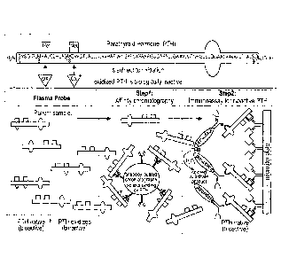
Fig.1B shows a schematic representation of the new method for measuring
parathyroid
hormone in human samples.
Fig.2A shows a NanoLC-ESI-FTMS total ion chromatogram of non-digested oxidized
synthetic hPTH(1-84)ox.
Fig.2B shows a magnified summed FTMS spectrum for retention time interval
18.30 to
20.50 minutes which spectrum comprises several different charged analyte ions
belonging to PTHox and its fragments.
Fig.3A shows a NanoLC-ESI-FTMS total ion chromatogram of a flow through
fraction
from the affinity column which binds oxidized synthetic PTH(1-84)ox.
Fig.3B is a magnified summed FTMS spectrum for retention time interval of
16.50-
18.50 minutes which spectrum does not show any analyte masses belonging to
PTH or oxidized PTH.
Fig.4A shows a NanoLC-ESI-FTMS total ion chromatogram of an eluate from the
affinity column comprising non-digested oxidized synthetic hPTH(1-84)ox.
Fig.48 is a magnified summed l- ____________________________________ I MS
spectrum for retention time interval of 16.50-
18.50 minutes comprising several different charged analyte ions of PTH.
Fig.5 shows for comparison the enlarged spectra of the starting material
comprising
non-digested oxidized synthetic hPTH(1-84)ox (Fig.1B) and the corresponding
eluate after binding to an affinity column (Fig. 3B).
Fig.6 is a bar diagram comparing directly determined "intact PTH values" in
serum of
patients on dialysis (blue bars), for further detail see also Table 2, and
after
removal of misfolded and oxidized PTH peptides from the sample.
CA 02865182 2014-08-21
WO 2013/124462
PCT/EP2013/053632
7
DETAILED DESCRIPTION OF THE INVENTION
[019] The oxidation of parathyroid hormone (PTH) peptide at methionine
residues 8 and/or 18 results in a loss of biological activity. (Galceran T et
al., Absence
of biological effects of oxidized parathyroid hormone-(1-34) in dogs and rats.
Endocrinology 1984;115(6):2375-2378. Horiuchi N et al., Effects of oxidation
of human
parathyroid hormone on its biological activity in continuously infused,
thyroparathyroid-
ectomized rats. J Bone Miner Res 1988;3(3):353-358. Zull JE et al., Effect of
methionine oxidation and deletion of amino-terminal residues on the
conformation of
parathyroid hormone. Circular dichroism studies. J Biol Chem 1990;
265(10):5671-
5676). Thus, studies by independent groups have shown that the oxidation of
PTH
diminishes its interaction with the respective receptor and that oxidized PTH
peptides
cannot stimulate the PTH receptor to generate cAMP, the second messenger of
PTH.
WO 2002/082092 (Roth HJ et al) discloses a two-site immunoassay which can
distinguish between oxidized PTH and "bioactive PTH" and wherein masking
antibodies are added which bind to oxidized methionine 8 or 18 so that an
antibody of
the two-site immunoassay can no longer binding to a nearby site comprising the
parathyroid receptor binding domain due to steric hindrances. Further studies
showed
that such masking antibodies must overcome with the immunological problem that
the
oxidation of methionine gives rise to two different stereoisomers, methionine
S-
sulfoxide (Met-S-0) and methionine R-sutfoxide (Met-R-0) with the sulfur being
a chiral
center, or even methionine sulfone (Met02) so that such antibodies must bind
to a
plethora of primary structures, in addition to the problem that a multiplicity
of reactive
oxygen species (ROS) are possibly involved in the oxidation of the parathyroid
hormone.
020] Methionine sulfoxide oxidation is inhibited in vivo by lower molecular
weight antioxidants (LMWA) such as glutathion, histidin dipeptide, uric acid,
bilirubin,
ascorbic acid or tocopherol. Once PTH has been oxidized comprising a Met-S-0
and
Met-R-0 the endogenous methionine sulfoxide reductase type A (MRSA) can reduce
Met-S-0 only but not Met-R-0. Whether there is a methionine sulfoxide
epimerase or
other routes for reducing the Met-R-0 stereoisomer remains to be shown. Thus,
the
oxidation of PTH is only partly reversible, depending whether the oxidation
resulted in
Met-S-0, Met-R-0 or Met02. The oxidation to Met02 however is not reversible.
It was
however found by the present inventors that any methionine oxidation of PTH
impacts
its folding and tertiary structure as oxidized methionines are less
hydrophobic and more
CA 02865182 2014-08-21
WO 2013/124462
PCT/EP2013/053632
8
polar. This may explain why intact PTH assays conventionally used in clinical
practice
poorly reflect PTH-related bone and cardiovascular abnormalities.
[021] The present disclosure provides a fast and reliable method to remove all
forms of oxidized or misfolded PTH polypeptides from serum or plasma samples,
say
all PTH molecules which have taken on a new tertiary structure due to
oxidative stress
and/or methionine oxidation. The present disclosure provides a method for
measuring
the amount or concentration of correctly folded bioactive PTH molecules in a
serum
sample which is particularly important for patients on dialysis. In the
examples below,
we used the herein disclosed method and assay strategy in a patient population
known
to be exposed to oxidative stress: end-stage: renal disease patients on
intermittent
hemodialysis (Witko-Sarsat V et al, Advanced oxidation protein products as a
novel
marker of oxidative stress in uremia. Kidney Int. 1996 May;49(5):1304-13). The
present
disclosure demonstrates that established ways of measuring PTH generally
result in
too high plasma concentrations of active PTH as compared to results
considering the
folding and oxidation status of PTH. Moreover, the correlation proved to be
very weak
between conventional PTH measurements and measurements after removal of all
oxidized and misfolded PTH polypeptide chains.
[022] The present disclosure further provides an antibody for a common
conformational epitope which is specific for all forms of oxidized parathyroid
hormone
and fragments thereof, at least comprising the amino acid sequence from 3 to
34 of
parathyroid hormone and being biologically inactive. This definition shall
encompass
all forms of oxidized human parathyroid hormone, particularly oxPTH(aa 1-84),
oxPTH(aa 1-52), oxPTH(aa 1-34), oxPTH(1-36), oxPTH(aa 1-37), oxPTH(1-38),
oxPTH(aa 3-84), oxPTH(aa 3-38) etc. The conformational epitope specific for
misfolded and/or oxidized human parathyroid hormone is therefore composed of
structures present in the aminoterminal portion of parathyroid hormone. All
oxidized
forms of the human parathyroid hormone seem to be inactive and misfolded.
Thus, the
disclosure comprises the information that the aminoterminal portion of the
human
parathyroid hormone can flip into an alternative tertiary conformation which
is
biologically inactive. The alternative conformation flip can likely be brought
about too by
a deletion of the second utmost or more (6) amino acids at the aminoterminus
or by an
oxidation of the methionine residues at positions 8, 18 or both, which
oxidations make
the hydrophobic side chain of methionine more polar and hydrophilic, or even
by an
oxidation of tryptophan at position 23. Due to the low amounts of parathyroid
hormone
in serum, it is however completely unclear which of those "degradation or
inactivation
CA 02865182 2014-08-21
WO 2013/124462
PCT/EP2013/053632
9
mechanisms" are physiologically more relevant. In other words, it remains to
be
examined whether the "large PTH fragments" in serum are degradation products
of
previously oxidized parathyroid hormone or vice versa, and whether the
oxidation
points to a biological mechanism for inactivation.
[023] The present disclosure also relates to a method for obtaining an
antibody which specifically binds to a conformational epitope or antigenic
determinant
of inactivated, misfolded or oxidized human parathyroid hormone. The
disclosure
further provides a reagent for removal of inactivated, misfolded or oxidized
human
parathyroid hormone from body fluids such as serum, plasma or whole blood. A
preferred embodiment relates to a column material with a covalently linked
antibody
recognizing a conformational epitope specific for inactive, oxidized and/or
misfolded
parathyroid hormone or fragments thereof, comprising at least amino acids 3 to
34 of
PTH. The disclosure provides an antibody which does not recognize biologically
active
hPTH(aa 1-84) or biologically active fragments thereof, but only inactive PTH
peptides
which are such modified or oxidized at any one position in the aminoterminal
portion 1
to 38 of the parathyroid hormone so that this portion flips into another
tertiary
conformation in which it is inactive and cannot bind to its receptor.
[024] The disclosure thus provides methods and means for measuring the
active parathyroid hormone concentration in serum or plasma of patients,
notably
patients on dialysis and subject to reactive oxygen species (ROS) and
oxidative stress.
EXAMPLES
EXAMPLE 1
Oxidation of hPTH(aa 1-84)
[025] 200 pg human PTH(1-84) purchased from Bachem AG (Bubendorf,
Switzerland) was dissolved in 400 pl of 0.1 M acetic acid (final concentration
of 0.5
pg/pl), mixed 1:1 with 30% hydrogen peroxide and incubated for 45 min at 37 C
to
obtain a mixture of PTH(1-84) peptides oxidized at methionines 8, 18, and
both.
Afterwards, the mixture was cooled on ice, divided into aliquots and
lyophilized.
CA 02865182 2014-08-21
WO 2013/124462
PCT/EP2013/053632
Oxidation of hPTH(aa 1-38) conjugate
[026] Human PTH(aa 1-38) peptide (Art.No. A1105AG.1, lmmundiagnostik
AG, Bensheim, Germany) was coupled to bovine thyreoglobulin by the
carbodiimide
method, dissolved in 1.0 ml 0.1% acetate buffer, pH 5.0, mixed 1:1 with 30%
hydrogen
5 peroxide and incubated for 18 hours at 37 C to obtain oxPTH(aa 1-38)
conjugate.
Oxidation of Biotin-hPTH(aa 1-38)
[027] Human PTH(aa 1-38) peptide (Art.No. A1105AG.1, lmmundiagnostik
AG, Bensheim, Germany) was dissolved in 1.0 ml 0.1% acetate buffer, pH 5.0,
mixed
1:1 with 30% hydrogen peroxide and incubated for two hours at 37 C to obtain
10 oxPTH(aa 1-38) peptides. Following lyophilisation, the oxPTH(aa 1-38)
was conjugated
to biotin using water-soluble biotin-sulfosuccinimidyl ester.
EXAMPLE 2
Monoclonal antibodies against a conformation epitope of oxidized PTH(aa 1-38)
[028] Monoclonal antibodies were raised in BALB/c-mice. The mice were
immunized with the oxPTH(aa 1-38) thyreoglobulin conjugate at 200 pg for both
primary and secondary immunizations with incomplete Freund's (mineral oil
only) in the
intraperitoneal cavity. Each of the antisera was tested for binding to non-
oxidized
biotin-hPTH(1-38). To detect antibodies specifically recognizing oxPTH(aa 1-
38)
peptides, we used the double antibody separation technique and as tracer
biotin-
oxPTH(aa1-38) labelled with 1251-streptavidin. After cell fusion and HAT
selection,
selected hybridomas were screened in the same way, namely for binding to human
oxidized PTH(aa 1-84) but not to human PTH(aa 1-84).
[029] For ultimate characterization of the specificity of the monoclonal
antibodies (MAB) and for identification of a monoclonal antibody recognizing a
conformation epitope common to oxidized hPTH(aa 1-38) peptides, say common to
all
forms of oxidized hPTH(aa 1-38) independently from oxidation status and
chirality
(Met-R-0, Met-S-0, and Met02 at positions 8, 18 and both), the antibody was
immobilized on CNBr-activated Sepharose 4B (GE Healthcare Bio-Sciences,
Uppsala,
Sweden). Hundred pl aliquot of the slurry was filled in a column
(MobiSpinColumn,
MoBiTec, GOttingen, Germany) and equilibrated with PBS buffer, pH 7.4. Then
2.5 pg
of lyophilized oxidized hPTH(1-84) were dissolved in 300 pl of equilibrating
buffer and
CA 02865182 2014-08-21
WO 2013/124462
PCT/EP2013/053632
11
applied on the column. The column was incubated end-over-end for 1 h at room
temperature, washed with 300 pl of equilibrating buffer, followed by 3 washes
with 300
pl of distilled water, and then eluted 2 times with 200 pl of elution buffer
(0.1% TFA).
Flow-through, wash fractions (equilibrating buffer and water) as well as
eluate of the
column were collected separately, lyophilized and analyzed by nanoLC-ESI-FT-
MS.
Since oxidized hPTH(aa1-38) regularly results in a variety of oxidized PTH
fragments,
oxidized at positions 8, 18 or both, an antibody or antibody clone can be
selected which
binds oxidized parathyroid hormone independently from the specific type of
protein
oxidation. Consequently, a monoclonal antibody ("oxPTH-ConforMAB") recognizing
a
conformation epitope present on all forms of oxidized hPTH(aa 1-84) and
fragments
thereof was selected for further analysis and characterization. The selected
oxPTH-
ConforMAB specifically recognized with high affinity all forms of oxidized and
misfolded
hPTH fragments, but not non-oxidized PTH (aa 1-84).
EXAMPLE 3
nanoLC-ES1-FT-MS/MS
[030] In order to investigate the oxidation of human PTH(aa 1-84) of example
1 the sample was analyzed directly by high resolution nanoLC-ESI-FT-MS/MS to
determine the masses of the whole molecule species and after cleavage by three
endoproteases (ArgC, LysC and chymotrypsin) to characterize methionine
oxidations
at positions 8 and/or 18.
[031] The non-digested human PT}-I(aa 1-84) and oxPTH(aa 1-84) samples
were directly applied to nanoLC-ESI-FT-MS after acidification with 2% formic
acid.
[032] The digested oxidized human PTH(aa 1-84) samples (1 nmol) were
denatured prior digestion by 8 M urea containing 20 mM TCEP (tris[2-carboxyq-
phosphine) reducing agent for 30 min. lodoacetamide was added to 50 mM final
concentration and the mixtures incubated in the dark for another 20 min. After
dilution
to 0.8 M urea, the samples were digested with ArgC, LysC and chymotrypsin,
respectively, in accordance with SOPs of Proteome Factory, Berlin, DE. Enzyme
to
protein ratio (w/w) was 1:50 in each digest. The acidified peptide digests
(ArgC, LysC
and chymotrypsin) were pooled and applied to nano-LC-ESI-MS (LTQ-FT, Thermo
Scientific) analysis using a 35 min nanoLC gradient (Agilent 1100 nanoLC
system) with
solvent A (0.1% formic acid ( 5% acetonitrile 1 94.9% ddH20) and solvent B
(0.1%
formic acid / 99.9% acetonitrile).
CA 02865182 2014-08-21
WO 2013/124462
PCT/EP2013/053632
12
[033] For testing the synthetic oxidized hPTH(1-84) of example 1 was
subjected to affinity-chromatography on a column comprising the specific
monoclonal
oxPTH-conformation antibody (MAB) which binds to an antigenic determinant only
present on oxhPTH(aa 1-84) and oxidized hPTH(aa 1-38) polypeptide chains but
not
on correctly folded hPTH, which antigenic determinant does not encompass
methionine
sulfoxide or methionine sulfone. No oxidized hPTH(1-84) or fragments thereof
were
detectable after removal of oxidized PTH molecules in the sample by nanoLC-ESI-
FT-
MS so that all oxidized PTH forms of the given sample were recognized by the
oxPTH-
ConforMAB on the immunoaffinity column and quantitatively removed from the
flow-
through. The mass accuracy was better than 5 ppm for MS data. The MS data were
analyzed by MASCOT (Matrixscience) and Qualbrowser (Thermo Scientific)
according
to the predicted peptide masses. Results are shown in Table 1 and Figures 2
and 3.
TABLE 1
Deduced masses of charged peaks in the spectra of non-digested
hPTH(aa 1-84)ox and eluate (column-bound oxPTH-fragments)
MASS CHARGE MW [DA] MW
[MiZ] Z INCREASE
728.16 13 9453.08 +32
729.39 13 9469.07 +48
________________ 730.62 13 9485.06 +64
731.85 13 9501.05 +80
780.50 12 9354.00 +32
781.83 12 9369.96 +48
783.17 , 12 9386.04 +64
788.76 12 9453.12_ 4-32
790.09 12 , 9469.08 +48
791.42 12 , 9485.04 +64
792.75 12 9501.00 +80
851.36 11 9353.96 +32
852.82 11 9370.02 +48
854.27 , 11 9385.97 +64
860.28 11 9452.08 +32
861.73 11 9468.03 +48
863.19 11 9484.09 +64
864.64 11 9500.04 - +80
CA 02865182 2014-08-21
WO 2013/124462
PCT/EP2013/053632
13
[034] No significant mass peaks were observed that can be assigned to any of
the hPTH(1-84)ox species by nanoLC-ESI-FT-MS analysis of the flow-through and
wash fractions (equilibrating buffer and water) of the column (Fig. 3A,B),
whereas
several mass peaks corresponding to the different oxidized states of hPTH(1-
84)ox
were detected in the eluate (Fig. 4A,B; Table 1). Comparison of the spectra of
the
starting material, non-digested oxidized synthetic hPTH(1-84)ox (Fig. 2B), and
the
eluate from the affinity column of non-digested oxidized synthetic hPTH(1-
84)ox (Fig.
4B) on Fig. 5 revealed the same profile despite the difference in peak
intensity. The
results demonstrate that synthetic oxidized hPTH(1-84) consisted of a
considerable
variety of products corresponding to the different oxidized methionines.
However, the
column with the monoclonal antibody (MAB) raised against the oxidized human
PTH
was specific for all oxidized forms of hPTH(1-84) and removed them all from
the probe.
[035] More precisely, the intact oxidized hPTH(1-84) sample showed TIC-
peaks at 18 - 20 min. The molecular masses corresponded to values shifted by
+16,
+32, +48, +64 Da caused by methionine oxidation (suifoxide, +16 Da and
sulfone, +32
Da for each residue, and combinations thereof, maximal +64 Da) and by +80 Da
for the
additional oxidation of tryptophan 23. Figure 2A shows a NanoLC-ESI-FTMS total
ion
chromatogram of non-digested oxidized synthetic hPTH(aa 1-84) and Fig. 2B the
corresponding magnified summed FTMS spectrum for retention time interval 18.30-
20.50 minutes. Several different charged analyte ions have been marked.
[036] Fig 3 shows the analysis of the flow through fraction of non-digested
oxidized synthetic hPTH(1-84)ox after binding to the immunosorption column.
Fig. 3A
shows a nanoLC-ESI-FTMS total ion chromatogram of the flow-through and Fig. 38
the
corresponding magnified summed FTMS spectrum for retention time interval of
16.50-
18.50 minutes. The spectrum does not show any analyte masses belonging to
oxidized
PTH.
[037] Figure 4 concerns the eluate from the affinity column of non-digested
oxidized hPTH(1-84)ox. Fig. 4A shows the nanoLC-ESI-FTMS total ion
chromatogram
of the eluate and Fig. 48 again the corresponding magnified summed FTMS
spectrum
for retention time interval of 16.50-18.50 minutes. Several different charged
analyte
ions of oxPTH(aa 1-84) were detectable in the eluate.
[038] Thus, the examples confirm that all oxidized, misfolded forms of human
parathyroid hormone and fragments thereof had a characteristic conformation
epitope
CA 02865182 2014-08-21
WO 2013/124462
PCT/EP2013/053632
14
which can be used for removal of these fragments from a sample for
determination of
the biologically active concentration of parathyroid hormone.
EXAMPLE 4
[039] We studied specimens from 18 patients on intermittent haemodialysis
treated in our dialysis unit. Specimens (EDTA-whole blood) were taken before
start of
the dialysis session, centrifuged and immediately stored at -80 C until
further analysis
after obtaining of plasma. The study was approved by the local hospital
ethical
committee. Written informed consent was obtained in each case. Patient's
characteristics were obtained from patients clinical records. Serum
phosphorus,
calcium and C-reactive protein (CrP) were analyzed on an automatic analyzer of
the
clinical laboratory of the university hospital Charite.
[040] The intact-PTH electrochemiluminescence immunoassay (ECLIA;
Roche PTH, Intact jiPTHD was used for measuring the PTH concentration. The
intact-
PTH ECLIA of Roche uses a biotinylated monoclonal antibody, which reacts with
amino
acids 26-32, and a capture ruthenium-complexed monoclonal antibody, which
reacts
with amino acids 55-64. The determinations were performed on Roche Modular E
170 . The intraassay CV was 4.1% and the interassay CV was 5.8% at
concentrations
of 35.0 and 180.0 ng/L, respectively.
[041] Human samples were either measured directly (named iPTH in Table 2)
or after removal of oxidized PTH by a column which removes oxidized PTH using
the
selected monoclonal oxPTH conformation antibody described in example 2 which
recognizes all forms of oxidized PTH and oxidized PTH fragments. More
precisely, the
oxPTH-ConforMAB binding column was used with samples from 18 patients on
dialysis
followed by a classical sandwich PTH ECLIA as it is used in daily clinical
practice.
0
t,..)
--o
TABL 2
Z..::,
t,)
" RENAL DISEASE AGE TIME ON
-SEX IPTH REAL ox- RATIO TOTAL CA P CRP 4...
0
.1===
A YEARS DIALYSIS (NG/L) IPTH
i PT H IPTH/ (MMOLJL) (MMOL/L) (MG/DL)
Z -4 (YEARS) i
1J
; (NG/L) (ng/L) REAL-IPTH
0 m
1 Hypertensive Nephropathy 62 0.3 m 43.63 8.9 34.73
0.204 2.58 1.24 0.43
2 Diabetic Nephropathy 73 4.0 '. m ' 796.2 70.62
725.6 0.089 2.2 2.15 -.
3 Unknown 37 0.1 m 52.84 10.35 42.49
' 0.196 2.53 0.81 0.03
4 Diabetic Nephropathy 68 2.1 f 70.8 11.18 59.62
0.158 2.23 0.91 ' 4.08
Acute Kidney Injury 64 ' 0 ' m ' 46.49 9.45
37.04 0.203 2.17 1.32 3.26 P
_
6 Diabetic Nephropathy 63 1.6 f 42.13 5.37 36.76
0.127 2.08 1.43 ' 12.2 ' 7 ' ADPKD 70 3.3 f '
1029.00 ' 74.76 954.2 ' 0.073 2.1 1.37 0.53 in
- -
VI
.
8 Cardio-Renal-Syndrom 70 3.4 m 240.4 41.89 198.5
0.174 2.38 1.57 0.32
9 'Unknown ' 70 9.0 ' m 105.00 18.48
86.52 ' 0.176 2.26 1,5 3.12 -
=,-
_.
Diabetic Nephropathy 65 7.0 ' m '1301.00 445.30
855.7 0.342 2.53 2.23 1.74 .
-
11 Membraneous GN 45 5.4 ' f 311.80 24.44
287.4 0.078 1.57 2.06 0.52 '
12 Membranoproliferative GN ( Typ1) 52 1.5 m 144.10 19.24
124.9 0.134 1.87 0.73 0.17
13 'Hypertensive Nephropathy 61 4.1 m 73.45 15.92 57.53
0.217 2.15 2.35 0.67
_ . 14 ' ADPKD 57 1.2 m 281.9
44.02 237.9. 0.156 2.18 1.35 ' 13.4
. _
_
' Diabetic Nephropathy 73 4.0 m 116.9 19.73 97.17
0.169 2.38 1.66 4
16 Mesangioproliferative GN 69 8.1 m 70.81 18.51 52.3
0.261 2.62 2.28 6.7 -o
_
n
17 ' Interstitial Nephritis 61 2.6 f 76.28 11.21 65.07
0.147 2.21 1.61 2.9 - --i
4-1
18 Unknown 56 10.6 ' m 487.1 76.12 411 ,
0.156 2.35 2.41 0.17 -0
e
t,/
--,
7.
GO
= \
C=4
14
CA 02865182 2014-08-21
WO 2013/124462
PCT/EP2013/053632
16
[042] For sample preparation, 100 pl aliquots of the slurry with immobilized
monoclonal oxPTH conformation antibody (oxPTH-ConforMAB) were filled in
MobiSpin-columns equilibrated with PBS buffer, pH 7.4. Then 500 pl of each
sample
were applied on the column, respectively. The columns were incubated mixing
end-
over-end for 2 h at room temperature, washed with 250 pl of 0.1 M ammonium
acetate
buffer pH 7.0, followed by a wash with 250 pl of 0.1 M ammonium acetate buffer
pH
7.0, containing 20% acetonitrile, and then eluted twice with 200 pl of elution
buffer
(0.05 M formic acid, pH 3.5). Flow-through, wash fractions as well as eluate
of the
column were separately collected and lyophilized. The samples were
reconstituted in
500 pl of PBS buffer, pH 7.4 and aliquots analyzed by the intact-PTH ECLIA of
Roche
(Elecsys PTH, Intact assay, Roche, Penzberg, Germany). Table 2 shows the
basic
clinical characteristics and laboratory data of the studied patients on
dialysis as well as
concentrations of directly measured iPTH (ng/L) and after removal of
misfolded,
oxidized PTH (real iPTH).
[043] The results have been summarized in Figure 6. Dark (blue) bars show
PTH concentrations in serum directly determined by a conventional intact-PTH
ELCIA
of Roche. When oxidized forms of PTH were removed from the sample, the
measured
PTH concentrations were completely different (grey/red bars). While the
measured
PTH concentrations were substantially lower after immunosorption and removal
of
oxidized PTH forms the relationship between directly measured PTH
concentrations
and PTH concentrations after removal of oxidized PTH forms varied highly with
patients. In some patients only 7% of directly measured PTH was free of
oxidation and
misfolding, whereas in other patients 34% of the directly measured PTH was
intact
PTH. Thus, the data show a surprising variation of oxidized to biologically
active
parathyroid hormone in our patients, possibly in accordance with exposed
oxidative
stress among the studied patients, the amount of ROS present, the activity of
the
methionine sulfo reductase type A or the reductive potential in blood and
circulation.
Controls
[044] For determining recovery, in a separate series of measurements 500 pl
of each sample was spiked with 1 ng of oxidized hPTH (aa 1-84) of example 1.
The
spiked samples were treated as described in the sample preparation part. The
splicing
had no impact on the measured PTH value when oxidized PTH and fragments
thereof
were removed as described. The recovery of added oxidized PTH (aa 1-84) was in
the
range from 65 to 105 %, if directly determined by the iPTH ECLIA.
CA 02865182 2014-08-21
WO 2013/124462
PCT/EP2013/053632
17
[045] In order to be sure that the oxPTH columns remove specifically oxidized
PTH only, we analyzed some samples after purification with a column able to
bind
1,25-dihydroxyvitamin 03. More precisely, we subjected serum samples affinity
columns comprising a monoclonal antibody binding to 1,25-dihydroxy vitamin 03
(Art No. K1107-737, Immundiagnostik AG, Bensheirn, DE). A treatment with such
a
column had little impact on the measured PTH concentration as shown by Table
3.
TABLE 3
1PTH IPTH RATIO
NG/L POST VIT. D COLUMN
(NG/L)
43,63 32,43 0,74
796,2 684,83 0,86
52,84 47,45 0,89
46,49 41,86 0,90
70,6 61,99 0,87
[046] The data of Table 3 show that the non-specific binding of PTH
accounted roughly for 14% for an immunosorption column comprising a non-
specific
antibody. Moreover, the non-specific binding of PTH was in all samples about
the same
so that the affinity column on its own did not significantly influence PTH
measurements
except for a typical loss of recovery. In other words the column on its own
did not
significantly influence the results.
[047] To rule out that the tested monoclonal oxPTH conformation antibody
MAB is released from the column and interferes with the PTH quantification in
the iPTH
ELICA of Roche, free monoclonal oxPTH conformation antibody (MAB) was added
sample from two patients in a final concentration of 1.8 pg MAB per ml. The
samples
were then analyzed using the iPTH immunoassay. Those samples where only
solvent
was added had measured iPTH concentrations of 43.63 [ng/L] (patient a) and
796,20
[ng/L] (patient b), respectively. Adding the monoclonal antibodies to the
samples did
not alter significantly the results. In the samples with antibodies we
measured 35,70
[ng/L] (patient a) and 753,20 [ng/L] (patient b). Thus, even if monoclonal
antibodies
(MAB) against oxidized human PTH are released these antibodies do not
significantly
interfere with final iPTH quantification.
CA 02865182 2014-08-21
WO 2013/124462
PCT/EP2013/053632
18
Clinical Data
[048] The clinical characteristics are shown in Table 2. We included 17
patients on chronic hemodialysis as well as one patient requiring dialysis due
to acute
renal failure. We analyzed the clinical specimens with the iPTH immunoassay.
In all
patients the measured PTH concentrations were substantially lower when
considering
oxidized forms of parathyroid-hormone (see Table 2 and Figure 6). It is of
note,
however, that the relationship between PTH concentrations determined directly
with the
iPTH immunoassay and those concentrations measured after removal of the
oxidized
PTH forms is not constant, by contrast the relationship varies substantially
probably
due to the different degree of oxidative stress among the studied patients. In
some
patients only 7% of traditionally measured PTH were free of oxidation, whereas
in
another patient 34% of the traditionally measured PTH were real intact PTH.
Taken
together, without considering oxidation status of PTH the conventionally
measured
PTH concentrations using a modern sandwich detection system are detected
several
fold higher as the concentrations when considering oxidation of PTH. The
effect of
oxidation of PTH is highly variable among these patients requiring dialysis.
There is
only a very weak correlation between traditionally measured PTH and oxidized
PTH.
[049] In some patients we used beside the iPTH immunoassay from Roche
also the PTH(1-84) assay system from Roche. We got basically similar results
as
described above with the iPTH assay system. Without considering oxidation
status of
PTH, the traditionally measured PTH concentrations were several fold higher as
compared to the concentrations which take due account of the oxidation of PTH.
[050] Using very sensitive mass spectroscopy approaches, the current study
demonstrated that oxidation of human PTH(1-84) results in the formation of a
variety of
products corresponding to the different oxidized methionine resides at
position 8 and/or
18 within the parathyroid hormone. A column with the monoclonal antibody (MAB)
raised against the hPTH(1-34)ox fragment is specific for all oxidized forms of
hPTH(1-
84) and removed them all from the sample. The clinical part of our study
demonstrated
that without considering oxidation status of PTH, the traditionally measured
PTH
concentrations based on current gold standard methods resulted in much higher
PTH
concentrations in the clinical samples as compared to the concentrations when
considering oxidation of PTH. The effect of PTH oxidation is highly variable
among the
patients requiring dialysis. There is only a very weak correlation between
traditionally
measured PTH and PTH data considering the oxidation of this hormone. Given the
fact
that oxidized PTH (Figure 1) does not stimulate the PTH receptor anymore to
generate
CA 02865182 2014-08-21
WO 2013/124462
PCT/EP2013/053632
19
cAMP, and is thus most likely biological inactive, clinical strategies for the
treatment of
hyperparathyroidism in dialysis patients based on measurements of PTH using
classical third generation sandwich ELISA techniques are most likely prone to
incorrect
decision making.
[051] It is known for example that in uremic patients highly specific assays
have measured a 2.5-fold increase in the non-suppressible fraction of PTH
compared
with healthy subjects. Moreover, PTH concentrations measured in uremic serum
apparently overestimated PTH-related bone abnormalities also by a factor of 2-
2.5. It
was suggested that in patients with chronic renal failure, the presence of
high
circulating levels of non-1-84 PTH fragments (most likely 7-84 PTH) detected
by the
second generation assay and the antagonistic effects of 7-84 PTH on the
biological
activity of 1-84 PTH may explain this. However, this hypothesis was never
proven in
adequately designed clinical studies using for example HPLC coupled to mass
spectrometry to really distinguish between different PTH fragments. Our data
on the
other hand using modern liquid chromatography linked to tandem mass
spectroscopy
to detect PTH suggest that this well-known overestimation of PTH in patients
on
dialysis might be most likely due to the presence of oxidized, biologically
inactive forms
of PTH in patients on dialysis.
[052] Reactive oxygen species (ROS) such as hydrogen peroxide (H202) or
hypochlorus acid (HOC), and free radicals such as hydroxyl radical (OH) or
others are
continuously formed in vivo. Additional imbalance between formation of ROS and
potent antioxidative defence mechanism creates oxidative stress. Uraemia in
general is
associated with enhanced oxidative stress, and haemodialysis or peritoneal
dialysis
may in particular contribute to oxidative stress and reduced antioxidant
levels in such
patients.
[053] One of the preferred highly sensitive targets for oxidation is
methionine.
The oxidation product methionine sulfoxide can be reversed by reduction with
chemicals or enzymatically, whereas oxidation to the methionine suifone is
biologically
irreversible. Oxidation of methionine residues can lead to an activation or
inactivation of
a functional protein, respectively, and the resulting methionine sulfoxide can
be
reversed enzymatically by a specific reductase. Methionyl sulfoxide reductase
has
been found in E. coil and in mammalian tissues. Oxidation of methionine and
its
reversal may serve as a regulator for protein activities. The parathyroid
hormone
contains two methionine residues in the amino-terminal region (position 8 and
18),
responsible for the biological activity of the peptide, accessible to
alterations trough
CA 02865182 2014-08-21
WO 2013/124462
PCT/EP2013/053632
oxidation. The secondary structure of the parathyroid hormone seems to be
essential
for its receptor binding. The methionine residue 8 is important for the
folding of the
hormone and proves the key role for this residue in the structure of the amino-
terminal
domain and its biological activity. Thus oxidation of methionine residue 8,
producing
5 fundamental
chances in secondary structure of PTH, is implicated both in binding and
in activation of adenylyl cyclase.
[054] Based on published data and our results, we suggest that methionine
residues in different peptide hormones, like human growth hormone, somatomammo-
tropin, luteotropin as well as PTH may be subject to oxidation resulting in
loss of
10 biological
activity or receptor affinity. Methionine oxidation may be a general principle
in
regulation of hormone activity. However, this hypothesis needs to be proved in
detail.
[055] Our new assay system is - for the first time - able to differentiate
between oxidized and non-oxidized forms of PTH by removing oxidized PTH
fragments
with a highly specific antibody able to detect and bind all forms of oxidized
PTH. The
15 removal of
oxidized forms of PTH can be done either ¨ as it was done in the present
study ¨ prior to analysis by a coated column followed by a third generation
PTH assays
(for assay principle see figure 6) or even as an integrative part of a third
generation
sandwich ELISA system. It should also be feasible to combine our approach with
modern techniques like liquid chromatography coupled to tandem mass
spectrometry
20 (LC MS/MS) in
clinical practice in the near future by immunocapture oxidized PTH
fragments prior to LC-MS/MS. This will improve the diagnostic performance of
LC-
MS/MS PTH approaches.
[056] In conclusion, by means of nanoLC-ESI-FT-MS we were able to
demonstrate that oxidation of human PTH(1-84) resulted in the formation of a
variety of
products corresponding to the different oxidized methionine residues at
position 8
and/or 18 within the parathyroid hormone. We screened for a monoclonal
conformation
antibody against a common antigenic determinant of oxidized human parathyroid
hormone and oxidized fragments thereof and found one specific for all oxidized
forms
of hPTH(1-84) which allows a removal of oxidized parathyroid hormone and
fragments
thereof from the serum samples of human patients. We also disclose herein that
traditionally measured PTH concentrations based on current gold standard
methods,
which do not account for the oxidation status of PTH, resulted in much higher
PTH
concentrations in clinical samples specimens as compared to the concentrations
when
considering oxidation of PTH. The effect of PTH oxidation is further highly
variable
among the patients requiring dialysis. Given the impact of vascular
calcification in end-
CA 02865182 2014-08-21
WO 2013/124462
PCT/EP2013/053632
21
stage renal disease patients on morbidity and mortality the present results
support that
measuring whole PTH without "contamination" of oxidized PTH forms will greatly
improve clinical decision making with respect to PTH-related bone and
cardiovascular
abnormalities.
Conclusions
[057] Thus, the present application is provides a disclosure of a method of
obtaining antibody molecules specific for oxidatively inactivated human
parathyroid
hormone and circulating fragments thereof, comprising a step of obtaining
antibodies
against human parathyroid hormone peptide by immunizing a non-human animal
with
an immunogen comprising as hPTH hapten a hPTH peptide oxidized at positions 8,
18
or both, or a respective fragment thereof, and recovering said antibodies from
said non-
human animal; a step of selecting or purifying said antibodies from antibody
molecules
that bind to bioactive human parathyroid hormone peptide under physiological
conditions to obtain antibodies that specifically bind oxidized hPTH peptide
or
respective circulating fragments thereof; a step of selecting or purifying
said antibodies
specific for oxidized hPTH peptide from antibody molecules binding to an hPTH
amino
acid sequence (primary protein structure) comprising at positions 8, 18 or
both
methionine R-sutfoxide, methionine L-sulfoxide or methionine sulfone, to
obtain
antibody molecules having a complementary determining region which
specifically
binds to a conformational epitope (tertiary protein structure) common to
inactive
oxidized human parathyroid hormone peptides and circulating fragments thereof.
[058] The antibody molecules may be further purified or selected by a step
wherein they are further tested for their binding to a primary hPTH structure
comprising
an oxidized tryptophan at position 22 or which hPTH structure is lacking the
utmost
aminoterminal amino acids at positions 1 and 2 or both of the hPTH sequence.
[059] The antibodies subjected to these selection or purification steps may be
monoclonal antibodies produced by mouse or rat cell clones. A person skilled
in the art
Will appreciate that the antibodies for screening and selection may also be
recombinant
antibody molecules or antibody fragments or single-chain antibodies from a
synthetic
antibody library. if the antibodies are recovered from a non-human animal, the
immunogen for eliciting these antibodies is preferably is a carrier protein
having bound
as hapten any one of synthetic oxidized human parathyroid hormone, synthetic
oxidized fragment of human parathyroid hormone or synthetic oxidized peptide
CA 02865182 2014-08-21
WO 2013/124462
PCT/EP2013/053632
22
comprising the amino acid sequence 1 to 38 of human parathyroid hormone or a
substantial portion, fragment or variant thereof.
[060] Further preferred embodiments and the scope of the present invention
are pointed out in the appending claims.