Note: Descriptions are shown in the official language in which they were submitted.
CA 02865553 2014-08-26
WO 2013/136182
PCT/1B2013/000848
1
ACTIVATED IMMUNOSTIMULATORY
CELL COMPOSITION AND USES THEREOF
CROSS-REFERENCE TO RELATED APPLICATIONS
[001] This application claims the benefit of U.S. Provisional
Application No. 61/611,202, filed March 15, 2012, which is incorporated
herein by reference.
DESCRIPTION OF THE INVENTION
FIELD AND BACKGROUND
[002] The present invention relates to activated imnriunostimulatory
cell compositions, methods of preparing those compositions, and to uses of
the compositions to treat conditions that may benefit from immunostimulation,
such as cancer.
[003] Many tumors possess tumor-specific and tumor-associated
antigens. Tumor specific antigens (TSA) are uniquely expressed on the tumor
and not present on the normal cells while tumor-associated antigens (TAA)
are expressed at higher levels on tumors than on normal cells (Wang, RF
2002). Tumor antigens are cleaved within tumor cells into small peptides that
are presented to immune cells as a complex with class I Major
Histocompatibility molecules (MHC class l) also called Human Leukocyte
Antigens (HLA class I, e.g. HLA A, B, or C). The MHC class I-tumor peptide
complex is recognized by the T Cell Receptor (TCR) complex of cytotoxic T
lymphocytes (CTLs), which are CD8+ T cells. The interaction of TCR with
MHC I-tumor peptide complex causes release of substances from CTLs that
destroy tumor cells (perforin, granzymes, and granulysin) (Mami-Chouaib F,
2002). CTLs also send death or apoptotic signals (through FAS receptor) to
tumor cells. (Wu JY 2006.)
[004] The initial CTL response is short-lived and requires amplification
support from CD4+ T helper (Th) lymphocytes to continue. Differentiation and
proliferation of Th cells occurs through their interaction with professional
Antigen Presenting Cells (APCs), such as macrophages or interstitial dendritic
CA 02865553 2014-08-26
WO 2013/136182
PCT/1B2013/000848
2
cells (DCs) (Wang RF. 2002). APCs engulf tumor antigens or apoptotic tumor
cells, process these antigens by cleaving them into small peptides, then
present the peptides to naïve T cells. The process is similar to CTL
stimulation by MHC class I + peptide, but CD4+ T cells recognize peptides
presented in the context of MHC class II (e.g. HLA-DR). Upon recognition of
MHC class II-peptide complexes by TCR, T cells acquire Th functions and
produce IL-2, a cytokine that in turn amplifies the CTL response and induces
development of T memory cells for sustained immunity (Keene JA, 1982;
Fujiwara, H, 1986).
[005] T cell stimulation is thought to occur in at least two steps.
(MareIli-Berg FM, 2007; Chambers CA 2001.) In the first step (signal 1),
MHC/peptide complexes interact with the TCR. This step is not sufficient to
fully stimulate the T cells. Instead, a second interaction between one of the
APC-expressed co-stimulatory molecules (such as CD86, CD83, CD80 and
CD40) and a corresponding ligand on T cells (signal 2) is required. T cells
that receive signal 1 in the absence of signal 2 are unable to acquire helper
functions (Chappert & Schwartz, 2010).
[006] The interaction of naïve T helper cells with APCs such as DCs
polarizes the Th cells into Th1 and Th2 subsets, which differ by the patterns
of
cytokines that they produce. Th1 cells produce cytokines such as interferons
and IL-2 that activate proliferation of CTLs and cause tumor rejection. Th2
cells produce the cytokines IL-4, IL-6, IL-10 and IL-13. Increased levels of
Th2 cytokines are found in sera of cancer patients with poor clinical
prognosis.
[007] Like Th cells, monocytes/macrophages phenotypes polarize in a
process that is dependent on the balance between Th1/Th2 cytokines. The
nomenclature for these polarized cells mirrors the Th1 and Th2 nomenclature
and, like Th1 and Th2 T cells, M1 and M2 macrophages produce different
patterns of cytokines. M1 macrophages predominantly produce IL-12, IL-23,
TNFa, IL-1, IL-6; whereas M2 macrophages produce high levels of IL-10 and
IL-13 (Cassetta L, 2011). M1 macrophages also express high levels of HLA-
DR, while M2 macrophages express high levels of CD163 antigen. Fully
CA 02865553 2014-08-26
WO 2013/136182
PCT/1B2013/000848
3
polarized M1 and M2 macrophages are the extremes of a continuum of
functional states. Notably, macrophages that infiltrate tumor tissues are
driven by the tumor environment to acquire a polarized M2 phenotype which
plays a key role in subversion of adaptive immunity and inflammatory circuits
to promote tumor growth and progression. In contrast, M1 macrophages elicit
an anti-tumor effect.
[008] Another APC that is thought to also differentiate from peripheral
blood monocytes is the myeloid dendritic cell (DC). Mature, activated DCs are
extremely potent APCs. DC maturation is associated with up-regulation of
MHC molecules, co-stimulatory molecules (CD86, CD83 CD80, CD40)
(Tuyaerts S, 2007), adhesion molecules, such as CD11b, CD11c, and CD54,
and the chemokine receptor CCR7 (Alvarez D, 2008). The latter enables the
DC to migrate from the peripheral tissue through the vessel walls to the T
cell
areas (Forster R, 2008). DC maturation not only ensures expression of
molecules relevant for T cell stimulation, it also permits DC to reach the
appropriate anatomical compartments in secondary lymphoid organs so that
they can present antigens to naïve T cells. Cognate signals from T cells
further activate DC (Cavanagh LL, 2002).
[009] Mature myeloid DCs are characterized by their ability to make
IL-12 (Mariotti S, 2008). Because IL-12 promotes Th1 polarization, DCs that
produce IL-12 are used in cancer vaccine development (Trinchieri G: 2003).
[010] In addition to stimulating the adaptive, antigen-specific immune
responses described above, DCs also play a role in stimulating innate (MHC-
unrestricted) immunity, including stimulating several antitumor cell types.
These cells include classical natural killer cells (NK cells), which do not
express TCR (CD3-/CD56+ cells) and cytokine-induced killer T cells
(CD3+/CD56+). NK cells can directly induce tumor cell apoptosis via the
perforin-granzyme pathway or by expressing death-receptor ligands such as
Fas ligand (Bryceson YT, 2011). IL-12-producing DCs can induce
proliferation of NK cells (Walzer T, 2005). NK cells also cells release
cytokines that promote differentiation of DCs (Ferlazzo G, 2009).
CA 02865553 2014-08-26
WO 2013/136182
PCT/IB2013/000848
4
[011] Thus, mature DC are an important cell type in generating both
effective adaptive (T cell) and innate (NK, NKT cell) anti-tumor immune
responses. Indeed, much research has focused on the application of DC-
based vaccines as a therapy for a variety of tumor types and several clinical
trials are underway. DC-based vaccines for metastatic melanoma using
cytokine activated, antigen-pulsed DC have in particular shown promising
results (e.g., Cornforth, Lee & Dillman, 2011). Nevertheless, the complexity
of
the immune system makes it important to ensure that cellular therapies
possess functional properties that lead to tumor cell lysis. For example,
immature DCs have the potential to inhibit, rather than stimulate, tumor
immunity in vivo. Accordingly, there is a great need for compositions
comprising fully mature dendritic cells and other activated immune cells for
treatment of conditions that may benefit from immunostimulation, such as
cancer therapy.
SUMMARY OF THE INVENTION
SUMMARY OF CERTAIN EMBODIMENTS
[012] The invention relates to activated immunostimulatory cell
compositions, methods of preparing those compositions, and to uses of the
compositions to treat conditions that may benefit from immunostimulation,
such as cancer.
[013] Accordingly, in one aspect the invention is directed to methods
for making an activated immunostimulatory cell composition, comprising: (a)
incubating human leukocytes under conditions of time and temperature to
activate the leukocytes; (b) subjecting the activated leukocytes to hypo
osmotic shock; (c) adding to the leukocytes a salt solution in an amount
effective to restore isotonicity; (d) mixing the leukocytes with a supportive
medium; and (e) incubating the leukocytes in the supportive medium for a
period of time to at least induce maturation of dendritic cells, thereby
making
an activated immunostimulatory composition.
[014] In still another aspect, the invention is directed to methods of
making an activated immunostimulatory cell composition comprising
CA 02865553 2014-08-26
WO 2013/136182
PCT/IB2013/000848
incubating non-quiescent (i.e., at least partially activated) leukocytes in a
supportive medium under conditions of time and temperature that induce
maturation of dendritic cells, thereby making an activated innmunostimulatory
composition.
5 [015] In yet another aspect, the invention is directed to methods for
making a composition comprising mature dendritic cells (DCs) comprising: (a)
providing leukocytes, (b) allowing the leukocytes to transition from a
quiescent
to an active state by maintaining the leukocytes at room temperature for about
8 to 20 hours, (c) subjecting the leukocytes to hypo-osmotic shock, and (d)
incubating the shocked leukocytes for 36 hours to 14 days in a supportive
medium to thereby make a composition comprising mature DCs.
[016] In some embodiments of these aspects of the invention, the
leukocytes are incubated in the supportive medium for about 36 to 84 hours.
[017] In some embodiments of these aspects of the invention, the
leukocytes are incubated in the supportive medium for about 48-72 hours.
[018] In some embodiments of these aspects of the invention, the
composition is enriched in mature dendritic cells.
[019] In some embodiments of these aspects of the invention, the
composition further comprises activated lymphocytes.
[020] In some embodiments of these aspects of the invention, the
composition further comprises T helper cells enriched in Th1 phenotype.
[021] In some embodiments of these aspects of the invention, the
composition is enriched in Thl cytokines.
[022] In some embodiments of these aspects of the invention, the
composition further comprises active macrophages enriched in the M1
phenotype.
[023] In some embodiments of these aspects of the invention, the
composition is enriched in M1 cytokines.
[024] In some embodiments of these aspects of the invention, the
leukocytes are isolated from peripheral blood, placental blood, cord blood,
bone marrow, or lymphoid tissue.
CA 02865553 2014-08-26
WO 2013/136182
PCT/IB2013/000848
6
[025] In some embodiments of these aspects of the invention, the
supportive medium is serum or plasma.
[026] In some embodiments of these aspects of the invention, the
supportive medium is serum or plasma that does not comprise exogenously
added cytokines or interferons.
[027] In some embodiments of these aspects of the invention, the
methods further comprise adding at least one tumor antigen to the supportive
medium.
[028] In some embodiments of these aspects of the invention, the
mature DC express at least one of HLA-DR, CD86, CD54, CD40, CD80,
CD83 or CCR7 at levels higher than levels on a monocyte.
[029] In some embodiments of these aspects of the invention, the
methods further comprise removing the supportive medium and resuspending
the leukocytes in a physiologically acceptable carrier.
[030] In another aspect, the invention is directed to methods of making
a cell-free composition, comprising a further step of collecting the
supportive
medium used in any of the various aspects for producing an activated
immunostimulatory composition following incubation of the leukocytes, and
removing the cells.
[031] In another aspect, the invention is directed to compositions
produced by any of the methods of preparing an activated immunostimulatory
composition, including a cell-free portion of an immunostimulatory
composition.
[032] In still another aspect, the invention is directed to a composition
comprising mature dendritic cells, activated helper T cells, cytolytic T
cells,
and at least one other leukocyte cell type.
[033] In some embodiments of these aspects of the invention, at least
50% of the dendritic cells express at least one of HLA-DR, CD86, or CD54.
[034] In some embodiments of these aspects of the invention, at least
5% of the dendritic cells express at least one of CCR7, CD40, CD80, or
CD83.
CA 02865553 2014-08-26
WO 2013/136182
PCT/1B2013/000848
7
[035] In some embodiments of these aspects of the invention, at least
5% of the dendritic cells express CD8.
[036] In some embodiments of these aspects of the invention, the
composition comprises at least about 5 pg/mL IL-12.
[037] In some embodiments of these aspects of the invention, the
composition comprises at least about 1500 pg/mL IL-2.
[038] In some embodiments of these aspects of the invention, the
composition comprises at least about 100 pg/mL IFN-gamma.
[039] In some embodiments of these aspects of the invention, the
composition is depleted of cells.
[040] In some embodiments of these aspects of the invention, the
composition (including the cell-free composition) comprises at least about 5
pg/mL IL-12, at least about 1500 pg/mL IL-2, at least about 100 pg/mL IFN-
gamma, or a combination of any two or three of these cytokines.
[041] In one aspect, the invention is directed to methods of reducing
the number of tumor cells in a subject having a tumor, comprising
administering to the subject a composition of the invention, wherein the
composition further comprises at least one antigen of the tumor.
[042] In a similar aspect, the invention is directed to the use of any of
the compositions of the invention in the preparation of a medicament for use
in reducing the number of tumor cells in a subject having a tumor, wherein the
composition further comprises at least one antigen of the tumor.
[043] In a similar aspect, the invention is directed to compositions of
the invention for reducing the number of tumor cells in a subject having a
tumor, wherein the composition further comprises at least one antigen of the
tumor.
[044] In another aspect, the invention is directed to methods of
stabilizing or regressing a tumor in a patient comprising: (a) collecting
leukocytes from a patient afflicted with the tumor; (b) culturing the
leukocytes
at about 37 C in a supportive rriedium that contains antigens from the
patient's tumor but lacks exogenously added cytokines or growth factors to
CA 02865553 2014-08-26
WO 2013/136182
PCT/IB2013/000848
8
form mature dendritic cells and activated lymphocytes; and (c) administering a
therapeutically effective amount of the composition to the patient.
[045] In a similar aspect, the invention is directed to the use of any of
the compositions of the invention in the preparation of a medicament for
stabilizing or regressing a tumor in a patient.
[046] In another similar aspect, the invention is directed to any of the
compositions of the invention for stabilizing or regressing a tumor in a
patient.
[047] In some embodiments of these aspects of the invention, the
administration is by systemic injection, intratumoral injection, or local
injection
into a lymph node draining the tumor.
[048] In some embodiments of these aspects of the invention, the
tumor is melanoma, metastatic melanoma, basal cell carcinoma, squamous
cell carcinoma, Merkel cell carcinoma, breast cancer, colon cancer, rectal
cancer, cervical cancer, oral cancer, Hodgkin's lymphoma, non-Hodgkin's
lymphoma, sarcoma, cancer of the head and neck, esophageal cancer,
bladder cancer, prostate cancer, or cancer of the peritoneal lining
(nnesothelioma).
[049] In some embodiments of these aspects of the invention, the
tumor is melanoma.
[050] In still another aspect, the invention is directed to tumor vaccines
comprising a composition of the invention, a tumor antigen, and an adjuvant.
[051] Additional objects and advantages of the embodiments in the
application appear in part in the following description and in part will be
obvious from the description, or they may be learned in practice. The objects
and advantages of the embodiments will manifest themselves by means of
the elements and combinations particularly pointed out in the appended
claims.
[052] Unless otherwise defined, all technical and scientific terms used
herein have the same meaning as commonly understood by one of ordinary
skill in the art to which this invention belongs. Although methods and
materials
similar or equivalent to those described herein can be used in the practice or
testing of the present invention, suitable methods and materials are described
CA 02865553 2014-08-26
WO 2013/136182
PCT/1132013/000848
9
below. In case of conflict, the patent specification, including definitions,
will
control. In addition, the materials, methods, and examples are illustrative
only
and not intended to be limiting.
BRIEF DESCRIPTION OF THE DRAWINGS
[053] The invention is herein described, by way of example only, with
reference to the accompanying drawings. With specific reference now to the
drawings in detail, it is stressed that the particulars shown are by way of
example and for purposes of illustrative discussion only. In this regard, no
attempt is made to show structural details of the invention in more detail
than
is necessary for a fundamental understanding of the invention, the description
taken with the drawings making apparent to those skilled in the art how the
several forms of the invention may be embodied in practice.
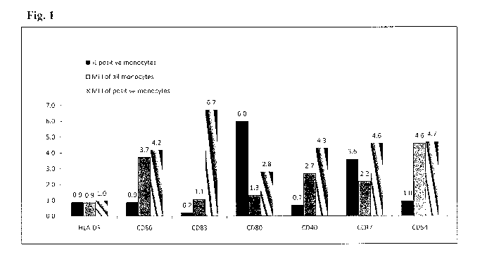
[054] Figure 1 shows dendritic cell (DC) markers on monocytes
expressed as fold increase after incubation at 37 C for 48 hours.
[055] Figure 2 compares expression of DC markers before and after
incubation at 37 C for 48h in adherent and non-adherent culture conditions.
[056] Figures 3A-3B compares expression of CD8 on monocytes
before and after incubation at 37 C for 48 hours. Fig. 3A presents % CD8
positive cells. Fig. 3B presents the Mean Fluorescence Intensity (MFI) of
CD8.
[057] Figures 4A-4B shows expression of the activation marker CD69
on lymphocyte subsets before and after incubation at 37 C for 48 hours in the
presence or absence of the superantigen, Staphylococcus aureus Enterotoxin
B. Fig. 4A shows the % positive cells among all lymphocytes, T cells, or CD3
negative cells. Fig. 4B presents the Mean Fluorescence Intensity (MFI) of
CD69 on positive cells in each population.
[058] Figure 5 shows expression of IL-2 receptor (CD25) on
lymphocyte subsets before and after incubation at 37 C for 48 hours in the
absence (48h) and presence (48 h SA) of superantigen, Staphylococcus
aureus Enterotoxin B.
CA 02865553 2014-08-26
WO 2013/136182
PCT/IB2013/000848
[059] Figures 6A-6C shows expression of CD40 on monocytes before
and after incubation at 37 C for 48 hours in the absence and presence of
superantigen, Staphylococcus aureus Enterotoxin B. Fig. 6A present the %
CD40 positive cells. Fig. 6B presents the Mean Fluorescence Intensity. Fig
5 6C shows the fold increase after 48 hour incubation.
[060] Figure 7A shows IL-12 production before and after incubation at
37 C for 48 hours. Figure 7B shows IL-12 production in the absence and
presence of superantigen, Staphylococcus aureus Enterotoxin B, before and
after incubation at room temperature (RT) or 37 C for 48 hours.
10 [061] Figure 8 shows IL-2 content in AICC and IL-2 production by
washed cells of AICC resuspended in fresh serum after incubation at 37 C for
48 hours in the absence and presence of superantigen, Staphylococcus
aureus Enterotoxin B.
[062] Figure 9 shows IFN-gamma production in the presence of
superantigen, Staphylococcus aureus Enterotoxin B, after incubation at 37 C
for 48 hours and IFN-gamma production by washed cells resuspended in
fresh serum after incubation at 37 C for 48 hours in the presence of
Enterotoxin B.
DESCRIPTION OF THE EMBODIMENTS
[063] As described in more detail below, the present invention relates
to activated immunostimulatory cell compositions (AICCs), methods of
preparing AICCs, and methods of using AICCs.
[064] An AICC of the invention includes functionally active monocytes
differentiated into mature DCs, as shown by their cell surface marker
profiles,
their ability to present antigens such as superantigens to T cells, and their
release of IL-12, a key factor promoting preferential Th1 polarization. T
cells
in the AICC are also activated. The interaction of the mature DC with T cells
in an AICC in the presence of antigen causes upregulation of IL-2 receptor on
T cells and release of IL-2 and IFN-g. When DCs in an AICC are exposed to
antigen, IL-12 production drastically increases. Accordingly, an AICC of the
current invention is a powerful tool for immune stimulation. For example,
CA 02865553 2014-08-26
WO 2013/136182
PCT/IB2013/000848
11
when administered in vivo, an AICC can change the cytokine balance in the
tumor environment to favor Th1 cytokines (e.g., interferons, IL-2), which
activate proliferation of CTLs and cause tumor rejection.
[065] Without being bound by theory, an AICC of the invention
polarizes monocytes/macrophages into an M1 phenotype. As demonstrated
in the working examples, the majority of monocytes/macrophages in AICC
express high levels of HLA-DR and produce IL-12 and other M1 cytokines.
M1 cytokines are known to overcome inhibitory effect of tumor environment on
cellular immunity and promote tumor rejection. Accordingly, the cytokines in
an AICC are useful in tumor therapies.
[066] The principles and operation of the present invention may be
better understood with reference to the drawings and accompanying
descriptions. Before explaining at least one embodiment of the invention in
detail, it is to be understood that the invention is not limited in its
application to
the details set forth in the following description or exemplified by the
Examples. The invention is capable of other embodiments or of being
practiced or carried out in various ways.
[067] It is to be understood that the phraseology and terminology
employed herein is for the purpose of description and should not be regarded
as limiting. Further, the term "about" as used in connection with any and all
values (including lower and upper ends of numerical ranges) includes a range
of deviation of +/- 0.5% to +/- 20% (and values therebetween, e.g., 1%,
1.5%, 2%, 2.5%, 3%, 3.5%, 4%, 4.5%, 5%, 5.5%, 6%,
6.5%, 7%, 7.5%, 8%, 8.5%, 9%, 9.5%, 10%, 10.5 ')/0, 11%,
11.5%, 12%, 12.5%, 13, 13.5%, 14%, 14.5%, 15%, 15.5%,
16%, 16.5%, 17%, 17.5%, 18%, 18.5%, 19%, 19.5%, and
20%).
l. Methods of Making an Activated lmmunostimulatory Cell
Composition
[068] Leukocytes require activation to mediate an immune response.
As used here, an activated immunostimulatory cell composition refers to a
composition comprising at least one type of activated leukocyte. In this
=
CA 02865553 2014-08-26
WO 2013/136182
PCT/1B2013/000848
12
context, "activated" means that a cell has acquired one or more functional or
phenotypic characteristics of an activated cell. Examples of characteristics
of
an activated (or "matured") dendritic cell include, but are not limited to,
production of IL-12; absent or low level production of IL-10, expression of
one
or more of the costimulatory molecules CD80, CD86, CD83, CD40, or CD1c
(BDCA1), of one or more adhesion molecules such as CD56, CD11b, CD11c,
or IGSF4 (SynCam and Nectin-like-2), of one or more lectin receptors such as
CLEC9A (DNGR-1), of one or more chemokine receptors such as CCR7, of
one or more Toll receptors such as TLR1, TLR3, or TLR6, of one or more
endocomal protein such as DC-LAMP, or of one or more transcription factors
such as Id2, IRF8, or ICSBP; ability to activate naïve T cells via antigen
presentation; and ability to induce B cell differentiation into antibody
secreting
(plasma) cells. Examples of characteristics of an activated T cell include,
but
are not limited to, production of one or more of IL-2, IFN-gamma, IFN-alpha,
or IFN-beta; expression of IL-2R; upregulation of T cell activation markers
such as one or more of CD69, CD71 (transferrin receptor 1), CD28, or CD4OL;
and proliferation following exposure to antigen, cytotoxic function, or helper
function.
[069] In one embodiment, an AICC is prepared from peripheral blood.
Peripheral blood generally contains not only red blood cells (RBC) and
platelets, but also leukocytes. Leukocytes, also known as "white blood cells,"
include monocytes (a "precursor" cell that differentiates into macrophages of
various tissues and dendritic cells), lymphocytes (which includes T cells, B
cells, natural killer (NK) cells, and natural killer T cells (NKT cells)), and
granulocytes (which includes neutrophils, basophils, and eosinophils).
[070] Although whole peripheral blood is a convenient source of
leukocytes, in alternate embodiments, an AICC is prepared using leukocytes
isolated from blood from a central line, umbilical cord blood, placental
blood,
lymph, bone marrow, or lymphoid tissue such as lymph node or spleen.
Leukocytes may be prepared by leukopheresis. Accordingly, the source of
the leukocytes is not believed to be critical.
CA 02865553 2014-08-26
WO 2013/136182
PCT/IB2013/000848
13
[071] When whole blood is used, letlikocytes can be partially
separated from red blood cells and platelets by preparing a "buffy coat" using
density gradient separation of the different cell types. Accordingly, in some
embodiments, the amounts of platelets and red blood cells present in an AICC
are lower than that in whole blood.
[072] The starting materials for producing an AICC may be obtained
from autologous or allogeneic sources. In one embodiment, an AICC is
prepared from the patient who will ultimately be treated with the AICC; that
is,
the source is autologous. In other embodiments, an AICC is prepared from
an individual other than the intended AICC recipient. In this case, the source
is allogeneic.
[073] In those embodiments, involving allogeneic starting materials,
these may be conveniently obtained from a blood bank. The samples may be
screened by the blood bank for blood type (ABO, Rh) or specific human
leukocyte antigen alleles such as, but not limited to, A2, B12 and C3,
irregular
antibodies to red cell antigens, and transfusion-transmittable diseases. More
specifically, screening can be conducted with antibodies using an Abbott
PRISM instrument against: Hepatitis B, C, HIV 1/2, HTLV and Syphilis (-HCV;
HbsAg; anti-HIV 1/2 0+; and anti-HTLV I/11). The samples can also be
screened for HIV, HCV and HBV by molecular methods (NAT-Nucleic Acid
Testing). Molecular screening can be accomplished using commercially
available instrumentation, e.g., the TIGRIS system of Chiron or any other
methods which may be suitable forms of testing for such diseases.
[074] In one embodiment involving allogeneic sources, the sample is
obtained from donors with the same blood type as the intended AICC
recipient. In one embodiment, the donor(s) and recipient patient can be
matched based on one or more HLA allele type. Alternatively, plasma
samples can be obtained from donors with AB+ blood and the leukocytes can
be obtained from donors with 0- blood. Donors with AB+ blood are universal
donors for plasma and donors with 0- blood are universal donors for
leukocytes. The plasma can be fresh, stored (e.g., at 1-6 C for less than 24
hours), dried, or otherwise pre-treated (e.g., pathogen-reduced plasma and
CA 02865553 2014-08-26
WO 2013/136182 PCT/IB2013/000848
14
solvent/detergent (SD) treated plasma). Regardless of the source, all
necessary processing of the sample(s) can be carried out without the need for
highly specialized equipment.
[075] In some embodiments, activated immunostinnulatory cell
composition may be prepared from smaller volumes of blood samples, with
commensurate decreases in volumes of all solutions and use of smaller bags
or other incubation vessels. Furthermore, use of these different size
= incubation vessels yields AICC with similar compositions. Use of smaller
volumes provides the clinician with the ability to perform blood collection
autonomously, without using an external blood bank. This may be useful
when treating patients with otherwise healthy immune systems but suffering
from some type of a small cancerous lesion.
[076] In some embodiments, a method of preparing an AICC
comprises a) activating human leukocytes; b) incubating the activated
leukocytes in an incubation composition under conditions of time and
temperature to induce differentiation and maturation of dendritic cells (DC),
thus producing an AICC. In one embodiment, step (b) also induces activation
of lymphocytes.
[077] In one embodiment, the method further comprises contacting the
DC with antigen or an antigenic peptide. In one embodiment, the antigen or
antigenic peptide is contacted with the DC as they differentiate and mature in
the incubation composition. That is, antigen or antigenic peptide is added
during a part or all of the incubation of step (b). In one embodiment, the
antigen or antigenic peptide is contacted with the DC after the incubation in
the incubation composition is concluded. That is, the method further
comprises a step (c) in which antigen or antigenic peptide is added to the
AICC for a period of time sufficient to load DC with antigenic peptide.
[078] In one embodiment, an Activated Leukocyte Composition
produced using the methods of WO 2010/100570, is used in preparing the
AICC. In this embodiment, the Activated Leukocyte Composition corresponds
to step (a) of the above embodiment of the method.
CA 02865553 2014-08-26
WO 2013/136182
PCT/1B2013/000848
[079] In some embodiments, a method of preparing an AICC
comprises a) isolating human leukocytes; b) optionally subjecting the
leukocytes to hypo-osmotic shock; and c) incubating the shocked leukocytes
in an incubation composition under conditions of time and temperature to
5 induce differentiation and maturation of dendritic cells (DC), thus
producing an
AICC. In one embodiment, step (c) also induces activation of lymphocytes.
[080] In one embodiment, the method further comprises contacting the
DC with antigen or an antigenic peptide. In one embodiment, the antigen or
antigenic peptide is contacted with the DC as they differentiate and mature in
10 the incubation composition. That is, antigen or antigenic peptide is
added
during a part or all of the incubation of step (c). In one embodiment, the
antigen or antigenic peptide is contacted with the DC after the incubation in
the incubation composition is concluded. That is, the method further
comprises a step (d) in which antigen or antigenic peptide is added to the
15 AICC for a period of time sufficient to load DC with antigenic peptide.
[081] In one embodiment, an Activated Leukocyte Composition
produced using the methods of WO 2010/100570, is used in preparing the
AICC. In this embodiment, the Activated Leukocyte Composition corresponds
to steps (a) and (b) of the above embodiment of the method.
[082] In some embodiments, the method comprises a) incubating
human leukocytes under conditions of time and temperature to activate the
leukocytes; b) optionally subjecting the leukocytes to hypo-osmotic shock; c)
adding to the leukocytes of step b a physiologically acceptable salt solution
in
an amount effective to restore isotonicity; d) mixing the leukocytes of step c
with a medium to form a second incubation composition; and e) incubating the
second incubation composition under conditions of time and temperature to
induce differentiation and maturation of dendritic cells (DC), thus producing
an
AICC. In one embodiment, step (e) also induces further activation of
lymphocytes.
[083] In one embodiment, the method further comprises contacting the
dendritic cells (DC) of step (e) with antigen or an antigenic peptide. In one
embodiment, the antigen or antigenic peptide is contacted with the DC as they
CA 02865553 2014-08-26
WO 2013/136182
PCT/1B2013/000848
16
differentiate and mature in the incubation composition. That is, antigen or
antigenic peptide is added during part or all of the incubation of step (e).
In
one embodiment, the antigen or antigenic peptide is contacted with the DC
after the incubation in the incubation composition is concluded. That is, the
5 method further comprises a step (f) in which antigen or antigenic peptide
is
added to the AICC for a period of time sufficient to load DC with antigenic
peptide.
[084] In one embodiment, an Activated Leukocyte Composition
produced using the methods of WO 2010/100570,.is used in preparing the
10 AICC. In this embodiment, the Activated Leukocyte Composition
corresponds
= to steps (a) through (d) of the above embodiment of the method.
[085] In some embodiments, the method comprises a) incubating
human leukocytes at room temperature for up to about 20 hours to activate
the leukocytes; b) subjecting the leukocytes to hypo-osmotic shock; c) adding
15 to the leukocytes of step b a physiologically acceptable salt solution
in an
amount effective to restore isotonicity; d) mixing the leukocytes of step c
with
a medium to form a second incubation composition; and e) incubating the
second incubation composition under conditions of time and temperature to
induce differentiation and maturation of dendritic cells (DC), thus producing
an
20 AICC. In one embodiment, step (e) also induces further activation of
lymphocytes.
[086] In one embodiment, the method further comprises contacting the
dendritic cells (DC) of step (e) with antigen or an antigenic peptide. In one
embodiment, the antigen or antigenic peptide is contacted with the DC as they
25 differentiate and mature in the incubation composition. That is, antigen
or
antigenic peptide is added during part or all of the incubation of step (e).
In
one embodiment, the antigen or antigenic peptide is contacted with the DC
after the incubation in the incubation composition is concluded. That is, the
method further comprises a step (f) in which antigen or antigenic peptide is
30 added to the AICC for a period of time sufficient to load DC with
antigenic
peptide.
CA 02865553 2014-08-26
WO 2013/136182
PCT/IB2013/000848
17
[087] In one embodiment, an Activated Leukocyte Composition
produced using the methods of WO 2010/100570, is used in preparing the
AICC. In this embodiment, the Activated Leukocyte Composition corresponds
to steps (a) through (d) of the above embodiment of the method.
[088] In some embodiments, the method comprises a) activating
human leukocytes; b) mixing the leukocytes of step a with a medium to form a
second incubation composition; and c) incubating the second incubation
composition under conditions of time and temperature to induce differentiation
and maturation of dendritic cells (DC), thus producing an AICC. Activation of
leukocytes is indicated by a change in expression levels or in the number of
leukocytes expressing an activation marker of leukocytes, such as CD11b
and/or CD62L. Accordingly, in one embodiment, activation of the leukocytes
is indicated by increased expression of CD11b in the leukocyte population.
Increased expression of CDllb can be detected, for example, by flow
cytometry. Increased expression of CDllb encompasses an increase in the
mean fluorescence intensity for CDllb on leukocytes, for example, the mean
fluorescence intensity may be increased by at least about 5%, 10%, 15%,
20%, 25%, 30%, 35%, 40%, 45%, 50%, 55%, 60%, 65%, 70%, 75%, 80%,
85%, or 90%. Increased expression of CDllb also encompasses an increase
in the percentage of leukocytes expressing CD11 b (e.g., after correcting for
background staining using an isotype control). For example, the percentage
of leukocytes expressing CD11 b may increase at least about 5%, 10%, 15%,
20%, 25%, 30%, 35%, 40%, 45%, 50%, 55%, 60%, 65%, 70%, 75%, 80%,
85%, or 90% relative to expression of CDllb expression on leukocytes in a
buffy coat. In one embodiment, activation of the leukocytes is indicated by
reduced expression of CD62L in the leukocyte population. Reduced
expression of CD62L can be detected, for example, by flow cytometry.
Reduced expression of CD62L encompasses a decrease in the mean
fluorescence intensity for CD62L on leukocytes, for example, the mean
fluorescence intensity may be reduced by at least about 5%, 10%, 15%, 20%,
25%, 30%, 35%, 40%, 45%, 50%, 55%, 60%, 65%, 70%, 75%, 80%, 85%, or
90%. Reduced expression of CD62L also encompasses a decrease in the
CA 02865553 2014-08-26
WO 2013/136182
PCT/IB2013/000848
18
percentage of leukocytes expressing CD62L (e.g., after correcting for
background staining using an isotype control). For example, the percentage
of leukocytes expressing CD62L may decrease at least about 5%, 10%, 15%,
20%, 25%, 30%, 35%, 40%, 45%, 50%, 55%, 60%, 65%, 70%, 75%, 80%,
85%, or 90% relative to expression of CD62L expression on leukocytes in a
buffy coat. In one embodiment, CD62L and/or CD11b is measured on
leukocytes that are granulocytes. In one embodiment, CD62L and/or CD11b
is measured on leukocytes that are monocytes. In one embodiment, step (c)
also induces further activation of lymphocytes.
[089] In addition, in any of the other embodiments involving activation
of leukocytes, changes in expression of CD11b and/or CD62L, as discussed
above, may be used alone, together, or in combination with additional
markers and assays, as discussed elsewhere in the application, as an
indicator of leukocyte activation.
[090] In one embodiment, the method further comprises contacting the
dendritic cells (DC) of step (c) with antigen or an antigenic peptide. In one
embodiment, the antigen or antigenic peptide is contacted with the DC as they
differentiate and mature in the incubation composition. That is, antigen or
antigenic peptide is added during part or all of the incubation of step (c).
In
one embodiment, the antigen or antigenic peptide is contacted with the DC
after the incubation in the incubation composition is concluded. That is, the
method further comprises a step (d) in which antigen or antigenic peptide is
added to the AICC for a period of time sufficient to load DC with antigenic
peptide.
[091] In one embodiment, an Activated Leukocyte Composition
produced using the methods of WO 2010/100570, is used in preparing the
AICC. In this embodiment, the Activated Leukocyte Composition corresponds
to steps (a) and (b) of the above embodiment of the method.
[092] In general, in any of the methods of preparing an AICC, any
composition in which the leukocytes have transitioned from a quiescent to a
functionally active state can be used for the hypo-osmotic shock step. For
example, as described in WO 2010/100570, leukocytes can be transitioned
CA 02865553 2014-08-26
WO 2013/136182
PCT/IB2013/000848
19
from a quiescent to functionally active state by incubating them at room
temperature for up to about 20 hours. In one embodiment, the transition
occurs by incubating the leukocytes for about 90 minutes to about 20 hours at
room temperature. In one embodiment, the transition occurs by incubating
the leukocytes for about 8 hours to about 20 hours at room temperature. In
one embodiment, the transition occurs by incubating the leukocytes overnight
at room temperature. In other embodiments, the temperature may be about
37 C. As noted, the details of this "transitioning" step are not essential and
leukocytes may be obtained by any method for use in preparing an AICC.
[093] Further, since hypo-osmotic shock is a type of stress, in those
embodiments of the method that include a step of hypo-osmotic shock other
methods may be employed to stress the cells. That is, since various types of
stress elicit cellular responses through the same highly conserved signaling
pathway consisting of protein kinase cascades that result in the activation of
mitogen-activated proteins kinases (MAPKs), other stressors may be used in
place of hypo-osmotic shock in any of the embodiments that mention hypo-
osmotic shock. For example, in some embodiments, the methods of
preparing an AICC comprise an optional step (in place of, or in addition to,
hypo-osmotic shock) of subjecting the leukocytes to a stressor chosen from
heat shock, hypoxia, treatment with any one or more of chlorpromazine,
caffeine, vanadate, zymolyse, Congo red, calcofluor, rapamycin, or a mating
pheromone, or by induction of actin depolymerization. Leukocyte activation
also causes an increase in intracellular calcium, and there are many agonists
that mimic this response. Accordingly, in still other embodiments, the
methods of preparing an AICC comprise an optional step of subjecting the
leukocytes to a calcium ionophore such as FMLP or PMA in place of or in
addition to hypo-osmotic shock.
[094] In any of the embodiments of the various methods of producing
an AICC, an incubation under "conditions of time and temperature to induce
differentiation and maturation of dendritic cells (DC)" (which may optionally
also activate lymphocytes and other cells) generally is an incubation of from
CA 02865553 2014-08-26
WO 2013/136182
PCT/IB2013/000848
about 24 hours to about 14 days at from about room temperature to about
37 C.
[095] In some embodiments, the incubation to induce differentiation
and maturation of dendritic cells (DC) is at a temperature of about room
5 temperature, i.e., in the range of about 12 C to about 28 C. In one
embodiment, the incubation is at a temperature of from about 16 C to about
C. In one embodiment, the incubation is at a temperature of from
about 18-25 C. In yet another embodiment, the incubation is at a temperature
of from about 20-25 C.
10 [096] In some embodiments, the incubation to induce differentiation
and maturation of dendritic cells (DC) is at a temperature of about 37 C. In
one embodiment, the incubation is at about 35 C to about 38 C. In one
embodiment, the incubation is at 37 C +/- 0.5 C.
[097] In some embodiments, the time period of incubation to induce
15 differentiation and maturation of dendritic cells (DC) is from about 24
hours to
14 days. Thus, in some embodiments the incubation is for about 24, 30, 36,
42, 48, 54, 60, 66, 72, 84, 96, 108, 120, 132, 144, 156, 168, 192, 216, 240,
264, 288, 312, or about 336 hours, or for about any range of hours in between
these values.
20 [098] In some embodiments, the time period of incubation to induce
differentiation and maturation of dendritic cells (DC) is from about 48 to
about
72 hours. In one embodiment, the incubation is for about 48 to about 72
hours at about 37 C. In one embodiment, the incubation is for about 48 hours
at about 37 C. In one embodiment, the incubation is for about 72 hours at
25 about 37 C. In one embodiment the incubation is for about 24 to about 72
hours at room temperature.
[099] In some embodiments, the incubation is in a cell incubator in an
atmosphere containing 5% CO2 and at 100% humidity. In some
embodiments the incubation is in gas-permeable bags and the bags are
placed in a cell incubator in an atmosphere containing 5% CO2 and 100%
humidity. In other embodiments, tissue culture flasks or dishes are used in
the method. In still other embodiments, combinations of bags systems and
CA 02865553 2014-08-26
WO 2013/136182
PCT/1132013/000848
21
culture dishes or flasks are used in the method. In those embodiments
involving incubation in a bag system, the bag system may be one of those
described in W02010/100570, or it can be a bag made from a different
material, such as but not limited to fluorinated ethylene propylene (FEP) or
Ethyl Vinyl Acetate (EVA), or in a tissue culture vessel or in any vessel.
[0100] Any vessel used for incubation may also be treated or otherwise
modified so that it becomes adhesive for leukocytes, which could be beneficial
for leukocyte activation, differentiation of monocytes and activation/priming
of
lymphocytes. In one embodiment, one or more of the culture vessels used in
the methods of preparing an AICC are non-adherent for dendritic cells. In
another embodiment, the culture vessels used in the methods of preparing an
AICC are treated to reduce cell adherence. In still another embodiment, one
or more culture vessels used in the methods of preparing an AICC are treated
to increase the adherence of cells.
[0101] Any vessel used for an incubation may also contain scaffolds.
The scaffolds may be in different shapes and in particular could be
microbeads, biodegradable or not biodegradable, e.g., made of collagen, or
made of PLA, PGA (polylactic acid, polyglycolic acid) or similar synthetic
polymers, hydrogel scaffolds made of gelatin, hyaluronic acid alginated,
fibrin
sealer. Scaffolds or bags could be coated with adhesion receptors,
extracellular matrix proteins such as fibronectin or laminin or with active
binding peptides from extracellular matrices, such as RGD. Scaffolds or
microbeads could be also coated with activating stimuli or stimulating
antibodies,.such as but not limited to activating antibodies against CD3,
CD28, or CD40. In at least one embodiment, however, the method does not
comprise microbeads or scaffolds coated with one or more activating stimuli
or with one or more antibodies against CD3, CD28, or CD40.
[0102] In some embodiments, the medium used for the incubation to
produce an AICC is plasma or serum. In those embodiments utilizing serum,
the serum may be obtained from a sample of plasma, which may be obtained
from the same or a different whole blood sample (i.e., from the same or a
different human) as the leukocytes, that has been contacted with a
CA 02865553 2014-08-26
WO 2013/136182
PCT/1B2013/000848
22
coagulating agent at about 37 C. In some embodiments, the serum or plasma
is obtained from a commercial or non-profit supplier and may be either fresh
or in a storage-compatible form, such as frozen.
[0103] In addition, although serum (particularly human serum) is often
used in the incubation composition as the supportive medium, other
supportive media may be used as well so long as it is a physiologic medium
that supports release of cytokines, growth factors, and/or other soluble
components from the activated leukocytes. For example, plasma may be
used instead of serum. Other incubation medium that may also be used as
supportive medium include culture medium, saline, or buffered saline
solutions with optional addition of sugars and other components essential for
cell viability and function such as amino acids (e.g. Lactated Ringer's
solution,
Acetated Ringer's solution, Hank's balanced salt solution (HBSS), Earle's
balanced salt solution (EBSS), Standard saline citrate (SSC), HEPES-
buffered saline (HBS), Gey's balanced salt solution (GBSS)). Saline solutions
and culture medium may also be supplemented with human serum or clinical
grade animal serum, or serum substitutes. The incubation composition may
alternatively, or in addition, contain serum proteins such as human or bovine
albumin, gamma-globulin, transferrin or other proteins from different tissues,
plant proteins, or plant extracts.
[0104] In certain embodiments, leukocyte agonists such as complement
proteins, chemokines, interferon-alpha, interferon-gamma, cytokines such as
interleukin-4, granulocyte-macrophage¨colony stimulating factor (GM-CSF),
or interleukin-12, are added to the incubation. Monocyte differentiation to
DCs in vitro can be induced using well-defined cytokine cocktails (Jensen SS,
Gad M. 2010). Accordingly, in one embodiment, an incubation may be
performed in the presence of cytokines such as interleukin-4 or GM-CSF. In
other embodiments, an incubation may be performed with other substances
that increase differentiation and maturation of dendritic cells and activation
of
lymphocytes and NK cells. For example, the CD40 co-stimulatory receptor on
monocytes may be ligated by antibodies to CD40 or by a CD40 ligand (CD54)
in the absence of cytokines (Brossart P, 1998). A CD40 independent
CA 02865553 2014-08-26
WO 2013/136182
PCT/1B2013/000848
23
activation of DC maturation can be induced by interaction with activated CD8
positive T cells (Ruedl C., 1999; Wirths, 2002). Accordingly, in one
embodiment, exogenous, activated CD8 positive T cells may be added to the
incubation. DC differentiation from monocytes in vitro can also be induced by
DC interaction with NKT cells. DC differentiation results from NKT cell
secretion of GM-CSF and IL-13, cytokines that were produced by the NKT
cells upon activation by monocytes (Hegde, 2007). Accordingly, in one
embodiment, exogenous, activated NKT cells may be added to the incubation.
In other embodiments, DC differentiation and maturation may be promoted by
the addition of one or more of GM-CSF, IL-4, IFN-gamma, IL-2, IFN-alpha,
and TNF-alpha; and/or by addition of one or more bacterial products that
interact with Toll receptors on DCs, such as but not limited to
lipopolysaccharide (LPS), peptidoglycan (murein), double-stranded RNA or its
synthetic analog polyinosinic:polycytidylic acid (poly I:C), Resiquimod (R-
848),
and Picibanil (0K-432).
[0105] It is also expressly contemplated that, in one or more
embodiments of the methods, incubation occurs in the absence of one or
more of the exogenously added factor(s) described above as involved in DC
maturation. In one embodiment, all of the components needed for DC
maturation are provided endogenously and no additional stimuli are added to
the incubation composition. Nevertheless, various cytokines may be present
in the incubation composition because they are released upon leukocyte
activation during incubation. For example. CD40 ligand may be found in
serum and on platelets that are part of the incubation composition. Similarly,
activated CD8 + T cells and NKT cells that are endogenously present in the
incubation composition can interact with monocytes to support dendritic cell
differentiation and maturation.
[0106] Accordingly, in one embodiment, the incubation composition for
producing an AICC does not include exogenous GM-CSF, exogenous IL-4,
exogenous TNF, or an exogenous interferon (although one or more of GM-
CSF, IL-4, TNF, or an interferon may be produced endogenously during the
incubation). Thus, in one embodiment a method of preparing an AICC
CA 02865553 2014-08-26
WO 2013/136182
PCT/1B2013/000848
24
excludes the addition of one or more exogenous cytokine or interferon, the
addition of reagent(s) that crosslink CD3 and/or CD28, the addition of
reagent(s) that crosslink CD40, and/or the addition of other exogenous agents
that promote dendritic cell maturation during the production of the A1CC.
Examples of exogenously added cytokines and exogenously added
interferons that may be excluded from the practice of the method include any
one or more of GM-CSF, IL-4, IFN-gamma, IL-2, IFN-alpha, or IL-2.
Examples of exogenously added bacterial products that may be excluded
from the practice of the methods include those known to interact with Toll
receptors on DCs, such as but not limited to lipopolysaccharide (LPS),
peptidoglycan (murein), double-stranded RNA or its synthetic analog
polyinosinic:polycytidylic acid (poly I:C), Resiquimod (R-848), and Picibanil
(0K-432).
[0107] In some embodiments inhibitors of angiogenesis targeting VEGF
signaling are added to the incubation composition. These include but are not
limited to anti-VEGF antibodies (e.g. bevacizumab, ranibizumab), antibodies
against VEGF receptors (e.g. Brivanib, targets VEGFR-2 and FGFR),
inhibitors of the tyrosine kinase activity of the VEGF receptors (e.g.,
Sorafenib, Cediranib, Sunitinib), soluble receptor-decoys (e.g, VEGF Trap,
also called aflibercept), or vascular-disrupting agents (e.g., ZD6126).
[0108] In some embodiments adjuvants are added to the incubation
composition. Examples of adjuvants include but are not limited to aluminium
hydroxide, aluminium phosphate and calcium phosphate, adjuvants based on
oil emulsions (Freund's emulsified oil adjuvants (complete and incomplete),
Arlacel A, Mineral oil, emulsified peanut oil adjuvant (adjuvant 65), products
from bacteria (their synthetic derivatives as well as liposomes) or gram-
negative bacteria, endotoxins, cholesterol, fatty acids, aliphatic amines,
paraffinic and vegetable oils, monophosphoryl lipid A, 1SCOMs with Quil-A,
and Syntex adjuvant formulations (SAFs).
[0109] As discussed elsewhere, in some embodiments, any of the
methods may further comprise a contacting step wherein one or more antigen
or antigenic peptide is introduced. Examples of antigens include tumor-
CA 02865553 2014-08-26
WO 2013/136182
PCT/IB2013/000848
specific and tumor-associated antigens, stem cell/ cancer stem cell antigens,
and superantigens (e.g., staphylococcal enterotoxins). Generally speaking,
antigens or antigenic peptides will enhance differentiation of monocytes into
dendritic cells and prime lymphocytes specific for that antigen. Further,
since
5 antigen presentation on the cellular level involves an antigenic peptide
presented in the context of a class I or class II molecule, the terms
"antigen,"
"antigen peptide," and "antigenic peptide" should not be construed as
requiring contact with an intact antigen or a particular peptide. Instead, the
terms are used broadly to indicate that an antigen presenting cell is
contacted
10 with antigenic material that it may then either directly, or after
further
processing, present in the context of class I or class II molecules.
[0110] Antigens and antigenic peptides, whether prepared from cell
lysates or by recombinant expression of a protein or peptide, are incubated
with an AICC or during the production of an AICC at various concentrations
15 for about 1 hour to about 24 hours at room temperature to about 37 C.
Examples of antigens/peptides include those listed below and any
antigen/peptide used in the Examples section.
[0111] Recently, high-throughput technologies have enabled the
identification of mutated gene in cancers. The number of these genes is high,
20 with a functional heterogeneity broader than previously thought.
(Stratton et
al., Nature 458:719-24 (2009); Pleasance et al., Nature 463:191-96 (2010).)
These studies have been performed in breast, colorectal, pancreatic, and lung
cancers, as well as in glioblastomas, and overall have identified almost 400
candidate cancer genes (CAN-genes).
25 [0112] Some
examples of shared antigens that are normally associated
with spermatocytes or spermatogonia of the testis, placenta, and ovary
include the cancer-testis (CT) antigens BAGE, GAGE, MAGE, NY-ESO-1, and
SSX. These antigens are found in melanoma, lymphoma, lung, bladder,
colon, and breast carcinomas. Shared antigens normally found in
melanocytes, epithelial tissues, prostate, and colon also include the
differentiation antigens Gp100, Melan-A/Mart-1, Tyrosinase, PSA, CEA, and
Mammaglobin-A. These antigens are found in melanoma, prostate cancer,
CA 02865553 2014-08-26
WO 2013/136182
PCT/1B2013/000848
26
and in colon and breast carcinomas. Shared antigens that are ubiquitously
expressed at low levels may be overespressed in cancers. Examples of
overexpressed antigens include p53, HER-2/neu, livin, and survivin, found in
esophagus, liver, pancreas, colon, breast, ovary, bladder, and prostate
carcinomas. Other antigens are unique, such as 13 -catenin-m, f3-Actin/4/m,
Myosin/m, HSP70-2/m, and HLA-A2-R170J, which are associated with one or
more of melanoma, non-small cell lung cancer, and renal cancer. Still other
antigens are the tumor-associated carbohydrate antigens that are normally
found in epithelia tissues such as renal intestinal and colorectal tissues.
These antigens include GM2, GD2, GD3, MUC-1, sTn, abd globo-H, which
can be found in melanoma, neuroblastoma, colorectal, lung, breast, ovarian,
and prostate cancers.
[0113] Some additional exemplary antigen/peptides that may be used
in the various aspects of the invention include MART-1, MAGE-1, MAGE-3,
TYR, and gp100 antigens/peptides, which are associated with metastatic
melanoma (e.g., as described in Butterfield et al., J. lmmunotherapy 2008;
31:294-309; Markowicz et al., J Clin Oncol 27:15s, 2009 (suppl; abstr 9039));
TADG-12, CA125, hepsin, and other antigens/peptides associated with
ovarian cancer (e.g., as described in U.S. Patent Nos. 8,097,242 and
7,935,531); the carcinoembryonic antigen (CEA), which is associated with
colorectal, gastric, and pancreatic carcinomas, some breast cancers, and
many non-small cell lung cancers (e.g., as described in U.S Patent No.
8,012,468); antigens associated with neural cancers (e.g., glioblastoma
multiforme and astrocytomas), such as the antigens tyrosine-related protein
(TRP), melanoma-associated antigen-1 (MAGE-1), HER-2, AIM-2, IL-13
receptor alpha 2, or gp100 antigens and their peptide epitopes described in
U.S. Patent No. 8,097,256; hTERT (human telomerase reverse transcriptase),
including the peptides described in U.S. Patent No. 8,003,773; prostate
specific antigen (PSA), prostate-specific membrane antigen (PSMA), and
prostatic acid phosphatase (PAP) antigen, which are associated with prostate
cancer (e.g., Tartour et al., Immunol Lett 2000; 74(1): 1-3); HPV (human
papilloma virus) antigen (associated with cervical carcinoma); prostate
CA 02865553 2014-08-26
WO 2013/136182
PCT/IB2013/000848
27
specific G protein coupled receptor (PSGR) and six-transmembrane epithelial
antigen of prostate STEAP described in U.S. Patent No. 7,906,620 as
associated with prostate and colon cancer; and/or PAGE4, which is
associated with reproductive cancers such as prostate, uterine, and testicular
cancer (as described in U.S. Patent No. 7,910,692). In some embodiments,
the intact antigen is used, whereas in other embodiments a peptide epitope of
the antigen (prepared either by proteolytic digestion or recombinantly) is
used.
[0114] In some embodiments, dendritic cells in an AICC are transfected
with mRNA isolated from tumor or stem cells with known RNA sequence for
tumor-specific antigens through electroporation, for example, using
exponential decay wave or square-wave electroporators or other RNA pulsing
apparatuses. In another embodiment, one or more antigen or antigen peptide
is introduce into antigen presenting cells, such as dendritic cells in an
AICC,
using microparticle-based transfection, for example, as described in U.S.
Patent No. 8,097,243. In still other embodiments, one or more antigen or
antigen peptide is introduced using adenovirus-based transduction, for
example, as described in U.S. Patent No. 8,012,468 and in Butterfield et al.,
J.
Immunotherapy 2008; 31:294-309; or using a retroviral vector as described in
U.S. Patent No. 8,003,773.
[0115] In any of the embodiments, the method may result in one or
more of differentiation of monocytes into maturate DC's, activation of
lymphocytes, activation and/or proliferation of NK cells, or activation and/or
proliferation of NKT cells.
[0116] In one embodiment, an AICC comprises "mature" DCs if the
AICC includes cells that can stimulate activation (priming) of naïve T cells
(as =
shown by expression of one or more of the T cell activation markers CD69, IL-
2R, CD28, CD71, CD49d, CD4OL, and/or by production of IL-2, IFN-alpha,
IFN-beta, or IFN-gamma) and differentiation and proliferation of T helper and
cytotoxic T cells in the presence of antigen. In another embodiment, the AICC
comprises mature DC if there is an increase in production of IL-12. In another
embodiment, the AICC comprises mature DC if there is an increase in the
expression of one or more of the markers CD80, CD86, CD83, CD40, CD1c,
CA 02865553 2014-08-26
WO 2013/136182
PCT/1B2013/000848
28
CD56, CD11 b, CD11 c, IGSF4, CLEC9A, CCR7, TLR1, TLR3, TLR6, DC-
LAMP, Id2, IRF8, or ICSBP on monocytes in the AICC. In one embodiment,
an increase in expression is an increase in the number of one or more of
molecules on the surface of a DC in the AICC (e.g., an increase in the mean
fluorescence intensity (MFI) as determined by flow cytometry). An increase in
the number of molecules is determined by comparing the MFI for that
particular marker on a DC in the AICC that is suspected of being mature. In
one embodiment, an AICC comprises "mature" DCs if there is an increase in
the percentage of monocytes expressing one or more of the markers. In one
embodiment, a monocyte is identified based on characteristic side scatter
[SSC] and positive staining for the pan-leukocyte marker CD45 by flow
cytometry, or by monocyte-specific CD14 staining. In one embodiment, the
method results in an AICC in which both the MFI and the percentage of
monocytes expressing at least one of the cell surface markers HLA-DR,
CD54, CD86, CD83, CD80, CD40, and CCR7 increases. In one embodiment,
the method results in an AICC in which both the MFI and the percentage of
monocytes expressing any combination or all of the cell surface markers HLA-
DR, CD54, CD86, CD83, CD80, CD40, and CCR7 increases. In one
embodiment, an increase is assessed relative to the starting leukocyte
composition used to produce the AICC (e.g., leukocytes in a buffy coat). In
one embodiment, an increase is assessed relative to the cell composition
used to being the incubation under conditions of time and temperature to
induce differentiation and maturation of DC.
[0117] In one embodiment, an AICC will present antigens to naive T
cells, causing the naïve T cells to differentiate into CD4 positive and CD8
positive cells, proliferate, produce IL-2, express IL-2 receptor, and produce
interferons and other Thl cytokines.
[0118] In another aspect, the methods may further comprise enriching
an AICC of the invention for one or more cell populations. Compositions
enriched for dendritic cells, T cells, NK cells, NKT cells, or other cell
types can
be prepared by cell sorting, panning, MACS, etc., using either positive or
negative marker selection according to known methods.
CA 02865553 2014-08-26
WO 2013/136182
PCT/IB2013/000848
29
[0119] In an additional embodiments, the methods may further
comprise separating the cellular portion of the AICC from the liquid portion.
In
one embodiment, both the cellular and liquid (supernatant) portions are
recovered. This may be accomplished, for example, by centrifuging the AICC
and transferring the supernatant to a separate vessel. As described in the
Examples, the supernatant is useful for therapy even in the absence of cells
because of the cytokines and other soluble factors it contains. The cells in
the
pellet that forms following centrifugation may then be resuspended in any
desired carrier. In other embodiments, the cells are removed from the liquid
portion without recover of the cells, for example, by filtration. In still
other
embodiments, the cells are recovered without recovery of the supernatant, for
example, by pelleting the cells and aspirating the supernatant.
[0120] In any of the embodiments, once an AICC is produced, the cells
in the AICC may be isolated, either with or without additional concentration,
and suspended in a carrier such as serum (which may be autologous or
allogeneic with respect to recipient) or some other physiologically acceptable
isotonically normal liquid suitable for storing and administering cells.
Examples of such solutions are described below, and include solutions used
to restore isotonicity, cell culture medium, buffered saline, or any other
biocompatible fluid or specially formulated clinically acceptable cell storage
or
cell cryopreservation medium.
11. Activated lmmunostimulatory Cell Compositions
[0121] In another aspect, the invention relates to an Activated
Immunostimulatory Cell Composition (AICC), which refers to any of the
compositions produced by the methods of making an AICC described above.
Accordingly, while "AICC" often refers to a cellular composition in the same
carrier used in the incubation, as described above, an AICC also
encompasses the cellular component in any carrier or excipient, as well as the
liquid component separated from the cellular component.
[0122] Leukocytes in an AICC have certain characteristics that may be
used to distinguish the individual leukocyte cell types or the composition as
a
CA 02865553 2014-08-26
WO 2013/136182
PCT/IB2013/000848
whole. For example, monocytes in an AICC may express higher levels of
CD54, HLA-DR and/or CD86 compared to freshly isolated monocytes.
Further, monocytes in the AICC may express additional activation markers,
such as one or more of CD8-alpha, CD83, CD80, CCR7, and/or CD40
5 compared to freshly isolated monocytes. An AICC composition may comprise
a higher percentage or number of monocytes that have differentiated and
matured into DCs than in a freshly prepared sample of, for example,
peripheral blood leukocytes. Likewise, an AICC may contain a greater
number or percentage of cells that are capable of activating/priming naïve
10 lymphocytes than in a freshly prepared sample of, for example,
peripheral
blood leukocytes. Lymphocytes in the AICC may express higher surface
levels of additional markers compared to freshly isolated lymphocytes, such
as one or more of CD69, CD25, CD28, CD154, CD107a, and/or CD42d. In
addition, leukocytes in the AICC may exhibit an increased ability to produce
15 cytokines, such as one or more of IL-2, IFN gamma, IFN alpha, IFN beta,
TNF
alpha, TNF beta, and/or IL-12, compared to freshly isolated leukocytes.
[0123] As noted, in some embodiments of the method, an Activated
Leukocyte Composition (ALC) produced using a method of WO 2010/100570
is used in the method of preparing the Activated lmmunostimulatory Cell
20 Composition (AICC). Although leukocytes in the ALC may be at least
partially
activated, as described in the Examples the leukocytes in the AICC may
achieve higher levels of activation than in the ALC. The higher level of
activation of leukocytes in an AICC compared to leukocytes in an ALC may be
shown by any one or more of the characteristics noted above for an AICC
25 using the ALC as the comparator.
[0124] As shown in the working examples, an AICC may also be
characterized and distinguished from known compositions in terms of
minimum activation level of DCs, e.g., as indicated by surface expression of
HLA-DR, CD86, CD83, CD80, CD40, CD54, and CCR7 on monocytes;
30 minimum activation level of lymphocytes, e.g., as indicated by surface
expression of CD69, CD25 (IL-2R), CD28, CD154/CD4OL, and CD49d; and
minimum activation levels of NK and NKT cells, e.g., as indicated by surface
CA 02865553 2014-08-26
WO 2013/136182
PCT/1132013/0008.18
31
expression of CD56, CD57, and CD107a. Surface expression of markers may
be evaluated either as the percentage of cells expressing the marker or as the
level of marker per cell.
[0125] In some embodiments, the final content of Activated
. 5 Immunostimulatory Cell Composition (AICC) includes, in terms of the
populations of leukocytes present, granulocytes, monocytes and lymphocytes.
Specific amounts and relative percentages of the cells may differ based on
the analysis techniques employed and on sample-to-sample variation. For
example, when analysis is performed using a Cell Dyn Analyzer, the AICC
generally contains about 45% to about 72% granulocytes (including
neutrophils, eosinophils and basophils), about 3% to about 10% monocytes,
and about 25% to about 50% lymphocytes. When analysis is performed using
FACS (e.g., using a side-scatter versus a forward-scatter dot plot analysis or
versus CD45 and/or CD14 fluorescence), the AICC generally contains about
50% to about 70% granulocytes; about 5% to about 15% monocytes, and
about 15% to about 35% lymphocytes.
[0126] Granulocytes include neutrophils, eosinophils and basophils. In
some embodiments, an AICC contains about 25% to about 85% neutrophils,
about 0 to about 9% eosinophils; about 1.5 to about 4% basophils, about 2%
to about 40% monocytes (including dendritic cells), and about 4% to about
70% lymphocytes, based on the total number of leukocytes in the AICC.
[0127] In any of the embodiments, an AICC may further contain
residual levels of red blood cells, generally in the amount of about 0.05 to
about 0.2 million per microliter, and/or residual levels of platelets,
generally in
the amount about 1 to about 100 thousand per microliter.
[0128] In some embodiments, the subpopulations of lymphocytes in the
AICC are in the general ranges as follows: about 20% to about 80% T cells
(CD3+); about 5% to about 40% B cells (CD19+); about 5% to about 35% NK
cells (CD3-/CD56+), and/or about 0.1% to about 35% of NKT cells
(CD3+/CD56+). In some embodiments, among T cells there are about 5% to
about 65% T helper cells (CD4+/CD3+) and about 5% to about 75% cytotoxic
T lymphocytes (CTLs, CD8+/CD3+).
CA 02865553 2014-08-26
WO 2013/136182
PCT/IB2013/000848
32
[0129] In other embodiments, there about 40% to about 60% T cells
(CD3+); about 15% to about 30% B cells (CD19+); about 15% to about 30%
NK cells (CD3-/CD56+), about 2% to about 20% of NKT cells (CD3+/CD56+).
In some embodiments, among T cells there are about 15% to about 40% of T
helper cells (CD4+/CD3+) and about 25% to about 50% of CTL (CD8+/CD3+).
[0130] The ratio between Th cells and CTLs is usually about 0.5 to 1.5.
[0131] In any of the embodiments, the levels of DC, lymphocyte, NK,
and NKT cell markers, as well as percentages of cells expression those
markers, may be determined as described in the methods of preparing an
AICC, or as described in the Examples.
[0132] In one embodiment, an AICC comprises DCs, wherein at least
about 5%, 10%, 15%, 20%, 25%, or 30% of the DC express CD8, as detected
by flow cytometry compared to an isotype control.
[0133] In one embodiment, at least about 1%, 5%, 10%, 15%, 20%,
25%, 30%, 35%, 40%, 45%, 50%, 55%, or 60% of the monocytes in the AICC
are positive for the marker CCR7, as detected by flow cytometry compared to
an isotype control.
[0134] In one embodiment, at least about 1%, 5%, 10%, 15%, 20%,
25%, 30%, 35%, 40%, 45%, 50%, 55%, or 60% of the monocytes in the AICC
are positive for the marker CD40, as detected by flow cytometry compared to
an isotype control.
[0135] In one embodiment, at least about 1%, 5%, 10%, 15%, 20%,
25%, 30%, 35%, 40%, 45%, 50%, 55%, or 60% of the monocytes in the AICC
are positive for the marker CD80, as detected by flow cytometry compared to
an isotype control.
[0136] In one embodiment, at least about 1%, 5%, 10%, 15%, 20%,
25%, 30%, 35%, 40%, 45%, 50%, 55%, or 60% of the monocytes in the AICC
are positive for the marker CD83, as detected by flow cytometry compared to
an isotype control.
[0137] In one embodiment, the mean fluorescence intensity (MFI) of
total monocytes for the marker CD86 is at least about 0.5, 1.0, 1.5, 2.0, 2.5,
3.0, 3.5, 4.0, 4.5, 5.0, 5.5, 6.0, 6.5, 7.0, 7.5, 8.0, 8.5, 9.0, 9.5, or 10
fold higher
CA 02865553 2014-08-26
WO 2013/136182
PCT/1B2013/000848
33
fresh than on peripheral blood monocytes. In one embodiment, the mean
fluorescence intensity (MFI) of monocytes for the marker CD86 is at least
about 0.5, 1.0, 1.5, 2.0, 2.5, 3.0, 3.5, 4.0, 4.5, 5.0, 5.5, 6.0, 6.5, 7.0,
7.5, 8.0,
8.5, 9.0, 9.5, or 10 fold higher than on monocytes in an ALC prepared
according to WO 2010/100570. In these embodiments, total monocytes are
determined by SSC and staining for a pan-leukocyte marker.
[0138] In one embodiment, the mean fluorescence intensity (MFI) of
total monocytes for the marker CD83 is at least about 0.5, 1.0, 1.5, 2.0, 2.5,
3.0, 3.5, 4.0, 4.5, 5.0, 5.5, 6.0, 6.5, 7.0, 7.5, 8.0, 8.5, 9.0, 9.5, or 10
fold higher
fresh than on peripheral blood monocytes. In one embodiment, the mean
fluorescence intensity (MFI) of monocytes for the marker CD83 is at least
about 0.5, 1.0, 1.5, 2.0, 2.5, 3.0, 3.5, 4.0, 4.5, 5.0, 5.5, 6.0, 6.5, 7.0,
7.5, 8.0,
8.5, 9.0, 9.5, or 10 fold higher than on monocytes in an ALC prepared
according to WO 2010/100570. In these embodiments, total monocytes are
determined by SSC and staining for a pan-leukocyte marker.
[0139] In one embodiment, the mean fluorescence intensity (MFI) of
total monocytes for the marker CD80 is at least about 0.5, 1.0, 1.5, 2.0, 2.5,
3.0, 3.5, 4.0, 4.5, 5.0, 5.5, 6.0, 6.5, 7.0, 7.5, 8.0, 8.5, 9.0, 9.5, or 10
fold higher
fresh than on peripheral blood monocytes. In one embodiment, the mean
fluorescence intensity (MFI) of monocytes for the marker CD80 is at least
about 0.5, 1.0, 1.5, 2.0, 2.5, 3.0, 3.5, 4.0, 4.5, 5.0, 5.5, 6.0, 6.5, 7.0,
7.5, 8.0,
8.5, 9.0, 9.5, or 10 fold higher than on monocytes in an ALC prepared
according to WO 2010/100570. In these embodiments, total monocytes are
determined by SSC and staining for a pan-leukocyte marker.
[0140] In one embodiment, the mean fluorescence intensity (MFI) of
total monocytes for the marker CD40 is at least about 0.5, 1.0, 1.5, 2.0, 2.5,
3.0, 3.5, 4.0, 4.5, 5.0, 5.5, 6.0, 6.5, 7.0, 7.5, 8.0, 8.5, 9.0, 9.5, or 10
fold higher
fresh than on peripheral blood monocytes. In one embodiment, the mean
fluorescence intensity (MFI) of monocytes for the marker CD40 is at least
about 0.5, 1.0, 1.5, 2.0, 2.5, 3.0, 3.5, 4.0, 4.5, 5.0, 5.5, 6.0, 6.5, 7.0,
7.5, 8.0,
8.5, 9.0, 9.5, or 10 fold higher than on monocytes in an ALC prepared
CA 02865553 2014-08-26
WO 2013/136182
PCT/1B2013/000848
34
according to WO 2010/100570. In these embodiments, total monocytes are
determined by SSC and staining for a pan-leukocyte marker.
[0141] In one embodiment, the mean fluorescence intensity (MFI) of
total monocytes for the marker CCR7 is at least about 0.5, 1.0, 1.5, 2.0, 2.5,
3.0, 3.5, 4.0, 4.5, 5.0, 5.5, 6.0, 6.5, 7.0, 7.5, 8.0, 8.5, 9.0, 9.5, or 10
fold higher
fresh than on peripheral blood monocytes. In one embodiment, the mean
fluorescence intensity (MFI) of monocytes for the marker CCR7 is at least
about 0.5, 1.0, 1.5, 2.0, 2.5, 3.0, 3.5, 4.0, 4.5, 5.0, 5.5, 6.0, 6.5, 7.0,
7.5, 8.0,
8.5, 9.0, 9.5, or 10 fold higher than on monocytes in an ALC prepared
according to WO 2010/100570. In these embodiments, total monocytes are
determined by SSC and staining for a pan-leukocyte marker.
[0142] In one embodiment, the mean fluorescence intensity (MFI) of
total monocytes for the marker CD54 is at least about 0.5, 1.0, 1.5, 2.0, 2.5,
3.0, 3.5, 4.0, 4.5, 5.0, 5.5, 6.0, 6.5, 7.0, 7.5, 8.0, 8.5, 9.0, 9.5, or 10
fold higher
fresh than on peripheral blood monocytes. In one embodiment, the mean
fluorescence intensity (MFI) of monocytes for the marker CD54 is at least
about 0.5, 1.0, 1.5, 2.0, 2.5, 3.0, 3.5, 4.0, 4.5, 5.0, 5.5, 6.0, 6.5, 7.0,
7.5, 8.0,
8.5, 9.0, 9.5, or 10 fold higher than on monocytes in an ALC prepared
according to WO 2010/100570. In these embodiments, total monocytes are
determined by SSC and staining for a pan-leukocyte marker.
[0143] In one embodiment, the mean fluorescence intensity (MFI) of
total monocytes for the marker CD8 is at least about 0.5, 1.0, 1.5, 2.0, 2.5,
3.0, 3.5, 4.0, 4.5, 5.0, 5.5, 6.0, 6.5, 7.0, 7.5, 8.0, 8.5, 9.0, 9.5, or 10
fold higher
fresh than on peripheral blood monocytes. In one embodiment, the mean
fluorescence intensity (MFI) of monocytes for the marker CD8 is at least about
0.5, 1.0, 1.5, 2.0, 2.5, 3.0, 3.5, 4.0, 4.5, 5.0, 5.5, 6.0, 6.5, 7.0, 7.5,
8.0, 8.5,
9.0, 9.5, or 10 fold higher than on monocytes in an ALC prepared according to
WO 2010/100570. In these embodiments, total monocytes are determined by
SSC and staining for a pan-leukocyte marker.
[0144] In one embodiment, at least about 5%, 10%, 15%, 20%, 25%,
30%, or 35% of the CD3 positive lymphocytes in an AICC are positive for the
marker CD69, as detected by flow cytometry compared to an isotype control,
CA 02865553 2014-08-26
WO 2013/136182
PCT/1B2013/000848
when the AICC is prepared in the presence of a superantigen. In one
embodiment, the superantigen is Staphylococcal Enterotoxin B (SEB) at 100
ng/mL.
[0145] In one embodiment, the mean fluorescence intensity (MFI) of
5 CD3 positive lymphocytes for the marker CD69 is at least about 0.5, 1.0,
1.5,
2.0, 2.5, 3.0, 3.5, 4.0, 4.5, or 5.0 fold higher when the AICC is prepared in
the
presence of a superantigen that mediates T cell-APC interaction. In one
embodiment, the superantigen is Staphylococcal Enterotoxin B (SEB) at 100
ng/mL.
10 [0146] In one embodiment, at least about 5%, 10%, 15%, 20%, 25%,
30%, 35%, 40%, or 45% of the CD3 negative lymphocytes in an AICC are
positive for the marker CD69, as detected by flow cytometry compared to an
isotype control, when the AICC is prepared in the presence of a superantigen.
In one embodiment, the superantigen is Staphylococcal Enterotoxin B (SEB)
15 at 100 ng/mL.
[0147] In one embodiment, the mean fluorescence intensity (MFI) of
CD3 negative lymphocytes for the marker CD69 is at least about 0.5, 1.0, 1.5,
2.0, 2.5, 3.0, 3.5, 4.0, 4.5, or 5.0 fold higher when the AICC is prepared in
the
presence of a superantigen that mediates T cell-APC interaction. In one
20 embodiment, the superantigen is Staphylococcal Enterotoxin B (SEB) at
100
ng/mL.
[0148] In one embodiment, at least about 5%, 10%, 15%, 20%, 25%,
30%, or 35% of the CD3 positive lymphocytes in an AICC are positive for the
marker CD25, as detected by flow cytometry compared to an isotype control,
25 when the AICC is prepared in the presence of a superantigen. In one
embodiment, the superantigen is Staphylococcal Enterotoxin B (SEB) at 100
ng/mL.
[0149] In one embodiment, the mean fluorescence intensity (WI) of
CD3 positive lymphocytes for the marker CD25 is at least about 0.5, 1.0, 1.5,
30 2.0, 2.5, 3.0, 3.5, 4.0, 4.5, 5.0, 5.5, or 6.0 fold higher when the AICC
is
prepared in the presence of a superantigen that mediates T cell-APC
interaction than in the preincubation composition or in AICC incubated without
CA 02865553 2014-08-26
WO 2013/136182
PCT/IB2013/000848
36
superantigen. In one embodiment, the superantigen is Staphylococcal
Enterotoxin B (SEB) at 100 ng/mL.
[0150] In one embodiment, at least about 5%, 10%, 15%, 20%, 25%,
30%, or 35% of the CD3 negative lymphocytes are positive for the marker
CD25, as detected by flow cytometry compared to an isotype control, when
the AICC is prepared in the presence of a superantigen. In one embodiment,
the superantigen is Staphylococcal Enterotoxin B (SEB) at 100 ng/mL.
[0151] In one embodiment, the mean fluorescence intensity (MFI) of
CD3 negative lymphocytes for the marker CD25 is at least about 0.5, 1.0, 1.5,
2.0, 2.5, 3.0, 3.5, 4.0, 4.5, or 5.0 fold higher when the AICC is prepared in
the
presence of a superantigen than in the preincubation composition or when it is
prepared in the absence of a superantigen. In one embodiment, the
superantigen is Staphylococcal Enterotoxin B (SEB) at 100 ng/mL.
[0152] In one embodiment, at least about 30%, 35%, 40%, 45%, 50%,
55%, 60%, 65%, 70%, 75%, or 80% of the monocytes in an AICC incubated
with a superantigen are positive for the marker CD40, as detected by flow
cytometry compared to an isotype control, when the AICC is prepared in the
presence of a superantigen. In one embodiment, the superantigen is
Staphylococcal Enterotoxin B (SEB) at 100 ng/mL.
[0153] In one embodiment, the mean fluorescence intensity (MFI) of
monocytes for the marker CD40 is at least about 1.0, 2.0, 3.0, 4.0, 4.5, 5.0,
6.0, 7.0, 8.0, 9.0, or 10 fold higher than in the preincubation composition or
when the AICC is prepared in the absence of a superantigen. In one
embodiment, the T cell-APC superantigen is Staphylococcal Enterotoxin B
(SEB) at 100 ng/mL.
[0154] In one embodiment, the concentration of IL-12 in an AICC is at
least about 4, 5, 6, 7, 8, 9, 10, 11, 12, 13, 14, 15, 16, 17, 18, 19 or 20
picograms per milliliter, as determined by ELISA. In one embodiment, the
concentration of IL-12 in an AICC incubated with a superantigen is at least
about 0.5, 1.0, 1.5, 2.0, 2.5, 3.0, 3.5, 4.0, 4.5, or 5.0 fold higher when the
AICC is prepared in the presence of a superantigen that mediates T cell-APC
interaction than when it is prepared in the absence of a superantigen. In one
CA 02865553 2014-08-26
WO 2013/136182
PCT/IB2013/000848
37
embodiment, the superantigen is Staphylococcal Enterotoxin B (SEB) at 100
ng/mL.
[0155] In one embodiment, the concentration of IL-2 in an AICC is at
least about 100, 500, 1000, 1100, 1200, 1300, 1400, 1500, 1600, 1700, 1800,
1900, 2000, 2100, 2200, 2300, 2400, 2500, 2600, 2700, 2800, 2900, 3000,
3100, 3200, 3300, 3400, 3500, 3600, 3700, 3800, 3900, 4000 or more
picograms per milliliter, as determined by ELISA. In one embodiment, the
concentration of IL-2 is determined in an AICC incubated with a superantigen
that mediates T cell-APC interaction. In one embodiment, the superantigen is
Staphylococcal Enterotoxin B (SEB) at 100 ng/mL. In one embodiment, the
superantigen is present during a 48 hour incubation at 37 C used to produce
an AICC.
[0156] In one embodiment, the concentration of IFN-gamma (IFN-g) in
an AICC is at least about 80, 90, 100, 110, 120, 130, 140, 150, 160, 170, 180,
190, 200, 210, 220, 230, 240, 250, 260, 270, 280, 290, or 300 picograms per
milliliter, as determined by ELISA. In one embodiment, the concentration of
IFN-g in an AICC incubated with a superantigen is at least about 2.0, 2.5,
3.0,
3.5, 4.0, 4.5, 5.0, 5.5, 6.0, 6.5, or 7.0 fold higher when the AICC is
prepared in
the presence of a superantigen that mediates T cell-APC interaction than
when it is prepared in the absence of a superantigen. In one embodiment, the
superantigen is Staphylococcal Enterotoxin B (SEB) at 100 ng/mL.
[0157] In one embodiment, an AICC supports antigen presentation to
naïve CD4 T cells, as shown by proliferation of the T cells when co-cultured
with an AICC in the presence of antigen. In one embodiment, an AICC
supports generation of T cells with a Th1 phenotype, as shown by the
secretion of IL-2 and IFN-gamma by the T cells following co-culture with
AICC.
[0158] In some embodiments, an AICC comprises T cell subsets in a
ratio that is altered compared to the ratio of those same T cell subsets in
peripheral blood. In one embodiment, an AICC comprises a ratio of CD4 T
cells to CD8 T cells (CD4/CD8 ratio) that is less than about 1:1.
CA 02865553 2014-08-26
WO 2013/136182
PCT/1B2013/000848
38
[0159] In one embodiment, an AICC has the characteristics of an AICC
as described in the Examples with respect to one or more cell surface
markers and/or cytokines. In one embodiment, an AICC has one or more of
the characteristics of an "average" AICC presented in the Tables (that is, the
characteristic reflects the mean +/- any standard deviation given in the
Tables). In one embodiment, an AICC has one or more characteristics of a
representative AICC as shown in the Figures, although any numbers in the
Figures should be construed as "about" that number. In one embodiment, an
AICC has a characteristic as described in the Examples following stimulation
of the AICC with a superantigen.
[0160] In some embodiments, an AICC of the invention may be
enriched for one or more cell populations. In one embodiment, an AICC is
enriched for dendritic cells by negative selection of lymphocytes, for
example,
using anti-CD3 and anti-CD19 antibodies. In one embodiment, an AICC is
enriched for lymphocytes by negative selection of monocytes and dendritic
cells, for example using cell adherence or using anti-HLA-DR or anti-CD40, or
anti-CD14 antibodies, or combinations thereof. In one embodiment, an AICC
is enriched for T cells by positive selection, for example, using anti-CD3
antibody on coated beads in MACS or anti-CD2 antibody in FACS.
[0161] Any of the AICC may be used therapeutically in the inventive
methods. But as described in detail elsewhere, in some embodiments an
AICC is incubated with antigens from tumor cells produced from the patient's
tumor tissue or tumor cell lines, or produced by recombinant methods or any
other means of deriving or isolating antigens or antigenic peptides from a
tumor, for example by eluting peptides from tumor cells. In some
embodiments, the AICC, or the DCs within the AICC, is incubated with
antigens common for cancer stem cells. In some embodiments, an AICC or
DC within the AICC, is incubated with superantigens such as bacterial
products. The addition of antigen/peptides will result in further maturation
of
DC from monocytes and more specific activation/priming of T cells present in
the AICC.
CA 02865553 2014-08-26
WO 2013/136182
PCT/1B2013/000848
39
[0162] An AICC may be separated into cellular and liquid components.
Accordingly, in one embodiment, an AICC comprises the cellular and liquid
components resulting directly from any of the methods. In one embodiment,
an AICC comprises the cellular component separated from the liquid
component, although the cellular component may be (re)-formulated in one or
more carriers or excipients. In one embodiment, an AICC comprises the liquid
component separated from the cellular component. It is believed that both the
cellular components and the liquid components possess therapeutically
beneficial properties. For example, the cellular component comprises
matured DC and other cell types and the liquid component comprises various
cytokines, such as IL-2 and IL-12.
[0163] In some embodiments adjuvants are added to an AICC to make
a tumor vaccine. In one embodiment, the vaccine further comprises at least
one tumor antigen/peptide. Examples of adjuvants include but are not limited
to aluminium hydroxide, aluminium phosphate, calcium phosphate, Freund's
complete adjuvant, Freund's incomplete adjuvant, Arlacel A, Mineral oil,
emulsified peanut oil adjuvant (adjuvant 65), lipopolysaccharide (LPS),
liposomes, endotoxins, cholesterol, fatty acids, aliphatic amines, paraffinic
and vegetable oils, monophosphoryl lipid A, ISCOMs with Quil-A, and Syntex
adjuvant formulations (SAFs).
[0164] In some embodiments, the vaccine may further comprise
angiogenesis inhibitors. These include but are not limited to anti-VEGF
antibodies (e.g. bevacizumab, ranibizumab), antibodies against VEGF
receptors (e.g. Brivanib, targets VEGFR-2 and FGFR), inhibitors of the
tyrosine kinase activity of the VEGF receptors (e.g., Sorafenib, Cediranib,
Sunitinib), soluble receptor-decoys (e.g, VEGF Trap, also called aflibercept),
or vascular-disrupting agents (e.g., ZD6126).
[0165] AICC compositions may, if desired, be presented in a pack or
dispenser device, such as an FDA approved kit, which may contain one or
more unit dosage forms containing the active ingredient. The pack may, for
example, comprise metal or plastic foil, such as a blister pack. The pack or
dispenser device may be accompanied by instructions for administration. It
CA 02865553 2014-08-26
WO 2013/136182
PCT/IB2013/000848
may also be accommodated by a notice associated with the container in a
form prescribed by a governmental agency regulating the manufacture, use or
sale of pharmaceuticals, reflecting approval by the agency of the form of the
composition or human or veterinary administration. Such notice, for example,
5 may be of labeling approved by the U.S. Food and Drug Administration for
prescription drugs or of an approved product insert.
[0166] In an additional aspect, the invention provides an AICC
formulated as a vaccine for treating a tumor. In one embodiment the vaccine
further comprises at least one adjuvant as described above. In one
10 embodiment, the vaccine comprises an AICC formulated with at least one
of
the tumor antigens described above. In one embodiment, the vaccine
comprises an AICC that comprises matured dendritic cells, activated
lymphocytes, at least 5 pg/mL IL-12, at least 1500 pg/mL IL-2, and at least
100 pg/mL IFN-gamma.
15 [0167] In another aspect, the invention provides an AICC for
stimulating
an immune response. The embodiments of this aspect include those
described with respect to an AICC per se and those described with respect to
the therapeutic uses of an AICC. For example, this aspect also relates to an
AICC for treating a tumor or for stimulating an immune response to a tumor.
20 [0168] In an additional aspect, the invention provides for the use of
any
AICC in the preparation of a medicament for stimulating an immune response.
The embodiments of this aspect include those described with respect to an
AICC per se and those described with respect to the therapeutic uses of an
AICC. For example, this aspect also relates to the use of an AICC for
25 preparing a medicament for treating a tumor or for stimulating an immune
response to a tumor.
111. Therapeutic Uses
[0169] In another aspect, the invention provides methods in which an
AICC is used as an immunostimulatory composition. According, the invention
30 encompasses methods of stimulating an immune response to at least one
CA 02865553 2014-08-26
WO 2013/136182
PCT/1B2013/000848
41
tumor antigen, comprising administering an AICC of the invention to a subject.
Subject includes both human and veterinary subjects.
[0170] In one aspect, the invention is directed to stimulating an immune
response to a tumor. "Tumor" refers to all neoplastic cell growth and
proliferation, whether malignant or benign, and all pre-cancerous and
cancerous cells and tissues.
[0171] An AICC may be administered to treat any type of tumor.
Examples of malignancies amenable to treatment include but are not limited
to skin cancer such as melanoma, basal cell carcinoma, squamous cell
carcinoma, Merkel cell carcinoma, breast cancers, colon cancers, rectal
cancers, cervical cancers, oral cancers, liver cancers, pancreatic cancers,
localized lymphomas, such as Hodgkin's lymphoma and various types of non-
Hodgkin's lymphoma; sarcomas, cancers of the head and neck, esophageal
cancers, bladder cancers, prostate cancers, gastric carcinoma, epipharyngial
carcinoma, sigmoid carcinoma, rectal carcinoma, breast carcinoma, pelvic
carcinoma, endometrial carcinoma, and peritoneal lining (mesothelioma). The
tumors treated may be at various stages (stage I-IV) and grades (grades 1-4)
according to TNM (American Joint Committee on Cancer (AJCC) staging
system) and histological classification respectively. For example skin cancer
can penetrate the epidermis, dermis, or the subcutaneous tissue without or
with regional lymph node involvement. It could be well-differentiated,
moderately differentiated, poorly differentiated, and undifferentiated (high
grade).
[0172] An AICC may be used to treat primary tumors, tumor
metastases, as well as ulcers/lesions that arise in various malignant
pathologies, such as the benign skin lesions associated with Kaposi's
Sarcoma. In one embodiment, an AICC inhibits residual malignant cells after
tumorectomy.
[0173] In some embodiments, an AICC is used directly in a method of
stimulating an immune response to a tumor without further manipulation. In
other embodiments, prior to administration an AICC is incubated (pulsed) with
antigens from tumor cells produced from the patient's tumor tissue or from
CA 02865553 2014-08-26
WO 2013/136182
PCT/IB2013/000848
42
related tumor cell lines, with antigens common for cancer stem cells, such as
CD166, CD133, nestin, CD44, CD24 and ALDH1, or with peptide antigens
known to be associated with the patient's tumor type. Other examples of
tumor antigens and peptide antigens for particular tumor types are described
in the method of preparing an AICC. In still other embodiments, an AICC is
incubated with one or more "superantigen," such as bacterial products, prior
to administration. Contacting an AICC with an antigen or superantigen prior
to administration will result in further and more specific activation/priming
of T
cells present in the AICC.
[0174] Additional examples and details regarding addition of antigen
and antigen peptides are presented in the description of methods of preparing
an AICC.
[0175] In some embodiments adjuvants are added to the AICC prior to
administration. Examples of adjuvants include but are not limited to
aluminium hydroxide, aluminium phosphate, calcium phosphate, Freund's
complete adjuvant, Freund's incomplete adjuvant, Arlacel A, Mineral oil,
emulsified peanut oil adjuvant (adjuvant 65), lipopolysaccharide (LPS),
liposomes, endotoxins, cholesterol, fatty acids, aliphatic amines, paraffinic
and vegetable oils, monophosphoryl lipid A, ISCOMs with Quil-A, and Syntex
adjuvant formulations (SAFs).
[0176] In some embodiments, angiogenesis inhibitors are added to the
AICC prior to administration. These include, but are not limited to, anti-VEGF
antibodies (e.g. bevacizumab, ranibizumab), antibodies against VEGF
receptors (e.g. Brivanib, targets VEGFR-2 and FGFR), inhibitors of the
tyrosine kinase activity of the VEGF receptors (e.g., Sorafenib, Cediranib,
Sunitinib), soluble receptor-decoys (e.g, VEGF Trap, also called aflibercept),
or vascular-disrupting agents (e.g., ZD6126).
[0177] In general, application of the activated immunostimulatory cells
composition is accomplished by one or more administrations of an AICC. In
one embodiment, an AICC is administered systemically. Examples of
systemic administration include intravenous, intramuscular, intraperitoneal,
subcutaneous, and intradermal injection. In one embodiment, an AICC is
CA 02865553 2014-08-26
WO 2013/136182
PCT/1B2013/000848
43
=
administered locally, for example, intratumorally or intradermally or
subcutaneously around the tumor or lymph nodes. In one embodiment, an
AICC is administered intranodally; that is, an AICC is injected into one or
more
lymph nodes associated with (for example, draining) a tumor.
[0178] In some embodiment, an AICC is administered at a single site.
In other embodiments, an AICC is administered at multiple sites.
[0179] In some embodiments, an AICC is administered by injection
using a suitable syringe and needle (e.g., a 2 ml syringe fitted with an 18G
or
25G needle). In those embodiments in which an AICC is injected directly into
the tumor and/or the tissue surrounding the tumor and/or to regional draining
lymph nodes, administration of the AICC may be through a catheter or
endoscopic device under surveillance of ultrasound, X-ray and other similar
technologies. In some embodiments, injection occurs about every one
centimeter to about every three centimeters for the entire length of the
tumor.
In another embodiment, the injection is into healthy tissue surrounding the
tumor, for example, tissue associated with lymph nodes draining the tumor. In
one embodiment, injection occurs about every one centimeter to about every
three centimeters in the area of healthy tissues associated with lymph nodes
draining the tumor.
[0180] For injection, an AICC may be used directly. In this embodiment
the incubation composition, which may contain cytokines that may help
stimulate a tumor killing immune response, is administered along with the
cellular portion of the AICC. In alternate embodiments, the liquid portion of
the AICC may be removed, for example by centrifugation, and the cellular
portion of an AICC then formulated in an aqueous solution (optionally more
concentrated than the AICC), for example, in physiologically compatible
buffers such as Hank's solution, Ringer's solution, or other physiological
salt
buffer, or in serum or plasma, including serum or plasma from the patient.
[0181] In another embodiment, an AICC is absorbed onto a
physiologically inert and/or resorbable matrix or scaffold (e.g., collagen)
and
inserted by means of a press fit, into the lesion. This allows for a sustained
CA 02865553 2014-08-26
WO 2013/136182
PCT/IB2013/000848
44
delivery of the AICC into the site which benefits the patient in that the
cells
have a longer period in situ.
[0182] Depending on the severity and responsiveness of the condition
to be treated, dosing can be of a single or a plurality of administrations,
with
course of treatment lasting from several days to several weeks, or until
diminution of the disease state is achieved. Accordingly, in one embodiment
an AICC is administered only once. In another embodiment, an AICC is
administered at least two, three, four, five, or up to ten times or more. When
the administration comprises at least two administrations, each administration
may be 0, 1, 2, 3, 4, 5, 6, 7, 8, 9, 10, 11, 12, 13, 14, 15, 16, 17, 18, 19,
20, 21,
22, 23, 24, 25, 26, 27, 28, 29, or 30 days apart. Alternatively, each
administration may be about one, two, three, four, five, or six months apart.
[0183] An AICC may be administered more than once if a clinician
determines another application is necessary. Factors that may be taken into
account include tumor size, spread of the tumor into surrounding tissues or
draining lymph nodes, ulceration, suppuration, pyrexia or any other sign or
symptom indicating an infection, or any clinical test(s) suggesting that re-
treatment is warranted. In addition to re-treatment, referral for surgical
treatment may be indicated at any point the clinician deems appropriate.
[0184] In the case of multiple administrations, the individual
administrations may all be via the same route, or different routes of
administration may be utilized for different administrations during the course
of
therapy.
[0185] The amount of an AICC to be administered will, of course, be
dependent on the individual being treated, the severity of the affliction, the
manner of administration, the judgment of the prescribing physician, etc. The
dosage and timing of administration will be responsive to a careful and
continuous monitoring of the individual's changing condition. Further,
treatment algorithms should not be limited by the severity or type of
malignancy, since an AICC may be more efficacious in patients presenting
with advanced stage and poorly differentiated cancers.
CA 02865553 2014-08-26
WO 2013/136182
PCT/1B2013/000848
[0186] In one embodiment, an AICC is used as a single treatment
modality, where it may be administered once or more than once. In other
embodiments, however, the AICC is administered as part of a combined
treatment approach. In those embodiments involving combination therapy, an
5 AICC is administered before, after, or in combination with other
treatment
modalities. An AICC may be used alone or in conjunction with any other
conventional treatment for the specific type of cancer. Examples of
conventional treatments include, but are not limited to radiation,
chemotherapy, surgical tumorectomy, tumor oblation by electrophysiological
10 methods such as laser treatment and photodynamic therapy, local,
regional,
and whole-body hyperthermia, therapy with angiogenesis inhibitors (such as
anti-VEGF antibodies (e.g. bevacizumab, ranibizumab), antibodies against
VEGF receptors (e.g. Brivanib, targets VEGFR-2 and FGFR), inhibitors of the
tyrosine kinase activity of the VEGF receptors (e.g., Sorafenib, Cediranib,
15 Sunitinib), soluble receptor-decoys (e.g, VEGF Trap, also called
aflibercept),
or vascular-disrupting agents (e.g., ZD6126)), therapy with cytokines such as
IL-2 and interferon alpha, therapy with adjuvants such as BCG, therapy with
antibodies such as Rituxan (for treatment of non-Hodgkin's lymphoma) or
Trastuzumab (for treatment of breast cancer), therapy with G-CSF
20 (Neupogen), erythropoietin (Epogen) and IL-11 to increase white, red and
platelets cell counts respectively, bone marrow or hematopoietic stem cell
transplantation, gene therapy, and therapy with molecularly targeted drugs.
[0187] Stimulation of an immune response against a tumor by an AICC
can be demonstrated in various ways. For example, in one embodiment,
25 administration of an AICC to a subject causes a reduction in the size of
the
tumor. Tumors may be evaluated for length, width, and height measurements
and a 10%, 20%, 30%, 40%, 50%, 60% or more decrease in the sum of the
products of the measurements indicates a reduction in tumor size.
Alternatively or in addition, in one embodiment administration of an AICC to a
30 subject inhibits progression of any preexisting lesions so that the
tumor
remains localized or encapsulated with no metastases. In still other
embodiments, an AICC stimulates an immune response to a tumor when
CA 02865553 2014-08-26
WO 2013/136182
PCT/1B2013/000848
46
clinical tests usually used to monitor the status and/or progression of a
particular tumor, such as X-rays, CT scans, MR1s, PET and PET/CTs,
ultrasound, LDH testing, photoacoustic detection, or the level of a tumor
biomarker (such as PSA for prostate cancer), demonstrate improvement or
remission.
[0188] In any of the embodiments related to tumor therapy,
responsiveness to the tumor therapy is measured by decreased serum
concentrations of tumor specific markers. In certain embodiments,
responsiveness to the tumor therapy is measured by one or more of an
increased overall survival time, an increased progression-free survival, a
decreased tumor size, a decrease in bone turnover metastasis markers, an
increased impact on minimal residual disease, an increased induction of
antibody response to the cancer cells that have been rendered proliferation-
incompetent, an increased induction of delayed-type-hypersensitivity (DTH)
response to injections of autologous tumor, or an increased induction of T
cell
response to autologous tumor or candidate tumor-associated antigens.
[0189] Irrespective of the nature of the treatment, an AICC may be
useful in improving disease outcomes in patients. In addition, an AICC may
also provide an analgesic effect.
EXAMPLES
[0190] Reference is now made to the following examples, which
together with the above descriptions illustrate the invention in a non-
limiting
fashion.
[0191] Unless otherwise noted, the nomenclature used and the
laboratory procedures utilized include standard techniques. See, for example,
"Current Protocols in Molecular Biology'' Volumes I-111 Ausubel, R. M., ed.
(1994); "Cell Biology: A Laboratory Handbook", Volumes 1-111 Cellis, J. E.,
ed.
(1994); "Current Protocols in Immunology" Volumes 1-111 Coligan J. E., ed.
(1994); Stites et al. (eds), "Basic and Clinical Immunology" (8th Edition),
Appleton & Lange, Norwalk, CT (1994); "Animal Cell Culture" Freshney, R. L,
ed. (1986); "Methods in Enzymology" Vol. 1-317, Academic Press; and
CA 02865553 2014-08-26
WO 2013/136182
PCT/IB2013/000848
47
Marshak et al., "Strategies for Protein Purification and Characterization - A
Laboratory Course Manual" CSHL Press (1996).
EXAMPLE 1: Preparation of Exemplary Activated lmmunostimulatory
Cell Compositions (AICC)
[0192] Exemplary AICC were prepared as follows.
[0193] As an initial step, an activated leukocyte composition was
prepared in accordance with the methods described in W02010/100570.
Briefly, buffy coat derived from a single unit of blood was incubated for 8-12
hours at room temperature. The cells were then subjected to hypo-osmotic
shock ("HOS") by addition of water for 40-45 sec, and isotonicity restored by
adding a NaCI solution. The cells were pelleted by centrifugation and the cell
pellet was resuspended in 50 mL serum obtained from the plasma fraction
derived from the same blood unit. The leukocyte suspension in serum was
then incubated for 90 min at 37 C. The serum was then'discarded and fresh
serum added to the leukocytes to make a final concentration of 3-4 million/mL.
This initial composition is referred to in the examples as the "Preincubation
Composition" (PC).
[0194] Although the examples utilize a PC that was incubated in serum
for 90 min at 37 C, it is expressly contemplated that leukocytes pelleted
after
hypo-osmotic shock may also be resuspended in, for example, serum, and
directly incubated for a period of time, e.g., 48 hours, to form the AICC.
[0195] In the current examples, the PC was concentrated by gentle
centrifugation, removal of excess serum, and gentle resuspension of the
leukocyte pellet in a volume of serum so that the leukocyte concentration was
approximately 10 million/mL. The composition was then incubated for 48 or
72 hours (as indicated in the particular example) in a cell incubator at 37 C,
5% CO2 and 100% humidity. The incubation was performed in gas-
permeable FEP bags.
[0196] The current examples present results for AICC prepared using a
concentrated PC so that the resulting AICC is also concentrated. A
concentrated AICC may be better suited for clinical applications since the
more concentrated the cells the smaller the injection volume needed to
CA 02865553 2014-08-26
WO 2013/136182 PCT/IB2013/000848
48
administer a sufficient amount of cells. But a comparison using non-
concentrated PC showed that the leukocyte concentration did not affect the
leukocyte composition in the AICC, either as assessed by percentage of cell
types or by expression levels of specific markers. Accordingly, it is also
possible to use a non-concentrated PC and the examples should in no way be
read as limited to an AICC prepared using a concentrated PC.
[0197] In the examples that follow, leukocytes in the Preincubated
Composition (PC) were compared to leukocytes in the AICC prepared from
the concentrated PC, unless otherwise clearly specified in the example.
EXAMPLE 2: Cell Population Analysis of an Activated
lmmunostimulatory Cell Composition (AICC)
[0198] The cell composition of the AICC was compared to that of the
PC using two different cell counting methods: differential cell count on a
Cell-
Dyn Ruby Hematology Analyzer (Abbott Diagnostics) and flow. cytometry
analysis on a FACSCaIiburTM (BD Biosciences). The Cell Dyn counts
compare cell populations present in the PC ("before incubation") and in an
AICC after incubation for 48 hours at 37 C. In the table, WBC denotes white
blood cells, or leukocytes. Both the total WBC count and the percentage of
leukocyte types in the WBC were determined. The numbers of red blood cells
(RBC) and platelets in the sample were also determined. The results of the
Cell Dyn counts are summarized in Table 1.
[0199] Table 1. Cell Dyn Hematology Analysis
WBC % WBC RBC
platelets
Sample
x106/mL granulocytes lymphocytes monocytes eosinophils basophils x106/mL
x103/mL
before
10.4 1.7 62.2 8.4 22.3 5.4 11.4 4.2 2.3 1.2 1.8 1.3
0.1 0.1 118 232
incubation
48h
5.1 1.1 54.2 9.7 36.3 10.7 5.2 1.9 1.7 2.0 2.7 0.2
0.1 0.02 44 32
incubation
[0200] The data presented are mean SD of 5 experiments each
performed with a blood sample from a different blood donation. Each Cell
CA 02865553 2014-08-26
WO 2013/136182
PCT/IB2013/000848
49
Dyn Hematology Analyzer measurement was performed in triplicate and the
average count was calculated.
[0201] The percentages of leukocyte subtypes present in the PC
("before incubation") and in an AICC after incubation for 48 hours at 37 C
were also determined by flow cytometry. Sampled cells were stained with
pan-leukocyte antigen CD45 conjugated to peridinin chlorophyll protein
(PerCP). CD45 staining permitted better resolution of leukocyte populations.
Flow cytometry was performed using FACSCaIiburTM (BD Biosciences) and
population and marker analysis was done using FACSDiva TM software (BD
Biosciences). Leukocyte populations were gated based on SSC and FL3
(CD45-PerCP) signals.
[0202] The percentage of granulocytes, lymphocytes, and monocytes in
the leukocyte population was then determined. The results of the flow
cytometry analysis of leukocyte populations are presented in Table 2 as
mean+SD of 5 experiments.
[0203]Table 2. Flow Cytometry Analysis of Leukocytes
% WBC
sample
Granulocytes Lymphocytes Monocytes
before
62.2 12.9 21.5 5.5 9.7 4.3
incubation
48h
59.0 11.0 25.7 6.8 10.6 4.8
incubation
[0204] The two counting methods produce slightly different results.
Nevertheless, each method provides an acceptable measure of leukocyte
subsets. The Cell-Dyn system is often used in clinical applications. But flow
cytometry, as described below, can be advantageously used to determine the
expression levels of individual molecules on the surface of an individual
cell.
In general, the AICC differed little from the PC in terms of percentages of
the
leukocyte subsets. The Cell-Dyn counts, however, indicate that the total
number of leukocytes decreases during incubation to prepare the AICC.
CA 02865553 2014-08-26
WO 2013/136182 PCT/IB2013/000848
[0205] The lymphocyte subset gated as described above based on
SSC and CD45-PerCP fluorescence was further analyzed by flow cytometry.
To separate T and B lymphocytes, samples were stained with an anti-CD3
antibody conjugated to fluorescein isothiocyanate (FITC) and with an anti-
5 CD19 antibody conjugated to allophycocyanin (APC). Lymphocytes that were
CD3+/CD19- were counted as T cells, while cells that were CD3-/CD19+ were
counted as B cells. Samples were also triple-stained with anti-CD4-APC,
anti-CD8-phycoerythrin (PE), and anti-CD3-FITC antibodies. CD3+
lymphocytes were then separated into CD4+ T helper cells and CD8+ cytotoxic
10 T cells (CTLs). NK and NKT cells were identified by double staining with
anti-CD3-FITC and a mixture of anti-CD56 and anti-CD16 antibodies
conjugated to PE ((CD56+CD16)-PE antibodies). NK cells were identified as
CD37(CD56+CD16)+ cells and NKT cells were identified as
CD3+/(CD56+CD16)+ cells. The results of 4-7 experiments performed with
15 AICC produced from different blood donations are summarized in Table 3
and
presented as mean+SD.
[0206]Table 3. Lymphocyte Subset Analysis by Flow Cytometry.
%B
%T cells % Th cells % CTL % NKT cells % NK cells
condition cells
__________________________________ CD4/CD8 ______________________
s/ cell CD3+/ CD37
ratio CD3'
types CD3+ CD3+/CD4+ CD3+/CD8. (CD56+CD16) (CD56+CD16)
1CD19+
before
38.8+10.
incubatio 27.5+13.0 33.8+15.1 1.1+0.8 9.0+7.5
20.5+5.8 29.2+6.5
6
48h
incubatio 48.4+9.1 28.7+10.9 36.5+11.9 0.9+0.5 10.7+7.0 20.7+5.9
22.7+6.8
[0207] These results demonstrate a statistically significant increase
from 38.8% to 48.4% (p=0.18) in the percentage of T-cells (CD3 positive) in
20 the AICC compared to the percentage of T cells in the starting
composition.
CA 02865553 2014-08-26
WO 2013/136182
PCTAB2013/000848
51
Example 3. Activation and Differentiation of Dendritic Cells (DCs)
Analysis of Dendritic Cell Markers on Monocytes
[0208] Differentiation of monocytes into dendritic cells (DC) was
assessed by flow cytometry analysis of expression of the DC-specific markers
HLA-DR, CD54, CD86, CD83, CD80, CD40 and CCR7 on monocytes.
Monocytes were analyzed first in Preincubated Composition (PC) and then in
AICC produced using 48 or 72 hour incubations in gas-permeable FEP bags.
Sampled cells were double-stained with antibodies against each of the DC-
specific markers and with an antibody to the pan-leukocyte antigen CD45
conjugated to peridinin chlorophyll protein (PerCP). The latter was used for
better resolution of leukocyte populations. Anti-HLA-DR and anti-CCR7
antibodies were conjugated to Allophycocyanin (APC). The rest of the DC-
specific antibodies were conjugated to Phycoerythrin (PE).
[0209] Cells from each time point were washed with FACS staining
solution (PBS, 2% Normal Mouse Serum; 0.02% Sodium Azide), aliquoted at
0.5x10⁶/tube and incubated with appropriate monoclonal antibodies for 30 min
at 4 C in the dark. The second antibody (anti-CD45-PerCP) was added for 15
min at 4 C in the dark. After incubation, the cells were washed,
resusupended in PBS, and analyzed on a FACSCaIIbUrTM flow cytometer (BD
Biosciences). Cells stained with irrelevant but isotype-matched antibodies
under the same conditions were used as negative controls. The results were
analyzed by FACSDiVaTM software (BD Biosciences). Leukocyte populations
were distinguished based on SSC and FL3 (CD45 positive, red fluorescence)
signals.
[0210] The results of flow cytometry analysis of DC markers on
monocytes are summarized in Table 4 and a representative experiment is
shown in Figure 1.
CA 02865553 2014-08-26
WO 2013/136182 PCT/1B2013/000848
52
[0211]Table 4. Analysis of DC-Specific Markers on Monocytes
samples HLA-DR CD54 CD86 CD83 CD80 CD40 CCR7
before 46618 6149 3150 5080 442 295 208
11003 656 721 203 340 189 76
after 48h 31930 33183 17689 1541 944 1287 741
14702 10424 3492 1623 432 1151 315
fold
0.6 0.3 5.6 2.4 6.0 1.8 2.9 2.5 3.9 3.9 4.7
3.6 3.7 1.8
increase
after 72h 47751 2826 1428 1236 905 1910 990
23925 2 22334 2 290 492 217 646 57
fold
1.1 0.4 5.0 4.4 2.4 3.1 0.8 1.0 1.6 1.8 5.4
4.8 8.1 6.5
increase
[0212] There are two parameters that can be used to characterize
marker expression: 1) percentage of cells expressing the marker (`)/0 positive
cells), and 2) the Mean Fluorescence Intensity (MFI) of the marker, which
depends on the number of marker molecules per cell. The data in Table 4
present the MFI of all monocytes for each marker, presented as mean+SD of
7-8 experiments. Fold increase was calculated for each marker in each
experiment, then nnean+SD calculated. As shown by the standard deviation,
there is variability among experiments. Nevertheless, the MFI is several fold
higher in the AICC (whether incubated for 48 hours or 72 hours) compared to
the PC for most of the markers related to DC differentiation and maturation,
confirming monocyte differentiation into DC during a 48 hour incubation of PC
at 37 C.
[0213] Figure 1 presents the fold increase for AICC prepared using a
48 hour incubation compared to the PC starting composition for a
representative experiment. Fold increase is shown for three parameters for
each marker: 1) the number of cells expressing a certain marker (% positive
cells, first bar), 2) MFI for all monocytes (middle bar) and 3) MFI of
"positive"
monocytes (third bar). MFI represents the fluorescence distribution in the
monocyte population. A measure of MFI for positive monocytes is included so
that any increase in MFI in a population of cells that is a small percentage
of
CA 02865553 2014-08-26
WO 2013/136182
PCT/IB2013/000848
53
total monocytes can still be detected. HLA¨DR expression was already high
in the preincubation composition (PC) used to prepare the AICC. In the PC,
94+3 /0 of monocytes were HLA-DR positive and the MFI was also high. HLA-
DR expression did not increase further in the AICC. For markers CD86,
CD83, CD40, and CD54, the "'A positive cells" remained about the same or
even decreased as in the case of CD83 upon incubation (fold increase ¨0.2-
1); however the MFI of all monocytes increased 2-5 fold mainly because the
MFI of the positive cells increased many fold. For markers CD80 and CCR7
both % positive cells and MFI increased several fold.
[0214] These results show that AICC is enriched for mature DC
compared to the starting composition.
[0215] The effect of incubating cells under conditions that promote cell
adherence was also investigated. AICC was prepared using two different
types of bags, one type with a regular surface and the other type with a
surface treated to promote cell adhesion. Expression of DC-specific markers
was then compared for AICC prepared using the adherent and non-adherent
bags. The results of this experiment are shown in Figure 2. Surprisingly,
incubation in non-adherent bags resulted in higher MFI for DC markers
compared to incubation in adherent bags. The effect was particularly notable
for CD83, CD40, and CCR7.
Analysis of CD8-alpha Expression on Monocytes
[0216] While monocytes express low levels of CD8-alpha, a
subpopulation of differentiated DCs express high levels of this marker (Merad
M, 2000). CD8a(+) dendritic cells (DCs) are important in vivo for cross-
presentation of antigens derived from intracellular pathogens and tumors
because they secrete high levels of IL-12 (Mashayekhi M, 2011) and promote
a Th1 phenotype of T helper cells (Maldonado-Lopez R, 1999; Maldonado-
LOpez R, 2001).
[0217] CD8 expression on monocytes was measured by flow
cytometry. Monocytes were analyzed in the Preincubated Composition (PC)
and in AICC produced using 48 hour incubations in gas-permeable FEP bags.
Sampled cells were double-stained with antibodies against CD8 conjugated to
CA 02865553 2014-08-26
WO 2013/136182 PCT/1B2013/000848
54
PE and with an antibody to the pan-leukocyte antigen CD45 conjugated to
PerCP. Leukocyte populations were distinguished based on SSC and FL3
(CD45) signals so that CD8 expression in the monocyte population could be
analyzed.
[0218] Table 5 summarizes the results of 3 experiments. Figure 3
presents a representative experiment.
[0219]Table 5. Expression of CD8 on Monocytes
conditions % CD8 + monocytes MFI
before incubation 1.0+0.6 65283+9906
48h incubation 14.6+15.2 71244+23900
[0220] AICC prepared by incubation of the preincubation composition
(PC) for 48 hours at 37 C had an increased percentage of monocytes
expressing CD8 compared to the percentage of CD8 cells in the PC (Table 5
and Figure 3A). The MFI of CD8 also increased (Table 5 and Figure 3B).
Example 4. Effects of Superantigen Presentation by DC to T cells in
AICC.
[0221] To confirm the functionality of DCs produced from monocytes
during incubation of PC to produce AICC, Staphylococcal Enterotoxin B, a
superantigen (SA), was added to the incubation mixture. Superantigens
(SAs) resemble processed antigen peptides as they too engage MHC Class II
molecules on antigen presenting cells and T cell receptor on T lymphocytes.
SAs are advantageous, however, because unlike other antigens, they don't
require intracellular processing. In addition, SAs stimulate about 20% of T
cells bearing a certain family of T cell receptors; in contrast, most antigen
peptides stimulate only around 0.001% of T cells because they only stimulate
antigen-specific T cells. (Bhardwaj N, 1993). Thus, SAs can be used as a
substitute for peptide antigen to test the ability of antigen presenting
cells,
such as DCs, to stimulate T cells.
CA 02865553 2014-08-26
WO 2013/136182
PCT/IB2013/000848
Analysis of T Cell Activation Markers CD69 and CD25 (IL-2R)
[0222] Activation of lymphocytes in AICC in the presence of SA was
assessed by flow cytometry analysis of the expression of a well known
lymphocyte activation marker, CD69. CD69 expression increases rapidly on
5 activated T cells, with peak expression occurring 18-48 hours after
stimulation. (Simms & Ellis 1996.) Lymphocytes were analyzed first in
Preincubated Composition (PC) and then in AICC following 48 or 72 hours
incubation in serum in cell incubator (at 37 C, 5% CO2 and 100% humidity) in
the absence or presence of different doses of the SA Staphylococcal
10 Enterotoxin B (SEB) (Sigma Aldrich). The PC was produced and
concentrated as described above in the Example 1. Phytohemagglutinin
(PHA), which is a polyclonal T cell stimulator that does not require
interaction
with antigen presenting cells such as DC, was used as a control.
[0223] Lymphocytes were first double stained with anti-CD69-FITC
15 antibody and anti-CD3-APC antibody (both from eBioscience), and then
with
anti-CD45-PerCP antibody. Cells were analyzed by flow cytometry.
Mean+SD of 4 experiments is shown in Table 6. Figure 4 presents a
representative experiment.
[0224]Table 6. CD69 Expression on SA-Stimulated Lymphocytes
CD3pos CD69pos CD3 neg CD69pos
conditions % pos % pos
MFI MFI
cells cells
before incubation 11.4+1.7 563+132 32.9+3.4
676+91
48h SA10 8.9+10.8 2157+301
16.0+14.0 1441+932
48h SA100 15.3+13.2 2954+30
26.7+13.9 1883+1056
48h PHA 7.1+1.5 1389+191 10.0+1.6 649+1
20 [0225] AICC at 48
hours contained a reduced number of CD69-positive
T cells (CD3) and non-T cells (CD3-) in the absence of SA. Addition of SA
stimulated both subsets of lymphocytes, causing a dose-dependent increase
in the percentage of CD69-positive cells (Table 6 and Figure 4A) and in the
CA 02865553 2014-08-26
WO 2013/136182
PCT/1B2013/000848
56
Mean Fluorescence Intensity (MFI) of those cells (Table 6 and Figure 4B).
An increase in MFI reflects an increase in the number of CD69 molecules on
each activated cell. The effect of SA was via a DC-dependent mechanism
because phytohemagglutinin (PHA), an activator of lymphocytes that acts
independent of antigen presentation by DCs, failed to produce similar effect,
suggesting specific effect of SA via DC-dependent mechanism (Table 6).
[0226] The effect of SA on expression of IL-2 receptor (CD25) was also
studied. Antigen presentation causes release of IL-2 from lymphocytes and
upregulation of IL-2 receptor on their surface resulting in amplification of
the
immune response. The preincubation composition was incubated in serum for
48 hours at 37 C in the presence of 1.00 ng/mL SA. The resulting AICC was
double stained with anti-CD25-PE antibody and anti-CD3-FITC antibody (both
from eBioscience), and then with anti-CD45-PerCP antibody. The results of 4
experiments are summarized in Table 7 and a representative experiment is
shown in Figure 5.
[0227]Table 7. IL-2R Expression Following Superantigen
Stimulation
CD25; all
CD25; T cells CD25; CD3 neg
cells
lymphocytes
conditions
MFI % positive MFI % positive MFI
positive
Before
13.4+3.8 123+31 3.3+2.3 80+25 16.4+6.5 158+47
incubation
48 hour
13.1+3.9 162+40 2.8+1.4 81+12 12.2+7.3
236+124
incubation
48 hour plus
22.9+11.5 550+319 5.6+4.0 529+359 18.8+14.3 606+334
SA 100 ng/ml
[0228] Presentation of SA by DCs to lymphocytes increases the
percentage of lymphocytes expressing IL-2R (Table 7, Figure 5A) and the
number of IL-2R on each cell (MFI) (Table 7, Figure 5B). This effect was
seen in both T cells and CD3-negative cells.
CA 02865553 2014-08-26
WO 2013/136182 PCT/1B2013/000848
57
Analysis of Monocyte Differentiation Marker CD40
[0229] Addition of the superantigen (SA) during the 48 hour incubation
used to make AICC not only promoted activation of lymphocytes but also
stimulated further maturation of DCs. CD40 is a key co-stimulatory molecule
on DCs that interacts with CD4OL on T cells and induces production of IL-12
from DCs. Expression of CD40 therefore is an indication that DCs are
functionally mature. To assess the maturation state of DC, levels of this
marker were measured on monocytes in the preincubation composition and in
AICC prepared either in the absence or presence of different amounts of
superantigen. Table 8 and Figure 6 summarize the results of 3 independent
experiments performed with AICC from different blood donations.
[0230]Table 8. CD40 Expression on Monocytes
CD40 positive monocytes
Conditions
A Positive MFI
before incubation 24.7 33.0 683 175
48h incubation 14.2 14.6 3055 74
48h SA 10 ng/ml 51.8 22.2 6927 1508
48h SA 100 ng/ml 61.9 35.6 7358 2239
48H PHA 104g/m1 13.3 12.7 3237 259
[0231] The percentage of CD40 positive cells in the AICC increased
with increasing amounts of SA (Table 8 and Figure 6A). Expression levels
(MFI) of CD40 were also enhanced by addition of SA in dose-dependent
manner (Table 8 and Figure 6B). As shown by the MFI results, although
levels of CD40 were high in AICC compared to the preincubation composition,
the presence of SA resulted in even greater enhancement of CD40 levels on
the cells (Table 8, "MFI" and Figure 6C). Phytohemagglutinin (PHA), an
activator of lymphocytes that acts independently of antigen presentation by
DCs, failed to produce this effect.
Analysis of IL-12 Content in AICC
[0232] When fully matured DCs interact with T cells, the DCs produce
IL-12. Accordingly, the levels of IL-12 were also determined.
CA 02865553 2014-08-26
WO 2013/136182
PCT/IB2013/000848
58
[0233] In the first experiment, the IL-12 concentration was measured in
the PC and in AICC prepared using a 48 hour incubation at 37 C in FEP
bags. Ten mL of concentrated PC was added to a bag and cells were
incubated for 48 hours to prepare AICC. PC and AICC compositions were
centrifuged at 3,500xg for 30 min at 15 C and IL-12 concentrations were
measured in the supernatants with DiacloneTm-ELISA for IL12 p70 (Gen-
Probe, Inc) according to the manufacturer's instructions. Serum without
addition of the cells, either obtained on the day of PC production or
incubated
simultaneously with AICC for 48 hours at 37 C, was centrifuged the same
way. The OD values of the serum were used as a background and subtracted
from the OD values of PC and AICC samples, respectively.
[0234] As shown in Figure 7A, the IL-12 concentration was increased
in the AICC. Accordingly, the results provide additional evidence that
monocytes differentiated into DCs that are capable of producing 1L-12 in the
AICC. Considering that monocytes comprise only about 10% of all the
leukocytes in AICC, these results are quite significant.
[0235] In the second experiment (Figure 76), PC was incubated at
37 C for 48 hours in the absence and in the presence of 100 ng/mISA to
produce AICC. Parallel cultures were incubated at room temperature (RT).
Again the IL-12 content increased during 48 hour incubation at 37 C. In the
presence of SA, however, IL-12 concentration increased more than 2 fold.
These results suggest that further maturation of DC occurs in the presence of
SA. When incubation was performed at RT, no SA effect was seen,
confirming that IL-12 release was a biological process dependent on cell
function (Figure 7B).
Analysis of IL-2 Production by AICC Lymphocytes
[0236] Naïve T cells activated by DCs via antigen presentationacquire a
CD4 postive Th1 phenotype and produce large amounts of the T cell mitogen
cytokine 1L-2. Accordingly, if the AICC included activated T cells, those T
cells should release 1L-2 in the presence of antigen since the AICC contains
mature DC.
CA 02865553 2014-08-26
WO 2013/136182
PCT/IB2013/000848
59
[0237] To detect IL-2 release, IL-2 concentrations were measured in
the preincubation composition (PC) and in the AICC at the end of incubation
for 48 hours at 37 C in the presence and absence of 100 ng/ml SA. Samples
were prepared as described for IL-12. After cell pelleting, IL-2
concentrations
were measured in the supernatants using an IL-2 ELISA kit (eBioscience). As
described above, pre-incubated and incubated serum samples were used as
controls. In addition, the incubated cells (plus/minus SA) were pelleted,
washed with culture medium, resuspended in fresh serum with no additives at
5x10⁶/nnL, and placed in an incubator for 3 hours. The release of IL-2 into
the
fresh serum was then measured. The results of 3 experiments using different
batches of PC as the starting composition are summarized in Table 9.
[0238]Table 9. IL-2 Release Following Superantigen Stimulation
IL-2 IL-2 release into
Conditions concentration fresh serum [pg/mL/3
[pg/mL] hours]
before incubation below sensitivity below sensitivity
48 hour incubation below sensitivity below sensitivity
48 hour incubation with
2562+1206 131+50
SA
[0239] The data in Table 9 show that DC in AICC are functionally active
and present SA to T cells causing robust release of IL-2. The results in Table
9 also demonstrate that the activated lymphocytes continue IL-2 release after
SA and other biologically active agents in AICC have been washed off. As
shown in Figure 8, the concentrations of IL-2 in AICC produced in the
presence of SA were high in all three samples derived from different subjects.
There was a correlation between the concentration of IL-2 in incubated
samples and the ability of lymphocytes to release IL-2 into fresh serum.
Negligible amounts of IL-2 were produced when SA was not added to
incubation medium (i.e., in the absence of antigen presentation).
CA 02865553 2014-08-26
WO 2013/136182 PCT/IB2013/000848
Analysis of IFN-gamma Production by AICC Lymphocytes
[0240] CD4 T helper cells and CD8 cytotoxic T lymphocyte (CTL)
effector T cells activated by DCs via antigen presentation produce large
amounts of interferon gamma(IFN-g). 1FN-g is a cytokine that is critical for
5 innate and adaptive immunity and for tumor control. Accordingly, if the
AICC
included activated T cells, it should contain IFN-g.
[0241] IFN-g release during an incubation for 48 hours in the presence
of SA was measured in the preincubation composition (PC) and in the AICC at
the end of incubation for 48 hours at 37 C in the presence and absence of
10 100 ng/ml SA. Samples were prepared as described for IL-12. The
concentrations of IFN-g was measured in supernatants after cell pelleting
using IFN-g ELISA kit (eBioscience). Pre-incubated and incubated serum
samples were used as controls. The amount of IFN-g naturally present in the
serum ranged from 1.5 to 5 pg/mL. These background serum concentrations
15 were subtracted from the values obtained for experimental samples. In
addition, the incubated cells (plus/minus SA) were pelleted, washed with
culture medium and resuspended in fresh serum with no additives at
5x10⁶/mL. The cells were incubated for 3 hours and the release of IFN-g into
the fresh serum was measured. The results of 4 experiments with different
20 batches of PC are summarized in Table 10.
[0242]Table 10. IFN-g Release Following Superantigen
Stimulation
IFN-g IFN-g release into
Conditions concentration fresh serum [pg/mL/3
[pg/mL] hours]
before incubation below sensitivity below sensitivity
48 hour incubation below sensitivity below sensitivity
48 hour incubation with
190.2+86.6 39.5+15.2
SA
[0243] The data in Table 10 show that DC in AICC are functionally
active and present SA to T cells causing robust release of 1FN-g. The data in
CA 02865553 2014-08-26
WO 2013/136182
PCT/I62013/000848
61
Table 10 also demonstrate that the activated lymphocytes continue to release
IFN-g after SA and other biologically active agents in the AICC have been
washed off. As shown in Figure 9, there was a correlation between the
concentrations of IFN-g in incubated samples and the ability of lymphocytes to
release IFN-g into fresh serum. Negligible amounts of IFN-g are produced
when SA is not added to incubation medium and no antigen presentation
occurs.
Example 5. Expression of NK Cell Specific Activation Markers
[0244] Expression of two known NK cell activation markers, CD57 and
CD107a, was also evaluated in the preincubation composition, and in AICC
prepared by incubating for 48 hours at 37 C in the absence and presence of
SA. Leukocytes were stained with antibodies against CD57 conjugated to
APC or with antibodies against CD107a conjugated to PE. A second antibody
against CD14 marker on monocytes conjugated to FITC or APC respectively
was added in order to achieve better resolution between leukocyte
populations. Cells within a lymphocyte gate were analyzed. The results of
two experiments are shown in Table 11.
Table 11. Expression of NK Markers on Lymphocytes
NK markers on Lymphocytes
CD57 lymph CD107a lymph
conditions ________________________________________________
% positive MFI % positive MFI
before
43.8+1.3 14359+5353 47.5+2.6 420+94
incubation
48 hour
49+1.0 16502+190 46.6+12.4 247+75
incubation
48 hour
incubation 42.6+8.4 12798+951 66.4+12.1 363+89
with SA
CA 02865553 2014-08-26
WO 2013/136182
PCT/1B2013/000848
62
[0245] No significant differences were found in MFI for either marker.
The percentage of CD107a positive cells increased (66.4% vs 47.5%) when
incubation was performed in the presence of SA.
Example 6. Stimulation of an Immune Response Against Solid Tumors
in Animal Models
[0246] An AICC can be evaluated in animal models to demonstrate that
the soluble portions of the AICC stimulate an immune response to a tumor.
Animal models for use in this assay include healthy mice with intact immune
systems implanted with syngeneic tumor cells. One model uses the murine
B16 melanoma cell line that is syngeneic to the C57BU6 (H-2b) mouse strain.
Another model that Tay be used is orthotopic or ectotopic implantation of 4T1
cells, which were originally derived from a spontaneous mammary tumor of a
BALB/c mouse.
[0247] Briefly, an AICC is prepared as detailed above, for example,
from blood donations of healthy human subjects. The AICC is produced by
incubation for about 48 to 72 hours at 37 C in the presence of a super
antigen, such as 100 ng/mL Staphylococcus Entertoxin B. At the end of the
incubation, cells in the AICC are removed, for example, by centrifugation, and
the supernatant (i.e., the cell-free fraction of the AICC) is used for
injection
into mice bearing syngeneic tumors. After implantation, the cell-free AICC is
administered to the animal one or more times. In one group of mice, the
administration is systemic, while in another group the administration is by
injection into the area around the tumor. An additional group of tumor bearing
mice are administered AICC produced without superantigen. An additional
group is administered human serum used for the incubation of activated
leukocytes during production of the AICC. An untreated group serves as a
control group.
[0248] Tumor size and animal survival are measured. Weight change
and other cytotoxic effects are also monitored. The frequency of murine
activated T cells producing IFN-gamma is measured by murine IFN-gamma
enzyme-linked immunospot (ELISPOT) assay. A cell-free AICC is
demonstrated to stimulate an immune response to a tumor if survival is
CA 02865553 2014-08-26
WO 2013/136182
PCT/IB2013/000848
63
prolonged, if tumor growth is inhibited, or if an animal administered an AICC
exhibits an increased immune response compared to animals implanted with
the tumor but not administered AICC or administered human serum alone.
Example 7. Treatment of Malignant Melanoma
[0249] The activated immunostimulatory cell composition is particularly
useful for treatment of skin cancers and in particular melanoma. Melanoma is
a malignant tumor of melanocytes. Melanocytes are cells that produce the
dark pigment, melanin, which is responsible for the color of skin. They
predominantly occur in skin, but are also found in other parts of the body,
including the bowel and the eye. Although melanoma accounts for less than
5% of skin cancer cases, 75% of skin cancer deaths are due to melanoma.
The incidence of melanoma has been steadily increasing for the past 30 years
(American Cancer Society. 2009 Cancer Facts and Figures). In 2010,
114,900 new cases of melanoma were diagnosed in the US. (American
Academy of Dermatology. Melanoma Fact Sheet. Accessed November 1,
2010.)
[0250] To treat a melanoma, an AICC is prepared from a patient
diagnosed with a melanoma. Alternatively an AICC is prepared from the
peripheral blood of a healthy allogeneic donor, matched for blood type (ABO,
Rh) and specific human leukocyte antigen (HLA) alleles such as HLA-A2,
HLA-B12 and HLA-Cw7. These are the most frequent alleles among
melanoma patients (Fensterle et al., BMC Med. 2006, 13;4:5).
[0251] The AICC is stimulated (either during preparation or in a
separate incubation afterward) with tumor antigens from the patient's
melanoma biopsy or from tissue obtained from surgical resection of the tumor.
Tumor cell lysates are prepared. Alternatively, primary melanoma cell lines
derived from biopsy material are subjected to melanoma chemotherapeutic
agents such as dacarbazine and temozolomide according to the protocol
described in Naumann et al., Br J Cancer. 2009 Jan 27;100(2):322-33.
Apoptosis of the treated cells is confirmed, for example by flow cytometry
analysis for caspase 3 activity using the assay described in He et al., J
CA 02865553 2014-08-26
WO 2013/136182
PCT/IB2013/000848
64
Immunol Methods. 2005 Sep;304(1-2):43-59). The melanoma ysates or
apoptotic cells are then loaded onto DCs in the AICC.
[0252] AICC may also be stimulated with known melanoma antigens.
Exemplary melanoma antigens include MART-1, MAGE-1, MAGE-3, TYR,
and gp100 antigens.
[0253] The functional activity of cytotoxic T cells in AICC loaded with
melanoma antigens is assessed in an ex-vivo cytotoxicity assay. For this
assay, primary melanoma cell lines are established from the same biopsy or
surgical material. Cells are isolated by enzymatic dissociation with
collagenase and DNase and cell suspensions are cultured in Dulbecco
modified Eagle medium 10% fetal calf serum (Gibco-BRL), HEPES (1:500),
penicillin-streptomycin (1:100), and glutamine (1:100). Adherent cultured
melanoma cells are incubated with AICC at various ratios for 6 hours at 37 C.
Melanoma cells are then washed and analyzed by flow cytometry using
caspase 3 activity for evidence of apoptosis.
[0254] The AICC composition is then aspirated into a sterile syringe of
any size, using an 18-gauge (18G) needle. Aspiration is performed slowly to
minimize damage to the cells. While the size of the syringe and needle are by
no means limiting, a large gauge needle is preferred for aspiration. This
facilitates the transfer and reduces cell damage.
[0255] The AICC is administered by injecting the composition into
and/or intradermally around the tumor. The AICC may also be injected into
regional lymph nodes for a systemic effect. Alternatively, the entire syringe
can be injected at one time into a single site within the tumor. The clinician
may choose to administer additional AICC if it is determined to be necessary
based on clinical parameters.
[0256] The patient is monitored and the effect of the AICC on
parameters such as tumor size, number of metastases, progression-free
survival, immune response to tumor antigens, etc. are determined. The
frequency of melanoma-specific CD8+ T cells in the patient's peripheral blood
is monitored, for example, with IFN-gamma enzyme-linked immunospot
(ELISPOT) assay.
CA 02865553 2014-08-26
WO 2013/136182
PCT/IB2013/000848
Example 8. Preparation of Peptide-Pulsed AICC for Stimulating an Anti-
Tumor Immune Response in a Cancer Patient
[0257] A tumor biopsy is obtained from a patient with a tumor, along
with a sample of peripheral blood. Alternatively, peripheral blood may be
5 obtained from a donor other than the patient, particular if the donor is
matched
for one or more HLA antigens with the patient. Peripheral blood may be
obtained by standard phlebotomy/blood banking techniques, including
leukopheresis. In those embodiments involving allogeneic starting materials,
these may be conveniently obtained from a blood bank. The samples may be
10 screened by the blood bank for blood type (ABO, Rh) or specific human
leukocyte antigen alleles such as, but not limited to, A2, B12 and C3.
[0258] Tumor tissue is disrupted under sterile processing conditions to
obtain tumor cells. Tumor cells are either then used immediately to prepare
tumor antigen, cultured (passaged), or cryopreserved for later use. If
desired,
15 expression of one or more tumor antigen associated with the particular
tumor
type can be verified by flow cytometry, PCR, or other standard techniques.
[0259] An AICC is prepared from the patient's peripheral blood sample
as described above. Antigen is added either during a part of the incubation to
prepare an AICC, or following preparation of an AICC. The AICC is pulsed for
20 at least an hour with tumor cell antigens prepared from the patient's
tumor
cells. If the tumor cell antigens are added during preparation of the AICC,
then pulsing lasts for the duration of the AICC incubation. As an alternative
to
tumor cell antigens, one or more peptide antigen associated with the
particular tumor type may be used, for example, at a concentration of about
25 10 microgram/ml (per peptide antigen). When a tumor antigen preparation
or
one or more peptide antigens is added following production of the AICC, the
AICC is generally pulsed with the antigen or peptide antigen should for about
16-20 hours at 37 C. Because the AICC contains not only dendritic cells but
also other cell types such as activated T lymphocytes as well as cytokines and
30 other growth factors, the AICC may be further incubated at 37 C to allow
the
DC to present the tumor antigen(s) or tumor peptide antigen(s) to T cells in
the AICC and the cytokines to act to further promote activation of the cells.
CA 02865553 2014-08-26
WO 2013/136182
PCT/1B2013/000848
66
[0260] Following incubation, the AICC is administered to the patient.
The antigen-pulsed AICC may be infused, or it may be administered by
injection at a site local to the tumor. When the antigen-pulsed AICC is
injected, it may optionally be concentrated to reduce the volume so that the
AICC contains about 107 to 108 DC. If the AICC volume is reduced for
injection, it may be desirable to administer the AICC supernatant by a
separate injection or by infusion.
[0261] Following administration of the AICC, the patient is monitored for
tumor response.
[0262] All publications cited in the specification, including patent
publications and non-patent publications, are indicative of the level of skill
of
those skilled in the art to which this invention pertains. All these
publications
are herein incorporated by reference to the same extent as if each individual
publication were specifically and individually indicated as being incorporated
by reference.
[0263] Other embodiments of the invention will be apparent to those
skilled in the art from consideration of the specification and practice of the
invention disclosed herein. It is intended that the specification and examples
be considered as exemplary only, with a true scope and spirit of the invention
being indicated by the following claims.