Note: Descriptions are shown in the official language in which they were submitted.
CA 02866089 2016-05-24
ENHANCED ELECTRONIC EXTERNAL FETAL MONITORING SYSTEM
RELATED APPLICATION DATA
[0001] The present application claims benefit of US provisional patent
application
number 61/605,519, filed March 1, 2012.
BACKGROUND OF THE INVENTION
1. Field of the Invention
[0002] The present invention relates to fetal monitoring and, more
particularly, to an
electronic external fetal monitoring system that includes a self adhering
single use dermal
patch including embedded sensors that can be attached to the skin of an
expectant maternal
patient and is configured to record fetal heart rate, uterine activity, and
uterine integrity.
2. Description of the Related Art
[0003] Accurately evaluating the well-being of a fetus during labor and
delivery is
tantamount in providing a plan of care that will ensure the most desired
outcome (i.e., healthy
newborn and mother). Electronic Fetal Monitoring technology/devices, EFM, was
developed
in the 1960's, and became routinely used in hospitals by the late 1970's. EFM
is used to
evaluate fetal well-being during the labor process by recording fetal
heartbeat and frequency
of uterine contractions via two monitors - an ultrasound device (US) and
tocodynameter
(TOCO), respectively.
[0004] Today EFM is the most common obstetrical procedure in the US,
estimated at
8 million applications annually. Virtually every woman undergoes EFM during
pregnancy,
and labor and delivery. EFM has also become the standard of care in
obstetrical settings
worldwide.
[0005] In brief, during labor, care providers perform a Leopold Maneuver on
the
gravid abdomen to try to detect the lie of the fetus in the uterus. Placing
the US over the fetal
back, once detected, is usually the best location to record consistent fetal
heart beat.
Ultrasonic gel must be applied between the US device and the skin surface to
function
properly. The US is held to the abdomen with an elastic belt. The TOCO is
applied to the
abdomen above the umbilicus where the fundal height of the uterus is palpated,
and is held to
the upper part of the abdomen by an elastic belt.
[00061 EFM technology has not changed much since its inception, although
care
providers are relying more heavily on the data this technology provides and
some
improvements have been made in the interpretation of this data. It is
therefore very important
to record the most consistent, accurate, and reliable data possible.
Physicians are under
CA 02866089 2014-08-29
WO 2013/130979 PCT/US2013/028628
2
tremendous pressure from lawsuits to intervene when any indication elicited
from the EFM
technology alerts the physician to a change in fetal well-being during labor.
Whenever there
is inconsistent data from the ITIVI technology, care providers often have to
choose the plan of
care with the least amount of risk to the fetus. In many cases this means
cesarean birth.
[0007] Current
interpretation of data gathered by conventional EFM technology
involves subjective interpretation of the data by the clinician, i.e., the
clinician uses his/her
trained eye to monitor a strip of data indicating fetal heart rate and uterine
contractions over a
specific period of time (as should he understood by those skilled in the art).
[0008] The first thing a
clinician determines when interpreting EFM data is the
baseline fetal heart rate. "[he baseline is defined as the average heartbeat
between
contractions, consistent for 10 or more minutes; for example, 140 hpm (beats
per minute).
The next step is to determine variability. Variability is the variance in the
baseline, also
described in beats per minute, hpm. There are four categories of variability:
Absent: none
detected; minimal: 1-5 hpm; moderate: 6-25 hpm; and marked: >25 bpm. Moderate
variability indicates fetal well-being, while absent, minimal, or marked
variability can
indicate fetal distress.
[0009] The next step is
to determine the presence or not of accelerations in the fetal
heart rate. For neonates, accelerations are defined as an increase in fetal
heartbeat by 15 hpm
over baseline for a duration of 15 seconds. As an example clinical assessment,
the clinician
may note fetal heartbeat 140 hpm, moderate variability, with accelerations
noted.
[0010] Finally, the
clinician determines the presence and nature of decelerations.
There are four categories of decelerations. (i) Early deceleration is a
decrease in the fetal
heart heat 15 bpin under baseline for the peak of the contraction. Early
decelerations return
to baseline as the contraction ends. Early decelerations are benign, and
usually indicate head
compression. (ii) Variable deceleration, can be abrupt decreases in baseline
up to 25 hpm
below baseline, usually at the peak of a contraction. Variable decelerations
return to baseline
at the end of the contraction. Variable decelerations usually indicate
umbilical cord
compression. Variable decelerations require close monitoring, as they can he
benign, or
become indication of fetal distress. (ii) Late decelerations are defined as a
gradual decrease in
fetal heart beat of 1-15 hpm below baseline, which occur after the nadir of a
uterine
contraction, and return to baseline after the contraction is completed. Late
decelerations
usually indicate placental insufficiency, and indicate fetal distress.
Delivery should be
imminent when a repetitive pattern of late decelerations is noted, especially
if variability is
also minimal. (iii) Prolonged decelerations are defined as deceleration 1-25
hpm below
CA 02866089 2014-08-29
WO 2013/130979 PC1711S2013/028628
3
baseline for 2 or more minutes. Prolonged decelerations are indicators of
fetal distress, and
fetal hypoxia may be suspected.
[0011] EFM also records uterine activity. Interpretation of uterine
activity in EFM
includes the frequency and duration of contractions. Frequency is determined
by counting
the minutes between the start of one contraction, to the start of the next
contraction. Duration
is the time (generally indicated in seconds) between the beginning and end of
a contraction.
For example, uterine contractions can be every 3 minutes, lasting 60 seconds.
External
monitoring cannot measure strength of contractions quantitatively. Absent a
quantitative
measure, clinicians judge the strength of contractions by palpating the fundus
during a
contraction, observing the patients response to the contraction, and
considering the
progression of cervical change.
[0012] Description of the Related Art Section Disclaimer: To the extent
that specific
patents/publications are discussed above in this Description of the Related
Art Section or
elsewhere in this Application, these discussions should not be taken as an
admission that the
discussed patents/publications are prior art for patent law purposes. For
example, some or all
of the discussed patents/publications may not be sufficiently early in time,
may not reflect
subject matter developed early enough in time and/or may not be sufficiently
enabling so as
to amount to prior art for patent law purposes. To the extent that specific
patents/publications
are discussed above in this Description of the Related Art Section and/or
throughout the
application, they are all hereby incorporated by reference into this document
in their
respective entirety(ies).
SUMMARY OF THE INVENTION
[0013] The present invention recognizes that there are potential problems
and/or
disadvantages in the conventional EFM technology. For example, the main
problems of
conventional EFM technology include (i) inconsistent data acquisition; (ii)
patient
discomfort; (iii) lack of automatic data synthesis and interpretation; and
(iv) hygienic
concerns.
[0014] Inconsistent data acquisition occurs because current EFM technology
does not
record data consistently when the fetus, mother, or monitor moves. Without
consistent data
acquisition, care providers are unsure if the fetus is at risk for
complications resulting from
intolerance of labor. Data gathered from the use of ELM and interpretation of
this data is the
best tool care providers have to determine fetal well-being; however, it is
only valuable if it is
consistent data. Care providers must be able to witness consistent fetal heart
rate patterns and
responses of fetal heart rate to uterine contractions to determine if the plan
of care is the most
CA 02866089 2014-08-29
WO 2013/130979 PCT/US2013/028628
4
prudent medical care. Additionally, uterine contractions can be difficult to
record
when the mother has copious abdominal fat, since the TOCO operates by sensing
the
hardening of the uterus during a contraction. When a care provider does not
have consistent
data relating to fetal well-being during labor, often times the most prudent
action is to opt for
a cesarean birth. Cesarean birth rates have risen in recent years, in part
because of
questionable fetal well-being, which is directly related to inconsistent
monitoring. Cesarean
births can cost hospitals, insurance companies, and physicians more money than
vaginal
births when argument is initialed on whether a cesarean birth was indeed
necessary.
[OM] As with any surgical scar, tissue that has been incised has potential
to lose its
integrity under strain. After a woman has a cesarean birth, she is advised to
have all future
births via cesarean method. This is because there is no monitoring available
to gather
consistent reliable data to allow a clinician to determine if the uterine scar
from the previous
cesarean will remain intact during labor until it is too late (i.e., an
obstetric emergency with
risk to fetal and maternal life). Today, most obstetricians do not offer trial
of labor for
vaginal birth, after cesarean birth for this reason. This has not always been
the case, and
there are many women who are good candidates to attempt vaginal birth after
cesarean.
[0016] EFM is a challenge in obese patients, as noted above. Conventional
EFM
technology/devices do not provide consistent data in obese patients. In order
to get the best
fetal heart rate signal, a nurse has to palpate the fetus in the woman's
uterus to determine fetal
lie, enabling placement of the EFM device over the fetal back. Often this is
not possible, and
the nurse has to place the EFM device in one quadrant at a time to search for
the fetal
heartbeat. "[he ultrasound in the conventional EFM device is approximately 2.5
inches in
diameter, and ultrasonic gel is applied. Currently, EEM devices record
presence of uterine
contractions by use of a pressure sensitive disc. This is the tocodynamometer.
When placed
over the :fundus, distal end of the uterus, during a contraction the fundus
presses the disc. The
pressure is translated into a bell curve on the EFM record. In obese patients,
there is often
difficulty in recording contractions because the fundus cannot be palpated
through thick
layers of adipose tissue.
[0017] Every time there is inconsistent data or loss of recording of
contractions or
fetal heart rate. which frequently occurs, a care provider must readjust the
monitor
placement. This is often a repetitive disruption for the laboring patient
causing patient
anxiety and patient discomfort. Also, as discussed above, current EFM
monitoring is
done with two separate bulky plastic reusable transducers held to the abdomen
with elastic
belts. These transducers move constantly, thus interrupting the recording of
fetal heart beat
CA 02866089 2014-08-29
WO 2013/130979 PCT/US2013/028628
and contraction frequency. The transducers move because they are not adhered
to the skin.
When a patient changes position in bed, to sit up for a drink of water, or lay
on her side,
the belts may loosen or tighten, and the transducer moves. Patients report
discomfort
related to belts being too tight, feeling like they can't move freely, and
feeling wet from
the ultrasound gel. Patients also complain that frequent readjustments are
necessary to
record fetal heart beat and contraction pattern. Each time the monitors
require adjustment
the patient's gown is lifted to expose the abdomen. Care is taken to protect
patients
privacy, and ensure modesty, however many patients verbalize being
uncomfortable being
exposed frequently in front of their labor coaches. A patient currently must
ring a call bell
for the nurse to conic unhook the monitors if she needs to use the restroom.
Waiting for a
nurse to come to the room can he a source of frustration for the patient, as
their autonomy
is damaged. They cannot carry out the simple task of going to the bathroom by
themselves
because of the monitor. Some women may experience embarrassing bowel
occurrences in
labor, such as diarrhea or constipation, and having to ask for 'permission' to
use the
restroom is often a source of discomfort. The patient discomfort is also
related to ultrasonic
gel often being applied all over the abdomen to record the fetal heart rate.
[0018] Further, without automatic data synthesis -- particularly of
contraction and
fetal heart rate data sets ¨ unnecessary subjectivity for healthcare decision-
making results.
"Eye-balling" a chart of contraction vs. time and fetal heart rate versus time
to lead to a
conclusion concerning baby health is archaic.
[0019] Moreover, since current EEM devices are reusable, they can be
sources of
infectious blood and other bodily fluids creating a potentially hazardous
hygienic problem.
[0020] Various embodiments of the present invention may be advantageous in
that
they may solve or reduce one or more of the potential problems and/or
disadvantages
discussed above in this Summary of the Invention section.
[0021] It is therefore a principal object and advantage of the present
invention to
provide an electronic external fetal monitoring system that can provide
clinicians with more
consistent and accurate data (as compared with conventional EFM technology) to
enable
clinical decisions to be made more effectively and efficiently and to ensure
fetal and maternal
well-being during labor, thereby decreasing fetal and maternal mortality
rates.
[0022] It is a further object and advantage of the present invention to
provide an
electronic external fetal monitoring system that can provide greater patient
comfort, allow
more freedom of patient movement during labor and delivery, decrease patient
anxiety, and
provide Unproved hygiene over conventional EFM technology.
CA 02866089 2014-08-29
WO 2013/130979 PCT/US2013/028628
6
[0023] It is a further object and advantage of the present invention to
provide an
electronic external fetal monitoring system that can provide physicians
accurate, consistent,
reliable data to form a well-reasoned and effective plan of care.
[0024] It is an additional object and advantage of the present invention to
provide an
electronic external fetal monitoring system including enhanced signal
stability compared with
conventional EFM devices, and one time application without the need for
ultrasonic gel.
[0025] It is a further object and advantage of the present invention to
provide an
electronic external fetal monitoring system that can provide a new standard of
monitoring
that would support clinical decisions made by labor and delivery personnel.
[0026] It is an additional object and advantage of the present invention to
provide an
electronic external fetal monitoring system that can standardize protocols for
clinical
decisions in vaginal births, and thus improve outcomes in vaginal births.
[0027] In accordance with the foregoing objects and advantages and as
described
further in the Detailed Description Section herein, an embodiment of the
present invention
relates to an electronic external fetal monitoring system that includes a
single use
use/disposable self adhering dermal pad or patch containing sensors configured
to allow the
detecting/gathering/recording of uterine contractions (e.a., through
application of a strain
gauge), uterine integrity (at the area of a previous cesarean section scar on
uterus) and fetal
heartbeat (e.g., through polymeric ultrasonic transduction), without the use
of belts. An
embodiment of the present invention contemplates an electronic external fetal
monitoring
system that can also monitor the mother's heartbeat. Similar to electrode pads
used for an
EKG, an embodiment of the present invention contemplates a TOCO that is re-
designed to
sense uterine contractions by resistive changes to the strain-gauge. This can
improve
consistency in recording uterine contractions, particularly in overweight
patients.
[0028] An ultrasonic Doppler Flow imaging sensor (which is configured to be
small
enough to fit in the strip discussed further below) can be implemented as part
of an electronic
external fetal monitoring system of an embodiment of the present invention to
monitor
uterine integrity, which is especially important where trial of labor is
indicated for an attempt
at VBAC (vaginal birth after cesarean). The ultrasound is capable of showing
motion, and
muscle contraction, color flow Doppler of blood flow, and tissue spectral
analysis. This
technology can give physicians a tool to maintain patient safety during VBAC.
[0029] It is contemplated that in a preferred embodiment, the self adhering
patch of
an electronic external fetal monitoring system will appear as a single
disposable soft piece of
foam the thickness of about two quarters that comfortably adheres to the lower
aspect of the
CA 02866089 2014-08-29
WO 2013/130979 PCT/US2013/028628
7
abdomen. The patch will have impregnated sensors within a gel layer that would
record fetal
heart rate, and uterine muscle activity, and uterine integrity. The fetal
heart rate can be
measured by interpretation of signal acquired from the pitch catch (sometimes
called pulse-
receive) ultrasound technique. Alternatively, in the way in which an
echocardiogram
visualizes the beating heart, fetal heart beat could be detected by having
real time ultrasound
embedded in the patch. Fetal position for vaginal birth is cephalic vertex, or
head first.
Therefore, it is envisioned that such a sensor over the lower abdomen would
capture signal
from fetal carotid pulse, or heartbeat. Uterine activity can be monitored by
the same real time
ultrasound. Just as in and echocardiogram, the muscles of the heart can he
visualized as
moving during contraction, uterine contractions could also be viewed.
Ultrasonic Doppler
flow imaging technology already existing, could be employed to monitor the
flow of blood
within the uterine wall. In an echocardiogram, blood can be watched as red or
blue matter
flowing between the valves of the heart. In cases of dysfunctional valves, the
red and blue
blood mix, and the diagnosis is apparent. Detecting blood flow from outside
the uterine wall
would tell clinicians the uterus may rupture, and immediate surgical birth
intervention can
make the difference between a live, and still barn infant.
[0030] As an electronic external fetal monitoring system of an embodiment
of the
present invention can utilize piezoelectric thin film polymeric ultrasonic
transducers, acoustic
coupling gel may be needed and should be contained in the device.
Additionally, gel can be
required for those functions of the electronic external fetal monitoring
system of an
embodiment of the present invention involving the sending and/or receiving of
acoustic
signals, which include heart rate monitoring (fetal and maternal) and "pulse-
echo"
measurement of the contractions kir obese patients. For other patients,
contraction strength
can be measured by sensor flexure (resistive changes (of strain gauges)),
which does not
require application of ultrasonic gel. Further, the gel can be included at the
interface between
sensor and patient in much the same fashion as EKG and EEG electrodes. Thus, a
disposable,
paper-based ply can be removed prior to use, exposing both gel and adhesive
for the
application. These modifications can greatly improve data recording, patient
comfort and
hygiene.
[0031] In accordance with an embodiment of the present invention, PVDF
sensors are
embedded into the patch. The sensors are configured in multiple modes (e.g., 2
modes): one
mode for detecting fetal heart and maternal heart beats, and the other mode to
detect uterine
activity utilizing pitch catch method. Signal processing will sort the fetal
and maternal heart
rates. The algorithm described herein will work with this patch to analyze the
data. This
CA 02866089 2014-08-29
WO 2013/130979 PCT/US2013/028628
8
embodiment can also have maternal pulse oximetry capabilities, and non
invasive fetal pH
monitors.
[0032] In accordance with an embodiment of the present invention, a uterine
integrity monitor is contemplated, which can include a self adhering gel
layered patch with
color flow doppler/spectroscopy technology sensor (ultrasonic Doppler flow
imagine
technology) to monitor uterine integrity at the site of a previous cesarean
birth scar. It can be
configured to provide real time monitor/observation of the uterine scar during
labor to look
for leakage of blood or amniotic fluid from the scar during contractions. In
the case of
evidence of fluid leaking from the uterine scar, or any indication of scar
separation during
labor, a clinician can intervene accordingly with more reaction time than
without the monitor.
[0033] In accordance with an embodiment of the present invention, a
combination
patch, combining the patches described in the two previous paragraphs is
contemplated.
[0034] In accordance with an embodiment of the present invention, use of a
rechargable battery for the patch is contemplated. A hydrogen peroxide
sterilization
procedure, used increasingly in hospitals, can be employed to
disinfect/sterilize the cell phone
sized, for example, battery pack the patient can wear while she is being
monitored. The patch
can have a metal coil around its perimeter that will power the unit in concert
with the battery
pack.
[0035] En accordance with an embodiment of the present invention, data
transmission
can occur pursuant to blue tooth technology. For example, a network device can
sit at the
nurse's station, or patients room creating a zig-bee mesh through which the
data can be
transmitted to MD/clinicians secured iphone devices. Care can be taken to
ensure
HIPPA specifications will he met. In a rural setting, where patients are 50-
100+ miles from
care facilities, clients could attach the device to the abdomen, and the data
could
be viewable by MD/clinician from his/her alternate remote location.
[0036] In accordance with an embodiment of the present invention, a data
synthesis
algorithm is provided that can be used to assist with an objective
interpretation or synthesis of
data venerated (e.g., related to fetal heart rate and uterine contractions) by
conventional EFM
technology and/or by an electronic external fetal monitoring system of an
embodiment of the
present invention. The data can be quantitatively analyzed with algorithms to
interpret the
data within the direction of the American College of Obstetricians and
Ciynecologists,
ACO(i. In a preferred embodiment, the algorithm can be programmed into
firmware (as
should he appreciated and understood by those skilled in the art), or in a
software program
9
running on a computer receiving the data, and displayed in real time on a
graph, as well as the
quantitative analysis.
10036a1 Accordingly then, in one aspect, there is provided an electronic
external fetal
monitoring system comprising: a planar dermal patch comprising a first side
configured to
adhere to the surface of the skin of a pregnant patient, said first side
further comprising: i) a
first portion embedded in said first side and configured to monitor and detect
heart rate data;
ii) a second portion embedded in said first side and configured to monitor and
detect uterine
activity data; and iii) a third portion embedded in said first side and
configured to monitor
and detect uterine integrity data comprising an acoustic ultrasonic Doppler
flow imaging
sensor configured to detect blood or amniotic fluid leaking outside the
uterine wall, and
wherein said uterine integrity data is selected from the group consisting of
previous cesarean
section integrity data and uterine scarring integrity data; and a computer
device configured to
temporally combine fetal heart rate data acquired from the first portion and
uterine activity
data acquired from the second portion.
[0036b] In another aspect, there is provided an electronic external fetal
monitoring
system comprising: a planar dermal patch comprising a first side configured to
adhere to the
surface of the skin of a pregnant patient, said first side further comprising:
a first portion
embedded in said first side and configured to monitor and detect heart rate
data, uterine
activity data, and uterine integrity data wherein the uterine integrity data
is monitored and
detected by an acoustic ultrasonic Doppler flow imaging sensor embedded in the
first portion
and configured to detect blood or amniotic fluid leaking outside the uterine
wall, and wherein
said uterine integrity data is selected from the group consisting of previous
cesarean section
integrity data and uterine scarring integrity data; and a computer device
configured to
temporally combine fetal heart rate data and uterine activity data acquired
from the first
portion.
[0037] In accordance with an embodiment of the present invention, a
method of using
an electronic external fetal monitoring system of an embodiment of the present
invention is
also provided.
BRIEF DESCRIPTION OF THE DRAWINGS
[0038] The present invention will be more fully understood and
appreciated by
reading the following Detailed Description in conjunction with the
accompanying drawings,
in which:
CA 2866089 2018-12-18
CA 02866089 2016-05-24
9a
[0039] Fig. 1 is a schematic representation of an electronic external fetal
monitoring
system in accordance with an embodiment of the present invention.
[0040] Fig. 2 is a schematic representation of an electronic external fetal
monitoring
system in accordance with an embodiment of the present invention.
[0041] Fig. 3 is a graphical representation of a user interface used in
conjunction with
the enhanced electronic fetal monitoring system of an embodiment of the
present invention.
[0042] Fig. 4 is a schematic representation of a moving window of a
particular time
frame over a particular fetal heart rate response (including absent, minimal,
moderate, and
marked) to a particular uterine contraction, in accordance with an embodiment
of the present
invention.
[0043] Fig. 5 shows a schematic representation of the major fetal heart
rate responses
to a contraction, in accordance with an embodiment of the present invention.
[0044] Fig. 6 is a schematic representation of an electronic external fetal
monitoring
system in accordance with an embodiment of the present invention.
[0045] Fig. 7 is a schematic representation of a patch portion of an
electronic external
fetal monitoring system in accordance with an embodiment of the present
invention.
[0046] Fig. 8 is a schematic representation of a patch portion of an
electronic external
fetal monitoring system in accordance with an embodiment of the present
invention.
[0047] Fig. 9 is a schematic representation of a patch portion of an
electronic external
fetal monitoring system in accordance with an embodiment of the present
invention.
[0048] Fig. 10 is a schematic representation of a patch portion of an
electronic
external fetal monitoring system in accordance with an embodiment of the
present invention.
CA 02866089 2014-08-29
WO 2013/130979 PCT/US2013/028628
DETAILED DESCRIPTION
[0049] The present invention will be more fully understood and appreciated
by
reading the following Detailed Description in conjunction with the
accompanying drawings,
wherein like reference numerals refer to like components.
[0050] Turning to Fig. 1, a perspective view of an electronic external
fetal monitoring
system 100 is shown in accordance with an embodiment of the present invention.
The
electronic external fetal monitoring system 100 includes a preferably single
use, disposable,
self adhering dermal patch 10 which includes one or more of the following: an
adhesive
material 5 for attaching the patch 10 to the skin of the patient, a portion 15
for collecting and
recording data related to fetal heart rate and/or the heart rate of the
expectant mother, a
portion 25 for collecting and recording data related to uterine activity, a
portion 35 for
collecting and recording data related to uterine integrity, and a portion 45
for data
transmission purposes (i.e., a portion configured to transmit data related to
fetal/expectant
mother heat rate, uterine activity, and/or uterine integrity to a monitor
device 55 and/or a
computer 65 with a display screen (e.g., laptop, desktop, smart phone, cell
phone, computer
tablet, and/or other portable computer like device) and running a computer
program for
Further analysis. Fig. 1 also shows the self adhering dennal patch 10 attached
to the skin of
the expectant mother patient 20 toward the lower part of the abdomen. The
patch can also
contain a battery or batteries, not shown.
[0051] In a preferred embodiment, a piezoelectric polymer strip can be used
at
portion 15 to record fetal heartbeat. can enable consistent recording
regardless of
patient, fetal movement, and would eliminate monitor movement. Piezoelectric
polymer strip
incorporation would allow for a broader sensing of heartbeat, and can be
smaller than current
ultrasound devices use for hand-held imaging. Uterine activity can be recorded
using a
pressure disc, a strain gauge, or physiologic change sensors at portion 25.
Uterine integrity
can be further monitored via real time ultrasound at portion 35, with an
additional monitor
applied to the patient undergoing a trial of labor after cesarean delivery.
The real time
ultrasound can be employed on the lower transverse section of the abdomen to
visualize
uterine integrity during trial of labor. This particular uterine activity
monitoring can be
utilized in the case of a premature labor patient with a previous cesarean to
determine her
plan of care (i.e.; tocolysis versus delivery). Use of the real time
ultrasound monitor could
also be used in patients at risk for premature separation of the placenta from
the uterine wall,
known as abruption, which is also an obstetric emergency.
CA 02866089 2014-08-29
WO 2013/130979
PCT/US2013/028628
= 11
[0052] In accordance with an embodiment of the present
invention, a single ultrasonic
strip can be used to record data relating to two or more of the following:
maternal heart rate,
fetal heart rate, uterine activity, and/or uterine integrity (e.g., a thin
film poly(vinylidene
fluoride) (PVDF) strip at portion 15 only; PVDF is a preferred embodiment of
the
piezoelectric polymer strip/film). This single strip monitoring can be
accomplished through
the use of modes of data gathering that can be controlled at a point away from
the dermal
patch 10, e.g., at the monitor 55 or the computer 65. That is, the computer
can have a
mechanism that allows the user to select/change data gathering/recording mode
depending
upon which type of data is sought to he monitored (e.g., mode I = maternal
hear rate; mode 2
= fetal heart rate; mode 3 = uterine activity; and mode 4 = uterine
integrity).
[0053] In a preferred embodiment, the patch 10 is about 1/4
inches or smaller in
profile, 2-4 inches in width, and about 4-8 inches in length. The patch 10 can
transmit the
data it gathers/records through a wire attached to a monitor 55/computer 65 or
the patch 10
can transmit this data wirelessly. The wireless transmission can be
accomplished through any
=wireless protocol/technology, including, but not limited to, ZieBee standards-
based protocol.
Bluetooth technology, and/or Wi-Fi technology. The monitor and computer can be
located in
the same room, in a different room in the same building, and/or in a
completely different
building and location from the patient wearing the patch 10.
[0054] Turning to Fig. 2, another illustration of the self
adhering single use dermal
patch 10 including embedded sensors attached to the skin of an expectant
maternal patient 20
toward the lower part of the abdomen is shown. The patch 10 is shown with an
adhesive
(e.g., Hypafix tape, other medical tape or glue like adhesive material). A
sensor portion 15'
is also shown (which can be a single portion or multiple portions) and can
include sensors
such as a PVDF contact microphone, and/or a piezoelectric ultrasonic
transducer. Gel can be
applied to the sensor portion(s). A monitor 55 and a computer/display screen
65 are. also
shown.
[0055] The principles of use of an electronic external fetal
monitoring system of an
embodiment of the present invention include utilizing microphone and pitch-
catch methods
of ultrasonic sensing to detect heart beat and to measure uterine
activity/contractions.
Pressure discs, strain gage, and/or physiologic change sensors can be used to
fine tune data
collection.
[0056] In accordance with an embodiment of the present
invention, an electronic
external fetal monitoring system is constructed and used in a particular
manner that can he
especially helpful in obese patients. For example, an electronic external
fetal monitoring
CA 02866089 2014-08-29
WO 2013/130979
PCT/US2013/028628
12
system is provided that employs a broader, more sensitive ultrasound
capability that would
pick up fetal heart beat, without necessarily being directly Over the fetal
back. In addition,
the electronic external fetal monitoring system can record uterine activity at
the low
transverse section of the abdomen, at the distal end of the uterus. Rather
than employ
pressure sensitive disc, uterine contractions can be recorded with ultrasound
(as described
above). This can be accomplished by real time ultrasound, translating muscle
movement into
a bell curve, or measurement of other physiologic changes.
100571 Figure 3 shows a user interface used in conjunction
with the enhanced
electronic fetal monitoring system of an embodiment of the present invention.
The top graph
displays a time series of acquired fetal heart rate data while the bottom
graph displays the
contraction magnitude, each continuously sweeping with the passage of time.
Additional
features shown are the ultrasonic Doppler flow imaging assessment of uterine
scar health
(bottom) and algorithm-based assessment of fetal health in color form (upper
right).
[0058] In accordance with an embodiment of the present
invention, real time
quantitative analysis of fetal heart rate and uterine contraction data is
contemplated in order
to eliminate subjective interpretation of this data. It is critical to have a
recording of the fetal
heart beat and the precise beginning and end of a contraction to determine
fetal well-being,
and an electronic external fetal monitoring system of an embodiment of the
present invention
can deliver a far superior recording than existing EFM. Furthermore and as
further described
below, objective data synthesis is possible. A data synthesis algorithm has
been developed by
combining contraction and fetal heart rate signals into useful knowledge that
can guide
clinicians in objectifying otherwise subjective data syntheses as described
above.
[0059] Event parameterization. Critical to the algorithm for
objective assessment of
an embodiment of the present invention is first parameterizing both the fetal
heart rate signal
and the uterine contraction signal, each obtained from the patch 10 of an
electronic external
fetal monitoring system 100 (as described above, for example). To begin, the
variability in
fetal heart rate signal, shown at the different possible levels of
variability, will be quantified
using the standard deviation fommla with a moving window (shown in Fig. 4) of
60 seconds,
for example. Figure 4 shows a range of time-series graphs of the fetal heart
rate signal during
the period of time absent any contraction. Indicated to the right of each
trace are descriptions
commonly used in practice to describe the different levels of heart rate
variability, including
"absent" (no variability whatsoever), "minimal" (slight variability detected),
"moderate"
(moderate variability detected), and "marked" (significant variability
detected). 'these are
subjective classifications. Needed are objective measurements, which this
invention, in part,
=
CA 02866089 2014-08-29
WO 2013/130979
PCT/US2013/028628
= 13
addresses. The "moving window" shown indicates the time domain used for
objective
quantification of fetal heart rate variability. Other time frames may be used
for the moving
window as deemed appropriate by the clinician.
VN ( Yi Yaw )2
[0060] In particular, the variability measure will be: VAR =
where yi is the fetal heart rate measurement, y,õ is the average fetal heart
rate in the sampling
window, and N is the number of data in the window. This quantity will have
units of bpin and
is a commonly accepted measure of variability for use in statistical analyses.
[0061] As described above, beyond baseline regions of no
contraction activity, the
fetal heart rate responses are very telling indicators of fetal health. Thus,
combined analysis
of fetal heart rate and uterine contraction activity is necessary.
Importantly, contractions and
fetal heart rate responses alike can be described mathematically by either
Gaussian or log-
normal functions provided that the parameters of each are adjusted to best fit
the acquired
data. Such fitting can be done "on the fly" with embedded computing to yield
the function
parameters. Further, parameters of these fits can be combined (or compared) in
an algorithm
to measure fetal health. Fig. 5 shows a schematic representation of the major
fetal heart rate
responses to a contraction, in accordance with an embodiment of the present
invention.
[0062] As illustrated in Fig. 5, a heart rate response
characteristic or characteristics is
shown associated with direction (aced/up or decal/clown), magnitude, position
in time
relative to contraction, and shape or duration. To capture these
characteristics quantitatively
in accordance with an embodiment of the present invention, each heart rate
signal is fit with a
Gaussian or log-normal distribution function (whichever gives the best fit as
ascertained by
the sum of squares correlation coefficient, commonly given the symbol R2¨ only
prolonged
deeds will be best fit with log-normal; all others Gaussian) to yield function
parameters.
Each data set will be collected as triggered by the onset of a contraction and
data collected
until the end of a contraction plus 2-3 minutes (to be determined during
algorithm
optimization).
[0063] The Gaussian (eqn. (I)) and log-normal distribution (eqn.
(2)) functions are as
follows:
(r I õ)2
1 ¨ _____________________________ I" __ CXD
,127ro ___________________________________ 2o-2 j+ I'ff"' (1)
CA 02866089 2014-08-29
WO 2013/130979 PCT/US2013/028628
14
(in t ¨
I = I exp ___ + (2)
onser
ta.,1271- 2o-2
The position, magnitude, and shape/width are parameterized for either the
contraction or the
fetal heart rate response with tõ. (where M = ln(t0)), 10, and a, with toffs,
being adjusted to the
baseline signal. The scheme below shows this parameterization graphically.
110
to
With each contraction and associated fetal heart rate response parameterized
as shown, fetal
health is assessed with an as yet unspecified (to be optimized) functional
relationship:
Fetal Health =f(to.c,lo.õ cre ; t. 10.1,( 3 ,VAR ) (3)
where, the "c" and "f" subscripts refer to contraction or fetal heart rate
parameters,
respectively. In light of the description above concerning subjective
assessment, several
combinations of the parameters clearly indicate different fetal health
conditions and these are
now listed.
[0064] Aced l (okay): Both and f and laf are positive quantities and to,
and tõf are
within several seconds of each other.
[0065] Early Decel (okay): /or and /of are opposite in sign. iõfis less
than a threshold
magnitude of 15 bpm. to, and 10f are within several seconds of each other.
[00661 Variable Decel (concern): Iõ, and /of are opposite in sign. /4 is
greater than a
threshold magnitude of 20-25 bpm. tõ and t-01 are within several seconds of
each other.
[0067] Late Decel (significant concern): Iõ and /of are opposite in sign.
10j is greater
than a threshold magnitude of 10 bpm. tof is later than t0, by more than 5
seconds.
[0068] Prolonged Decel (significant concern): /õ. and 'of are opposite in
sign. lofts
greater than a threshold magnitude of 10 hpm. t01 is later than t, by more
than 5 seconds and
o-f is larger than 15 seconds. Log-normal is a better tit than the Gaussian
function.
[0069] All of these above-referenced measures of fetal health will also
account for
variability (VAR) in the baseline heart rate signal. All of these measures can
be indicated on
the monitor 65 with appropriate concern levels (green, yellow, or red)
indicated with
prominence.
CA 02866089 2014-08-29
WO 2013/130979 PCT/US2013/028628
[0070] Procedurally, the algorithm implementation of an embodiment of the
present
invention can adopt one or more of the following activities, ultimately
embedded in
firmware, or in a software program that is implemented by a computer
processor, for
example, : ( I) Measure baseline fetal heart rate and variability after most
recent contraction;
(2) Contraction monitor exceeds a threshold change. This threshold should be
determined
during patient-specific calibration at the time of initial sensor application
and equipment set-
up; the threshold should be small enough so that a real contraction triggers
data acquisition
but large enough so that spurious motion by lhe patient does not prematurely
trigger data
acquisition), triggering data acquisition; (3) Upon return of contraction
signal to baseline,
data is continued to be acquired for the window width (in seconds) times two;
(4) Contraction
and fetal heart rate data sets are each fit with Gaussian and log-normal
distribution functions
(Eqns. ( I) and (2)) and the one yielding the smallest error is selected for
its parameters; and
(5) Parameters from the fitting procedure are combined as shown above to
objectively assess
fetal health.
[0071] As will be appreciated by one skilled in the art, aspects of the
present
invention may be embodied as a system, method or computer program product.
Accordingly,
aspects of the present invention may take the form of an entirely hardware
embodiment, an
entirely software embodiment or an embodiment combining software and hardware
aspects
that may all generally he referred to herein as a "circuit," "module" or
"system."
Furthermore, aspects of the present invention may take the form of a computer
program
product embodied in one or more computer readable medium(s) having computer
readable
program code embodied thereon.
[0072] Any combination of one or more computer readable medium(s) may he
utilized. The computer readable medium may be a computer readable signal
medium or a
computer readable storage medium. A computer readable storage medium may be,
for
example, but not limited to, an electronic, magnetic, optical,
electromagnetic, infrared, or
semiconductor system, apparatus, or device, or any suitable combination of the
foregoing.
More specific examples (a non-exhaustive list) of the computer readable
storage medium
would include the following: an electrical connection having one or more
wires, a portable
computer diskette, a hard disk, a random access memory (RAM), a read-only
memory
(ROM), an erasable programmable read-only memory (EPROM or Hash memory), an
optical
fiber, a portable compact disc read-only memory (CD-ROM), an optical storage
device, a
magnetic storage device, or any suitable combination of the foregoing. In the
context of this
document, a computer readable storage medium may be any tangible medium that
can
16
contain, or store a program for use by or in connection with an instruction
performance system,
apparatus, or device.
[0073] The program code may perform entirely on the user's computer,
partly on the
user's computer, as a stand-alone software package, partly on the user's
computer and partly on
a remote computer or entirely on the remote computer or server. In the latter
scenario, the
remote computer may be connected to the user's computer through any type of
network,
including a local area network (LAN) or a wide area network (WAN), or the
connection may
be made to an external computer (for example, through the Internet using an
Internet Service
Provider).
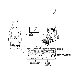
[0074] Turning to Fig 6, another schematic representation of an
electronic external
fetal monitoring system in accordance with an embodiment of the present
invention is shown.
patch 10 is shown wirelessly transmitting information (as described above) per
arrow 75 to a
computer system 65" (including a smart phone, tablet or other portable
computer) in a
patient's home, for example. Arrow 75 shows the wireless transmission of the
information to
a monitor/integrated hardware unit 55' configured to perform the monitoring
etc.
functionality discussed above and work with the diagnostic algorithms. Arrows
72, 74, and
76 show the wireless transmission of information (as described herein) to a
computer system
and display screen 65, 65' and 65" (including a smart phone 65", tablet 65' or
other portable
computer; and a central nursing station display 65) with a color graphical
user interface (e.g.,
"Red-Yellow-Green").
[0075] Fig. 7 is a schematic representation of a patch portion 10' of an
electronic
external fetal monitoring system in accordance with an embodiment of the
present invention.
Wireless transmitter 115, thin film piezo-electric ultrasound sensor array
102, ultrasound
microcontroller 104, patch adhesive 106 (reverse side only) electro-conductive
semi-solid gel
coating 108 (reverse side only), color-flow Doppler blood leakage sensor array
110, Doppler
micro controller 112, and micro batteries 114 are shown.
[0076] Fig. 8 is a schematic representation of a patch portion 10' of an
electronic
external fetal monitoring system in accordance with an embodiment of the
present invention.
Color-flow Doppler blood leakage sensor array 110 is shown, which is
configured to (and/or
assist with) monitor uterine integrity, migrate existing cardiac Doppler
system to uterus, test
and validate robustness of data capture and diagnosis of uterine integrity,
and finalize
interface with partner/existing hardware system(s).
[0077] Fig. 9 is a schematic representation of a patch portion 10' of an
electronic
external fetal monitoring system in accordance with an embodiment of the
present invention.
CA 2866089 2017-12-13
17
Thin film Ppiezo-electric ultrasound sensor array 102, which is configured to
(and/or assist
with) monitor maternal heart rate, monitor fetal heart rate, monitor uterine
contractions,
finalize sensor system to find fetal and maternal heart rates, create an
algorithm to separate
fetal and maternal heart rates, finalize sensors and algorithm to find uterine
contractions, test
and validate robustness of data capture with placement and movement, finalize
interface with
partner/existing hardware system(s), and create algorithms to diagnose
condition of mother
and baby ("red-yellow-green").
[0078] Fig. 10 is a schematic representation of a patch portion 10' of
an electronic
external fetal monitoring system in accordance with an embodiment of the
present invention.
Wireless transmitter 115 and a monitor/integrated hardware unit 55' is shown.
The wireless
transmitter system, designated by components 104, 115 and 114, is configured
to utilize
existing wearable transmitter, battery pack and set-top receiver, develop
inexpensive, micro-
sized power and transmitter and integrate into disposable strip, and migrate
to the
monitor/integrated hardware unit 55'.
[0079] Other potential uses for the devices contemplated herein include
health and
wellness monitoring such as patches created for athletes, and military service
men. Patches
could be created to monitor heartbeat, respiration, pulse oximetry, to track
endurance, and
surveillance of health. PVDF patches could be made to allow for ultrasound
diagnostics in
transit in military battlefield applications. Medic on site would apply patch,
and data could
be viewed at remote treatment facility or hospital while injured is en route.
[0080] While several embodiments of the invention have been discussed,
it will be
appreciated by those skilled in the art that various modifications and
variations of the present
invention are possible. Such modifications do not depart from the scope of the
present
invention.
CA 2866089 2017-12-13