Note: Descriptions are shown in the official language in which they were submitted.
CA 02866595 2014-09-08
WO 2013/131991
PCT/EP2013/054566
-1-.
Means and methods for determining neurotoxin activity based on a modified
luciferase
[0001] The present invention is concerned with test systems for determining
the activity of
neurotoxin polypeptides. Specifically, it relates to a polynucleotide encoding
a single chain
luciferase fusion polypeptide comprising: (i) a LuxB subunit, (ii) a linker
comprising a
neurotoxin cleavage site, and (iii) a LuxA subunit and a polypeptide encoded
by said
polynucleotide. Further provided in accordance with the invention are a vector
and a host cell
comprising said polynucleotide. Moreover, the present invention relates to a
method for
determining a proteolytically active neurotoxin polypeptide in a sample and a
kit for carrying
out said method.
[0002] Clostridium botulinum and Clostridium tetani produce highly potent
neurotoxins, i.e.
botulinum toxins (BoNTs) and tetanus toxin (TeNT), respectively. These
Clostidial
neurotoxins specifically bind to neuronal cells and disrupt neurotransmitter
release. Each
toxin is synthesized as an inactive unprocessed approximately 150 kDa single-
chain protein.
The posttranslational processing involves formation of disulfide bridges, and
limited
proteolysis (nicking) by bacterial protease(s). Active dichain neurotoxin
consists of two
chains, an N-terminal light chain of approx. 50 kDa and a heavy chain of
approx. 100 kDa
linked by a disulfide bond. Neurotoxins structurally consist of three domains,
i.e. the catalytic
light chain, the heavy chain encompassing the translo cation domain (N-
terminal half) and the
receptor binding domain (C-temiinal half), see Krieglstein 1990, Eur 3 Biochem
188, 39;
Krieglstein 1991, Eur J Biochem 202, 41; Krieglstein 1994, J Protein Chem 13,
49.
[0003] Clostridium botulinum secretes seven antigenically distinct serotypes
designated A to
G of the BoNTs. All serotypes together with the related TeNT secreted by
Clostridium tetani,
are zinc (Zn2+)-dependent endoproteases that block synaptic exocytosis by
cleaving SNARE
proteins and, in particular in the case of BoNT/A, C or E, SNAP-25. BoNTs
cause, inter alia,
the flaccid muscular paralysis seen in botulism, see Fischer 2007, PNAS 104,
10447.
[0004] Despite its toxic effects, BoNTs have been used as a therapeutic agents
in a large
number of diseases. BoNT serotype A (BoNT/A) was approved for human use in the
United
States in 1989 for the treatment of strabism, blepharospasm, and other
disorders. It is
commercially available as a protein preparation, for example, under the
tradename BOTOX
CA 02866595 2014-09-08
WO 2013/131991
PCT/EP2013/054566
- 2 -
(Allergan Inc) under the tradename DYSPORT (Ipsen Ltd). For therapeutic
application the
complex is injected directly into the muscle to be treated. At physiological
pH, the toxin is
released from the protein complex and the desired pharmacological effect takes
place. An
improved BoNT/A preparation being free of complexing proteins is available
under the
tradename XEOMIN (Merz Pharmaceuticals GmbH).
[0005] BoNTs, in principle, weaken voluntary muscle strength and are,
therefore, effective
therapeutic agents for the therapy of diseases such as strabism, focal
dystonia, including
cervical dystonia, and benign essential blepharospasm or spasticity. They have
been farther
shown to relief hemifacial spasm, and focal spasticity, and moreover, to be
effective in a wide
range of other indications, such as gastrointestinal disorders, hyperhidrosis,
and cosmetic
wrinkle correction, see Jost 2007, Drugs 67, 669.
[0006] The determination of the biological activity is important as a safety
measure, for
quality control and for quantification purposes. The mouse LD50 assay is
currently the only
reliable assay for quantifying the biological activity of neurotoxins and for
assessing their
therapeutic potential and/or their toxicity. Said assay is also accepted for
quality control
purposes during manufacture of neurotoxin. In the mouse LD50 bioassay, lethal
and sub-
lethal concentrations of a sample containing the neurotoxin polypeptide have
to be injected
into at least 120 animals. The number of killed animals over an observation
period of 72
hours allows determining the neurotoxin polyp eptide concentration in the
sample. Apparent
drawbacks of this assay are the high number of animals which will be
sacrificed and the high
level of stress and pain for said animals during the test.
[0007] In vitro assays which have been proposed so far are based on
determining SNAP-25
cleavage in a cell free system or on neurotoxin exposure to primary neurons.
However, these
assay are less reliable and/or do not take into account all of the desired
neurotoxin functions.
Thus, at present, the LD50 bioassay described above is the only reliable assay
which is
described in the monograph for BoNT/A in the European phafinacopeia. However,
there is a
need for a reliable assay for measuring neurotoxin activity which avoids the
drawbacks of the
LD50 bioassay.
[0008] Therefore, the technical problem underlying the present invention could
be seen in
the provision of means and methods for complying with the aforementioned
needs. The
technical problem is solved by the embodiments characterized in the claims and
herein
described below.
CA 02866595 2014-09-08
WO 2013/131991
PCT/EP2013/054566
- 3 -
[0009] The present invention relates to a polynucleotide encoding a single
chain luciferase
fusion polypeptide comprising: (i) a LuxB subunit, (ii) a linker comprising a
neurotoxin
cleavage site, and (iii) a LuxA subunit.
[0010] The term "polynucleotide" as used herein refers to single- or double-
stranded DNA
molecules as well as to RNA molecules. Encompassed by the said term is genomic
DNA,
cDNA, hnRNA, inRNA as well as all naturally occurring or artificially modified
derivatives
of such molecular species. The polynucleotide may be in an aspect a linear or
circular
molecule. Moreover, in addition to the nucleic acid sequences encoding the
polypeptide of the
present invention, a polynucleotide of the present invention may comprise
additional
sequences required for proper transcription and/or translation such as 5 r- or
3 '-UTR
sequences. The nucleic acid sequences encoding the polypeptide of the present
invention can
be derived from the amino acid sequence envisaged for the single chain
luciferase fusion
polypeptide of the invention by a skilled artisan without further ado. In
light of the
degeneracy of the genetic code, optimized codons may be used in the nucleic
acid sequences
encoding the single chain luciferase fusion polypeptide in the polynucleotide
of the present
invention. Thereby, optimal expression in, e.g., a host cell of the present
invention can be
achieved.
[0011] The term "single chain luciferase fusion polypeptide" as used herein
refers to a
polypeptide comprising within a single polypeptide chain a LuxB subunit of a
luciferase as
well as a LuxA subunit. The said subunits are separated by a linker that
comprises a
neurotoxin cleavage site which is specifically recognized and cleaved by a
proteolytically
active neurotoxin polypeptide as referred to elsewhere herein. The single
chain luciferase
fusion polypeptide, in an aspect, has the LuxB and LuxA subunits arranged in a
manner
which prevents or reduces the formation of a biologically active luciferase
holoenzyme when
the subunits are present in the single chain luciferase fusion polypeptide. In
an aspect, a
biologically active luciferase holoenzyme is, thus, formed only or formed more
efficiently
after cleavage of the single chain luciferase fusion polypeptide by the
neurotoxin protease. If
the enzymatic activity, in an aspect, is prevented, essentially no activity
shall occur when the
single chain luciferase fusion polypeptide is in the single chain state. A
reduced enzymatic
activity as referred to herein, in an aspect, means a statistically
significant reduced activity. In
an aspect, the reduction of the activity in the single chain state versus the
holoenzyme formed
after cleavage is at least 10%, at least 20%, at least 30%, at least 40%, at
least 50%, at least
60%, at least 70% or at least 80%.
CA 02866595 2014-09-08
WO 2013/131991
PCT/EP2013/054566
- 4 -
[0012] In an aspect, the LuxB and LuxA subunits in the single chain luciferase
fusion
polypeptide are arranged in a manner which prevents or reduces the formation
of a
biologically active luciferase holoenzyme in that the order of the subunits in
the single chain
luciferase fusion polypeptide is from N to C-terminus: (i) a LuxB subunit,
(ii) a linker
comprising a neurotoxin cleavage site, and (iii) a LuxA subunit. Such an order
of the subunits
has been described to efficiently prevent luciferase activity in a single
chain fusion protein
(see Olsson 1989, Gene 81: 335-347).
[0013] In another aspect, the LuxB and LuxA subunits in the single chain
luciferase fusion
polypeptide are separated in addition to the linker by further polypeptide
elements which
prevent or reduce the capability of the subunits to interact with each other.
This can be
achieved, in an aspect, by inserting ankyrin repeats into the single chain
luciferase fusion
polypeptide either between the LuxB subunit and the linker or between the LuxA
subunit and
the linker or both. Ankyrin repeats are known to reduce the flexibility of a
polypeptide chain
and, thus, will reduce the capability of the separated luciferase subunits in
the single chain
luciferase fusion polypeptide of the invention to interact with each other.
Ankyrins are well
known in the art and belong into the family of adaptor proteins mediating the
attachment of
transmembrane proteins and cytoskeletal proteins. In mammals, three different
ankyrins are
known, ANK1, ANK2 and ANK3, occurring in various splice forms. The ankyrins
have an N-
terminal domain comprising the so-called ankyrin repeats which are to be
applied in
accordance with the present invention (see Wetzel 2008, J Mol Biol. 376(1):
241-57;
Michaely 1995, J. Biol Chem. 270(52):31298-302; Batrukova 2000, Biochemistry
(Mose).
65(4):395-408.).
[0014] In another aspect, a prevention or reduction of the capability of the
subunits to
interact with each other can be achieved by inserting protein domains or
proteins either
between the LuxB subunit and the linker, between the LuxA subunit and the
linker or both
which due to their molecular size sterieally interfere with the interaction of
the subunits with
each other. In an aspect, globular proteins or domains thereof can be applied
for this purpose.
In an aspect, the said globular protein or domain thereof is selected from the
group consisting:
glutathione S-transferase (GST), maltose binding protein, small ubiquitin-like
modifier
(SUMO) protein, green fluorescent protein (GFP), red fluorescent protein
(RFP), yellow
fluorescent protein (YFP), blue fluorescent protein (BFP), and the like. In
another aspect, a
fluorescent protein may be bound to the neurotoxin and/or luciferase
polypeptides specified
elsewhere herein, whereby said fluorescent protein e.g. alters light emission
of said
polypeptide and/or is be used as internal control to monitor expression,
number of cells,
loading control, and the like. In yet an aspect, the linker can be comprised
in a globular
protein or domain thereof as described before.
CA 02866595 2014-09-08
WO 2013/131991
PCT/EP2013/054566
- 5 -
[0015] It will be understood that the neurotoxin cleavage site comprised in
the linker shall
be made available by the single chain luciferase fusion protein to a
neurotoxin polypeptide
such that the neurotoxin protease can recognize, bind to and cleave the single
chain luciferase
fusion polypeptide under suitable conditions. The skilled artisan is well
aware of how a
suitable arrangement within the single chain luciferase fusion polypeptide can
be designed.
Moreover, the single chain luciferase fusion protein can be tested for
cleavage by
proteolytically active neurotoxin polypeptide as described in the accompanying
Examples. In
an aspect, the single chain luciferase fusion polypeptide of the invention has
an amino acid
sequence as shown in any one of SEQ ID NOs: 1 to 3.
[0016] The tem' "luciferase" as used herein refers to enzymes belonging into a
class of
enzymes capable of catalyzing a light-emitting reaction. Luciferases occur
naturally as firely
or bacterial luciferases. Such luciferases as enzymatically (biologically)
active holoenzymes
are composed of different subunits. In an aspect, the luciferase referred to
herein is a bacterial
luciferase and the subunits to be inserted into the single chain luciferase
fusion polypeptide
are LuxA and LuxB. In yet another aspect, the said LuxA and/or LuxB subunits
are from
Vibrio fischeri or Vibrio harveyi. How to arrange such subunits in a singly
chain luciferase
fusion protein without additional linker according to the present invention is
known in the art
and described in Olsson 1989, loc cit.. The structure of the aforementioned
luciferases and
their subunits as well as nucleic acid sequences encoding them are well known
in the art and
describe, e.g., in Chon 1985, J Biol Chem. 260(10):6139-46 and Johnston 1986,
J Biol Chem.
261(10:4805-11. In an aspect, the LuxA subunit of Vibrio fischeri as referred
to herein has
an amino acid sequence as shown in SEQ ID NO: 4 or a variant thereof In
another aspect, the
LuxB subunit of Vibrio fischeri has an amino acid sequence as shown in SEQ 1D
NO: 5 or a
variant thereof. It will be understood that the present invention also
encompasses variants of
such specific amino acid or nucleic acid sequences encoding them as long as
these variant
sequences also allow for the formation of an enzymatically active luciferase
holoenzyme. In
an aspect, a sequence variant as used herein differs from the specific amino
acid sequence or a
specific nucleic acid sequence as specified before by one or more amino acid
or nucleotide
substitutions, additions and/or deletions. In another aspect, the said variant
sequence is at least
50%, at least 60%, at least 70%, at least 80%, at least 90%, at least 95%, at
least 98% or at
least 99% identical to the specific sequence over the entire length or over at
least a stretch of
half of the length of the specific sequence. The term "identical" as used
herein refers to
sequence identity characterized by determining the number of identical amino
acids between
sequences wherein the sequences are aligned so that the highest order match is
obtained. It
can be calculated using published techniques or methods codified in computer
programs such
CA 02866595 2014-09-08
WO 2013/131991
PCT/EP2013/054566
- 6 -
as, for example, BLASTP or FASTA (Altschul 1990, J Mol Biol 215, 403). The
percent
identity values are, in one aspect, calculated over the entire amino acid
sequence or over a
sequence stretch of at least 50% of the query sequence. A series of programs
based on a
variety of algorithms is available to the skilled worker for comparing
different sequences. In
this context, the algorithms of Needleman and Wunsch or Smith and Waterman
give
particularly reliable results. To carry out the sequence alignments, the
program PileUp
(Higgins 1989, CABIOS 5, 151) or the programs Gap and BestFit (Needleman 1970,
J Mol
Biol 48; 443; Smith 1981, Adv Appl Math 2, 482), which are part of the GCG
software packet
(Genetics Computer Group 1991, 575 Science Drive, Madison, Wisconsin, USA
53711), may
to be used. The sequence identity values recited above in percent (%) are
to be determined, in
another aspect of the invention, using the program GAP over the entire
sequence region with
the following settings: Gap Weight: 50, Length Weight: 3, Average Match:
10.000 and
Average Mismatch: 0.000, which, unless otherwise specified, shall always be
used as standard
settings for sequence alignments.
[0017] The term "neurotoxin cleavage site" as used herein refers to a cleavage
site which is
recognized and cleaved by the endogenous protease of a neurotoxin polypeptide.
Cleavage
sites which are recognized by the neurotoxin proteases are well known in the
art (see, e.g., EP
1 926 744 B1). In an aspect, the said neurotoxin cleavage site is selected
from the group
consisting of: a neurotoxin cleavage site from SNAP-25, a neurotoxin cleavage
site from
VAMP, a neurotoxin cleavage site from syntaxin, and the autocatalytic cleavage
sites from
neurotoxin polypeptides.
[0018] In principle, a neurotoxin cleavage site can be a cleavage site which
naturally occurs
in a substrate or which is an artificially designed cleavage site recognized
and cleaved by the
neurotoxin polypeptides protease. It will be understood that the properties of
the neurotoxin
cleavage site govern the kind of neurotoxin which can activate the polypeptide
of the present
invention. Neurotoxin polypeptides referred to herein, M an aspect, encompass
BoNT/A,
BoNT/B, BoNT/C1, BoNT/D, BoNT/E, BoNT/G, BoNT/F or TeNT all of which are well
known in the art. For example, if a neurotoxin cleavage site is used which is
specifically
recognized and cleaved by BoNT/A, only the BoNT/A protease will be capable of
activating
the polypeptide of the present invention and, in particular, its luciferase
activity, whereas if a
neurotoxin cleavage site is used which is specifically recognized and cleaved
by BoNT/E,
only the BoNT/E protease will be capable of activating the polypeptide of the
present
invention and, in particular, its caspase activity. In an aspect of the
invention, the neurotoxin
cleavage site is cleaved by mature BoNTs. In yet another aspect, it is cleaved
by muteins of
CA 02866595 2014-09-08
WO 2013/131991
PCT/EP2013/054566
- 7 -
BoNTs, in an aspect, by muteins comprising or consisting of the BoNT light
chain exhibiting
the BoNT protease activity.
[0019] A neurotoxin cleavage site recognized and cleaved by the BoNT/A
protease, in an
aspect of the invention, is derived from a protein that is sensitive to
cleavage by BoNT/A. In
an aspect, such a protein is human SNAP-25A or -25B or a homolog, paralog or
ortholog
thereof from rat, mouse, bovine, Danio, Carassius, Xenopus, Torpedo,
Strongylocentrotus,
Loligo, Lymnaea or Aplysia. Suitable cleavage sites derived from said proteins
are disclosed
in EP 1 926 744 Bl.
[0020] A neurotoxin cleavage site recognized and cleaved by the BoNT/B
protease, in an
aspect of the invention, is derived from a protein that is sensitive to
cleavage by BoNT/B. In
an aspect, such a protein is human or mouse VAMP-1, VAMP-2 and VAMP-
3/cellubrevin,
bovine VAMP-2, rat VAMP-2 or VAMP-3, chicken VAMP-1, VAMP-2 or VAMP-3,
Torpedo VAMP-1, Strongylocentrotus VAMP, Drosophila sybA, synB, synC, synD, or
syn,
Hirudo VAMP, Xenopus VAMP-2 or VAMP-3, Danio VAMP-1 or VAMP-2, Loligo VAMP,
Lymnaea VAMP, Aplysia VAMP or Caenorhabditis SNB1-like or any ortholog,
paralog or
homolog thereof. Suitable cleavage sites derived from said proteins are
disclosed in EP 1 926
744 Bl.
[0021] A neurotoxin cleavage site recognized and cleaved by the BoNT/C1
protease, in an
aspect of the invention, is derived from a protein that is sensitive to
cleavage by BoNT/C1. In
an aspect, such a protein is human and mouse Syntaxin 1A, Syntaxin 1B1,
Syntaxin 2-1,
Syntaxin 2-2, Syntaxin 2-3, Syntaxin 3A or Syntaxin 1B2, bovine or rat
Syntaxin 1A,
Syntaxin 1Blor Syntaxin 1B2, rat Syntaxin 2 or Rat syntaxin 3, mouse Syntaxin
1A, Syntaxin
1B1, Syntaxin 1B2, Syntaxin 2, Syntaxin 3A, Syntaxin 3B or Syntaxin 3C,
chicken Syntaxin
IA or Syntaxin 2; Xenopus Syntaxin lA or Syntaxin 1B, Danio Syntaxin 1A,
Syntaxin 1B or
Syntaxin 3, Torpedo Syntaxin lA or Syntaxin 1B, Strongylocentrotus Syntaxin lA
or
Syntaxin 1B, Drosophila Syntaxin IA or Syntaxin 1B, Hirudo Syntaxin lA or
Syntaxin 1B,
Loligo Syntaxin IA or Syntaxin 1B, Lymnaea Syntaxin lA or Syntaxin 1B or any
ortholog,
paralog or homolog thereof Suitable cleavage sites derived from said proteins
are disclosed in
EP 1 926 744B1.
[0022] A neurotoxin cleavage site recognized and cleaved by the BoNT/D
protease, in an
aspect of the invention, is derived from a protein that is sensitive to
cleavage by BoNT/D. In
an aspect, such a protein is human or mouse VAMP-1, VAMP-2 and VAMP-
3/cellubrevin,
bovine VAMP-2, rat VAMP-2 or VAMP-3, chicken VAMP-1, VAMP-2 or VAMP-3,
Torpedo VAMP-1, Strongylocentrotus VAMP, Drosophila sybA, synB, synC, synD, or
syn,
CA 02866595 2014-09-08
WO 2013/131991
PCT/EP2013/054566
- 8 -
Hirudo VAMP, Xenopus VAMP-2 or VAMP-3, Dania VAMP-1 or VAMP-2, Loligo VAMP,
Lymnaea VAMP, Aplysia VAMP or Caenorhabditis SNB1-like or any ortholog,
paralog or
homolog thereof. Suitable cleavage sites derived from said proteins are
disclosed in EP 1 926
744B1.
[0023] A neurotoxin cleavage site recognized and cleaved by the BoNT/E
protease, in an
aspect of the invention, is derived from a protein that is sensitive to
cleavage by BoNT/E. In
an aspect, such a protein is, such a protein is human SNAP-25A or B or a
homolog, paralog or
ortholog thereof from rat, mouse, bovine, Danio, Carassius, Xenopus, Torpedo,
Strongylocentrotus, Loligo, Lymnaea or Aplysia. Suitable cleavage sites
derived from said
proteins are disclosed in EP 1 926 744 B1 .
[0024] A neurotoxin cleavage site recognized and cleaved by the BoNT/F
protease, in an
aspect of the invention, is derived from a protein that is sensitive to
cleavage by BoNT/F. In
an aspect, such a protein is, such a protein is human or mouse VAMP-1, VAMP-2
and
VAMP-3/cellubrevin, bovine VAMP-2, rat VAMP-2 or VAMP-3, chicken VAMP-1, VAMP-
2 or VAMP-3, Torpedo VAMP-1, Strongylocentrotus VAMP, Drosophila sybA, synB,
synC,
synD, or syn, Hirudo VAMP, Xenopus VAMP-2 or VAMP-3, Danio VAMP-1 or VAMP-2,
Loligo VAMP, Lymnaea VAMP, Aplysia VAMP or Caenorhabditis SNB1-like or any
ortholog, paralog or homolog thereof. Suitable cleavage sites derived from
said proteins are
disclosed in EP 1 926 74481.
[0025] A neurotoxin cleavage site recognized and cleaved by the BoNT/G
protease, in an
aspect of the invention, is derived from a protein that is sensitive to
cleavage by BoNT/G. In
an aspect, such a protein is, such a protein is human or mouse VAMP-1, VAMP-2
and
VAMP-3/cellubrevin, bovine VAMP-2, rat VAMP-2 or VAMP-3, chicken VAMP-1, VAMP-
2 or VAMP-3, Torpedo VAMP-1, Strongylocentrotus VAMP, Drosophila sybA, synB,
synC,
synD, or syn, Hirudo VAMP, Xenopus VAMP-2 or VAMP-3, Danio VAMP-1 or VAMP-2,
Loligo VAMP, Lymnaea VAMP, Aplysia VAMP or Caenorhabditis SNB1-like or any
ortholog, paralog or homolog thereof. Suitable cleavage sites derived from
said proteins are
disclosed in EP 1 926 744 Bl.
[0026] A neurotoxin cleavage site recognized and cleaved by the TeNT protease,
in an
aspect of the invention, is derived from a protein that is sensitive to
cleavage by TeNT. In an
aspect, such a protein is human or mouse VAMP-1, VAMP-2 and VAMP-
3/cellubrevin,
bovine VAMP-2, rat VAMP-2 or VAMP-3, chicken VAMP-1, VAMP-2 or VAMP-3,
Torpedo VAMP-1, Strongylocentrotus VAMP, Drosophila sybA, synB, synC, synD, or
syn,
Hirudo VAMP, Xenopus VAMP-2 or VAMP-3, Danio VAMP-1 or VAMP-2, Loligo VAMP,
CA 02866595 2014-09-08
WO 2013/131991
PCT/EP2013/054566
- 9 -
Lymnaea VAMP, Aplysia VAMP or Caenorhabditis SNB1-like or any ortholog,
paralog or
homolog thereof. Suitable cleavage sites derived from said proteins are
disclosed in EP 1 926
74481.
[0027] A neurotoxin cleavage site recognized and cleaved by the BoNT
proteases, in
another aspect of the invention, is derived from the autocatalytie cleavage
sites found in the
BoNT proteins. In aspects, a neurotoxin cleavage site to be used in accordance
with the
present invention and which is derived from the autoeatalytic cleavage site of
a given BoNT
or TeNT comprises at least 6, at least 8, at least 10 or at least 15
consecutive residues of
including the BoNT/A residues 250Tyr-251Tyr, the BoNT/B residues 256Phe-
257Phe, the
BoNT/C1 residues 257Phe-258Tyr, the BoNT/D residues 2.57Phe-258Phe, the BoNT/E
residues 239Pro-240Leu, the BoNT/F residues 254Pro-255Leu, the BoNT/G residues
256Phe-
257Phe, the TeNT residues 25911e-260Tyr, the BoNT/A residues Phe266-G1y267,
the
BoNT/B residues Phe272-G1y273, the BoNT/C1 residues Phe273-G1y274, the BoNT/D
residues Phe273-Gly274, the BoNT/E residues Phe255-G1y256, the BoNT/F residues
Phe270-
G1y271, the BoNT/G residues Phe272-G1y273 or the TeNT residues Phe275-Gly276.
Suitable
cleavage sites derived from said BoNTs and TeNT are disclosed in EP 1 926 744
Bl.
[0028] In yet an aspect of the invention, said neurotoxin cleavage site is a
SNAP-25 derived
cleavage site as shown in any one of SEQ ID NOs: 6 to 8.
[0029] Advantageously, the single chain luciferase fusion polypeptide of the
present
invention allows for the determination of the qualitative or quantitative
protease activity of a
given neurotoxin polypeptide in a cell culture system or in a cell free assay.
Thus, expensive
and unnecessary animal testing can be avoided or reduced thanks to the present
invention.
Moreover, the single chain luciferase fusion polypeptide can be applied in
automated high
throughput screening assays. The single chain luciferase fusion polypeptide
also allows for
establishing an assay in neuroblastoma cell lines rather than primary cells.
Accordingly, the
use of primary neurons as required in other cell based neurotoxin assays can
be avoided. This
is another advantage since the preparation of primary neurons is cumbersome
and inefficient.
[0030] It is to be understood that the definitions and explanations of the
terms made above
apply mutatis mutandis for all aspects described in this specification in the
following except
as otherwise indicated.
[0031] The present invention also relates to a vector comprising the
polynucleotide of the
invention.
CA 02866595 2014-09-08
WO 2013/131991
PCT/EP2013/054566
- 10 -
[0032] The term "vector", preferably, encompasses phage, plasmid, viral or
retroviral
vectors as well as artificial chromosomes, such as bacterial or yeast
artificial chromosomes.
Moreover, the term also relates to targeting constructs which allow for random
or site-
directed integration of the targeting construct into genomic DNA. Such target
constructs, in
an aspect, comprise DNA of sufficient length for either homologous or
heterologous
recombination as described in detail below. The vector encompassing the
polynucleotides of
the present invention, in an aspect, further comprises selectable markers for
propagation
and/or selection in a host cell. The vector may be incorporated into a host
cell by various
techniques well known in the art. For example, a plasmid vector can be
introduced in a
precipitate such as a calcium phosphate precipitate or rubidium chloride
precipitate, or in a
complex with a charged lipid or in carbon-based clusters, such as fullerens.
Alternatively, a
plasmid vector may be introduced by heat shock or electroporation techniques.
Should the
vector be a virus, it may be packaged in vitro using an appropriate packaging
cell line prior to
application to host cells. Retroviral vectors may be replication competent or
replication
defective. In the latter case, viral propagation generally will occur only in
complementing
ho st/cells.
[0033] Moreover, in an aspect of the invention, the polynucleotide is
operatively linked to
expression control sequences allowing expression in prokaryotic or eukaryotic
host cells or
isolated fractions thereof in the said vector. Thus, in an aspect, the vector
is an expression
vector. Expression of the polynucleotide comprises transcription of the
polynucleotide into a
translatable mRNA. Regulatory elements ensuring expression in host cells are
well known in
the art. In an aspect, they comprise regulatory sequences ensuring initiation
of transcription
and/or poly-A signals ensuring termination of transcription and stabilization
of the transcript.
Additional regulatory elements may include transcriptional as well as
translational enhancers.
Possible regulatory elements permitting expression in prokaryotic host cells
comprise, e.g.,
the lac-, trp- or tac- promoter in E. coli, and examples for regulatory
elements permitting
expression in eukaryotic host cells are the A0X1- or the GAL1- promoter in
yeast or the
CMV-, SV40-, RSV-promoter (Rous sarcoma virus), CMV-enhancer, SV40-enhancer or
a
globin intron in mammalian and other animal cells. Moreover, inducible
expression control
sequences may be used in an expression vector encompassed by the present
invention. Such
inducible vectors may comprise tet or lac operator sequences or sequences
inducible by heat
shock or other environmental factors. Suitable expression control sequences
are well known
in the art. Beside elements which are responsible for the initiation of
transcription such
regulatory elements may also comprise transcription termination signals, such
as the SV40-
poly-A site or the tk-poly-A site, downstream of the polynucleotide. In this
context, suitable
expression vectors are known in the art such as Okayama-Berg cDNA expression
vector
pcDV1 (Phannacia), pBluescript (Stratagene), pCDM8, pRc/CMV, pcDNA1, pcDNA3
CA 02866595 2014-09-08
WO 2013/131991
PCT/EP2013/054566
- 11 -
(Invitrogen) or pSPORT1 (Invitrogen). Preferably, said vector is an expression
vector and a
gene transfer or targeting vector. Expression vectors derived from viruses
such as
retroviruses, vaccinia virus, adeno-associated virus, herpes viruses, or
bovine papilloma virus,
may be used for delivery of the polynucleotide or vector of the invention into
a targeted cell
population. Methods which are well known to those skilled in the art can be
used to construct
recombinant viral vectors; see, for example, the techniques described in
Sambrook, Molecular
Cloning A Laboratory Manual, Cold Spring Harbor Laboratory (1989) N.Y. and
Ausubel,
Current Protocols in Molecular Biology, Green Publishing Associates and Wiley
Interscience,
N.Y. (1994).
[0034] Moreover, the present invention relates to a host cell comprising the
polynucleotide
or the vector of the present invention.
[0035] The term "host cell" as used herein encompasses prokaryotic and
eukaryotic host
cells. In an aspect the host cell is a bacterial cell. In one aspect, the said
bacterial host cell is
an E.coli host cell. Such a bacterial host cell may be used, e.g., for
reproduction of the
polynucleotide or the vector of the present invention.
[0036] A eukaryotic host cell, in an aspect, is a cell which comprises the
polypeptide and
either the polynucleotide or the vector of the present invention wherein said
polynucleotide or
vector are expressed in the host cell in order to generate the polypeptide.
The polynucleotide
may be introduced into a host cell either transiently or stably. In an aspect,
the eukaryotic host
cell may be a cell of a eukaryotic host cell line which stably expresses the
polynucleotide of
the invention. In another aspect, the host cell is a eukaryotic host cell
which has been
transiently transfected with the polynucleotide or vector of the invention and
which expresses
the polynucleotide of the invention. In an aspect the host cell is a
eukaryotic host cell which is
capable of translocating a neurotoxin polypeptide into its cytoplasm. In an
aspect, a cell
capable of uptaking neurotoxin polypeptides can be a cell produces
endogenously all
necessary components for the neurotoxin polypeptide uptake. In an aspect, the
said cell is a
neuronal cell. In an aspect, the said cell is selected from the group
consisting of:
neuroblastoma cell lines, embryonic stem cells, and in an aspect non-human
embryonic stem
cells, induced pluripotent stem cells, and primary neurons, e.g. SH-SY5Y,
SiMa, PC12,
CHP134, LA-N-5, SK-N-BE(2), and the like. In another aspect, the said cell is
a cell which
has been genetically engineered to produce the components necessary for the
neurotoxin
polypeptide uptake. How such cells can be genetically engineered by molecular
biology
techniques is well known to the skilled person.
CA 02866595 2014-09-08
WO 2013/131991
PCT/EP2013/054566
- 12 -
[0037] The present invention relates to a polypeptide encoded by the
polynucleotide of the
present invention.
[0038] The term "polypeptide" as used herein encompasses isolated or
essentially purified
polypeptides being essentially free of other host cell polypeptides. The term,
in another
aspect, includes polypeptide preparations comprising the polypeptide of the
present invention
and other proteins in addition. Moreover, the term includes, in an aspect,
chemically modified
polypeptides. Such modifications may be artificial modifications or naturally
occurring
modifications. The polypeptide of the present invention shall have the
biological properties
referred to above. The polypeptide of the invention, in an aspect, can be
manufactured by
chemical synthesis or recombinant molecular biology techniques well known for
the skilled
artisan. In an aspect, such a method of manufacturing the polypeptide of the
invention
comprises (a) culturing the host cell of the present invention described
elsewhere herein in
more detail and (b) obtaining from the said host cell the polypeptide of the
present invention.
In an aspect of this method, the polypeptide can be obtained by conventional
purification
techniques from a host cell lysate including affinity chromatography, ion
exchange
chromatography, size exclusion chromatography and/or preparative gel
electrophoresis.
[0039] The present invention relates to a method for determining a
proteolytically active
neurotoxin polypeptide in a sample comprising:
a) contacting the host cell of or the polypeptide of the invention with
a sample
suspected to comprise said proteolytically active neurotoxin polypeptide under
conditions which allow for proteolytic cleavage of the single chain luciferase
fusion protein into separate Lux13 and LuxA subunits;
b) allowing
the said LuxB and LuxA subunit to form a biologically active
luciferase; and
c) determining the said luciferase.
[0040] The method of the present invention can be assisted by automation.
Specifically, in
an aspect, step a) and /or b) may be assisted by robotic devices and automated
reader systems
for mixing compounds and measuring the luciferase activity. Suitable systems
are known in
the art and depend on the type of response to be determined. Moreover, the
method may
comprise additional steps pertaining to the sample preparation or generation
of the
polypeptide of the present invention.
[0041] The term "contacting" as used herein refers to bringing at least two
different
compounds in physical proximity as to allow physical and/or chemical
interaction of said
compounds. In the aforementioned method, the polypeptide according to the
present invention
is contacted with a sample suspected to comprise a biologically active
neurotoxin
CA 02866595 2014-09-08
WO 2013/131991
PCT/EP2013/054566
- 13 -
polypeptide. The polypeptide shall be contacted for a time and under
conditions sufficient to
allow cleavage of the neurotoxin cleavage site in the polypeptide of the
present invention by
the neurotoxin polypeptide comprised by the sample. Contacting as used herein,
in an aspect,
occurs in a host cell of the present invention containing the polypeptide of
the present
invention. Thus, in an aspect, said polypeptide is comprised by a host cell
and, in an aspect,
the host cell of the present invention. The said time and conditions will
dependent on the
amount of neurotoxin polypeptide comprised by the sample as well as on the
uptake of the
neurotoxin polypeptide by the host cell. The person skilled in the art is well
aware of which
conditions need to be applied dependent on the host cell, kind of sample, and
kind of
neurotoxin which shall be determined. In another aspect, contacting occurs in
a cell free
system comprising the polypeptide of the invention as well as a substrate of
the polypeptide
of the present invention. The cell free system shall allow for measuring the
activity of the
polypeptide of the present invention, i.e. luciferase activity, upon
contacting the system with a
sample and, thus, allows for determining the neurotoxin protease activity in
said sample.
[0042] In an aspect, said luciferase is deteimined by measuring the enzymatic
conversion of
a luciferase substrate. The latter one can be measured in an aspect by
detecting the intensity of
the light emitted during the conversion reaction. Suitable systems for
measuring the light
emission that occurs during the conversion reaction catalyzed by luciferases
are well known
in the art and commercially available. Moreover, suitable substrates which can
be used for the
luciferases are also well known and commercially available. In another aspect,
however, the
luciferase can be measured either by determining the amount of the formed LuxA
ad/or B
subunits or the formed holoenzyme. In an aspect, this determination is carried
out by an
antibody-based immunoassay using antibodies which specifically recognize a
subunit or the
holoenzyme, e.g., immunoblots or ELISA, or by SDS PAGE.
[0043] The term "sample" refers to a sample suspected to comprise neurotoxin
polypeptide.
The sample, in an aspect, is an aqueous solution. Such a sample may be a
biological sample or
may be a sample of an artificially generated aqueous solution. Such solutions,
in an aspect,
are obtained at different stages during neurotoxin manufacture, either for
quality control
and/or activity determination/specification purposes or for safety control. It
is envisaged that
the neurotoxin present in the said sample shall exhibit at least the
neurotoxin protease activity.
In another aspect, the neurotoxin is fully biologically active. In an aspect
the said fully
biologically active neurotoxin is required for entering the cell and for
activating the read out
based on the single chain luciferase fusion polypeptide of the present
invention. Accordingly,
such a fully biologically active neurotoxin is to be applied if a host cell is
to be contacted with
the sample to be analyzed by the method of the invention. In another aspect,
the sample to be
applied for the method of the invention comprises neurotoxin polypeptides or
fragments
CA 02866595 2014-09-08
WO 2013/131991
PCT/EP2013/054566
- 14 -
thereof which merely exhibit neurotoxin protease activity. Such neurotoxin
polypeptides or
fragments are, in an aspect, muteins of neurotoxin polypeptides comprising or
consisting
essentially of a proteolytically active light chain. It is to be understood
that samples
comprising neurotoxin polypeptides or fragments thereof which merely exhibit
neurotoxin
protease activity shall be used if the sample is to be contacted to a cell
free system as
specified elsewhere herein in detail.
[0044] The neurotoxin polypeptide in a sample can be determined quantitatively
or
qualitatively. For a qualitative determination, in an aspect of the invention,
the presence or
absence of a neurotoxin polypeptide is determined. For a quantitative
detection, in an aspect,
the amount of the neurotoxin polypeptide is determined. In an aspect, the
quantitative
deteimination encompasses a determination of the absolute amount or a relative
amount, i.e.
an amount which is nollnalized such that the amount found in different samples
can be
compared with each other. In an aspect, this can be achieved by comparison of
a measured
luciferase activity for a test sample to a calibration curve which is to be
established by
subjecting calibration samples having predetermined amounts of the neurotoxin
polypeptide
to the method of the present invention.
[0045] It will be understood that in an aspect, the neurotoxin cleavage site
comprised in the
single chain luciferase fusion polypeptide is recognized by the
proteolytically active
neurotoxin polypeptide to be determined in the sample. In yet an aspect, said
neurotoxin
polypeptide is selected from the group consisting of: BoNT/A, BoNT/B, BoNT/C1,
BoNT/D,
BoNT/E, BoNT/F, BoNT/G or TeNT.
[0046] In another aspect of the method of the invention, the activity of more
than one
neurotoxin polypeptide shall be determined. To this end, a host cell can be
applied which
comprises a polynucleotide according to the present invention encoding a first
single chain
luciferase fusion polypeptide comprising: (i) a LuxB subunit of a first
luciferase, (ii) a linker
comprising a neurotoxin cleavage site for a first neurotoxin polypeptide, and
(iii) a LuxA
subunit of said first luciferase and another polynucleofide according to the
present invention
encoding a second single chain luciferase fusion polypeptide comprising: (i) a
LuxB subunit
of a second luciferase, (ii) a linker comprising a neurotoxin cleavage site
for a second
neurotoxin polypeptide, and (iii) a LuxA subunit of said second luciferase. It
will be
understood that the first and the second neurotoxin polypeptides are different
and recognize
and cleave different cleavage sites. Moreover, in an aspect it will be
understood that the first
luciferase holoenzyme comprising said first LuxB and LuxA subunits and the
second
luciferase holoenzyme comprising said second LuxB and LuxA subunits utilize
generate
different light emissions which can be distinguished from each other, e.g.,
emission maxima
CA 02866595 2014-09-08
WO 2013/131991
PCT/EP2013/054566
- 15 -
at different wavelengths. In an aspect, said first luciferase holoenzyme
and/or said second
luciferase holoenzyme can be bound to a fluorescent protein, e.g. green
fluorescent protein
(GFP), yellow fluorescent protein (YFP), red fluorescent protein (RFP), blue
fluorescent
protein, and the like. In an aspect, the first subunits are from Vibrio
harveyi and the second
subunits referred to before are from Vibrio fischeri.
[0047] Thus, the method of the present invention allows for an efficient
determination of a
biologically active neurotoxin polypeptide in a sample and can, therefore, be
applied in high
throughput screenings or quality control approaches. Thanks to the method of
the present
invention, neurotoxin polypeptide determination can be automated due to the
use of cell
culture cells rather than primary neurons. Moreover, the use of stably
transfected host cell
lines allows for a comparable quality of the readout system, i.e. the host
cell to be applied in
the method of the invention. Accordingly, the method of the present invention
may serve as
an alternative or may at least significantly reduce animal testing in the
context of neurotoxin
polypeptide development or quality control.
[0048] Further encompassed by the present invention is the use, in general, of
the
polynucleotide, the vector, the host cell or the polypeptide of the invention
for determining a
proteolytically active neurotoxin polypeptide in a sample in vitro.
[0049] Finally, the present invention contemplates a kit for determining a
proteolytically
active neurotoxin polypeptide in a sample comprising the polynucleotide, the
vector, the host
cell and/or the polypeptide of the present invention and, preferably, a
detection agent for
measuring the enzymatic conversion of a luciferase substrate and a luciferase
substrate.
[0050] The term "kit" as used herein refers to a collection of means
comprising the
polypeptide, the polynucleotide, the vector and/or the host cell of the
present invention which
are provided in separate or common vials in a ready to use manner for carrying
out the
method of the present invention. In an aspect, the kit comprises additional
means for carrying
out the method of the present invention, in an aspect, calibration standard
solutions
comprising neurotoxin polypeptide and/or means for measuring the luciferase
activity such as
detection agents for luciferase or substrates converted by luciferase.
Furthermore, in an
aspect, the kit comprises instructions for carrying out the method of the
present invention.
These instructions can be provided as a manual or can be in the foul' of an
computer-
implementable algorithm on a data storage medium which upon implementation is
capable of
governing one or more steps of the method of the invention. In an aspect, the
kit is to be used
for carrying out the method of the invention specified above.
CA 02866595 2014-09-08
WO 2013/131991
PCT/EP2013/054566
- 16 -
[0051] All references cited in this specification are herewith incorporated by
reference with
respect to their entire disclosure content and the disclosure content
specifically mentioned in
this specification.
FIGURES
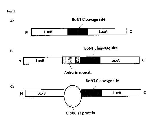
[0052] Figure 1 shows a schematic drawing of the single chain luciferase
fusion
polypeptide of the in vention, (A) prevention of the self interaction of the
subunits by a
specific order of the said subunits; (B) prevention of the self interaction by
ankyrin repeats;
(C) prevention of the self interaction by globular proteins.
[0053] Figure 2 shows the expression of MRZ_LuxABO and MRZ LuxBA3 in different
E. coli expression strains at 22 C, whereby soluble and insoluble fractions
were separated.
Load per lane: 1/6 x 0D600 unit with 1 x 0D600 unit defined as 1 ml of a
culture with an 0D600
of 1Ø Selected marker sizes are depicted to the left. A shows the 12% SDS-
PAGE analysis
of the expression of MRZ_LuxABO and MRZ_LuxBA3 in the E. colt strains BL21 and
Rosetta (DE3) / pRARE2. Gels were stained with Coomassie Blue. B shows the
Western-blot
analysis of a 12% gel; detection was performed with anti-Strep-tag antibody,
secondary
antibody anti mouse-AP, developed with NBT / BCIP.
[0054] Figure 3 shows the expression of MRZ_LuxABO and MRZ LuxBA3 in E. coli
expression strain BL21 at 16 C, whereby soluble and insoluble fractions were
separated. Load
per lane: 1/6 x 0D600 unit with 1 x 0D600 unit defined as 1 ml of a culture
with an 0D600 of
1Ø Selected marker sizes are depicted to the left. A shows the 12% SDS-PAGE
analysis of
the expression of MRZ_LuxABO and MRZ_LuxBA3 in the E. coli strain BL21. Gels
were
stained with Coomassie Blue. B shows the Western-blot analysis of a 12% gel;
detection was
performed with anti-Strep-tag antibody, secondary antibody anti mouse-AP,
developed with
NBT / BCIP.
[0055] Figure 4 shows E. coli strain BL21 LuxABO fed-batch fermentation. One 5
1
fermentation was run to generate 62 g (wcw) biomass.
[0056] Figure 5 shows. 0D600 throughout fed-batch fermentation of A E. coli
strain BL21
LuxABO and BE. coif strain BL21 LuxBAl.
[0057] Figure 6 shows SDS-PAGE and Western blotting of samples from before
induction
and at time of harvest of A E. coil strain BL21 LuxABO and B E. coil strain
BL21 LuxBAL
CA 02866595 2014-09-08
WO 2013/131991
PCT/EP2013/054566
- 17 -
[0058] Figure 7 shows Strep-tag affinity batch-purification of BL21-MRZ-LuxABO
and
BL21-MRZ-LuxBAL Load per lane is 1.25 ul undiluted sample of fractions Load,
FT1, and
ET2, and 2.5 ul undiluted sample of Pool El, Pool E2, and Pool E3. Selected
marker sizes are
depicted to the left. A SDS-PAGE analysis; Gel 12%, stained with Coornassie
Blue. B
Western-blot analysis; Gel 12%, detection with anti-Strep antibody (StrepMAB-
Classic, HRP
conjugate, IBA, Cat. No. 2-1509-001, dilution 1:10000 in PBS-Tween containing
2% BSA).
[0059] Figure 8 shows the analysis of expression in small scale E. coil shake
flask cultures.
Target vectors were expressed in the host strains BL21(DE3) and BL21(DE3)
Rosetta, and
cultured at 37 C. Samples for analysis were drawn just before induction as
well as 2, 4, and
hours after induction of target expression. Load per lane: 0.25 x 0D600 units
with 1 x
0D600 unit defined as 1 ml of a culture with an 0D600 of 1Ø Left side shows
BL21(DE3) and
right side shows BL21(DE3) Rosetta. A shows LuxBA3-Strep (84.9 kDa), B GST-
LuxBA3-
15 Strep (111.2 kDa), and C 9xHis-LuxBA3-Strep (86.8 kDa).
[0060] Figure 9 shows the analysis of 9xHis-Xa-LuxBA3-Strep protein
expression. 11
shake flask culture grown at 25 C (before and after induction). 12 % SDS-PAGE
followed by
Coll. Coomassie-staining and anti-Strep-tag Western blotting. Load per lane:
0.25 x 0D600
20 units with 1 x 0D600 unit defmed as 1 ml of a culture with an 0D600 of
1Ø Marker:
Fermentas PageRulerTM Prestained Protein Ladder.
[0061] Figure 10 shows the analysis of final LuxBA3 protein samples. 12% SDS-
PAGE
and Coll. Coomassie staining. The two different gels refer to two independent
FPLC runs (left
panel: 1.89 mg total; right panel: 1.90 mg total) but from the same starting
material.
EXAMPLES
[0062] The invention will now be illustrated by Examples which shall, however,
not be
construed as limiting the scope of the invention.
[0063] Example 1: Expression, fermentation and purification of different
Luciferase
constructs in Escherichia coil
[0064] Expression of MRZ_LuxABO and MRZ_LuxBA3 in E. coil
The constructs MRZ LuxABO (SEQ ID NO: 9) and MRZ LuxBA3 (SEQ ID NO: 10) are
CA 02866595 2014-09-08
WO 2013/131991
PCT/EP2013/054566
- 18 -
pASK-IBA3-plus vector constructs, encoding for two different Luciferase-
targets both
carrying a C-terminal Strep-tag II. The expected molecular weight of
Luciferase ABO carrying
a C-terminal Strep-tag-II is 78.6 KDa, with an estimated pI of 5.22. The
expected molecular
weight of Luciferase BA3 carrying a C-terminal Strep-tag-II is 84.9 KDa, with
an estimated
pI of 5.20.
[0065] Expression of MRZ LuxABO and MRZ_LuxBA3 at
22 C
For analyzing the expression MRZ LuxABO and MRZ_LuxBA3 (both Amp') were
transformed into the E. colt strains BL21 and Rosetta (DE3) / pRARE2 (Cam').
Cells were
grown at 37 C in LB medium supplemented with 200 pig/rn1 ampicillin (and 30
jig/nil
chloramphenicol for the respective strains) until an 0D600 of 0.4 (BL21) or an
0D600 of 0.2
(Rosetta (DE3) / pRARE2) was reached. Each culture was shifted to 22 C and
grown until an
0D600 of 0.65 (BL21) or an 0D600 of 0.3 (Rosetta (DE3) / pRARE2) was reached.
Cultures
were induced with 0.2 pg/rn1 anhydrotetracycline and grown for another 24
hours at 22 C.
After 0, 1, 3, 5, and 24 hours of induction samples were taken and treated
with Bug buster HT
solution (Novagen) to break the cells and separate soluble and insoluble
protein fractions.
Samples were analyzed via SDS-PAGE analysis on 12% Gels (and via Western-blot
with
anti-Strep-tag antibody), as seen in Figures 2A and 2B.
[0066] Expression of MRZ_LuxABO and MRZ_LuxBA3 at
16 C
For analyzing the expression MRZ_LuxABO and MRZ_LuxBA3 (both AmpR) were
transfoimed into E. colt BL21. Cells were grown at 25 C in LB medium
supplemented with
200 jig/m1 ampicillin until an 0D600 of 0.15 was reached. Each culture was
shifted to 16 C
and grown until an 0D600 of 0.2 (MRZ_LuxABO) or an 0D600 of 0.35 (MRZ_LuxBA3)
was
reached. Cultures were induced with 0.2 mg/m1 anhydrotetracycline and grown
for another 24
hours at 16 C. After 0, 1, 3, 5 and 24 hours of induction samples were taken
and treated with
Bug buster HT solution (Novagen) to break the cells and separate soluble and
insoluble
protein fractions. Samples were analysed via SDS-PAGE analysis on 12% Gels
(and via
Western-blot with anti- Strep-tag antibody), as seen in Figures 3A and 3B.
[0067] To clearly relate bands in the Coomassie stained gels shown in Figures
2A and 3A to
the Luciferase ABO and BA3 target and to estimate the amount of soluble
compared to
insoluble target, western blot analysis with anti- Strep-Tag antibody was
performed. The
western-blot analysis revealed the presence of a protein running between the
75 and the 100
kDa molecular weight marker band (corresponding with the expected molecular
weight of
78.6 kDa of the translated Luciferase ABO protein) in the insoluble protein
fraction of the
BL21 / LuxABO expression, as shown in Figures 2B and 31R. The LuxABO-construct
showed
the best soluble expression in BL21 after 24 hours of induction at 16 C. In
both Figures no
specific western-blot signal was detected by the anti-Strep-tag antibody in
the LuxBA3
CA 02866595 2014-09-08
WO 2013/131991
PCT/EP2013/054566
- 19 -
expression strains. In Figure 3B, however, a very weak signal can be seen
after 24 hours of
expression in BL21 in the soluble protein fraction.
[0068] High cell density batch-fermentation of the E. coli strains BL21 LuxABO
and
BL21
LuxBA1
E. coil strains BL21 LuxABO and BL21 LuxBA1 were separately grown in batch
fermentation mode. A single colony of each strain was picked from an LB plate,
inoculated in
2 x 100 ml LB medium, grown at 25 C, 175 rpm for 16 hours. These preculture
were used to
inoculate 4.5 1 fermentation medium. Ampicillin was added in all cultures to
100 ug/nal.
Growth was recorded throughout the fermentation (Figures 4, 5A, and 5B).
Fennenter settings
are summarized in Table 1.
[0069] Table 1 summarizes ferinenter settings.
Ferrnenter settings summary:
Preculture volume 125 ml
Initial fermentation volume 4.5 1
PH 7.4 (adjusted using ammonia/phosphoric acid)
P02 20 % (stir airflow cascade)
Temperatur 23 C whole cultivation period
Antifoam reagent Antifoam A (Sigma) at 1 m1/1
Antibiotic Ampicillin at 100 ug/m1
Inducer Anhydro-tetracycline at 1 mg/1
[0070] The fermentation medium was made as follows (per liter): For YTG base,
to 900 ml
of H20 add 12 g bacto-tryptone, 24 g bacto-yeast extract, and 4 mL glycerol.
In a separate
flask dissolve in 90 mL H20 2.31 g KH2PO4 tnonobasie, 12.54 g K2HPO4 dibasic,
and adjust
volume to 100 mL with 1120. Both solutions were autoclaved separately and
mixed only after
cooling down to below 60 C.
[0071] The fermenter cultures were inoculated to a starting 0D600 of about 0.1
at 23 C
which was kept throughout the whole fermentation process. The culture of E.
coli strain BL21
LuxABO had reached an 0D600 of 0.96 after 7 hours, wherein the culture of E.
coli strain
BL21 LuxBA1 had reached an 0D600 of 1.2 after 8 hours. Then the inducer
anhydro-
tetracycline was added (1.0 mg/1 final concentration). The cultures were
harvested after an
additional 18 hours (LuxABO) or 15 hours (LuxBA1) by centrifugation at 8.000 g
for 20 min
at 4 C. The supernatant was discarded, the cell pellets snap frozen in liquid
nitrogen and then
stored at -80 C until further use. The final 0D600 were 10.8 (LuxABO) and
13.5 (LuxBA1)
with a culture volume of about 5 1. The biomass yield were 62 g (wcw, LuxABO)
and 70 g
(wcw, LuxBA1).
CA 02866595 2014-09-08
WO 2013/131991
PCT/EP2013/054566
- 20 -
[00721 During the fermentation, two samples were drawn (just before anhydro-
tetracycline
addition and at the time of harvest), the cells pelleted by centrifugation,
and then also stored at
-80 C until further use. These two samples were processed for analysis using
Bugbuster
(Novagen) to separate soluble from insoluble material. Comparable amounts were
analyzed
by SDS-PAGE and subsequent Colloidal Coomassie staining and Western blotting,
respectively (Figures 6A and 6B; load per lane: 0.25 x 0D600 units for
Coomassie staining and
0.5 x 0D600 units for anti-Strep-tag Western blotting, with 1 x 0D600 unit
defined as 1 ml of a
culture with an 0D600 of 1Ø)
[0073] Strep-tag affinity batch-purification of LuxABO and LuxBA1
62 g (wcw, LuxABO) fermenter biomass or 70 g (wcw, LuxBA1) fermenter biomass
were
resuspended in 150 ml resuspension buffer (100 mM Tris/HC1 pH 8.0, 150 mM
NaCI, and 1
mM EDTA). The cells were broken by passing them two times through a
microfluidizer.
Unbroken cells were removed by centrifugation at 4 C, 10000 x g for 30 minutes
(pellet was
discarded, supernatant = Load). 1 ml (bed volume) Strep-Tactin Superflow
matrix from IBA
was added to the crude extract (supernatant, Load) and binding was performed
for 30 minutes
with gentle shaking at 4 C. The suspension was centrifuged at 4 C, 2000 x g
for 10 minutes.
The matrix was transferred to a gravity column, the flovvthrough was collected
(FT1). The
column was washed one time with 5 ml of resuspension buffer (wash was added to
the FT1)
followed by six elution steps with 500 j.tl resuspension buffer containing 2.5
mM D-
desthiobiotin (El ¨ E6, first Elution). The flow-trough (FT1) was loaded on
the column again.
The flow-through of this step was collected again (FT2). The column was washed
one time
with 5 ml of resuspension buffer (wash was added to the FT2). A second elution
was
performed consisting of six elution steps with 500 pi resuspension buffer
containing 2.5 mM
D-desthiobiotin (El ¨ E6, second Elution). The second flow-trough (FT2) was
loaded on the
column again. A third elution was performed consisting of six elution steps
with 500 pi
resuspension buffer containing 2.5 mM D-desthiobiotin (El ¨ E6, third
Elution). The
following elution fractions were pooled: Pool El (El -E6 from elution 1), Pool
E2 (El -E6
from elution 2), and Pool E3 (El -E6 from elution 3). Samples were analysed
via 12% SDS-
PAGE analysis and western blot using Strep-tag antibody (StrepMAB-Classic, HRP
conjugate, IBA, Cat. No. 2-1509-001), as shown in Figure 7.
[0074] Protein concentrations of the elution fractions were determined using
Bradford
analysis. Each elution fraction pool was split in two halves (with 1,5 ml each
respectively)
and stored at 4 C. The total protein yield of the elution fractions were
approximately 6 mg for
the LuxABO construct (purified out of 62g wcw fermenter biomass) and
approximately 3 mg
for the LuxBA1 construct (purified out of 70g wcw fermenter biomass), as shown
in Table 2.
CA 02866595 2014-09-08
WO 2013/131991
PCT/EP2013/054566
- 21 -
[0075] Table 2 shows total protein yield for LuxABO and LuxBA1 constucts.
Eluation pool Concentration [ug/u1] Volume [ml] Total protein yield
[mg]
LuxABO El 1.00 3.00 3.00
LuxABO E2 0.53 3.00 1.59
LuxABO E3 0.45 3.00 1.35
LuxBA1 El 0.40 3.00 1.20
LuxBA1 E2 0.36 3.00 1.08
LuxBA1 E3 0.25 3.00 0.75
[0076] Example 2: Cloning of different LuxBA3-Strep-tag expression vectors and
expression in E. coli
[0077] Cloning of expression
vectors
Using the template DNA (SEQ ID NO: 10), the target sequence was amplified and
subeloned
into pET-based expression vectors. The resulting target vectors were named
accordingly
(Table 3). E. coli transformants were screened, and plasmid DNA from several
candidates
was isolated and sequenced. Their target sequences were verified by DNA
sequencing.
[0078] Table 3 shows nomenclature and SEQ ID NOs. of generated expression
vectors.
Abreviations: His, Histidin; Strep, Streptavidin-tag; Kan, Kanamycin; Amp,
Ampieillin.
Expression vector Protein features (N- to Resis Protein sequence DNA
vector
C-terminal) tance sequence
pTZ E02_LuxBA3 LuxBA3-Strep Kan SEQ ID NO: 11
pTZ E3O_LuxBA3 GST-LuxBA3-Strep Amp SEQ ID NO: 12
pTZ_E47_LuxBA3 9xHis-LuxBA3-Strep Kan SEQ JD NO: 13
SEQ ID NO: 14
[0079] Expression in small scale E. coli shake flask
cultures
Each of the three constructs listed in Table 3 were expressed in two different
host strains.
Samples for analysis were drawn at 4 timepoints (just before IPTG addition as
well as 2, 4,
and 20 hours post-induction). A single colony was picked from an LB plate,
inoculated in 5
ml LB medium (incl. the appropriate antibiotics), and gown overnight at 37 C,
175 rpm.
From these, fresh 30 ml LB cultures were inoculated to a starting 0D600 of
0.1. When the
cultures reached an 0W600 of about 0.4, each culture was kept at 37 C and
target expression
was induced in all cultures by the addition of 1PTG (0.5 mM). All samples were
processed in
the same manner using Bugbuster (Novagen) to separate soluble from insoluble
material.
Comparable amounts were analyzed by SDS-PAGE and subsequent Colloidal
Coomassie
staining and anti-Strep Western blotting, respectively (Figures 8A-C).
CA 02866595 2014-09-08
WO 2013/131991
PCT/EP2013/054566
- 22 -
[0080] Example 3: Expression of LuxBA3 construct and purification of LuxBA3
protein
[0081] Expression of the LuxBA3
construct
The construct pTZ_E47_LuxBA3 (9xHis-Xa-LuxBA3-Strep protein; see Table 3) was
expressed in E. colt strain BL21(DE3). A single colony was picked from an LB
plate,
inoculated in 5 nil LB medium (incl. Kanamycin, 25 jig/m1), and grown
overnight at 25 C,
175 rpm. On the next morning, 100 ml LB shake flask cultures were inoculated
to a starting
0D600 of 0.1. When the cultures reached an 0D600 of about 0.6, target
expression was induced
in all cultures by the addition of IPTG (0.02, 0.10, and 0.25 mM 1PTG,
respectively). Samples
for analysis were drawn just before IPTG addition and 20 hours post-induction.
These
expression conditions were used to generate additional biomass (3 x 11).
[0082] All samples were processed in the same manner using Bugbuster (Novagen)
to
separate soluble from insoluble material. Comparable amounts were analyzed by
SDS-PAGE
and subsequent Colloidal Coomassie staining and Western blotting, respectively
(Figure 9).
Beyond the sampling over the time course, the cultures were harvested 20 hours
post-
induction by centrifugation at 5.000 g for 15 min. Cell pellets were stored at
-20 C.
[0083] Purification of the target protein 9xHis-Xa-LuxBA3-Strep (87 kDa) from
the
insoluble fraction under denaturing conditions - refolding on column
For the purification from the insoluble fraction, all three cell pellets (see
above) were
combined and processed at once. After binding of the denatured target protein
on a NiNTA
chromatography column, the target was refolded on column and then eluted.
Final yield of
purification was 3.8 mg with an estimated purity of 90 %..
[0084] The following protocol was used to solubilize the insoluble protein
fraction: The
biomass was resuspended in PBS pH 7.4 including protease inhibitors.
Mechanical cell lysis
was performed by passing the resuspended biomass through a microfluidizer.
Cell were
centrifuged to separate insoluble and soluble fraction. After centrifugation
in PBS, the pellet
(insoluble fraction) was resuspended, urea was added to 8 M final
concentration, and the
mixture was incubated for one hour at room temperature with stirring. After a
centrifugation
at room temperature an urea-insoluble (pellet) and an urea-soluble fraction
(supernatant) were
obtained. The urea-soluble fraction (supernatant) was loaded onto Nickel-
chelating resin
(FPLC) using a loading buffer containing 8 M urea). In the next step a linear
gradient starting
from 8 M to 0 M urea follows over 2 hours. After washing with PBS (no urea
from this step
onwards) a second washing with PBS and 20 mM Imidazole followed. The proteins
are
elutated by a linear gradient from 20 to 500 mM Imidazole (in PBS). The final
samples were
CA 02866595 2014-09-08
WO 2013/131991
PCT/EP2013/054566
- 23 -
analyzed by SDS-PAGE and Coll. Coomassie staining (Figure 10). Purified target
protein was
stored in PBS pH 7.4, residual imidazole. Samples were aliquoted. About one
half each was
stored at +4 C and the other half frozen and stored at -20 C.
[0085] Further, analysis of the 9xHis-Xa-LuxBA3-Strep protein revealed that
the protein is
cleavable by BoNT/A activity but that this cleavage did not result in any
luciferase activity as
desired.