Note: Descriptions are shown in the official language in which they were submitted.
CA 02866907 2014-10-08
Perm selective membrane for treating vascular calcification
in chronic hemodialysis patients
Technical Field
The present disclosure relates to a hemodialysis membrane
for the treatment of vascular calcification in hemodialysis
patients, especially in chronic hemodiaiysis patients. The
present disclosure further relates to methods of treating
vascular calcification in hemodialysis patients, wherein
the hemodialysis membrane is characterized in that it
comprises at least one hydrophobic polymer and at least one
hydrophilic polymer and in that it has a MWRO of between 15
and 20 kD and a MWCO of between 170-320 kD, or, in the
alternative, has a MWRO of between 8.5 and 14 kD and a MWCO
of between 55 kD and 130 kD.
Description of the Related Art
Patients with impaired renal function due to chronic kidney
diseases face one of the highest risks for cardiovascular
morbidity and death that continuously increases as kidney
function declines. This is true for patients with pre-end-
stage renal failure, on dialysis or after successful renal
transplantation. It is the most common cause for death in
patients with a functional allograft, and prevents many
dialysis patients from being engrafted (Goldsmith et al.
(2001): "Coronary artery disease in patients with renal
failure", int J din Pract 55, 196-210). The prevailing
metabolic milieu in moderate-to-severe chronic renal
failure and on dialysis strongly seems to favor an
CA 02866907 2014-10-08
- 2 -
increased rate of atherosclerosis and
atherosclerotic/thrombotic events in these patients. There
is now evidence that vascular smooth muscle cells can
become chondrocyte or osteobiast-like and lay down and
mineralize collagen and non-collagenous proteins in
arteries. Resulting vascular calcification remains one of
the major unsolved problems in uremic patients. Arterial
calcium load seems to be a strong predictor for
cardiovascular complications in this population.
Vascular calcification is common in physiologic and
pathologic conditions such as aging, diabetes,
dyslipidemia, genetic diseases, and diseases with
disturbances of calcium metabolism. However, in CKD
patients, vascular calcification is even more common,
developing early and contributing to the markedly increased
cardiovascular risk. Increased knowledge about the
mechanisms of calcification together with improved imaging
techniques have provided evidence that vascular
calcification should be divided into two distinct entities
according to the specific site of calcification within the
vascular wall. Intimal calcification is advanced
atherosclerosis, occurring in medium-to-large conduit
arteries without smooth muscle cells. Plaques develop and
arterial occlusion occurs. Medial calcification, known also
as Monckeberg's arteriosclerosis, occurs in elastin fibers
around smooth muscle cells in the absence of
atherosclerosis or inflammation and is seen primarily in
chronic renal failure or diabetes. It is typically less
occlusive of the arterial lumen than intimal calcification.
(Nakamura et al (2009): "Coronary Calcification in Patients
with chronic kidney disease and coronary artery disease".
din J Am Soc Nephrol 4, 1892-1900). Also, medial
calcification occurs in arteries of any size, including
small arteries in which atherosclerosis does not occur.
CA 02866907 2014-10-08
- 3 -
Renal failure increases the extent of calcification in
atherosclerotic plaques, but the effect on medial
calcification is probably greater as it rarely occurs in
individuals without renal insufficiency under the age of 60
years. The histological prevalence of medial calcification
in radial arteries was 45-fold greater in patients with CKD
compared with those without CKD (O'Neill et al. (2010):
"Recent progress in the treatment of vascular
calcification". Kidney /nt. 78(12), 1232-1239).
In general, the presence of vascular calcifications in end-
stage renal disease (ESRD) patients is associated with
increased stiffness of large, capacitive, elastic-type
arteries like the aorta and common carotid artery (CCA)
(Blacher et al. (2001): "Arterial Calcifications, Arterial
Stiffness, and Cardiovascular Risk in End-Stage Renal
Disease". Hypertension 38, 938-942), together with higher
pulse wave velocity (PWV), earlier return of wave
reflections from the periphery to the ascending aorta
during systole and abnormal increase of aortic systolic
blood pressure, with reduced diastolic blood pressure and
high pulse pressure. In the general population and in
patients with CKD, electron-beam computed tomography (EBCT)
has proven coronary artery calcjfication (CAC) as a potent
predictor of cardiac events. Both the prevalence and
intensity of CAC are increased in patients with CKD.
Traditional cardiac risk factors do not appear to entirely
account for the elevated cardiovascular morbidity seen in
advanced CKD. Hyperphosphatemia, elevated Ca x P product,
and hyperparathyroidism have been associated with
cardiovascular disease (CVD) risk and mortality in advanced
CEO. In addition, uremia is believed to confer
nontraditional CVD risks such as, for example, a
CA 02866907 2014-10-08
- 4 -
broinflammatory state and dysregulation of calcification
inhibitors and inducers.
Calcification in patients with end-stage renal disease
(ESRD) was previously believed to occur as a result of
passive mineral deposition processes. Although the
pathophysiology is not completely understood, it is clear
that it is a multifactorial process involving altered
mineral metabolism, as well as changes in systemic and
local factors that can promote or inhibit vascular
calcification, and all of these are potential therapeutic
targets. Molecular Mechanisms involved in vascular
calcifaction are described, for example, in Mizobuchi et
al. (2009): "Vascular Calcification: The Killer of Patients
with Chronic Kidney Disease", J Am Soc Nephrol 20, 1454,
namely ectopic osteogenesis and elastin degradation.
Inducers of vascular calcification are also reviewed in
Mizobuchi et al. (2009): "Vascular Calcification: The
Killer of Patients with Chronic Kidney Disease", J Am Soc.
Nephrol 20, 1453-1464, where it is stated that compared
with the general population, patients with CKD have a
disproportionately high occurrence of vascular
calcification. One hypothesis to account for this is the
altered Ca and P2-- metabolism seen in these patients. This
is the most important contributor to the progression of
vascular calcification in the uremic condition. Another
factor mentioned are uremic toxins. Uremic serum was found
to upregulate the expression of, for example, Cbfal/Runx2
and its target protein OPN, and increases secretion of a
mediator of osteoblastic differentiation, BMP-2, resulting
in the mineralization of VSMCs into osteoblast-like cells,
see also Moe et at. (2008), Figure 1. Mizobuchi et al. .also
mention oxidative stress and inflammation and other
inducers such as leptin. The bone proteins osteonectin,
osteopontin, bone sialoprotein, type 1 collagen, and
CA 02866907 2014-10-08
- 5 -
alkaline phosphatase have also been identified in multiple
sites of extraskeletal calcification. Interestingly, in
cell culture, vascular smooth muscle cells and vascular
pericytes are capable of producing these same boneforming
transcription factors and proteins, and can be induced to
do so wiLh high concentrations of phosphorus, uremic serum,
high glucose, oxidized lipids, and several other factors
(Moe et al. (2008): "Mechanisms of Vascular Calcification
in Chronic Kidney Disease", J Am Soc Nefohrol 19, 213-216.
Therefore, current therapeutic approaches are directed to
preventing disordered bone and mineral metabolism in
advanced kidney disease and mainly involve lowering the
circulating levels of both phosphate and calcium. The
efficacy of compounds that specifically target
calcification, such as bisphosphonates and thiosulfate, has
been shown in animals but only in small numbers of humans,
and safety remains an issue (O'Neill et al. (2010); "Recent
progress in the treatment of vascular calcification".
Kidney Int. 78(12), 1232-1239). Additional therapies, such
as pyrophosphate, vitamin K, and lowering of pH, are
supported by animal studies, but are yet to be investigated
in further detail (O'Neill et al. (2010)). In any case,
potential anticaloification therapies always carry the risk
of adversely acting on normal calcification, for example in
bones and teeth.
Interestingly, not all dialysis patients seem to develop
arterial calcifications, despite similar exposure to these
risk factors, and importantly, do not develop calcification
with increased duration of dialysis (Moe et al. (2008)).
For the efficient treatment of CKD patients, it is
therefore helpful to which patients have a high risk for a
cardiovascular event. Patients with calcification having a
higher risk for future coronary events than an age-gender-
CA 02866907 2014-10-08
- 6 -
specific percentile ranking can then be treated with
available therapies. The Kidney Disease Improving Global
Outcomes (KDIGO) suggests (see Kidney International (2009)
76 (Suppl 113), S1-S2) that patients with CKD stages 3-5
with known vascular/valvular calcification should generally
be considered at highest cardiovascular risk. Coronary
artery calcification score (CACS) may be used as a
quantitative assessment of calcified atherosclerosis which
is detectable by electron-beam (EBCT) or multislice
computed tomography (CT). The score which is also referred
to as Agatston score is calculated using a weighed value
assigned to the highest density of calcification in a given
coronary artery. Details on how the Agatston score as used
herein can be determined and analyzed are given in
Halliburton et al. (2010): "Noninvasive quantification of
coronary artery calcification: methods and prognostic
value", Cleve Clin J Med. 69 (suppl. 3), S6 -S11. A more
elaborate method to determine the Agatston score of a
patient is shown in van der Bijl et al. (2010), AJR 195,
1299-1905, An Agatston score of 0 is normal. In general,
the higher the score, the more likely it is to have a
coronary heart disease (Cl-ID) . A score of 0 to 10 is
associated to a low risk, a score of 11 to 100 to an
intermediate risk. A score of more than 100 (>100)
describes an intermediate to high risk. A score of more
than 400 (>400) describes persons with a very high risk
(Halliburton at al. (2010)).
So far, current therapeutic approaches are, apart from
nutritional aspects, based mainly on a certain medication
of the patients in need of a treatment. The dialysis
treatment, in contrast, has mainly been discussed with
regard to the risks it imposes on a CKD patient and the
development of vascular calcification. However, because of
the risks and drawbacks associated with the compound based
CA 02866907 2014-10-08
- 7 -
anticalcification therapies directed to preventing
disordered bone and mineral metabolism in advanced kidney
disease, it would clearly be desirable to devise a dialysis
system which is able to lower or reduce calcification in
CKD patients already during the extracorporeal treatment
and before vascular calcification develops. If a system can
be devised which is able to reduce the onset of
calcification in CKD patients or reduces existing
calcification in patients who already have to undergo
medication, said medication and potential side effects
related thereto could be reduced or omitted completely.
Currently available membranes and filters for use in
hemodialysis, hemodiafiltration or hemofiltration could so
far not contribute effectively to avoiding or reducing
vascular calcification in uremic patients. For the
avoidance of doubt, if not expressly indicated otherwise,
the expression "hemodialysis" as used herein encompasses
hemodialysis, hemodiafiltration and hemofiltration methods.
Based on the findings on molecular mechanisms involved in
vascular calcification as described above and reviewed, for
example, in Mizobuchi et al. (2009), the present inventors
have focused their attention on the key mediators for the
mineralization of cells in the vessel wall and on methods
for the removal of such mediators rather than addressing
calcification problems by administering inhibitors of such
mediators. As a result of their studies, the inventors have
found that newly developed membranes, so-called high cut-
off membranes can be used for eliminating from CKD patients
in need said pro-calcifying mediators which induce and/or
promote calcification. It was found as a consequence that
calcification can be reduced or delayed by using said high
cut-off membranes in the extracorporeal hemodialysis
treatment of uremic patients.
CA 02866907 2014-10-08
- 8 -
In general, dialysis membranes are designed to accomplish
the removal of uremic toxins and excess water from the
blood of patients with chronic renal failure while
balancing the electrolyte content in the blood with the
dialysis fluid. Uremic toxins are usually classified
according to their size and physicochemical characteristics
in small water-soluble compounds (e.g., urea and
creatinine), protein-bound solutes (e.g., p-cresyl sulfate)
and middle molecules (e.g., b2-microglobulin and
interleukin-6) (1-4). While the removal of small molecules
takes place mainly by diffusion due to concentration
differences between the blood stream and the dialysis fluid
flow, the removal of middle molecules is mainly achieved by
convection through ultrafiltration. The degree of diffusion
and convection depends on the treatment mode (hemodialysis,
hemofiltration or hemodiafiitration) as well as on the
membrane type (low-flux, high-flux, protein leaking, or
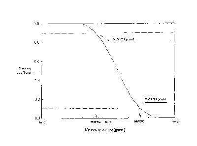
high cut-off membranes). The sieving property of a
membrane, i.e., its permeability to solutes, is determined
by the pore size and sets the maximum size for the solutes
that can be dragged through the membrane with the fluid
flow. The sieving coefficient for a given substance could
be simply described as the ratio between the substance
concentration in the filtrate and its concentration in the
feed (i.e., the blood or plasma), and is therefore a value
between 0 and 1. Assuming that the size of a solute is
proportional to its molecular weight, a common way to
illustrate the properties of membranes is by creating a
sieving curve, which depicts the sieving coefficient as a
function of the molecular weight. The molecular weight cut-
off (MWCO) is defined as the molecular weight where the
sieving coefficient is 0.1 (Figure 1). The sieving curve
determined for a polydisperse dextran mixture can be
considered a standard characterization technique for a
membrane. Conventional dialysis membranes are classified as
CA 02866907 2014-10-08
- 9 -
low-flux or high-flux, depending on their permeability. A
third group, called protein leaking membranes, is also
available on some markets. These three membrane groups were
described in a review by Ward (2005), J Am Sac Nephrol 16,
2421-2430. Lately a fourth type has emerged, the above-
mentioned high cut-off membranes, which have particular
characteristics (Boschetti-de-Fierro et al. (2013):
"Extended characterization of a new class of membranes for
blood purification: The high cut-off membranes", Int J
Art:if Organs 36(7), 455-463). A concise summary of the
generai classification and performance of said membranes as
is shown in Table I of Boschetti-de-Fierro et al. (2013)
and shall be valid also for describing the present
invention. The latest step in membrane development is a
membrane type which in terms of classification could be
positioned in between the so-called high flux and the high
cut-off membranes. Said membranes are therefore also
referred to as "medium cut-off" membranes (see also Table
II). These membranes and how they can be prepared are
described in detail in European Patent Application No.
14154175.5.
Table I: General classification and typical performance of
hemodialysis membranes
Dia- Water Sieveing FLC Album
lyzer perme- Coefficient b Clearance' in
type abilitya Loss
ml/ (m2hram (g)d
Hg)
132- Albumin Kappa Lambda
Micro-
globulin
Low- 10-20 <0.01 0
flux
High- 200-400 0.7-0.8 <0.01 <10 <2
<0.5
flux
CA 02866907 2014-10-08
- 10 -
Pro- 50-500 0.9-1.0 0.02- 2-6
tein 0.03
lea-
king
High 1100 1.0 0.2 38 33 28
cut-
off
with 0.9 wt.- sodium chloride at 37 1 C and Q 100-500
ml/min
l'according to EN1283 with Q. max and UF 20,5
'Serum Free Light Chains, Clearance in vitro, Qb 250 mi/min and
500 ml/min, UF 0 ml/min, Bovine Plasma, 60 g/1, 37 C, Plasma
Level: human K 500 mg/l, human A. 250 mg/l. All clearances in
ml/min, measured for membrane areas between 1.7 and 2.1 M:
' measured in conventional hemodialysis, after a 4-h session,
with Q. 250 ml/min and Q, 530 ml/min, for membrane areas between
1.7 and 2.1 m2
The most evident difference among the types of membranes is
their position along the molecular weight axis (Figure 2).
High-flux membranes have a sieving curve more similar to
that of the glomerular membrane, removing toxins of small
molecular weight such as urea and also allowing some
removal of relatively large toxins, such as p2-
microglobulin and myoglobin. High cut-off membranes show a
sieving curve located at higher molecular weights than that
for the glomerular membrane. Although the high cut-off
sieving profile resembles that of the glomerular membrane
up to 20 kDa, the high cut-off membranes are open toward
molecular weights higher than 20 kDa. This means that the
high cut-off membranes allow some passage of proteins. WO
2004/056460 already discloses certain early high cut-off
membranes which could be used for the treatment of sepsis
in dialyzers by eliminating sepsis-associated inflammatory
mediators. Advanced dialyzers with high cut-off membranes
which are currently on the market are, for example,
CA 02866907 2014-10-08
- 11 -
HCO11000, septe>C4 and Theralite0, all available from
Gambro Lundia AB. Known uses of high cut-off membranes
include treatment of sepsis, chronic inflammation (EP 2 161
072 Al), amyloidosis and rhabdomyolysis and treatment of
anemia (US 2012/0305487 Al), the most explored therapy to
date being the treatment of myeloma kidney (US 7,875,183
B2). In this case, the removal of the free light chains in
patients with multiple myeloma on chemotherapy has allowed
the recovery of kidney function in a significant number of
patients. Due to the loss of up to 40 g of albumin per
session with the above-mentioned dialyzers, high cut-off
membranes have been used for acute applications, although
some physicians have contemplated benefits of using them in
chronic applications, possibly in conjunction with albumin
substitution.
The expression "molecular weight cut-off" or "MWCO" or
"nominal molecular weight cut-off" as used herein is a
value for describing the retention capabilities of a
membrane and refers to the molecular mass of a solute where
the membranes have a rejection of 90% (see above and Figure
1), corresponding to a sieving coefficient of 0.1. The MWCO
can alternatively be described as the molecular mass of a
solute, such as, for example, dextrans or proteins where
the membranes allow passage of 10% of the molecules. The
shape of the curve depends, for example, on the pore size
distribution and is thus linked to the physical form of
appearance of the membrane.
As already mentioned, sieving curves give relevant
information in two dimensions: the shape of the curve
describes the pore size distribution, while its position on
the molecular weight axis indicates the size of the pores.
Molecular weight cut-off (MWCO) limits the analysis of the
sieving curve to only one dimension, namely to the size of
CA 02866907 2014-10-08
- 12 -
the pores where the sieving coefficient is 0.1. To enhance
membrane characterization the molecular weight retention
onset (MWRO) is used herein for characterizing high cut-off
and medium cut-off membranes. The MWRO is defined as the
molecular weight at which the sieving coefficient is 0.9,
as schematically shown in Figure 1. It is analogous to the
MWCO and describes when the sieving coefficient starts to
fall from 1 to 0. Defining two points on the sieving curves
allows a better characterization of the sigmoid curve,
giving an indication of the pore sizes and also of the pore
size distribution. The expression "molecular weight
rejection onset" or "MWRO" or "nominal molecular weight
rejection onset", as used herein, therefore refers to the
molecular mass of a solute where the membranes have a
rejection of 10%, or, in other words, allow passage of 90%
of the solute, corresponding to a sieving coefficient of
0.9.
The use of dextran sieving curves together with the
respective MWCO and MWRO values based thereon allows
differentiating the existing dialyzer types low-flux, high-
flux, protein leaking, medium cut-off or high cut-off
(Figure 3). Compared to the high-flux dialyzers, which are
the standard for current dialysis treatment, the low-flux
diaiyzers are depicted in a group with low MWRO and MWCO.
The other two families - protein leaking and high cut-off
dialyzers - have different characteristics. While the
protein leaking dialyzers are mainly characterized by a
high MWCO and a low MWRO, the high cut-off family can be
strongly differentiated due to the high ir vitro values for
both MWRO and MWCO (Table II).
TABLE II: General classification of hemodialysis membranes
based on dextran sieving
CA 02866907 2014-10-08
- 13 -
Dialyzer Structural Characteristics
type
MWRO [kDa] MWCO [kDa] Pore radius
[mu]
Low-flux 2-4 10-20 2-3
High-flux 5-10 25-65 3.5-5.5
Protein 2-4 60-70 5-6
leaking
High cut-off 15-20 170-320 8-12
Medium cut- 8.5-14.0 55-130 5.5 < pore
off radius < 8.0
The applicants have found that high cut-oft membranes as
defined above and in Table II as well as medium cut-off
membranes as defined above and described in further detail
in EP 14154175.5 can be used to effectively treat vascular
calcification in chronic hemodialysis patients. Especially
the high permeability of the high cut-off membranes but
also the characteristics of the medium cut-off membranes
seem to allow for an increased clearance of relevant
mediators in comparison to the high-flux dialyzers which
currently are the standard for treating chronic
nemodialysis patients. More specifically, even though the
uremic milieu is characterized by a multitude of known and
so far unidentified substances, the inventors were able to
show a reduction of mediators in the serum of patients
having been treated with high cut-off or medium cut-off
membranes, which serum otherwise could be shown to induce
osteoblastic phenotype and osteoblastic differentiation in
mesenchymal stem cells (MSC).
Summary of the Invention
CA 02866907 2014-10-08
- 14 -
It is the object of the present invention to provide for a
method of treating vascular calcification in a hemodialysis
patient, comprising withdrawing and bypassing the blood
from the patient in a continuous flow into contact with one
face of an hemodialysis membrane, simultaneously passing
dialysate solution in a continuous flow on an opposite face
of the hemodialysis membrane to the side of the
hemodialysis membrane in contact with the blood, the flow
of the dialysate solution being countercurrent to the
direction of flow of blood, and returning the blood into
the patient, wherein the hemodialysis membrane is
characterized in that it comprises at least one hydrophobic
polymer and at least one hydrophilic polymer and in that it
has a MWRO of between 15 and 20 kD and a MWCO of between
170-320 kD or in that it has a MWRO of between 8.5 kD and
14.0 kD and a MWCO of between 55 and 130 kD. The MWRO and
MWCO for a given membrane is based on dextran sieving
experiments before blood contact of the membrane as
described by Boschetti-de-Fierro et al. (2013) (see
"Materials and Methods" section of the reference).
Brief Description of the Drawings
Figure 1 is a representation of a dextran sieving curve
where the values of molecular weight retention onset (MWRO,
achieved at SC=0.9) and molecular weight cut-off (MWCO,
achieved at SC=0.1) are illustrated.
Figure 2 shows characteristic dextran sieving curves for
the different types of dialysis membranes: low flux, high
flux and high cut-off. The data for the glomerular membrane
(as reported by Axeisson et al. (2009): Loss of size
selectivity of the glomerular filtration barrier in rats
following laparotomy and muscle trauma. American Journal of
= CA 02866907 2014-10-08
- 15 -
Physiology - Renal Physiology, 297, F577-F582) has been
added for illustration.
Figure 3 shows a mapping of different types of blood
membranes based on the molecular weight retention onset and
molecular weight cut-off from dextran sieving curves. The
dotted line squares approximately represent the boundaries
that delimit the diaiyzer families.
Figure 4 depicts the effect of dialysis with high cut-off
membranes on serum induced osteoblast differentiation and
calcification of mesenchymal stromal cells (MSC). MSCs were
induced with osteoblast induction medium containing serum
from dialysis patients treated either with conventional
high-flux dialyzers (Conventional) or from the same
patients after a 3 weeks course of dialysis with high cut-
off membranes (HCO). (A) Alkaline phosphatase (ALP)
activity normalized to protein content measured on single-
patient level (n=16). (B) ALP activity measurements from
the same patients with conventional treatment set 1Ø (C)
Quantification of deposited calcium normalized to protein
content. Sera from a total of 16 patients were combined to
3 different serum pools for both treatment modalities. Each
serum pool was applied to 4 MSC preparations from different
healthy donors. (D) Calcium measurements from the same
serum pools and cell preparations with conventional
treatment set 1Ø ***P<0.001.
Figure 5 depicts the dose-dependent induction of
osteoblastic differentiation in mesenchymal stromal cells
(MSC) by pro-inflammatory cytokines, fibroblast growth
factors (FGF), and full-length parathyroid hormone (PTH1-
84). (A-E) Alkaline phosphatase (ALP) activity in MSCs
treated with different concentrations of IL-l3 (A), TNF-a
(8), FGF-2 (C), FGF-23 (D) or PTH1-84 E]) in osteoblast
CA 02866907 2014-10-08
- 16 -
induction medium (OM) for 7 days. (F-J) Calcium deposited
by MSCs cultured for 3 weeks in OM with increasing
concentrations of IL-1 p (F), TNF-a (G), FGF-2 (H), FGF-23
(I) or PTH1-84 (J). "Fold CMAX" denotes the x-fold
concentration of the highest reported concentration found
in dialysis patients (I1--1P) CMAX = 1.7 pg/L; TNF-a CMAX =-
408 ng/L; FGF-2 CMAX = 19.5 ng/L; FGF-23 CMAX = 255.2 ng/L;
PTH1-84 CMAX = 2.4 pg/L). ALP activity and calcium were
normalized to sample protein content. All values are
expressed relative to OM without cytokine (set 1.00). Means
+ SEM from 4 - 6 independent experiments are shown.
*P<0.05, **P<0.01, ***P<0.001.
Figure 6 shows the relative calcification which was
measured in vitro in vascular smooth muscle cells (VSMC)
upon incubation with plasma samples from healthy donors or
from 48 patients who were dialysed with both high-flux and
high cut-off membranes for three weeks (Example 3.6).
Calcification was assessed after 10 days with alkaline
phosphatase and alizarin staining. A reduction of
calcification was measured in VSMC incubated with serum
after the high cut-off phase compared to the high-flux
phase.
Figure 7 shows the relative calcification of VSMC upon
incubation with plasma samples from healthy donors and from
samples obtained from in vitro dialysis experiments with
high cut-off, medium cut-off and high-flux membranes,
respectively. The results of the in vitro study support the
observation of the clinical trial (Figure 6, Example 3.6).
Vascular calcification was reduced by 36% in high cut-off
probes and by 32% in medium cut-ott probes compared to
high-flux probes.
CA 02866907 2014-10-08
- 17 -
Detailed Description
Patients with impaired renal function due to chronic kidney
diseases face one of the highest risks for cardiovascular
morbidity and death that continuously increases as kidney
function declines. This is true for patients with pre-end-
stage renal failure, on dialysis or after successful renal
transplantation.
The present disclosure therefore relates to a high cut-off
or medium cut-off hemodialysis membrane for the treatment
of vascular calcification in hemodialysis patients,
especially in chronic hemodialysis patients and especially
in a CKD stage 3-5 patient having an Agatston score of more
than 11. The method comprises withdrawing and bypassing the
blood from the patient in a continuous flow into contact
with one face of an hemodialysis membrane, simultaneously
passing dialysate solution in a continuous flow on an
opposite face of the hemodialysis membrane to the side of
the hemodialysis membrane in contact with the blood, the
flow of the dialysate solution being countercurrent to the
direction of flow of blood, and returning the blood into
the patient, wherein the hemodialysis membrane is
characterized in that it comprises at least one hydrophobic
polymer and at least one hydrophilic polymer and in that it
has a MWRO of between 15 and 20 kD and a MWCO of between
170-320 kD. Alternatively, the membrane has a MWRO of
between 8.5 kD and 14.0 kD and a MWCO of between 55 and 130
kD. The MWRO and MWCO for a given membrane is based on
dextran sieving experiments as described by Boschetti-de-
Fierro et al. (2013)(see "Materials and Methods" section of
the reference) and refers to values obtained before blood
contact of the membrane.
CA 02866907 2014-10-08
=
- 18 -
As described before in this document, patients with CKD
have a disproportionately high occurrence of vascular
calcification. One hypothesis to account for this is the
altered Ca'2' and 1,)- metabolism seen in these patients.
Another factor mentioned is uremic toxins and uremic serum
was found to upregulate the expression of, for example,
Cbfal/Runx2 and its target protein OPN, and increases
secretion of a mediator of osteoblastic differentiation,
BMP-2, resulting in the mineralization of VSMCs into
osteoblast-like cells. Oxidative stress and inflammation
and other inducers such as leptin are also discussed as
possible inducers. The bone proteins osteonectin,
osteopontin, bone sialoprotein, type I collagen, and
alkaline phosphatase have also been identified in multiple
sites of extraskeletal calcification. Interestingly, in
cell culture, vascular smooth muscle cells and vascular
pericytes are capable of producing these same boneforming
transcription factors and proteins, and can be induced to
do so with high concentrations of phosphorus, uremic serum,
high glucose, oxidized lipids, and several other factors
(Moe et al. (2008): "Mechanisms of Vascular Calcification
in Chronic Kidney Disease", J Am Soc Nephrol 19, 213-216.
As cells of mesenchymal origin including endothelial and
vascular smooth muscle cells are prime targets of uremic
solutes, mesenchymal stromal cells (MSCs) as common
progenitors of both are a suitable model to identify
mechanisms by which the uremic milieu interior may disturb
vascular health. The model can also be used to determine
the effect of the use of high cut-off membranes/dialyzers
on vascular calcification indicators in the uremic
retention solutes. For the present invention, effects of 64
individual uremic retention solutes (URS) on osteoblastic
transformation of MSCs were systematically studied in order
CA 02866907 2014-10-08
- 19 -
to identify therapeutic strategies feasible for targeting
of vascular calcification.
Bone marrow derived MSCs were separately treated with 64
individual URS at uremic concentrations in osteobiastic
induction medium. Osteoblastic differentiation was measured
by alkaline phosphatase activity, western blot,
immunocytochemistry, and calcium deposition. In an
additional translational approach, ostecblastic potential
of serum obtained upon high cut-off dialyzer treatment was
compared to that obtained upon conventional dialysis. It
was found in said approach that substance removal with high
cut-off dialyzers had favorable effects on the attenuation
of osteoblastic differentiation and calcium deposition by
MSCs. The findings emphasize importance of larger
molecules, sometimes also referred to as "middle
molecules", in mediating uremic calcifying MSC phenotype.
Since conventional dialysis strategies fail to effectively
remove this group of URS, targeted dialysis modalities,
possibly in combination with specific pharmacologic
interventions were found to be useful for addressing the
unsolved problem of vascular calcification in chronic
kidney diseases.
In the present invention, the question was addressed, if
maintenance dialysis with membranes characterized by a
higher molecular weight cut-off and consequently a greater
capacity for the removal of larger molecules compared to
conventional high flux dialyzers could improve serum
composition and reduce its pro-osteoblastic and pre-
calcifying effect on MSCs. Serum from patients that had
been dialyzed with high cut-off membranes for 3 weeks with
serum from the same patients obtained during a period when
they had been dialyzed with conventional high-flux
membranes (Example 3). Overall, the potential for induction
CA 02866907 2014-10-08
- 20 -
of osteoblastic differentiation in MSCs was reduced when
serum was obtained during H00 dialysis compared to
conventional high-flux dialysis as indicated by alkaline
phosphatase (ALP) activity (48.67 1.6 U/g protein versus
65.02 4.2 tug protein; Figure 4A). This effect was
detectable in the paired serum samples of each patient
(Figure 4B). Finally, MSCs treated with serum from patients
treated with high cut-off membranes deposited 40% less
calcium compared to serum obtained during a period of
conventional dialysis (Figure 4C). Reduced calcification
was consistently present in each single experiment (Figure
4D).
The concentrations of certain molecules found in a dialysis
patient and which are deemed to play a role in the
mediation of vascular calcification are sometimes extremely
high and will not be encountered frequently in stable
patients on maintenance dialysis. Therefore, in order to
get an insight on dose effects of certain mediators on the
development of calcification, it was tested whether or not
also lower concentrations of inducers of MSC osteoblast
differentiation have detectable effects at least in the
present in vitro model (Example 4). Dose-dependent
increases were identified in both, ALP activity and calcium
deposition induced by the pro-inflammatory cytokines
(Figure 5, A and E) and TNF-a (Figure 5, B and F). The
dose-response curve for FGF-2 also revealed induction of
osteoblastic differentiation and calcium deposition at
concentrations below CN[lx (Figure 5, C and G). It is obvious
from these results that high cut-off membranes according to
the invention that high cut-off dialyzers will have an
effect not only in high risk patients or those patients who
already suffer from severe vascular calcification, but also
on patients who have not yet developed a severe vascular
calcification and/or do not show high levels of mediators
CA 02866907 2014-10-08
- 21 -
suspected of inducing vascular calcification. In the latter
case, the onset and development of vascular calcification
can be prevented or delayed.
As can be seen from Figure 2, the high cut-off dialysis
membrane allows for the limited passage, in whole blood, of
molecules with a molecular weight of above 60 kD, including
also, to a certain limited extend, albumin with a molecular
weight of 68 kD. High-flux membranes, in contrast, allow
only for the passage of molecules up to 25kD in whole
blood. For this reason, filters based on and comprising
high cut-off membranes can be efficiently used to remove
larger molecules in the range of between 25 and 60 k0,
which cannot be efficiently addressed with conventional
dialysis based on low flux or high flux dialyzer.
It was thus found, in the present invention, that in
hemodialysis patients with CKD stages 3-5 and with an
Agatston score of 11 and more, the use of high cut-off or
medium cut-off membranes leads to a reduction of mediators
inducing and/or governing vascular calcification in CKD
patients. Said use thus leads to an effective treatment of
patients suffering from vascular calcification and/or to an
improved, preventive treatment of patients having a
moderate to high risk of developing vascular calcification
and related cardiovascular diseases, respectively.
The use of high cut-off or medium cut-off membranes for
treating hemodialysis patients was found to be especially
favorable for patients with CKD stages 3-5 and with an
Agatston score of >100 to 400. The treatment according to
the invention is especially indicated for patients with CKD
stages 3-5 and with an Agatston score of >400. The use of
the high cut-off or medium cut-off membrane in the dialysis
treatment of patients of Agatston scores of 11-100, 100-400
CA 02866907 2014-10-08
- 22 -
and >400 is also very advisable for patients with CKD
stages 4-5. The use according to the invention of high cut-
off or medium cut-off membranes in the treatment of
hemodialysis patients is especially indicated for patients
with CKD stage 5 and an Agatston score of >100.
The expression -vascular calcification" as used herein
refers to the process of dedifferentiation Or
transformation of vascular smooth muscle cells (VSMC) into
osteo/chondrocytic-like cells, whereupon
osteo/chondrocytic-like VSMC become calcified in a process
similar to bone formation. The calcification involves
deposition of collagen and noncollagenous proteins in the
intima or media and incorporation of calcium and phosphorus
into matrix vesicles to initiate mineralization and further
mineralization into hydroxyapatite. The expression
"vascular calcification" thus encompasses, on a clinical
level, arterial stiffening, higher pulse wave velocity
(PWV), earlier return of wave reflections from the
periphery to the ascending aorta during systole and
significant increase of aortic systolic blood pressure with
reduced diastolic blood pressure and high pulse pressure.
The vascular calcification is quantitatively described by
determining the Coronary Artery Calcification Score (CACS)
as detectable by electron-beam or multislice computed
tomography (CT) and as described before. For the avoidance
of doubt, the expression "Agatston score" as used herein is
equivalent to the expression Coronary Artery Calcification
Score (CACS) as used herein.
In the context of the present invention, the expression
"CKD patients" refers to patients with CKD (KDOQI) stages
3-5, if not indicated otherwise. Stage 3 refers to
moderately reduced kidney function with GFR (Glomerular
Filtration Rate, normalized to an average surface area
CA 02866907 2014-10-08
- 23 -
(size) of 1.73 m2) values of 45-59 (3A) and 30-44 (32).
Stage 4 refers to severely reduced kidney function with GFR
values of 15-29. Stage 5 refers to very severe or endstage
kidney failure and GFR values below 15.
The expression "high cut-off membrane" or "high cut-off
membranes" as used herein refers to membranes comprising at
least one hydrophobic polymer and at least one hydrophilic
polymer and having a MWRO of between 15 and 20 kD and a
MWCO of between 170-320 kD. The membranes can also be
characterized by a pore radius, on the selective layer
surface of the membrane, of between 8-12 nm. The expression
"medium cut-off membrane" as used herein refers to
membranes comprising at least one hydrophobic polymer and
at least one hydrophilic polymer and having a MWRO of
between 8.5 and 14.0 kD and a MWCO of between 55 kD and 130
kD. The membranes can also be characterized by a pore
radius, on the selective layer surface of the membrane, of
above 5.5 nm and below 8.0 nm. For the avoidance of doubt,
the determination of MWRO and MWCO for a given membrane is
according to the methods of Boschetti-de-Fierro et al.
(2013); see "Materials and Methods" section of the
reference. The expression "high cut-off membrane" as used
herein otherwise comprises membranes characterized by the
performance parameters as shown in Table I of this
document, without wanting to limit the definition of high
cut-off membranes to the single performance parameters
disclosed in Table I for said membranes. The high cut-off
or medium cut-off membranes can be processed into
hemodialysis filters by methods generally known in the art,
for example, into hemodialysis filters having a design in
terms of housing, area, fiber and bundle geometry, packing
density and flow characteristics, similar to or the same as
products already available on the market such as, for
example, HCO11000 or TheraliteG, both comprising high cut-
CA 02866907 2014-10-08
- 24 -
off membranes. Accordingly, the use of the expression "high
cut-off membrane" or "medium cut-off membrane" in the
context of the present invention encompasses the use of the
membrane within an adequate filter device fit for being
used in/on an extracorporeal dialysis machine.
In one embodiment of the invention, the high cut-off
membranes for the treatment of vascular calcification are
characterized by a pore radius, on the selective surface
layer of the membrane, of between 8-12 nm.
In a further embodiment of the invention, the high cut-off
dialysis membrane is characterized by a clearance (ml/min)
for K -PLC of from 35 to 40, and for K-PLC of from 30 to 40
it as determined according to the method described in Table I.
In yet another embodiment of the invention, the high cut-
off dialysis membranes for the treatment of vascular
calcification are characterized by allowing the passage of
molecules having a molecular weight of up to 45 kDa with a
sieving coefficient of from 0.1 to 1.0 in presence of whole
blood, based on EN1238 with Qb max and UP 20%.
In yet another embodiment of the invention, the high cut-
off dialysis membrane is characterized by sieving
coefficients of from 0.9 to 1.0 for k-microglobulin and of
from 0.8 to 1.0 for myoglobin, when measured according to
EN 1283 with Qr max and UP 20%.
In yet another embodiment of the invention, the medium cut-
off dialysis membrane is characterized as set forth in
European Patent Application No. 14154175.5.
It is a further object of the present invention to provide
for a method for reducing and/or preventing vascular
CA 02866907 2014-10-08
- 25 -
calcification in hemodialysis patients having an Agatston
score of more than 11, comprising withdrawing and bypassing
the blood from the patient in a continuous flow into
contact with one face of an hemodialysis membrane,
simultaneously passing dialysate solution in a continuous
flow on an opposite face of the hemodialysis membrane to
the side of the hemodialysis membrane in contact with the
blood, the flow of the dialysate solution being
countercurrent to the direction of flow of blood, and
returning the blood into the patient, wherein the
hemodialysis membrane is characterized in that it comprises
at least one hydrophobic polymer and at least one
hydrophilic polymer and has a MWRO of between 15 and 20 kD
and a MWCO of between 170-320 kD, or that it comprises at
least one hydrophobic polymer and at least one hydrophilic
polymer and has a MWRO of between 8.5 and 14 kD and a MWCO
of between 55 kD and 130 kD
It is a further aspect of the present invention to provide
for a method for reducing and/or preventing vascular
calcification in hemodialysis patients having an Agatston
score of more than 100. It is another aspect of the present
invention to provide for a method for reducing and/or
preventing vascular calcification in hemodialysis patients
having an Agatston score of between 100 and 400. It is yet
a another aspect of the present invention to provide for a
method for reducing and/or preventing vascular
calcification in hemodialysis patients having an Agatston
score of more than 400. It is also an aspect of the present
invention to provide for a method for reducing and/or
preventing vascular calcification in hemodialysis patients
having an Agatston score of more than 11 and with CKD
stages 3-5, especially 4-5. It is a further aspect of the
present invention to provide for a method for reducing
and/or preventing vascular calcification in hemodialysis
CA 02866907 2014-10-08
- 26 -
patients having an Agatston score of more than 100 and with
CKD stages 4-5, especially 5.
it is another aspect of the present invention to provide
for a dialysis membrane comprising at least one hydrophobic
polymer and at least one hydrophilic polymer, wherein the
membrane allows the passage of molecules having a molecular
weight of up to 45 kDa with a sieving coefficient of from
0.1 to 1.0 in presence of whole blood, based on EN1238 with
Q max and UF 20%, for treating vascular calcification in a
hemodialysis patient, especially in hemodialysis patients
with an Agatston score of >11.
It is also an aspect of the present invention to provide
for a dialysis membrane comprising at least one hydrophobic
polymer and at least one hydrophilic polymer, wherein the
membrane has a molecular weight retentioE onset (MWRO) of
between 15 and 20 kD and a MWCO of between 170-320 kD for
treating vascular calcification in hemodiaiysis patients,
especially in hemodialysis patients with an Agatston score
of >11, wherein the membrane has a pore radius, on the
selective layer, of between 8 and 12 nm.
Ic is also an aspect of the present invention to provide
for a dialysis membrane comprising at least one hydrophobic
polymer and at least one hydrophilic polymer, wherein the
membrane has a molecular weight retention onset (MWRO) of
between 8.5 kD and 14 kD and a mWCO of between 55 kD and
130 kD for treating vascular calcification in hemodialysis
patients, especially in hemodialysis patients with an
Agatston score of >11, wherein the membrane has a pore
radius, on the selective layer, of between 8 and 12 nm.
In another embodiment of the invention, the hemodialysis
treatment regime is performed with a high cut-off or medium
CA 02866907 2014-10-08
- 27 -
cut-off membrane which has a urea clearance of at least 170
ml/min at a QB of 200 ml/min and a (2,.- of 500 ml/min (UP - 0
ml/min). In yet another embodiment of the invention, the
dialysis treatment according to the invention must ensure a
Kt/V of >1.2.
In yet another embodiment of the invention, a patient's
total albumin loss does not exceed about 60 g per week, and
preferably does not exceed 40 g per week.
In one embodiment of the invention, the hemodialysis
treatment with the membranes according to the invention is
performed from 2 to 4 times per week for a period of from 2
to 6 hours, respectively, with a membrane according to the
invention. A hemodialysis patient suffering from vascular
calcification, especially a CKD patient with stage 3-5, is
thus being treated, for a certain period of time, only with
such hemodialysis filter according to the invention. In one
embodiment of the invention, the treatment may continue
until the signs of vascular calcification have been stayed
or have decreased. In another embodiment of the invention,
the patient receives a continual standard hemodialysis
treatment with a hemodialysis filter comprising a medium
cut-off membrane. In the context of the present invention,
"stayed" and/or "decreased" refers to a constant Agatston
score or the reduction of the Agatston score, respectively.
According to another embodiment of the invention, the
treatment regimen as described may be applied for a period
of from 4 to 12 weeks. In yet another embodiment of the
invention, the treatment may continually be used for a
hemodialysis patient with stage 3-5, especially a patient
who belongs to a medium to high or high risk group as
defined by the Agatston score.
CA 02866907 2014-10-08
- 28 -
In another embodiment of the invention, one of three
hemodialysis treatments per week is performed for a period
of 2 to 6 hours with a membrane according to the invention,
whereas two of three hemodialysis treatments per week
comprise the use of a standard high-flux hemodialysis
membrane. Said treatment may be used in cases where
standard dialysis is recommended in addition to using a
hemodialysis filter according to the invention. In one
embodiment of the invention, the treatment may continue
until the signs of vascular calcification have been stayed
or have decreased, or until an Agatston score of below 100,
preferably below 50 has been reached. In another embodiment
of the invention, the treatment regime as described may be
applied for a period of from 4 to 12 weeks. In yet another
embodiment of the invention, the treatment may continually
be used for a hemodialysis patient with CDK stage 3-5,
especially a patient who belongs to a medium to high or
high risk group as defined by the Agatston score.
In a further embodiment of the present invention, the
hemodialysis treatment for a period of 2 to 6 hours is
performed with a dialysis filter comprising a membrane
according to the invention every other dialysis treatment,
whereas the other hemodialysis treatment comprises the use
of a standard high-flux hemodialysis membrane. Said
treatment may be used in cases where standard dialysis is
recommended in addition to using a hemodialysis filter
according to the invention. In one embodiment of the
invention, the treatment may continue until the signs of
vascular calcification have been stayed or have decreased,
or until an Agatston score of below 100, preferably below
50 has been reached.
Depending on the specific condition of a patient, such
treatment regimens or routines can be applied singularly or
CA 02866907 2014-10-08
- 29 -
dynamically, i.e they may be interchanged or subsequently
be used for certain periods of time.
Accordingly, the above method also provides for a
possibility to reduce or suspend the further development of
vascular calcification in hemodialysis patients. The
treatment according to the invention is designed to reduce
or remove such molecules which are connected to the
condition of vascular calcification as discussed before.
The amelioration of the condition of the patient based on
the present treatment will allow reducing medication which
has to be administered to the patients and the risk going
hand in hand with such medication as described before. The
respective reduction rates upon using a high cut-off or
medium cut-off membrane according to the invention at least
lie in the range of more than 10% relative to the Agatston
score determined at the beginning of treating a given
patient according to the invention. It is an object of the
present invention to achieve reduction rates of more than
20%, preferably more than 30%. At least the use of high
cut-off or medium cut-off membranes and filter devices
comprising them is connected to no further increase of the
Agatston score determined at the beginning of treating a
given patient according to the invention.
In one embodiment of the invention, the hemodialysis
treatment according to the invention can be supplemented by
a state of the art medication which would otherwise be
prescribed to a patient suffering from vascular
calcification.
Dialysis machines which can be used for performing a
treatment according to the invention are standard dialysis
machines which can accurately control and monitor the
ultrafiltration rate. Examples for such devices are the AK
CA 02866907 2014-10-08
- 30 -
AK 200'''' S and AK 200 ULTRA S,
PrismafieX eXeed''' or
the Artis7K dialysis machines of Gambro Lundia AB. However,
any other dialysis machine having UF control can also be
used for the treatment.
Parameters for performing a treatment according to the
invention can be adjusted to standard dialysis treatment or
medium cut-off parameters and the specifications of the
high cut-off or medium cut-off membrane. Typical flow rates
used for the present treatment may vary. It is advantageous
to use flow rates with a QB (blood flow) of 100-500,
preferably 250-400 ml/min and a QD (dialysate flow rate) of
100-1000, preferably 300-500 ml/min.
Membrane passage of a solute, such as a protein which needs
to be removed from blood, is described by means of the
sieving coefficient S. The sieving coefficient S is
calculated according to S = (2C1)/(Cõ + where Cr is
the concentration of the solute in the filtrate and C., is
the concentration of a solute at the blood inlet side of
the device under test, and CB, is the concentration of a
solute at the blood outlet side of the device under test. A
sieving coefficient of S=1 indicates unrestricted transport
while there is no transport at all at S=0. For a given
membrane each solute has its specific sieving coefficient.
In addition, the sieving curves may serve as a basis for
determining, for example, the average or mean pore size or
pore size distribution of a membrane on the selective
layer. There is a factual and mathematical correlation
between the sieving characteristics of a membrane and its
pore structure. The mean pore size or pore size
distribution can, for example, be determined according to
Aimar et al (1990) from the dextran sieving curve.
CA 02866907 2014-10-08
- 31 -
In one embodiment, the membrane allows for the passage of
free light chains (FLC). That is, the K or N free light
chains pass through the membrane. High flux membranes, with
smaller pore sizes, sometimes also referred to as "protein-
leaking membranes", have been observed to remove some free
light chains. However, this appears to be primarily due to
binding of the FLC onto the dialysis membranes. FLC may be
used as markers of middle molecular weight proteins.
Although clearing of free light chains is not a primary
target of the invention, their reduction can be used as an
indicator of membrane functionality.
It is provided, in a further aspect of the invention,
dialysis system wherein the membrane has a clearance
(ml/min) for K-FLC of from 30 to 45, and for A.-FLC of from
28 to 40. Clearance is determined in vitro (i 20%) with 1:).;,
= 250 ml/min, Q, = 500 ml/min, UF - 0 ml/min in bovine
plasma having a protein level of 60 q/1 at 37 C. The plasma
level for human K = 500 mg/1 and for human A = 250 mg/i.
In one aspect of the present invention, the dialysis
membrane according to the invention comprises at least one
hydrophilic polymer and at least one hydrophobic polymer.
In one embodiment, at least one hydrophilic polymer and at
least one hydrophobic polymer are present in the dialysis
membrane as domains on the surface of the dialysis
membrane.
The hydrophobic polymer may be chosen from the group
consisting of polyarylethersulfone (PAES), polypropylene
(PP), polysulfone (PSU), polymethylmethacrylate (PMMA),
polycarbonate (PC), polyacrylonitrile (PAN), polyamide
(PA), polytetrafluorethylene (PTFE) or combinations
thereof. In one embodiment of the invention, the
hydrophobic polymer is chosen from the group consisting of
CA 02866907 2014-10-08
- 32 -
polyarylethersulfone (PAES), polypropylene (PP),
polysulfcne (PSU), polycarbonate (PC), polyacrylonitrile
(RAN), polyamide (PA) polytetrafluorethylene (PTFE) or
combinations thereof. In another embodiment of the
invention, the hydrophobic polymer is chosen from the group
consisting of polyarylethersulfone (PAES) and polysulfone
(PSU).
The hydrophilic polymer may be chosen from the group
consisting of polyvinylpyrrolidone (PVP),
polyethyleneglycol (PEG), polyvinylalcohol (PVA), and
copolymer of polypropyleneoxide and polyethyleneoxide (PPO-
PEO). In one embodiment of the invention, the hydrophilic
polymer may be chosen from the group consisting of
polyvinylpyrrolidone (PVP), polyethyleneglycol (PEG) and
polyvinylalcohol (PVA). In one embodiment of the invention,
the hydrophilic polymer is polyvinylpyrrolidone (PVP).
In one embodiment of the invention, the high cut-off
dialysis membrane is a hollow fiber having a symmetric
(sponge-like) or an asymmetric structure with a separation
layer present in the innermost layer of the hollow fiber.
In one embodiment of the invention, the high cut-off
dialysis membrane has at least a 3-layer asymmetric
structure, wherein the separation layer has a thickness of
less than 0.5 pm. In one embodiment, the separation layer
contains pore channels having an average pore size of more
than 7 nm, generally between 8 and 12 nm as based on
dextran sieving coefficients (see also Boschetti-de-Fierreo
et al. (2013), Table III). The average pore size (diameter)
is generally above 8 nm for this type of membrane (Figure
6). The next layer in the hollow fiber membrane is the
second layer, having the form of a sponge structure and
serving as a support for said first layer. In a preferred
embodiment, the second layer has a thickness of about 1 to
CA 02866907 2014-10-08
- 33 -
15 um. The third layer has the form of a finger structure.
Like a framework, it provides mechanical stability on the
one hand; on the other hand a very low resistance to the
transport of molecules through the membrane, due to the
S high volume of voids. During the transport process, the
voids are filled with water and the water gives a lower
resistance against diffusion and convection than a matrix
with a sponge-filled structure having a lower void volume.
Accordingly, the third layer provides mechanical stability
to the membrane and, in a preferred embodiment, has a
thickness of 20 to 60 pm.
In one embodiment, the high cut-off dialysis membrane also
includes a fourth layer, which is the outer surface of the
hollow fiber membrane. In this embodiment, the outer
surface has openings of pores in the range of 0.5 to 3 pm
and the number of said pores is in the range of from 10.000
to 150.000 pores/mm2, preferably 20.000 to 100.000
pores/mm. This fourth layer preferably has a thickness of
1 to 10 pm.
The manufacturing of a high cut-off dialysis membrane
follows a phase inversion process, wherein a polymer or a
mixture of polymers is dissolved in a solvent to form a
polymer solution. The solution is degassed and filtered and
is thereafter kept at an elevated temperature.
Subsequently, the polymer solution is extruded through a
spinning nozzle (for hollow fibers) or a slit nozzle (for a
flat film) into a fluid bath containing a non-solvent for
the polymer. The non-solvent replaces the solvent and thus
the polymer is precipitated to an inverted solid phase.
To prepare a hollow fiber membrane, the polymer solution
preferably is extruded through an outer ring slit of a
nozzle having two concentric openings. Simultaneously, a
CA 02866907 2014-10-08
- 34 -
center fluid is extruded through an inner opening of the
nozzle. At the outlet of the spinning nozzle, the center
fluid comes in contact with the polymer solution and at
this time the precipitation is initialized. The
precipitation process is an exchange of the solvent from
the polymer solution with the non-solvent of the center
fluid.
By means of this exchange the polymer solution inverses its
phase from the fluid into a solid phase. In the solid phase
the pore structure, i.e. asymmetry and the pore size
distribution, is generated by the kinetics of the
solvent/non-solvent exchange. The process works at a
certain temperature which influences the viscosity of the
polymer solution. The temperature at the spinning nozzle
and the temperature of the polymer solution and center
fluid is 30 to 80 C. The viscosity determines the kinetics
of the pore-forming process through the exchange of solvent
with non-solvent. The temperature in the given range should
be chosen in way to be some degrees higher than the
temperature which would have been chosen for the same
recipe in order to obtain a standard high-flux membrane.
Subsequently, the membrane is preferably washed and dried.
By the selection of precipitation conditions, e. g. tempe-
rature and speed, the hydrophobic and hydrophilic polymers
are "frozen" in such a way that a certain amount of
hydrophilic end groups are located at the surface of the
pores and create hydrophilic domains. The hydrophobic
polymer builds other domains. A certain amount of
hydrophilic domains at the pore surface area are needed to
avoid adsorption of proteins. The size of the hydrophilic
domains should preferably be within the range of 20 to 50
nm. In order to repel albumin from the membrane surface,
the hydrophilic domains also need to be within a certain
CA 02866907 2014-10-08
- 35 -
distance from each other. By the repulsion of albumin from
the membrane surface, direct contact of albumin with the
hydrophobic polymer, and consequently the absorption of
albumin, are avoided.
The polymer solution used for preparing the membrane
preferably comprises 10 to 20 wt.- , of hydrophobic polymer
and 2 to 11 wt.-% of hydrophilic polymer. The center fluid
generally comprises 45 to 60 wt.-% of precipitation medium,
chosen from water, glycerol and other alcohols, and 40 to
55 wt.-% of solvent. In other words, the center fluid does
not comprise any hydrophilic polymer.
In one embodiment, the polymer solution coming out through
the outer slit openings is, on the outside of the
precipitating fiber, exposed to a humid steam/air mixture.
Preferably, the humid steam/air mixture has a temperature
of at least 15 C, more preferably at least 30 C, and not
more than 75 C, more preferably not more than 60 C.
Preferably, the relative humidity in the humid steam/air
mixture is between 60 and 100%. Furthermore, the humid
steam in the outer atmosphere surrounding the polymer
solution emerging through the outer slit openings
preferably includes a solvent. The solvent content in the
humid steam/air mixture is preferably between 0.5 and 5.0
wt-%, related to the water content. The effect of the
solvent in the temperature-controlled steam atmosphere is
to control the speed of precipitation of the fibers. When
less solvent is employed, the outer surface will obtain a
denser surface, and when more solvent is used, the outer
surface will have a more open structure. By controlling the
amount of solvent within the temperature-controlled steam
atmosphere surrounding the precipitating membrane, the
amount and size of the pores on the outer surface of the
CA 02866907 2014-10-08
- 36 -
membrane are controlled, i.e. the size of the openings of
the pores is in the range of from 0.5 to 3 um and the
number of said pores is in the range of from 10,000 to
150,000 pores/mm2. A fourth layer of a high cut-off
dialysis membrane is preferably prepared by this method.
Before the extrusion, suitable additives may be added to
the polymer solution. The additives are used to form a
proper pore structure and optimize the membrane
permeability, the hydraulic and diffusive permeability, and
the sieving properties. In a preferred embodiment, the
polymer solution contains 0.5 to 7.5 wt.-% of a suitable
additive, preferably chosen from the group comprising
water, glycerol and other alcohols.
The solvent may be chosen from the group comprising N-me-
thylpyrrolidone (NMP), dimethyl acetamide (DMAC), dimethyl
sulfoxide (DMSO) dimetny1 formamide (DMF), butyrolactone
and mixtures of said solvents.
Medium cut-off membranes can be prepared as described in HP
14154175.5.
Membranes which can also effectively be used according to
the invention and methods for preparing them are also
described in EP 2 253 367 Al. Dialysis filters which can be
used according to the invention are shown, for example, in
Table II of Boschetti-de-Fierro et al (2013) and identified
as "High cut-off" dialyzer.
It will be readily apparent to one skilled in the art that
various substitutions and modifications may be made to the
invention disclosed herein without departing from the scope
and spirit of the invention.
CA 02866907 2014-10-08
- 37 -
The present invention will now be illustrated by way of
non-limiting examples in order to further facilitate the
understanding of the invention.
Examples
Example 1
High cut-off membrane preparation
Two solutions are used for the formation of the membrane,
the polymer solution consisting of hydrophobic and
hydrophilic polymer components (21 wt-%) dissolved in N-
methyl-pyrrolidone, and the center solution being a mixture
of N-methyl-pyrrolidone and water. The polymer solution
contains polvethersulfone (PES 14.0 wt-%) and
polyvinylpyrrolidone (PVP 7.0 wt-%) as membrane building
components. The solution further contains NMP (77.0 wt-%)
and water (2.0 wt-%). The center solution contains water
(53.0 wt-%) and NMP (47.0 wt-%).
During the membrane formation process polymer and center
solution are brought in contact with a spinneret or jet and
the membrane precipitates. A defined and constant
temperature (58 C) is used to support the process. The
precipitated hollow fiber falls through a humidified shaft
filled with steam (100% relative humidity, 54 C) into a
washing bath (20 C, -4 wt-% NMP). The membrane is further
washed in two additional water baths (70 C - 90 C) with
counter current flow (250 l/h). Membrane drying is
performed online, wherein remaining water is removed.
Fibers used in the following tests had an inner diameter of
215 pm, a wall thickness of 50 pm, and an effective
membrane area of, for example 1.1 m2 (as in HC011000) or
2.1 m2 (as in Theralitee).
= CA 02866907 2014-10-08
- 38 -
Example 2
Preparation of hand bundles, mini-modules and filters
The preparation of a membrane bundle after the spinning
process is necessary to prepare the fiber bundle for
following performance tests with mini-modules. The first
process step is to cut the fiber bundles to a defined
length of 23 cm. The next process step consists of melting
the ends of the fibers. An optical control ensures that all
fibers are well melted. Then, the ends of the fiber bundle
are transferred into a potting cap. The potting cap is
fixed mechanically and a potting tube is put over the
potting caps. Then the fibers are potted with polyurethane.
After the polyurethane has hardened, the potted membrane
bundle is cut to a defined length and stored dry before it
is used for the different performance tests.
Mini-modules [= fiber bundles in a housing] are prepared in
a similar manner. The mini-modules ensure protection of the
fibers and are used for steam-sterilization. The manufactu-
ring of the mini-modules comprises the following specific
steps:
(A) The number of fibers required is calculated for an
effective surface A of 360cm2 according to equation (1)
A = it x di x 1 x n [cml (1)
Wherein d, is the inner diameter of fiber [cm], n
represents the amount of fibers, and 1 represents the
effective fiber length [cm].
(B) The fiber bundle is cut to a defined length of 20 cm.
(C) The fiber bundle is transferred into the housing before
the melting process
(D) The mini-module is put into a vacuum drying oven over
night before the potting process.
CA 02866907 2014-10-08
- 39 -
For in vivo studies standard format dialysis filters are
needed. Such filters can be prepared from the hollow fiber
membranes of Example 1 according to methods known in the art.
Fiber geometry is as said before in Example 1. The blood flow
1-) range can he from 100-400 ml/min. For example, the HC01100G
dialyzer has a blood flow range of 200-500 ml/min, the
Theralitee dialyzer has a blood flow range of 100 to 400
ml/min. Dialysate flow range is from 300 to 800 mi/min. For
example, the HC011000 dialyzer has a dialysate flow range
of 300-800 ml/min, the Theralite0 dialyzer has a dialysate
flow range of up to 800 ml/min.
Example 3
Effects of dialysis with high cut-off membranes on the
ability of serum to induce osteoblastic differentiation in
MSCs
The study was conducted in accordance with the Declaration
of Helsinki and had been approved by local ethic
authorities. Al1 subjects provided written informed
consent.
3.1 Isolation and culture of MSCs
MSCs were isolated from bone marrow aspirates obtained from
20 healthy bone marrow donors (7 female, 13 male) median
age 31 years (range 0.5 - 42) as described previously
(Lange et al. (2007), J Cell Physiol 213, 18-26). All
subjects provided written informed consent. In brief, bone
marrow mononuclear cells were purified by Percoli density
gradient centrifugation, plated at 400,000 cells/cm- and
cultured in a-MEM (#E15-862, PAA) supplemented with 100
U/mL penicillin (PAA), 100 pg/mL streptomycin (PAA), 2
I0/m1 heparin (Ratiopharm), and 5% freshly thawed platelet
lysate at 37 C and 5% CO,.. Nonadherent cells were washed
off with PBS after 2-3 days. Medium was changed twice a
week. When cultures reached about 90% confluence, cells
CA 02866907 2014-10-08
- 40 -
were detached with 0.05% Trypsin/0.02% EDTA (PAA), counted,
and re-plated at 500 cells/cm2 in 175 flasks
(Saarstedt). For all MSC preparations, mesenchymal
multilineage differentiation capacity, expression of
characteristic surface marker proteins (CD59, CD90, 0D105),
and lack of hematopoetic markers were confirmed
(supplemental Figure S1) according to the standard criteria
for MSC research.
3.2 Induction of osteoblastic differentiation
Passages 2 to 5 were used for experiments. MSCs were seeded
in complete a-MEN at 141,000 cells per well in 6-well-
plates. The next day, medium was changed to osteoblast
induction medium (OM) consisting of Dulbecco's Modified
Eagle's Medium (DMEM; PAA) supplemented with 2 mM glutamine
(PAA), penicillin/streptomycin (PAA), 1% FCS (Biochrome),
10 mM r3-glycerophosphate, 500 pM ascorbic acid, and 100 nM
dexamethasone (all from Sigma).
3.3 High cut-off versus conventional dialysis membranes
For the assessment of enhanced removal of relevant
mediators of vascular calcification by dialysis and the
effects on MSC osteoblastic differentiation, serum from 16
dialysis patients treated with either conventional
(Polyflux 210H, Gambro) or high cut-off membranes according
to the invention (HC011000, in line with a Polyfluxe 14L
dialyzer for reaching a sufficient Kt/V due to the limited
membrane area of the HC011000 dialyzer) were tested. Serum
was obtained immediately before a dialysis session after a
dialysis-free interval of 3 days. One serum sample was
taken after at least 3 weeks of dialysis treatment with the
conventional high-flux (HFL) membrane. Another serum sample
was drawn from the same patients after they had been
dialyzed for 3 weeks with high cut-off membranes. One half
of the patients were treated with the HFL dialyzer prior to
-41-
the high cut-off dialyzer. The other half received the different
treatments in opposite sequence. OM was supplemented with 2.5%
patient serum instead of 1% FCS. Medium was changed every 2-3 days.
3.4 Alkaline phosphatase activity
Activity of alkaline phosphatase (ALP) in MSCs was determined after
exposure to the different experimental conditions for 7 days. cells
were washed with PBS and lysed with 400
pl ALP lysis buffer
(150 mM Tris'm pH 10.0, 0.1 mM ZnC12, 0.1 mM MgCl2, 1% Triton-
X100) at room temperature under constant agitation for 30
minutes. Supernatants were collected and aliquots were
immediately frozen at -80 C. For measurement of ALP activity, an
aliquot was thawed and centrifuged for 10 min at 12,000 rpm and
4 C. Each sample was measured in triplicate. 50 pl per well of a
96-well-plate were mixed with 200 pl substrate solution (ALP buffer
with freshly dissolved p-Nitrophenyl phosphate at 2.7 mM) that was
pre-warmed to 37'C. Optical densities (OD) were measured at 405 nm
and followed every 10 min over a 1-h incubation period at 37 C.
AOD values to baseline ODs at one chosen time point during the
linear phase were divided by the protein concentration of the
sample as determined with the DC Protein Assay (Bio-Rad). Each
AOD/protein ratio was related to the AOD/protein ratio of the
appropriate control.
3.5 Calcium deposition
Extracellular calcium deposition by differentiating MSCs was
assessed after 3 weeks of incubation with OM and different
experimental substances or earlier if the cells started dying
due to extensive calcification. After supernatants were
discarded calcified cells were scraped off in 500 pL 0.6 M HC1,
transferred to microtubes and incubated overnight under
constant agitation at 4'C to solubilize the calcium. Samples
were then centrifuged for 60 min at 20,000g and 4 C. Supernatants
were transferred to new microtubes and pellets were dissolved in
25 pl 0.1 M NaOH/O.1% SDS solution for protein quantification with
the DC protein assay (Bio-Rad). Supernatants were assayed in
duplicate in 96-well-plates. 10 pL either of a calcium standard
curve ranging from 5 to 25 mg/dL or sample were mixed with 150 pL
Date Recue/Date Received 2021-04-20
-42-
color reagent (0.1 mg/mL ortho-cresophthalein complexone, 1 mg/mL
8-hydroxy-quinoline, 0.7 M HC1) and 150 pl AMP buffer (15% 2-
amino-2-methyl-l-propanol in 1120, pH 10.7, adjusted with HC1).
After incubation for 15 min at room temperature OD was measured
at 540 nm. Blank absorption was subtracted and calcium
concentrations were calculated by means of the standard curve.
Extracellular calcium was finally expressed as pg calcium per mg
protein.
3.5 Statistics
All data are expressed as mean + SEM. The screening
experiments were evaluated with the Wilcoxon signed-rank test or,
after confirming normal distribution of the data with the
Kolmogorov-Smirnov test, with the t-test. 1-way ANOVA followed by
Dunnett's post-test was used to evaluate dose-response curves.
The Wilcoxon matched pairs test was performed to
compare the
effect of the two dialysis membranes. All analyses were performed
with GraphPacP Prism"' version 5.02 for Windows, GraphPacP
Software, San Diego California USA. Significance was considered at
a value of p<0.05.
3.6 In vitro calcification of VSMC
During the study 48 patients were dialyzed with both high-
flux and high cut-off membranes for three weeks. After each
phase plasma serum samples were drawn and incubated with 35
calcifying smooth muscle cells as described before. After ten
days calcification was assessed with alkaline phosphatase and
alizarin staining. A reduction of calcification was measured in
VSMC which had been incubated with serum alter the high cut-off
phase compared to high-flux phase (Figure 6).
Apart from the clinical trial an in vitro dialysis model was
established. Briefly, plasma samples were obtained from healthy
donors and incubated with lipopolysacharide for 3 hours.
Afterwards the plasma samples were dialyzed with high cut-off,
high-flux and medium cut-off membranes in an in vitro model. The
plasma samples obtained were incubated in the cell culture model
Date Recue/Date Received 2021-04-20
- 43-
described above. The incubation of VSMC with plasma samples from
the in vitro dialysis supports the observations of the clinical
trial. Vascular calcification was reduced by 36% with high cut-off
probes and by 32% with medium cut-off probes compared to high flux
dialysis (Figure 7).
***
In some aspects, described herein are one or more of the
following items:
1. Use of a hemodialysis membrane for the treatment of
vascular calcification in a hemodialysis patient, wherein blood
from the patient is withdrawn and bypassed in a continuous flow
into contact with one side of a hemodialysis membrane, wherein
dialysate solution is simultaneously passed in a continuous flow
on a side opposite to the one side of the hemodialysis membrane
in contact with the blood, wherein the flow of the dialysate
solution is countercurrent to the direction of the flow of the
blood, and the blood is to be returned into the patient, wherein
the hemodialysis membrane comprises at least one hydrophobic
polymer and at least one hydrophilic polymer and has a molecular
weight retention onset (MWRO) of between 15 and 20 kD and a
molecular weight cut-off (MWCO) of between 170-320 kD as
determined by dextran sieving before blood contact of the
hemodialysis membrane, and wherein the hemodialysis membrane
comprises a pore radius, on the side of the hemodialysis membrane
in contact with the blood, of between 8-12 nm.
2. Use of a hemodialysis membrane for the treatment of
vascular calcification in a hemodialysis patient, wherein blood
from the patient is withdrawn and bypassed in a continuous flow
into contact with one side of a hemodialysis membrane, wherein
dialysate solution is simultaneously passed in a continuous flow
on a side opposite to the one side of the hemodialysis membrane
Date Recue/Date Received 2022-02-25
- 43a-
in contact with the blood, wherein the flow of the dialysate
solution is countercurrent to the direction of the flow of the
blood, and the blood is to be returned into the patient, wherein
the hemodialysis membrane has a molecular wight rejection onset
(MWRO) of between 8.5 kD and 14.0 kD and a molecular weight cut-
off (MWCO) of between 55 kD and 130 kD as determined by dextran
sieving before blood contact of the hemodialysis membrane, and
wherein the hemodialysis membrane comprises a pore radius, on
the side of the hemodialysis membrane in contact with the blood,
of more than 5.5 and less than 8.0 nm.
3. The use of item 1 or 2, wherein the hemodialysis patient
is classified in any one of chronic kidney disease (CKD) stages
3, 4 or 5 and has an Agatston score of above 10.
4. The use of item 1 or 2, wherein the hemodialysis patient
is classified in any one of chronic kidney disease (CKD) stages
4 or 5 and has an Agatston score of above 100.
5. The use of any one of items 1 to 4, wherein the hemodialysis
membrane allows passage of molecules having a molecular weight
of up to 45 kDa with a sieving coefficient of from 0.1 to 1.0
in presence of whole blood, based on EN1238 with QB max and UF
20%.
6. The use of any one of items 1 to 5, wherein the use is
performed 2 times per week.
7. The use of any one of items 1 to 5, wherein the use is
performed 3 times per week.
8. The use of any one of items 1 to 5, wherein the use is
performed 4 times per week.
9. The use of any one of items 1 to 8, wherein the use is
performed for a period of 2 hours.
10. The use of any one of items 1 to 8, wherein the use is
performed for a period of 3 hours.
11. The use of any one of items 1 to 8, wherein the use is
performed for a period of 4 hours.
12. The use of any one of items 1 to 8, wherein the use is
performed for a period of 5 hours.
Date recue / Date received 2021-12-01
- 43b-
13. The use of any one of items 1 to 8, wherein the use is
performed for a period of 6 hours.
Date recue / Date received 2021-12-01