Note: Descriptions are shown in the official language in which they were submitted.
81783164
1
SPRAY EJECTOR MECHANISMS AND DEVICES PROVIDING CHARGE
ISOLATION AND CONTROLLABLE DROPLET CHARGE, AND LOW DOSAGE
VOLUME OPTHALMIC ADMINISTRATION
RELATED APPLICATIONS
This Patent Cooperation Treaty patent application claims the benefit of the
filing date
of U.S. Provisional Application Nos. 61/736,948, filed December 13, 2012;
61/722,589, filed
November 5, 2012; 61/642,867, filed May 4, 2012; and 61/622,148, filed April
10, 2012.
BACKGROUND OF THE DISCLOSURE
Using spray devices to administer products in the form of mists or sprays is
an area
with large potential for safe, ease-of-use products. However, a major
challenge in providing
such a device is to provide consistent and accurate delivery of suitable
doses. An important
area where spray devices are needed is in delivery of eye medications.
Traditional application of fluids to the eye, as in the case of eye drops, has
always
posed a problem, particularly for children and animals that tend to blink or
jerk at the critical
moment of administration, causing the droplet to land on the eyelid, nose or
other part of the
face. The impact of a large drop or drops of fluid on the eyeball,
particularly when the fluid
is at a different temperature, also tends to produce a blinking reaction.
Elderly, disabled, and
stroke victims also often lose the dexterity and coordination necessary to
properly administer
eye drops. In addition, with unfavorable administration, subject compliance
can be
problematic.
More particularly, a typical medical droplet as dispensed by an eye dropper
bottle can
vary, depending on the viscosity and surface tension of the fluid. In order to
control the
amount of active ingredient that is administered in a single droplet, the
concentration of the
active ingredient is adjusted by volume. Once the concentration is defined, a
correct dosage
may require one drop or more. However, since the human eye can typically
retain only 7 ul
of fluid at a time, even a single medical droplet can result in overflow and
loss of part of the
medication from the eye. Multiple drop dosage often compounds the problem of
medication
retention in the eye. Subjects will typically administer all droplets required
for a dosage in
one sitting, which exacerbates the problem and can result in 50 to 90% of the
medication
overflowing and leaking out of the eye.
Another problem is that a single droplet of the defined concentration marks
the lower
limit of a dose and, as such, the amount of active ingredient that can be
administered at the
CA 2870181 2019-06-25
CA 02870181 2014-10-09
WO 2013/155201
PCT/US2013/036002
2
defined concentration. For example, pediatric applications where lower doses
are often
advisable are an illustration of where the size/dose of a droplet can be
problematic.
Accordingly, there is a need to develop a delivery device that provides safe,
suitable,
and repeatable dosages to a subject for ophthalmic, topical, oral, nasal, or
pulmonary use.
SUMMARY OF THE DISCLOSURE
In certain aspects, the disclosure relates to methods for delivering a low
dosage
volume medicament composition to the eye of a subject in need thereof by
controlling droplet
charge, droplet size and/or droplet deposit parameters of the medicament
composition. In
this regard, using ejector devices of the present disclosure, low dosage
volume medicament
compositions may be deposited on the eye of a subject in a reproducible
manner, e.g., as
compared to standard eyedropper use and dosage volumes.
In one aspect, the disclosure relates to a method of delivering a low dosage
volume
medicament composition to an eye of a subject in need thereof, as compared to
dosage
volume of a standard eyedropper, the method comprising: (a) generating
droplets including
the low dosage volume medicament composition with a controllable droplet
charge; and
(b) delivering the droplets including the low dosage volume medicament
composition to the
eye of the subject, wherein the controllable droplet charge improves delivery
of the droplets
to the eye of the subject, as compared to delivery via standard eyedropper.
In another aspect, the disclosure relates to a method of delivering a low
dosage
volume medicament composition to an eye of a subject in need thereof, as
compared to
dosage volume of a standard eye dropper, the method comprising: (a) generating
droplets
including the low dosage volume medicament composition, wherein said droplets
have an
average drop size of between about 15 microns and about 100 microns in
diameter and an
average ejecting velocity of between about 0.5 m/s to about 20 m/s; and (h)
delivering the
droplets including the low dosage volume medicament composition to the eye of
the subject,
wherein between about 80% to about 100% of the ejected mass of the droplets
are deposited
on the eye.
In certain other aspects, the disclosure relates to a spray ejector mechanism
and
ejector device which controllably charges ejected droplets to thereby improve
delivery of
ejected droplets to a desired surface of administration, e.g., tissue or
biological surface.
In certain embodiments, the spray ejector mechanism and ejector device is
configured
to provide controllable charge to ejected droplets upon administration of the
ejected droplets,
without charging droplet fluid while in storage prior to administration,
thereby minimizing
81783164
3
potential physical and chemical interactions, degradation, denaturing, etc. of
the droplet fluid during
storage due to charge.
In yet other aspects, the disclosure relates to an ejector mechanism and
ejector device which
provides suppression or elimination of electro-wetting, induction charging
induced droplet recapture,
and chemical alterations due to localized charging and discharging of fluids.
In an embodiment, there is provided a charge isolated ejector mechanism for
generating
droplets of a fluid comprising: a generator plate having a plurality of
openings formed through its
thickness; a piezoelectric actuator operable to directly or indirectly
oscillate the generator plate upon
application of a voltage; a first conducting layer; a second conducting layer;
and a dielectric layer;
wherein the first conducting layer separates the piezoelectric actuator from
the dielectric layer, the
dielectric layer separates the first conducting layer from the second
conducting layer, and the second
conducting layer separates the dielectric from the generator plate, thereby
charge isolating and
grounding the generator plate.
In an embodiment, there is provided use of the charge isolated ejector
mechanism as described
herein for generating said droplets.
In an embodiment, there is provided the charge isolated ejector mechanism as
described
herein, for use in delivering said droplets to an eye of a subject, wherein
the droplets comprise a low
dosage volume medicament. The mechanism may be for the charge isolated ejector
mechanism as
described herein, wherein the ejector mechanism is for generating a directed
stream of said droplets
with an average ejected droplet diameter greater than 15 microns and a low
entrained airflow. The
mechanism may be for the charge isolated ejector mechanism as described
herein, wherein the ejector
mechanism is for generating said droplets comprising the low dosage volume
medicament with a
controllable droplet charge.
These and other aspects of the invention will become apparent to one of skill
in the art.
BRIEF DESCRIPTION OF THE DRAWINGS
FIG. 1 shows a cross-sectional view of an ejector device according to certain
aspects of the
disclosure.
FIGS. 2A-2B illustrate a cross-sectional view of an activated ejector plate
for an ejector device
according to certain aspects of the disclosure.
FIG. 3A is a schematic view of an ejector mechanism according to certain
aspects of the
disclosure.
FIG. 3B is a disassembled view of an ejector mechanism according to certain
aspects of the
disclosure.
CA 2870181 2019-06-25
81783164
3a
FIG. 3C is a plan view of an ejector mechanism according to certain aspects of
the disclosure.
FIGS. 4A-4D illustrate induced charging and tribocharging of a droplet, and
associated ejector
system drive signals according to certain aspects of the disclosure.
FIG. 5 shows a three-dimensional expanded view of a charge isolated ejector
mechanism
.. according to certain aspects of the disclosure.
FIG. 6 shows a three-dimensional expanded view of a differential signal
compatible, grounded
ejector surface, droplet ejector mechanism having a charge isolated design
according to certain aspects
of the disclosure.
FIG. 7A-7B shows a cross section of FPC bonded to copper/PEEK/copper ejector
surfaces for
(A) no under copper design and (B) under copper design according to certain
aspects of the disclosure.
FIG. 8 illustrates an exploded top view of the embodiment of FIG. 7B according
to certain
aspects of the disclosure.
FIG. 9A-9B shows a cross section of FPC bonded to DLC coated SS316L ejector
surfaces for
(A) no under copper design and (B) under copper design according to certain
aspects of the disclosure.
CA 2870181 2019-06-25
CA 02870181 2014-10-09
WO 2013/155201
PCMJS2013/036002
4
FIG. 10 illustrates exemplary performance evaluation of sample charge isolated
ejector mechanisms according to certain aspects of the disclosure.
FIG. 11A-11B shows a cross section of multilayer flex circuit ejector (bonded
to
copper/PEEK/copper ejector surface) with and without flying lead connections
according to
certain aspects of the disclosure.
FIG. 12 illustrates a FPC/PEEK charge isolated ejector assembly and process
according to certain aspects of the disclosure.
FIG. 13 illustrates a FPC/SS charge isolated ejector assembly and process
according
to certain aspects of the disclosure.
FIGS. 14A-14C illustrate alternative implementations of ejector mechanisms,
ejector
system drive signals and associated electric fields according to certain
aspects of the
disclosure. 14A: a single ended drive applied only to the top terminal of a
piezoelectric with
the ejector surface grounded and drive signals and associated fields; 14B: a
differentially
driven ejector system where both the piezoelectric and the ejector surface are
alternatively
driven by a voltage while the other electrode is grounded and drive signals
and associated
fields;14 C: a charge isolated implementation having an added third conductor
and dielectric
and drive signals and associated fields.
FIG. 15A illustrates mean % change in dilation from baseline using an ejector
device
of the disclosure (W) vs. standard eyedropper (E); dosages shown apply to each
of
phenylephrine 2.5% and tropic amide 1%.
FIG. 15B illustrates mean differences in % dilation using an ejector device of
the
disclosure (W) vs. standard eyedropper (E); dosages shown apply to each of
phenylephrine
2.5% and tropicamide 1% (relative to baseline dilation @ t = 0 minutes).
FIG. 16A illustrates intraocular pressure in one dog treated with 1.5 pl of
0.005%
latanoprost via a spray ejector device of the disclosure (Whisper MDD).
FIG. 16B illustrates pupil diameter in one dog treated with 1.5 pl of 0.005%
latanoprost via a spray ejector device of the disclosure (Whisper MDD).
FIG. 17A illustrates changes in intraocular pressure in one dog treated with
3.0 IA of
0.005% latanoprost via a spray ejector device of the disclosure (Whisper MDD).
FIG. 17B illustrates pupil diameter in one dog treated with 3.0 p..1 of 0.005%
latanoprost via a spray ejector device of the disclosure (Whisper MDD).
FIG. 18A illustrates intraocular pressure in animals treated with 9.0 p.1 of
0.005%
latanoprost via a spray ejector device of the disclosure (Whisper MDD),
compared to
traditional eyedropper administration (Eye Dropper).
CA 02870181 2014-10-09
WO 2013/155201
PCMJS2013/036002
FIG. 18B illustrates pupil diameter in animals treated with 9.0 pl of 0.005%
latanoprost via a spray ejector device of the disclosure (Whisper MDD),
compared to
traditional eyedropper administration (Eye Dropper).
FIG. 19A illustrates intraocular pressure in animals treated with 12.0 pl of
0.005%
5 latanoprost via a spray ejector device of the disclosure (Whisper),
compared to traditional
eyedropper administration (Generic).
FIG. 19B illustrates changes in intraocular pressure in animals treated with
12.0 pl of
0.005% latanoprost via a spray ejector device of the disclosure (Whisper),
compared to
traditional eyedropper administration (Generic).
FIG. 19C illustrates pupil diamter in animals treated with 12.0 p.1 of 0.005%
latanoprost via a spray ejector device of the disclosure (Whisper), compared
to traditional
eyedropper administration (Generic).
FIG. 20 illustrates intraocular pressure in 2 dogs treated with 30 pl of
0.005%
latanoprost via a spray ejector device of the disclosure (Whisper MDD),
compared to
traditional eyedropper administration (Eye Dropper).
FIG. 21A illustrates intraocular pressure in animals treated with 9.0 p.1 of
0.004%
travoprost via a spray ejector device of the disclosure (Whisper MDD),
compared to
traditional pippette administration (Micropipettior).
FIG. 21B illustrates pupil diameter in one dog treated with 9.0 p.1 of 0.004%
travoprost via a spray ejector device of the disclosure (Whisper MDD),
compared to
traditional pippette administration (Micropipettior).
FIG. 22A illustrates intraocular pressure in animals treated with 18.0 pl of
0.004%
travoprost in the morning via a spray ejector device of the disclosure
(Whisper MDD),
compared to traditional eyedropper administration (Eye Dropper).
FIG. 22B illustrates pupil diameter in one dog treated with 18.0 pl of 0.004%
travoprost in the morning via a spray ejector device of the disclosure
(Whisper MDD),
compared to traditional eyedropper administration (Eye Dropper).
FIG. 22C illustrates intraocular pressure in animals treated with 18.0 ittl of
0.004%
travoprost in the evening via a spray ejector device of the disclosure
(Whisper MDD),
compared to traditional eyedropper administration (Eye Dropper).
FIG. 22D illustrates pupil diameter in one dog treated with 18.0 pl of 0.004%
travoprost in the evening via a spray ejector device of the disclosure
(Whisper MDD),
compared to traditional eyedropper administration (Eye Dropper).
CA 02870181 2014-10-09
WO 2013/155201
PCMJS2013/036002
6
FIG. 23A illustrates intraocular pressure in animals treated with 6.0 III of
0.03%
bimatoprost via a spray ejector device of the disclosure (Whisper MDD),
compared to
traditional eyedropper administration (Eye Dropper).
FIG. 23B illustrates pupil diameter in one dog treated with 6.0 pi of 0.03%
bimatoprost via a spray ejector device of the disclosure (Whisper MDD),
compared to
traditional eyedropper administration (Eye Dropper).
FIG. 24A illustrates intraocular pressure in animals treated with 6.0 jul of
0.025%
(5X) latanoprost via a spray ejector device of the disclosure (Whisper MDD),
compared to
traditional eyedropper administration of 0.005% latanoprost (Eye Dropper).
FIG.24B illustrates pupil diameter in animals treated with 6.0 pi of 0.025%
(5X)
latanoprost via a spray ejector device of the disclosure (Whisper MDD),
compared to
traditional eyedropper administration of 0.005% latanoprost (Eye Dropper).
FIG. 25A illustrates intraocular pressure in animals treated with 12.0 jul of
0.005%
latanoprost twic daily via a spray ejector device of the disclosure (Whisper
MDD), compared
to traditional eyedropper administration (Eye Dropper).
FIG. 25B illustrates pupil diamter in animals treated with 12.0 iLt1 of 0.005%
latanoprost twice daily via a spray ejector device of the disclosure (Whisper
MDD),
compared to traditional eyedropper administration (Eye Dropper).
FIG. 26A illustrates weekly average levels of acid of latanoprost present in
the AH
after administration of 9.0 ill of 0.005% latanoprost via a spray ejector
device of the
disclosure (Whisper MDD), compared to traditional eyedropper administration
(Eye
Dropper).
FIG. 26B illustrates latanoprost present in the AH after administration of 9.0
.1 of
0.005% latanoprost twice daily via a spray ejector device of the disclosure
(Whisper MDD),
compared to traditional eyedropper administration (Eye Dropper).
FIG. 27A shows a plot of single ended drive waveforms measured for an
implementation of an ejector system of FIG. 14A showing a small periodic
voltage in the
fluid resulting from current flow through the oscillating plate.
FIG. 27B shows a plot of single ended drive waveforms measured for an
implementation of an ejector system of FIG. 14B showing a large periodic
voltage in the fluid
due to the direct contact with the alternating potential of the oscillating
plate.
FIG. 27C shows a plot of single ended drive waveforms measured for an
implementation of an ejector system of FIG. 14C showing a small periodic
voltage in the
CA 02870181 2014-10-09
WO 2013/155201
PCMJS2013/036002
7
fluid due to the current flow through the oscillating plate that is one half
or less the level of
the standard system of FIG. 14A.
FIG. 28 illustrates a droplet experiencing electro-wetting while oscillating
in an
opening of a charged oscillating plate according to an embodiment of the
present disclosure.
FIG. 29 shows a plot of mass deposition for a charge isolated ejector device
according
to an implementation according to the present disclosure.
FIG. 30A-30C shows images of the effect of driving the oscillating plate and
grounding the piezoelectric electrode according to an implementation according
to the
present disclosure.
FIG. 31 shows an image of the surface of an ejector after applying a square
waveform
according to an implementation of an ejector of FIG. 14A with a fluid having
the drug
latanoprost.
FIG. 32 shows an image of the surface of an ejector after applying a square
waveform
according to an implementation of an ejector of FIG. 14A with a fluid having
the drug
tropicamide.
FIGS. 33A-33I illustrate controllable droplet charging according to certain
aspects of
the disclosure.
FIGS. 34A-34G illustrate pupil diameter and intraocular pressure following
administration of travoprost (Travatan) via a spray ejector device with
controllable droplet
charge of the disclosure (Whisper ¨ Positive, Whisper ¨ Negative, Whisper -
Neutral),
compared to traditional pippette administration (Pippette - Neutral).
DETAILED DESCRIPTION
The present disclosure relates to ejector mechanisms and devices for
generating a
directed stream of droplets, as well as improved methods for delivering an
ejected stream of
droplets to a target. The device and methods may be useful for the delivery of
fluid for
ophthalmic, topical, oral, nasal, or pulmonary use, more particularly, for use
in the delivery of
ophthalmic fluid to the eye.
Certain aspects of the disclosure relate to devices and methods for the
delivery of a
therapeutically effective low dosage volume medicament composition to a
target, e.g., by
controlling charge, droplet size and/or droplet deposit parameters of the
medicament
composition. In certain aspects, the ejected stream of droplets may be
provided via an ejector
device of the present disclosure. However, the disclosure is not so limited
and any suitable
manner of providing a directed stream of droplets with controllable charge,
droplet size
and/or droplet deposit parameters may be used. For instance, in certain
embodiments, an
CA 02870181 2014-10-09
WO 2013/155201
PCT/1JS2013/036002
8
ophthalmic pipette configured with charging electrodes and grounding surfaces
(e.g., a
wooden tip), may be used.
In certain other aspects, ejector devices include a charge isolated ejector
mechanism,
which generates a directed stream of droplets. In certain aspects, devices and
methods may
provide a controllable charge on the ejected stream of droplets. In yet other
aspects, the
devices and methods may provide improved delivery and dosing strategies of an
ejected
stream of droplets to a target. In accordance with the disclosure, delivery
targets may include
any biological tissue surfaces of interest, e.g., epithelial and mucosa'
surfaces including oral
mucosa, Kiesselbach's Plexus, nasopharynx, oropharynx, larynx, trachea,
bronchial tree and
alveoli. In addition, the directed stream of droplets can be used to treat the
mucosa of the
gastrointestinal tract and urogenital tract.
In certain embodiments, devices and methods are provided for the reproducible
delivery of therapeutically effective low dosage volume medicament
compositions to a
desired target (e.g., an eye of a subject in need thereof, as compared to
standard eye dropper
use and dosage volumes). In certain aspects, therapeutically effective low
dosage volumes
may be delivered to an eye of, e.g., 3/4, 1/2, 1/4, 1/6, 1/8, (e.g., -0.02-
0.75) etc. of the volume
of a standard eye dropper volume. By way of example, in certain embodiments,
from 0.5 pl -
10 pl of medicament composition may be delivered to the eye of a subject, as
compared to
approximately 25 il to approximately 70 pi by way of a standard eye dropper,
while
obtaining equivalent or improved therapeutic efficacy.
In addition, in certain aspects, therapeutically effective low dosage volume
medicament compositions comprising lower concentrations of active agents,
e.g., as
compared to standard eye dropper compositions, may be utilized to deliver a
comparable
therapeutic dosage of active agent to a subject in need thereof. In this
regard, due to the
particular controlled delivery methodologies of the present disclosure,
therapeutically
effective low dosage volume medicament compositions may be delivered to the
eye of a
subject in a reproducible manner such that the dosage and volume of active
agent required for
delivery may be reduced, as compared to standard eye droppers. Without
intending to be
limited by theory, in this way, safety and efficacy may be improved and
unwanted side
effects may be minimized.
Dosing strategies also may incorporate various approaches to initiating
treatment,
stopping treatment, switching treatment and responding to different subject
states. Examples
of dosing modes or strategies include once-a-day dosing, twice-a-day dosing,
three times-a-
day dosing, continuous dosing, bolus dosing, weekly dosing, monthly dosing,
taper dosing,
CA 02870181 2014-10-09
WO 2013/155201
PCMJS2013/036002
9
need-based dosing, and feedback dosing by a physician, provider, subject, or
family. In
addition, dosing schemes may include dosing per eye, as needed. The clinical
scenarios
where these can be employed include chronic disease, disease exacerbation,
need for
suppression treatment, need for recurrence treatment, or state of treatment
like medicament
tolerance.
One embodiment provides a method of delivering a therapeutically effective low
dosage volume medicament composition to an eye of a subject in need thereof,
as compared
to dosage volume of a standard eye dropper, the method comprising: (a)
generating a
directed stream of droplets of the low dosage volume medicament composition,
wherein the
.. droplets have a desired average drop size and average initial ejecting
velocity; and
(b) delivering a therapeutically effective amount of the droplets of the low
dosage volume
medicament composition to the eye of the subject, wherein the droplets deliver
a desired
percentage of the ejected mass of the droplets to the eye. In certain aspects,
the directed
stream of droplets may be ejected with a controllable charge, to thereby
improve delivery of
the droplets to the eye.
Devices capable of providing and delivering therapeutically effective low
dosage
volume medicament compositions to the eye are described herein. By way of
example, the
directed stream of droplets may be generated via an ejector mechanism, the
ejector
mechanism comprising a generator plate and a piezoelectric actuator, wherein
the generator
plate includes a plurality of openings foliated through its thickness. The
piezoelectric actuator
may be operable to directly or indirectly oscillate the generator plate, at a
frequency to
generate a directed stream of droplets of the low dosage volume medicament
composition. In
certain aspects, the ejector mechanism may be charge isolated, and may provide
a
controllable charge to the ejected droplets.
More particularly, the stream of droplets may be generated by devices
described
herein in a controllable distribution of sizes, each distribution having an
average droplet size.
In certain embodiments, the average droplet size may be in the range of about
15 microns to
about 100 microns, about 20 microns to about 100 microns, greater than 20
microns to about
100 microns, about 20 microns to about 80 microns, about 25 microns to about
75 microns,
about 30 microns to about 60 microns, about 35 microns to about 55 microns,
etc. However,
the average droplet size may be as large as 2500 microns, depending on the
intended
application. Further, the droplets may have an average initial ejecting
velocity of about 0.5
m/s to about 20 m/s, e.g., about 0.5 nVs to about 10 tiVs, about 1 m/s to
about 10 m/s, about 1
nVs to about 5 m/s, about 1 m/s to about 4 m/s, about 2 m/s, etc. As used
herein, the ejecting
CA 02870181 2014-10-09
WO 2013/155201
PCMJS2013/036002
size and the ejecting initial velocity are the size and velocity of the
droplets when the droplets
leave the ejector plate. The stream of droplets directed at a target will
result in deposition of
a percentage of the mass of the droplets including their composition onto the
desired location.
In certain aspects of the disclosure, the ejector devices will eject droplets
without
5 substantial evaporation, entrainment of air, or deflection off a target
surface (e.g., the surface
of an eye), which facilitates consistent dosing. Average ejecting droplet size
and average
initial ejecting velocity are dependent on factors including fluid viscosity,
surface tension,
ejector plate properties, geometry, and dimensions, as well as operating
parameters of the
piezoelectric actuator including its drive frequency. In some implementations,
about 60% to
10 about 100%, about 65% to about 100%, about 75% to about 100%, about 80%
to about
100%, about 85% to about 100%, about 90% to about 100%, about 95% to about
100%, etc.,
of the ejected mass of droplets are deposited on the surface of the eye, such
deposition being
repeatable independent of operating and use conditions. The direction of flow
of the stream
of droplets may be horizontal, or any direction a user chooses to aim the
actuation mechanism
during use.
Droplet performance is generally related to particle diameter. Without
intending to be
limited, ejected droplets are slowed to a stop by air drag (i.e., stopping
distance of the ejected
droplets). Ejected droplets also fall vertically due to gravity. After a short
acceleration time,
the droplets reach terminal velocity where the drag force equals the force of
gravity. The
ejected droplets may carry air along with them, which creates an entrained
airstream, which
aids to then carry the ejected droplets beyond the calculated stopping
distance. However,
increased levels of entrained air may cause the ejected droplets to flow
across an impact
surface (e.g., an eye surface) because the entrained airflow must turn 90
degrees at such a
surface. Small, ejected droplets (e.g., droplets having an average diameter
less than about 17
.. microns, less than about 15 microns, etc.) are carried along the surface of
the eye by the
airstream and may not impact the surface. Contrasted to this, larger ejected
droplets create
less entrained air than an equivalent mass of smaller droplets, and have
enough momentum to
impact the surface. The ejected droplet stopping distance is a measure of this
effect.
Also provided is a method of delivering a therapeutically effective low volume
dosage
medicament composition to a subject in need thereof, by controlling droplet
charge, droplet
size and/or deposit parameters of the low dosage volume medicament
composition, the
method comprising: (a) determining a desired dosage of the low dosage volume
medicament
composition for the subject in need thereof; (b) generating a directed stream
of droplets of the
low dosage volume medicament compositions having the desired dosage, wherein
the
CA 02870181 2014-10-09
WO 2013/155201
PCMJS2013/036002
11
droplets have a desired charge, average drop size, average initial ejecting
velocity, or a
combination thereof; and (c) delivering a therapeutically effective amount of
the droplets of
the low dosage volume medicament composition to the eye of the subject in a
single
application or multiple applications based on the determined desired dosage,
wherein the
droplets deliver a desired percentage of the ejected mass of the droplets to
the eye.
Many factors, including those described herein, can influence the desired
dosage.
Once the desired dosage is determined, and also if needed, desired frequency,
such doses can
be delivered. Frequency of dosing can vary by number of times, periodicity or
both.
In yet other aspects, the disclosure includes devices and methods for
controlling
charge on the ejected stream of droplets to thereby improve delivery of the
fluid to a target.
In this regard, the methods comprise providing an ejected stream of droplets
via an ejector
device configured to controllably charge ejected droplets, thereby improving
delivery of the
ejected droplets to a desired site of administration.
By way of example and without intended to be limited, a controllable charge on
ejected droplets may improve administration of the ejected droplet stream to a
target delivery
site by increasing the adherence, distribution, residence time, absorption,
transportation,
biotransformation and/or bioavailability of the ejected droplets upon
administration to a
desired surface. More particularly, controlled droplet charge may improve
delivery of ejected
droplets due, at least in part, to interactions between the droplet change and
the charged
properties of the surface of administration, e.g., the surface of the eye,
oral mucosa, lungs, or
other tissue of interest. For instance, positively charged droplets spread
onto and pass
through the net negatively charged surface of the eye, e.g., thereby enhancing
bioavailability
of medicaments comprised in the droplets.
Ejector devices and ejector mechanisms are disclosed, which controllably
charge
ejected droplets and/or control droplet size and droplet deposit parameters,
to thereby
improve delivery of the ejected droplets to a desired site of administration.
However, the
disclosure is not so limited and any suitable manner of providing a directed
stream of droplets
having controllable charge, droplet diameter, and/or droplet deposit
parameters, may be used.
By way of example, piezoelectric actuated ejector devices configured to
controllably charge
ejected droplets via induced charging and/or tribocharging may be used.
For instance, in certain aspects, an ejector device or ejector mechanism of
the
disclosure may charge ejected droplets via induction. Such devices and ejector
mechanisms
may be configured to generate an electric field that causes a controllable
charge, positive or
negative, on an ejected fluid. In certain configurations, the droplet fluid is
not charged or
CA 02870181 2014-10-09
WO 2013/155201
PCMJS2013/036002
12
exposed to an electric field other than during ejection (e.g., prior to
ejection). The droplet
fluid is only charged, controllably and repeatedly, during ejection to a
desired site of
administration.
In certain embodiments, the methods described herein may be used to treat,
ameliorate, or prevent various eye diseases, conditions, discomforts,
infections, and disorders
in a subject in need thereof, including but not limited to glaucoma.
Medicament
compositions include, without limitation, any suitable composition for use in
connection with
administration to the eye of a subject, which composition, e.g., may be a
suspension or
emulsion and may have any suitable viscosity in a range capable of droplet
formation using
an ejector mechanism of the disclosure. As explained in further detail herein,
in accordance
with certain aspects of the present disclosure, the ejector mechanism of an
ejector device may
foun a directed stream of droplets, which may be directed toward a target.
In this regard, any suitable medicament showing a desired ophthalmic activity
may be
administered. In an aspect, the medicament is available by prescription. In
another aspect,
.. the medicament is available over the counter. In an aspect, the medicament
is or comprises a
biologic agent. In an aspect, the biologic agent is selected from the group
consisting of a full-
length antibody, an active fragment of a full-length antibody, a peptide, a
pegylated peptide,
and an enzymatic ingredient. In another aspect, the biologic ingredient is
selected from the
group consisting of bevacizumab, ranibizumab, FV fragments, bi-specific
antibodies, fusion
molecules, pegaptanib, plasmin, and microplasmin. In a further aspect, the
biologic agent is
selected from the group consisting of ranibizumab antibody FAB ( including
LucentisTm),
VEGF Trap fusion molecule (including VEGF"frap-EyeTm), microplasmin enzyme
(including OcriplasminTm), macugen pegylated polypeptide (including
PegaptanibTm), and
bevacizumab (including AvastinTm).
In another aspect, a medicament to be administered is or comprises a small
molecule.
For instance, the medicament to be administered may comprise cyclosporine,
neomycin,
biomonidine, and aminoglycoside antibiotics, including for example,
tobramycin,
gentamycin, and latanoprost.
In an aspect, the medicament to be delivered comprises a medicament selected
from
the group consisting of carboxymethylcellulose sodium, tetrahydrozoline HC1,
pheniramine
maleate, ketotifen fumarate, oxymetazoline HC1, naphazoline HC1, pheniramine
maleate,
moxifloxacin hydrochloride, bromfenac, proparacaine hydrochloride,
difluprednate,
gatifloxacin, travoprost, bepotastine besilate, gatifloxacin, loteprednol
etabonate, timolol
ophthalmic, olopatadine hydrochloride, phenylephrine hydrochloride,
levofloxacin, ketorolac
CA 02870181 2014-10-09
WO 2013/155201
PCMJS2013/036002
13
trometliamine, latanoprost, bimatoprost, and BAK-free latanoprost. In another
aspect, the
medicament is selected from the group consisting of Refresh Tears TM, Visine
Advanced
Relief , Naphcon Alm, Sensitive EyesTm, RenuTm, Opti-free' rewetting drops,
Visine
A.C.Tm, Hypo tears', AlawayTm, Visine L.R. TM, VisineTm original, Rohto Cool',
Soothe
XP, ZaditorTm, Bausch & Lomb Advanced Eye Relief RednessTm, Visine ATm, Opcon-
ATm, Walgreens artificial tears, Visine' dry eye relief, Advanced Eye Relief
Dry EyeTm,
Opti-free ReplenishTm, Clear EyesTm redness relief, VigamoxTm, BromdayTm,
DurezolTm,
ZymaxidTm, Travatan Z-Tm, TropicamideTm, BepreveTm, ZymarTm, LotemaxTm.
IstalolTm,
PatadayTm, AK-Dilate', ToradolTm, XalatanTm, and LumiganTm.
In another aspect, the medicament to be delivered comprises a medicament
selected
from the group consisting of fluorosilicone acrylate, sodium
carboxymethylcellulose,
hydroxypropyl methylcellulose, tetrahydrozoline HCl, carboxymethylcellulose
sodium,
propylene glycol, hypromellose, zinc sulfate, dorzolamide HC1 timolol maleate,
azithromycin, brimonidine tartrate, nepafenac, brinzolamide, besifloxacin,
dorzolamide HC1,
prenisone acetate, loteprednol etabonate, tobramycin/dexamethasone, and
cyclosporine. In a
further aspect, the medicament is selected from the group consisting of Tears
Naturale II,
Optimum NWNTm, Thera TearsTm, Systane UltraTm, GenTealTm, Systane Lubricant
Eye
DropsTm, Blink' tears, Visine Max Redness Relief', Refresh OptiveTm,
Muro128Tm,
Systane BalanceTM, Rohto Hydra', Rohto Ice', Walgreens sterile artificial
tears, Rohto
ArcticTm, Clear Eyes TM natural tears lubricant, SimilasanTm pink eye relief,
SimilasanTm
allergy eye relief, CosoptTm, AzaSiteTm, Alphagan PTm, NevanacTm, AzoptTm,
BesivanceTm,
TrusoptTm, AlrexTm, and RestasisTm.
In an aspect, an ophthalmic medicament to be delivered is used to treat
glaucoma. In
an aspect, a glaucoma medicament is selected from the group consisting of
travoprost,
timolol ophthalmic, latanoprost, bimatoprost, dorzolamide HCl timolol maleate,
brimonidine
tartrate, brinzolamide, dorzolamide HCl, and BAK-free latanoprost. In a
further aspect, a
medicament is selected from the group consisting of travoprost, timolol
ophthalmic,
latanoprost, bimatoprost, and BAK-free latanoprost. In another aspect, a
medicament is
selected from the group consisting of dorzolamide HCl timolol maleate,
brimonidine tartrate,
brinzolamide, and dorzolamide HCl. In an aspect, a glaucoma medicament is
selected from
the group consisting of TravatanTm, IstalolTm, XalatanTm, LumiganTm, CosoptTm,
Alphagan
PTm, AzoptIm, and TrusoptTm. In another aspect, a medicament is selected from
the group
consisting of TravatanTm, IstololTm, XalatanTm, and LumiganTm. In a further
aspect, a
CA 02870181 2014-10-09
WO 2013/155201
PCMJS2013/036002
14
medicament is selected from the group consisting of CosoptTm, Alphagan PTm,
Azoptim, and
Dorzolamide HC1Tm.
The term "therapeutically effective" amount refers to an amount of an active
agent
used to twat, ameliorate, prevent, or eliminate the identified ophthalmic
condition (e.g.,
disease or disorder), or to exhibit a detectable therapeutic or preventive
effect. The effect can
be detected by, for example, chemical markers, antigen levels, or time to a
measurable event,
such as morbidity or mortality. The precise effective amount for a subject
will depend upon
the subject's body weight, size, and health; the nature and extent of the
condition; and the
therapeutic or combination of therapeutics selected for administration.
Effective amounts for
a given situation can be deteimined by routine experimentation that is within
the skill and
judgment of the clinician. Any of the agents can be provided in an effective
amount.
For any active agent, the effective amount can be estimated initially either
in cell
culture assays, e.g., in animal models, such as rat or mouse models. An animal
model may
also be used to determine the appropriate concentration range and route of
administration.
Such information can then be used to determine useful doses and routes for
administration in
humans.
In an aspect, the concentration of an active ingredient in a medicament is
measured as
a percentage of the active ingredient in solution. In an aspect, the
concentration of active
ingredient ranges from about 0.0001% to about 5%. In another aspect, the
concentration of
active ingredient in a medicament ranges from about 0.0005% to about 1%. In
other aspects,
the concentration of active ingredient ranges from about 0.0005% to about
0.0001%, from
about 0.0001% to about 0.001%, or from about 0.0005% to about 0.001%. In other
aspects,
the concentration of active ingredient ranges from about 0.005% to about
0.001% or from
about 0.001% to about 0.01%. In another aspect, the concentration of active
ingredient
ranges from about 0.001% to about 0.5%. In various other aspects, the
concentration of
active ingredient is selected from the group consisting of about 0.0001%,
about 0.0005%,
about 0.001%, about 0.0025%, about 0.005%, about 0.01%, about 0.025%, about
0.05%,
about 0.1%, about 0.2%, about 0.3%, about 0.4%, about 0.5%, about 0.75%, about
1%, about
1.5%, about 2%, about 2.5%, about 3%, about 4%, and about 5% measured as a
percentage of
the solution. However, given the lower dosing amounts afforded by the methods
of the
present disclosure, higher concentrations may be used depending on the
intended use. For
example, about 10%, about 20%, about 25%, of the active ingredient in the
medicament,
measured as a percentage of the solution, may be utilized.
- 81783164
In other aspects, the disclosure generally relates to ejector devices useful,
e.g., in the
delivery of a directed stream of droplets for ophthalmic, topical, oral,
nasal, or pulmonary
use, more particularly, for use in the delivery of ophthalmic fluid to the
eye. Droplets may be
formed by an ejector mechanism from fluid contained in a reservoir coupled to
the ejector
5 mechanism.
Except as otherwise described herein, the ejector mechanism and reservoir may
be disposable or reusable, and the components may be packaged in a housing of
an ejector
device. More particularly, exemplary ejector devices and ejector mechanisms
are illustrated
in U.S. Application No. 61/722,589, filed November 5, 2012, entitled Charge
Isolated Ejector
Mechanisms, Ejector Devices, and Methods of Use; U.S. Application No.
13/712,784, filed
10 December 12,
2012, entitled "Ejector Mechanism, Ejector Device, and Methods of Use," and
U.S. Application No. 13/712,857, filed December 12, 2012, entitled "High
Modulus
Polymeric Ejector Mechanism, Ejector Device, And Methods of Use".
For example, referring to FIG. 1, ejector assembly 1600 may include an ejector
15 mechanism 1601
and reservoir 1620. Ejector mechanism 1601 may include an oscillating
plate assembly or hybrid mechanism with ejector plate 1602 coupled to
generator plate 1632
including one or more openings 1626, which can be activated by (e.g.
piezoelectric) actuator
1604. Actuator 1604 vibrates or otherwise displaces ejector plate 1602 to
deliver fluid 1610
from reservoir 1620, as droplets 1612 from one or more openings 1626 to form a
stream of
droplets ejected from one or more openings 1626, along direction 1614.
In some applications, ophthalmic fluid may be ejected toward eye 1616, for
example
in a human adult or child, or an animal. The fluid may contain a
pharmaceutical agent to treat
a discomfort, condition, or disease of the human or an animal, either in the
eye or on skin
surface, or in a nasal or pulmonary application.
The attachment of ejector 1604 to ejector plate 1602 may also affect operation
of
ejection assembly 1600, and the creation of single droplets or streams
thereof. In the
implementation of FIG. 1, for example, ejector 1604 (or a number of individual
ejector
components 1604) may be coupled to a peripheral region of ejector plate 1602,
on
surface 1622 opposite reservoir 1620.
Central region 1630 of ejector plate 1602 includes ejection region 1632 with
one or
more openings 1626, through which fluid 1610 passes to fowl droplets 1612.
Ejection region
(or droplet generator) 1632 may occupy a portion of central region 1630, for
example the
center, or the ejection hole pattern of ejector region 1632 may occupy
substantially the entire
area of central region 1630. Further, open region 1638 of reservoir housing
1608 may
CA 2870181 2019-06-25
CA 02870181 2014-10-09
WO 2013/155201
PCMJS2013/036002
16
correspond substantially to the size of ejection region 1632, or open region
1638 may be
larger than ejection region 1632.
As shown in FIG. 1, ejector plate 1602 is disposed over or in fluid
communication
with reservoir 1620, containing fluid 1610. For example, reservoir housing
1608 can be
coupled to ejector plate 1602 at a peripheral region 1646 of the first major
surface 1625,
using a suitable seal or coupling such as 0-rings 1648a to seal against
reservoir wall 1650. A
portion 1644 of reservoir housing 1608 may also be provided in the form of a
collapsible
bladder. However, the disclosure is not so limited, and any suitable bladder
or reservoir may
be used.
Prior to excitation, droplet generation device (or ejection mechanism) 1600 is
configured in a resting state. When a voltage is applied across electrodes
1606a and 1606b
on opposite surfaces 1634 and 1636 of (e.g., piezoelectric) actuator 1604,
ejector plate 1602
deflects to change between relatively more concave shape 1700 and relatively
more convex
shape 1701, as shown in FIGS. 2A and 2B, respectively.
When driven with an alternating voltage, actuator 1604 operates to reverse the
convex
and concave shapes 1700 and 1701 of ejector plate 1602, inducing periodic
movement
(oscillation) of ejector plate 1602 in ejection region (droplet generator)
1632. Droplets 1612
are formed at apertures or openings 1626, as described above, with the
oscillatory motion of
ejection region 1632 causing one or more droplets 1612 to be ejected along
fluid delivery
(ejection) direction 1614, for example in a single-droplet (droplet on demand)
application, or
as a stream of droplets.
The drive voltage and frequency may be selected for improved performance of
the
ejection mechanism, as described above. In particular aspects, the oscillation
frequency of
actuator 1604 may be selected at or near a resonance frequency of ejector
plate 1602, or at
one or more frequencies selected to oscillate ejector plate 1602 at such a
resonance via
superposition, interference, or resonant coupling.
When operated at or near a resonant frequency (for example, within the full
width at
half maximum of a resonance), ejector plate 1602 may amplify the displacement
of ejector
region (droplet generator) 1632, decreasing the relative power requirements of
the actuator,
as compared to a direct-coupling design. The damping factor of the resonance
system,
including ejector plate 1602 and droplet generator 1632, may also be selected
to be greater
than the piezoelectric actuator input power, in order to reduce fatigue and
increase service life
without substantial failure.
= 81783164
17
Exemplary hybrid ejector mechanisms are disclosed in U.S. Application No.
13/712,784, filed December 12, 2012, entitled "Ejector Mechanism, Ejector
Device, and
Methods of Use," and U.S. Application No. 13/712,857, filed December 12, 2012,
entitled
"High Modulus Polymeric Ejector Mechanism, Ejector Device, And Methods of
Use".
In one particular embodiment, ejector plate mechanism 1601 may include
a rotationally symmetric ejector plate 1602 coupled to a generator plate-type
actuator 1604, for example as shown in FIG. 3A. However, the disclosure is not
so
limited. In the particular configuration of FIG. 3A, generator plate-type
actuator 1604
incorporates one or more individual piezoelectric devices or other actuator
elements, as
described above, for driving rotationally symmetric ejector plate 1602. Drop
generator
(ejector) region 1632 of ejector plate 1602 includes or is formed by a pattern
of openings
1626 in center region 1630, and is driven using a suitable drive signal
generator circuit as
described below. Exemplary techniques for generating drive voltages are
illustrated in U.S.
Provisional Patent Application No. 61/647,359, "Methods, Drivers and Circuits
for Ejector
Devices and Systems," filed May 15, 2012.
Fla 3B is a disassembled view of symmetric ejector mechanism 1601. In this
embodiment, ejector plate 1602 utilizes a discrete (separate) drop generator
element (ejector
region) 1632, as shown on the left and right of FIG. 5B from back (face down)
surface 1625
and front (face up) surface 1622, respectively. Drop generator element 1632 is
mechanically
coupled to ejector plate 1602 in central aperture 1652, and includes a pattern
of openings
1626 configured to generate a stream of fluid droplets when driven by
generator-plate type
actuator 1604, as described above.
FIG. 3C is a plan view of symmetric ejector mechanism 1601. Ejector mechanism
1601 includes ejector plate 1602 with mechanical couplings 1604C to generator
plate-type
actuator 1604 and droplet generator 1632 with a pattern of openings 1626 in
central region
1630, as described above. Ejector mechanism 1601 may be coupled to a fluid
reservoir or
other ejection device component via apertures 1651 in tab-type mechanical
coupling elements
1655, or using another suitable connection as described above with respect to
FIG. 3.
As shown in FIG. 3C, ejector mechanism 1601 and ejector plate 1602 may be
defined
by overall dimension 1654, for example about 21 mm, or in a range of about 10
mm or less to
about 25 mm or more, depending upon application. Suitable materials for
ejector plate 1602
and drop generator 1632 include, but are not limited to, flexible stress and
fatigue-resistant
metals such as stainless steel.
CA 2870181 2019-06-25
CA 02870181 2014-10-09
WO 2013/155201
PCMJS2013/036002
18
For orientation purposes, the different elements of ejector mechanism 1601 as
shown
in FIGS. 3A-3C may be described relative to the location of fluid 1610 or
reservoir 1620, as
described above with respect to FIG. 1. In general, the proximal elements of
mechanism
1601 are located closer to fluid reservoir 1620 and the distal elements are
located farther from
fluid reservoir 1620, as defined along the droplet stream or ejection
direction 1614.
In certain aspects, the ejector devices include a piezoelectric ejector
mechanism
configured to generate an electric field that causes a controllable charge,
positive or negative,
on a fluid to be ejected as a directed stream of droplets. In various
embodiments, the ejector
devices include an ejector assembly including a charge isolated ejector
mechanism
configured to generate a controllable stream of droplets of fluid with a
controllable charge.
When a piezoelectric element has an electric field applied to it with
alternating
polarity, periodic movement of the element occurs. In certain configurations,
applying an
electric field to a piezoelectric device may be accomplished by connecting two
different
voltages, or potentials, to the two electrodes of the device. In certain
instances, voltages over
60 volts may be necessary to adequately drive piezoelectric devices. In
battery powered
systems, high output voltages are difficult to create due to input voltage and
voltage converter
limitations. It can be difficult to drive many piezoelectric battery powered
systems in a single
ended configuration. (i.e., configurations having only one electrode driven by
an electrical
signal, with the other being grounded).
Differential signaling increases the effective voltage swing from a single
supply, and
may be used to overcome battery limitations on the electric field that can be
applied to the
piezoelectric (i.e., equal amplitude and opposite polarity electrical signals
are applied to each
electrode of the piezoelectric). However, differential signaling is not
without consequence in
a device configured to eject a directed stream of droplets, in that fluid in
direct contact with a
differentially driven surface can charge and discharge with time. For
instance, if the surface
potential oscillates in time, the fluid will charge and discharge.
Electromotive forces can also
pull generated droplets towards the alternating potential surface upon
ejection, thereby
reducing system ejection performance and resulting in fluid deposition on the
ejection
surface. Electro-wetting can also occur in electrolytic fluids, pulling fluid
out of ejector holes
and flooding the ejector surface.
In certain embodiments, the droplet ejector device may be a piezoelectric
actuated
droplet ejector device including a droplet ejector plate, wherein the
potentials from driving
the piezoelectric are completely isolated from the droplet ejector plate. In
this regard, the
ejector mechanism may be a charge isolated ejector mechanism that is
configured so as to
CA 02870181 2014-10-09
WO 2013/155201
PCMJS2013/036002
19
allow differential signaling, e.g., for portable, battery powered devices,
while maintaining a
grounded ejector surface. In general, the piezoelectric element is bounded on
one side by
metallization and the other side a conducting ring. The conducting ring is
electrically
isolated from the grounded ejector plate by a thin dielectric washer. This
system provides
.. two separate terminals for electrically driving the piezoelectric element,
while still
electrically grounding the ejector plate. In certain embodiments, the grounded
ejector plate
may itself comprise openings for generation of droplets of fluid, or it may be
coupled to a
generator plate which comprises openings, etc.
Except as otherwise described herein, exemplary device configurations may
include
charge isolated ejector mechanisms. Charge isolation, as well as prevention of
electro-
wetting, may be maintained with any static potential on the isolated charge
ejector plate. In
certain aspects, the potential, whether it is ground, a positive voltage, or
negative voltage,
will preferably be static, i.e., not change, during ejection. In this regard,
the ejector plate may
be insulator or conductor. However, if the ejector plate is an insulator, an
electrode must be
.. in contact with the plate at some point to provide static potential.
In one embodiment, the charge isolated ejector surface comprising a generator
plate
may have a proximal surface in contact with a fluid, and the generator plate
may have one or
more openings. In an aspect, the ejector surface may be in contact with a
dielectric layer,
separating the ejector surface from a conducting layer. In an aspect, the
conducting layer
may separate the dielectric layer from a piezoelectric actuator that is
operable to oscillate
upon application of a voltage.
In other aspects, ejector device configurations and mechanisms for applying a
controllable electric field to ejecting fluid droplets upon administration are
disclosed. In
these configurations, the fluid is not charged or exposed to an electric field
prior to
administration. The fluid is only charged during ejection, to a controllable
and repeatable
charge, positive or negative, that is beneficial for deposition, drug
transport, and
bioavailability at the target site of administration. In certain embodiments,
such ejector
device configuration may include a charge isolated ejector mechanism, as
described herein.
In certain aspects, when a droplet is ejected in accordance with the
disclosure, the
electric field causes charges in the fluid to separate so as to align with the
field. As the static
potential surface is kept at a set potential via an electrical source or
ground, charge in contact
with the ejector plate is stripped and drained by the supply/ground as the
droplet leaves the
ejector plate. The droplet retains a net electrical charge, which is positive
if the ejector
surface is at the higher potential and negative if the ejector surface is at a
lower potential than
CA 02870181 2014-10-09
WO 2013/155201
PCMJS2013/036002
the reference electrode. This process is shown in FIG. 4A-4B, with exemplary
potentials and
an inductive charging based ejection system. In this configuration, the fluid
is not charged
until the instant the droplet leaves the ejector plate. By moving the
reference surface closer,
the strength of the charging may be increased. The difference in potentials
between the
5 reference and the ejector plate can also he increased to increase the
electric field. The charge
deposited on each droplet is repeatable and is generally linear with applied
field. Static
potentials can be applied all the time or just during ejection.
More particularly, in certain embodiments, FIGS. 4A and 4B illustrate
inductive
charging of a droplet. FIG. 4A shows exemplary electrical signals,
demonstrating
10 piezoelectric drive signals which alternate between the maximum output
voltage and ground.
The ejector plate may be at any defined potential between the maximum output
voltage and
the negative of the maximum output voltage. FIG. 4B shows the E-field lines
between an
ejector plate and ground. As the droplet leaves the charged ejector plate, the
charge in the
liquid redistributes in the field and is stripped by the constant potential
surface, leaving a
15 charged droplet upon ejection.
Other embodiments of the disclosure may impart charge via tribocharging.
Tribocharging is a known phenomenon where charge is stripped from a surface
through
friction as a material rubs against it at a certain velocity. Tribocharging is
conventionally
thought to be random, but this is only the case if potential of the ejecting
surface remains
20 floating (no defined potential from electrical source or ground). This
is shown in FIG. 4C,
where a droplet strips charge from the material, but as there is no electrical
source or ground
to drain off the charge imbalance. In this configuration, droplets are
randomly charged
positively or negatively in order to balance the charge on the ejector
surface.
Contrasted to this, and in accordance with certain aspects of the disclosure,
by
controlling the potential of the ejector surface and equalizing the charge of
the ejector surface
between each droplet ejection, the tribocharge can be made always positive or
always
negative, relative to the system potential. Furthermore, by controlling the
velocity of the
droplet ejection, the amount of charge imparted to each droplet can be
controlled. More
particularly, with reference to FIG. 4C-4D, ejector mechanisms are illustrated
wherein (C)
the ejector surface charge is floating, i.e. has no defined potential, thus
with each ejection,
charge is randomly stripped from droplet or ejector surface to equalize charge
on the ejector
surface and, (D) the ejector surface is grounded to drain negative charge off,
thus allowing
charge stripping due to friction to maintain the same sign and magnitude with
each ejection.
CA 02870181 2014-10-09
WO 2013/155201
PCMJS2013/036002
21
A charge isolated ejector mechanism of the disclosure may be used to control
tribocharging
in this manner.
An exemplary charge isolated ejector mechanism capable of providing a
controllable
charge is shown in FIG. 5. In the embodiment shown, a ground layer forms the
external
reference potential. The ejector mechanism includes isolated electrodes for
the piezoelectric
drive signals and an electrode in contact with the ejector plate for static
potential control. A
positive potential on the ejector plate will result in a positive charge in
this embodiment,
while a negative potential will result in a negative charge.
As illustrated, the ejector plate may be isolated from the AC drive potentials
and a
static potential can be placed on the ejector plate. In certain embodiments,
an external
electrode can be placed in close proximity to the ejector plate with a
different reference
potential, and a constant voltage may be supplied to the ejector plate. An
electric field may
then be developed between the two surfaces. The polarity of the field is
determined by which
surface has the larger potential. The magnitude of the field is determined by
the difference in
voltages between the surfaces and their separation in the relationship E = V/d
for parallel
surfaces. As the fluid to be ejected is only in contact with one charged
surface and
completely contained by insulators, the fluid will not conduct current itself.
During ejection,
the drug accelerates due to mechanical motion and experiences an electric
field, E= V/d, at
the instant it passes the plane of the ejection plate. Charge redistributes to
align with the
electric field, and due to the ejection velocity, the polarity charge of
electrostatic electrode 1
is stripped from the fluid and drained off by electrostatic electrode 2. The
fluid is then
ejected as droplets, retaining the desired charge in transit.
In certain embodiments, a retainer flex circuit ground layer may form the
reference
potential with the surface of the ejector plate. A static voltage is applied
to the ejector plate,
resulting in a charge being applied to the droplets as they are ejected but
not while the fluid is
in storage. The charge can be varied linearly by changing the surface
potential in this
configuration. Additionally, zero charge can be imparted to the fluid during
ejection by
keeping the ejector surface grounded. The ejector surface can be electrically
insulating or
conductive. Electrically insulating layers require an electrode connection and
cannot be left
floating (electrically). Plastic parts between the retainer flex and ejector
surface do not
significantly affect charging.
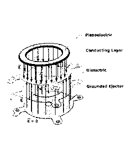
With reference to FIG. 6, an ejector mechanism 1601 may comprise a distal
piezoelectric actuator 1604, separated from a proximal ejector plate 1602
first by a
conducting layer 1660 and then by a dielectric layer 1662. In an aspect, the
distal side of
CA 02870181 2014-10-09
WO 2013/155201
PCMJS2013/036002
22
piezoelectric actuator 1604 is bounded on one side by metallization and on the
proximal side
by a conducting layer 1660. In an aspect, distal side of the dielectric layer
1662 is located
proximal to the conducting layer 1660 and dielectric layer 1662 both separates
and charge
isolates ejector plate 1602. The proximal side of ejector plate 1602 contacts
the fluid 1610
and is in fluid communication with the reservoir 1620. The distal side of
ejector plate 1602 is
in contact with the proximal side of dielectric layer 1662. In an aspect, the
ejector plate 1602
may be grounded and electrically isolated from the conducting layer 1660 by
the dielectric
layer 1662. In an aspect, ejector mechanism 1601 provides two separate
terminals for
electrically driving the piezoelectric, electrode 1606a on surface 1634 (e.g.,
distal surface)
and electrode 1606b on surface 1636 (e.g., proximal surface). In an aspect,
dielectric layer
1662 separating conducting layer 1662 provides for a charge isolated ejector
plate 1602. In a
further aspect, the charge isolated ejector plate 1602 may be grounded.
In an aspect according the present disclosure the dielectric layer 1662 may
comprise a
plastic, glass, porcelain, etc. The dielectric layer may be any suitable size
and shape to
accommodate the piezoelectric element and ejector surface (exemplary
dimensions are shown
in FIG. 8), but the disclosure is not so limited), but not so as to impede
droplet generation and
ejection. In certain aspects, the dielectric layer may range in thickness from
10 ttm to 30 ttm,
12 gm to 25 gm, 15 gm to 25 gm, etc. In preferred configurations, the
dielectric layer is
concentric in shape with the piezoelectric element and/or ejector surface, and
its thickness is
minimized to reduce stiffness of the charge isolated ejector mechanism.
In an aspect, the dielectric layer may be a plastic selected from polystyrene,
polyvinyl
chloride, or nylon. In an aspect, the dielectric layer may be selected from
the group
consisting of polyester (PES), polyethylene terephthalate (PET), polyethylene
(PE), high-
density polyethylene (HDPE), polyvinyl chloride (PVC), polyvinylidene chloride
(PVDC).
low-density polyethylene (LDPE), polypropylene (PP), polystyrene (PS), pigh
impact
polystyrene (HIPS), polyamides (PA)(e.g., nylon), acrylonitrile butadiene
styrene (ABS),
polycarbonate (PC), polycarbonate/acrylonitrile butadiene styrene (PC/ABS),
polyurethanes
(PU), melamine formaldehyde (MF), plastarch material, phenolics (PF),
polyetheretherketone
(PEEK), polyetherimide (PEI) (Ultem), polylactic acid (PLA), polymethyl
methacrylate
(PMMA), polytetrafluoroethylene (PTFE), or urea-formaldehyde (UF).
In an aspect according the present disclosure the conducting layer 1660 may
comprise
a metal, graphite, or a polymer. The conducting layer may be any suitable size
and shape to
accommodate the piezoelectric element and ejector surface, but the disclosure
is not so
limited), but not so as to impede droplet generation and ejection. In certain
aspects, the
CA 02870181 2014-10-09
WO 2013/155201
PCMJS2013/036002
23
conducting layer may range in thickness from 10 m to 30 lam, 12 lam to 25
[tm, 15 itim to 25
tim, etc. In preferred configurations, the conducting layer is concentric in
shape with the
piezoelectric element and/or ejector surface, and its thickness is minimized
to reduce stiffness
of the charge isolated ejector mechanism.
In an aspect the conducting layer 1660 may be copper, aluminum, silver, or
gold. In
an aspect, the polymer may be a melanin. In another aspect, the polymer may be
a
poly(fluorene), a polyphenylene, a polypyrene, a polyazulene, a
polynaphthalene, a
poly(pyrrole)(PPY), a polycarbazole, a polyindole, polyazepine, a polyaniline
(PAM), a
poly(thiophene) (PT), a poly(3,4-ethylenedioxythiophene) (PEDOT), a poly(p-
phenylene
sulfide) (PPS), a poly(acetylene) (PAC), or a poly(p-phenylene vinylene)
(PPV).
In aspects of the disclosure, at least one or more layers of the charge
isolated ejector
mechanism may be configured as a flexible printed circuit (FTC), e.g., two
signal layers and a
ground layer. Operation of the ejection mechanism may generally be impacted by
stiffness of
the materials of construction. Stiffness is generally impacted by the use of
adhesives, their
rigidity, and their thickness. As such, in certain aspects, construction and
configuration of the
charge isolated ejector mechanism optimized to improve performance in this
regard. In
certain embodiments, the FPC layers may then be coupled to the remaining
layers of the
charge isolated ejector mechanism, e.g., an ejector surface, piezoelectric
element, etc.
The layers of the charge isolated ejector mechanism configured as a FPC can be
designed and fabricated in any suitable manner. In certain aspects, the FPC
may be
configured so as minimize its thickness. In this regard, adhesiveless
construction may be
preferred, but the disclosure is not so limited. In certain embodiments, the
PVC layers may
comprise the dielectric layer and the conducting layer of the charge isolated
ejector
mechanism, as well as bonding layers (e.g., adhesiveless bonding layers),
flying lead
connections, etc. to aid in fabrication and assembly, as explained in further
detail herein.
With reference to FIGS. 7A-7B, exemplary conducting layer/dielectric layer
designs
are shown. As shown in FIGS. 7A-7B, the core of the circuit is built from dual
copper clad
polyimide laminate (adhesiveless). The polyimide may be punched or drilled
out, along with
any copper under it. A photoresist coating may be applied and imaged to allow
patterning of
the copper on both sides. Finally, an LPI (liquid photo-imageable) soldermask
or polyimide
cover coating may be applied to provide electrical protection of the top
copper and bottom.
In certain embodiments, the LPI may be selected from a crosslinked photoresist
used to
provide electrical isolation without adhesive in a very thin layer. Electrical
connection to the
piezoelectric may be ensured by mixing epoxy with 5% nickel powder to give
anisotropic
CA 02870181 2014-10-09
WO 2013/155201
PCMJS2013/036002
24
conduction between the copper and piezoelectric (does not conduct sideways,
only up and
down). The top of the piezoelectric may be connected to the outside top copper
ring in any
suitable manner, e.g., run down tin/solder (large solder drop that cools as it
runs down side of
piezoelectric, connecting electrode on FPC and top of piezoelectric), by
metallized epoxy
applied from the top of the piezoelectric down the side and onto the FPC
electrode, etc.
As shown, certain embodiments retain an additional under metal layer (FIG 7B),
and
certain embodiments etch away and remove the under layer of metal (FIG 7A)
that is
electrically floating between the dielectric and the ejector surface. Without
intending to be
limited by theory, this under metal layer acts to keep the FPC flat, to
thereby assist in
piezoelectric bonding, and to allow metal to metal bonding (rather than
polymer to metal
bonding). However, the addition of a metal layer adds to the stiffness of the
FPC. As such,
design parameters may be selected depending on the desired end use of the FPC
and the
charge isolated ejector mechanism.
FIG. 8 shows an exploded top view of the embodiment of FIG 5B, illustrating
exemplary configurations of the layers, which may conform to an exemplary
piezoelectric
element/ejector surface shape, and may include floating leads to aid in
bonding.
With reference to FIGS. 9A-9B, an FPC can be made in a similar manner with the
same general configuration, and bonded to stainless steel (e.g., DLC (diamond
like carbon)
coated SS316L), gold, or other suitable ejector surface. Exemplary perfoimance
curves of
fabricated devices are shown in FIG 10.
An alterntive FPC configuration utilizing adhesives is illustrated in FIGS. 9A
and 9B.
With reference to FIGS. 11A and 11B, a multilayer FPC bonded to
copper/PEEK/copper
ejector surfae with and without flying lead connections (i.e., floating metal
leads that connect
to the top of the piezoelectric) is shown. In alternative embodiments (not
shown), this type of
FPC can also be bonded to S5316L, in which case Layer 2 copper may optionally
not be
included.
Any suitable manner for bonding together of an FPC and an ejector surface may
be
used. In one embodiment, the bonding together a flexible printed circuit with
an ejector
surface (e.g., ejector plate coupled to a generator plate) may be achieved,
e.g., through
surface treatment (roughening through plasma etch, wet etch, mechanical
sanding, etc.) and
heat pressing past the plastic glass transition temperature at a high
compression (typical
values 750F/350psi polyimide, 350F/350 psi PEEK, etc.), by applying a thin
sheet of
adhesive which is cured under heat and pressure specific to the adhesive, or
other bonding.
CA 02870181 2014-10-09
WO 2013/155201
PCMJS2013/036002
By way of example, FIG 12 illustrates an exemplary process for generating a
charge
isolated ejector mechanism from a FPC and a copper/PEEK/copper ejector
surface.
Generator plate openings may be laser micro-machined out of the PEEK after
device
fabrication (all photolithography and etching steps). Likewise, FIG. 11
illustrates a general
5 process of bonding an ejector plate (passivated stainless steel sheet) to
an FPC. The
generator plate (active ejector mesh containing ejector openings) may be
subsequently
bonded using flexible medical adhesive (flexible glue may be preferred for
active area to
allow full moding and good ejection). The FPC may also be punched out and
bonded to an
ejector surface (e.g., stainless steel annuli or PEEK annuli) that are pre-
punched, EDM,
10 etched, laser machined, or otherwise fabricated.
In an alternative embodiment, each of the layers of the FPC may be separately
cut,
e.g., using laser, EDM, etched, punching, or other suitable technique, and
then each aligned
and bonded separately together with adhesive.
The present disclosure provides for and includes methods for generating
droplets of a
15 fluid using a charge isolated ejector surface. In some implementations,
the method includes
applying a voltage to a piezoelectric actuator operable to oscillate an
ejector surface so as to
generate droplets of a fluid. In an aspect, the ejector area may have a charge
isolated,
grounded ejector surface comprising a generator plate. In certain aspects, the
charge isolated
ejector surface may include an ejector plate coupled to a generator plate. In
some further
20 aspects, the charge isolated ejector surface may be grounded. In an
aspect, the ejector
surface, e.g., comprising an ejector plate and/or generator plate may be
coated with an inert
material.
Many implementations of the inventions have been disclosed. This disclosure
contemplates combining any of the features of one implementation with the
features of one or
25 more of the other implementations. For example, any of the ejector
mechanisms or reservoirs
can be used in combination with any of the disclosed housings or housing
features, e.g.,
covers, supports, rests, lights, seals and gaskets, fill mechanisms, or
alignment mechanisms.
Further variations on any of the elements of the inventions disclosed and
within the scope of
ordinary skill are contemplated by this disclosure. Such variations include
selection of
materials, coatings, or methods of manufacturing.
Any of the electrical and electronic technology can be used with any of the
implementations without limitation. Furthermore, any networking, remote
access, subject
monitoring, e-health, data storage, data mining, or internet functionality is
applicable to any
and all of the implementations and can be practiced therewith. Further still,
additional
CA 02870181 2014-10-09
WO 2013/155201
PCMJS2013/036002
26
diagnostic functions, such as performance of tests or measurements of
physiological
parameters may be incorporated into the functionality of any of the
implementations.
Perfotmance of glaucoma or other ocular tests can be performed by the devices
as a part of
their diagnostic functionality. Other methods of fabrication known in the art
and not
explicitly listed here can he used to fabricate, test, repair, or maintain the
device.
Furthermore, the device may include more sophisticated imaging or alignment
mechanisms.
For example, the device or base may be equipped with or coupled to an iris or
retina scanner
to create a unique identification to match a device to the user, and to
delineate between eyes.
Alternatively, the device or base may be coupled to or include sophisticated
imaging devices
for any suitable type of photography or radiology.
To assist in understanding the present inventions, the following examples are
included. The experiments described herein should not, of course, be construed
as
specifically limiting the invention and such variations of the invention, now
known or later
developed, which would be within the purview of one skilled in the art are
considered to fall
within the scope of the invention as described herein and hereinafter claimed.
EXAMPLES
EXAMPLE 1
This example relates to a clinical dilation study using an ejector device of
the
disclosure on human subjects. This example demonstrates that the delivery of a
low dosage
.. volume medicament composition as a directed stream of droplets via an
ejector device of the
disclosure provides equivalent dilation to that of a standard eye dropper
using only 1/4 of the
dosage volume of the eye dropper.
MATERIALS AND METHODS
In the first arm of the study, thirty-five subjects are dosed with 2 x 3 1.1.1
doses of
phenylephrine 2.5% and 2 x 3 l doses of tropicamide 1% from an ejector device
in one eye
(3 minutes apart), and 1 x drop (¨ 26 p1) of phenylephrine 2.5% and 1 x drop
(¨ 26 1) of
tropicamide 1% from a standard eyedropper in the fellow eye. In the second arm
of the
study, thirty-three subjects are dosed with 1 x 6 pl dose of phenylephrine
2.5% and 1 x 6 pl
dose of tropicamide 1% from an ejector device in one eye, and 1 x drop of
phenylephrine
.. 2.5% and 1 x drop of tropicamide 1% from an eyedropper in the fellow eye.
In the third arm
of the study, thirty-four subjects are dosed with 1 x 1.5 pl dose of
phenylephrine 2.5% and 1
x 1.5 .1 dose of tropicamide 1% from an ejector device in one eye, and 1 x
drop of
phenylephrine 2.5% and 1 x drop of tropicamide 1% from an eyedropper in the
fellow eye.
CA 02870181 2014-10-09
WO 2013/155201
PCMJS2013/036002
27
The effectiveness of the three different dosages of 1 x 1.5 [11, or 1 x 6
iLtl, or 2 x 3 p.1 of
phenylephrine 2.5% delivered by the ejector device versus one drop of
phenylephrine 2.5%
from the eyedropper, together with lx 1.5 pi, or lx 6 iLtl, or 2 x 3 pi of
tropicamide 1%
delivered by the ejector device versus one drop of tropic amide 1% from the
eyedropper, are
assessed by measuring the percentage increase in dilation of the subject's
pupils at 10
minutes, 20 minutes, and 60 minutes after administering the dosage(s) relative
to the pre-
treatment baseline.
RESULTS
FIG. 15A illustrates the mean percentage change in dilation measured from the
pre-
treatment baseline for delivery via a spray ejector device of the disclosure,
as compared to
traditional eyedropper administration.. FIG. 15B illustrates the mean
percentage difference in
dilation relative to the pre-dosage dilation baseline for the spray ejector
device as compared
to the eyedropper. Differences are calculated on a subject-specific basis and
are then
averaged.
DISCUSSION
FIG. 15A indicates that the dilation drugs delivered by both the ejector
device (e.g., a
direct stream of droplets of a low dosage volume medicament composition) and
the standard
eyedropper materially dilated subjects' eyes, and that the average degree of
dilation increases
monotonically as more time passed after administering the dosage, up to the
maximum post-
dosage measurement point of 60 minutes.
FIG. 15B indicates that while the 1 x 1.5 il dosage from the ejector device
did not
achieve a statistically equivalent degree of dilation as the 1 x drop from the
eyedropper (2-
tailed p-values on equivalence <0.001), all the 1 x 6 iLt1 dosages and two of
the three 2 x 3111
dosages from the ejector device did achieve a statistically equivalent degree
of dilation as the
eyedropper (2-tailed p-values for 1 x 6 ill are all > 0.20; 2-tailed p-values
for 2 x 3 i.t1 at 10
minutes and 60 minutes are 0.17 and 0.10, respectively). Moreover, for the 2 x
3 pl dosage it
was the case that 20 minutes after dosing, the ejector device achieved a
statistically
significantly higher mean dilation than did the eyedropper (2-tailed p-value =
0.05).
EXAMPLE 2
This example relates to a clinical study using glaucomatous beagles to
evaluate the
effects of latanoprost on intraocular pressure (I0P). More particularly, this
study evaluates
the reduction in IOP and pupil diameter (PD) following once-daily instillation
of 1.5 Ill of
CA 02870181 2014-10-09
WO 2013/155201
PCMJS2013/036002
28
0.005% latanoprost via a spray ejector device of the disclosure, as compared
to an untreated
eye.
MATERIALS AND METHODS
One glaucomatous beagle dog (female, aged 3 years) from the University of
Florida
MacKay Colony of Glaucomatous Beagles was chosen for this study. The animal
was rested
a minimum of one week before starting the study. Prior to the commencement of
the study,
the animal was assigned one eye to receive 1.5 ul of 0.005% latanoprost and
the contralateral
eye to remain untreated as a control. The basic measurement protocol was
followed at all
times and performed by the same operator. PD was measured via Jameson Caliper
(mm
horizontally). IOP was measured using a TonoVet instrument with clean probe
(iCare).
IOP and PD were measured at time 0, 1, 2, 4, 7 and 18 hours daily for two
days. Immediately
after the time 0 measurement, the animal received 1.5 ul of 0.005% latanoprost
in its assigned
eye with a spray device. The contralateral eye was left untreated. The spray
device was
checked for accuracy before and after every use, and required a 10% delivered
dose accuracy
in pretreatment. Standard measurements continued throughout each day at time
1, 2, 4, 7 and
18 hours. Possible endpoints included, but were not limited to, excessive
ocular irritation,
ocular damage, and other illness or injury. No statistical crossover was
utilized for this study.
RESULTS
IOP in the treated eye was reduced to 11 mmHg, a maximum decrease of 16 mmHg
from the initial value, by hour 2 (FIG. 16A). PD was reduced to pinpoint (0)
by hour 1 and
remained there through hour 7 (FIG. 16B).
DISCUSSION
The 1.5 ul dose of 0.005% latanoprost delivered via the WhisperTM MDD device
was
demonstrated to be as effective at lowering IOP and constricting the pupil as
traditional
therapy. This study confirms that the spray ejector device is capable of
delivering a 1.5 [1,1
dose of latanoprost consistently.
EXAMPLE 3
This example relates to a clinical study using glaucomatous beagles to
evaluate the
effects of latanoprost on intraocular pressure (10P). More particularly, this
study evaluates
the reduction in IOP and pupil diameter (PD) following once-daily installation
of 3.0 IA of
0.005% latanoprost using a spray ejector device of the disclosure, as compared
to an
untreated eye.
CA 02870181 2014-10-09
WO 2013/155201
PCT/ES2013/036002
29
MATERIALS AND METHODS
One glaucomatous beagle dog (female, aged 3 years) from the University of
Florida
MacKay Colony of Glaucomatous Beagles was chosen for this study. Prior to the
commencement of the study, the animal was assigned one eye to receive 3.0 1
of 0.005%
latanoprost and the contralateral eye to remain untreated as a control. The
basic measurement
protocol was followed at all times and performed by the same operator. PD was
measured
via Jameson Caliper (mm horizontally). IOP was measured using a TonoVet
instrument with
clean probe (iCare). IOP and PD were measured at time 0, 1, 2, 4, 7 and 18
hours daily for
two days. Immediately after the time 0 measurements, the animal was
administered 3.0 ttl of
0.005% latanoprost in its assigned eye using a spray ejector device. The
contralateral eye
was left untreated. The spray ejector device was checked for accuracy before
and after every
use, and required a 10% delivered dose accuracy in pretreatment. Standard
measurements
continued throughout each day at time 1, 2, 4, 7 and 18 hours. Possible
endpoints included,
but were not limited to, excessive ocular irritation, ocular damage, and other
illness or injury.
No statistical crossover was utilized for this study.
RESULTS
IOP in the treated eye was reduced to 19 mmHg, a decrease of 12 mmHg from the
initial value, by hour 2 (FIG. 17A). The maximum decrease was seen at hour 7
on day 2. PD
was reduced to pinpoint (0) by hour 1 and remained there throughout hour 7
(FIG. 17B).
DISCUSSION
Results indicate that, as with the 1.5 p1 dose, the 3.0 j.il dose of 0.005 %
latanoprost is
as effective at lowering IOP and constricting the pupil as traditional
eyedropper therapy.
This study confirms that the spray ejector device is capable of delivering a
3.0 p1 dose of
latanoprost consistently.
EXAMPLE 4
This example relates to a clinical study using glaucomatous beagles to
evaluate the
effects of latanoprost on intraocular pressure (I0P). More particularly, this
study evaluates
the effects on intraocular pressure (lOP) of once-daily installation of 9 j.il
of latanoprost via a
spray ejector device of the disclosure, as compared to an average of 26 1 of
latanoprost
delivered once daily by traditional eyedropper.
MATERIALS AND METHODS
Six glaucomatous beagle dogs (4 males and 2 females, aged 3-8 years) from the
University of Florida MacKay Colony of Glaucomatous Beagles were chosen for
this study.
CA 02870181 2014-10-09
WO 2013/155201
PCMJS2013/036002
Conditions for inclusion included elevated TOP and documented glaucomatous
disease state
as deteimined by gross eye exam by a boarded veterinary ophthalmologist.
All animals were rested for a minimum of one week before starting the trials.
IOP and
pupil diameter (PD) were measured 5 times (at 0, 1, 2, 4, and 7 hours) daily
for four days
5 (study days 1-4) to establish initial baseline levels. The basic
measurement protocol was
followed at all times and performed by the same operator. PD was measured via
Jameson
Caliper (mm horizontally). IOP was measured with a Tono Vet instrument with
clean probe
(iCare). After three rest days, one eye was randomly assigned to receive 9 jtl
of 0.005%
latanoprost delivered via a spray ejector device, and the contralateral eye
was administered
10 traditional latanoprost from a traditional eyedropper container (26-30
1).
On study days 5-9, IOP and PD measurements were taken at approximately 0900h
(time 0). Immediately after time 0 measurements, each animal received a once-
daily dose of 9
of generic 0.005% latanoprost in its assigned eye via the spray ejector
device. The
contralateral eye received a clinical dose (one drop) of the same commercially
available
15 topical drug from a traditional eyedropper (positive control). The spray
ejector device was
checked for accuracy before and after every use and required a 10% delivered
dose accuracy
in pretreatment. Standard measurements continued throughout each day at 0, 1,
2, 4, and 7
hours. Possible endpoints included, but were not limited to, excessive ocular
irritation, ocular
damage, and other illness or injury. No statistical crossover was utilized for
this study.
20 RESULTS
Results for the spray ejector device and generic eyedropper treatments were
similar.
The 9 1 dose was as effective at lowering IOP and constricting the pupil as
traditional
therapy. The duration of action of the eyedropper dose was greater, but the
spray ejector
device therapy maintained values significantly lower than baseline for IOP
(FIGS. 18A and
25 18B). Irritation in eyes treated using the spray ejector device was less
than that observed in
the eyedropper-treated eyes.
DISCUSSION
Administration of 9 jrl of 0.005% latanoprost in a single daily morning dose
via a
spray ejector device was as effective at controlling TOP as traditional
eyedropper therapy, at
30 approximately one-third the typical eyedropper dose over a seven-hour
study period for five
days. This pharmacodynamic (PD) study is the companion study to the
pharmacokinetic
(PK) study of Example 12, comparing the bioavailability of the acid of
latanoprost in the
aqueous humor following delivery of 9 !al of latanoprost via a spray ejector
device, as
compared to 26111 of latanoprost delivered via eyedropper.
CA 02870181 2014-10-09
WO 2013/155201
PCMJS2013/036002
31
EXAMPLE 5
This example relates to a clinical study using glaucomatous beagles to
evaluate the
effects of latanoprost on intraocular pressure (I0P). More particularly, the
study evaluates
the effectiveness of lower doses of latanoprost delivered via a spray ejector
device in
lowering 10P in a side-by-side, randomized, crossover study in six
glaucomatous beagles.
During the study, IPO of glaucomatous beagles is measured when latanoprost is
delivered in
two 6 IA doses using an ejector device of the disclosure (e.g., as a low
dosage volume
directed stream of droplets) versus when delivered as a single drop (¨ 24 IA)
from a standard
eyedropper.
MATERIALS AND METHODS
Six glaucomatous animals (4 males and 2 females aged 3-8 years) from the
University
of Florida (UF) MacKay glaucomatous Beagle colony are chosen for this study.
Conditions
for inclusion include elevated intraocular pressure (TOP) and documented
glaucomatous
disease state as determined by gross eye exam by a boarded veterinary
ophthalmologist.
All animals are rested a minimum of one week before starting the trials.
Intraocular
pressure (TOP), pupil diameter (PD), and heart rate (HR) are measured 6 times
(at 0, 0:30,
0:45, 1, 2, 7 hrs) daily for five days, for initial baseline levels (Study
days 1-5). The basic
measurement protocol is followed and performed by the operator. PD is measured
via
Jameson Caliper (mm horizontally). IOP is measured with a TonoVet instrument
with clean
probe (iCare). HR is monitored via palpation of the femoral vessels. After two
rest days, one
eye is randomly assigned to receive the drug delivered via the ejector device
and the
contralateral eye is given traditional latanoprost from an eye dropper
container.
Active drug delivery begins on study day eight. For five days, TOP and PD
measurements are taken at 0900. Immediately after 0900 measurements (time 0),
each
animal receives 6 ul of generic latanoprost in its assigned eye with the
ejector device,
followed by a second dosing 3 minutes later of an additional 61u1 (to avoid
overloading the
conjunctival sac). The contralateral eye receives a clinical dose (one drop)
of the same
commercially-available topical drug from FDA approved container (positive
control). The
ejector device is checked for accuracy before and after every use (Table 1).
Standard
measurements continue throughout each day (at 0:30, 0:45, 1, 2, 7 hrs).
Possible endpoints
include, but are not limited to, excessive ocular irritation, ocular damage,
or other illness and
injury.
CA 02870181 2014-10-09
WO 2013/155201
PCT/1JS2013/036002
32
Table 1: Calibration measurements for exemplary sample
Pre-Spray Post-Spray Pre-Spray Post-Spray Standard Percent
#1 #1 #2 #2 Average Deviation Variance
6.8 6 5.9 6 6.175 0.419 6.79%
6.6 5.5 6.9 6 6.25 0.624 9.99%
6.8 6.4 6.7 6.5 6.6 0.182 2.77%
6.7 6.5 6 6.1 6.325 0.330 5.22%
7.7 6 6.4 6.6 6.55 0.500 7.63%
6.9 6 6.1 6.1 6.275 0.419 6.68%
* Units in ul
After 7 days of rest, starting on study day 22, the treatments are repeated
with the only
change being the use of opposite eyes for statistical control. All initially
assigned drug eyes
are now used as positive controls and receive a clinical dose (one drop) of
the same
commercially-available topical drug from FDA approved eye dropper container.
The
contralateral eyes now receive 6 il of generic latanoprost in its assigned
eye, followed by a
second dosing 3 minutes later of an additional 6 il, for a total of 12 pi of
latanoprost.
RESULTS
FIG. 19A-19C illustrate that the ejector device results generally align with
those
obtained using a conventional eyedropper. IOP pressure over the course of the
study is
shown in FIG. 19A, the change in IOP over the course of the study is shown in
FIG. 19B, and
pupil diameter over the course of the study is shown in FIG. 19C.
Both the ejector device (e. g. , a direct stream of droplets of a low dosage
volume
medicament composition) and traditional eyedropper latanoprost treatments
reduced TOP and
caused iridal miosis. The ejector device changes in IOP became significantly
different from
baseline at timepoint 0:45 on day one. The maximum change in TOP during the
first 7 hours
was -34.8 mmHg (69%). The traditional treatment showed significance in IOP at
timepoint
0:45 as well. The maximum change in TOP during the first 7 hours for the
traditional
treatment was -38.0 mmHg (72%). There were no significant differences between
the ejector
device treatments and the traditional treatments for IOP.
Significant pupil diameter changes were seen with the ejector device treatment
at
timepoint 0:30 on day one. The iris reached pinpoint status (maximum miosis)
at 1 hour, and
remained there for the remainder of the seven hours of monitoring. Significant
pupil
diameter changes were seen with the traditional latanoprost treatment at
timepoint 0:30. The
iris reached pinpoint status at timepoint 1 hour, and remained there for the
remainder of the
seven hours of monitoring.
CA 02870181 2014-10-09
WO 2013/155201
PCMJS2013/036002
33
There were no changes in heart rate throughout the study with average values
around
25 beats/15 seconds (100 bpm). There were no reports of excessive eye
irritation or other eye
issues. Some conjunctival irritation was noted in the traditional treatment
eyes, related to
prostaglandin use.
DISCUSSION
In summary, 0.005% latanoprost at low dosage volume significantly lowers 10P
in the
glaucomatous beagle when instilled once daily, commensurate with standard
eyedropper.
The reduction in IOP ranged from about 20 mm Hg (45%) to 27 mm Hg (60%). The
results
show that the reduction in canine IOP achieved by an ejector device of the
disclosure (e.g., as
a low dosage volume directed stream of droplets) is statistically
indistinguishable from that of
the dosage volume of a standard eyedropper, even though the two 6 ill doses
delivered via the
ejector device of the disclosure is approximately 1/2 of the volume delivered
via the standard
eyedropper.
EXAMPLE 6
This example relates to a clinical study using glaucomatous beagles to
evaluate the
effects of latanoprost on intraocular pressure (I0P). More particularly, the
study evaluates
the effects on intraocular pressure (I0P) of once-daily instillation of 301.11
of 0.005%
latanoprost via a spray ejector device of the disclosure, as compared to once-
daily instillation
of approximately 26-30 IA by traditional eyedropper.
MATERIALS AND METHODS
Two glaucomatous beagle dogs (2 females, aged 3-8 years) from the University
of
Florida MacKay Colony of Glaucomatous Beagles were chosen for this study. Both
animals
were rested a minimum of one week before starting the trials. Prior to the
commencement of
the study, each animal was assigned one eye to receive either 30 IA of 0.005%
latanoprost
delivered by a spray ejector device or one eye drop of 0.005% latanoprost. The
basic
measurement protocol was followed at all times and performed by the same
operator. IOP
was measured at time 0, 1, 2, 4, and 7 hours daily for two days using a
TonoVet instrument
with clean probe (iCare). Immediately after the time 0 measurements, each
animal was
administered 30 !al of 0.005% latanoprost in its assigned eye using the spray
ejector device.
The contralateral eye received a clinical dose (one drop) of the same
commercially available
topical drug from a traditional eyedropper (positive control). The spray
ejector device was
checked for accuracy before and after every use, and required a 10% delivered
dose accuracy
in pretreatment. Standard measurements continued throughout each day at time
1, 2. 4, and 7
CA 02870181 2014-10-09
WO 2013/155201
PCMJS2013/036002
34
hours. Possible endpoints included, but were not limited to, excessive ocular
irritation, ocular
damage, and other illness or injury. No statistical crossover was utilized for
this study.
RESULTS
Reduction in IOP for animals treated with the spray ejector device and with
the
eyedropper were similar (FIG. 20). The 30 lid dose delivered via the spray
ejector device was
as effective at lowering IOP and constricting the pupil as the traditional
eyedropper therapy.
DISCUSSION
Results confiim that the spray ejector device is capable of delivering a
301.11 dose of
0.005% latanoprost. In this pilot study, drug delivery via the spray ejector
device was
demonstrated to be as effective in controlling IOP as traditional eyedropper
therapy, with a
trend toward increased duration of action. This study completes the spectrum
of the effective
dosage range for the delivery of prostaglandin pro-drugs, which is 1.5 1.11 to
30 1.11 in this series
of studies.
EXAMPLE 7
This example relates to a randomized cross-over clinical study using
glaucomatous
beagles to evaluate the effects of travoprost on intraocular pressure (I0P).
More particularly,
the study compares the effectiveness of a 9 Ill dose of 0.004% travoprost
(Travatan ZO,
Alcon Laboratories, Fort Worth, TX USA) delivered by a spray ejector device of
the
disclosure, as compared to delivery by micropipette in suppressing and
flattening the 24-hour
intraocular pressure (UR) dose/response curve and controlling the morning
intraocular
pressure (I0P) spikes. This study also compares delivery effectiveness of the
spray ejector
device, as compared to standard micropipette by monitoring IOP and ocular
irritation.
Prior to the development of the spray ejector devices and methods of the
disclosure,
investigational and commercially available prostaglandin agonists have been
delivered by
eyedropper in volumes of approximately 26 pl. Patients who are intolerant to
preservatives
or who have concomitant ocular surface diseases (OSD) have been treated with
single-dose
pipettes using eyedropper-equivalent doses of pharmacy-compounded preservative-
free
drugs. This method is fraught with safety and ease-of-use issues. Recently,
single-dose
.. delivery of eyedropper-equivalent doses of preservative-free, IOP-lowering
drugs has been
marketed in sterile blow-fill pipettes. Eye injury and usability issues,
however, remain.
For drugs delivered in low volume doses by a spray ejector device of the
disclosure,
the exposure to preservatives per dose may be reduced, and preservative-
induced ocular
irritation is further reduced by once-daily dosing.
CA 02870181 2014-10-09
WO 2013/155201
PCT/1TS2013/036002
MATERIALS AND METHODS
Six glaucomatous beagles (3 males and 3 females, aged 3-8 years) from the
University of Florida MacKay Colony of Glaucomatous Beagles were chosen for
this study.
Conditions for inclusion included elevated TOP and documented glaucomatous
disease state
5 as determined by gross eye exam by a boarded veterinary ophthalmologist.
All animals were rested a minimum of one week before starting the trials.
Intraocular
pressure (I0P), pupil diameter (PD), and heart rate (HR) were measured daily
at time 0, 12,
13, 14, 16, and 19 hours for five days starting at approximately 2100h to
establish initial
baseline levels (study days 1-5). The basic measurement protocol was followed
at all times
10 and performed by the same trained operator. PD was measured via Jameson
Caliper (mm
horizontally). IOP was measured using a TonoVet instrument with clean probe
(iCare). HR
was monitored via palpation of the femoral vessels. After two rest days, one
eye of each
animal was randomly assigned to receive 9 pl of 0.004% travoprost delivered
via a spray
ejector device, and the contralateral eye was assigned treatment with an
identical dose
15 delivered via micropipette.
Active drug delivery began on study day 8. For five days, IOP and PD
measurements
were taken at approximately 2100h. Immediately after this measurements (time
0), each
animal received 9.0 0.9 IA of 0.004% travoprost (Travatan Z , Alcon
Laboratories, Fort
Worth, TX USA) in its assigned eye via the spray ejector device. The
contralateral eye
20 received an identical dose (9.0 ul) of the same drug (positive control)
administered via
micropipette. The spray ejector device and micropipette were checked for
accuracy before
and after every use. Standard measurements continued throughout each day at
12, 13, 14, 16,
19 hours. Possible endpoints included, but were not limited to, excessive
ocular irritation,
ocular damage, and other illness or injury.
25 After 7 days of rest, starting on study day 22 the treatments were
repeated as above
except using opposite eyes for statistical control. All initially assigned
drug eyes were now
used as positive controls and received 9 IA of 0.004% travoprost via
micropipette. The
contralateral eyes received 9 ul of 0.004% travoprost via the spray ejector
device.
RESULTS
30 The spray ejector device caused significant changes in IOP from baseline
at time 12
hours on study day 1. The maximum change in IOP during the first 24 hours was
¨20.0
mmHg (59%). The initial average TOP level on day 1 was 33.75 mmHg; after 5
days of
treatment, the highest average daily peak observed was 18.83 mmHg, well below
similar
baseline levels of 30.83 mmHg (FIG. 21A). Significant PD changes were seen
with the spray
CA 02870181 2014-10-09
WO 2013/155201
PCMJS2013/036002
36
ejector device treatment at time 12 hours on study day 1. The iris reached
maximum miosis
(1.17 mm) at time 12 hours and began rising to baseline levels over the
remaining hours of
monitoring (FIG. 21B). There were no changes in HR throughout the study, with
values
averaging 25 beats/15 seconds (100 bpm). There were no reports of excessive
eye irritation
or other eye issues.
Both the spray ejector device and the micropipette functioned well. There were
no
statistically significant differences between the treatments throughout the
study (p = 0.7546).
This demonstrates the effectiveness of travoprost at low volume doses,
regardless of delivery
method. However, issues relating to delivery methods should be noted. Both
treatment
methods showed similar levels of overall irritation.
DISCUSSION
Travoprost is the most effective of the prostaglandin agonists studied for
lowering of
IOP over a 24 hour period day after day including excellent control of morning
IOP spike
often seen with treatment with prostaglandin pro-drugs.
EXAMPLE 8
This example relates to a randomized cross-over clinical study using
glaucomatous
beagles to evaluate the effects of travoprost on intraocular pressure (I0P).
More particularly,
the study evaluates the effects on intraocular pressure (TOP) following once
daily, morning or
evening, instillations of 18 Ill of 0.004% travoprost by a spray ejector
device of the
disclosure, as compared to an average of 26 [1.1 delivered once daily by
traditional eyedropper.
MATERIALS AND METHODS
Six glaucomatous beagle dogs (4 males and 2 females, aged 3-8 years) from the
University of Florida MacKay Colony of Glaucomatous Beagles were chosen for
this study.
Conditions for inclusion included elevated intraocular pressure (I0P) and
documented
glaucomatous disease state as determined by gross eye exam by a boarded
veterinary
ophthalmologist.
All animals were rested a minimum of one week before starting the trials. IOP
and
pupil diameter (PD) were measured at time 0, 1, 2, 4, 7, and 12 hours daily
for four days
(study days 1-4) to establish initial baseline levels. The basic measurement
protocol was
followed at all times and performed by the same operator. PD was measured via
Jameson
Caliper (mm horizontally). IOP was measured using a Tono Vet instrument with
clean probe
(iCare). After three rest days, one eye was randomly assigned to receive 18
[11 travoprost
CA 02870181 2014-10-09
WO 2013/155201
PCMJS2013/036002
37
delivered via a spray ejector device in two 9 jil doses, and the contralateral
eye was
administered a dose of travoprost (average 26 pl) from a traditional
eyedropper container.
On study days 5 and 6, IOP and PD measurements were taken at approximately
0900h. Immediately after this measurements (time 0), each animal was
administered 18 il of
travoprost in its assigned eye via the spray ejector device. The contralateral
eye received a
clinical dose (one drop) of the same commercially available topical drug from
a traditional
eyedropper (positive control). The spray ejector device was checked for
accuracy before and
after every use and required a 10% delivered dose accuracy in pretreatment.
Standard
measurements continued throughout each day at time 0. 1, 2, 4, 7, and 12
hours.
On study days 7-9, immediately after the time 12 hour (approximately 2100h)
measurements, each animal was administered 18 ill of travoprost in its
assigned eye via the
spray ejector device. The contralateral eye received a clinical dose (one
drop) of the same
commercially available topical drug from a traditional eyedropper (positive
control). The
spray ejector device was checked for accuracy before and after every use and
required a 10%
delivered dose accuracy in pretreatment. Standard measurements continued
throughout each
day at time 0, 1, 2, 4, 7, and 12 hours. Possible endpoints included, but were
not limited to,
excessive ocular irritation, ocular damage, and other illness or injury. No
statistical crossover
was utilized for this study.
RESULTS
Results for the spray ejector device and traditional eyedropper were similar.
The 18
dosage delivered by the spray ejector device was as effective at lowering IOP
and
constricting the pupil as the traditional therapy. The difference in duration
of IOP lowering
following drug delivery with the spray ejector device, as compared with the
eyedropper was
not significant. Both treatments maintained IOP values much lower than
baseline for a full
.. 24 hours throughout the duration of the study (FIGS. 22A-22D). Irritation
in eyes treated
using the spray ejector device was less than that observed in the eyedropper-
treated eyes.
DISCUSSION
The delivery of an 18 1.11 morning or evening dose of travoprost via a spray
ejector
device is as effective at controlling IOP as traditional therapy, even at
approximately two-
thirds of the average eyedropper-delivered dose (26 gl). Post-treatment IOP
remained
consistently below 20 mmHg. Based upon mathematical models, including the
effect of
diurnal variation in IOP and prior studies in this series, once-daily evening
dosing of
travoprost should provide the best 24- hour lowering of TOP. The noted lack of
ocular
irritation with the spray ejector device indicate its use in prostaglandin pro-
drug¨sensitive
CA 02870181 2014-10-09
WO 2013/155201
PCMJS2013/036002
38
patients, patients with concomitant ocular disease, and patients with severe
glaucoma
requiring optimal control.
EXAMPLE 9
This example relates to a clinical study using glaucomatous beagles to
evaluate the
effects of bimatoprost on intraocular pressure (10P). More particularly, the
study evaluates
the effects on intraocular pressure (I0P) and pupil diameter (PD) of once-
daily morning
instillations of 6 pl of 0.03% bimatoprost (Lumigan0; Allergan, Irvine, CA
USA) by a spray
ejector device of the disclosure, as compared to delivery once daily of an
average of 26 p1 by
traditional eyedropper.
MATERIALS AND METHODS
Six glaucomatous beagle dogs (4 males and 2 females, aged 3-8 years) from the
University of Florida MacKay Colony of Glaucomatous Beagles were chosen for
this study.
Conditions for inclusion included elevated TOP and documented glaucomatous
disease state
as detemiined by gross eye exam by a boarded veterinary ophthalmologist.
All animals were rested a minimum of one week before starting the trials. IOP
and PD
were measured at time 0, 1, 2, 4, and 7 hours daily for four days (study days
1-4) to establish
initial baseline levels. The basic measurement protocol was followed at all
times and
performed by the same operator. PD was measured via Jameson Caliper (mm
horizontally).
IOP was measured using a Tono Vet instrument with clean probe (iCare). After
three rest
days, one eye was randomly assigned to receive 6 p1 of 0.03% bimatoprost
delivered via a
spray ejector device, and the contralateral eye was assigned to receive
traditional bimatoprost
from a traditional eyedropper container (26-30 1).
On study days 5-9, IOP and PD measurements were taken at approximately 0900h
(time 0). Immediately after these measurements, each animal received 6 p1 of
0.03%
bimatoprost in its assigned eye via the spray ejector device. The
contralateral eye received a
clinical dose (one drop) of the same commercially available topical drug from
a traditional
eyedropper (positive control). The spray ejector device was checked for
accuracy before and
after every use and required a 10% delivered dose accuracy in pretreatment.
Standard
measurements continued throughout each day at 0, 1, 2, 4, and 7 hours.
Possible endpoints
included, but were not limited to, excessive ocular irritation, ocular damage,
and other illness
or injury. No statistical crossover was utilized for this study.
RESULTS
CA 02870181 2014-10-09
WO 2013/155201
PCMJS2013/036002
39
Results for the spray ejector device and the traditional eyedropper were
similar. The
6 ul dose was as effective at lowering IOP and constricting the pupil as
traditional therapy.
The duration of the dose administered by eyedropper was greater, but the spray
ejector device
therapy maintained IOP values that were lower than baseline (FIGS. 23A-23B).
Markedly
less irritation was observed in eyes treated using the spray ejector device,
as compared to the
eyes receiving the traditional eye drop treatment
DISCUSSION
The once-daily morning delivery of 6 ul of 0.3% bimatoprost via a spray
ejector
device is as effective at controlling IOP as is the traditional therapy, even
at less than one-
fourth of the average eyedropper dose of 26 1. Lack of ocular irritation was
noted during the
study.
EXAMPLE 10
This example relates to a clinical study using glaucomatous beagles to
evaluate the
reduction in IOP achieved by low dosage volume medicament compositions
comprising
modified active agent concentrations, as compared to standard eyedroppers.
More
particularly, this study evaluates the effectiveness of lower-volume, higher-
concentration
latanoprost delivered via a spray ejector device in lowering IOP in a side-by-
side, randomized
crossover study in six glaucomatous beagles.
MATERIALS AND METHODS
Six Beagle dogs with differing levels of hereditary glaucoma were used. All
animals
were from an existing colony of glaucomatous Beagles housed at the University
of Florida. 4
males and 2 females aged three to nine years were selected for this study. All
dogs weighed
at least 5 kg. No acclimation or quarantine was necessary. Animals were
identified by
tattoo, microchip and markings.
Dogs were examined to ensure that they were healthy before placement on study.
Dogs were housed in indoor runs large enough to exempt them from exercise
requirements.
Animals were exposed to natural environmental elements, such as temperature
and humidity.
Housing and sanitation were performed according to University of Florida
Animal Care
Services (UF ACS) protocols.
Dogs were provided a laboratory canine diet (Teklad Global 21% Protein Dog
Diet).
Diet certification and analysis were provided by the vendor, Harlan Teklad.
Dogs were
provided tap water ad libitum. No contaminants were known to exist in the
water and no
analysis outside that provided by the local water district and as specified in
UF ACS
CA 02870181 2014-10-09
WO 2013/155201
PCMJS2013/036002
protocols was performed. Environmental parameters (temperature and humidity)
were
measured daily.
Ophthalmic examinations (slit lamp with fluorescein and indirect
ophthalmoscopy)
were performed on the eyes of each animal prior to Study Day 1. Ocular
findings were
5 scored according to the McDonald-Shadduck Score System. Ocular findings
were recorded
using a standardized data collection sheet. Macroscopic observations were
recorded in
accordance with the Draize Scale for Scoring Ocular Lesions. Animals were
assigned
treatments based on basic randomization prior to study day one.
A 6.0 0.6 pl 5X concentration (0.025%) of latanoprost, formulated by Westlab
10 Pharmacy, was instilled in one randomized eye of each animal using a
spray ejector device.
Each 6 jul dose from the ejector device included 5X the active agent
concentration of standard
eyedropper latanoprost. The refoimulated latanoprost was targeted to contain
the same
amount of active agent as that of a standard drop from an eyedropper, but have
only
approximately 1/4 the volume of liquid. The contralateral eye received one
drop of
15 commercially available generic latanoprost with supplied eye dropper.
All spray ejector
device treatments were verified for delivery accuracy using pretreatment and
post treatment
calibration measurements. All pretreatments were expected to be within 10%
(0.6) of the
target 6.0 pl before use.
Following a 7 day rest period, the eye choice was reversed. Measurements of
TOP,
20 heart rate and pupil diameter (PD) were made at 0:15, 0:30, 0:45, 1, 2,
4, 7, and 18 hours in
the primary and crossover study. A veterinary ophthalmologist evaluated each
test subject
before and after the study for evidence of ocular side effects.
RESULTS
Both the spray ejector device (6.0 p 1 at 0.025%) and generic latanoprost
(0.005%)
25 eyedropper had similar results. There were no significant differences
between intraocular
pressure (I0P) (p=0.823) nor pupil diameter (PD) (p=0.943) at any point in
this study. IOP
decreased approximately 45.4 mmHg the first day of the study and 24.7 mmHg on
subsequent days. PD decreased as predicted to pinpoint size (0 mm) by time 1
hour each day
(FIGS. 24A and 24B). There were no abnormal or unexpected outcomes. Normal
irritation
30 caused by prostaglandin analogues was present.
DISCUSSION
In summary, IOP and PD results for 0.025% latanoprost delivery via a spray
ejector
device at 6.0 0.6 pl are statistically equivalent to traditional delivery of
1 drop
(approximately 26.0 10.0 pl) latanoprost once daily. Both treatments
significantly lower
CA 02870181 2014-10-09
WO 2013/155201
PCMJS2013/036002
41
IOP in the glaucomatous Beagle when instilled daily. Latanoprost delivered via
spray ejector
device in five times (5X) the concentration of that delivered by eyedropper
resulted in no
more irritation than that occurring following eyedropper administration. Use
of equivalent
doses in lower-volume, higher-concentration formulations delivered via a spray
ejector
device may improve therapeutic outcomes by allowing for lower preservative
amounts per
dose and reducing systemic toxicity due to less outflow into the pharynx.
EXAMPLE 11
This example relates to a clinical study using glaucomatous beagles which
evaluates
the effects on intraocular pressure (I0P) of ql2h instillation (BID) of 12 1
of latanoprost by
a spray ejector device, compared to once-daily instillation of an average of
26 IA via
traditional eyedropper.
The current study instills a known effective dose of two 6 !al doses of 0.005%
latanoprost, which is less than 50% of the dose volume delivered by
traditional eyedropper.
The 6 IA doses were administered q 12h by a spray device in a side-by-side
randomized
crossover comparison with once-daily, eyedropper-instilled 0.005% latanoprost
in an attempt
to decrease 24-hour fluctuations in IOP.
MATERIALS AND METIIODS
Four beagle dogs with differing levels of hereditary glaucoma were used. All
animals
were from an existing colony of glaucomatous beagles housed at the University
of Florida.
Two males and two females aged 3-9 were selected for this study. All dogs
weighed at least
5 kg. No acclimation or quarantine was necessary. Animals were identified by
tattoo,
microchip, and markings.
Ophthalmic examinations (slit lamp with fluorescein and indirect
ophthalmoscopy)
were performed on the eyes of each animal prior to study day 1. Ocular
findings were scored
according to the McDonald-Shadduck score system and macroscopic observations
were
recorded in accordance with the Draize scale for scoring ocular lesions.
Ocular findings were
recorded using a standardized data collection sheet.
The study used commercially available latanoprost (0.005%) instilled by a
spray
ejector device and by traditional eyedropper. The study eye was randomized.
Study animals
received one dose via spray ejector device in the study eye and one drop via
traditional
eyedropper in the contralateral eye. Beginning on study day 1, baseline data
were collected
at time 0, 1, 2, 4, 7, and 12 hours for 5 days. On study day 7, the first
dosing was
administered at approximately 0900h (study hour 0). A spray ejector device was
used to
CA 02870181 2014-10-09
WO 2013/155201
PCMJS2013/036002
42
deliver 6 ul into one randomly assigned study eye of each dog. After 5
seconds, a second 6
tl dose was given to the same eye. The contralateral eye (positive control)
received one drop
of generic 0.005% latanoprost via traditional eyedropper. Beginning at study
hour 0 on study
day 7, pupil diameter (PD), intraocular pressure (I0P), and heart rate (HR)
were monitored
daily at hours 0, 1, 2, 4, 7, and 12. Dosing was performed after the
measurements at hours 0
and 12 each day. This procedure was repeated for 5 days. The study ended after
the study
hour 24 measurement.
RESULTS
Baseline IOP, measured over 5 days, had a daily average of 48.4 1.16 mmHg.
Baseline PD was 6.4 0.15 mm on average. There was no significant difference
between
morning and evening readings. Some trends in TOP indicated a slight diurnal
effect, with
lower pressures measured in the evening. Study day 7, time 0 measurements
reflected the
baseline data.
In eyes treated with a spray ejector device, TOP declined rapidly on study day
7, the
first day of instillation, with a maximum decrease of 28 mmHg to a level of
15.5 2.2 mmHg
at hour 7. On day 8, the maximum IOP was 17.2 3.0 mmHg, and the minimum was
13.6
1.2 mmHg at hour 7. On day 9, the maximum TOP was 26.2 6.3 mmHg, and the
minimum
16.2 1.5 mmHg at hour 4. On day 10, the maximum IOP was 27.5 3.5 mmHg, and
the
minimum was 14.0 1.3 at hour 4. Day 11 was similar, with a maximum IOP of
26.5 2.8
mmHg and a minimum that day of 14.7 0.9 mmHg at hour 7 (FIG. 25A).
Eyes treated with the eyedropper also showed a decrease in TOP. These changes
tracked published results, with a maximum decrease of 27.5 mmHg over the 5
days of the
study. Daily maximum values were generally higher than in eyes treated with
the spray
ejector device. The average daily change for the eyedropper-treated eyes
(maximum minus
minimum) was 14.6 mmHg; for eyes treated using a spray ejector device, average
daily
change was 7.6 mmHg.
PD changes were similar for both treatments: PD reached a minimum diameter
(pinpoint) by time 1 hour on most days. Eyedropper-treated eyes returned to
normal PD
within 24 hours; in eyes treated using a spray ejector device, PD remained
constricted much
longer (FIG. 25B). There were no reported changes in heart rate. There were no
significant
reports of ocular irritation, injury, or discomfort with either treatment.
DISCUSSION
In summary, 0.005% latanoprost delivery via a spray ejector device
significantly
lowered IOP in glaucomatous beagles when instilled BID. Twice-daily
instillation of
CA 02870181 2014-10-09
WO 2013/155201
PCT/1JS2013/036002
43
latanoprost resulted in fewer daily IOP fluctuations, while using less than
50% of the
standard doses. Dosing with a spray ejector device consistently prevented the
anticipated
morning IOP spike as compared to eyedropper delivery.
The lower dosage was associated with less irritation to the eye often seen
with heavy
use of prostaglandin analogues. The average fluctuation in KW was 7.6 mmHg
(30%),
representing a substantial reduction relative to once-daily dosing. PD
remained low for a
longer duration following spray ejector device delivery, as compared to that
following
eyedropper delivery of latanoprost.
A distinct trend is shown in this series of studies (Examples 1-11),
illustrating that
drugs delivered via a spray ejector device of the disclosure are effective in
lower total doses,
lower total dosage volumes, and higher dosage concentrations, with less ocular
irritation.
EXAMPLE 12
This example relates to a pharmacokinetic clinical study using glaucomatous
beagles
to compare the levels of acid of latanoprost in the aqueous humor (AH)
following delivery of
9 1.11 of 0.005% latanoprost via a spray ejector device of the disclosure, as
compared to
delivery of an average of 26 p1 of 0.005% latanoprost via traditional
eyedropper.
MATERIALS AND METIIODS
Eight normal laboratory beagles were chosen for this study. Conditions for
inclusion
included normal overall health and normal ophthalmic state as deteimined by
gross eye exam
by a boarded veterinary ophthalmologist. Pre- and post-dosing mass deposition
calibrations
of the spray device were consistently within 10% of the intended dose of 9 IA
A 9.0 IA dose
of a commercially available prostaglandin pro-drug, latanoprost, was instilled
topically in two
eyes of each of eight normotensive beagle dogs using the spray ejector device.
Levels of the
acid of latanoprost were measured in aqueous humor (AH) obtained by
aqueocentesis over a
7-hour period each day for 5 days. This protocol was repeated for latanoprost
0.005%
delivered by eyedropper (average dose 26 1).
Part 1
All dogs received a washout and acclimation period of two weeks. In Week 1,
all
animals were assigned to a group, with two animals each in Groups 1, 2, 4, and
7. On study
day 1, at hour 0 ( 15 minutes) each animal received 9 IA of 0.005%
latanoprost in each eye
via the spray ejector device. At hour 1, two Group 1 dogs were sedated
(TorbugesicC), 0.1 to
1 mg/kg) and given topical ocular anesthetic (Proparacaine Hydrochloride
Ophthalmic
Solution USP, 0.5%). IOP and PD were measured while the topical anesthetic
took effect.
CA 02870181 2014-10-09
WO 2013/155201
PCMJS2013/036002
44
Sedation protocol was modified using dexmedetomidine 1 mcg/kg with excellent
results
when anxiety and movement was noted, preventing researchers from obtaining
four aliquots
of AH for testing. Each eye was then gently cleansed with a 0.1% betadine
solution. In each
eye, a 27 g or 30 g needle was used to carefully collect a minimum of 50-75 of
AH from
the anterior chamber. Each sample was placed in a sterile 1.5m1 eppendorf
tube, labeled, and
stored first on dry ice and later in a ¨80 C ultra-freezer. The needle
puncture was held with
direct pressure from a sterile swab. Topical antibiotics were applied to the
eye as a final
caution each day. This was repeated for each pair of grouped dogs at hours 2,
4, and 7 (16
samples per day for 5 days). A total of 76 AH samples were collected over a 5-
day period.
Week 2 was used as a washout and healing week for the animals.
Part 2
Week 3 followed the Week 1 protocol, with the substitution of eyedropper-
delivered
0.005% latanoprost at an average dose of 26 tl at time 0. A total of 74 AH
samples were
collected in 80 aqueocentesis attempts over a period of 5 days. All All
samples were frozen
to ¨80 C, stored overnight on dry ice, and then analyzed for the acid of
latanoprost using a
latanoprost-specific HPLC methodology. At the conclusion of the study, the
animals were
physically examined and prepared for adoption or other uses as deemed
appropriate.
RESULTS
Part 1
Following dosing with 9 jil of 0.005% latanoprost delivered by the spray
ejector
device, AH samples taken at 1, 2, 4, and 7 hours were found to have acid of
latanoprost levels
of 0.43 0.11 jig/ml, 0.54 0.10 jig/ml, 0.28 0.08 jig/ml, and 0.30 0.06
jig/ml,
respectively (Table 2).
Table 2: Average levels of acid of latanoprost in the AH after instillation of
9 ul of
0.005% latanoprost via a spray ejector device of the disclosure
Acemge ffv,!] (gpfm]) Std. Dv.
1 0.432:9529 f;`;49%
2 0.537137 t3.1026815
4 0.2790176 0.011.1741
7 0.3043631
Part 2
Following administration of an average dosage of 26 IA of 0.005% latanoprost
delivered by eyedropper, AH samples taken at 1, 2, 4, and 7 hours were found
to have acid of
CA 02870181 2014-10-09
WO 2013/155201
PCMJS2013/036002
latanoprost levels of 0.50 0.14 litg/ml, 0.15 0.03 jig/nil, 0.28 0.09
jig/ml, and 0.27 0.04
jig/ml, respectively (Table 3).
Table 3: Average levels of acid of latanoprost in the AH after instillation of
an
average of 26 ittl of 0.005% latanoprost via traditional eyedropper.
EMI AVM:F.7,e level Dev..
0:4914E4 0.1.43. ;618
0.1543346 0.0270988
0.2311312 CI.M12668
7 0.26499 ON02795
5
Weekly and daily average levels of acid of latanoprost for parts 1 and 2 of
the study
are shown in FIGS 26A and 26B. A mathematical analysis of FIG. 26A (Table 4)
highlights
the greater maximum concentration (CMAX) and area under the curve (AUC) for
the
pharmacologically active acid of latanoprost levels following dosing via the
spray ejector
10 device.
Table 4: Mathematical analysis of FIG. 26A
lInatr
.17?i4gte.r'' Thsax nours 2.32773
Cmax.
;:axlzi= :it=E
EFRRZwp.pes. Ts= hzrrs I.L.c3cA5
El.4:74334
.Cman
Asez Diffeaure: (1.56273
DISCUSSION
After 1 hour, the level of acid of latanoprost in AH is higher following the
delivery of
15 26 !al latanoprost by eyedropper than after delivery of 9 pl of
latanoprost by a spray ejector
device of the disclosure. At hour 2 the acid of latanoprost level in eyes
receiving the
latanoprost delivered by the spray ejector device rose by more than 25%, while
the level in
eyes treated by eyedropper decreased by more than 80%. At hours 4 and 7, the
amount of
acid of latanoprost delivered by the spray ejector device leveled out at 50%
of its initial level,
20 and eyedropper-delivered latanoprost in its acid form leveled out at
approximately 40%. IOP
reduction and decrease in PD were comparable for both delivery methods (9 1.11
delivered by
the spray ejector device and 26 jil delivered by eyedropper) over the 7-hour
study period in
the companion study of Example 4.
CA 02870181 2014-10-09
WO 2013/155201
PCMJS2013/036002
46
The results of this study suggest that passive diffusion, osmosis, and active
transport
are occurring with both delivery systems, and that the prostaglandin pro-drug
delivered by the
spray ejector device is absorbed at a higher (three-fold) rate and has a
longer duration than
eyedropper-delivered drug in higher doses. Droplet size, droplet momentum,
volume of dose,
and other factors appear to influence the pharmacokinetics and the TOP- and PD-
decreasing
effects of the sprayed prostaglandin pro-drug latanoprost.
EXAMPLE 13
This example evaluates the charging of an ejector surface and fluid loading of
a
charge isolated spray ejector device of the disclosure.
With reference to FIG 14A-14C, ejector system signals on each conducting
surface
for 14A: a single ended drive applied only to the top terminal of a
piezoelectric with the
ejector surface grounded, 14B: a differentially driven ejector system where
both the
piezoelectric and the ejector surface are alternatively driven by a voltage
while the other
electrode is grounded, and 14C: where a third conductor and dielectric are
added to drive the
piezoelectric differentially while grounding the ejector surface.
A standard piezoelectric ejector topology is shown in FIGS. 14A-14B, where a
piezoelectric is bonded to an ejector plate that may be a hybrid ejector or a
single membrane
with ejector nozzles. In the device in FIG. 14A: a standard piezoelectric
ejector topology is
shown where the ejector surface remains grounded. The other electrode of the
piezoelectric is
driven by a single ended electrical signal, i.e. it oscillates with equal and
opposite polarity
with respect to the ground electrode. Due to the constant potential of the
ejector surface,
which is grounded, no voltage is applied to the fluid and the electric field
in the fluid is or
nearly is zero.
FIG. 27A shows single ended drive waveforms measured in the lab for the
ejector
system of FIG 14A. A small periodic voltage develops in the fluid due to the
current flow
through the ejector plate. The voltage developed in the fluid (distilled
water) is lower than the
voltage induced on an infinitely small dipole by the Electric field locally
around the fluid
reservoir (directly on top) and orders of magnitude smaller than the signal
applied to the
piezoelectric. Fig. 27B shows that while the ejector plate is grounded, a
finite voltage still
develops on the ejector plate due to resistive losses when current flows
through the plate to
ground. The fluid thus experiences a small voltage fluctuation lower than the
voltage induced
on an infinitely small dipole by the local Electric field around the fluid
reservoir and
approximately two orders of magnitude below the piezoelectric drive signal.
CA 02870181 2014-10-09
WO 2013/155201
PCMJS2013/036002
47
In the device in FIG. 14B, a standard piezoelectric ejector topology is shown
where
the ejector surface and the piezoelectric electrode are driven differentially,
i.e. with equal
signals offset by a half period. While one electrode is driven, the other
electrode is either
grounded equal and opposite polarity. No part of the ejector system is
directly grounded in
this system. Voltages on each electrode are referenced to the power supply
ground and fields
form between the varying potentials of the two electrodes, exciting the
piezoelectric. Due to
the varying potential of the ejector surface, which is not grounded, a voltage
is periodically
applied to the fluid corresponding to the ejector electrode and an alternating
polarity electric
field exists in the fluid which is extremely large amplitude relative to the
driving signal. An
actual laboratory measurement of this configuration with distilled water shows
that under
differential drive, the fluid acts as a capacitor which stores energy and
returns it back to the
ejector surface. This induces current flow in the fluid which extremely
haunful for fluids
with electrolytic properties, as the current flow increases for these fluids.
FIG. 27B shows a differential drive waveforms measured in the lab for the
ejector
system of FIG. 14B. A large periodic voltage develops in the fluid due to the
direct contact
with the alternating potential of the ejector plate. The fluid charges and
discharges
periodically similar to a capacitor following the ejector surface potential.
Electrolytic fluids
are more similar to resistors and will follow the waveform directly.
Contrary to this, in accordance with the present disclosure, in the device of
FIG. 14C,
a charge isolated ejector topology is shown where the ejector surface remains
grounded while
the piezoelectric element is driven differentially. The electric field is
largely confined
between the differentially driven electrodes in the case of opposite polarity,
equal amplitude
signals on each electrode. The ejector surface remains grounded. In the case
of alternating
same polarity signals, as shown in FIG. 14C the electric field at the ejector
surface is zero
when the piezoelectric electrode is driven and one fourth of the single ended
case of FIG.
14A (half voltage of single ended drive on that electrode and field split by
1/2, 1/2 goes to
piezoelectric electrode and other 1/2 goes to ejector surface). The result of
this configuration is
near perfect field screening with respect to the fluid when both electrodes
are driven with
equal and opposite polarity signals. When both electrodes are driven time
offset with
alternating ground and positive polarity signals, as shown in FIG. 14C, lab
measurements,
shown in Fig. 27C, demonstrate that less than one half the voltage seen in the
single ended
case of Fig. 27B and over two orders of magnitude less than the piezoelectric
signal. For true
differential drive, the voltage imparted into the fluid would be further
reduced an order of
magnitude or more.
CA 02870181 2014-10-09
WO 2013/155201
PCMJS2013/036002
48
FIG. 27C shows a charge isolated ejector waveforms measured in the lab for the
ejector system of FIG. 14C in accordance with an embodiment of the present
disclosure. A
small periodic voltage develops in the fluid due to the current flow through
the ejector plate
that is one half or less the level of the standard system of FIG. 14A. The
voltage developed
.. in the air shows how well the system screens the electric field produced by
standard systems
near the fluid reservoir.
The fluid in contact with the ejector alternately charges and discharges while
in its
containment reservoir. This charging and discharging can catalyze corrosive
reactions for
unpassivated, conducting surfaces. Upon oscillation in an ejection hole, the
charge of the
.. ejector can reverse polarity with respect to the fluid, causing electric
fields from the drop to
the ejector surface which result in electromotive force pulling the fluid onto
the ejector
surface (electro-wetting). After electro-wetting has formed a fluid bead on
the surface of the
ejector, the process can continue pumping non ejected fluid toward the surface
which
interferes with subsequent ejected droplets. Vibration of the mesh in a
spraying mode only
enhances this problem. FIG. 28 demonstrates the process of electro-wetting.
When a voltage (V) is applied between the substrate and the fluid the contact
angle 0
decreases (to a certain critical or "saturated" value) according to the
following relationship.
cos(V) = ces0(0) Vz
2ty
In the equation above E, Eo, are the peimittivity of the insulating film at
the interface
.. between the fluid and conducting substrate and the permittivity of free
space respectively.
The thickness of the insulating film is represented by t, the termY is the
surface tension of
the fluid. Fringing electric field lines toward the edge of the droplet force
the fluid closer to
the surface (contact angle 0 decreases as a function of applied voltage) which
increases the
area of the droplet in contact with the substrate. This phenomenon is called
electro-wetting.
When a drug electrowets the surface of the mesh a film is left over after the
bulk of the
leaked drug has evaporated that degrades and/ or prevents fluid ejection.
EXAMPLE 14
This example evaluates the mass deposition achieved by a charge-isolated
ejector
.. mechanism of the disclosure. To measure the mass deposition of an ejector
device, an ejector
device is clamped in a testing apparatus, a ground wire and positive wire of
the device is
connected to an operational amplifier and a current probe and voltage probe
are connected to
an oscilloscope. The frequency and voltage are set, for example, to a 90V peak
to peak
(90Vpp) sine wave at a frequency of 50 kilohertz (kHz) and the spray from the
ejector device
CA 02870181 2014-10-09
WO 2013/155201
PCMJS2013/036002
49
is measured 5 times on a 24 mm x 60 mm No. 1 glass coverslip using a scale
with a 1
milligram (mg) sensitivity and calibrated with a 1 mg class 1 weight with
traceable
certificate. For each measurement, the coverslip is placed on the scale and
the scale is
zeroed. The slide is placed in the spray path of the ejector device and the
voltage applied for
a predetermined time. The slide is returned to the scale and the mass is
determined and
recorded. The coverslip is cleaned, the scale re-zeroed before each
measurement. A total of
5 measurements are recorded for each frequency. The process is repeated with
the frequency
incrementally changed based on a predetermined step size (noimally 1 kHz).
The mass deposition profile of a charge isolated ejector device was
determined. The
results are presented in FIG. 29 which shows excellent performance. With a
charge isolated
ejector mechanism configured as illustrated in FIG 14C, the ejector surface is
always
grounded, preventing charging effects and electro-wetting induced beading
while still
allowing the electrical benefits of differential signaling.
EXAMPLE 15
This example evaluates the electro-wetting and corrosion of the surface of a
charge
isolated ejector mechanism of the disclosure. To examine electro-wetting of a
charge isolated
ejector mechanism by an oscillating electric field, an ejector device is set
up according to
FIG. 14C with either the ejector plate or the piezoelectric actuator grounded.
In the
experiments, the fluid is either distilled water (nonconductive) or water with
salt added to
make it conductive. To uncouple effects due to electro-wetting from injection
induced
droplet foimation, the ejector device is driven at a frequency of 1 kHz that
is a non-ejection
frequency.
In a first experiment, an ejector surface is grounded and the piezoelectric
actuator is
driven by a 0 to 70V square wave form. There is no change observed either with
distilled
water or salt water because the fluid does not experience any electric field
because the ejector
plate surface is held at a constant potential of zero (ground).
In a second experiment, a piezoelectric actuator is grounded and the ejector
surface is
driven by a 0 to 70V square wave form. The ejector surface is driven with a 0
to 70V square
waveform with the piezoelectric actuator tied to ground to ensure that the
fluid behind the
ejector surface sees an electric field similar to the one applied under actual
operating
conditions. When the fluid is distilled water, there are no observed changes
as the distilled
water is non-conducting,
CA 02870181 2014-10-09
WO 2013/155201
PCMJS2013/036002
In a third experiment, a piezoelectric actuator is grounded and the ejector
plate 1602
is driven by a 0 to 70V square wave form as in experiment two above, but the
distilled water
is exchanged with salt water. As shown in FIGS. 30A-30C, changes are observed
almost
immediately (Compare initial image FIG. 30A to intermediate image FIG. 30B)
where a
5 droplet of liquid begins to form. As the experiment continues, a chemical
reaction becomes
evident as seen in FIG. 30C.
To assess the potential electro-wetting and corrosive effects of a
pharmaceutical
solutions on an charge isolated ejector mechanism, the experiments are
repeated using
exemplary pharmaceutical solutions rather than water/saltwater. The
pharmaceutical
10 solutions include various salts, and may be partially acidic, which
makes them similar to
saltwater.
In an experiment, the fluid comprises the pharmaceutical, latanoprost. The
ejector
surface is driven by a 0 to 70V square wave form and the piezoelectric
actuator is grounded
as described above. After 2 minutes, the fluid containing latanoprost can be
observed on the
15 surface of the ejector plate as shown in FIG. 31.
In another experiment, fluid comprises the pharmaceutical active agent,
tropicamide.
The ejector surface is driven by a 0 to 70V square wave form and the
piezoelectric actuator is
grounded as described above. After 2 minutes, the fluid containing tropic
amide can be
observed on the surface of the ejector plate and the drug reacts chemically
with the surface of
20 the ejector plate as shown in FIG. 32.
EXAMPLE 16
This example evaluates the charging of droplets ejected via a spray ejector
mechanism.
25 MATERIALS AND METHODS
All measurements were made using a faraday cup housed in a faraday cage
connected
to an electrometer with devices ejecting saline or other test fluid. All
ejection surfaces were
set 1-2 cm above the faraday cup. Charge measurements were normalized to
ejection mass to
account for mass differences in ejection methods. When necessary, ground
surfaces were
30 arranged above and below the ejector mechanism.
The faraday cup output is connected to the electrometer input while the
electrometer
ground is referenced to a common ground rail. The faraday cage is also
referenced to
conmon ground throughout the experiment. A conductive tube is placed on top of
the
faraday cage to increase the signal to noise ratio by limiting electrical
interference that is
CA 02870181 2014-10-09
WO 2013/155201
PCMJS2013/036002
51
received inside the faraday cage. The dual sides of the DC power supply are
connected in
series to facilitate voltages up to 125V DC. A resistor is placed at the
output of the power
supply as a safety measure. The lab stand is arranged to hold the mechanical
sprayer at a set
height of 1 lmm above the faraday cage. The ejector plate of the ejector
mechanism serves as
.. an electrode to inductively charge the reservoir. The electrode is
connected to either polarity
of the power supply while the opposite polarity is connected to common ground.
This allows
the foimation of an electric field in the space between the electrode and the
faraday cage.
The electrometer is watined up and zeroed, meanwhile the selected ejector is
mounted
to the mechanical sprayer and reservoir filled with saline (or other test
fluid). The average
mass per drop from the ejector is measured and recorded. A baseline is taken
from the
floating reservoir, then a grounded reservoir with the electrometer zeroed
after each
measurement. For the inductive charging analysis, once the baseline has been
taken the
electrode is connected to the power supply and measurements are taken from 25V
to 125V in
25V steps from each polarity. The procedure is repeated for each ejector
assembly. To
.. suppress the effects of tribocharging, velocity was reduced to near zero by
forcing fluid
through mesh holes using a syringe. Droplet velocity was as close to zero as
possible.
For the tribocharging analysis, the faraday cup is tied to the same potential
as the
ejector surface. The ejector surface is swept in potential from -125V to +125V
to determine
tribocharge effects.
RESULTS AND DISCUSSION
As shown, in accordance with aspects of the disclosure, inductive charging of
droplets
is controllable and repeatable, with charge polarity and amplitude being
controllable. Several
configurations of ejector mechanisms of the disclosure were evaluated in this
regard.
For instance, certain configurations comprise a hybrid ejector mechanism with
a
.. generator plate mounted on top of stainless steel ejector plate. In certain
embodiments, the
stainless steel ejector plate is passivated with a diamond-like coating (DLC)
on the side that
is in contact with fluid. In certain embodiments, the generator plate is
mounted on the DLC
passivated side of the ejector plate, which for this case would be the fluid.
In other
configurations, the generator plate is mounted opposite the DLC/fluid side.
Certain
embodiments include PEEK generator plates, while others include passivated
plated NiCo
generator plates. The configurations may include various combinations of non-
passivated or
DLC stainless steel ejector plates, PEEK or gold passivated NiCo generator
plate, and fluid or
non-fluid side mounting of the generator plate, etc. Other embodiments include
a non-hybrid
ejector mechanism comprising a generator plate including perforations in
monolithic PEEK
CA 02870181 2014-10-09
WO 2013/155201
PCMJS2013/036002
52
or "PIMP". PIMP configurations are may be set as a 4-post mounted, full piece
of PEEK
either "virgin" or talc filled with an ejector hole pattern drilled in the
center.
In certain configurations, the ejector mechanism may be "shielded", in that
the
stainless steel ejector plate is held at ground (0) potential. In non-hybrid
configurations, a
stainless steel ring is mounted on top of the PIMP ejector and held at ground
(0) potential. In
this configuration, the stainless steel ring is used because PEEK is an
insulating material, and
draining charge from it would be difficult. The shielded configurations were
useful to test the
tribocharging effects of the generator plate alone.
In certain configuration, the placement of charging electrodes is also varied.
In one
configuration, the annulus (ejector plate) is held at a high/hot or positive
potential and the
flex circuit attached to the retainer plate (retainer flex) is held to ground
("AHFG"). Other
configurations are the reverse of AHFG, in that the annulus (ejector plate) is
held to ground
and the retainer flex is held at a hot potential ("AGFH").
FIGS. 33A-33C show the charge imparted in pico-Coulombs per gram versus the
applied voltage to the ejector surface. For each ejector mechanism tested
(descriptions
above), a linear relationship is shown between applied potential and measured
charge on the
droplets. Notably, when the ejector surface is kept at the same potential as
the exterior
electrode, no electric field is present and no measurable charge is imparted
to the droplets.
FIG. 33D illustrates the charge imparted via tribocharging with near zero
velocity
ejection for various ejector mechanism configurations (described above). As
shown, the
imparted tribocharge is at least 3 orders of magnitude lower than inductive
charging.
FIGS. 33E-33G illustrate charging of both saline and a representative ocular
medication, latanoprost, establishing that controllable inductive charging of
droplets may be
achieved for a variety of representative fluids.
For comparison, FIGS. 33H-33I illustrate average charge data for pipette
delivered
droplets, showing an alternative mechanism for controlling charge on droplets.
EXAMPLE 17
The purpose of this study is to analyze the surface interaction of droplets
ejected via a
spray ejector device of the disclosure with a positively charged glass slide.
The study aims to
emulate the charge difference between the surface of the eyes and the spray
ejector device.
CA 02870181 2014-10-09
WO 2013/155201
PCMJS2013/036002
53
MATERIALS AND METHODS
For this experiment a glass slide is used to simulate the surface of the eyes.
A
positive charge is induced in the glass slide. The charge in the spray ejector
device is induced
via a DC offset charge. The charge in the spray is measured via a Faraday
Cage.
RESULTS
With a positive charge spray, the droplets tend to bead on the glass slide
surface.
With a negative charge spray, the droplets tend to wet the glass slide
surface.
DISCUSSION
There is a noticeable difference in behavior in surface interaction depending
on the
charge difference between the droplets and the glass slide surface. In this
regard, the surface
of the eyes have a net negative charge, and therefore a positively charge
spray can promote
liquid distribution and adherence to the ocular surface. Likewise, negative
charge can
promote reduced run out from the ocular surface due to equivalent polarities
of charge.
EXAMPLE 18
This example relates to clinical studies using glaucomatous beagles to
evaluate the
effect of droplet charge on treatment.
MATERIALS AND METHODS
In-vivo phammcodynamic (PD) studies in glaucomatous beagles and
pharmacokinetic
(PK) studies in notmal beagles have demonstrated superior intraocular pressure
(I0P)
lowering effects and significantly increased bioavailability of commercially
available
prostaglandin agonists sprayed onto the cornea with a spray ejector device of
the disclosure
(see Examples 2-15). The devices used for those studies were found to have a
tribocharge
and induced positive charge.
The studies of this example further investigate the effect of droplet charge
on
treatment. Animal selection and administration of drug may be performed as
described above
in Examples 2-15. In these studies, 0.002%. 0.004%, or 0.0005% travoprost
(Travatan Z .
Alcon Laboratories, Fort Worth, TX USA) were administered to animals via a
spray ejector
device of the invention with controllable droplet charge.
RESULTS
Controllable droplet charging via a spray ejector device of the disclosure
improves the
IOP-lowering effects, lengthens duration of effect, and increases
bioavailability of PF2a
prostaglandin agonist pro-drugs without the need for drug reformulation.
CA 02870181 2014-10-09
WO 2013/155201
PCMJS2013/036002
54
More particularly, as shown in FIGS. 34A-34H, lower doses of travoprost (3
microliters, 0.002% and 0.0005%) produce greater IOP lowering when delivered
via a spray
ejector device of the disclosure with controllable droplet charge.
Controllable droplet charge
spray administration achieve 3X IOP lowering effectiveness, with a
corresponding 3X
reduction in preservatives, and 4X bioavailability of the same class of PF2a
prostaglandin
agonist drugs, as compared to traditional delivery with eyedropper or pipette.
Controllable
droplet charge spray administration provides prolonged IOP lowering effects of
travoprost
(with lxqd@hs dosing), providing full 24 hour IOP lowering and reduces saw-
tooth IOP
curves observed with traditional administration. The data also suggests
enhanced ligand
.. receptor bias and possible enhanced biotransformation to the
pharmacologically active form
of travoprost occurs with controlled droplet charging administration.
FIGS. 34A-34D show less decrease of pupillary diameter and greater IOP
lowering
(compared to uncharged droplets) with very low dose (0.0005%, 0.002%),
positively charged
travoprost (3mc1), suggesting possible ligand GPCR receptor bias. FIGS. 34C
and 34D
FIGS. 34E and 34F show paradoxical TOP/pupil diameter effect of both positive
and
negative controllable droplet charging spray administration of travoprost,
suggesting binding
by multiple receptors (PF2a prostaglandin agonist receptors, cationic and
anionic receptors),
biased ligand (drug) intracellular signaling, and accelerated
biotransfonnation. FIGS. 34F
and 34G show greater TOP lowering (compared to uncharged droplets) with
0.004%,
positively and negatively charged travoprost, further suggesting that opposite
charge
adherence to the cornea is not the only mechanism of enhanced drug effect
While this invention has been described with reference to exemplary
embodiments, it
will be understood by those skilled in the art that various changes may be
made and
equivalents may be substituted for elements thereof, without departing from
the spirit and
scope of the invention. In addition, modifications may be made to adapt the
teachings of the
invention to particular situations and materials, without departing from the
essential scope
thereof. Thus, the invention is not limited to the particular examples that
are disclosed herein,
but encompasses all embodiments falling within the scope of the appended
claims.