Note: Descriptions are shown in the official language in which they were submitted.
CA 02870505 2014-10-09
WO 2013/166022 PCT/US2013/038880
1
MACHINE VISION SYSTEM FOR FROZEN ALIQUOTTER
FOR BIOLOGICAL SAMPLES
FIELD OF INVENTION
[0001] The present invention relates generally to machine
vision systems and methods, and more particularly to machine
vision systems for facilitating control of robotic systems for
taking multiple frozen sample cores from frozen samples in
containers without thawing the frozen samples.
BACKGROUND
[0002] Biological samples are commonly preserved to support
a broad variety of biomedical and biological research that
includes but is not limited to translational research, molecular
medicine, and biomarker discovery. Biological samples include
any samples which are of animal (including human), plant,
protozoal, fungal, bacterial, viral, or other biological origin.
For example, biological samples include, but are not limited to,
organisms and/or biological fluids isolated from or excreted by
an organism such as plasma, serum, urine, whole blood, cord
blood, other blood-based derivatives, cerebral spinal fluid,
mucus (from respiratory tract, cervical), ascites, saliva,
amniotic fluid, seminal fluid, tears, sweat, any fluids from
plants (including sap); cells (e.g., animal, plant, protozoal,
fungal, or bacterial cells, including buffy coat cells; cell
lysates, homogenates, or suspensions; microsomes; cellular
organelles (e.g., mitochondria); nucleic acids (e.g., RNA, DNA),
including chromosomal DNA, mitochondrial DNA, and plasmids
(e.g., seed plasmids); small molecule compounds in suspension or
solution (e.g. small molecule compounds in DMS0); and other
fluid-based biological samples. Biological samples may also
include plants, portions of plants (e.g., seeds) and tissues
(e.g., muscle, fat, skin, etc.).
[0003] Biobanks typically store these valuable samples in
containers (e.g., well plates or arrays, tubes, vials, or the
CA 02870505 2014-10-09
WO 2013/166022 PCT/US2013/038880
2
like) and cryopreserve them. Tubes, vials, and similar
containers can be organized in arrays and can be stored in well
plates, racks, divided containers, etc. Although some samples
are stored at relatively higher temperatures (e.g., about -20
degrees centigrade), other samples are stored at much lower
temperatures. For example some samples are stored in freezers at
-80 degrees centigrade, or lower using liquid Nitrogen or the
vapor phase above liquid Nitrogen) to preserve the biochemical
composition and integrity of the frozen sample as close as
possible to the in vivo state to facilitate accurate,
reproducible analyses of the samples.
[0004] From time to time, it may be desirable to run one or
more tests on a sample that has been frozen. For example, a
researcher may want to perform tests on a set of samples having
certain characteristics. A particular sample may contain enough
material to support a number of different tests. In order to
conserve resources, smaller samples known as aliquots are
commonly taken from larger cryopreserved samples (which are
sometimes referred to as parent samples) for use in one or more
tests so the remainder of the parent sample will be available
for one or more different future tests.
[0005] Biobanks have adopted different ways to address this
need to provide sample aliquots. One option is to freeze a
sample in large volume, thaw it when aliquots are requested and
then refreeze any remainder of the parent sample for storage in
the cryopreserved state until future aliquots are needed. This
option makes efficient use of frozen storage space; yet this
efficiency comes at the cost of sample quality. Exposing a
sample repeatedly to freeze/thaw cycles can degrade the sample's
critical biological molecules (e.g., RNA) and damage biomarkers,
either of which could compromise the results of any study using
data obtained from the damaged samples.
[0006] Another option is to freeze a sample in large
volume, thaw it when an aliquot is requested, subdivide the
CA 02870505 2014-10-09
WO 2013/166022
PCT/US2013/038880
3
remainder of the parent sample in small volumes to make
additional aliquots for future tests and then refreeze these
smaller volume aliquots to cryopreserve each aliquot separately
until needed for a future test. This approach limits the number
of freeze/thaw cycles to which a sample is exposed, but there is
added expense associated with the larger volume of frozen
storage space, labor, and larger inventory of sample containers
(e.g. tubes, vials, or the like) required to maintain the
cryopreserved aliquots. Moreover, the aliquots can be degraded
or damaged by even a limited number freeze/thaw cycles.
[0007] Yet another approach is to divide a large volume
sample into smaller volume aliquots before freezing them for the
first time. This approach can limit the number of freeze thaw
cycles to which a sample may be subjected to only one; yet,
there are disadvantages associated with the costs of labor,
frozen storage space, and sample container inventory
requirements with this approach.
[0008] U.S. pre-grant publication No. 20090019877, the
contents of which are hereby incorporated by reference,
discloses a system for extracting frozen sample cores from a
frozen biological sample without thawing the original (parent)
sample. The system uses a drill including a hollow coring bit to
take a frozen core sample from the original parent sample
without thawing the parent sample. The frozen sample core
obtained by the drill is used as the aliquot for the test. After
the frozen core is removed, the remainder of the sample is
returned to frozen storage in its original container until
another aliquot from the parent sample is needed for a future
test.
[0009] The present inventors have developed systems and
methods, which will be described below, that facilitate
automatic recognition of whether or not a frozen sample contains
any bores from previous extraction of one or more frozen sample
cores as well as the positions of any such bores to implement
CA 02870505 2014-10-09
WO 2013/166022
PCT/US2013/038880
4
automatic extraction of further frozen sample cores from the
sample.
SUMMARY
[0010] One aspect of the invention is a machine vision
system for use with a robotic system for taking a plurality of
frozen sample cores from frozen samples that are each contained
in a respective container. The machine vision system includes a
platform for supporting one or more of the containers. The
platform has a station for receiving at least one of the
containers and a pair of calibration marks on the platform in
fixed positions relative to the station. The system has a camera
for capturing an image of the container while the container is
received at the station. A processor is configured to receive
image data from the camera indicative of the image of the
container. The processor is configured to determine one or more
locations where a frozen sample core has already been taken from
a frozen sample contained in the container by: (a) evaluating
contrast in the image to identify one or more bore candidates;
and (b)using information about the position of the calibration
marks relative to the bore candidates to determine whether or
not the one or more candidates are likely to be artifacts
instead of real bores in the sample.
[0011] Another aspect of the invention is a method of
taking a frozen sample core from a frozen sample that is
contained in a container. The method includes positioning the
container at a station for receiving a container on a platform.
The platform has a pair of calibration marks on the platform in
fixed positions relative to the station. An image of the
container is captured while the container is received at the
station. One or more locations where a frozen sample core has
already been taken from the frozen sample contained in the
container is determined by: (a) evaluating contrast in the
image to identify one or more bore candidates; and (b) using
CA 02870505 2014-10-09
WO 2013/166022
PCT/US2013/038880
information about the position of the calibration marks relative
to the bore candidates to determine whether or not the one or
more candidates are likely to be artifacts instead of real bores
in the frozen sample. A frozen sample core is taken from the
sample at a location from which no frozen sample core has
already been taken, as determined in the determining step.
[0012] Yet another aspect of the invention is a machine
vision system for use with a robotic system for taking a
plurality of frozen sample cores from frozen samples that are
each contained in a respective container. The machine vision
system includes a platform and a camera for capturing an image
of one of the containers while it is on the platform. A
processor is configured to receive image data from the camera
indicative of the image captured by the camera. The processor is
configured to determine one or more locations where a frozen
sample core has already been taken from the frozen sample
contained in the container by: (a) evaluating contrast in the
image to identify one or more bore candidates; and (b)
determining whether or not the one or more bore candidates are
likely to be artifacts instead of real bores in the sample. The
processor is configured to use at least one of the following to
determine whether or not the one or more bore candidates are
likely to be artifacts: (i) the size of the bore candidate;
(ii) the distance between the bore candidate and a center axis
of the container; (iii) the angle formed between a first line
and a second line, the first line extending between the bore and
the center axis of the container and the second line extending
between the center axis of the container and another bore
candidate; (iv) the relation between the position of the one or
more bore candidates and an expected pattern of bores in the
sample; (v) the location of the one or more bore candidates
relative to a peripheral edge of the container; (vi) the number
of bore candidates identified; (vii) the amount of contrast
CA 02870505 2014-10-09
WO 2013/166022
PCT/US2013/038880
6
between the bore candidates and the area surrounding the bore
candidates; and (viii) combinations thereof.
[0013] Another aspect of the invention is a method of
taking a frozen sample core from a frozen sample that is
contained in a container. The method includes capturing an image
of the container. The captured image is used to determine one or
more locations where a frozen sample core has already been taken
from the frozen sample contained in the container by: (a)
evaluating contrast in the image to identify one or more bore
candidates; and (b) determining whether or not the one or more
bore candidates are likely to be artifacts instead of real bores
in the frozen sample. At least one of the following pieces of
information is used to determine whether or not the one or more
bore candidates are likely to be artifacts instead of real
bores: (i) the size of the bore candidate; (ii) the distance
between the bore candidate and a center axis of the container;
(iii) the angle formed between a first line and a second line,
the first line extending between the bore and the center axis of
the container and the second line extending between the center
axis of the container and another bore candidate; (iv) the
relation between the position of the one or more bore candidates
and an expected pattern of bores in the frozen sample; (v) the
location of the one or more bore candidates relative to a
peripheral edge of the container; (vi) the number of bore
candidates identified; (vii) the amount of contrast between the
bore candidates and the surrounding areas; and (viii)
combinations thereof. A frozen sample core is taken from the
sample at a location from which no frozen sample core has
already been taken, as determined in the determining step.
[0014] Another aspect of the invention is a calibration
system configured to calibrate a robotic system for taking a
plurality of frozen sample cores from frozen samples that are
each contained in a respective container. The calibration system
includes a platform for supporting the containers. The platform
CA 02870505 20110-139
WO 2013/166022
PCT/US2013/038880
7
having one or more fixed targets positioned thereon. A camera is
mounted on the robotic system for capturing an image of one or
more containers while the containers are supported by the
platform and for capturing images of the one or more fixed
targets positioned on the platform. A processor is configured to
receive image data from the camera indicative of images formed
by the camera. The processor is configured to calibrate the
robotic system using an image of the one or more fixed targets
on the platform.
[0015] Another aspect of the invention is a method of
calibrating a robotic system for taking a plurality of frozen
sample cores from frozen samples that are each contained in a
respective container. The method includes using a camera for
capturing an image of one or more containers while the
containers are supported by a platform of the robotic system to
determine whether or not one or more frozen sample cores has
already been taken from the frozen sample to capture an image of
one or more fixed targets on the platform. The image of the one
or more targets is used to calibrate the robotic system.
[0016] Another aspect of the invention is a machine vision
system for use with a robotic system adapted for taking a
plurality of frozen sample cores from frozen samples that are
each contained in a container. The machine vision system
includes a camera for capturing an image of a container while
the container is supported by a platform. The camera has an
optical axis. The system has a ring light for illuminating the
container on the platform. The ring light includes a plurality
of light sources arranged in an annular patter. The optical axis
of the camera extends through a central portion of the annular
pattern. A processor is adapted to receive image data from the
camera indicative of the image captured by the camera and to
determine one or more locations where a frozen sample core has
already been taken from the sample contained in the container by
evaluating contrast in the image.
CA 02870505 2014-10-09
WO 2013/166022 PCT/US2013/038880
8
[0017] Still another aspect of the invention is a method of
determining one or more locations where frozen sample core have
already been taken from frozen samples, each of the frozen
samples being contained in a respective container. The method
includes operating a robotic system to move a camera relative to
a first one of the containers so the camera is directed at the
frozen sample in the first container. The frozen sample is
illuminated using a ring light. The ring light has a plurality
of light sources arranged in an annular pattern. The camera has
an optical axis that extends through a central portion of the
annular pattern. The camera is used to capture an image of the
illuminated frozen sample. Contrast in the captured image is
evaluated and the image is processed to identify one or more
bore candidates in the captured image and determine whether or
not the bore candidates are likely to be artifacts or real bores
in the frozen sample. The robotic system is operated to move the
camera relative to a second of the containers so the camera is
directed at the frozen sample in the second container. The
imaging is repeated for the frozen sample in the second
container.
[0018] Yet another aspect of the invention is a machine
vision system for use with a robotic system for taking a
plurality of frozen sample cores from frozen samples that are
each contained in a container. The system includes a camera
configured for capturing monochrome images of the containers
while the containers are supported by a platform. A light is
positioned to illuminate the containers and the samples
contained therein while the containers are on the platform. A
processor is adapted to receive grayscale image data from the
camera indicative of images formed by the camera and determine
locations where frozen sample cores have already been taken from
the samples by evaluating contrast in the images. The light
emits light having a color other than white.
CA 02870505 2014-10-09
WO 2013/166022
PCT/US2013/038880
9
[0019] Another aspect of the invention is a method of
determining one or more locations where frozen sample core have
already been taken from frozen samples. Each of the frozen
samples is contained in a respective container. The method
includes operating a robotic system to move a camera relative to
a first one of the containers so the camera is directed at the
frozen sample in the first container. The frozen sample is
illuminated with a colored light. The camera is used to capture
a grayscale image of the illuminated frozen sample. Contrast in
the captured image is evaluated and the image is processed to
identify one or more bore candidates in the captured image and
determine whether or not the bore candidates are likely to be
artifacts or real bores in the frozen sample. The robotic system
is operated to move the camera relative to a second of the
containers so the camera is directed at the frozen sample in
said second container. The imaging is repeated for the frozen
sample in the second container.
[0020] Another aspect of the invention is a machine vision
system for use with a robotic system for taking a plurality of
frozen sample cores from frozen samples that are each contained
in a container. The system includes a camera for taking images
of the containers while the containers are supported by a
platform. A light is positioned to illuminate the containers and
the samples contained therein while the containers are on the
platform. The light has red light emitting elements, blue light
emitting elements, and green light emitting elements. The
intensity of light emitted from the red, blue, and green light
emitting elements is selectively adjustable to allow any of
multiple different colors of light to be selected as the color
of light to be emitted by the light. A processor is adapted to
receive image data from the camera indicative of images formed
by the camera and determine locations where frozen sample cores
have already been taken from the samples by evaluating contrast
in the images. The processor is adapted to receive input about
CA 02870505 2014-10-09
WO 2013/166022
PCT/US2013/038880
the color of the samples in the containers and adjust the color
of the light emitted by the light to reduce a difference between
the color of the samples and the color of the light emitted by
the light.
[0021] Another aspect of the invention is a method of
determining one or more locations where frozen sample core have
already been taken from frozen samples. Each of the frozen
samples is contained in a respective container. The method
includes operating a robotic system to move a camera relative to
a first one of the containers so the camera is directed at the
frozen sample in the first container. The frozen sample is
illuminated with a colored light. The color of the light is
selected to match the color of the frozen sample. The camera is
used to capture an image of the illuminated frozen sample.
Contrast in the captured image is evaluated and the image is
processed to identify one or more bore candidates in the
captured image and determine whether or not the bore candidates
are likely to be artifacts or real bores in the frozen sample.
The robotic system is operated to move the camera relative to a
second of the containers so the camera is directed at the frozen
sample in said second container. The imaging process is repeated
for the frozen sample in the second container.
[0022] Another aspect of the invention is a machine vision
system for use with a robotic system for taking a plurality of
frozen sample cores from frozen samples that are each contained
in a container. The machine vision system includes a platform
for supporting the containers. The platform has a station for
receiving one of the containers while a frozen sample core is
extracted from a frozen sample contained in the container. The
system has a camera for capturing images of containers while
they are received at the station on the platform. A light is
positioned to illuminate the containers from a position
providing at least one of back lighting and side lighting.
CA 02870505 2014-10-09
WO 2013/166022
PCT/US2013/038880
11
[0023] Yet another embodiment of the invention is a method
of determining one or more locations where frozen sample core
have already been taken from frozen samples. Each of the frozen
samples is contained in a respective container. The method
includes operating a robotic system to position one of the
containers on a platform at a station for receiving the
container while a frozen sample core is extracted from the
frozen sample contained in the container. A light is used to
provide at least one of back lighting and side lighting for the
container. A camera is used to capture an image of the frozen
sample while illuminated by the light. Contrast in the captured
image is evaluated and the image is processed to identify one or
more bore candidates in the captured image.
[0024] Another inventive aspect is a machine vision system
for use with a robotic system adapted for taking a plurality of
frozen sample cores from frozen samples that are each contained
in a container. The machine vision system includes a camera for
capturing an image of a container while the container is
supported by a platform at a station for receiving the container
while a frozen sample core is extracted from the frozen sample
contained therein. The system has a red light for illuminating
the container from above while it is on the platform at the
station with substantially monochromatic red light. A processor
is adapted to receive image data from the camera indicative of
the image captured by the camera and to determine one or more
locations where a frozen sample core has already been taken from
the sample contained in the container by evaluating contrast in
the image.
[0025] Yet another aspect of the invention is a method of
determining one or more locations where a frozen sample core
have already been taken from frozen samples. Each of the frozen
samples is contained in a respective container. The method
includes operating a robotic system to position one of the
containers on a platform at a station for receiving the
CA 02870505 2014-10-09
WO 2013/166022
PCT/US2013/038880
12
container while a frozen sample core is extracted from the
frozen sample contained in the container. The container is
illuminated from above while it is on the platform at the
station with substantially monochromatic red light. A camera is
used to capture an image of the frozen sample while illuminated
by the red light. Contrast in the captured image is evaluated
and the image is processed to identify one or more bore
candidates in the captured image.
[0026] Another aspect of the invention is a machine vision
system for use with a robotic system for taking a plurality of
frozen sample cores from frozen samples that are each contained
in a respective container. The machine vision system includes a
platform for supporting one or more of the containers. The
platform having a station for receiving at least one of the
containers. The system has a camera for capturing an image of
the container while the container is received at the station. A
processor is configured to receive image data from the camera
indicative of the image of the container. The processor is
configured to determine one or more locations where a frozen
sample core has already been taken from a frozen sample
contained in the container by evaluating contrast in the image
to identify one or more bore candidates and identify an edge of
the container and using information about the position of the
edge relative to the bore candidates to determine whether or not
the one or more candidates are likely to be artifacts instead of
real bores in the sample.
[0027] Another aspect of the invention is a method of
determining one or more locations where a frozen sample core
have already been taken from frozen samples. Each of the frozen
samples is contained in a respective container. The method
includes operating a robotic system to position one of the
containers on a platform at a station for receiving the
container while a frozen sample core is extracted from the
frozen sample contained in the container. A camera is used to
CA 02870505 2014-10-09
WO 2013/166022
PCT/US2013/038880
13
capture an image of the frozen sample. Contrast in the captured
image is evaluated to identify one or more bore candidates and
identify an edge of the container. Information about the
position of the edge relative to the bore candidates is used to
determine whether or not the one or more candidates are likely
to be artifacts instead of real bores in the sample.
[0028] One aspect of the invention is a machine vision
system for use with a robotic system for taking a plurality of
frozen sample cores from frozen samples that are each contained
in a respective container. The machine vision system includes a
platform for supporting one or more of the containers. The
platform has a station for receiving at least one of the
containers. The system includes a camera for capturing an image
of the container while the container is received at the station.
The system includes a fill level detection system adapted to
detect the positions of the surfaces of the frozen samples. A
processor is configured to receive signals from the fill level
detection system and use the signals to determine where to
position the camera to obtain an image of the frozen samples.
[0029] Another aspect of the invention is a method of
determining one or more locations where frozen sample core have
already been taken from frozen samples, each of the frozen
samples being contained in a respective container. The method
includes using a fill level detection system to determine the
position of a surface of the frozen sample that is spaced from a
bottom of the container. Information from the fill level
detection system is used to determine where to position a camera
so the camera has a predetermined position relative to the
surface of the sample and the camera is moved to that position.
An image of the frozen sample in the container is captured from
that position. The image is used to identify the location of one
or more bores in the sample.
CA 02870505 2014-10-09
WO 2013/166022
PCT/US2013/038880
14
[0030] Yet another aspect of the invention is a machine
vision system for use with a robotic system for taking a
plurality of frozen sample cores from frozen samples that are
each contained in a respective container. The machine vision
system includes a platform for supporting one or more of the
containers. The platform has a station for receiving at least
one of the containers. The system includes a coring probe for
taking frozen sample cores from the frozen samples. The system
includes a camera for capturing an image of the container while
the container is received at the station. A processor is
configured to receive image data from the camera indicative of
the image of the container and to determine one or more
locations where a frozen sample core has been taken from a
frozen sample contained in the container. The processor is
configured to move the coring probe into the open end of at
least one bore to clear the open end of the bore of debris.
[0031] Another aspect of the invention is a method of
taking a frozen sample core from a frozen sample that is
contained in a container. The method includes positioning the
container at a station for receiving a container on a platform.
An image of the container is captured while the container is
received at the station. One or more locations where a frozen
sample core has already been taken from the frozen sample
contained in the container is determined. The frozen sample core
is taken from the frozen sample at a location from which no
frozen sample core has already been taken, as determined in the
determining step. After taking the frozen sample core from the
frozen sample, a coring probe is inserted into the one or more
locations where a frozen sample core has been taken to clear the
one or more locations where a frozen sample core has been taken
of debris.
[0032] Still another aspect of the invention is a machine
vision system for use with a robotic system adapted for taking a
plurality of frozen sample cores from frozen samples that are
CA 02870505 2014-10-09
WO 2013/166022
PCT/US2013/038880
each contained in a container. The machine vision system
includes a camera for capturing an image of a container while
the container is supported by a platform. The system includes a
light for illuminating the container on the platform. A majority
of the light energy emitted by the light is selected from the
group consisting of red light with a wavelength in the range of
620nm to 750nm and green light with a wavelength in the range of
495nm to 570nm. A processor is adapted to receive image data
from the camera indicative of the image captured by the camera
and to determine one or more locations where a frozen sample
core has already been taken from the sample contained in the
container by evaluating the image.
[0033] Another aspect of the invention is a method of
determining one or more locations where frozen sample core have
already been taken from frozen samples, each of the frozen
samples being contained in a respective container. The method
includes operating a robotic system to move a camera relative to
a first one of the containers so the camera is directed at the
frozen sample in the first container. The frozen sample is
illuminated using a light, wherein a majority of the light
energy emitted by the light is selected from the group
consisting of red light with a wavelength in the range of 620nm
to 750nm and green light with a wavelength in the range of 495nm
to 570nm. The camera is used to capture an image of the
illuminated frozen sample. The image is used to identify one or
more bore candidates in the captured image and determine whether
or not the bore candidates are likely to be artifacts or real
bores in the frozen sample. The robotic system is operated to
move the camera relative to a second of the containers so the
camera is directed at the frozen sample in said second
container. The imaging is repeated for the frozen sample in said
second container.
[0034] Other objects and features will in part be apparent
and will in part be pointed out hereinafter.
CA 02870505 20110-139
WO 2013/166022 PCT/US2013/038880
16
BRIEF DESCRIPTION OF THE DRAWINGS
[0035] FIG. 1 is perspective of one example of a frozen
aliquotter including one embodiment of a machine vision system
of the present invention;
[0036] FIG. 2 is a top plan of the frozen aliquotter;
[0037] FIG. 3 is a top plan of the frozen aliquotter with
parts removed to avoid obstructing view of one embodiment of a
platform thereof;
[0038] FIG. 4 is an enlarged perspective of the platform
taken in a plane including line 4--4 on Fig. 4
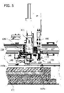
[0039] FIG. 5 is a perspective of a fragment of the frozen
aliquotter shown in cross section taken in a plane including
line 5--5 on Fig. 2;
[0040] FIG. 6 is a perspective of one embodiment of robotic
end effector for use with a frozen aliquotter;
[0041] FIG. 7 is a bottom plan view of the robotic end
effector illustrated in Fig. 6;
[0042] FIG. 8 is a schematic diagram showing some of the
components of the frozen aliquotter;
[0043] FIG. 9 is a schematic diagram illustrating bore
candidates that differ in size;
[0044] FIG. 10 is a schematic diagram illustrating one
embodiment of a geometric pattern according to which frozen
sample cores are extracted from a frozen sample;
[0045] FIG. 11 is a schematic diagram illustrating bore
candidates that spaced different distances from a center of a
container;
[0046] FIG. 12 is a schematic diagram illustrating bore
candidates that are positioned at various different angles
relative to one another from a center of the container;
[0047] FIG. 13 is a schematic diagram illustrating bore
candidates that do not follow an expected sequence planned for
extraction of frozen sample cores from a frozen sample;
CA 02870505 20110-139
WO 2013/166022 PCT/US2013/038880
17
[0048] FIG. 14 is a photograph of a container illustrating
use of an edge finding algorithm to identify the location of an
edge of the container from the image data;
[0049] FIG. 15 is a schematic diagram illustrating one
embodiment of using fixed calibration marks to identify the
location of features in the image data;
[0050] FIG. 16 is a photograph showing a pair of
calibration marks in fixed position relative to a container;
[0051] FIG. 17 is a photograph of one embodiment of a
density step target;
[0052] FIG. 18 is a schematic illustration of a coring
probe positioned over a bore in a frozen sample; and
[0053] Figure 19 is a schematic illustration of the coring
probe of FIG. 18 inserted into the bore.
[0054] Corresponding reference characters indicate
corresponding parts throughout the drawings.
DETAILED DESCRIPTION
[0055] Referring now to the drawings, first to Figs. 1-3 in
particular, one embodiment of a robotic system for taking frozen
sample cores from frozen samples contained in containers is
generally designated 101. The system 101 includes a platform 103
for supporting a plurality of containers 105 and a robotic end
effector 111 movable relative to the platform by a motorized
drive system 113 controlled by a processor 114 (Fig. 8). In the
illustrated embodiment, the robotic system 101 is a cartesian
gantry style robot, but this is not required and other types of
robotic systems can be used within the scope of the invention.
Additional details about robotic systems for taking frozen
sample cores from frozen samples are set forth in U.S. pre-grant
publication No. 20090019877, PCT application No.
PCT/U52011/61214, filed November 17, 2011, and U.S. Application
No. 13/359,301, filed January 26, 2012, the contents of which
are each hereby incorporated by reference.
CA 02870505 2014-10-09
WO 2013/166022
PCT/US2013/038880
18
[0056] In the illustrated embodiment, the platform 103
includes a recessed area 115 sized and shaped to receive one or
more removable trays 117 for holding the containers 105. For
example, one or more of the trays 117a are suitably source trays
that hold a plurality of source containers 105, each of which
contains a frozen sample core is to be taken, and one or more
other trays 117b are suitably destination trays that hold a
plurality of destination containers, each of which is for use
holding one or more frozen sample cores taken from the
containers on the source tray.
[0057] As illustrated in Figs. 3 and 4, the platform 103
also includes a source container station 107 adapted to receive
one of the source containers while a frozen sample core is
extracted from the frozen sample container therein and a
destination container station 109 adapted to receive an empty
container in which one or more frozen sample cores are
deposited. As illustrated in Fig. 4, the source container
station 107 includes a receptacle 106 for receiving containers
105 and a pair of clamping jaws 108, 110 on opposite sides at
the top of the receptacle. At least one of the jaws 108 is
selectively moveable, such as by a pneumatic actuator (not
shown), toward and away from the other jaw 110 for selectively
clamping containers 105 in position at the station 107 to hold
them in place during extraction of a frozen sample and releasing
the containers so they can be removed from the station and
replaced in the tray 117 afterward. Similar jaws can be used to
hold the container 105 at the sample receiving station 109 if
desired.
[0058] The system 101 illustrated in the drawings is
adapted for use with frozen samples that are stored in
individual containers 105. However, it is understood the system
could be adapted for use with well plates and arrays in which
multiple different frozen samples are stored in a single
container. For example, appropriate components can be provided
CA 02870505 2014-10-09
WO 2013/166022
PCT/US2013/038880
19
(e.g., on the end effector) for moving well plates or arrays
instead of individual containers and the stations 107, 109 for
receiving the containers can be adapted to receive well plates
or arrays without departing from the scope of the invention.
Likewise, the clamping system can be adapted to hold well plates
and arrays within the scope of the invention.
[0059] A washing station 119 for cleaning a sample coring
probe 121 used to extract frozen sample cores from the frozen
samples is also on the platform. Details concerning the
construction and operation of a suitable washing station are
provided in PCT application No. PCT/US2011/61214, filed November
17, 2011, and do not need to be repeated herein.
[0060] A cooling system 131 for keeping the frozen samples
and the frozen sample cores extracted therefrom frozen is
positioned under the platform 103 in the illustrated embodiment,
although the cooling system can be positioned elsewhere and/or
other cooling systems used without departing from the scope of
the invention. As illustrated in Figs. 5-7, the end effector 111
of the robotic system 101 includes a sample coring probe 121 and
a sample core extraction system 123 operable to move the sample
coring probe into one of the frozen samples contained in one of
the containers 105 and then withdraw the coring probe from the
frozen sample to obtain a frozen sample core from the frozen
sample. In the illustrated embodiment, for example, the sample
core extraction system 123 includes a motor 125 adapted to
rotate the sample coring probe 123 as the robotic drive system
113 lowers the sample coring probe into the container and then
raises it out of the container. Additional details about the
operation of a coring probe to extract frozen sample cores from
frozen samples are set forth in U.S. pre-grant publication No.
20090019877, PCT application No. PCT/US2011/61214, filed
November 17, 2011, and U.S. Application No. 13/359,301, filed
January 26, 2012 and do not need to be repeated herein. It is
understood any sample coring probe and sample extraction system
CA 02870505 2014-10-09
WO 2013/166022 PCT/US2013/038880
can be used within the scope of the invention, as long as they
can be operated to extract a frozen sample core from a frozen
sample while resulting in only limited to no thawing of the
frozen sample material and the frozen sample core extracted
therefrom.
[0061] The end effector 111 also includes a gripping system
127 operable to selectively hold and release containers 105 for
use by the robotic system 101 in moving containers back and
forth between the trays 117 and the stations 107, 109 on the
platform for the containers from which frozen sample cores are
being taken and into which frozen sample cores are being
deposited. Those skilled in the art will be familiar with
various commercially available gripping systems that can be
used. In the illustrated embodiment, for example, the gripping
system includes a plurality of moveable fingers 129 moveable by
one or more pneumatic actuators (not shown) under the control of
the processor 114. It is understood other gripping systems may
be used within the scope of the invention. For example, the
gripping system can be modified if desired to facilitate use of
the gripping system to move well plates or arrays containing
multiple frozen samples.
[0062] As illustrated schematically in Fig. 8, the robotic
system 101 cooperates with a machine vision system 141
configured to automatically recognize locations from which
frozen sample cores have already been taken from the frozen
samples in the containers 105 (if there are any) to facilitate
taking additional frozen sample cores from already-cored frozen
samples. At these locations, there will be a bore or hole in the
frozen sample. In some cases the bore may be empty, but in other
cases the bore may contain or be obscured by frost crystals that
have grown on the sample (e.g., while the sample was in frozen
storage), by debris, and/or for other reasons. The machine
vision system 141 is also configured to recognize the absence of
any bores in frozen samples that have not yet had any frozen
CA 02870505 2014-10-09
WO 2013/166022
PCT/US2013/038880
21
sample cores extracted from them. The machine vision system 141
facilitates use of the robotic system 101 to take frozen sample
cores from frozen samples that were previously sampled to obtain
an aliquot and then were returned to frozen storage for a period
of time before additional frozen sample cores from that sample
are requested to provide additional aliquots in later tests.
[0063] The machine vision system 141 includes a camera 143
mounted for capturing an image of a container 105 and a frozen
sample contained therein while the container is supported by the
platform 103. In the illustrated embodiment, the machine vision
system 141 includes a display 146 coupled to the processor 114
for displaying the captured and/or processed image data. The
camera 143 is suitably mounted on the robotic system 101 for
movement relative to the containers 105 by the robotic system.
For example, in the illustrated embodiment, the camera 143 is
mounted on the end effector 111 for movement with the end
effector. It is recognized the camera could be mounted in fixed
position relative to the platform within the scope of the
invention.
[0064] The camera 143 and processor 114 are configured to
communicate with one another so the processor can instruct the
camera to capture images at appropriate times and receive image
data from the camera indicative of the images captured by the
camera. The processor 114 for the vision system 141 can suitably
be the same processor that controls operation of the robotic
system 101, although separate processors could be used within
the scope of the invention. Various cameras can be used within
the broad scope of the invention. For example, the camera 143
can be a digital camera containing a CCD array (not shown) that
converts the captured image into electrical signals. The camera
143 in the illustrated embodiment is configured to capture
monochromatic (e.g., grayscale) images instead of color images
for reasons that will be discussed in more detail later, but the
CA 02870505 2014-10-09
WO 2013/166022
PCT/US2013/038880
22
camera can be configured to capture color images within the
broad scope of the invention.
[0065] The machine vision system 141 also includes a light
145 for illuminating the container 105 being imaged by the
camera 143. One or more lights having various different
configurations, arrangements, and colors can be used within the
broad scope of the invention. The lights can be moveable (e.g.,
mounted on the end effector 111) or fixed (e.g., secured to or
within the platform 103) within the scope of the invention. The
one or more lights can be positioned to provide bright field
illumination, dark field illumination, indirect lighting (e.g.,
side lighting), backlighting, direct lighting (i.e., lighting
directed perpendicular to the illuminated surface), and any
combinations thereof. Figure 4 illustrates three optional lights
181, 183, 185 that can be positioned at fixed locations relative
to the station for receiving the container 105 holding the
frozen sample. For example, fiber optic cables can be routed
through the platform or provided on the platform to provide
light at locations designated 181, 183, and/or 185. Other types
of lights could also be secured on or within the platform at
these locations.
[0066] The containers 105 are typically transparent or at
least translucent so light can pass through the side or bottom
of the container and interact with the frozen sample therein.
The light 181 at the bottom of the receptacle 106 for receiving
the container 105 provides a backlighting option. The light 183
at the top of the container is suitably secured within one of
the jaws 108, 110 to provide a side lighting and/or dark field
illumination option for the surface of the sample. The light 185
in the side of the receptacle 106 suitably provides a side
lighting option below or at the surface of the frozen sample.
When the side lighting and/or back lighting options are used,
the bores in the frozen sample will typically have a brighter
appearance than the frozen sample in the corresponding image.
CA 02870505 2014-10-09
WO 2013/166022
PCT/US2013/038880
23
Side lighting and/or back lighting can be useful in detecting
real bores that are either filled with or completely obscured by
frost or other debris. The machine vision system 141 can include
multiple different lights and the processor 114 can be
configured to operate the lights sequentially if desired to make
use of image data of the frozen sample under different lighting
conditions.
[0067] As illustrated in Figs. 6 and 7, the light 145 in
the illustrated embodiment is a ring light. The ring light 145
has a plurality of light sources 147 (e.g., LEDs) arranged in an
annular (e.g., circular) pattern. For example, the ring light
145 suitably has a hollow cylindrical housing 151 having a
groove 153 in one end. The light sources 147 are positioned in
the groove 153 in a recessed position so the housing blocks the
direct path of light from the light sources at wide angles
therefrom. A cover 149 such as a clear window, transparent or
translucent diffuser, or lens can be installed in the groove to
enclose the light sources 147 if desired.
[0068] In the embodiment illustrated in Figs. 6-7, the
camera 143 is positioned so an optical axis 155 of the camera
143 extends through a central portion of the annular pattern of
the ring light 151. For example, the annular ring light 145
suitably has a central axis that is co-linear with the optical
axis 155 of the camera. The ring light 145 and camera 143 are
suitably arranged so there is no direct path from the light
sources 147 in the ring light to the camera. In Fig. 5, for
example, the camera 143 has a forward end 157 for receiving
light from an object being imaged and the ring light 145 is
suitably positioned to extend farther forward than the camera so
the light emitted by the ring light is emitted from a position
in front of the camera. Also as illustrated in Fig. 5, the edge
of the housing 151 of the ring light 145 suitably extends
between the light sources 147 and the camera 143 to block the
path of light directly from the light sources into the camera.
CA 02870505 20110-139
WO 2013/166022 PCT/US2013/038880
24
[0069] The processor 114 is configured to receive image
data from the camera 143 indicative of the image of the
container 105 and the frozen sample therein and to use the image
data from the camera to determine one or more locations where a
frozen sample core has already been taken from the frozen sample
contained in the container. The processor 114 can be configured
to use various methods to make this determination. For example,
the processor 114 can suitably be configured to evaluate
contrast in the image to identify one or more bore candidates
and then determine whether or not the one or more bore
candidates are likely to be artifacts instead of real bores in
the sample using information from the image.
[0070] The processor 114 suitably processes the image
captured while the container 105 is illuminated with the light
145 in various ways to facilitate this determination. For
example, in one embodiment the processor 114 is configured to
perform a thresholding filter to the raw image data, apply one
or more morphological filters (e.g., erosion, dilation, opening,
and/or closing) to the thresholded image, and then apply
particle analysis to identify one or more bore candidates.
[0071] It is understood that the bore candidates identified
by the processor might include some features that are artifacts
instead of real bores. For example, the sample surfaces can be
blotchy or become blotchy over time (e.g., due to undesired
formation of frost crystals on the frozen sample, irregularities
in the surface contour of the frozen sample resulting from the
speed with which the sample was frozen, pieces of ice and other
debris that may accumulate on the upper surface of the frozen
sample such as by falling from the cap or sides of the
container, etc.). Further, although the real bores resulting
from extraction of frozen sample cores are typically very
uniform (e.g., circular) in appearance initially, frost crystals
that might grow on the frozen sample after it is returned to
cold storage can extend into the bore or over the opening at the
CA 02870505 2014-10-09
WO 2013/166022
PCT/US2013/038880
top of the bore and alter the appearance of the bore. Thus, it
has been found that a machine vision system that looks for nice
perfectly formed bores and excludes all else from the list of
bore candidates results in a significant risk of failure to
recognize actual bores that exist in the sample, particularly
when the frozen sample is replaced in cold storage for a long
time before additional frozen sample cores from that frozen
sample are requested.
[0072] Accordingly, the processor 114 is suitably
configured to use multiple types of information to determine
whether or not a bore candidate is likely to be a real bore
candidate or an artifact. For example, the processor 114 is
suitably configured to use information selected from the group
consisting of:
the size of the bore candidate;
the distance between the bore candidate and a center axis
of the container;
the angle formed between a first line and a second line,
the first line extending between the bore and the center
axis of the container and the second line extending between
the center axis of the container and another bore
candidate;
the relation between the position of the one or more bore
candidates and an expected pattern of bores in the sample;
the location of the one or more bore candidates relative to
a peripheral edge of the container;
the total number of bore candidates associated with a
particular container;
the amount of contrast between the bore candidates and the
area surrounding the bore candidates; and
combinations thereof to help determine whether or not a
bore candidate is likely to be an artifact or a real bore.
[0073] In many cases there will be an expected range of
size (e.g., diameter) for the bores formed by extracting a
CA 02870505 2014-10-09
WO 2013/166022
PCT/US2013/038880
26
frozen sample core from a frozen sample. However, some bore
candidates can be substantially larger or substantially smaller
than expected, as illustrated in Fig. 9. Thus, it is possible
the processor 114 may be able to determine certain bore
candidates are likely to be artifacts on the basis of the size
being either too large (e.g., having diameter D1 in Fig. 9) or
too small (e.g., having diameter D2 in Fig. 9).
[0074] In many cases frozen sample cores will be extracted
from the frozen samples according to a pre-determined geometric
pattern or a geometric pattern that can be recognized from the
captured image data by the processor 114. The geometric pattern
can vary depending on what objectives are to be achieved, such
as maximizing the number of frozen sample cores that can be
extracted from a frozen sample or taking as many frozen sample
cores as possible at a particular radial position from the
center. Figure 10 illustrates an example of one geometric
pattern in which five sample cores are extracted from a frozen
sample. The bores resulting from this pattern are all spaced
about the same distance D3 from the center of the container and
the angles 01 between the lines extending between corresponding
points (e.g., the center) in adjacent bores are all about equal.
The number of bores in the geometric pattern can vary within the
scope of the invention. Although the pattern in Fig. 10 is a
regular pattern, meaning the bores are all the same size, are
all spaced the same distance from the center, and are all spaced
at equal angles, it is understood that the pattern could be
irregular within the scope of the invention.
[0075] As illustrated in Fig. 11, some bore candidates can
be spaced too close to the center (e.g., see distance D4 in Fig.
11) or too far from the center of the container (e.g., see
distance D5 in Fig. 11), or conversely, spaced to far or close
to the edge of the container if the edge of the container can be
detected, to fall within the geometric pattern. Likewise, as
illustrated in Fig. 12 the angular spacing between one or more
CA 02870505 2014-10-09
WO 2013/166022 PCT/US2013/038880
27
of the bore candidates can be different (either too high 02 or
too low 03) from the expected angular spacing. Thus, the
processor 114 can determine certain bore candidates are likely
to be artifacts on the basis that the distance between the bore
candidate and a center axis of the container (or from an edge of
the container) is not within expected limits and/or that the
angle formed between lines extending between corresponding
points in the two bore candidates (e.g., the center, as
illustrated, or an edge) and the center axis of the container is
not within expected limits.
[0076] In many cases, frozen sample cores will be extracted
from the frozen samples according to a specific orderly
sequence. As illustrated in Fig. 10, for example, the frozen
sample cores are extracted starting at the top position and then
moving clockwise around the geometric patter until the last
sample core has been taken. As illustrated in Fig. 13, in some
cases one or more of the bore candidates may be out of order
even though it could be positioned at a correct place within the
geometric pattern. For example, there may be an empty gap 307 in
the pattern between one of the bore candidates 305 and other
bore candidates 301, 303 indicating that if all of the bore
candidates are real bores, the result would be the expected
sequence was not followed. In this case, the processor 114 can
determine a bore candidate is an artifact on the basis that it
is out of order with a sequence according to which frozen sample
cores are expected to be extracted from the frozen sample,
particularly when multiple bore candidates 301, 303 follow the
expected sequence and only one bore candidate 305 is out of
sequence.
[0077] In some cases, the number of bore candidates can be
larger than is expected. The processor 114 is suitably
configured to recognize this as an indication of a higher
likelihood that one or more of the bore candidates is an
artifact. The processor 114 can apply more rigorous standards to
CA 02870505 2014-10-09
WO 2013/166022
PCT/US2013/038880
28
help exclude likely artifacts when the number of bore candidates
is too high.
[0078] Sometimes the amount of contrast between a bore
candidate and its surrounding area can help distinguish between
real bores and artifacts. For example, a large contrast can be
indicative of a very good candidate for a real bore whereas a
smaller contrast might indicate one of a set of bore candidate
that is questionable on other accounts (e.g., there are too many
bore candidates, there are only two bore candidates and they do
not follow the expected geometric pattern, etc.) is more likely
than the other(s) to be an artifact.
[0079] One way the processor can evaluate the positions of
the bore candidates is with reference to the position of the
center axis of the container 105 holding the frozen sample or
alternatively relative to a peripheral edge of the container.
The processor 114 can be configured to identify the edge and/or
the center of the container 105 in various ways within the scope
of the invention. For example, one option is to use an edge
finding algorithm to identify the inner or outer peripheral edge
of the container 105 and then compute the geometric center of
that edge to identify the center of the container. For example,
Fig. 14 shows an image of a container 105 with an overlay
including a pair of concentric circles 163, 165 and a plurality
of radially extending scan lines 167 extending between the
circles. The circles 163, 165 define an area to be scanned in an
attempt to identify the edge of the container 105. The processor
114 is suitably configured to evaluate the image data to
determine points 169 along each line where there is sharp
contrast. Each point 169 potentially represents an intersection
between the edge of the container 105 and the respective scan
line 167. In the case of a successful attempt to identify the
edge of a container, a significant number of the points 169 will
lie on the same circle (or other shape if the containers do not
have circular shapes) in which case the processor 114 concludes
CA 02870505 2014-10-09
WO 2013/166022
PCT/US2013/038880
29
the points 169 lying thereon define the edge of the container
105. As used herein in the context of edge finding techniques
and using information about the edge of the container to
identify and/or evaluate bore candidates, it is understood the
edge of the container can refer to the edge of a well or other
discrete area within which one frozen sample is stored on a well
plate or other container adapted for holding multiple different
samples.
[0080] Although edge detection techniques can work very
well in certain circumstances, edge detection can be impaired
when there is low contrast between the edge of the container and
the background. This can present a problem when relying solely
on edge detection in the context of a machine vision system for
a frozen aliquotter because one of the most common colors for
containers is white or semi-transparent and white frost can form
on surfaces adjacent the containers, which leads to the
potential problem that there might be low contrast between the
edge of the container and the surroundings in the image.
Ultraviolet or infrared lighting can help enhance the contrast
between the edge of the container and the surroundings in the
image. This enhanced contrast improves detection and
identification of the container edges. In one embodiment, a
separate UV or IR light source can be positioned to illuminate
the container. The UV or IR light source can be moveable (e.g.,
mounted on the end effector 111) or fixed (e.g., secured to or
within the platform 103) within the scope of the invention. In
one embodiment, the separate UV or IR light source can be
positioned in the platform to provide indirect lighting (e.g.,
backlighting or side lighting) to the container to aid in edge
detection. In another embodiment, any one of or combination of
the lights 145, 181, 183, 185 can include a UV or IR light
source.
[0081] As illustrated in Figs. 15 and 16, a pair of
calibration marks 161 is suitably provided on the platform 103
CA 02870505 20110-139
WO 2013/166022
PCT/US2013/038880
at fixed positions relative to the source container station 107.
The calibration marks 161 can be any structure or marking that
has sufficient contrast with the background to allow the
calibration marks to be reliably identified by the processor 114
from the image data. For example, the calibration marks 161 can
suitably be dark openings, dark colored marking (e.g., dots), or
other structures. The calibration marks 161 can suitably be or
include heaters (e.g., small low-power resistance heaters) to
limit accumulation of frost on the calibration marks, which
might obscure the calibration marks.
[0082] The processor 114 is suitably configured to use the
calibration marks 161 (e.g., in combination with edge detection
or without edge detection) to determine whether or not the one
or more bore candidates are likely to be artifacts instead of
real bores in the frozen sample. The calibration marks 161 are
designed to ensure there is strong color contrast between the
calibration marks and the surrounding objects in the image even
if there is frost formation or other conditions that minimize
contrast between the edge of the container 105 and its
surrounding in the image data. Because the calibration marks 161
are at fixed positions relative to the station 107 for receiving
source containers 105, the processor 114 can determine the
position of the bore candidates by comparing their positions to
the positions of the calibration marks.
[0083] For example, the calibration marks 161 are suitably
positioned to form a triangle with the center of the container.
The angles a and p formed between a line connecting the
calibration marks and the respective lines connecting the
calibration marks to the center can be known before the vision
system 141 inspects a frozen sample. Accordingly, the processor
114 can be configured to identify the center axis of the
container by triangulating the center from the calibration
marks. A machine vision system 141 including a processor 114
that is configured to use calibration marks 161 to identify the
CA 02870505 2014-10-09
WO 2013/166022
PCT/US2013/038880
31
center of a container is not sensitive to errors in the rotation
of the camera or to errors in translation of the camera. In
cases where it is not practical to use edge detection to
determine the center of the container (e.g., because of low
contrast), the processor 114 can be configured to identify the
edge of the container 105 and/or the center axis of the
container as a function of the position of the calibration marks
without detecting any edges of the container. Alternatively, the
processor 114 can be configured to both use the calibration
marks and peripheral edge detection to identify the center of
the container. The processor 114 can be configured to compare
the positions of the bore candidates directly to the positions
of the calibration marks 161 to determine which bore candidates
are likely to be artifacts without computing the relative
distances between the bore candidates and the center of the
container or the edge of the container without computing the
center of the container and/or without computing the edge of the
container.
[0084] The processor 114 is configured to automatically
select a suitable location in the frozen sample from which the
robotic system 101 can take another frozen sample core (or in
the case of a frozen sample from which no frozen sample cores
have been taken yet, it is configured to automatically select
the location from which the initial frozen sample core will be
extracted) once the processor has determined from the image data
whether or not there are any bores in a particular frozen sample
and the locations of any such bores. This facilitates taking
frozen sample cores from samples that may have already been
subjected to previous extractions of frozen sample cores without
requiring the processor 114 to have access to any information
about the number of previous frozen sample cores that may have
been extracted from the sample or the locations within the
frozen sample from which any such sample cores have been taken.
This eliminates the need for manual intervention to orient the
CA 02870505 20110-139
WO 2013/166022
PCT/US2013/038880
32
containers 105 is a particular way and greatly reduces the
amount of data on a sample that needs to be tracked to
successfully manage and process samples when extracting frozen
sample cores from frozen samples to fill orders for sample
aliquots. It also facilitates the ability to install the robotic
system 101 in a bio-bank that has previously used one or more
different approaches to sample core extraction (e.g., using a
geometric pattern of sample cores that maximizes the number of
samples that can be obtained vs. using a geometric pattern of
sample cores that results in some or all of the samples being
taken from a part of the frozen sample that is a particular
radial distance from the center even if this reduces the maximum
number of samples cores that can be extracted). Thus, if a bio-
bank has previously employed one strategy or particular set of
operating procedures for extracting frozen sample cores, the
system 101 can still recognize bores in the frozen sample even
if the bores are not where they would be expected to be if the
previously extracted frozen sample cores had been extracted
according to the protocols of the system 101 instead of whatever
other protocols were previously in use.
[0085] For example, if the processor 114 detects one or
more pre-existing bores in the frozen sample, the processor can
be configured to select a location for the next frozen sample
core that continues the geometric pattern that has already been
started. Another option if it is desired that the next sample
core be taken from a particular radial location in the frozen
sample is that the processor 114 can be configured to select a
location that is the desired radial distance from the center of
the container and also sufficiently spaced from existing bores
in the frozen sample. The processor 114 can be configured so a
user can select which of these options is used for any
particular container or set of containers.
[0086] The processor 114 is also configured to select an
appropriate initial geometric pattern for the locations from
CA 02870505 20110-139
WO 2013/166022
PCT/US2013/038880
33
which a plurality of frozen sample cores will be extracted if
the processor determines there are no existing bores in the
frozen sample. The processor 114 can be configured to select a
geometric pattern that maximizes the number of frozen sample
cores that can be taken from the frozen sample and/or the
processor can be configured to select a geometric pattern that
results in one or more frozen sample cores (e.g., all frozen
sample cores) being taken from a particularly desired radial
distance from the center of the container. The processor 114 can
be configured to allow a user to select which of several
different strategies will be used for planning the geometric
pattern of the locations from which frozen sample cores are to
be extracted for different containers or sets of containers. If
desired, the processor 114 can be configured to display the
geometric pattern selected by the processor and/or the
location(s) selected by the processor as the site(s) for frozen
sample extraction(s) to facilitate confirmation and/or
intervention by a human operator.
[0087] It has been determined that the color of light
emitted by the light 145 can be important. In general, better
results are obtained when the light used to illuminate the
frozen sample matches the color of the frozen sample. For
example, the color of the light used to illuminate the frozen
sample is suitably the same as the color of the sample or no
more different from the color of the sample than one of the two
adjacent colors on an RGB color wheel having six colors arranged
in the following order extending around the wheel: red, yellow,
green, cyan, blue, magenta, and then back to red. For example, a
red light works well with red samples, orange samples, and
yellow samples. Because there are large numbers of blood (red)
and urine (yellow or orange) samples that have been frozen for
research, it is anticipated that it can be desirable for the
light 145 to emit red light for illuminating the frozen sample.
It is also anticipated that it will be desirable in some cases
CA 02870505 20110-139
WO 2013/166022
PCT/US2013/038880
34
for the light to emit green light or blue light. However, the
light can emit any color light within the broad scope of the
invention.
[0088] In one embodiment, the light 145 includes red light
emitting elements, blue light emitting elements, and green light
emitting elements and the intensity of light emitted from the
red, blue, and green light emitting elements is selectively
adjustable to allow any of multiple different colors of light to
be selected as the color of light that is used to illuminate the
samples. In one embodiment, the light 145 includes red light
emitting elements that emit red light having a wavelength in the
range of about 620nm to about 750nm (about 4001Hz to about
4841Hz). For example, the majority of the light energy emitted
by the light (e.g., substantially all of the light energy) is
suitably within the range of about 620nm to about 750nm. The
light sources can include LEDs that emit light concentrated in
the range of about 620nm to about 750nm in wavelength. In
another embodiment, the light 145 includes green light emitting
elements that emit green light having a wavelength in the range
of about 495nm to about 570nm (about 5261Hz to about 6061Hz).
For example, the majority of the light energy emitted by the
light (e.g., substantially all of the light energy) is suitably
within the range of about 495nm to about 570nm. The light
sources can include LEDs that emit light concentrated in the
range of about 495nm to about 570nm in wavelength. The light
sources 147 can include some light sources that emit only red
light, other light sources that emit only green light, and other
light sources that emit only blue light. Another possibility is
that the light sources include multicolor LEDs, each which is
operable to emit red light, green light, blue light and
combinations thereof.
[0089] In the case that the color of light can be adjusted
the processor 114 can suitably be configured to receive input
about the color of the samples in the containers and adjust the
CA 02870505 2014-10-09
WO 2013/166022
PCT/US2013/038880
color of the light emitted by the light to reduce a difference
between the color of the samples and the color of the light
emitted by the light. For example, the vision system 141 can
include a user interface configured to allow a user to input
information about the color of the samples and the processor 114
can be configured to adjust the color of the light to match the
color input by the user. Another option is that the camera 143
can be adapted to capture a color image of the sample (or a
representative sample of a group of samples) and the processor
114 can be configured to adjust the color of the light to match
the color of the sample in the captured color image. The
processor suitably adjust the color of the light used to
illuminate the sample to white when capturing the image that
will be used to determine the color of the sample to facilitate
accurate color detection and then adjusts the color of the light
to match the color of the sample. In some cases it may be known
that an entire set of samples will be similar in color, in which
case the processor can be configured to capture a color image of
one of the samples to assess the color of all of the samples in
that set and adjust the color once to match the color of all the
samples in the set.
[0090] Although the vision system can be configured so the
camera captures color images of the frozen samples and the
processor uses information from the color images to identify
where frozen sample cores have already been taken within the
scope of the invention, surprisingly good results are obtained
when the vision system is configured so the camera captures a
monochromatic (e.g., grayscale) image of the frozen sample (even
when the light illuminating the sample is other than white,
e.g., selected to match the color of the sample) and the
processor uses the grayscale image to determine whether or not
frozen samples cores have already been taken from the frozen
sample and, if so, to identify the locations from which the
frozen sample cores have already been taken. Digital cameras are
CA 02870505 20110-139
WO 2013/166022
PCT/US2013/038880
36
available that can capture both grayscale images and color
images, so it is possible that the camera captures one or more
color images (e.g., to identify the color of the sample so the
light used to illuminate the sample can be adjusted to match the
color of the sample) and also captures grayscale images for use
by the processor to identify locations where bores exist within
the frozen samples.
[0091] The machine vision system 141 is suitably a
component of a calibration system configured to calibrate the
robotic system 101. As illustrated in Figs. 1-3, the platform
103 has one or more fixed targets 171 positioned thereon. The
camera 143 is mounted on the robotic system 101 so it can be
moved to capture an image of each of the fixed targets 171. The
processor is suitably configured to receive image data from the
camera indicative of images of the target(s) formed by the
camera and calibrate the robotic system 101 using an image of
the one or more fixed targets 171 on the platform 103. As
illustrated in Fig. 1, at least one of the targets 173 has an
image (e.g., a cross hair) having a point or intersection of
lines for calibration in the x and y directions and a shape
(e.g., circle) having a known size for calibration in the z
direction. The calibration system suitably has a user interface
(not shown) configured to allow a user to guide the camera from
a position that is not in registration with one of the targets
(e.g., so a reticule overlaying the captured image is not
aligned with the cross hair) toward a position that is in
registration with said target (e.g., so the reticule is aligned
with the cross hair).
[0092] Also, as illustrated in Figs. 1-3, one of the
targets 171 (e.g., the target 173 having the cross hair and
circle) is secured to an upper surface of the work deck outside
the recessed area 115 for receiving the trays. Further, one of
the targets 175 is suitably secured to a bottom of the recessed
area (e.g., between the trays 117 and adjacent the stations 107,
CA 02870505 2014-10-09
WO 2013/166022 PCT/US2013/038880
37
109, as illustrated in Fig. 3) for receiving the containers 105.
In illustrated embodiment, the target 175 in the recessed area
115 also includes a cross hair.
[0093] Another of the targets in the illustrated embodiment
is suitably a density step target 177 (see Fig. 17) having an
image including one or more series of blocks arranged from
lighter shades to darker shades for use calibrating light/dark
settings for the camera 143.
[0094] The processor 114 is suitably configured to use
multiple additional features on the platform 103 as targets to
help calibrate the robotic system 101. For example, the
processor 114 is suitably configured to calibrate the robotic
system 101 using images of multiple features on the platform 103
selected from the group consisting of:
the station 107 for receiving a container 105 from which a
frozen sample core is to be taken;
the station 109 for receiving a container 105 in which a
frozen sample core is to be deposited;
the station 119 for cleaning a coring probe 121 of the
robotic system 101;
one or more sample trays 117 on the platform 103 for
holding the containers 105; and
combinations thereof.
[0095] For example, Fig. 3 illustrates 13 calibration
points that can be used according to one particular embodiment
of the calibration system, with each of calibration points being
labeled consecutively from 201-213. Point 201 corresponds to the
target 175 on the platform 103 in the recessed area 115. Points
201, 202, and 203 correspond to the stations 107, 109 for
receiving the containers 105 and the station 119 for washing the
coring probe 121, respectively. Points 204-208 correspond to
various points (e.g., points at the corners) of one of the trays
117a and points 209-212 correspond to various points (e.g.,
points at the corners) of another of the trays 117b. Point 213
CA 02870505 2014-10-09
WO 2013/166022
PCT/US2013/038880
38
corresponds to the target 173 on the deck of the platform 103.
The points included in the calibration are suitably selected so
they collectively extend over at least a substantial portion of
the operating envelop, but the specific points used in the
calibration process can vary within the scope of the invention.
[0096] The calibration system is suitably configured to
complete calibration of the robotic system 101 without any
physical contact between (i) the end effector 111 or and any
components moveable with the end effector and (ii) the platform
103 or components on the platform.
[0097] The calibration system is also suitably configured
to determine the positions of the camera 143, coring probe 143,
and gripper 127 relative to one another to compensate for
variations in the positional offsets associated with the camera,
probe, and gripper. For example, the calibration system is
suitably configured so a user can direct movement of the end
effector 111, to bring each of the camera 143, coring probe 121,
and gripping system 127 into registration with a target 171 or
other reference point and provide an indication to the processor
114 each time one of them is in registration therewith. This
allows the processor 114 to compute offsets between these
components that account for positional variations that may
result during assembly of the robotic system 101 or for any
other reason. This facilitates more accurate position of the
components of the robotic system 101.
[0098] During initial installation of the robotic system
101, and from time to time thereafter as may be needed, the
machine vision system 141 is suitably used to calibrate the
robotic system. The robotic drive system 113 moves the camera
143 into a position that is estimated by the processor to be in
registration with one of the targets 171. Then, image data from
the camera 143 is used (either by the processor or a user) to
instruct the robotic drive system 113 to adjust the position of
the camera until it is in registration with the target. If the
CA 02870505 2014-10-09
WO 2013/166022
PCT/US2013/038880
39
target 171 includes a circle or other shape having a known size
for calibration in the z-axis, the image data from the camera
143 is used to instruct the robotic drive system (either by the
processor or a user) to raise or lower the camera until the size
of the shape in the image captured by the camera indicates the
camera is the proper distance from the target in the z-
direction. Calibration in the Z-direction could instead be
achieved using a lens setting for the camera 143 having a known
focal length and then adjusting the height of the camera until
the image is in focus. When the camera 143 is in registration
with the target 171 and the correct distance from the target,
the processor records positional information from the robotic
system 101 (e.g., data from encoders and other devices that
provide positional feedback about the position of various
components of the robotic system) and designates that
information as corresponding to a set point corresponding to
respective target. The process is repeated for each of the
targets 171. For example, in the embodiment illustrated in Fig.
3 the process is repeated for each of the calibration points
201-213.
[0099] Although the targets 171 and/or calibration points
can be positioned at various locations on the platform 103, in
the illustrated embodiment, the targets suitably include one
target 173 on the upper surface of the deck of the platform and
another target 175 in the recessed area of the platform. The
targets 171/calibration points suitably include multiple targets
including at least one of:
the station 107 for receiving the container 105 from which
a frozen sample core is to be taken;
the station 109 for receiving the container 105 in which a
frozen sample core is to be deposited;
the station 119 for cleaning the coring probe 121;
one or more trays 117a, 117b on the platform 103 for
holding the containers 105; and
CA 02870505 2014-10-09
WO 2013/166022
PCT/US2013/038880
combinations thereof.
[00100] For example, in one embodiment the calibration
targets 171 and calibration points include each of points 201-
213 on Fig. 3.
[00101] The density step target 177 is also used during the
calibration process to adjust camera settings and light
intensity to provide standard image capturing conditions. The
light 145 is turned on and the diaphragm of the camera 143
and/or intensity of electric current supplied to the light are
adjusted while the camera captures images of the density step
target 177 until a particular shaded block on the density step
target is read as a certain gray level by the camera 143. For
example, good results have been obtained when the light 145 and
camera 143 are adjusted so the third lightest color block on a
standard density step target is read by the camera as 200 gray
level.
[00102] The robotic system 101 is also calibrated to adjust
for any variations in the offset between the positions of the
camera 143, the coring probe 121, and the gripping system 127.
For example, the camera 143 is first positioned in registration
with one of the targets 171 or other reference point on the
platform 103, at which point a user provides an indication to
the processor 114 that the camera is in registration therewith.
Then a user adjusts the position of the end effector 111 until
the coring probe 121 is in registration with the target 171 or
reference point and provides an indication to the processor 114
that the coring probe is in registration therewith. Finally, the
user adjusts the position of the end effector until the gripper
system 127 is in registration with the target 171 or other
reference point and provides an indication to the processor that
the gripper system is in registration therewith. The order in
the steps of this method is not important. The processor 114
determines the positional offset between the camera 143, coring
prove 121, and gripper assembly 127 using the information
CA 02870505 20110-139
WO 2013/166022
PCT/US2013/038880
41
provided in this process. The entire calibration process is
suitably completed without requiring any contact between the end
effector 111 or any components moveable with the end effector
and the platform 103 or any components on the platform.
[00103] To extract frozen sample cores from the frozen
samples, a set of containers 105 containing frozen samples is
placed on the platform 103. For example, one or more trays 117a
can be loaded with sample containers 105 and then placed on the
platform 103 (e.g., in the recessed area 115). A set of empty
containers 105 for receiving frozen sample cores after they are
extracted is loaded into one or more additional trays 117b and
placed on the platform 103. The robotic system 101 uses the
gripper system 127 to move one of the containers 105 containing
a frozen sample to the station 107 for receiving containers from
which frozen sample cores are being extracted and moves one of
the empty containers to the station 109 for receiving
destination containers into which the frozen sample cores are to
be deposited.
[00104] Then the robotic system moves the camera 143 into
position over the station 107 for holding the containers 105
containing frozen sample while frozen sample cores are extracted
from them. The robotic system suitably includes a fill level
detection system for detecting the level of an upper surface of
the frozen sample. Details concerning the construction and
operation of a suitable fill level detection system are provided
in U.S. Application No. 13/359,301, entitled Robotic End
Effector for Frozen Aliquotter and Methods of Taking a Frozen
Aliquot from Biological Samples, filed January 26, 2012, the
contents of which are hereby incorporated by reference. The fill
level detection system provides information about the position
of the upper surface of the frozen sample. The fill level
detection system can be used to position the camera 143 at a
desired level above the frozen sample to improve focusing of the
camera. For example, the processor 114 uses the information from
CA 02870505 2014-10-09
WO 2013/166022
PCT/US2013/038880
42
the fill level detection system about the position of the upper
surface of the frozen sample to determine the elevation at which
to position the camera for obtaining an image of the frozen
sample taken while the camera is within an optimal range of
distances from the upper surface of the sample. The light 145 is
used to illuminate the container 105 at the station 107 and the
frozen sample contained therein. If the machine vision system
141 includes the option of adjusting the color of the light 145,
the color of the frozen sample is determined (e.g., using image
data from the camera and/or user input) and the color of the
light is adjusted to match the color of the frozen sample, as
described above. For example, if the frozen sample is red,
orange, or yellow, the light 145 can be adjusted to emit red
light. Likewise, if additional lighting options are used,
additional images of the container 105 are captured with one or
more of the lights 181, 183, 185 providing illumination.
[00105] The camera 143 captures one or more raw images of
the illuminated container 105 and frozen sample. The raw image
is suitably processed to facilitate recognition of bore
candidates. For example, a thresholding filter is suitably
applied to the raw image obtained with illumination from light
145. A morphological filter is also applied to the image. After
the image has been filtered a particle analysis imaging
algorithm is performed to recognize any bore candidates. The
processor then uses the image data to evaluate whether or not
any bore candidates are actual bores or just artifacts in order
to determine whether or not any frozen sample cores have already
been taken from the frozen sample and, if so, to identify the
location(s) from which they were taken.
[00106] If the option of using calibration marks 161 to
evaluate the positions of the bore candidates is used, the
method suitably includes heating the calibration marks using the
low-power resistance heaters to ensure the calibration marks are
not obscured by frost.
CA 02870505 2014-10-09
WO 2013/166022
PCT/US2013/038880
43
[00107] Once the processor 114 has identified the bore
candidates and determined which bore candidates are likely to be
artifacts and which are likely to be real bores, the processor
automatically selects a position from which a frozen sample core
will be extracted, accounting for the position of pre-existing
bores in the frozen sample if there are any. Then the processor
114 instructs the robotic system 101 to move the coring probe
121 into position over the selected location and instructs the
sample extraction system to extract a frozen sample core from
the location. Consequently the coring probe 121 is extended into
the frozen sample (e.g., while rotating if the sample extraction
system 123 uses a drilling action) and then withdrawn from the
frozen sample with the frozen sample core contained therein. The
robotic system 101 moves the coring probe 121 into position over
the top of the container 105 at the station 109 for holding the
destination containers and ejects the frozen sample core from
the coring probe into the destination container. If more than
one frozen sample core is needed to provide enough material for
the aliquot that has been ordered, the frozen sample core
extraction process is repeated at another suitable location
within the frozen sample, as automatically determined by the
processor, until a sufficient amount of sample material has been
deposited in the destination container 105.
[00108]After a sufficient amount of sample material has
been deposited in the destination container 105, the frozen
parent sample is further processed to clear away any frost
crystals or other debris in each of the bores in the frozen
sample to ensure better accuracy and reliability in the machine
vision system 141 when the sample is retrieved again later from
frozen storage to obtain additional frozen sample cores. As
described above, the bores may contain or be obscured by frost
crystals that have grown on the sample (e.g., while the sample
was in frozen storage), by debris (e.g., from previously
drilling frozen sample cores), and/or for other reasons. To
CA 02870505 20110-139
WO 2013/166022
PCT/US2013/038880
44
improve the ability to recognize bores in the frozen parent
sample next time it is retrieved from frozen storage, the
processor 114 suitably uses the image data that was obtained
from the sample before the most recent frozen sample core was
extracted (i.e., the image data used to evaluate bore
candidates, the determination whether or not any frozen sample
cores had already been taken from the frozen sample, and if
frozen sample cores had already been taken, the location(s) from
which they were taken). The processor 114 suitably also uses
data obtained during the extraction of the most recent frozen
sample core (e.g., the geometric sampling pattern used to obtain
the most recent frozen sample core(s), information about the
location from which the most recent frozen sample cores were
taken). Using this image data about the location(s) of any
bores before the most recent extraction and the data about the
location(s) of any bores made during the most recent extraction,
the processor 114 determines the location(s) of every bore and
suspected bore in the frozen sample. Once the processor 114 has
identified all the bore(s) in the frozen sample, the processor
instructs the robotic system to move the coring probe 121 into
position over each bore in turn to reprocess or clean the bore
of any debris. The processor 114 may or may not have information
about whether a bore has any debris obstructing it or contained
therein, and therefore instructs the robotic system to move the
coring probe 121 into position over each bore in turn for
lowering into the bore regardless of whether any debris is
identified in the bore. In one embodiment, the processor 114
instructs the robotic system to reprocess or clean any bore(s)
identified using the image data that was obtained before the
most recent frozen sample core was extracted, and then
subsequently instructs the robotic system to reprocess or clean
any bore(s) made during the most recent frozen sample core
extraction. In another embodiment, the processor 114 instructs
the robotic system to reprocess or clean any bore(s) made during
CA 02870505 2014-10-09
WO 2013/166022
PCT/US2013/038880
the most recent frozen sample core extraction, and then
subsequently instructs the robotic system to reprocess or clean
any bore(s) identified using the image data that was obtained
before the most recent frozen sample core was extracted. In
another embodiment, the processor 114 instructs the robotic
system to reprocess or clean only any bore(s) identified using
the image data that was obtained before the most recent frozen
sample core was extracted. In another embodiment, the processor
114 instructs the robotic system to reprocess or clean only any
bore(s) made during the most recent frozen sample core
extraction. In yet another embodiment, the processor 114
instructs the robotic system to reprocess or clean any bore(s)
identified using the image data that was obtained before the
most recent frozen sample core was extracted prior to
instructing the robotic system to extract the most recent frozen
sample core.
[00109]Any order or combination of cleaning processes is
within the broad scope of the present invention. As illustrated
in Figs. 18 and 19, for example, the processor 114 may direct an
ejector pin 190 of the end effector 111 to move to an extended
position in which the ejector pin extends from a distal end of
the coring probe 121. The coring probe 121 is positioned over an
identified bore 192 (Fig. 18) and then lowered into the bore to
clear the bore of any debris (Fig. 19). The coring probe 121 is
lowered into the identified bore 192 whether or not there is
debris in the bore. If the ejector pin 190 encounters resistance
(e.g., the identified bore is actually an artifact and not a
real bore), this resistance is detected (e.g., using the
components of the fill level detection system) and the downward
motion of the coring probe 121 and ejector pin is stopped to
prevent damage to the frozen sample and to the robotic system.
The machine vision system 141 can include sensors as described
in U.S. Application No. 13/359,301, filed January 26, 2012, to
determine whether or not the ejector pin 190 is being lowered
CA 02870505 20110-139
WO 2013/166022
PCT/US2013/038880
46
into a bore instead of being lowered into contact with frozen
sample. As the ejector pin 190 enters the open end of the bore,
any frost, debris or other similar objects that may be
obstructing the view of the bore is knocked away from the open
end of the bore, either by being knocked into the bottom of the
bore or by being pushed aside. Clearing the bore(s) of debris
and frost makes it easier for the machine vision system 141 to
correctly identify bores in a frozen sample next time it is
retrieved from frozen storage when additional frozen sample
cores are required. Because the coring probe and ejector pin 190
are already required to contact the sample to complete other
parts of the process, there is substantially no added risk of
contaminating the sample by using the coring probe and ejector
pin to clear the debris away from the open ends of the bores.
[00110] The following discussion is intended to provide a
brief, general description of a suitable processing environment
in which aspects of the invention may be implemented. Although
not required, aspects of the invention will be described in the
general context of computer-executable instructions, such as
program modules, being executed by computers or processors in
network environments. Generally, program modules include
routines, programs, objects, components, data structures, etc.
that perform particular tasks or implement particular abstract
data types. Computer-executable instructions, associated data
structures, and program modules represent examples of the
program code means for executing steps of the methods disclosed
herein. The particular sequence of such executable instructions
or associated data structures represent examples of
corresponding acts for implementing the functions described in
such steps.
[00111] Those skilled in the art will appreciate that
aspects of the invention may be practiced in network computing
environments with many types of computer system configurations,
including personal computers, hand-held devices, multi-processor
CA 02870505 20110-139
WO 2013/166022
PCT/US2013/038880
47
systems, microprocessor-based or programmable consumer
electronics, network PCs, minicomputers, mainframe computers,
and the like. Aspects of the invention may also be practiced in
distributed computing environments where tasks are performed by
local and remote processing devices that are linked (either by
hardwired links, wireless links, or by a combination of
hardwired or wireless links) through a communications network.
In a distributed computing or processing environment, program
modules may be located in both local and remote memory storage
devices.
[00112] An exemplary system for implementing aspects of the
invention includes a general purpose computing device in the
form of a conventional computer, including a processing unit, a
system memory, and a system bus that couples various system
components including the system memory to the processing unit.
The system bus may be any of several types of bus structures
including a memory bus or memory controller, a peripheral bus,
and a local bus using any of a variety of bus architectures.
The system memory includes read only memory (ROM) and random
access memory (RAM). A basic input/output system (BIOS),
containing the basic routines that help transfer information
between elements within the computer, such as during start-up,
may be stored in ROM.
[00113] The computer may also include a magnetic hard disk
drive for reading from and writing to a magnetic hard disk, a
magnetic disk drive for reading from or writing to a removable
magnetic disk, and an optical disk drive for reading from or
writing to removable optical disk such as a CD-ROM or other
optical media. The magnetic hard disk drive, magnetic disk
drive, and optical disk drive are connected to the system bus by
a hard disk drive interface, a magnetic disk drive-interface,
and an optical drive interface, respectively. The drives and
their associated computer-readable media provide nonvolatile
storage of computer-executable instructions, data structures,
CA 02870505 20110-139
WO 2013/166022
PCT/US2013/038880
48
program modules, and other data for the computer. Although the
exemplary environment described herein employs a magnetic hard
disk, a removable magnetic disk, and a removable optical disk,
other types of computer readable media for storing data can be
used, including magnetic cassettes, flash memory cards, digital
video disks, Bernoulli cartridges, RAMs, ROMs, and the like.
[00114] Program code means comprising one or more program
modules may be stored on the hard disk, magnetic disk, optical
disk, ROM, and/or RAM, including an operating system, one or
more application programs, other program modules, and program
data. A user may enter commands and information into the
computer through keyboard, pointing device, or other input
devices, such as a microphone, joy stick, game pad, satellite
dish, scanner, or the like. These and other input devices are
often connected to the processing unit through a serial port
interface coupled to system bus. Alternatively, the input
devices may be connected by other interfaces, such as a parallel
port, a game port, or a universal serial bus (USB). A monitor
or another display device is also connected to system bus via an
interface, such as video adapter. In addition to the monitor,
personal computers typically include other peripheral output
devices (not shown), such as speakers and printers.
[00115] The computer may operate in a networked environment
using logical connections to one or more remote computers, such
as remote computers. Remote computers may each be another
personal computer, a server, a router, a network PC, a peer
device or other common network node, and typically include many
or all of the elements described above relative to the computer.
The logical connections include a local area network (LAN) and a
wide area network (WAN) that are presented here by way of
example and not limitation. Such networking environments are
commonplace in office-wide or enterprise-wide computer networks,
intranets and the Internet.
CA 02870505 20110-139
WO 2013/166022
PCT/US2013/038880
49
[00116]When used in a LAN networking environment, the
computer is connected to the local network through a network
interface or adapter. When used in a WAN networking
environment, the computer may include a modem, a wireless link,
or other means for establishing communications over the wide
area network, such as the Internet. The modem, which may be
internal or external, is connected to the system bus via the
serial port interface. In a networked environment, program
modules depicted relative to the computer, or portions thereof,
may be stored in the remote memory storage device. It will be
appreciated that the network connections shown are exemplary and
other means of establishing communications over wide area
network may be used.
[00117] Embodiments within the scope of the present
invention also include computer-readable media for carrying or
having computer-executable instructions or data structures
stored thereon. Such computer-readable media can be any
available media that can be accessed by a general purpose or
special purpose computer. By way of example, and not
limitation, such computer-readable media can comprise RAM, ROM,
EEPROM, CD-ROM or other optical disk storage, magnetic disk
storage, or other magnetic storage devices, or any other medium
that can be used to carry or store desired program code means in
the form of computer-executable instructions or data structures
and that can be accessed by a general purpose or special purpose
computer. When information is transferred or provided over a
network or another communications connection (either hardwired,
wireless, or a combination of hardwired or wireless) to a
computer, the computer properly views the connection as a
computer-readable medium. Thus, any such a connection is
properly termed a computer-readable medium. Combinations of the
above should also be included within the scope of computer-
readable media. Computer-executable instructions comprise, for
example, instructions and data which cause a general purpose
CA 02870505 2014-10-09
WO 2013/166022
PCT/US2013/038880
computer, special purpose computer, or special purpose
processing device to perform a certain function or group of
functions.
[00118] In one mode of operation, a frozen sample that is
contained in a container 105 is positioned (e.g., by the robotic
system) at the station 107 on the platform 103 having
calibration marks 161 in fixed positions relative to the
station. The camera 143 captures an image of the container 105
and the sample therein while the container 105 is received at
the station 107. The processor 114 determines one or more
locations where a frozen sample core has already been taken from
the frozen sample contained in the container 105 by: (a)
evaluating contrast in the image to identify one or more bore
candidates in the frozen sample; and (b) using information about
the position of the calibration marks relative to the bore
candidates to determine whether or not the one or more
candidates are likely to be artifacts instead of real bores in
the frozen sample. The frozen sample core is taken from the
sample at a location from which no frozen sample core has
already been taken, as determined by the processor.
[00119] In another mode of operation, the camera captures an
image of a container 105 containing a frozen sample. The
processor 114 uses the captured image to determine one or more
locations where a frozen sample core has already been taken from
the frozen sample contained in the container by: (a) evaluating
contrast in the image to identify one or more bore candidates;
and (b) determining whether or not the one or more bore
candidates are likely to be artifacts instead of real bores in
the frozen sample. To make this determination, the processor 114
uses information including at least one of the following:
the size of the bore candidate;
the distance between the bore candidate and a center axis
of the container 105;
CA 02870505 2014-10-09
WO 2013/166022 PCT/US2013/038880
51
the angle formed between a first line and a second line,
the first line extending between the bore and the center
axis of the container and the second line extending between
the center axis of the container and another bore
candidate;
the relation between the position of the one or more bore
candidates and an expected pattern of bores in the
frozen sample;
the location of the one or more bore candidates relative to
a peripheral edge of the container;
the number of bore candidates identified;
the amount of contrast between the bore candidates and the
surrounding areas; and
combinations thereof.
[00120] The system takes a frozen sample core from the
frozen sample at a location from which no frozen sample core has
already been taken, as determined by the processor.
[00121] In yet another mode of operation, the robotic system
101 is calibrated by using the camera 143 to capture an image of
one or more fixed targets 171 on the platform 103. The processor
114 uses an image of the one or more targets 171 to calibrate
the robotic system. Then the same camera 143 is used to capture
an image of one or more containers 105 while the containers are
supported by the platform to determine whether or not one or
more frozen sample cores has already been taken from the frozen
sample.
[00122] In still another mode of operation, the robotic
system 101 is operated to move the camera 143 relative to a
first one of the containers 105 so the camera is directed at the
frozen sample in the first container. The frozen sample in the
container 105 is illuminated using the ring light 145. The
camera 143 is used to capture an image of the illuminated frozen
sample. The processor 114 evaluates contrast in the captured
image and processes the image to identify one or more bore
CA 02870505 2014-10-09
WO 2013/166022 PCT/US2013/038880
52
candidates in the captured image. The processor 114 determine
whether or not the bore candidates are likely to be artifacts or
real bores in the frozen sample. The robotic system 101 moves
the camera relative to a second of the containers 105 so the
camera is directed at the frozen sample in the second container
and the process is repeated.
[00123] In yet another mode of operation, the robotic system
101 moves the camera 143 relative to a first one of the
containers 105 so the camera is directed at the frozen sample in
the first container. The frozen sample in the container 105 is
illuminated with a colored light. The camera 143 captures a
grayscale image of the illuminated frozen sample. The processor
114 evaluates contrast in the captured image and processes the
image to identify one or more bore candidates in the captured
image. The processor 113 determines whether or not the bore
candidates are likely to be artifacts or real bores in the
frozen sample. The robotic system 101 moves the camera 143
relative to a second of the containers 105 so the camera is
directed at the frozen sample in the second container. The
process is repeated.
[00124] In another mode of operation, the robotic system 101
moves the camera 143 relative to a first one of the containers
105 so the camera is directed at the frozen sample in the first
container. The frozen sample in the container 105 is illuminated
with a light 145 that has a color selected to match the color of
the frozen sample. The camera 143 captures an image of the
illuminated frozen sample. The processor 114 evaluates contrast
in the captured image and processes the image to identify one or
more bore candidates in the captured image. The processor 114
determines whether or not the bore candidates are likely to be
artifacts or real bores in the frozen sample. The robotic system
moves the camera 143 relative to a second of the containers 105
so the camera is directed at the frozen sample in the second
container and the process is repeated.
CA 02870505 2014-10-09
WO 2013/166022
PCT/US2013/038880
53
[00125] In another mode of operation the robotic system 101
to positions one of the containers 105 on the platform 103 at a
station 107 for receiving the container while a frozen sample
core is extracted from the frozen sample contained in the
container. One or more of the lights 181, 183, 185 provides at
least one of back lighting and side lighting for the container
105. In another embodiment, one or more lights can provide
direct lighting to the container 105. The camera 143 captures an
image of the frozen sample while it is directly or indirectly
(e.g., sidelit and/or backlit) lit. The processor 114 evaluates
contrast in the captured image and processes the image to
identify one or more bore candidates in the captured image.
[00126] The modes of operation described above can be used
in combination or they can be used separately within the scope
of the invention.
[00127] When introducing elements of the present invention
of the preferred embodiments thereof, the articles "a", "an",
"the", and "said" are intended to mean that there are one or
more of the elements. The terms "comprising", "including", and
"having" are intended to be inclusive and mean that there may be
additional elements other than the listed elements.
[00128] In view of the foregoing, it will be seen that the
several objects of the invention are achieved and other
advantageous results attained.
[00129]As various changes could be made in the above
constructions without departing from the scope of the invention,
it is intended that all matter contained in the above
description and shown in the accompanying drawings shall be
interpreted as illustrative and not in a limiting sense.