Note: Descriptions are shown in the official language in which they were submitted.
CA 02870599 2014-10-16
WO 2013/156505
PCT/EP2013/057958
AGENTS FOR TREATING DISORDERS INVOLVING MODULATION OF
RYANODINE RECEPTORS
FIELD OF THE INVENTION
The present invention relates to 1,4-benzothiazepine derivatives and their use
to treat
disorders and diseases associated with ryanodine receptors (RyRs) that
regulate calcium
channel functioning in cells. The invention also discloses pharmaceutical
compositions
comprising these compounds and uses thereof to treat diseases and conditions
associated with
RyRs, in particular cardiac, skeletal muscular and central nervous system
(CNS) disorders.
BACKGROUND OF THE INVENTION
The sarcoplasmic reticulum (SR) is a structure in cells that functions, among
other
things, as a specialized intracellular calcium (Ca2+) store. RyRs are channels
in the SR, which
open and close to regulate the release of Ca2+ from the SR into the
intracellular cytoplasm of
the cell. Release of Ca2+ into the cytoplasm from the SR increases cytoplasmic
Ca2+
concentration. Open probability of RyRs refers to the likelihood that a RyR is
open at any
given moment, and therefore capable of releasing Ca2 into the cytoplasm from
the SR.
There are three types of RyR, all of which are highly homologous: RyR1, RyR2,
and
RyR3. RyR1 is found predominantly in skeletal muscle as well as other tissues,
RyR2 is
found predominantly in the heart as well as other tissues, and RyR3 is found
in the brain as
well as other tissues. The RyR is a tetramer. Part of the RyR complex is
formed by four RyR
polypeptides in association with four FK506 binding proteins (FKBPs)
(calstabins),
specifically FKBP12 (calstabin 1) and FKBP12.6 (calstabin2). Calstabinl binds
to RyR1 and
RyR3 while calstabin2 binds to RyR2. The calstabins bind to the RyR (one
molecule per RyR
subunit), stabilize the RyR function, facilitate coupled gating between
neighboring RyRs and
prevent abnormal activation (Ca2+ leak) of the channel by stabilizing the
channel's closed
state.
Ryanodine Receptor 2 and Cardiac Diseases
.==
In cardiac striated muscle, RyR2 is the major Ca2+ release channel required
for
excitation-contraction (EC) coupling and muscle contraction. During EC
coupling,
CA 02870599 2014-10-16
WO 2013/156505
PCT/EP2013/057958
depolarization of the cardiac-muscle cell membrane during phase zero of the
action
potential activates voltage-gated Ca2+ channels. Ca2+ influx through the open
voltage-gated
channels in turn initiates Ca2+ release from the SR via RyR2. This process is
known as Ca2+-
induced Ca2+ release. The RyR2-mediated Ca2+-induced Ca2+ release then
activates the
contractile proteins in the cardiac cell, resulting in cardiac muscle
contraction.
Phosphorylation of RyR2 by protein kinase A (PKA) is an important part of the
"fight
or flight" response that increases cardiac EC coupling gain by augmenting the
amount of Ca2+
released for a given trigger. This signaling pathway provides a mechanism by
which
activation of the sympathetic nervous system (SNS), in response to stress,
results in increased
cardiac output. Phosphorylation of RyR2 by PKA results in partial dissociation
of calstabin2
from the channel, which in turn, leads to increased open probability, and
increased Ca2+
release from the SR into the intracellular cytoplasm.
Heart failure (HF) is characterized by a sustained hyperadrenergic state in
which
serum catecholamine levels are chronically elevated. One consequence of this
chronic
hyperadrenergic state is persistent PKA hyperphosphorylation of RyR2, such
that 3-4 out of
the four Ser2808 in each homotetrameric RyR2 channel are chronically
phosphorylated (Marx
SO, et al. Cell, 2000;101(4):365-376). In particular, chronic PKA
hyperphosphorylation of
RyR2 is associated with depletion of the channel-stabilization subunit
calstabin2 from the
RyR2 channel macromolecular complex. Depletion of calstabin results in a
diastolic SR Ca2+
"leak" from the RyR complex, which contributes to impaired contractility (Marx
et al., 2000) .
Due to the activation of inward depolarizing currents, this diastolic SR Ca2+
"leak" also is
associated with fatal cardiac arrhythmias (Lehnart et al, J Gin Invest.
2008;118(6):2230-
2245). Indeed, mice engineered with RyR2 lacking the PKA phosphorylation site
are
protected from HF progression after myocardial infarction (MI) (Wehrens XH et
al. Proc Nall
Acctd Sci USA. 2006;103(3):511-518). In addition, chronic PKA
hyperphosphorylation of
RyR2 in HE is associated with remodeling of the RyR2 macromolecular complex
that includes
depletion of phosphatases (Marx et al. 2000) PP1 and PP2a (impairing
dephosphorylation of
Ser2808) and the eAMP-specific type 4 phosphodiesterase (PDE4D3) from the RyR2
complex. Depletion of PDE4D3 from the RyR2 complex causes sustained elevation
of local
cAMP levels (Lehnart SE, et al., Cell 2005;123(1):25-35). Thus, diastolic SR
Ca2+ leak
contributes to HF progression and arrhythmias. Moreover, a recent report has
demonstrated
that RyR2-S2808D+/+ (aspartic acid replacing serine 2808) knock-in mice, that
mimic
constitutive PKA hyperphosphorylation of RyR2, show depletion of calstabin2
and leaky
2
CA 02870599 2014-10-16
WO 2013/156505 PCT/EP2013/057958
RyR2. RyR2-S2808D+/+ mice develop age-dependent cardiomyopathy, demonstrate
elevated
RyR2 oxidation and nitrosylation, a reduced SR Ca2 store content, and
increased diastolic SR
Ca2+ leak. After myocardial infarction, RyR2-S2808D+/+ mice exhibit increased
mortality
compared with WT littermates. Treatment with S107, a 1,4-benzothiazepine
derivative that
stabilizes RyR2-calstabin2 interactions (WO 2007/024717), inhibited the RyR2-
mediated
diastolic SR Ca2 leak and reduced HF progression in both WT and RyR2-
S2808D+/+ mice
(Shan et al., J Clin Invest. 2010 Dec 1;120(12):4375-87).
Moreover, RyR2 contains about 33 free thiol residues rendering it highly
sensitive to
the cellular redox state. Cysteine oxidation facilitates RyR opening and SR
Ca2+ leak. Shan et
al, 2010, demonstrated that oxidation and nitrosylation of RyR2 and
dissociation of the
stabilizing subunit calstabin2 from RyR2 induces SR Ca2+ leak.
Catecholaminergic polymorphic ventricular tachycardia (CPVT) is an inherited
disorder in individuals with structurally normal hearts. More than 50 distinct
RyR2 mutations
have been linked to CPVT. CPVT patients experience syncope and sudden cardiac
death
(SCD) from the toddler to adult ages, and by 35 years of age the mortality is
up to 50%.
Individuals with CPVT have ventricular arrhythmias when subjected to exercise,
but do not
develop arrhythmias at rest. CPVT-associated RyR2 mutations result in "leaky"
RyR2
channels due to the decreased binding of the calstabin2 subunit (Lehnart et
al., 2008). Mice
heterozygous for the R2474S mutation in RyR2 (RyR2-R2474S mice) exhibit
spontaneous
generalized tonic-clonic seizures (which occurred in the absence of cardiac
arrhythmias),
exercise-induced ventricular arrhythmias, and SCD. Treatment with S107
enhanced the
binding of calstabin2 to the mutant RyR2-R2474S channel, inhibited the channel
leak,
prevented cardiac arrhythmias and raised the seizure threshold (Lehnart et
al., 2008).
Ryanodine Receptor 1 and Skeletal Muscle Diseases
Skeletal muscle contraction is activated by SR Ca2+ release via RyR1.
Depolarization
of the transverse (T)-tubule membrane activates the dihydropyridine receptor
voltage sensor
(Cav1.1) that in turn activates RyR1 channels via a direct protein¨protein
interaction causing
the release of SR Ca2+ stores. Ca2+ binds to troponin C allowing actin-myosin
cross-bridging
to occur and sarcomere shortening.
In conditions of prolonged muscular stress (e.g., during marathon running) or
in a
disease such as heart failure, both of which are characterized by chronic
activation of SNS,
.=
skeletal muscle function is impaired, possibly due to altered EC coupling. In
particular, the
3
CA 02870599 2014-10-16
WO 2013/156505
PCT/EP2013/057958
amount of Ca2+ released from the SR during each contraction of the muscle is
reduced,
aberrant Ca2+ release events can occur, and Ca2+ reuptake is slowed (Reiken,
S, et al. 2003. J.
Cell Biol. 160:919-928). These observations suggest that the deleterious
effects of chronic
activation of the SNS on skeletal muscle might be due, at least in part, to
defects in Ca.2
signaling.
The RyR1 macromolecular complex consists of a tetramer of the 560-kDa RyR1
subunit that forms a scaffold for proteins that regulate channel function
including PKA and
the phosphodiesterase 4D3 (PDE4D3), protein phosphatase 1 (PP1) and calstabinl
A-kinase
anchor protein (mAKAP) targets PKA and PDE4D3 to RyR1, whereas spinophilin
targets PP1
to the channel (Marx et al. 2000; Brillantes et al., Cell, 1994, 77, 513-523;
Bellinger et
Clin. Invest'. 2008, 118, 445-53). The catalytic and regulatory subunits of
PKA, PP1, and
PDE4D3 regulate PKA-mediated phosphorylation of RyR1 at Ser2843 (Ser2844 in
the
mouse). It has been shown that PKA-mediated phosphorylation of RyR1 at Ser2844
increases
the sensitivity of the channel to cytoplasmic Ca2+, reduces the binding
affinity of calstabinl
for RyR1, and destabilizes the closed state of the channel (Reiken et al.,
2003; Marx, S.O. et
al., Science, 1998, 281:818-821). Calstabinl concentrations in skeletal muscle
are reported to
be approximately 200 nM and that PKA phosphorylation of RyR1 reduces the
binding affinity
of calstabinl for RyR1 from approximately 100-200 nM to more than 600 nM.
Thus, under
physiologic conditions, reduction in the binding affinity of calstabinl for
RyR1, resulting
from PKA phosphorylation of RyR1 at Ser2843, is sufficient to substantially
reduce the
amount of calstabinl present in the RyR1 complex. Chronic PKA
hyperphosphorylation of
RyR1 at Ser2843 (defined as PKA phosphorylation of 3 or 4 of the 4 PKA Ser2843
sites
present in each RyR1 homotetramer) results in "leaky" channels (i.e., channels
prone to
opening at rest), which contribute to the skeletal muscle dysfunction that is
associated with .=
persistent hyperadrenergic states such as occurs in individuals with heart
failure (Reiken et al.,
2003).
Moreover, regulation of RyR1 by posttranslational modifications other than
phosphorylation, such as by nitrosylation of free sulfhydryl groups on
cysteine residues (S-
nitrosylation), as well as channel oxidation, have been reported to increase
RyR1 channel
activity. S-nitrosylation and oxidation of RyR1 have each been shown to reduce
calstabinl
binding to RyR1.
4
CA 02870599 2014-10-16
WO 2013/156505 PCT/EP2013/057958
It was previously reported by Bellinger et al. (Proc. Natl. Acad. Sc!. 2008,
105(6):2198-2002) that during extreme exercise in mice and humans, RyR1 is
progressively
PKA-hyperphosphorylated, S-nitrosylated and depleted of PDE4D3 and calstabinl,
resulting
in "leaky" channels that cause decreased exercise capacity in mice. Treatment
with S107
prevented depletion of calstabinl from the RyR1 complex, improved force
generation and
exercise capacity, and reduced Ca2+-dependent neutral protease calpain
activity and plasma
creatinine kinase levels.
Duchenne muscular dystrophy (DMD) is one of the leading lethal childhood
genetic
diseases. DMD is X-linked, affecting 1 in 3,500 male births and typically
results in death by
¨30 y of age from respiratory or cardiac failure. Mutations in dystrophin
associated with
DMD lead to a complete loss of the dystrophin protein, thereby disrupting the
link between
the subsarcolemma cytoskeleton and the extracellular matrix. This link is
essential for
protecting and stabilizing the muscle against contraction induced injury.
Currently, there is no
cure for DMD and most treatments in the clinic are palliative. Emerging
interventions in
Phase 1/II clinical trials are exon skipping, myostatin inhibition, and up-
regulation of utrophin.
However, problems with systemic delivery, sustaining exon skipping, and up-
regulation of
utrophin exist. In addition, in Phase I/II clinical trials, inactivation of
myostatin to increase
muscle size did not show improved muscle strength or function. Sarcolemmal
instability due
to mutations in dystrophin has a cascade effect. One major effect is increased
cytosolic Ca2+
concentration, which leads to activation of Ca2+-dependent proteases
(calpains). Another effect
is inflammation and elevated iNOS activity, which can cause
oxidation/nitrosylation of
proteins, lipids, and DNA. DMD muscle pathology is progressive and far exceeds
the
instability of the sarcolemma. Thus the pathology is consistent with the
instability of the
sarcolemma increasing the susceptibility to further injury. It was recently
demonstrated that
excessive oxidation or nitrosylation of RyR1 can disrupt the interaction of
calstabinl with the
RyR1 complex, leading to RyR1 leakiness and muscle weakness in a mouse model
of
muscular dystrophy (mdx) and that treatment with S107 improves indices of
muscle function
in this mouse model (Bellinger, A. et al. 2009, Nature Medicine, 15:325-330).
Age-related loss of muscle mass and force (sarcopenia) contributes to
disability and
increased mortality. Andersson, D. et al. (Cell Metab. 2011 Aug 3;14(2):196-
207) reported
that RyR1 from aged (24 months) mice is oxidized, cysteine-nitrosylated, and
depleted of
calstabin 1, compared to RyR1 from younger (3-6 months) adults. This RyR1
channel
1
complex remodeling resulted in "leaky" channels with increased open
probability, leading to
5
CA 02870599 2014-10-16
WO 2013/156505 PCT/EP2013/057958
intracellular calcium leak in skeletal muscle. Treating aged mice with S107
stabilized binding
of calstabinl to RyRI, reduced intracellular calcium leak, decreased reactive
oxygen species
(ROS), and enhanced tetanic Ca2+ release, muscle-specific force, and exercise
capacity.
PCT International patent publications WO 2005/094457, WO 2006/101496 and WO
2007/024717 disclose 1,4-benzothiazepine derivatives and their use in treating
cardiac,
skeletal muscular and cognitive disorders, among others.
PCT International patent publication WO 2008/060332 relates to the use of 1,4-
benzothiazepine derivatives for treating muscle fatigue in subjects suffering
from pathologies
such as muscular dystrophy, or in subjects suffering from muscle fatigue as a
result of
sustained, prolonged and/or strenuous exercise, or chronic stress.
PCT International patent publication WO 2008/021432 relates to the use of 1,4-
benzothiazepine derivatives for the treatment and/or prevention of diseases,
disorders and
conditions affecting the nervous system.
PCT International patent publication WO 2012/019076 relates to the use of 1,4-
benzothiazepine derivatives for the treatment and/or prevention of cardiac
ischemia/reperfusion injury. Fauconnier et al., Proc Nall Acad Sc! USA, 2011,
108(32):
13258-63 reported that RyR leak mediated by caspase-8 activation leads to left
ventricular
injury after myocardial ischemia-reperfusion, and that treatment with S107
inhibited the SR
Ca2+ leak, reduced ventricular arrhythmias, infarct size, and left ventricular
remodeling at 15
days after reperfusion.
PCT International patent publication WO 2012/019071 relates to the use of 1,4-
benzothiazepine derivatives for the treatment and/or prevention of sarcopenia.
PCT International patent publication WO 2012/037105 relates to the use of 1,4-
benzothiazepine derivatives for the treatment and/or prevention of stress-
induced neuronal
disorders and diseases,
There is a need to identify new compounds effective for treating disorders and
diseases
associated with RyRs, including skeletal muscular and cardiac disorders and
diseases. More
particularly, a need remains to identify new agents that can be used to treat
RyR-associated
disorders by, for example, repairing the leak in RyR channels, and enhancing
binding of
calstabins to PKA-phosphorylated/oxidized/nitrosylated RyRs, and to mutant
RyRs that
otherwise have reduced affinity for, or do not bind to, calstabins.
6
CA 02870599 2014-10-16
WO 2013/156505 PCT/EP2013/057958
SUMMARY OF THE INVENTION
The present invention provides novel 1,4-benzothiazepine derivatives, and
their
pharmaceutically acceptable salts. In some embodiments, the compounds of the
present
invention are ryanodine receptor (RyR) calcium channel stabilizers, sometimes
referred to as
"RycalsTm," The present invention further provides methods of using these
compounds for
treating disorders and diseases associated with RyRs.
The compounds of the present invention are a selection from the 1,4-
benzothiazepine
derivatives described in WO 2007/024717. WO 2007/024717 describes structurally
similar
compounds, however, as further described herein, these compounds have been
found to be
highly unstable and thus their therapeutic utility as drugs is limited. The
problem underlying
the present application is thus to provide alternative 1,4-benzothiazepine
derivatives that are
not only pharmacologically active ¨ but also have favorable properties such as
high metabolic
stability, and thus are suitable as drugs in treating diseases and conditions
associated with the
RyR, for example cardiac, skeletal muscular and central nervous system (CNS)
disorders. It
has unexpectedly been discovered that compounds of formula (I) are stable as
well as
pharmacologically active thus providing a technical solution to the problem
underlying the
present invention.
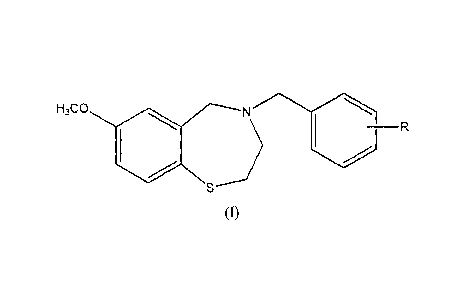
The compounds of the present invention are represented by the structure of
Formula
(I):
H3C0 N
(I)
wherein
R is COOH;
and pharmaceutically acceptable salts thereof.
The compounds of Formula (1) may be present in the form of a salt with a
pharmaceutically acceptable acid or base. Such salts are preferably selected
from the group
consisting of sodium, potassium, magnesium, hemifumarate, hydrochloride and
hydrobromide
7
CA 02870599 2014-10-16
WO 2013/156505 PCT/EP2013/057958
salts, with each possibility representing a separate embodiment of the present
invention. One
currently preferred salt is the sodium salt. Another currently preferred salt
is the
hemifumarate salt.
In some specific embodiments, the compound is selected from the group
consisting of
compound 1, compound 4 and compound 6, and pharmaceutically acceptable salts
thereof.
The structures of these compounds are described hereinbelow.
In a preferred embodiment, the compound is represented by the structure of
compound
(I):
,O
0
OH
(1)
or pharmaceutically acceptable salts thereof.
In some embodiments, compound 1 is provided as the parent compound. In other
embodiments, however, compound 1 is provided in the form of a salt with a
pharmaceutically
acceptable acid or base. Preferably, such salt is selected from the group
consisting of sodium,
potassium, magnesium, hernifumarate, hydrochloride and hydrobromide salts,
with each
possibility representing a separate embodiment of the present invention. One
currently
preferred salt is the sodium salt. Another currently preferred salt is the
hemifiimarate salt.
The present invention also provides methods for the synthesis of compounds of
the
invention, and salts thereof.
The present invention also provides pharmaceutical compositions comprising one
or
more of the compounds of the invention, and at least one additive or
excipient, e.g., fillers,
diluents, binders, disintegrants, buffers, colorants, emulsifiers, flavor-
improving agents,
gellants, glidants, preservatives, solubilizers, stabilizers, suspending
agents, sweeteners,
tonicity agents, wetting agents, emulsifiers, dispersing agents, swelling
agents, retardants,
lubricants, absorbents, and viscosity-increasing agents. The compositions may
be presented in
capsules, granules, powders, solutions, sachets, suspensions, or tablet dosage
form.
The present invention further provides methods of treating or preventing
various
disorders, diseases and conditions associated with RyRs, such as cardiac,
musculoskeletal
8
CA 02870599 2014-10-16
WO 2013/156505 PCT/EP2013/057958
cognitive, CNS and neuromuscular disorders and diseases, comprising
administering to a subject
in need of such treatment an amount of a compound of Formula (I) or a salt
thereof, effective to
prevent or treat a disorder or disease associated with an RyR. The present
invention also provides
a method of preventing or treating a leak in RyR (including RyR1, RyR2 and
RyR3) in a subject,
including administering to the subject an amount of a compound of Formula (I)
or a salt thereof;
effective to prevent or treat a leak in RyR.
In addition, the present invention provides a method of modulating the binding
of
RyRs and calstabins in a subject, including administering to the subject an
amount of a
compound of Formula (I) or a salt thereof; effective to modulate the amount of
RyR-bound
calstabin.
The present invention further relates to the use of a compound of Formula (I)
for the
manufacture of a medicament for the treatment and/or prevention of disorders,
diseases and
conditions associated with RyRs, such as cardiac, musculoskeletal and
cognitive/CNS
disorders and diseases. In another embodiment, the present invention relates
to the use of a
compound of Formula (I) for the manufacture of a medicament for preventing or
treating a
leak in RyR. In another embodiment, the present invention relates to the use
of a compound
of Formula (I) for the manufacture of a medicament for modulating the amount
of RyR-bound
calstabins.
The methods of the invention can be practiced on an in vitro system (e.g.,
cultured
cells or tissues) or in vivo (e.g., in a non-human animal or a human).
In some embodiments, the compounds of the invention are provided in
combination
with exon skipping therapy, e.g., antisense oligonucleotides (A0s) so as to
enhance exon
skipping in an mRNA of interest, e.g., the DMD gene, as further described
herein. Other
features and advantages of the present invention will become apparent from the
following
detailed description and figures.
BRIEF DESCRIPTION OF THE FIGURES
Figure 1A Immunoblot with calstabin2 antibody showing binding of
calstabin2 to
PKA-phosphorylated RyR2 in the absence (-) or presence of 100 nM
compound 1. (+): calstabin binding to non-PKA phosphorylated RyR2.
S36 (US 7,544,678), is used as a positive control.
9
CA 02870599 2014-10-16
WO 2013/156505 PCT/EP2013/057958
Figure 1B
Immunoblot with calstabin2 antibody showing binding of calstabin2 to
PKA-phosphorylated RyR2 in the absence (-) or presence of 100 tiM
compound 2, compound 3 or compound 4. (+): calstabin binding to
non-PKA phosphorylated RyR2. S36 is used as a positive control.
Figure 1C
Immunoblot with calstabinl antibody showing binding of calstabinl to
PKA-phosphorylated RyR1 in the absence (Neg) or presence of the
indicated concentrations of compound 1 or compound 4. (Pos):
calstabin binding to non-PKA phosphorylated RyR 1 . S36 is used as a
positive control.
Figure 2 Figure
2A: Immunoblot with calstabinl antibody showing the levels of
calstabinl in immunoprecipitated RyR1 complexes from tibialis lysates
in mice administered vehicle (50:50 DMSO/PEG), isoproterenol alone
(ISO) or isoproterenol together with the indicated concentrations of
compound 1 in osmotic pumps. S36 is used as control at 3.6 mM.
Figure 213: quantification of % calstabinl rebinding to RyR1.
Figure 3
Rat chronic heart failure model induced by ischemia-reperfusion (I/R)
injury. For I/R protocol, the left anterior descending (LAD) coronary
artery was occluded for 1 h.
Figure 4 Left
ventricular (LV) volumes and ejection fraction (EF) in rats treated
with compound 1 at 5 mg/kg/d (5MK) or 10 mg/kg/d (10MK) in
drinking water vs. vehicle (1120)-treated and sham-operated animals.
Chronic heart failure was induced by ischemia-reperfusion (I/R) injury.
LAD artery was occluded for 1 h; treatment started 1 week after
reperfusion and continued for 3 months. Echocardiographic parameters
were obtained after 1, 2 or 3 months of treatment. Figure 4A: LV End
Diastolic Volume; Figure 4B: LV End Systolic Volume; Figure 4C: EF.
Figures 4A and 4B: P <0.001 vs. sham; * P <0.05 vs. vehicle; t P
<0.001 vs. vehicle. Figure 4C: P <0.001 vs. sham, t P <0.001 vs.
vehicle.
CA 02870599 2014-10-16
WO 2013/156505 PCT/EP2013/057958
Figure 5 Figures 5A-C
depict body weight (BW) (5A), Infarct size (5B), and LV
weight (5C), and Figure 5D depicts collagen content in rats treated with
compound 1 at 5 mg/kg/d (5MK) and 10mg/kg/day (10MK) in drinking
water vs. vehicle(H20)-treated and sham-operated animals. Chronic
heart failure was induced by ischemia-reperfusion (1/R) injury. LAD
artery was occluded for 1 h; treatment started 1 week after reperfusion
and continued for 3 months. Parameters were measured after 3 months
of treatment. Figures 5A-C: not significant. Figure 5D:
P <0.001
vs. sham; * P <0.05 vs. vehicle.
Figure 6 Invasive
hemodynamics: Left ventricular systolic pressure (LV SP)
(GA), dP/dtmax (6B); and dP/dtmin (6C) in rats treated with compound
1 at 5 mg/kg/d (5MK) or 10mg/kg/day (10MK) in drinking water vs.
vehicle(H20)-treated and sham-operated animals. Chronic heart failure
was induced by ischemia-reperfusion (I/R) injury. LAD artery was
occluded for 1 h; treatment started 1 week after reperfusion and
continued for 3 months. Hemodynamic parameters were measured
after 3 months of treatment. Figure 6A: not significant. Figure 6B: P
<0.05 vs. sham; * P <0.05 vs. vehicle. Figure 6C: P <0.01 vs. sham;
* P <0.05 vs. vehicle.
Figure 7 Compound 1 plasma concentrations ( 114) vs. time of day.
Figure 8 EF in rats
treated with compound 1 or compound A at 5 mg/kg/d
(5MK) in drinking water vs. vehicle (I-120)-treated and sham-operated
animals. LAD artery was occluded for 1 h; treatment started 1 week
after reperfusion and continued for 3 months. Echocardiographic
parameters were obtained after 1, 2 or 3 months of treatment. P
<0.001 vs. sham; * P <0.05 vs. vehicle; t P <0.001 vs. vehicle.
Figure 9 Effect of
compound 1 on spontaneous physical activity of mdx and WT
mice as compared with vehicle (1-120)-treated controls. P<0.001 for
days 1-19 activity in nth mice dosed with 10 and 50 mg/kg/day (target
dose) administered in drinking water, compared to vehicle control.
Figure 10 Specific force-frequency relationship of EDL muscle. (A) mdx mice
treated with compound 1 (5, 10 and 50 mg/kg/d (target dose))
11
CA 02870599 2016-01-20
administered in drinking water, as compared with vehicle (H20)-treated
controls (n=5). p<0.05, for the 50 mg/kg/d dose, at frequencies of 150
Hz and above. (B) WT, C57BL/6, mice treated with compound 1 (50
mg/kg/d (target dose) administered in drinking water, as compared with
vehicle (H20)-treated controls (n=4)
Figure 11 Average body weight (12A) and average water consumption
(12B) of
mdx and WT mice treated with vehicle (H20) or compound 1 (50
mg/kg/d (target dose) administered in drinking water.
DETAILED DESCRIPTION OF THE INVENTION
It should be understood that the detailed description and the specific
examples while
indicating various embodiments of the invention are given by way of
illustration only, since
various changes and modifications will become apparent to those skilled in the
art from this
detailed description.
As used herein and in the appended claims, the singular forms "a," "an," and
"the"
include plural references unless the content clearly dictates otherwise.
The term "RycalsTm" refers to ryanodine receptor calcium channel stabilizers,
represented by compounds of the general Formula (I) or (IA) as provided by the
invention, as
well as the specific compounds designated by numerical numbers as provided by
the
invention, and herein collectively referred to as "compound(s) of the
invention".
Compounds
In some embodiments, the compounds of the present invention are represented by
the
structure of Formula (IA):
H3C0 N
-R
12
CA 02870599 2016-01-20
(IA)
wherein
R is COOH or a bioisostere thereof, COOR1 or CN; and
R1 is a Ci-C4 alkyl;
and pharmaceutically acceptable salts thereof.
In some preferred embodiments, R in Formula (IA) is a carboxylic acid (COOH).
In
other preferred embodiments, R in Formula (IA) is a carboxylic acid
bioisostere, for example
tetrazole. Alternatively, the carboxylic acid bioisostere may be an acidic
heterocycle such as
1,2,4-oxadiazol-5(4H)-one, 1,2,4-thiadiazol-5(4H)-one, 1,2,4-oxadiazole-5(4H)-
thione, 1,3,4-
oxadiazole-2(3H)-thione, 4-methyl-1H-1,2,4-triazole-5(4H)-thione, 5-
fluoroorotic acid, and
the like. Additional carboxylic acid bioisosteres are described in, e.g.,
Hamada, Y. et al.,
Bioorg. Med. Chem. Lett. 2006; 16:4354-4359; Herr, R.J. et al., Bioorg. Med
Chem. 2002; 10:
3379-3393; Olesen, P.H., Curr. Opin. Drug Discov. Devel. 2001; 4: 471; Patani.
G.A. et al.,
Chem. Rev. 1996; 96:3147; Kimura, T. et al. Bioorg. Med. Chem. Lett. 2006; 16:
2380-2386;
and Kohara, Y. et al. Bioorg. Med Chem. Lett. 1995; 5(17): 1903-1908.
In one preferred embodiment, the compounds of the present invention are
represented
by the structure of Formula (IA) wherein R is COOH and pharmaceutically
acceptable salts
thereof (i.e., a compound of formula (I)).
In other preferred embodiments, R in Formula (IA) is at position 4 of the
phenyl ring
(i.e., position 7 of the benzothiazepine ring). Each possibility represents a
separate
embodiment of the present invention. The compounds of Formula (IA) or (I) may
be present
in the form of a salt with a pharmaceutically acceptable acid or base. Such
salts are preferably
selected from the group consisting of sodium, potassium, magnesium,
hemifumarate,
hydrochloride and hydrobromide salts, with each possibility representing a
separate
embodiment of the present invention. One currently preferred salt is the
sodium salt. Another
currently preferred salt is the hemifumarate salt.
In some specific embodiments, the compound is selected from the group
consisting of
compound 1, compound 2, compound 3, compound 4, compound 5, compound 6,
compound
7, compound 8, compound 9, compound 10, compound 11, and compound 12, and
pharmaceutically acceptable salts thereof. These compounds are represented by
the following
structures:
13
CA 02870599 2014-10-16
WO 2013/156505
PCT/EP2013/057958
S
OH ;
(1)
0
0
I
S =
,
(2)
1010 1
0
S
5 0 3
(3)
OH
0 ) 1101 0
S ;
(4)
14
CA 02870599 2014-10-16
WO 2013/156505
PCT/EP2013/057958
\o
0
,,,..,.--n a )
II
S .
,
(5)
OH
0
,,,,,.--=c) 1110 ) II
S ;
(6)
N
\\
,...,/o 10 ) .
S ;
(7)
CA 02870599 2014-10-16
WO 2013/156505
PCT/EP2013/057958
/ N
//
S .
,
(8)
C) a )
Ill, __
------..N
S ;
(9)
N
\ NH
N.--------
õ,,,,,õ0 . )
IIII
S
;
(10)
16
CA 02870599 2014-10-16
WO 2013/156505 PCT/EP2013/057958
N
N.------- %
\ /N
NH
0 N
111
/ =
J
S
;and
(11)
,0 N
\\
N
/ 10
J N
H
S .
(12)
Chemical Definitions:
The term "alkyl" as used herein refers to a linear or branched, saturated
hydrocarbon
having from I to 4 carbon atoms ("CI-CI alkyl"). Representative alkyl groups
include, but are
not limited to, methyl, ethyl, propyl, isopropyl, butyl, sec-butyl, and tert-
butyl. The alkyl
group may be unsubstituted or substituted by one or more groups selected from
halogen,
haloalkyl, hydroxy, alkoxy, haloalkoxy, cycloalkyl, aryl, heteroeyelyl,
heteroaryl, amido,
alkylamido, dialkylamido, nitro, amino, cyano, N3, oxo, alkylamino,
dialkylamino, carboxyl,
thio, thioalkyl and thioaryl.
Compounds of the present invention may exist in their tautomeric form. All
such
tautomeric forms are contemplated herein as part of the present invention.
17
CA 02870599 2014-10-16
WO 2013/156505 PCT/EP2013/057958
All stereoisomers of the compounds of the present invention (for example,
those which
may exist due to asymmetric carbons on various substituents), including
enantiomeric forms
and diastereomeric forms, are contemplated within the scope of this invention.
Individual
stereoisomers of the compounds of the invention may, for example, be
substantially free of
other isomers (e.g., as a pure or substantially pure optical isomer having a
specified activity),
or may be admixed, for example, as racemates, or as mixtures enriched by one
stereoisomer.
The chiral centers of the present invention may have the S or R configuration
as defined by
the IUPAC 1974 Recommendations. The racemic forms can be resolved by physical
methods,
such as, for example, fractional crystallization, separation or
crystallization of diastereomeric
derivatives or separation by chiral column chromatography. The individual
optical isomers
can be obtained from the racemates by any suitable method, including without
limitation,
conventional methods, such as, for example, salt formation with an optically
active acid or
base, followed by crystallization.
Compounds of the present invention are, subsequent to their preparation,
preferably
isolated and purified to obtain a composition containing an amount by weight
equal to or
greater than about 90% of the compound, about 95% of the compound, and even
more
preferably greater than about 99% of the compound ("substantially pure"
compound), which is
then used or formulated as described herein. Such "substantially pure"
compounds of the
present invention are also contemplated herein as part of the present
invention.
Therapeutic Use
The present invention provides compounds that are capable of treating
conditions,
disorders and diseases associated with RyRs. More particularly, the present
invention
provides compounds that are capable of fixing a leak in RyR channels, which
may be RyR1,
RyR2 and/or RyR3 channels. In one embodiment, the compounds of the invention
enhance
association and/or inhibit dissociation of RyR and calstabin (e.g., RyR1 and
calstabinl; RyR2
and calstabin2; and RyR3 and calstabin1). "Conditions, disorders and diseases
associated
with RyRs" means disorders and diseases that can be treated and/or prevented
by modulating
RyRs and include, without limitation, cardiac disorders and diseases, muscle
fatigue,
musculoskeletal disorders and diseases, CNS disorders and diseases, cognitive
dysfunction,
neuromuscular diseases and disorders, cognitive function improvement, bone
disorders and
18
CA 02870599 2014-10-16
WO 2013/156505 PCT/EP2013/057958
diseases, cancer cachexia, malignant hyperthermia, diabetes, sudden cardiac
death, and sudden
infant death syndrome.
Thus, in one embodiment, the present invention relates to a method of treating
or
preventing a condition selected from the group consisting of cardiac disorders
and diseases,
muscle fatigue, musculoskeletal disorders and diseases, CNS disorders and
diseases, cognitive
dysfunction, neuromuscular diseases and disorders, bone disorders and
diseases, cancer
cachexia, malignant hyperthermia, diabetes, sudden cardiac death, and sudden
infant death
syndrome, or for improving cognitive function, the method comprising the step
of
administering to a subject in need thereof a therapeutically effective amount
of a compound of
Formula (I) or (IA) as described herein, or a salt thereof, to effectuate such
treatment. A
currently preferred compound is a compound of Formula (1).
In another embodiment, the present invention relates to the use of an
effective amount
of compound of Formula (I) or (IA), as described herein, or a salt thereof,
for the manufacture
of a medicament for treating or preventing a condition selected from the group
consisting of
cardiac disorders and diseases, muscle fatigue, skeletal muscular disorders
and diseases, CNS
disorders and diseases, neuromuscular diseases and disorders, cognitive
dysfunction, bone
disorders and diseases, cancer cachexia, malignant hyperthermia, diabetes,
sudden cardiac
death, and sudden infant death syndrome, or for improving cognitive function.
A currently
preferred compound is a compound of Formula (1).
In another embodiment, the present invention relates to a compound of Formula
(I) or
(IA) as described herein, or a salt thereof, for use in the manufacture of a
medicament for
treating or preventing a condition selected from the group consisting of
cardiac disorders and
diseases, muscle fatigue, skeletal muscular disorders and diseases, CNS
disorders and
diseases, cognitive dysfunction, neuromuscular diseases and disorders, bone
disorders and
diseases, cancer cachexia, malignant hyperthermia, diabetes, sudden cardiac
death, and sudden
infant death syndrome, or for improving cognitive function. A currently
preferred compound
is a compound of Formula (1).
In one embodiment, the condition, disorder or disease is associated with an
abnormal
function of RyR1. In another embodiment, the condition, disorder or disease is
associated with
an abnormal function of RyR2. In another embodiment, the condition, disorder
or disease is
associated with an abnormal function of RyR3. Each possibility represents a
separate
embodiment of the present invention.
19
CA 02870599 2014-10-16
WO 2013/156505 PCT/EP2013/057958
Cardiac disorders and diseases include, but are not limited to, irregular
heartbeat
disorders and diseases, exercise-induced irregular heartbeat disorders and
diseases, heart
failure, congestive heart failure, chronic heart failure, acute heart failure,
systolic heart failure,
diastolic heart failure, acute decompensated heart failure, cardiac
ischemia/reperfusion (I/R)
injury (including I/R injury following coronary angioplasty or following
thrombolysis during
myocardial infarction (MI)), chronic obstructive pulmonary disease, and high
blood pressure.
Irregular heartbeat disorders and diseases include, but are not limited to
atrial and ventricular
arrhythmia, atrial and ventricular fibrillation, atrial and ventricular
tachyaahythmia, atrial and
ventricular tachycardia, catecholaminergic polymorphic ventricular tachycardia
(CPVT), and
exercise-induced variants thereof.
The compounds of the invention are also useful in treating muscle fatigue,
which may
be due to prolonged exercise or high-intensity exercise, or may be caused by
musculoskeletal
diseases. Examples of muscular disorders and diseases include, but are not
limited to, skeletal
muscle fatigue, central core diseases, exercise-induced skeletal muscle
fatigue, bladder
disorders, incontinence, age-associated muscle fatigue, sareopenia, congenital
myopathies,
skeletal muscle myopathies and/or atrophies, cancer caehexia, myopathy with
cores and rods,
mitochondrial myopathies [e.g., Kearns-Sayre syndrome, MELAS (mitoehondrial
myopathy,
encephalopathy, lactic acidosis, and stroke) syndrome, and MERRF (myoclonus
epilepsy with
ragged-red fibers) syndrome], endocrine myopathies, muscular glycogen storage
diseases
[e.g., Pompe's disease, Andersen's disease, and Con's diseases],
myoglobinurias [e.g.,
McArdle's disease, Ta.rui disease, and DiMauro disease], clermatomyositis,
myositis
ossificans, familial periodic paralysis, polymyositis, inclusion body
myositis, neuromyotonia,
stiff-man syndrome, malignant hyperthermia, common muscle cramps, tetany,
myasthenia
gravis, spinal muscular atrophy (SMA), Spinal and bulbar muscular atrophy
(SBMA, also
known as spinobulbar muscular atrophy, bulbo-spinal atrophy, X-linked
bulbospinal
neuropathy (XBSN), X-linked spinal muscular atrophy type 1 (SMAX1), and
Kennedy's
disease (I(D)), and muscular dystrophy. Preferred skeletal muscular disorders
include, but
are not limited to exercise-induced skeletal muscle fatigue, a congenital
myopathy, muscular
dystrophy, age-related muscle fatigue, sarcopenia, central core disease,
cancer cachexia,
bladder disorders, and incontinence.
Examples of muscular dystrophy include, but are not limited to, Duchenne
Muscular
Dystrophy (DMD), Becker's Muscular Dystrophy (BMD), Limb Girdle Muscular
Dystrophy
(LGMD), Congenital Muscular Dystrophy (CMD), distal muscular dystrophy,
CA 02870599 2014-10-16
WO 2013/156505 PCT/EP2013/057958
facioscapulohumeral dystrophy, myotonic muscular dystrophy, Emery-Dreifuss
muscular
dystrophy, and oculopharyngeal muscular dystrophy, with DMD being currently
preferred.
Congenital muscular dystrophy as used herein refers to muscular dystrophy that
is
present at birth. CMD is classified based on genetic mutations: 1) genes
encoding for
structural proteins of the basal membrane or extracellular matrix of the
skeletal muscle fibres;
2) genes encoding for putative or demonstrated glycosyltransferases, that in
turn affect the
glycosylation of dystroglycan, an external membrane protein of the basal
membrane; and 3)
other. Examples of CMD include, but are not limited to Laminin-u2¨deficient
CMD
(MDC1A), Ullrich CMG (UCMDs 1, 2 and 3), Walker-Warburg syndrome (WWS), Muscle-
eye-brain disease (MEB), Fukuyama CMD (FCMD), CMD plus secondary laminin
deficiency
1 (MDC1B), CMD plus secondary laminin deficiency 2 (MDC1C), CMD with mental
retardation and pachygyria (MDC1D), and Rigid spine with muscular dystrophy
Type
(RSMD1).
Cognitive dysfunction may be associated with or includes, but is not limited
to
memory loss, age-dependent memory loss, post-traumatic stress disorder (PTSD),
attention
deficit hyperactivity disorder (ADHD), autism spectrum disorder (ASD),
generalized anxiety
disorder (GAD), obsessive compulsive disorder (OCD), Schizophrenia, Bipolar
disorder, or
major depression.
CNS disorders and diseases include, but are not limited to Alzheimer's Disease
(AD),
neuropathy, seizures, Parkinson's Disease (PD), and Huntington's Disease (HD).
Neuromuscular disorders and diseases include, but are not limited to
Spinocerebellar
ataxia (SCA), and Amyotrophic lateral sclerosis (ALS, Lou Gehrig's disease).
In some embodiments, the compounds of the present invention improve cognitive
function, which may be selected from short term memory, long term memory,
attention,
learning, and any combination thereof.
In some embodiments, the compounds of the present invention are useful in the
treatment of
cancer cachexia, i.e., muscle weakness which is associated with cancer in
general, and
preferably muscle weakness in metastatic cancer, such as bone metastases.
Muscle weakness
and muscle atrophy (cachexia) are common paraneoplastic symptoms in cancer
patients.
These conditions cause significant fatigue and dramatically reduce patients'
quality of life.
The present invention provides a method for treating and preventing muscle
weakness in a
21
CA 02870599 2014-10-16
WO 2013/156505 PCT/EP2013/057958
cancer patient, based, in part, on the discovery that, in certain types of
cancers, e.g., prostate
and breast cancer with bone metastases, RyR1 is oxidized which induces it to
become "leaky".
It has further been found that prevention of the leak by administration of
Rycal compounds
improves muscle function. Exemplary cancers include, but are not limited to,
breast cancer,
prostate cancer, bone cancer, pancreatic cancer, lung cancer, colon cancer,
and gastrointestinal
cancer.
Exon skipping therapy:
In some embodiments, the compounds of the present invention modulate (e.g.,
enhance)
mRNA splicing by enhancing antisense-mediated exon skipping. This modulation
of splicing
is accomplished in the presence of antisense oligonucleotides (A0s) that are
specific for
splicing sequences of interest. In some embodiments of the invention, the
compound of
formula (I) or (IA) and the AO can act synergistically wherein the compound of
formula (I) or
(IA) enhances AO mediated exon skipping.
Thus, in some embodiments, the present
invention relates to a pharmaceutical composition for use in the treatment or
prevention of any
of the conditions described herein that are associated with Leaky RyR, further
comprising the
use of an antisense AO which is specific for a splicing sequence in an mRNA
sequence, for
enhancing exon skipping in the mRNA of interest.
One particular embodiment for exon skipping enhancement by the compounds of
the present
invention pertains to Duchenne Muscular Dystrophy (DMD). DMD is a lethal X-
linked
recessive disease characterized by progressive muscle weakness over a
patient's lifetime.
DMD is primarily caused by out of frame multi-exon deletions in the DMD gene
that ablate
dystrophin protein production. Loss of dystrophin expression alone does not
explain DMD
pathophysiology. Disruption of the dystrophin-glycoprotein complex (DGC) also
results in
oxidative stress, mitochondrial Ca2+ overload and apoptosis, increased influx
of Ca2+ into the
muscle, and pathologic Ca2+ signaling. There are no curative therapies for
DMD, and the only
demonstrated pharmacological treatment is corticosteroids, which may prolong
ambulation,
but have substantial side effects. Antisense oligonucleotide¨mediated exon
skipping is a
promising therapeutic approach aimed at restoring the DMD reading frame and
allowing
expression of an intact dystrophin glyeoprotein complex. To date, low levels
of dystrophin
protein have been produced in humans by this method. Kendall et al. (Sci
Transl Med, 2012,
4(164), p. 164ra160) reported that certain small molecules such as Dantrolene
and other RyR
modulators, potentiate antisense oligomer-guided exon skipping to increase
exon skipping to
22
CA 02870599 2014-10-16
WO 2013/156505 PCT/EP2013/057958
restore the mRNA reading frame, the sarcolemmal dystrophin protein, and the
dystrophin
glycoprotein complex in skeletal muscle of mcix mice, a mouse model of DMD.
Thus, in one embodiment, the present invention relates to a method for
treating DMD,
by administering to a subject in need thereof a compound of formula (I) or
(IA) according to
the present invention, in combination with an antisense oligonucleotide (AO)
which is specific
for a splicing sequence of one or more exons of the DMD gene, for example exon
23, 45, 44,
50, 51, 52 and/or 53 of the DMD gene. Preferred AOs include, but are not
limited to, AOs
targeting DMD exon 23, 50 and/or 51 of the DMD gene, such as 2'-0-methyl
(2'0Me)
phosphorothioate or phosphorodiamidate morpholino (PMO) AOs. Examples of such
AOs
include, but not limited to, Pro051/GSK2402968, AVI4658/Eteplirsen, and PM0
E23
morpholino (5'-GGCCAAACCTCGGCTTACCTGAAAT-3').
The term an "effective amount," "sufficient amount" or "therapeutically
effective
amount" of an agent as used herein interchangeably, is that amount sufficient
to effectuate
beneficial or desired results, including clinical results and, as such, an
"effective amount" or
its variants depends upon the context in which it is being applied. The
response is in some
embodiments preventative, in others therapeutic, and in others a combination
thereof. The
term "effective amount" also includes the amount of a compound of the
invention, which is
"therapeutically effective" and which avoids or substantially attenuates
undesirable side
effects.
As used herein and as well understood in the art, "treatment" is an approach
for
obtaining beneficial or desired results, including clinical results.
Beneficial or desired clinical
results can include, but are not limited to, alleviation or amelioration of
one or more
symptoms or conditions, diminishment of extent of disease, stabilized (i.e.,
not worsening)
state of disease, preventing spread of disease, delay or slowing of disease
progression,
amelioration or palliation of the disease state and remission (whether partial
or total), whether
detectable or undetectable. "Treatment" can also mean prolonging survival as
compared to
expected survival if not receiving treatment.
Pharmaceutical Compositions
The compounds of the invention are formulated into pharmaceutical compositions
for
administration to human subjects in a biologically compatible form suitable
for administration
23
CA 02870599 2014-10-16
WO 2013/156505 PCT/EP2013/057958
in vivo. According to another aspect, the present invention provides a
pharmaceutical
composition comprising compounds of the invention in admixture with a
pharmaceutically
acceptable diluent and/or carrier. The pharmaceutically-acceptable carrier is
preferably
"acceptable" in the sense of being compatible with the other ingredients of
the composition
and not deleterious to the recipient thereof.
The compound may be administered alone, but is preferably administered with
one or
more pharmaceutically acceptable carriers. The pharmaceutically-acceptable
carrier employed
herein may be selected from various organic or inorganic materials that are
used as materials
for pharmaceutical formulations and which are incorporated as any one or more
of fillers,
diluents, binders, disintegrants, buffers, colorants, emulsifiers, flavor-
improving agents,
gellants, glidants, preservatives, solubilizers, stabilizers, suspending
agents, sweeteners,
tonicity agents, wetting agents, emulsifiers, dispersing agents, swelling
agents, retardants,
lubricants, absorbents, and viscosity-increasing agents.
The compounds of the present invention are administered to a human or animal
subject
by known procedures including, without limitation, oral, sublingual, buccal,
parenteral
(intravenous, intramuscular or subcutaneous), transdermal, per- or trans-
cutaneous, intranasal,
intra-vaginal, rectal, ocular, and respiratory (via inhalation
administration). The compounds
of the invention may also be administered to the subject by way of delivery to
the subject's
muscles including, but not limited to, the subject's cardiac or skeletal
muscles. In one
embodiment, the compound is administered to the subject by way of targeted
delivery to
cardiac muscle cells via a catheter inserted into the subject's heart. In
other embodiments, the
compounds may be administered directly into the CNS, for example by
intralumbar injection
or intreventricular infusion of the compounds directly into the cerebrospinal-
fluid (CSF), or by
intraventricular, intrathecal or interstitial administration.
Oral administration is currently
preferred.
The pharmaceutical compositions according to the invention for solid oral
administration include especially tablets or dragees, sublingual tablets,
sachets, capsules
including gelatin capsules, powders, and granules, and those for liquid oral,
nasal, buccal or
ocular administration include especially emulsions, solutions, suspensions,
drops, syrups and
aerosols. The compounds may also be administered as a suspension or solution
via drinking
water or with food. Examples of acceptable pharmaceutical carriers include,
but are not
limited to, cellulose derivatives including carboxymethyl cellulose, methyl
cellulose,
24
CA 02870599 2014-10-16
WO 2013/156505 PCT/EP2013/057958
hydroxypropyl cellulose, hydroxypropylmethylcellulose, ethyl cellulose and
microcrystalline
cellulose; sugars such as rnannitol, sucrose, or lactose; glycerin, gum
arabic, magnesium
stearate, sodium stearyl fumarate, saline, sodium alginate, starch, talc and
water, among
others.
The pharmaceutical compositions according to the invention for parenteral
injections
include especially sterile solutions, which may be aqueous or non-aqueous,
dispersions,
suspensions or emulsions and also sterile powders for the reconstitution of
injectable solutions
or dispersions. The compounds of the invention may be combined with a sterile
aqueous
solution that is isotonic with the blood of the subject. Such a formulation is
prepared by
dissolving a solid active ingredient in water containing physiologically-
compatible substances,
such as sodium chloride, glycine and the like, and having a buffered pH
compatible with
physiological conditions, so as to produce an aqueous solution, then rendering
said solution
sterile. The formulation is presented in unit or multi-dose containers, such
as sealed ampoules
or vials. The formulation is delivered by any mode of injection, including,
without limitation,
epifascial, intracapsular, intracranial, intracutaneous, intrathecal,
intramuscular, intraorbital,
intraperitoneal, intraspinal, intrasternal, intravascular, intravenous,
parenchymatous,
subcutaneous, or sublingual or by way of catheter into the subject's heart.
The pharmaceutical compositions for rectal or vaginal administration are
preferably
suppositories, and those for per- or trans-cutaneous administration include
especially powders,
aerosols, creams, ointments, gels and patches.
For transdermal administration, the compounds of the invention are combined
with
skin penetration enhancers, such as propylene glycol, polyethylene glycol,
isopropanol,
ethanol, oleic acid, N-methylpyrrolidone and the like, which increase the
permeability of the
skin to the compounds of the invention and permit the compounds to penetrate
through the
skin and into the bloodstream. The compound/enhancer compositions also may be
further
combined with a polymeric substance, such as ethylcellulose, hydroxypropyl
cellulose,
ethylene/vinylacetate, polyvinyl pyrrolidone, and the like, to provide the
composition in gel
form, which is dissolved in a solvent, evaporated to the desired viscosity and
then applied to
backing material to provide a patch.
The pharmaceutical formulations of the present invention are prepared by
methods
well-known in the pharmaceutical arts, including but not limited to wet and
dry granulation
methods, or by direct compression. The choice of carrier is determined by the
solubility and
CA 02870599 2014-10-16
WO 2013/156505 PCT/EP2013/057958
chemical nature of the compounds, chosen route of administration and standard
pharmaceutical practice.
The pharmaceutical compositions mentioned above illustrate the invention but
do not
limit it in any way.
In accordance with the methods of the present invention, any of these
compounds may
be administered to the subject (or are contacted with cells of the subject) in
an amount
effective to limit or prevent a decrease in the level of RyR-bound calstabin
in the subject,
particularly in cells of the subject. This amount is readily determined by the
skilled artisan,
based upon known procedures, including analysis of titration curves
established in vivo and
methods and assays disclosed herein. A suitable amount of the compounds of the
invention
effective to limit or prevent a decrease in the level of RyR-bound calstabin
in the subject
ranges from about 0.01 mg/kg/day to about 100 mg/kg/day (e.g., 1, 2, 5, 10,
20, 25, 50 or 100
mg/kg/day), and/or is an amount sufficient to achieve plasma levels ranging
from about 300
ng/rnl to about 5,000 ng/ml. Alternatively, the amount of compounds from the
invention
ranges from about 1 mg/kg/day to about 50 mg/kg/day. Alternatively, the
amount of
compounds from the invention ranges from about 10 mg/kg/day to about 20
mg/kg/day. Also
included are amounts of from about 0.01 mg/kg/day or 0.05 mg/kg/day to about 5
mg/kg/day
or about 10 mg/kg/day which can be administered.
Methods of Synthesis
The present invention provides, in a further aspect, processes for the
preparation of a
compound of the invention, and salts thereof. More particularly, the present
invention
provides processes for the preparation of compounds of Formula (I) or (IA),
e.g., compound 1,
compound 2, compound 3, compound 4, compound 5, compound 6, compound 7,
compound
8, compound 9, compound 10, compound 11, and compound 12, or salts thereof The
various
synthetic routes to the compounds are described in the examples. The general
route of
synthesis (ROS) is set forth in Scheme 1 below:
26
CA 02870599 2016-01-20
LI a
H3C0 NH (1,s.,:j¨HR3C 0 N
IP s j _________________________ b.
H3C0 N
S
Scheme 1
In Scheme 1, Ra is COOR1 or CN; R1 is a Ci-C4 alkyl, and L is a leaving group,
which is, by
way of example, a halogen, a sulfonate (OSO2R' wherein R' is alkyl or aryl,
e.g., OMs
(mesylate), OTs (tosylate)), and the like. The amine starting material is
reacted with the
alkylating agent (benzyl derivative shown above), preferably in the presence
of a base, to
yield the desired product or a precursor thereof (R=Ra). If desired, such
precursor may further
be reacted to convert the group Ra to the group R as exemplified in the
experimental section
herein below, or by any other method known to a person of skill in the art.
For example, an
ester precursor (Ra =COOR1 wherein R1 is a CI-C4 alkyl), can be converted into
the
corresponding carboxylic acid (R = COOH) by hydrolysis under acidic or basic
conditions in
accordance with known methods. Alternatively, a nitrile precursor (Ra = CN)
can be
converted into a tetrazole (a carboxylic acid isostere) by reaction with
sodium azide under
suitable conditions, or to a carboxylic acid (R = COOH) by hydrolysis.
The amine starting material may be prepared in accordance with the methods
described in WO 2009/111463 or WO 2007/024717, or by any other method known to
a
person of skill in the art. The nature of the base is not particularly
limiting. Preferred bases
include, but are not limited to, hydrides (e.g., sodium or potassium hydride)
and N,N-
diisopropylethylamine. Other suitable bases include, but are not limited to an
organic base
such as a tertiary amine, selected from the group consisting of acyclic amines
(e.g.,
trimethylamine, triethylamine, dimethylphenylamine diisopropylethylamine and
tributylamine), cyclic amines (e.g., N-methylmorpholine) and aromatic amines
(dimethylaniline, dimethylaminopyridine and pyridine).
The reaction may be conducted in the presence or absence of a solvent. The
nature of
the solvent, when used, is not particularly limiting, with examples including
solvents such an
27
CA 02870599 2016-01-20
ester (e.g., ethyl acetate), an ether (e.g., THF), a chlorinated solvent
(e.g., dichloromethane or
chloroform), dimethylformamide (DMF), and other solvents such as acetonitrile
or toluene or
mixtures of these solvents with each other or with water.
Salts of compounds of formula (I) wherein R=COOH may be prepared by reacting
the
parent molecule with a suitable base, e.g., NaOH or KOH to yield the
corresponding alkali
metal salts, e.g., the sodium or potassium salts. Alternatively, esters
(R=COOR1) may be
directly converted to salts by reactions with suitable bases.
Salts of compounds of formula (I) may also be prepared by reacting the parent
molecule with
a suitable acid, e.g., HC1, fumaric acid, or para-toluenesulfonic acid to
yield the corresponding
salts, e.g., hydrochloride, tosylate or hemi-fumarate.
EXAMPLES
The following examples are provided as illustrations of the some preferred
embodiments according to the invention.
EXAMPLE 1: Synthesis
Instruments:
NMR: Bruker AVANCETM III 400 or Varian MercuryTM 300
LC/MS: Waters DeltaTm 600 equipped with Autosampler 717Plus, Photo Diode Array
Detector 2996, and Mass Detector 3100, or ShimadzuTM 210
General procedure for the alkylation of 7-
methoxy-2,3,4,5-
tetrahydrobenzo[f][1,4]thiazepine ("Amine").
H3C0
0 JNH
S
Amine
Amine (structure shown above) (1 mmol) was dissolved in 3 ml dichloromethane.
To
the solution was added alkylation reagent (Immol), followed by N,N-
diisopropylethylamine
28
CA 02870599 2014-10-16
WO 2013/156505 PCT/EP2013/057958
(0.34 ml, 2 mmol). The mixture was stirred at room temperature overnight. The
solution was loaded onto column directly and eluted with hexane/Et0Ac (2:1,
v/v).
COOCH3
\
Sj
Compound 2
Methyl 3 -((7-methoxy-2,3-dihydrobenzo [f] [1,41thi azepin-4(5H)-
yl)methyl)benzoate: I HNMR
(300 MHz, CDC13): 7.96 (m, 211), 7.46 (m, 311), 6.70 (dd, J =8.4 Hz, 3.0 Hz,
1H), 6.50 (d, J
2.7 Hz, 111), 4.09 (s, 21-1), 3.90 (s, 3H), 3.72 (s, 31-1), 3.57 (s, 2H), 3.35
(m, 211), 2.72 (m, 211).
MS: 344(M+1)
N\
coo..3
Compound 3
Methyl 4-((7-methoxy-2,3-dihydrobenzo[f][1,4]thiazepin-4(5H)-
yl)methyl)benzoate: 1HNMR
(300 MHz, CDC13): 7.99 (d, J= 8.4 Hz, 2E1), 7.46 (d, J= 8.4 Hz, 114), 7.37 (d,
J= 8.7 Hz, 2H),
6.70 (dd, J ¨8.4 Hz, 3.0 Hz, 1H), 6.50 (d, J = 2.7 Hz, 1H), 4,09 (s, 211),
3.90 (s, 31-1), 3.72 (s,
3H), 3.57 (s, 2H), 3.35 (m, 211), 2.72 (m, 211). MS: 344(M+1)
H3COOC
=N
Si HCI
Compound 5
Methyl 2-((7-methoxy-2,3-dihydrobenzo[f][1,4]thiazepin-4(5H)-
yl)methyl)benzoate: The
compound was converted to hydrochloride salt with 2M HC1 in ether. iHNMR (300
MHz,
DMSO-d6): 10.33(br, 111), 8.08 (d, J= 7.5Hz, 1H), 7.80-7.65 (m, 3H), 7.51 (d,
J=8.1Hz, 1H),
7.14 (s, 1H), 6.99 (dd, J= 8.4, 2.1Hz, 111), 4.90-4.40 br,
4H), 3.88 (s, 311), 3.78 (s, 311),
3.40 (m, 211), 3.26 (m, 1H), 3.11 (m, 1H). MS: 344 (M+1)
29
CA 02870599 2014-10-16
WO 2013/156505 PCT/EP2013/057958
NC
N\
Compound 7
2((7-Methoxy-2,3-dihydrobenzo[f][1,4]thiazepin-4(5H)-yOmethypbenzonitrile:
iHNMR
(300 MHz, CDC13): 7.67-7.26 (m, 5H), 6.73 (d, J= 2.7 Hz, 1H), 6.74 (dd, .1=
2.7,8.4 Hz, 111),
4.14 (s, 2H), 3.78(s, 311), 3.70 (s, 2H), 3.36 (m, 2H), 2.76 (m, 211). MS :
311 (M+1)
CN
=
0 11#
N\
Compound 8
3-((7-Methoxy-2,3-dihydrobenzo[f][1,4]thiazepin-4(5H)-Amethyl)benzonitrile:
IHNMR
(300 MHz, CDC13): 7.64-7.42 (m, 5H), 6.74 (dd, .1= 2.7,8.4 Hz, 1H), 6.48 (d,
.1= 2.7 Hz, 111),
4.08 (s, 2H), 3.75(s, 3H), 3.57 (s, 2121), 3.36 (m, 2H), 2.76 (m, 211). MS :
311 (M+1)
0
411 N CN
Compound 9
4-((7-Methoxy-2,3-dihydrobenzo [11 [1,41thiazepin-4(5H)-ypmethyl)benzonitrile:
HNMR
(300 MHz, CDC13): 7.64 (d, .1.= 7.2Hz, 2H), 7.42 (m, 311), 6.74 (dd, .1=
2.7,8.4 Hz, 111), 6.48
(d, .1-= 2.7 Hz, 111), 4.08 (s, 211), 3.75(s, 311), 3.58 (s, 214), 3.36 (m,
2H), 2.76 (m, 211). MS:
311 (M+1)
Hydrolysis of ester (general procedure)
Methyl ester (3 mmol) was dissolved in 30 ml of TI-IF/methanol/1 M NaOH
(1:1:1,
v/v). The mixture was stirred for 8 hours and TLC showed complete
disappearance of the
ester. 1 ml Conc. HC1 was added to adjust to acidic pH. The organic solvent
was removed and
the formed solid was collected by filtration. The solid was dried in the air.
CA 02870599 2014-10-16
WO 2013/156505 PCT/EP2013/057958
COON
O,)
=
Compound 4
34(7-Methoxy-2,3-dihydrobenzo[f][1,41thiazepin-4(5H)-yl)methyl)benzoic acid:
This was
obtained by extraction with Et0Ac as solvent. IHNMR (300 MHz, CDC13): 8.10 (s,
111), 8.04
(d, J= 8.4 Hz, 1H), 7.80 (br, 1H), 7.46 (m, 2H), 6.80 (m, 2H), 4.40 (s, 2H),
3.90 (s, 2H), 3.76
(s, 3H), 3.42 (s, 2H), 2.86 (s, 2H). MS: 330 (M+1), 328 (M-1).
(30 N\
COOH
Compound 1
4((7-Methoxy-2,3-dihydrobenzo[f][1,4]thiazepin-4(5F1)-Amethypbenzoic acid:
This was
obtained by extraction with Et0Ac as solvent. 1HNMR (300 MHz, CDCI3): 8.02 (d,
J 8.4 Hz,
2H), 7.46 (d, .1= 8.4 Hz, 1H), 7.42 (d, J 8.7 Hz, 2H), 6.70 (dd, J =8.4 Hz,
3.0 Hz, 1H), 6.50
(d, J = 3.0 Hz, 1H), 4.11 (s, 2H), 3.72 (s, 3H), 3.62 (s, 2H), 3.35 (m, 211),
2.76 (m, 2H). MS:
330 (M+1), 328 (MA).
Compound 1, sodium salt:
The sodium salt of compound I was prepared from the parent molecule using 1
equivalent of
NaOH in Et0H (m.p. of the salt: > 290 C).
1HNMR (DMSO-D6, 600MHz), 6 (ppm) : 7.77 (211, in), 7.41 (1H, d), 7.13 (2H, m),
6.75 (111,
dd), 6.63 (1H, d), 4.00 (21-1, s), 3.70 (311, s), 3.49 (211, s), 3.18 (21-1,
m), 2.70 (211, m).
Compound 1, hemifumarate salt:
1.6 g of compound 1 (neutral form) and 265 mg of fumaric acid were introduced
in a round
bottom flask. After addition of 18 mL of acetone and 2 mL of water, the
reaction mixture was
refluxed. A partial solubilisation was observed (but no complete
clarification) followed by
precipitation. The reaction mixture was then refluxed overnight. After cooling
the residual
solid was isolated by filtration, washed with 3 mL of acetone and dried under
vacuum (40 C /
10 mbars) for 4 hours.
31
CA 02870599 2014-10-16
WO 2013/156505 PCT/EP2013/057958
1HNMR (DMSO-D6, 600MHz), 8 (ppm) : 12.97 (211, bs), 7.90 (2H, m), 7.43 (1H,
d), 7.40
(2H, m), 6.77 (114, dd), 6.64 (1H, d), 6.62 (1H, s), 4.03 (2H, s), 3.70(311,
s), 3.58 (21-1, s), 3.20
(2H, m), 2.72 (2H, m).
HO2C
dth N
S HCI
Compound 6
24(7-Methoxy-2,3-dihydrobenzo[f][1,4]thiazepin-4(5H)-y1)methy1)benzoic
acid: The
compound was converted to hydrochloride salt with 2M HC1 in ether. 1HNMR (300
MHz,
DMSO-d6): 10.10(br, 1H), 8.08 (d, J 7.5Hz, 111), 7.66-7.51 (m, 4H), 7.17 (d, J
2.1I1z, 114),
6.99 (dd, J 8.4, 2.1Hz, 111), 4.80-4.40 (m, br, 41-1), 3.78 (s, 311), 3.46 (m,
2H), 3.13 (m, 2H).
MS: 330(M+1), 328 (M-1).
Synthesis of tetrazole (general procedure)
Nitrile precursor (3.22 mmol), sodium azide (830 mg, 12.9 mmol) and
triethylamine
hydrochloride (1.72 g, 12.9 mmol) were stirred in 40 ml anhydrous DMF at 100 C
for 5 days.
The DMF was removed under high vacuum and the residue was mixed with water.
The water
solution was extracted with dichloromethane (3 x 100m1), The pure compound was
purified
by column chromatography (Et0Acimethanol).
N¨
õ0 ritt N\ 111P
Compound 10
4-(2-(1H-Tetrazol-5-yl)benzyl)-7-methoxy-2,3,4,5-
tetrahydrobenzo[f][1,4]thiazepine:
1HNMR (300 MHz, CDC13 and a drop of CD30D): 8.30 (d, .1= 8.7Hz, 111), 7.53 (m,
211).
7.14 (t, J 7.8Hz, 111), 7.20 (d, J= 7.5Hz,1H), 6.84 (dd, .T.= 2.7,8.4 Hz, 1H),
6.69 (d, .1= 2.7
32
CA 02870599 2014-10-16
WO 2013/156505 PCT/EP2013/057958
Hz, 1T-I), 4.46 (s, 211), 3.80(s, 2H), 3.75 (s, 2H), 3.43 (m, 211), 2.96 (m,
211). MS: 354(M+1),
352(M-1)
11
0
sjN
Compound 11
4-(3-(1H-Tetrazol-5-yl)benzyl)-7-methoxy-2,3,4,5-
tetranydrobenzo[f][1,4]thiazepine:
1HNMR (300 MHz, CDC13): 8.16 (s, 111), 7.90 (d, J=7.5Hz, 111), 7.40 (d, J=
8.4Hz, 1H),7.20
(m, 211), 6.74 (dd, .1= 2.7,8.4 Hz, 1H), 6.58 (d, J= 2.7 Hz, 1H), 4.18 (s,
211), 3.75(s, 5H), 3.36
(m, 2H), 2.76 (m, 211). ). MS: 354(M+1), 352(M-1)
siN 1,111,.
õ,
Compound 12
4-(4-(1H-Tetrazol-5-y1)benzyl)-7-methoxy-2,3,4,5-
tetrahydrobenzo[f][1,41thiazepine:
1HNMR (300 MHz, CDC13 and a drop of CD30D): 7.99 (d, J= 7.2Hz, 211), 7.42 (m,
3H),
6.74 (dd, I= 2.7,8.4 Hz, 111), 6.53 (d, J.¨ 2.7 Hz, 11-1), 4.10 (s, 2H),
3.71(s, 3H), 3.58 (s, 2H),
3.36 (in, 2H), 2.76 (m, 2H). ). MS: 354(M+1), 352(M-1)
Synthesis of 7-methoxy-2,3,4,5-tetrahydrobenzolf111,41thiazepine ("Amine").
111" SH HC1
0
.--
0
O 0
1.5 eq. K2CO3; 1.0 eq. DlEA S,NH2 _______________________ NI
THF, reflux, overnight NaHCO3, water A
1 DC1111, rt
2
10 eq.(HCHO)n ,.0 14\1'0 air 33% HBr in Acetic tic! 1101 HBr
---I
0.4 eq.PTSA s
S
3 4
2-(4-Methoxyphenylthio)ethanamine (1)
33
CA 02870599 2014-10-16
WO 2013/156505 PCT/EP2013/057958
4-Methoxythiophenol (50 g, 0.357 mol), 2-chloroethylamine monohydrochloride
(39.8 g,
0.343 mol.), K2CO3( 78.8 g, 0.57 mol) and diisopropyl ethylamine (32 mL, 0.178
mol) were
mixed in 200 mL of THF. The mixture was degassed for 5 min. under reduced
pressure and
refluxed under argon overnight. The solvent was removed and water (300 mL) was
added to
the flask. The mixture was extracted with dichloromethane (3 x 200 mL). The
organics were
collected, dichloromethane was removed and 50 mL conc. HC1 was added, followed
by 200
mL of water. The solution was extracted with 1:1 Et0Ac/hexane (3 x 200 mL).
The aqueous
layer was adjusted to pH 10 with 2 M NaOH, and was extracted with
dichloromethane (3 x
200 mL). The combined organic solution was dried over anhydrous sodium
sulfate. Removal
of solvent provided 61 g of the target compound as a colorless liquid, with a
yield of 97%.
1H-NMR (300 MHz, CDC13): 7.35(d, J 8.7 Hz, 211), 6.81 (d, J= 8.7 Hz, 2H), 3.77
(s, 3H),
2.88-2.80 (m, 41-1), 1.44 (s, 2H).
Benzyl 2-(4-methoxyphenylthio)ethylearbamate (2)
First method
To a the flask containing compound 1(8.0 g, 43.7 mmol), sodium bicarbonate
(12.1 g, 144
mmol), water (100 mL) and dichloromethane (200 mL) was added benzyl
chloroforrnate (8.2
g, 48.1 mmol, diluted in 100 mL of dichloromethane) dropwise at 0 C. After
the addition, the
mixture was stirred at r.t. for 5 hr. The organic layer was collected and
aqueous solution was
extracted with 100 mL of dichloromethane. The combined organic solution was
dried over
sodium sulfate. The solvent was removed and the resulting solid was triturated
with 200 mL
of THF/hexane (1:10). The solid was collected and dried leaving the target
product (12.9 g) in
the yield of 93%.
Alternative method
To the solution of compound 1(10 g, 54.6 mmol) and triethylamine (15 mL, 106
mmol) in
200 mL of dichloromethane was added benzyl chloroformate (7.24 mL, 51.5 mmol,
diluted in
100 mL of dichloromethane) dropwise at 0 C. After the addition, the solution
was stirred at
r.t. for one hour. The solid was removed by filtration. The solution was
extracted with 100
mL of 0.1 M HC1 and 100 mL of sat. sodium carbonate, and dried over anhydrous
sodium
sulfate. Removal of solvent provided a white solid that was stirred in 200 mL
of THF/hexane
34
CA 02870599 2014-10-16
WO 2013/156505 PCT/EP2013/057958
(1:20) for three hours. The solid was collected by filtration to give 14.2 g
of the target
compound in 87% yield.
11-I-NMR (300 MHz, CDC13): 7.35(m, 7H), 6.83 (d, J= 8.7 Hz, 2H), 5.07 (m, 3H),
3.77 (s,
3H), 3.10(q, J = 6.3 Hz, 2H), 2.92 (t, J=6.3 Hz, 2H).
Benzyl 7-methoxy-2,3-dihydrobenzo[f][1,41thiazepine-4(5H)-earboxylate (3)
A mixture of compound 2 (7.3 g, 23 mmol), paraformaldehyde (6.9 g 0.23 mol)
and p-
toluenesulfonic acid (1.45 g, 7.6 mmol) in 250 mL of toluene was stirred at 70
C overnight.
After cooling to r.t., the solid was filtered off The solution was extracted
with sat. sodium
carbonate (100 mL), and the organic layer was dried over anhydrous sodium
sulfate. The
target product (7.4 g) was obtained as a liquid after removal of the solvent
in 97% yield.
11-1-NMR (300 MHz, CDC13): 7.44 (d, J= 8.1 Hz, 0.77H), 7.32 (m, 5.60H), 7.07
(d, J= 2.7 Hz,
0.33H), 6.68 (m, 1.3011), 5.04 (s, 211), 4.59 (ss, 211), 3.96 (br, 1.80), 3.80
(ss, 1.23 H), 3.55 (s,
1.97H), 2.76 (m, 2H).
7-Methoxy-2,3,4,5-tetrahydrobenzo[f][1,4]thiazepine hydrobromide (Amine) (4
HBr salt)
First method
A solution of HBr (33% in acetic acid, 10 mL) was added to the compound 3 (4.2
g, 12.8
mmol). After the addition, carbon dioxide began to develop and a white solid
formed. The
mixture was let stand at r.t. for another 2 hours. Diethyl ether (150 mL) was
added to the
mixture, and it was stirred for 30 min. The solid was collected by filtration
and washed with
diethyl ether. The solid was dried under vacuum to give the 3.40 g of the
target compound
with the yield of 91.8%.
1H-NMR (300 MHz, DMSO-d6): 9.02 (br, 2H), 7.52 (d, .1= 8.1 Hz, 111), 7.27 (d,
J 3.3 Hz,
1H), 6.92 (dd, J= 8.4, 2.7 Hz, 1H), 4.41 (s, 211), 3.77 (s, 3H), 3.53 (m, 2H),
2.96 (m, 2H).
Alternative method (free base 4a)
Compound 3 (10 g, 30 mmol) was mixed with 50 mL of conc. HC1, 50 mL of water
and 30
mL of dioxane. The mixture was stirred at 100 C overnight. After cooling to
r.t., most of the
solvent and HC1 was removed under reduced pressure. Water (100 mL) was added
to the
solution and the solid was filtered off The aqueous solution was extracted
with
CA 02870599 2016-01-20
Et0Ac/hexane (1:1, 3 x100 mL) and basified by adding 15 g of NaOH. The mixture
was
extracted with dichloromethane (3 x 150 mL). The combined solution was dried
over
anhydrous sodium sulfate. Removal of solvent provided a liquid that solidified
after standing
at rt. leaving 6.2 g of target compound.
IH-NMR (300 MHz, CDC13): 7.42 (d, J= 8.1 Hz, 1H), 6.78 (d, J= 2.7 Hz, H), 6.68
(dd, J= 2.7,
8.1 Hz, 1H), 4.08 (s, 2H), 3.96 (br, 1.80), 3.76 (s, 3 H), 3.38 (m, 2H), 2.68
(m, 2H).
EXAMPLE 2: Binding of calstabin2 to PKA-phosphorylated RyR2
Cardiac SR membranes were prepared as previously described (Marx et al., 2000;
Kaftan et al., Circ. Res., 1996, 78:990-97). Immunoblotting of microsomes (50
f.tg) was
performed as described, with anti-calstabin antibody (1:1,000) (Jayaraman et
al., J. Biol.
Chem., 1992, 267:9474-77) for 1 hr at room temperature (Reiken et al.,
Circulation,
107:2459-66, 2003). After incubation with HRP-labeled anti-rabbit IgG (1:5,000
dilution;
Transduction Laboratories, Lexington, Ky.), the blots were developed using ECL
(Amersham
Pharmacia, Piscataway, N.J.) and detected on x-ray film, or exposed to
secondary antibodies
labeled with infrared Dye and visualized on equipment from Li-Cor
BiosciencesTm (model
Odyssey). Unless otherwise stated, compounds were tested at a concentration of
100 nM. A
representative calstabin2 binding assay is presented below.
A. PKA phosphorylation of cardiac sarcoplasmic reticulum (CSR)
Reaction mixture was set up in 1.5 ml microfuge tube. 200 g of cardiac SR were
added to a reaction mix of kinase buffer, PKA and ATP to a final volume of 100
IA
(Reaction mix below). ATP was added last to initiate the reaction.
Reaction mix:
20121= Sample (cardiac SR, 2 or 10 pg/11)
10 1= 10x Kinase buffer (80 mM MgCl2, 100 mM EGTA, 500 mM Tris/PIPES),
p117.0
20 1= PKA (2units/u1) (Sigma # P2645)
101.11 = 10x ATP (1.0 mM) (Sigma A 9187)
40 1= distilled H20
1. The tubes were incubated at 30 C for 30 minutes.
36
CA 02870599 2016-01-20
2. The reaction mix was then transferred to 0.5m1 thick walled glass tubes.
3. The glass tubes containing the reaction mix were centrifuged for 10 min at
50,000xg in
SorvallTM Centrifuge RCM120EX using S120AT3 rotor. Centrifugation at 50,000 x
g
for 10 min is sufficient to isolate the microsomes.
4. The resulting pellet was washed 4 times with binding buffer (10mM Imidazol
300mM Sucrose, pH =7.4). Each time 100 I of lx binding buffer was added to the
tube to wash the pellet. The pellet was resuspended by flushing up and down
using the
pipette tip. After the last spin 500 of binding buffer was added and the
pellets from all
tubes were pooled. The reaction was stored at ¨20 C.
5. Phosphorylation was confirmed by separating approximately 10 tg of CSR by
6%
Polyacylamide gel electrophoresis (PAGE) and analyzing the immunoblots for
both
total RyR (5029 Ab, 1:3000 dilution or Monoclonal Ab from Affinity
Bioreagents, Cat
# MA3-916, 1:2000 dilution) and PKA phosphorylated RyR2 (P2809 Ab, 1:10000
dilution).
6. Aliquots can be stored at -80C.
B. Calstabin Rebinding Assay
1. PKA-phosphorylated CSR (approximately 20pg) was incubated with 250nM
Calstabin
2 in 100 1 binding buffer (as described above) with or without compounds.
2. The reaction was set up in 0.5 ml thick walled glass tube (Hitachi
Centrifuge ware,
Catalog # B4105).
3. Calstabin2 was added as the last reagent in the reaction mix. Reaction was
carried out
at room temperature for 30 mins.
4. After the reaction, the tubes were centrifuged for 10 min at 100,000 g.
(SorvallTM
RCM120EX centrifuge with S120AT3 rotor).
5. The resulting pellet was washed 4 times in lx binding buffer at 4 C. After
each wash
the tubes were centrifuged at 50,000 g for 10 mins at 4 C.
6. After the final wash, supernatant was discarded.
7. 20 1 of sample buffer (2x) [6x sample buffer described below] were added
and the
pellet was resuspended with the tip and/or by brief vortexing. The suspension
was
transferred to 1.5ml microcentrifuge tube.
8. The tubes were heated at 90 C for 4min.
9. Proteins were separated using 15% SDS/PAGE.
37
CA 02870599 2016-01-20
10. Calstabin2 binding was detected with anti-FKBP (Jayaraman et at., J. Biol.
Chem.
1992;267:9474-77, 1:2000) primary antibody and appropriate secondary antibody.
6x Sample Buffer
7.0 ml 4x Tris-HCl/SDS, pH6.8
3.0 ml glycerol (30% final concentration)
1.0 g SDS (10% final concentration)
0.93 g DTT (0.6 M final)
lmg Bromophenol blue (0.001% final concentration)
Distilled water to 10 ml final volume.
Store in Iml aliquots at ¨70 C.
Results:
Figure IA depicts an immunoblot with calstabin2 antibody showing binding of
calstabin2 to PKA-phosphorylated RyR2 in the absence (-) or presence of 100 nM
compound
1. (+): calstabin binding to non-PKA phosphorylated RyR2. S36, a
benzothiazepine described
in US 7,544,678, is used as a control. As shown, compound 1, at a
concentration of 100 nM,
prevented the dissociation of calstabin2 from PKA-phosphorylated RyR2 and/or
enhanced the
(re)binding of calstabin2 to PKA-phosphorylated RyR.
As shown in Figure 1B, the following representative compounds were also found
to
prevent dissociation of calstabin2 from PKA-phosphorylated RyR2, and/or
enhance the
(re)binding of calstabin2 to PKA-phosphorylated RyR2 when tested in the
aforementioned
calstabin2 rebinding assay at 100 nM: compound 2, compound 3 and compound 4.
EXAMPLE 3: Binding of calstabinl to PKA-phosphorylated RyR1
SR membranes from skeletal muscle were prepared in a manner similar to Example
2,
and as further described in US patent application publication No.
2004/0224368.
Immunoblotting of microsomes (50 jig) was performed as described, with anti-
calstabin 1
antibody (Zymed) (1:1,000). The blots were developed and quantified as
described in
Example 2.
38
CA 02870599 2016-01-20
Figure 1C depicts an immunoblot with calstabinl antibody showing binding of
calstabin 1 to PKA phosphorylated RyR1 in the absence (Neg) or presence of the
indicated
concentrations of compound 1 or compound 4. (Pos): calstabin binding to non-
PKA
phosphorylated RyR1. S36 is used as a control. As shown, compound 1 and
compound 4
prevented the dissociation of calstabin 1 from PKA phosphorylated RyR1 and/or
enhanced the
(re)binding of calstabin 1 to PKA-phosphorylated RyR1 in a dose-dependent
manner, with an
estimated EC50 of about 100 nM and 150 nM, respectively.
EXAMPLE 4: Calstabinl Rebinding to RyR1 in Isoproterenol Treated Mice
Isoproterenol, a beta adrenergic receptor agonist, induces heart failure in
mice via
overstimulation of the beta adrenergic receptor. Concurrent with this is the
activation of PKA,
phosphorylation of the RyR2 on the SR, and decreased interaction of calstabin2
(FKBP12.6)
to RyR2. A similar cascade of events occurs in skeletal muscle, wherein RyR1
is
phosphorylated, leading to decreased binding of calstabinl (FKBP12) to RyR1.
As described in detail in International publication no. W02008/064264, chronic
isoproterenol treatment to a wild-type mouse offers a fast and reliable method
for inducing
changes in RyR biochemistry that could be readily quantified. These changes
include
increased RyR phosphorylation and concomitant decreased calstabin binding.
Animals and Reagents
C57131/6 mice were maintained and studied according to approved protocols. The
synthetic beta-adrenergic agonist, isoproterenol (ISO) was obtained from
SigmaTm (165627)
and prepared as a 100 mg/ml stock in water. Lysis buffer was made by adding
sucrose (1
mM), dithiothreitol (320 mM), and 1 protease inhibitor tablet (10X) to 10 ml
stock solution
(10 mM HEPES, 1 mM EDTA, 20 mM NaF, 2 mM Na3VO4).
Osmotic Pump Preparation and Surgical Implantation
39
CA 02870599 2014-10-16
WO 2013/156505 PCT/EP2013/057958
Mice were continually infused for five days with 10 mg/m1 isoproterenol (1
pl/hr) by
means of a subcutaneously implanted osmotic infusion pump (Alzet MiniOsmotic
pump,
Model 2001, Durect Corporation, Cupertino, CA).
For drug loading, the osmotic pump was held vertically and 200 pl drug
solution was
injected into the pump via a I ml syringe (attached to a cannula) that
contained an excess of
drug solution (¨ 250-300 pl). The drug solution was injected slowly downward,
while the
syringe was slowly lifted, until the pump was overfilled. Overflow of
displaced fluid upon
capping the pump confirmed that the pump was properly filled.
The loaded osmotic pumps were implanted subcutaneously by the following steps.
The recipient mouse was anesthetized with 1.5-2% isoflurane in 02 administered
at 0.6 Umin,
and its weight was then measured and recorded. The mouse was then placed chest-
down on
styrofoam, its face in the nose cone. The fur was clipped on the back of the
neck, extending
behind the ears to the top of the head. The area was wiped gently with 70%
alcohol, and a
small incision was made at the midline on the nape of head/neck. A suture
holder was
swabbed with alcohol, inserted into the cut, and opened to release the skin
from the underlying
tissue. To accommodate the pump, this opening was extended back to the
hindquarters. The
loaded pump was inserted into the opening, with its release site positioned
away from the
incision, and was allowed to settle underneath the skin with minimal tension.
The incision
was closed with 5.0 nylon suture, requiring about 5-6 sutures, and the area
was wiped gently
with 70% alcohol. Following surgery, mice were placed in individual cages to
minimize
injury and possible activation of the sympathetic nervous system.
Skeletal Muscle Isolation
Mouse skeletal muscle tissue was isolated as follows. The leg muscles were
exposed
by cutting the skin at the ankle and pulling upward. The tissue was kept
moistened with
Tyrode's buffer (10 mM HEPES, 140 mIVI NaCI, 2.68 mM KCI, 0.42 mM Na2HPO4, 1.7
mM
MgC12, 11.9 mM NaHCO3, 5 mM glucose, 1.8 mM CaCl2, prepared by adding 20 mg
CaC12 to
100 ml IX buffer made from a 10X solution without CaC12). The following
muscles were
isolated and frozen in liquid nitrogen. The extensor digitorum longus (EDL)
was isolated by
inserting scissors between lateral tendon and the X formed by the EDL and
Tibialis tendons,
cutting upward toward the knee; cutting the fibularis muscle to expose the fan-
shaped tendon
of gastrocnemius; inserting forceps under X and under the muscle to loosen the
EDL tendon;
CA 02870599 2014-10-16
WO 2013/156505 PCT/EP2013/057958
cutting the EDL tendon and pulling up the muscle; and finally cutting loose
the EDL. The
soleus was isolated by removing the fibularis muscle from top of
gastrocnemius; exposing the
soleus on the underside of the gastrocnemius by cutting and lifting up the
Achilles tendon;
cutting the soleus at the top of the muscle behind the knee; and finally
pulling the soleus and
cutting it away from the gastrocnemius muscle. The tibialis was isolated by
cutting the tibialis
tendon from the front of ankle, pulling the tendon upwards, and cutting it
away from the tibia.
The vastus (thigh muscle) was isolated from both legs, by cutting the muscle
just above the
knee and removing the muscle bundle. The samples were frozen in liquid
nitrogen.
RyR1 Immunoprecipitation from Tissue Lysates
RyR1 was immunoprecipitated from samples by incubating 200-500 1.tg of
homogenate with 2 ill anti-RyR1 antibody (Zymed) in 0.5 ml of a modified RIPA
buffer (50
mM Tris-HC1 (pH 7.4), 0.9% NaCl, 5.0 mM NaF, 1.0 mM Na3VO4, 0.5% Triton-X100,
and
protease inhibitors) at 4 C for 1.5 hr. The samples were then incubated with
Protein A
sepharose beads (Amersham Pharmacia Biotech, Piscatawy, NJ) at 4 C for 1 hour,
after which
the beads were washed three times with ice cold RIPA. Samples were heated to
95 C and size
fractionated by SDS-PAGE (15% SDS-PAGE for calstabin). Immunoblots were
developed
using an anti-FKBP antibody (FKBP12/12.6, Jayaraman et al., J. Blot
Chem.
1992;267:9474-77) at a 1:2,000 dilution. The antibodies were diluted in 5%
milk or TBS-T
(20 mM Tris-HC1, pH 7.5, 0.5 M NaC1, 0.05% Tween 20, 0.5% Triton X-100).
Results
Osmotic pumps containing isoproterenol with or without test compound were
implanted in mice as described above. The mice were osmotically perfused for
five days with
either vehicle alone (DMSO/PEG), isoproterenol alone (ISO) (0.5 mg/kg/hr), or
a combination
of isoproterenol (0.5 mg/kg/hr) and compound 1 at the indicated
concentrations. At day 6,
each mouse was sacrificed, and skeletal muscle tissue was isolated and used to
analyze
calstabinl binding in RyR1 immunoprecipates.
The effect of compound 1 on enhancing calstabinl binding to RyR1 in skeletal
muscle
isolated from isoproterenol treated mice is depicted in Figures 2A
(immunoblot) and 2B
(graphical quantification). As shown, compound 1 enhanced levels of calstabinl
bound to
41
CA 02870599 2014-10-16
WO 2013/156505 PCT/EP2013/057958
RyR1 in skeletal muscle membranes to a level similar to that observed by
administration of
3.6 mM S36, another benzothiazepine derivative used as a positive control
(W02008/064264). Similar results were obtained for compound 4 (data not
shown).
EXAMPLE 5: Effect of Compound 1 in a model of chronic post-ischemic heart
failure in
rat
Obiective
The objective of this study was to test the ability of compound 1 to reduce
cardiac
dysfunction and attenuate ventricular remodelling in a model of ischemia-
reperfusion induced
heart failure.
Methodology
Chronic heart failure was induced in male wistar rats (224-240 g, 10-1 1 weeks
of age) by
ischemia-reperfusion (I/R) injury. For 1/R protocol, the left anterior
descending (LAD) coronary
artery was occluded for 1 h. Drug treatment (5 mg/kg/d or 10 mg/kg/d in
drinking water) was
initiated 1 week after reperfusion and was maintained for a 3 month study
period. The efficacy of
compound 1 was assessed by echocardiography at one, two and three months after
treatment
began, and by invasive hemodynamics at 3 months in comparison with vehicle-
treated and sham
operated animals. Cardiac specimens were also analyzed to assess hypertrophy
and collagen
content. Blood was collected from each rat on the final study day to assess
drug plasma
concentrations as shown in Figure 3. The study design is depicted in Figure 3.
Experiments
were performed in a blinded manner.
Statistical methods
On parameters measured over time, comparison of Sham versus Vehicle and
comparison
of drug treatments are analyzed by 2 way ANOVAs with repeated measures. On
parameters
measured at sacrifice and morphometry, comparisons of Sham versus Vehicle are
analyzed by t-
test and comparisons of drug treatments by 1-way ANOVA followed by Dunnett
test.
Results
Vehicle-treated 1/R animals, compared to sham-operated animals, showed
increased left
ventricular (LV) end systolic (LV ESV) and end diastolic (LV EDV) volumes
(Figures 4 A and
B), depressed cardiac function as measured by decreased Ejection Fraction (EF)
(Figure 4C) and
42
CA 02870599 2014-10-16
WO 2013/156505 PCT/EP2013/057958
increased interstitial collagen content (Figure 5D). compound 1, administered
at 5 and 10
mg/kg/d, significantly increased EF, as well as decreased both LVESV and LVEDV
compared
to vehicle, from one to three months (Figures 4A-C), as well as reduced
interstitial collagen
content (Figure 5D).
Invasive hemodynamic study (at 3 months) showed a preservation of LV dP/dt max
and LV dP/dt min in the animals treated with compound 1 at 5 and 10 mg/kg/d
compared to
vehicle (Figures 6B and C), with no statistically significant change in LV
systolic pressure
upon treatment (Figure 6A).
No effects on body weight (BW), infarct size or hypertrophy (LV weight) were
observed upon treatment (Figures 5A-C). Drug plasma concentrations are
depicted in Figure
7.
The results show that compound 1, at concentrations as low as 5 mg/kg/d,
exerts a
beneficial effect on both systolic and diastolic cardiac function in a model
of chronic post-
ischemic heart failure in rat.
Compound 1 was significantly and surprisingly more active in comparison with
compound A, a structurally related benzothiazepine derivative described in WO
2007/024717.
As shown in Figure 8, compound A, administered at a concentration of 5 mg/kg/d
for 3
months, failed to improve systolic and diastolic cardiac function when
compared with
compound 1 in the chronic post-isehemic heart failure rat model at the end of
the study. Thus,
beneficial effects of compound 1, but not compound A, were observed at a dose
of 5 mg/kg/d
after 3 months of treatment in the rat CHF model.
OH
o j 0
Compound A
EXAMPLE 6: Effect of Compound 1 on muscle function in a mouse muscular
dystrophy
model (ndx)
Objective
43
CA 02870599 2014-10-16
WO 2013/156505 PCT/EP2013/057958
The objective of this study was to test whether treatment with compound 1
improves muscle
function in a dystrophin-deficient mouse model (mdx).
Methodology
C57BL/10SeSn-DMY"thea (abbreviated as mdx, n=5 per group) mice, 6 weeks and
approximately 20 grams at study initiation, were acclimated to wheel cages for
six days, prior
to randomization into groups receiving treatment with either vehicle (H20) or
target doses of
5 mg/kg/d, 10 mg/kg/d , or 50 mg/kg/d (actual doses: 7.9 mg/kg/d; 12.8
mg/kg/d; and 61.5
mg/kg/d, respectively, determined from weekly measured drug solution
consumption divided
by body weight) of the sodium salt of compound I (based on the weight of the
parent drug;
the sodium salt is referred to hereinafter in this Example as "compound 1")
administered in
the drinking water ad libitum for 4 weeks. Age-matched C57BL/6 (abbreviated as
WT, n=4
per group) mice, were randomized into groups receiving treatment with either
vehicle (H20)
or a target dose of 50 mg/kg/d (actual dose: 67.7 mg/kg/d) of the sodium salt
of compound 1.
Voluntary activity on wheel, body weight, and average water consumption were
measured in the first 3 weeks. Specific muscle force was measured after 4
weeks of treatment,
at the end of the study.
Distance traveled (Km/day) over a 24 hr period was analyzed as an index of
improved
functional activity (see, DMQM.2.1.002 SOP at http://www.treat-nind.eu/). At
the
conclusion of the study, Extensor digitorum longus (EDL) muscle was isolated
for muscle
force analysis as further described hereinbelow. Blood was collected from each
mouse by retro-
orbital bleeds at the end of the study (after end of dark cycle - about 7AM)
to assess drug plasma
concentrations. Experiments were blinded.
Force measurements
At the end of the study, EDL muscle was dissected from hind limbs for
isometric force
analysis using the 407A Muscle Test System from Aurora Scientific (Aurora,
Ontario,
Canada). A 6-0 suture were tied to each tendon and the entire EDL muscle,
tendon to tendon,
was transferred to a Ragnoti bath of 02/CO2 (95%/5%) bubbled Tyrode solution
(in mM:
NaCI 121, KCI 5.0, CaC12 1.8, MgC12, NaH2PO4, NaHCO3 24, and glucose 5.5).
Using the
sutures, one tendon was tied vertically to a stainless steel hook connected to
a force transducer
The other sutured tendon was clamped down into a moving arm on the Aurora
system. The
EDL muscle was stimulated to contract using an electrical field between two
platinum
44
CA 02870599 2014-10-16
WO 2013/156505 PCT/EP2013/057958
electrodes. At the start of each experiment, muscle length was adjusted to
yield the maximum
force. Force-frequency relationships were determined by triggering contraction
using
incremental stimulation frequencies (5-250 Hz for 200 ms at suprathreshold
voltage). Between
stimulations the muscle was allowed to rest ¨3 min. At the end of the force
measurement, the
length (L0) of the EDL muscle while sutured in the Aurora system was measured
excluding
the tendons. The EDL muscle was then removed from the system and weighed after
clipping
the end tendons and sutures off. The EDL muscle was then frozen in liquid
nitrogen. The
cross-sectional area (mm2) of the EDL muscle was calculated by dividing the
EDL muscle
weight by the EDL muscle length and the mammalian muscle density constant of
1.056 mg/m3
(Yamada, T., et al. Arthritis and rheumatism 60:3280-3289). To determine EDL
specific force
(kN/m2), the absolute tetanic force was divided by the EDL muscle cross-
sectional area.
Statistical Methods
To determine statistical significance, student's t-test was used for
comparison between
two groups. All pooled data was expressed as mean +/- SEM.
Results
The ability of compound 1 to improve voluntary exercise in mdx mice was
tested.
After acclimating the mice to the voluntary wheel cage, mouse activity on the
voluntary
wheels was monitored by a computer 24/7. Data collected was transcribed to
distance traveled
per day over 3 weeks. Mdx mice treated with 10 and 50 mg/kg/d (target dose) of
compound 1
traveled significantly longer distances on the wheel compared to mdx mice
treated with
vehicle (1120) alone(P < 0.001 from day 1 to day 19). Treatment effect
observed as early as 2-
3 days after treatment initiation, and continued throughout the activity
monitoring period. No
effect of compound 1 on travel distance was observed with WT mice treated with
50 mg/kg/d
compound 1 (Figure 9). In addition, as determined by in vitro force
measurements in EDL
muscle (Figure 10), compound 1 treatment increased specific force in mdx
muscle dose-
dependently. At stimulation frequencies of 150 Hz and above the 50 mg/kg/d-
treated mdx
mice showed statistically significant increase in specific muscle force
(P<0.05). No effect of
compound 1 treatment on specific muscle force was observed in WT mice.
As shown in Figure 11, compound 1 treatment did not affect body weight. No
dose-
dependent effects on water consumption were observed. Morning blood exposure
of
CA 02870599 2014-10-16
WO 2013/156505 PCT/EP2013/057958
compound 1 was (average SEM) 3.3 0.4 !LIM for the 5 mg/kg/d-dosed indx
mice, 10.7
0.9 ptM for the 10 mg/kg/d-dosed indx mice, 52.8 1.7 tiM for the 50 mg/kg/d-
dosed indx
mice and 72.8 7.011M for the 50 mg/kg/d-dosed WT mice.Taken together, the
results show
that, as compared with vehicle-treated controls, treatment with compound 1 at
10 mg/kg/d and
50 mg/kg/d (target dose) improved voluntary wheel exercise after 3 weeks and
specific muscle
force after 4 weeks in indx mice, a murine model of Duchenne muscular
dystrophy (DMD),
thereby demonstrating the utility of compound 1 and its analogs as claimed
herein, in the
treatment of muscular dystrophy.
EXAMPLE 7: Metabolic Stability
The metabolic stability of compound 1, a representative RyealIm according to
the
present invention, was compared to compound B and compound C, structurally
related
benzothiazepine derivative described in WO 2007/024717.
A. Metabolic stability in human hepatic microsomes
Methods:
Compound solubilization: Stock solutions were made in DMSO, and working
solutions in water containing lmg/m1 BSA.
Prediction of metabolic bioavailability: Metabolic bioavailability predictions
(MF%)
were based on in vitro metabolic stability measurements with hepatic
microsomes assuming
total absorption. Briefly, unchanged drugs were quantified by LC-MS-MS
following
incubation (10-7M) with rat and human hepatic microsomes (0.33 mg protein/ml)
after 0, 5,
15, 30 and 60 min of incubation in presence of NADPH (1mM). Enzymatic reaction
was
stopped with methanol (v/v) and proteins were precipitated by centrifugation.
The in vitro
intrinsic clearances (Clint_mic) expressed as ml/min/g protein were the slope
(after LN
linearization) of the unchanged drug remaining concentration versus incubation
time. In vitro
Clint were then scaled up to in vivo whole body (vivoClint) using 0.045 mg
prot/kg of liver
and liver weight of 11 g for the rat and 1.2 kg for Man. In vivo Clint were
then transformed
into hepatic clearances (HepC1) using the well-stirred model
(HepC1¨vivoC1int*HBF/
(vivoClint + HBF) where HBF (hepatic blood flow) were taken as 22 ml/min for
the rat and
1500 ml/min for Man. The MF% were then deducted from the extraction ratio with
the
following equation (MF%-1-FIepC1/HBF). The results are presented in Table 1:
46
CA 02870599 2014-10-16
WO 2013/156505 PCT/EP2013/057958
Table 1: Stability in human microsomes
Compound Structure Rat microsomes
Human microsomes
Clint_
Clintrnic MF mic MF
_
Class
Class
rat mic man mic
ill in/gprot % ml/mini %
gprot
o 111\ ver
very
823 5 y
low 285 6
low
very
very
low
1926 2
1326 2
low
8
401
Inter
1 10
30 media 9.1 75
high
OH to
a. Clint mic: in vitro intrinsic clearance in ml/min/gprotein
b. MF%: metabolic bioavailability in %
B. Metabolic stability in rat and human hepatic hepatocytes
Compound solubilization: Stock solutions were made in DMSO, and working
solutions in William medium containing 1/10 rat plasma or 1/4 human plasma.
Metabolic stability determination: Compounds were incubated at 10-7 M with
isolated
hepatocytes (6E+5 cells/ml for rat hepatocytes and 4E+5 cells/m1 for human
hepatocytes) at
37 C in plasma from the same species diluted in Wiliams medium (1/10 dilution
for rat and 'A
dilution for human). Sampling times were performed at 0, 10, 20, 30, 60 and
120 min and
enzymatic reaction stopped with methanol (v/v). Proteins were precipitated by
centrifugation
and the supernatant was analyzed by LC/MS/MS. Clint expressed as ml/min/g
protein were
calculated as for hepatic microsomes using a ratio of 0.134 mg protein/ml for
4E+5 cells/ml
for human and 0.201 mg protein/ml for 6E+5 cells/ml for rat. The presence of
the reference
drug and the potential metabolite was checked by LC/MS/MS during the assay in
each
sample. The results are presented in Table 2:
Table 2: Stability in rat and human hepatocytes
47
CA 02870599 2014-10-16
WO 2013/156505
PCT/EP2013/057958
Compound Rat hepatocytes Human hepatocytes
Clint MF rat Clint MF human
Q cellules/ml Q
cellules/ml
(ml/min/gprot) % (ml/min/gprot) %
1334 3 6.00E+05 693 3 4.00E+05
1 5 90 6.00E+05 0 100 4.00E+05
2610 2 6.00E+05 100 16 4.00E+05
a. Clint_mic: in vitro intrinsic clearance in ml/min/gprotein
b. MF%: metabolic bioavailability in %
C. Q: cells quantity per ml
C. Metabolic stability in mouse and rat microsomes
Materials and Methods
Dilution Buffer: 0.1M Tris HO buffer at pH 7.4 containing 5 mM EDTA.
NADPH cofactor solution: To a 50 mL falcon tube containing 2.79 mL of dilution
buffer
were added 0.429 rnL of NADPH-regenerating soln. A and 0.079 mL of NADPH-
regenerating
soln. B
Microsome Preparation: (1.5 mg/mL solution) A 50 mL falcon tube containing
3.32 mL
of dilution buffer was prewarmed at 37 C for 15 mm. (at least 10 min.) 0.178
mL of
microsome (24.6 ing/mL) were added to the prewarmed dilution buffer. The
protein
concentration of this microsome preparation was 1.25 mg/mL.
Sample (Test compound) - Original and intermediate Stock Solutions: A 1 mg/ mL
(0.5 mg/mL was used for compound 1) solution of the test compound in methanol
was
prepared. 100 trM intermediate solution of the test compound from the original
stock solution
were prepared using the dilution buffer. A 5 p.M solution was prepared by
diluting the 100 uM
intermediate solution using dilution buffer.
Experiment:
(The experiments were conducted in 1.5 mL eppendorf micro centrifuge tubes)
0 minutes incubation. Procedure:
a. Add 1004 of prewarmed niicrosomes
b. Add 50 [IL of 5 tiM solution of the test compound.
c. Add 500 mt of cold stop solution (ice cold Methanol)
d. Add 100 ttL of NADPH cofactor solution to the eppendorf.
a. Vortex mix the eppendorf.
48
CA 02870599 2014-10-16
WO 2013/156505 PCT/EP2013/057958
"t" minutes incubation
b. Add 100 I..tL of NADPH cofactor solution to the eppendorf.
c. Add 50 [i.L of 5 rtM solution of the test compound.
d. Add 100 1iL of prewarmed mierosomes
e. Incubate the eppendorf at 37 C 300 rpm for 't' min. on a thermomixer.
E Remove the eppendorf from thermornixer
g. Add 500 [iL of cold stop solution (ice cold Methanol)
b. Vortex mix the eppendorf.
Both the '0' and 't' minutes incubated samples were centrifuged at 15, 000 ref
at 4 C for
15 min. 500 lit of the supernatant solution was removed and subject it to
LC/MS analysis
(SIM ¨ Selected Ion Monitoring)
Results are expressed as % test compound remaining = (MS Area response of 't'
min
sample / MS Area response of '0' min sample) * 100. The MS area used is an
average of
duplicate injections.
Time points = 0, 15, 30 and 60 mm. for each test compound
Positive Control:
2 tiM Imipramine ¨ 5 min. and 2 i_tM Imipramine ¨ 15 min, incubation was used
as a
positive control for the rat and mouse liver microsome stability experiments.
The results are presented in Table 3:
Table 3: Stability in mouse and rat mierosornes
Compound Compound Compound
(1) (B) (C)
In Vitro Metabolic
Stability
Rat microsomes (% remaining)
15min 54% 1% 0%
30m in 17% 0% 0%
111. 2% 0% 0%
Mouse microsomes (% remaining)
15min 99% 0% 0%
30min 98% 0% 0%
lh 82% 0% 0%
49
CA 02870599 2016-01-20
Surprisingly, as seen in Tables 1-3, compound 1 was significantly more stable
in
mouse, rat and human microsomes, and in rat and human hepatocytes, as compared
with the
structural analogs compounds B and C disclosed in WO 2007/024717, both of
which have
been found to possess poor in-vitro metabolic stability in the tested systems,
making these
compounds unsuitable for development as drug candidates. Surprisingly and
unexpectedly,
the replacement of the H or OH moieties in the prior art compounds with a COOH
moiety
resulted in compound 1, which displayed high metabolic stability in all tested
systems. The
increased metabolic stability of Compound 1 compared with its structural
analogs was indeed
surprising and substantiates the unexpected benefits of this compound over
compounds known
in the art.
The foregoing description of the specific embodiments will so fully reveal the
general
nature of the invention that others can, by applying current knowledge,
readily modify and/or
adapt for various applications such specific embodiments without undue
experimentation and
without departing from the generic concept, and, therefore, such adaptations
and
modifications should and are intended to be comprehended within the meaning
and range of
equivalents of the disclosed embodiments. It is to be understood that the
phraseology or
terminology employed herein is for the purpose of illustration and not of
limitation. The
means, materials, and steps for carrying out various disclosed functions may
take a variety of
alternative forms without departing from the invention.