Note: Descriptions are shown in the official language in which they were submitted.
CA 02871123 2014-10-21
WO 2013/162762 PCT/US2013/032133
MOBILE IMAGING SYSTEM AND METHOD
CROSS-REFERENCE TO RELATED APPLICATIONS
[0001] This Application claims the benefit of the priority of commonly-owned
and co-pending
US Provisional Patent Appl. No. 61/637,733, filed on April 24, 2012, the
disclosure of which is
incorporated herein by reference.
BACKGROUND OF THE INVENTION
1. Field of the Invention
[0002] The present invention relates to devices and methods used to obtain
radiography
images, and to such devices in which a detector and a radiation source are in
communication
with a computer regarding orientation and location of the detector and the
radiation source.
2. Description of Related Art
[0003] Modern medical facilities such as hospitals or emergency care
facilities are often large
and complex organizations. A medical facility may be organized into various
departments or
branches that specialize in a particular type of patient care or expertise.
For example, a medical
facility may have a radiology department that handles various medical imaging
tasks such as
computed tomography (CT) systems, X-ray systems (including both conventional
and digital or
digitized imaging systems), magnetic resonance imaging (MRI) systems, positron
emission
tomography (PET) systems, ultrasound systems, nuclear medicine systems, and
the like. Such
systems provide invaluable tools for identifying, diagnosing and treating
physical conditions and
greatly reduce the need for surgical diagnostic intervention. In many
instances, these modalities
complement one another and offer the physician a range of techniques for
imaging particular
types of tissue, organs, physiological systems, and so forth. However,
patients requiring an X-ray,
for example, must often be transported to the radiology department or even a
separate and
geographically distant imaging center. This can present additional delays,
costs, and
inconveniences to the patient and the practitioners.
[0004] Digital imaging systems are becoming increasingly widespread for
producing digital data
that can be reconstructed into useful radiographic images. In one application
of a digital imaging
system, radiation from a source is directed toward a subject, typically a
patient in a medical
diagnostic application, and a portion of the radiation passes through the
subject and impacts a
detector. The surface of the detector converts the radiation to light photons,
which are sensed.
The detector is divided into an array of discrete picture elements or pixels,
and encodes output
- 1 -
CA 02871123 2014-10-21
WO 2013/162762 PCT/US2013/032133
signals based upon the quantity or intensity of the radiation impacting each
pixel region.
Because the radiation intensity is altered as the radiation passes through the
subject, the images
reconstructed based upon the output signals may provide a projection of
tissues and other
features similar to those available through conventional photographic film
techniques.
[0005] In use, the signals generated at the pixel locations of the detector
are sampled and
digitized. The digital values are transmitted to processing circuitry where
they are filtered, scaled,
and further processed to produce the image data set. The data set may then be
used to
reconstruct the resulting image, and display the image.
[0006] A number of devices have been conceived to address the needs of
portable
radiography, including developments in portable units, detectors, and related
digital imaging
features. For example, U.S. Patent No. 7,016,467 issued to Brooks discloses a
mobile x-ray
apparatus for generating a digital x-ray image and transmitting it to a remote
site. The device
includes a first computer, a flat panel detector in communication with the
first computer, and an
x-ray cart assembly removably supporting the first computer, which includes a
cart with a battery
charger and an x-ray machine in communication with the flat panel detector. It
further includes
an x-ray tube extendible from the cart, and a mechanism for framing a target
body area of a
patient.
[0007] U.S. Patent No. 7,342,998 issued to Kump, et al., discloses an x-ray
system quick-
connect connection to allow an end-user to de-couple a portable x-ray detector
from an x-ray
scanner/host.
[0008] U.S. Patent No. 7,43428,470 issued to Koren discloses a mobile computed
radiography
unit. This system includes a scanner adapted to acquire one or more images
from an image
recording medium, a frame that supports the scanner, an x-ray source mounted
to the frame, a
transport mechanism coupled to the frame and adapted to facilitate transport
of the mobile
apparatus between locations, and a display coupled to the frame and connected
to the scanner to
display the images acquired by the scanner.
[0009] U.S. Patent No. 7,783,008 issued to Jabri describes a techniquefor
placing markers on
digital radiographic images, such as projection x-ray and tomosynthesis
images. A tag encoding
data is disposed on or near a component of a radiographic imaging system, such
as on a digital
detector. The tag is read during an imaging session, and human readable
indicia for the marker is
generated that can be permanently included in the resulting images or
displayed when desired,
such as in an overlay.
- 2 -
CA 02871123 2014-10-21
WO 2013/162762 PCT/US2013/032133
[0010] U.S. Patent No. 7,798,710 issued to Barnes disclosues a mobile
radiographic unit with
improved x-ray scatter control. Improved x-ray scatter control is provided
through the
alignment of the system with the focal line of an anti-scatter grid. In a
preferred embodiment,
the system comprises an x-ray source assembly, a tube housing mounting, a
measuring system, a
motion control system and a processor in communication with the measuring
system and the
motion control system. The system attempts to establish an optimal alignment,
although it
discloses no means for controlling or preventing the emission of radiation.
[0011] U.S. Patent No. 7,817,040 issued to Homanfar, et al., discloses a radio
frequency
identification (RFID) system which detects conditions of alignment, wherein
the system may be
used with hand-held, fixed-in-place, stationary, and permanently mounted
apparatus. An RE
interrogator, an RF transponder, and an x-ray sensitive imaging device, and
its holder are
configured to be critically aligned to a dental x-ray machine head apparatus,
rendering repeat
imaging unnecessary. The x-ray emitter may be further configured to
automatically obtain a
desired x-ray image or configured so that the device cannot activate and
provide a radiograph
until alignment with the transponder and associated x-ray sensitive imaging
device has occurred.
A key limitation of this system is its reliance on RFID methods to determine
orientation and
location, because radio frequencies may interfere with other critical or life
support equipment
such as in an intensive care unit (ICU). There is also no mention of other
methods to detemine
orientation and location, such as inertial measurement units (IMU's), or other
features which
would make this device suitable for use in the context of an ICU or neonatal
ICU (NICU).
[0012] U.S. Patent No. 7, 947,960 issued to Wu, et al., discloses a portable
detector panel
including an x-ray detector assembly having an x-ray detecting surface on its
surface, a box-like
case that houses the x-ray detector assembly therein and whose upper part that
is opposite to the
x-ray detecting surface is x-ray transmissive.
[0013] U.S. Patent No. 8,04141,045 issued to Foos, et al., discloses a mobile
digital
radiography system including a mobile x-ray source; a mobile computer, the
computer having a
display for radiographic images and related information; and a digital
radiography detector, the
detector and x-ray source in communication with and under control of the
computer. No
alignment features are disclosed in this system, nor any functionality to
control or prevent the
emission of radiation based on alignment or location of the detector.
[0014] U.S. Publication No. 2002/015041415 invented by Barnes, et al.,
discloses a mobile
radiographic unit with improved x-ray scatter control. Improved x-ray scatter
control is
- 3 -
CA 02871123 2014-10-21
WO 2013/162762 PCT/US2013/032133
provided through the alignment of the system with the focal line of an anti-
scatter grid.The
system comprises an x-ray source assembly, a tube housing mounting, an
automatic measuring
means, a motion control means and a processing means in communication with the
automatic
measuring system and the motion control system. Although, the alignment of the
system occurs
with minimal input by the operator, there is no means provided which controls
or prevents the
emission of the radiation source based on the alignment condition.
[0015] U.S. Publication No. 2008/014242837 invented by Heath, et al.,
discloses a position
sensing apparatus for radiation imaging. The system includes a radiation head
with a radiation
source and an adjustable angular orientation. A radiation image detection
device has a
photostimulable medium (such as a detector) that records an image according to
radiation
emitted from the radiation source. A measurement sensor apparatus, preferably
inertial, is
coupled to the detector to provides three-dimensional data for determining the
orientation of the
photostimulable medium. There is at least one indicator responsive to the
orientation data from
the measurement sensor apparatus for indicating an orientation adjustment of
the radiation
source is needed in at least one direction. While this system attempts to
establish orientation of
the detector and radiation source, the system does not control or prevent the
emission of
radiation from the radiation source.
[0016] Despite the foregoing advances in the art, there remain significant
shortcomings in
existing systems used for diagnostic imaging. Current mobile
radiography/flouroscpoic imaging
systems are cumbersome and expensive. These mobile systems normally
incorporate a fixed,
mechanical C-Arm, or other mechanical configuration which connects the
radiation source and
the detector to one another, in order to mechanically fix the detector
relative to the x-ray source
to prevent misalignment outside of normally government-regulated, pre-
determined tolerances.
In addition, the spatial location of the detector is not always known relative
to the x-ray source,
as is the case in fixed, permanent digital radiography/flouroscopic (DR)
imaging systems.
Especially when the subject to be imaged is very fragile or largely immobile,
the need continues
to exist for mobile systems which comply with applicable governmental
regulations, while being
easy and safe to use in a variety of settings.
SUMMARY OF THE INVENTION
[0017] The present invention is deemed to meet this need, amongst others, in a
highly facile
and effective way. In particular, the present invention provides a mobile
system which enables
users to substantially continuously know the spatial location of the detector
relative to the x-ray
- 4 -
CA 02871123 2014-10-21
WO 2013/162762 PCT/US2013/032133
source. The x-ray source can more easily be aligned, and monitored for
maintenance of
alignment, with the portable detector within predetermined tolerances during
procedures. In
preferred embodiments, the invention further provides radiation interlock
switch to prevent the
emission of radiation if, for whatever reason, the x-ray source and detector
are not aligned within
the predetermine tolerance(s).
[0018] Thus, in one embodiment of this invention a mobile
radiography/fluoroscopic imaging
system is provided, comprising a portable radiation source operable to emit
radiation in a single
exposure (radiographic) or pulse (fluoroscopic) exposures, wherein the X-Ray
source is adapted
to move in all degrees of freedom; a portable detector operable to detect the
radiation in single
(radiographic) or pulse (fluoroscopic) emission from the radiation source,
wherein the detector
is adapted to move independently of the radiation source in all degrees of
freedom; and wherein
the radiation source and the detector each includes an alignment sensor in
communication with a
computer; wherein the computer is in communication with the radiation source
and the detector;
and wherein the position and/or orientation of the radiation source and the
detector are
established by the computer, and wherein the computer sends an activation
signal to the
radiation source to indicate when radiation may be emitted.
[0019] In a preferred embodiment, the radiation source and the detector each
includes an
motion tracking device (MTD) to detect position and/or orientation relative to
one another. For
purposes of the present disclosure, position refers to x and y axes location
of an object, distance
refers to the z axis delta between two objects' locations, and orientation
refers to the roll, pitch
and yaw of an object.
[0020] Advantageously, the detector may be visually obscured from the
radiation source.
[0021] In a further preferred embodiment, the radiation source is prevented
from emission of
radiation until the detector and the radiation source have achieved
predetermined alignment
conditions.
[0022] In another embodiment, emission of radiation from the radiation source
is
automatically performed upon and during achievement of predetermined alignment
conditions
between the detector and the radiation source.
[0023] Preferably, the radiation source is capable of emitting radiation in a
single emission and
in pulsed emissions.
- 5 -
CA 02871123 2014-10-21
WO 2013/162762 PCT/US2013/032133
[0024] The invention may further include an indicator adapted to notify an
operator when the
detector and the radiation source have achieved predetermined alignment
conditions, wherein
the indicator is a visible indicator or an audible indicator.
[0025] In a further embodiment, the indicator is adapted to notify an operator
when the
detector is within a predetermined range of the radiation source.
[0026] In a more preferred embodiment, the detector is a portable, flat panel,
digital X-ray
detector.
[0027] Preferably, the computer includes software adapted to receive position
and/or
orientation signals from the alignment sensors, and further adapted to send
alignment data from
the alignment sensors to the radiation source.
[0028] Yet another embodiment of the invention provides an improvement to a
medical
procedure which sends radiation through a subject in order to produce
radiological images of the
subject, the improvement comprising
[0029] placing the subject between a portable detector and a portable
radiation source, the
portable detector being operable to detect radiation from the portable
radiation source, wherein
the detector and the radiation source are each adapted to move independent of
one another and
to move in all degrees of freedom, and the radiation source and the detector
each comprises an
alignment sensor in communication with a computer;
[0030] placing the computer in communication with the radiation source and
the detector;
and operating the computer so as to establish the position, distance and/or
orientation of the
radiation source and the detector, and so as to send an activation signal to
the radiation source to
indicate when radiation may be emitted.
Preferably, the radiation source is also adapted to emit radiation in both
single and, alternatively,
pulse emissions (e.g., for use in fluoroscopic procedures).
[0031] In another embodiment, the aforesaid improvement further comprises
automatically
preventing the radiation source from emitting radiation until the detector and
the radiation
source have achieved one or more predetermined alignment conditions.
[0032] Yet another embodiment of the aforesaid improvement further comprising
automatically triggering the emission of radiation from the radiation source
upon and during
achievement of one or more predetermined alignment conditions between the
detector and the
radiation source.
- 6 -
CA 02871123 2014-10-21
WO 2013/162762 PCT/US2013/032133
[0033] These and still other embodiments, features and advantages of the
invention will now
become even more apparent from the accompanying figures, detailed description
and claims.
DESCRIPTION OF THE DRAWINGS
[0034] For a further understanding of the nature, embodiments and advantages
of the present
invention, reference should be had to the following detailed description, read
in conjunction with
the following drawings, wherein like reference letters or numerals denote like
elements.
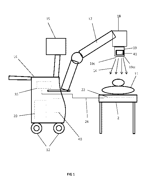
[0035] FIG. 1 illustrates a side view of a mobile imaging system applying
radiation to a subject
in accordance with a preferred embodiment of the invention.
[0036] FIG. 2 illustrates a more detailed side view of the device of FIG. 1
(with the subject
present) depicting the position and orientation sensors.
[0037] FIG. 3 illustrates more particularly in a side view features of the
articulating support
arm of the device of FIG. I.
[0038] FIG. 4 illustrates range of movement of the articulating arm of the
device of FIG. 1, to
provide for x-ray source positioning.
[0039] FIG. 5a illustrates an enlarged, side, partially phantom view of the
portable radiation
source of the device if FIG. I.
[0040] FIG. 5b illustrates an enlarged, top, partially phantom view of the
portable x-ray
detector used in conjunction with the device of FIG. I.
[0041] FIG. 6 is a schematic diagram of the computer and certain sensor inputs
and outputs
present in the device of FIG. I.
[0042] FIG. 7A-7D depict example representations of different views of the LCD
monitor
display in the embodiment of Figure I.
[0043] FIG. 8 is a work flow diagram for a typical X-ray examination employing
the device of
Figure 1.
[0044] FIG. 9 is a schematic representation of alignment dimensions and
tolerances for a
radiation detector and a stream of radiation emitted by a radiation source.
DETAILED DESCRIPTION OF THE INVENTION
- 7 -
CA 02871123 2014-10-21
WO 2013/162762 PCT/US2013/032133
[0045] To fully understand the invention in its various embodiments and the
improvements
the invention provides, first we have to review a number of key challenges a
mobile
radiographic/fluoroscopic system must address.
[0046] A mobile radiography imaging system, comprising a portable radiation
source (such as,
e.g., an X-ray source 18 as seen in FIG. 1) adapted to move in all degrees of
freedom; a portable
detector (such as an portable detector 22 as seen in F IG. 1) operable to
detect the radiation
from the radiation source, wherein the detector is adapted to move
independently of the
radiation source in all degrees of freedom. The patient may not necessarily be
in a horizontal
position for the X-Ray exanimation, but may be at an angle, depending on the
type of
exanimation required and the ability to move the patient for the exanimation.
More
importantly, if an X-Ray radiograph is captured and the portable detector and
X-Ray source are
not aligned within one or more predetermined tolerances, the quality and
amount of radiation
could be comprised, usually causing a retake of the X-Ray radiograph,
requiring the patient to
receive additional radiation dose. To perform fluoroscopic procedures, certain
governmental
agencies, e.g., the US FDA, may require that the x-ray source and portable
detector must be
aligned within one or more predetermined tolerances. Thus if the x-ray source
and portable
detector are not aligned within the predetermine tolerance(s), in accordance
with this invention a
radiation source exposure interlock 18A (as seen, e.g., on FIG. 6) should be
activated preventing
the x-ray source from emitting radiation into the subject or patient.
[0047] Before the subject invention is further described, it is to be
understood that the
invention is not limited to the particular embodiments of the invention
described below, as
variations of the particular embodiments may be made and still fall within the
scope of the
appended claims. It is also to be understood that the terminology employed is
for the purpose
of describing particular embodiments, and is not intended to be limiting.
Instead, the scope of
the present invention will be established by the appended claims.
[0048] In this specification and the appended claims, the singular forms "a,"
"an," and "the"
include plural reference unless the context clearly dictates otherwise. Unless
defined otherwise,
all technical and scientific terms used herein have the same meaning as
commonly understood to
one of ordinary skill in the art to which this invention belongs.
[0049] Although a radiographic system is described in this description, the
concepts are equally
applicable to a radiographic/fluoroscopic system as well. In fact, these
system of this invention
is in many respects particularly and preferably adapted for use in
fluoroscopic procedures,
- 8 -
CA 02871123 2014-10-21
WO 2013/162762 PCT/US2013/032133
because of the need for safe control of x-ray emissions from a pulsed
radiation source employed
during fluoroscopic procedures. The safety features of this system facilitate
the judicious use of
and exposure to x-ray radiation during fluoroscopic procedures carried out
using a mobile
system, and is particular beneficial when applied to subjects who are immobile
or fragile and
cannot be ported easily for radiological procedures.
[0050] Referring generally to Figure 1, a mobile X-ray imaging system is
presented, referenced
generally by reference numeral 16. In the illustrated embodiment, the mobile X-
ray imaging
system 16 is a digital X-ray system that is designed both to acquire
radiographic and/or
fluoroscopic image data and to process the image data for display in
accordance with the present
techniques. In particular, the system 16 is operable to produce both
radiographic images and
fluoroscopic images.
[0051] In the preferred embodiment of Figure 1, the mobile radiology imaging
system 16
generally comprises a portable cart having caster wheels 12, a radiation (X-
ray) source 18
operatively attached to a manipulatable arm 17 and capable of moving in all
degrees of freedom,
and a portable flat-panel digital radiation (X-ray) detector 22. Importantly,
the x-ray source 18
and the detector 22 are capable of producing both radiographic (via single
radiation emissions)
and fluoroscopic X-ray images (via pulse radiation emissions). The imaging
system 16 also
includes a collimator 19 attach to the radiation source 18, which permits a
controlled stream of
radiation 14 to pass into a region in which a patient 11 is positioned on a
table 2. For
fluoroscopic procedures a Lead aperture 19c and lead aperture interlock 19cc
ensures that the
stream of radiation 14 does not exceed the size of the active image area 22a
of the detector 22
further described. The controlled stream of radiation 14 passes through the
patient 11 and
impacts the detector 22. The detector 22 converts the X-ray photons received
on its surface to
lower energy photons, and subsequently to electric signals, which are acquired
and processed to
reconstruct an image of the features within the patient 11. As can be
appreciated from Figure 1,
alignment between the radiation source 18 and the detector 22 and size of
stream of radiation 14
is of critical importance. If the radiation source 18 and the detector 22 are
not aligned, a portion
of the stream of radiation 14 may not pass through the patient 11 at the
intended position,
orientation or angle, so the stream of radiation 14 cannot be properly
received by the detector 22,
and an accurate image of the patient 11 cannot be obtained. Furthermore, even
if the detector 22
is directly in line with the radiation source 18, the detector 22 must be
oriented such that its plane
is perpendicular to the radiation source 18 for proper detection of the
radiation 14. In addition
for fluoroscopic procedures, alignment and stream of radiation 14 must conform
to regulatory
- 9 -
CA 02871123 2014-10-21
WO 2013/162762 PCT/US2013/032133
standards for alignment of the radiation stream size of x-ray source 18 to
detector 22, if x-ray
source 18 is not within alignment tolerance, or stream of radiation 14 is not
the proper size, the
alignment system must inhibit x-ray source 18 from producing radiation 14. The
tolerances may
vary, but will typically be 2% of the distance between the radiation source
and detector (SID;
source image distance). The predetermined alignment conditions of this
invention also may vary,
but typically in the United States, for example, will be one or more of SID is
usually set at 40
inches, ( 40 inches x .2 = .8 inches total) radiation source and detector can
not be more than .4
inch off the center axis. In this regard, see Figure 9 further illustrating
such tolerances in a
schematic of the detector area and the radiation stream area.
[0052] In an operating configuration, a patient 11 is positioned on a table or
other patient
support 2 and located between the radiation source 18 and the detector 22. The
detector 22 can
be coupled via data cable 24 to a workstation computer 35 which commands
acquisition of the
signals generated in the detector 22, although wireless communication between
the detector 22
and the computer is the more preferred method. As the detector receives
radiation 14 that pass
through the patient 11, imaging data is transmitted to the workstation
computer 35. In most
cases, the workstation computer 35 may also execute various signal processing
and filtration
functions, such as for initial adjustment of dynamic ranges, interleaving of
digital image data, and
so forth. The workstation 35 also enables a user to control the operation of
the system to
produce a desired image. Images processed by the workstation 35 are displayed
on a monitor 15.
Electrical power for the radiation source 18, workstation computer 35, and the
digital detector 22
is provided by a conventional power supply 20 located within the cart, and
which may be
provide by batteries or electrically connected to any available 110VAC power
source.
[0053] Because movement of the detector 22 is independent of the radiation
source 18, it is
possible for the stream of radiation 14 to strike the detector 22 at an angle
or not centered to the
detector 22, producing inaccurate images of the patient 11. As shown more
clearly in Figure 2,
the radiation source 18 and the detector 22 each have an alignment
sensor/transmitter 43 , 42 in
the form of, for example, an motion tracking device (MTD), which establishes
both the location
and orientation of the respective radiation source 18 and detector 22 relative
to one another.
The sensor/transmitters 42 , 43 are used to align the detector 22 with the
radiation source 18 to
ensure that the radiation 14 from the radiation source 18 strikes the detector
22 at the correct
angle, position and orientation.
- 10 -
CA 02871123 2014-10-21
WO 2013/162762 PCT/US2013/032133
[0054] As further illustrated in Figure 6, alignment sensor/transmitters 42,
43 transmit data to
the computer 40 from the signals produced by the alignment sensor/transmitters
42, 43. Those
signals are processed by alignment system software located within computer 40
to ascertain the
orientation distance and location of the detector 22 relative to the radiation
source 18 to
determine if the detector 22 is aligned normal to the path of stream of
radiation 14 being emitted
from the radiation source 18. The alignment system software within computer 40
sends process
data to LCD display 41, and data received by LCD display 41 visually displays
the location and
orientation of detector 22 and the radiation source 18. When alignment in
accordance with
predetermined conditions is achieved, the computer 40 sends an activation
signal to the radiation
source 18, whereupon an audible indicator 37 and/or visual indicator 41 will
activate to notify
the operator that radiation 14 may be administered. The alignment
sensor/transmitters 42, 43
may also be operable to indicate when the detector 22 is within range of the
radiation source 18.
Finally, the system 16 may be connected to the Internet or other communication
network so that
the images produced by the system 16 may be sent to a remote user, such as a
radiologist's
workstation. Importantly, the computer 40 may also be used to control the
radiation source 18,
such that emission of radiation 14 is prohibited until and unless the proper
alignment conditions
are achieved. Similarly, the computer 40 and the alignment software may be
programmed to
automatically permit emission of a radiation 14 dose for either radiographic
or fluoroscopy
images immediately upon achieving the predetermined alignment conditions.
Thus, the present
invention may be used to limit the patient's 11 exposure to unnecessary or
excessive radiation 14
in a particular situation due to improper alignment. Until development of this
system, such
control over the emission of radiation by establishing this "interlock" 18A
between alignment
conditions and the radiation source has not been available in portable
radiology imaging systems,
fluoroscopy systems and particularly in the context of intensive care unit and
neonatal intensive
care unit applications.
[0055] FIG 3 illustrates the articulating tube support arm consist of vertical
travel arm 17a, fix
vertical support arm 17b, tube support arm pivot assembly 17c, tube support
arm rotation
assembly 17f, tube support arm longitudinal bearing assembly 17g, & 17h.
Vertical travel arm 17a
provides support and vertical movement of X-Ray tube assembly 18 (via, e.g.,
gas springs for
biasing) to produce a counterbalancing force so x-ray tube assembly 18 and x-
ray collimator 19
will remain in the vertical position they are place throughout the movement
range allowed by
pivot assembly 17c. Fix vertical arm 17b support the pivot assembly 17c for
vertical travel arm
17a. Rotation shaft 17e provides transversal movement of X-Ray tube assembly
18. Longitudinal
-11 -
CA 02871123 2014-10-21
WO 2013/162762 PCT/US2013/032133
bearing track assembly 17g provides longitudinal movement for the x-ray tube
assembly 18.
Yoke 17d provides x-ray tube assembly 18 to rotate around the axis of vertical
travel arm 17a.
Tube rotation assembly 17e provides x-ray tube assemble to rotate longitudinal
about is axis.
[0056] FIG 4 shows range of movement the articulating arm provides for x-ray
source
positioning in the device of FIG 1. Thus, directions of rotational movement
17g, 17h, 17i, 17j
and 17k illustrate the rotational motions of which the device is made capable
for positioning the
radiation source in a mobile radiologic or fluoroscopic application.
[0057] FIG 5a illustrates the portable x-ray source of the device FIG 1,
comprising X-Ray tube
head 18, X-Ray beam collimator 19, LCD monitor 41, collimator light 19B, laser
positioning
cross hair 19A, and lead aperture 19C. Positioning sensor 43 is housed or fix
mounted within the
X-Ray beam collimator enclosure 19.
[0058] FIG 5b illustrates the portable detector 22 of the device, comprising
the active imaging
panel 22A, power supply 22B, and positioning sensor 42. All components are
housed within the
detector enclosure 22. It will be appreciated that the position detector
sensor 42 and radiation
source sensor 43 may be comprised of various sensors or electronic devices,
including for
example RFID tags, internal measurement units (IMUs), mobile tracking devices
(MTDs),
microelectromechanical systems (MEMS), or the like, including combinations of
two or more of
the foregoing. Particular configurations will be determined by the design
criteria and economics
of a given system.
[0059] FIG 6 is a schematic diagram of the FIG 1 positioning system's sensors
and computer
controls, comprising detector sensor 42; radiation source sensor 43, alignment
system
computer/cpu 40, an alarm in the form of audible buzzer 37, radiation source
exposure interlock
18A, and a user interface in the form of LCD monitor 41. As previously noted,
radiation source
and detector position information is fed from sensors 43 and 42, respectively,
to computer 40,
which in turn controls buzzer 37, interlock 18A and the output to user
interface such as LCD
monitor 41. A wide variety of control system software known to those of skill
in the art can be
adapted for execution on computer 40 to receive the signals from sensors 42
and 43 and control
the operation of an alarm such as buzzer 37, interlock 18A and a user
interface such as LCD
monitor 41.
[0060] FIG 7A-7D illustrate four (4) alternative examples of the LCD monitor
41 display of
information to assist the operator with alignment of X-Ray source 18 to the
portable detector 22.
These screenshots illustrate examples of the type of information that may be
available to the
- 12 -
CA 02871123 2014-10-21
WO 2013/162762 PCT/US2013/032133
operator for positioning of radiation source 18 to the portable detector 22,
or portable detector
22 to the radiation source 18. Various icons, LEDs, bar graph, or graphic
symbols can be used
to display position or orientation of radiation source 18 and detector 22 on
LCD monitor 41.
FIG 7A shows LCD monitor positioning data if detector is placed at an oblique
angle 41A
"Start Icon" initiates sensors calibration and calculation of present position
of detector 22 and
radiation source 18. 41C displays distance between detector and radiation
source, 41D is
radiation source icon, 41E is detector icon, 41F is LED bar graph to show
longitudinal position,
41G is LED bar graph to show transverse position, FIG 7C shows LCD monitor
positioning
data if detector is place perpendicular: 41H is the detector, 41J is the
radiation source FIG 7B &
7D shows LCD monitor positioning data when detector and radiation source are
not aligned.
[0061] In Figure 8 the work flow for a typical X-Ray exanimation employing a
device of this
invention is illustrated. In step 80, the operator first places the portable
detector 22 under the
patient (note: the portable detector 22 is usually no longer visible to the
operator after
placement). The portable detector 22 is positioned to ensure the body part to
be examined is
within the active imaging area 22A of the portable detector. In the next step
81, the operator
then positions the portable radiation source 18 with the aid of data provide
on LCD 41.
Computer 40 may be automatically carrying out, or activated to carry out,
calculations of
radiation source 18 and portable detector 22 alignment via
sensors/transmitters 43 and 42
installed on radiation source 18 and detector 22, respectively. System will
prompt operator when
calibration and positioning calculation are complete. Then in step 83, the
system will accurately
display location of portable detector 22 with respect to radiation source 18,
and provide data of
direction, angle, orientation, and/or distance operator has to move radiation
source and/or
portable detector to position radiation source to detector within predetermine
tolerance(s).
Upon successful alignment of radiation source to detector, at step 84 the
system will active a
visual 41B and/or audible signal 44 confirming alignment is within the
predetermine tolerance(s).
At step 85, the system will then release "Radiation interlock" 18A, and in
step 86 operator will
press laser cross hair button 19b to verify patient is align with radiation
source 18. If patient is
not aligned operator moves patient for proper alignment, and in step 87
operator presses the
collimator light button to active collimator light source which displays a
representation of the
size of stream of radiation 14, and the operator adjusts the light size for
size for body part being
examined. In step 88, in addition, if operator has selected to perform a
fluoroscopic procedure
operator must installed lead aperture 19c to release lead aperture exposure
interlock 19cc.
- 13 -
CA 02871123 2014-10-21
WO 2013/162762 PCT/US2013/032133
When all conditions are met in step 89 operator can now initiate an X-ray
exposure and capture
the digital radiograph.
[0062] All references cited in this specification are herein incorporated by
reference as though
each reference was specifically and individually indicated to be incorporated
by reference. The
citation of any reference is for its disclosure prior to the filing date and
should not be construed
as an admission that the present invention is not entitled to antedate such
reference by virtue of
prior invention.
[0063] It will be understood that each of the elements described above, or two
or more
together may also find a useful application in other types of methods
differing from the type
described above. Without further analysis, the foregoing will so fully reveal
the gist of the
present invention that others can, by applying current knowledge, readily
adapt it for various
applications without omitting features that, from the standpoint of prior art,
fairly constitute
essential characteristics of the generic or specific aspects of this invention
set forth in the
appended claims. The foregoing embodiments are presented by way of example
only; the scope
of the present invention is to be limited only by the following claims.
- 14-