Note: Descriptions are shown in the official language in which they were submitted.
CA 02871950 2014-10-29
WO 2013/173926
PCT/CA2013/050398
PATIENT-SPECIFIC INSTRUMENTATION AND METHOD FOR ARTICULAR JOINT
REPAIR
CROSS-REFERENCE TO RELATED APPLICATION
[0001] This patent application claims priority of US provisional
Application Serial
No. 61/651,061, filed on May 24, 2012, United States Provisional Patent
Application
No. 61/671,990, filed on July 16, 2012, and United States Provisional Patent
Application
No. 617/87579, filed on March 15, 2013.
FIELD OF THE APPLICATION
[0002] The present application relates to patient-specific
instrumentation for
articular joint repair.
BACKGROUND OF THE ART
[0003] In arthroplasty, a damaged joint, such as a knee joint, is
replaced with
prosthetic implants. Prior to implantation of the implant, the damaged region
of the joint
is typically prepared by treating regions of the bones to provide surfaces
that can align
with and therefore accommodate the implant.
[0004] Accuracy in the alignment of the implant is important in the
arthroplasty
procedure. In knee replacement surgery, this entails proper alignment of the
knee so the
centre of the hip, knee and ankle are aligned in a straight line. This in turn
ensures faster
patient rehabilitation and better knee function. For this purpose, mechanical
jigs, which
ensure accurate position and orientation of finishing instruments used during
bone
resection, are typically used during arthroplasty procedures, such as knee
replacements.
However, such conventional jigs may lack precision as they may rely on the
user's
judgment to assess proper positioning of the devices. In addition, each
patient's anatomy
being different, proper component sizing may be required for optimizing the
outcome of
the surgery. Still, conventional components only allow patient customization
to a certain
degree. As such, the use of conventional instrumentation can lead to
misalignment and
result in instability and potential wear or even premature failure of the
prosthetic
implants.
[0005] There is therefore a need for improved patient-specific
instrumentation for
use during articular joint repair procedures.
- 1 -
CA 02871950 2014-10-29
WO 2013/173926
PCT/CA2013/050398
SUMMARY OF THE APPLICATION
[0006] It is therefore an aim of the present invention to provide novel
patient-
specific instrumentation and method.
[0007] Therefore, in accordance with the present application, there is
provided a
patient-specific rotational guide for guiding a positioning of a tool on a
resected surface
of a bone in an articular joint repair procedure, the rotational guide
comprising a body
comprising a tool attachment member adapted to be secured to the tool; and a
bone
contacting member having a bone contacting surface shaped using patient-
specific
modeling to conform to a shape of an articular surface of the bone for
matingly
contacting the articular surface when the tool is positioned on the resected
surface.
[0008] Still further in accordance with the present application, the
tool attachment
member has an inner contour conforming to a shape of the tool for retaining
the tool
attachment member in position relative to the tool when the tool attachment
member is
secured to the tool.
[0009] Still further in accordance with the present application, the
tool attachment
member has an outer contour conforming to a perimeter of the tool.
[0010] Still further in accordance with the present application, the
tool attachment
member comprises at least one attachment means securing the tool attachment
member
to the tool.
[0011] Still further in accordance with the present application, the
body
comprises at least one alignment element located on the body using patient-
specific
modeling for guiding the positioning of the tool on the resected surface of
the bone.
[0012] Still further in accordance with the present application, the at
least one
alignment element is indicative of at least one anatomic direction of the
bone.
[0013] Still further in accordance with the present application, the at
least one
alignment element is indicative of at least one of a mechanical axis, a medio-
lateral
direction, and an anterior-posterior direction of the bone.
[0014] Still further in accordance with the present application, the
tool has
formed therein at least one aperture and the at least one alignment element
comprises
at least one opening adapted to cooperate with the at least one aperture when
the tool
attachment member is secured to the tool, the cooperating at least one
aperture and at
least one opening adapted to receive therein at least one fixation for
securing the tool to
the resected surface.
[0015] Further in accordance with the present application, there is
provided a
patient-specific jig for preparing an articular surface of a bone in an
articular joint repair
procedure, the patient-specific jig comprising at least one bone contacting
member
having a mating surface shaped using patient-specific modeling to conform to a
shape of
- 2 -
CA 02871950 2014-10-29
WO 2013/173926
PCT/CA2013/050398
the articular surface, the mating surface adapted to matingly contact a
portion of the
articular surface; and a cutting guide adjacent the at least one bone
contacting member
and adapted to receive therein a saw blade for resecting the articular
surface.
[0016] Still further in accordance with the present application, the at least
one bone
contacting member has formed therein at least one clearance shaped to conform
to a
shape of at least one selected area of the articular surface for preventing
contact
between the mating surface and the at least one selected area.
[0017] Still further in accordance with the present application, the at least
one clearance
is shaped to prevent contact between the mating surface and at least one of
cartilage,
soft tissue, osteophytes, and menisci.
[0018] Still further in accordance with the present application, the mating
surface of the
at least one bone contacting member is adapted to matingly contact a distal
surface of a
lateral femoral condyle, a distal surface of a medial femoral condyle, an
anterior surface
of the lateral femoral condyle, and an anterior surface of the medial femoral
condyle.
[0019] Still further in accordance with the present application, mating
surface of the at
least one bone contacting member is adapted to matingly contact a proximal
surface of a
lateral tibial plateau, a proximal surface of a medial tibial plateau, and an
anterior
proximal tibial surface.
[0020] Still further in accordance with the present application, the mating
surface of the
at least one bone contacting member is shaped to conform to a shape of a
tibial
intercondylar eminence for securing a medio-lateral position and a rotation of
the jig
relative to the bone when the mating surface matingly contacts the articular
surface.
[0021] Still further in accordance with the present application, the mating
surface of the
at least one bone contacting member has a first size proportional to a second
size of the
bone.
[0022] Still further in accordance with the present application, the at least
one bone
contacting member comprises at least one alignment element defined on the at
least one
bone contacting member using patient-specific modeling for guiding a
positioning of the
jig on the resected surface of the bone.
[0023] Still further in accordance with the present application, the at least
one alignment
element is indicative of at least one anatomical direction of the bone.
[0024] Still further in accordance with the present application, the at least
one alignment
element is indicative of at least one of an anterior-posterior direction of
the bone and a
mechanical axis of the bone.
[0025] Still further in accordance with the present application, the at least
one alignment
element is indicative of a plane along which the articular surface is to be
resected.
- 3 -
CA 02871950 2014-10-29
WO 2013/173926
PCT/CA2013/050398
[0026] Still further in accordance with the present application, the cutting
guide
comprises an opening for receiving therein an insert, the insert comprising a
first
member and a second member coupled to the first member and spaced therefrom
for
defining an aperture adapted to receive therein the saw blade.
[0027] Further
in accordance with the present application, there is provided a cut
slot for use in resecting an articular bone surface in an articular joint
repair procedure,
the cut slot comprising a housing adapted to be positioned adjacent the
articular bone
surface, the housing having an opening defined therein; and an insert adapted
to be
fitted into the opening, the insert having defined therein an aperture adapted
to receive a
saw blade for resecting the articular bone surface.
[0028] Still further in accordance with the present application, the insert
comprises a first
member and a second member coupled to the first member, the second member
spaced
from the first member for defining the aperture.
[0029] Still further in accordance with the present application, the first
member and the
second member each have a first end and a second end opposite the first end
and are
each provided with a first attachment means at the first end and with a second
attachment means at the second end, the first attachment means of the first
member
adapted to cooperate with the first attachment means of the second member and
the
second attachment means of the first member adapted to cooperate with the
second
attachment means of the second member for coupling the first member to the
second
member.
[0030] Still further in accordance with the present application, the housing
comprises at
least one crush rib for securing a position of the insert within the opening,
the at least
one crush rib adapted to be resiliently deformed in response to a pressure
being exerted
thereon as the insert is fitted into the opening.
[0031] Still further in accordance with the present application, the housing
comprises a
first, a second, a third, and a fourth crush rib and further wherein, with the
insert fitted
into the opening, the first crush rib is positioned adjacent the first end of
the first
member, the second crush rib is positioned adjacent the first end of the
second member,
and the third and fourth crush ribs are positioned adjacent a lower surface of
the second
member.
[0032] Still further in accordance with the present application, the housing
has formed
therein at least one opening adapted to receive therein a drill bit for
drilling at least one
hole into the articular bone surface, the at least one hole adapted to receive
at least one
fixation for securing the cut slot on the articular bone surface.
[0033] Still further in accordance with the present application, the cut slot
further
comprises at least one bushing adapted to be received in the at least one
opening, the at
- 4 -
CA 02871950 2014-10-29
WO 2013/173926
PCT/CA2013/050398
least one bushing configured to accommodate therein the drill bit and to
prevent residual
debris resulting from the drilling of the at least one hole.
[0034] Still further in accordance with the present application, the insert
has a
substantially constant width along a length thereof.
[0035] Still further in accordance with the present application, the first and
second
members are made of a stamped sheet of metal.
[0036] Still further in accordance with the present application, the housing
is made of a
plastic material.
[0037] Further in accordance with the present application, there is
provided a
patient-specific plate for preparing a resected surface of a bone to receive
thereon a
prosthesis component in an articular joint repair procedure, the patient-
specific plate
comprising a body shaped using patient-specific modeling and having a contour
conforming to a perimeter of the resected surface, the body comprising a bone
contacting face adapted to be positioned on the resected surface, and a
prosthesis
receiving face opposite the bone contacting face, the prosthesis receiving
face
comprising at least one alignment element defined using patient-specific
modeling for
guiding a positioning of the prosthesis component on the prosthesis receiving
face.
[0038] Still further in accordance with the present application, the
prosthesis receiving
face has delineated thereon an outline of the prosthesis component.
[0039] Still further in accordance with the present application, the at least
one guiding
element is indicative of at least one anatomic direction of the bone for
guiding the
positioning of the prosthesis component relative to the at least one anatomic
direction.
[0040] Still further in accordance with the present application, the at least
one guiding
element is indicative of at least one of a medio-lateral direction and an
anterior-posterior
direction of the bone.
[0041] Still further in accordance with the present application, the at least
one guiding
element comprises at least one aperture indicative of at least one location on
the
resected surface at which to drill at least one hole, the at least one hole
adapted to
receive therein at least one fixation for securing on the resected surface a
tool for use in
the articular joint repair procedure.
[0042] Further in accordance with the present application, there is
provided a
method for manufacturing a patient-specific jig for use in preparing an
articular surface of
a bone in an articular joint repair procedure, the method comprising acquiring
image data
of the bone; generating a three-dimensional model of the bone using the
acquired image
data; providing a jig blank model; and applying a Boolean operation for
removing
material from the jig blank model thereby deconstructing the jig blank model,
a shape of
- 5 -
CA 02871950 2014-10-29
WO 2013/173926
PCT/CA2013/050398
the deconstructed jig blank model conforming to a shape of the three-
dimensional model
of the bone.
[0043] Still further in accordance with the present application, applying the
Boolean
operation causes the deconstructed jig blank model to have at leas one bone
contacting
surface conforming to the articular surface of the bone, the at least one bone
contacting
surface adapted to be positioned on the articular surface.
BRIEF DESCRIPTION OF THE DRAWINGS
[0044] Figure 1a is a flow chart of a method for performing bone
resection during
arthroplasty using a patient-specific jig in accordance with the present
disclosure;
[0045] Figure lb is a flow chart of the step of creating a patient-
specific jig of
Figure la;
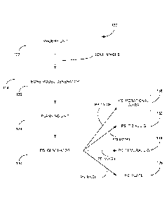
[0046] Figure 2 is a block diagram of a patient-specific instrumentation
computer-assisted system for arthroplasty in accordance with the present
disclosure;
[0047] Figure 3 is a side perspective view of a patient-specific femoral
jig on a
femur in accordance with the present disclosure;
[0048] Figure 4 is a bottom perspective view of the patient-specific
femoral jig on
the femur of Figure 2;
[0049] Figure 5 is a perspective view of a femur showing femoral jig
contacting
areas in accordance with the present disclosure;
[0050] Figure 6 is a top perspective view of a tibial jig in accordance
with the
prior art;
[0051] Figure 7 is a perspective view of the tibial jig of Figure 5;
[0052] Figure 8 is a top perspective view of a patient-specific tibial
jig on a tibia in
accordance with the present disclosure;
[0053] Figure 9 is a front perspective view of a tibia showing tibial
jig contacting
areas in accordance with the present disclosure;
[0054] Figure 10a is a bottom perspective view of a patient-specific
femoral jig
on a femur in accordance with an alternative embodiment of the present
disclosure;
[0055] Figure 10b is a side view of the femoral jig of Figure 10a;
[0056] Figure 11 a is a rear perspective view of a patient-specific
tibial jig on a
tibia in accordance with an alternative embodiment of the present disclosure
[0057] Figure llb is a top view of the tibial jig of Figure 11a;
[0058] Figure 11c is a top perspective view of the tibial jig of Figure
11a;
[0059] Figure 12 is a front perspective view of a patient-specific
tibial plate in
accordance with the present disclosure;
- 6 -
CA 02871950 2014-10-29
WO 2013/173926
PCT/CA2013/050398
[0060] Figure 13a is a perspective view of a rotational guide and of a
sizing plate
coupled to a plate handle in accordance with a first embodiment of the present
disclosure;
[0061] Figure 13b is a perspective view of the rotational guide of
Figure 13a
coupled to the plate handle and sizing plate;
[0062] Figure 13c is a side view of the rotational guide of Figure 13b;
[0063] Figure 13d is a perspective view of the rotational guide of
Figure 13c
mated with a bone surface in accordance with a first embodiment of the present
disclosure;
[0064] Figure 13e is a perspective view of a rotational guide and of a
sizing plate
in accordance with a second embodiment of the present disclosure;
[0065] Figure 13f is a bottom perspective view of the rotational guide
of Figure
13e;
[0066] Figure 13g is a top perspective view of the rotational guide of
Figure 13e
coupled to the sizing plate;
[0067] Figure 13h is a bottom perspective view of the rotational guide
and sizing
plate of Figure 13g;
[0068] Figure 14a is a top perspective view of a cut slot in accordance
with the
present disclosure;
[0069] Figure 14b is a front view of the cut slot of Figure 14a;
[0070] Figure 14c is a perspective view of the cut slot of Figure 14a;
[0071] Figure 14d is a perspective view of the cut slot of Figure 14a
inserted into
a cut guide in accordance with the present disclosure;
[0072] Figure 14e is a perspective view of the cut slot of Figure 14d
showing
assembly pins in accordance with the present disclosure;
[0073] Figure 14f is a perspective view of the cut slot of Figure 14e
receiving a
drop rod adaptor in accordance with the present disclosure; and
[0074] Figure 14g is a front view of the cut slot of Figure 14d with a
close-up
view of crush ribs in accordance with the present disclosure.
DESCRIPTION OF THE EXEMPLARY EMBODIMENTS
[0075] Referring to Figure la, a method 100 for preparing a bone using
patient-
specific instrumentation (PSI), and more particularly patient-specific femoral
and tibial
jigs, prior to performing an arthroplasty procedure, such as knee replacement,
will now
be described. Although described herein as relating to total knee replacement,
it should
be understood that the method 100 is also suitable for partial knee
replacement, or other
articular joint repair procedures known to those skilled in the art. It should
also be
- 7 -
CA 02871950 2014-10-29
WO 2013/173926
PCT/CA2013/050398
understood that the method 100 may be suitable for repairing other articular
joints, such
as an elbow, shoulder, wrist, or hip.
[0076] The first step 102 of the method 100 illustratively comprises pre-
operative
planning, during which image data of the patient's anatomy, e.g. the hip,
knee, and ankle
regions when total knee replacement is concerned, may be obtained before
surgery. The
image data may be obtained from scans generated using Magnetic Resonance
Imaging
(MRI), Computed Tomography (CT), ultrasound, x-ray technology, optical
coherence
tomography, or the like. Once the images are obtained, a computer software
creates a
three dimensional (3D) model of the patient's damaged knee joint (step 104),
which may
be sent to a user over a suitable communication network, such as the Internet.
The user
may then visualize the 3D model using a computer (not shown) to plan bone
resection
and prosthesis component placement at the damaged joint region (step 106). The
model
further enables the user to determine the prosthesis sizing and shape option,
e.g.
thickness, length, width, or curvature, best adapted to the patient given the
latter's age,
weight, gender, and other pertinent information.
[0077] Referring to Figure lb in addition to Figure la, once bone
resection and
prosthesis selection and placement have been planned by the user, the user's
computer
plan may be used to manufacture patient-specific jigs (step 108). The patient-
specific
jigs may be manufactured using a jig blank model as a starting point for the
machining
process (step 112). The jig blank model may be made of any suitable material,
including
but not limited to a polymer, a metal, a cross-linked polymer, a ceramic, and
an alloy. In
this case, a jig blank model of a given size, e.g. small, medium, or large,
selected from a
library of blanks and adapted to the anatomy of the patient's damaged joint
may be
deconstructed. Parts of the blank model may be removed using a Boolean
operation to
carve out the desired shape of the patient-specific jigs (step 114). A jig
having a surface
conforming to the joint surface to which the prosthesis is designed to mate,
and thus
precisely-fitting the patient's anatomy, may then be obtained (step 116).
[0078] A rapid prototyping manufacturing process may further be used to
manufacture the patient-specific jigs. In this technique, a computer software
may section
the 3D representations of an object to be manufactured into a plurality of
distinct two-
dimensional (2D) layers. A 3D printer then fabricates a layer of material for
each layer
sectioned by the software. The fabricated layers together form a prototype of
the desired
object.
[0079] During surgery, the thus manufactured jigs may be precisely
fitted over
the patient's knee bones, namely the femur and tibia, at the damaged region of
the knee
joint for guiding the bone resection (step 110). In this manner, customized
bone
preparation may be performed as previously planned on the computer by the
user.
- 8 -
CA 02871950 2014-10-29
WO 2013/173926
PCT/CA2013/050398
Optimal placement of the best fitting size and shape of the replacement
prosthesis may
therefore be achieved.
[0080] Referring to Figure 2, a PSI computer-assisted system for
arthroplasty is
generally shown at 120. The system 120 illustratively receives at an imagery
unit 122
images of the patient's hip, knee, and ankle regions from any appropriate
imaging
technology, such as MRI or CT. The imaging technology apparatus (not shown)
may be
part of the system 120. The bone images are then sent to a processor unit 124,
which
illustratively comprises a bone model generator 126, a planning unit 128, and
a PSI
generator 130. The processor unit 124 has a processor to run an application
that will
generate PSI models used to manufacture PSI, such as a PSI tibial jig 132, a
PSI
femoral jig 134, a PSI plate 136, and a PSI rotational guide 138 for use
during the
arthroplasty procedure, as will be discussed further below. The processor unit
124 may
be any appropriate computer or processing unit. User interfaces, such as a
monitor,
screen, touch-screen, keyboard, or mouse, may be part of the processor unit
124 for the
involvement of an operator in the creation of the PSI models.
[0081] The bone model generator 126 is illustratively used to interpret
the bone
images received from the imagery unit 122 in order to create a 3D model of the
patient's
damaged articular joint, e.g. the knee joint. For this purpose, input may be
provided by
an operator via the user interfaces to ensure proper adequate segmentation
between
bone and tissue as well as bone and cartilage, thus increasing the accuracy of
the
generated bone model.
[0082] The planning unit 128 may then be used to visualize the bone
model and
to plan bone resection as well as prosthesis component placement at the
damaged joint.
The prosthesis size and shape best-suited to the patient's unique anatomy may
also be
determined at the planning unit 128. According to the generated bone model and
pre-
operative planning, the PSI generator 130 may produce the PSI models, which in
turn
may be used to manufacture at least one of the PSI tibial jig 132, the PSI
femoral jig 134,
the PSI plate 136, and the PSI rotational guide 138, the latter being adapted
to be placed
over a resected bone portion for guiding the position or rotation of a
prosthesis
component thereon. For this purpose, patient-specific modeling may be used to
design
PSI tools, e.g. the PSI tibial jig 132, the PSI femoral jig 134, the PSI plate
136, and the
PSI rotational guide 138, such that each PSI tool has a mating surface that is
a replica of
or otherwise precisely conforms to a surface of a bone the tool is to be
positioned on. In
this manner, the PSI tool matingly contacts the bone surface and precisely
fits the
patient's anatomy. The PSI models may be in any appropriate format to allow
the
manufacture of PSI. For instance, the PSI models may be formatted into
numerical
control (NC) machine files, technical data, visual or digital models, etc.
- 9 -
CA 02871950 2014-10-29
WO 2013/173926
PCT/CA2013/050398
[0083] Referring now to Figure 3 and Figure 4, a femoral jig 200 may
first be
used to prepare a distal femoral surface prior to attaching a prosthesis
component
thereon, in most cases with a bone cement. The femoral jig 200 illustratively
comprises a
bone contacting portion 202 adapted to be positioned on the patient's femur
204 in flush
contact with an articular surface 206 thereof.
[0084] The femoral jig 200 illustratively comprises a cut-slot portion
208 having a
cutting slot 210 formed therein and adapted to receive a saw blade (not shown)
used to
execute the pre-planned bone cuts. When the femoral jig 200 is in position
over the
femur 204 following exposure of a distal end thereof during surgery, the cut-
slot portion
208 illustratively extends along the anterior-posterior direction A. In this
manner, when
the femoral jig 200 is in place, the slot 210 is positioned adjacent to the
trochlear groove
212 at a lower portion of the femur 204, which typically mates with an upper
portion of
the patient's tibia (not shown) at the knee joint. The saw blade may therefore
be used to
resect the lower portion of the femur 204.
[0085] The bone contacting portion 202 of the femoral jig 200 further
comprises
a first pair of pegs 214a and 214b, which are respectively positioned adjacent
to the
medial and lateral femoral condyles 216a and 216b and extend away from the
femur 204
along the cranial-caudal direction B when the femoral jig 200 is in place. The
pegs 214a
and 214b each have elongated guide bores 218a and 218b running therethrough
and
adapted to receive therein the drill bit of a surgical drill (not shown). In
this manner, the
user may drill elongated holes (not shown) into the femur 204. The thus
machined holes
are adapted to receive therein fixations, such as pins, screws, or the like,
to couple the
femoral jig 200 to the femur 204 prior to resection thereof. Proper alignment
of the pegs
214a and 214b along the medio-lateral direction C may further be verified
using a
device, such as a cut guide 220. For this purpose, the cut guide 220 may be
positioned
adjacent the pegs 214a and 214b and proper alignment with features, as in
222a, 222b
provided on the cut guide 220, may be verified. In particular, the bores 218a
and 218b
may be used as guides to drill holes in the distal end of the femur 204 for
positioning the
cut guide 220 in a pre-planned position. Any cuts required to position the
prosthesis
component in the preplanned position may be subsequently performed.
[0086] The bone contacting portion 202 of the femoral jig 200 further
illustratively
comprises a second pair of pegs 224a and 224b, which are positioned adjacent
the cut-
slot portion 208 and extend away from the femur 204 along the anterior-
posterior
direction A when the femoral jig 200 is in place. The pegs 224a and 224b each
have
elongated guide bores (not shown) running therethrough and adapted to receive
therein
fixations, such as pins, screws, or the like, to further securely attach the
femoral jig 200
to the femur 204 prior to resection thereof. Fixations are illustratively
first inserted into
- 10-
CA 02871950 2014-10-29
WO 2013/173926
PCT/CA2013/050398
the pegs 224a and 224b for attaching the femoral jig 200 to the femur 204,
followed by
insertion of fixations into the pegs 214a and 214b for further stabilizing the
femoral jig
200 in place.
[0087] Once the femoral jig 200 has been secured and stabilized, the
fixations
inserted into the pegs 214a and 214b may be removed to enable resection of the
distal
end of the femur 204. After the femur 204 has been resected, fixations may be
inserted
back into the pegs 214a and 214b for attaching a standard cutting block (not
shown) to
the resected bone. In this manner, additional cuts, e.g. remaining ones of the
five
femoral cuts to be performed during total knee arthroplasty, may be effected
on the
femur 204.
[0088] As can be seen from Figure 3 and Figure 5, the use of the femoral
jig 200
advantageously enables the user to have an improved lateral view of the femur
204, in
addition to having a clear view of the trochlear groove 212. Indeed, a distal
clearance
(reference 211a in Figure 4) is illustratively formed in the bone contacting
portion 202 in
the area between the cut-slot portion 208 and the pegs 214a and 214b. The
clearance
211a may be shaped and sized to conform to a shape and size of the trochlear
groove
212. A lower surface 213 of the bone contacting portion 202 may also be shaped
so as
to contact a reduced surface of the femur 204. In particular, the shape of the
lower
surface 213 illustratively defines a medial clearance 211b and a lateral
clearance (not
shown). The medial clearance 211b and the lateral clearance provide viewing
spaces
that enable a user to evaluate the degree of contact between the femoral jig
200 and the
distal surface of the medial and lateral femoral condyles 216a and 216b. The
distal
clearance 211a similarly enables to evaluate contact over the surface on the
anterior
part (not shown) of the condyles 216a and 216b. As seen in Figure 5, when the
femoral
jig 200 is in position, the bone contacting portion 202 illustratively makes
contact with the
femur 204 at a medial femoral condyle contact area 226a, a lateral femoral
condyle
contact area 226b, and an anterior surface contact area 228. The femoral
condyle
contact areas 226a and 226b are illustratively positioned on the distal part
of the medial
and lateral femoral condyles 216a and 216b, respectively, while the anterior
surface
contact area 228 is positioned on the anterior part of the condyles 216a and
216b. In one
embodiment, the anterior surface contact area 228 is defined by tabs (not
shown)
provided on the femoral jig 200 and extending over the flank of the lateral
femoral
condyle 216b and over the medial side (not shown) of the femur 204 accessible
during
the surgical procedure. The contact areas 226a, 226b, and 228 are
illustratively sized so
as to be proportional to the size of the femur 204. Visibility of the
trochlear groove 212
further ensures that the femoral jig 200 is properly positioned on the femur
204.
- 11 -
CA 02871950 2014-10-29
WO 2013/173926
PCT/CA2013/050398
[0089] The areas of contact between the femoral jig 200 and the femur
204 may
vary from one patient to the next and are illustratively proportional to bone
size. Still, the
femoral jig 200 is illustratively designed such that, when the femoral jig 200
is in place,
no contact is made with areas of the femur 204 where cartilage or soft tissues
and
osteophyte formation resulting from osteoarthritis may be present. For
instance, in the
embodiment illustrated in Figure 5, the contact areas 226a and 226b are
located on the
articular surface of the femur 204 and avoid the medial and lateral margins
(not shown)
where osteophytes may be present. This in turn optimizes the precision of the
arthroplasty procedure and improves an outcome thereof. Indeed, it is
desirable for the
femoral jig 200 to make contact with as few areas of the exposed femur 204 as
possible
so as to reduce the inaccuracies in the surgical cuts to be effected.
[0090] Referring now to Figure 6 and Figure 7, a prior art tibial jig
300 will now be
described. The tibial jig 300 is adapted to be precisely fit on an articular
surface 302 of
the patient's tibia 304. The tibial jig 300 illustratively comprises a tibia
contacting portion
306 and a pair of attachment arms 308a and 308b each having respectively
formed at an
end portion thereof plateau contacting portions 310a and 310b. When the tibial
jig 300 is
placed over the tibia 304 following exposure of a proximal end thereof, the
tibia
contacting portion 306 is adapted to contact the tibia 304. The attachment
arms 308a
and 308b then extend away from the tibia 304 along the anterior-posterior
direction A
with the plateau contacting portion 310a making contact with the articular
surface of the
medial tibial plateau 312a and the plateau contacting portion 310b making
contact with
the articular surface of the lateral tibial plateau 312b.
[0091] The tibial jig 300 further comprises a cut-slot portion 314
having a slot 316
adapted to receive therein a saw blade 318 used by the user to execute the pre-
planned
bone cuts. When the tibial jig 300 is in place on the tibia 304, the cut-slot
portion 314 is
positioned adjacent an upper portion of the tibia 304, which typically mates
with a lower
portion of the femur 204 at the knee joint. In this position, the slot 316
extends along the
transverse plane and the saw blade 318 may be inserted through the slot 316 to
resect
the upper portion of the tibia 304. The resected surface of the tibia 304 is
in most cases
perpendicular to the shaft axis Y of the tibia 304 in the corona! plane. The
slot 316 may
further be machined into the cut-slot portion 314 so as to enable proper reach
of the saw
blade 318 during the bone resection.
[0092] The pair of attachment arms 308a and 308b may respectively
comprise
pegs 320a and 320b, which extend away from the tibia 304 along the cranial-
caudal
direction B. The pegs 320a and 320b each have elongated guide bores 322a and
322b
running therethrough and adapted to receive therein a surgical drill bit (not
shown) used
to drill elongated holes into the tibia 304. The thus machined holes are then
mated with
- 12-
CA 02871950 2014-10-29
WO 2013/173926
PCT/CA2013/050398
holes provided in a provisional tibial sizing plate 324 used to determine the
proper size of
a prosthetic tibial tray (not shown). The holes in the sizing plate 324 are
adapted to
receive therein fixations, such as pins, screws, or the like, to securely
attach the sizing
plate 324 to the resected portion of the tibia 304. For this purpose, the
elongated guide
bores 322a and 322b are spaced and sized to match the spacing and size of
bores 326a
and 326b machined into the sizing plate 324. The bores 322a and 322b thus aid
in
setting the position and rotation of the sizing plate 324 on the resected
portion of the
tibia 304. With the proper sizing plate 324 selected and held in place, the
proximal tibia
can be further drilled and/or broached to accommodate supporting components,
such as
stems, of the prosthetic tibial tray.
[0093] A second pair of pegs 328a and 328b illustratively project from
the tibia
contacting portion 306 and extend away from the tibia 304 along the anterior-
posterior
direction A when the tibial jig 300 is in place. The pegs 328a and 328b each
have
elongated guide bores (not shown) running therethrough and adapted to receive
therein
a device, such as a drop rod (not shown), for confirming, subsequent to the
tibial cut,
that proper rotation and alignment of the cut relative to the overall tibial
shaft axis Y have
been achieved using devices, such as traditional tibial component sizing jigs
and
rotational jigs.
[0094] Referring to Figure 8 and Figure 9, a tibial jig 400 in
accordance with an
illustrative embodiment will now be described. The tibial jig 400
illustratively comprises a
pair of attachment arms 402a and 402b each having a surface contacting portion
(not
shown) so that the arms 402a, 402b may respectively make contact with the
articular
surfaces 404a and 404b of the medial tibial plateau 312a and of the lateral
tibial plateau
312b. The slope of the tibial intercondylar eminence 405 may further be used
to lock the
medio-lateral position as well as the rotation of the tibial jig 400. In
particular, when the
tibial jig 400 is in place, the attachment arms 402a and 402b may contact the
sides of
the intercondular eminence 405, thereby providing stability to the tibial jig
400.
[0095] The tibial jig 400 further comprises a cut-slot portion 406
having a slot 408
adapted to receive therein a saw blade (not shown) used to perform resection
of the tibia
304. A tibia contacting portion (not shown) is further provided adjacent the
cut-slot
portion 406 for making contact with the tibia 304 on a region 410 of the
medial and
lateral tibial plateaus 312a and 312b adjacent the tibial tuberosity 411. When
the tibial jig
400 is in position on the tibia 304, the cut-slot portion 406 is
illustratively adjacent an
upper portion of the tibia 304 with the slot 400 being parallel to the
transverse plane. At
least one line indicator 412 may further be provided on the cut-slot portion
406 adjacent
the attachment arm 402a. The line indicator 412 may be locate on the jig 400
using
patient-specific modeling. Alignment of the indicator 412 with the tibial
tuberosity 411
- 13-
CA 02871950 2014-10-29
WO 2013/173926
PCT/CA2013/050398
may be used to confirm that the tibial jig 400 is positioned at a desired
rotational angle
relative to the tibia 304. In one embodiment, a first and second indicator as
in 412 may
be respectively provided on the anterior and distal sides cut-slot portion 406
to indicate
alignment with the anterior-posterior direction A. A cut slot plane indicator
(not shown)
may also be provided on the medial side of the cut-slot portion 406 to
indicate alignment
with the plane along which the pre-planned bone cuts are to be performed.
[0096] Although the areas of contact between the tibial jig 400 and the
tibia 304
may vary from one patient to the next, a lower surface (not shown) of the
attachment
arms 402a and 402b is illustratively sized and/or shaped such that no mating
of the tibial
jig 400 is made on an area 413 of the tibia 304 where meniscus may be present,
thus
avoiding any soft tissues remaining on the tibia 304 following exposure
thereof. For
instance, the arms 402a, 402b may be provided with varus-valgus shapes. Also,
the tibia
contacting portion is illustratively sized and/or shaped so as to avoid
possible
osteophytes that may be present on the anterior proximal ridge (not shown) of
the tibia
304.
[0097] The tibia contacting portion illustratively has formed therein a
pair of pegs
414a and 414b, which project away from the tibia contacting portion along the
anterior-
posterior direction A when the tibial jig 400 is in place. Elongated guide
bores (not
shown) may be machined into the pegs 414a and 414b for receiving fixations
used to
secure the tibial jig 400 to the tibia 304. If, subsequent to resection of the
tibia 304 using
the saw blade, it is determined that an insufficient amount of bone has been
resected, a
standard cutting block (not shown) may be secured to the pegs 414a and 414b
for
performing additional bone cuts. A clearance (not shown) having a shape and
size
conforming to the shape and size of the area 413 may further be formed in the
tibia
contacting portion to ensure that no contact is made with the area 413 of the
tibia 304.
[0098] Figures 10a to 10b and Figures 11a to 11c illustrate alternate
embodiments of the femoral jig 200 and of the tibial jig 400. Figure 10a and
Figure 10b
illustrate an alternate embodiment 500 of the femoral jig 200 shown in Figures
3 and 4.
In this embodiment, the femoral jig 500 comprises a bone contacting portion
502
adapted to be positioned on the patient's femur 204 in flush contact with an
articular
surface 206 thereof, as produced by patient-specific fabrication. The bone
contacting
portion 502 further comprises a first pair of pegs 504a and 504b, which are
respectively
positioned adjacent to the medial and lateral femoral condyles (not shown) and
extend
away from the femur 204 along the cranial-caudal direction (reference B in
Figure 3)
when the femoral jig 500 is in place. The pegs 504a and 504b each have
elongated
guide bores (not shown) running therethrough and adapted to receive therein
the drill bit
of a surgical drill (not shown).
- 14-
CA 02871950 2014-10-29
WO 2013/173926
PCT/CA2013/050398
[0099] The bone contacting portion 502 also comprises a second pair of
pegs
506a and 506b, which extend away from the femur 204 along the anterior-
posterior
direction (reference A in Figure 3) when the femoral jig 500 is in place. The
pegs 506a
and 506b each have elongated guide bores (not shown) running therethrough and
adapted to receive therein fixations, such as pins, screws, or the like, to
further securely
attach the femoral jig 500 to the femur 204 prior to resection thereof.
[00100] A clearance 508 may be formed in the bone contacting portion 502
in an
area between the pegs 506a and 506b and the pegs 504a and 504b. A lower
surface
510 of the bone contacting portion 502 may also be shaped so as to contact a
reduced
surface of the femur 204.
[00101] The femoral jig 500 further comprises a line indicator 511 used
to indicate
the desired location of the bone cut to be performed on the femur 204. The
femoral jig
500 may further comprise a mechanical axis pointer 512 provided in between the
pegs
504a and 504b. The pointer 512 illustratively provides an indication as to
whether the
femoral jig 500 is properly positioned on the femur 204. In particular, proper
positioning
is achieved if the pointer 512 points towards the femur's mechanical axis (not
shown), as
defined during the pre-operative planning phase. A pair of transepicondylar
line
indicators 514a and 514b may further be provided on the bone contacting
portion 502
adjacent the pegs 504a and 504b, respectively. The indicators 514a and 514b
illustratively protrude away from the bone contacting portion 502 to align
with the lateral
and medial epicondyles (not shown).
[00102] Figure 11 a and Figure lib illustrate an alternate embodiment 600
of the
tibial jig 400 shown in Figure 8. In this embodiment, the tibial jig 600
comprises a pair of
attachment arms 602a and 602b each having a surface contacting portion (not
shown)
so that the arms 602a, 602b may respectively make contact with the articular
surfaces
(not shown) of the medial tibial plateau 312a and of the lateral tibial
plateau 312b. A tibia
contacting portion 604 is positioned adjacent an upper portion of the tibia
304 and
comprises elongated guide bores 606a and 606b adapted to receive fixations for
securing the tibial jig 600 to the tibia 304. A line indicator 608 may further
be provided on
the tibial jig 600 adjacent the tibia contacting portion 604. Proper
positioning of the tibial
jig 600 relative to the tibia 304 may be confirmed by alignment of the
indicator 608 with
the tibial tuberosity (not shown). Patient-specific modeling may be used to
determine the
position of the indicators 511, 514a, 514b, and the pointer 512 on the femoral
jig 500 as
well as the position of the indicator 608 on the tibial jig 600.
[00103] Use of the femoral jigs 200, 500 and the tibial jigs 400, 600
advantageously decreases errors and enables more precise prosthesis placement
compared to traditional mechanical jigs. In addition, fewer instruments need
to be
- 15-
CA 02871950 2014-10-29
WO 2013/173926
PCT/CA2013/050398
sterilized for use during the surgical procedure. For instance, the use of
patient-specific
jigs as in 200, 400, 500, or 600 precludes the need for inserting rods through
the
intramedullary canal of the bone during the arthroplasty procedure. This in
turn reduces
the risk of perioperative fat embolism syndrome. Moreover, the use of patient-
specific
jigs as in 200, 400, 500, or 600 further allows for smaller incisions to be
made in the
patient's body, thus facilitating completion of minimally invasive surgery and
allowing the
prosthesis to more quickly restore the patient's joint to a generally pre-
deteriorated state.
[00104] Referring now to Figure 12, a patient-specific plate 700 may be
placed
over the resected portion of the tibia 304 for guiding the position and/or
rotation of the
sizing plate as in 324 over the resected bone area. For this purpose, a
contour 702 of
the plate 700 may be designed so as to closely follow a contour of the tibial
cut.
Apertures as in 704a and 704b may further be machined into the plate 700 for
guiding
the user as to where in the tibia 304 holes should be drilled so as to receive
fixations of
the sizing plate 324, thereby attaching the latter to the tibia 304.
[00105] In order to ensure a proper fit of the tibial tray (not shown) on
the resected
portion of the tibia 304, and accordingly a proper positioning of the
prosthesis, an outline
or contour 706 of the tibial tray may be etched or otherwise delineated onto
an upper
surface 708 of the plate 700. Such a contour 706 may be obtained as a result
of the
user's pre-operative planning, during which the size and shape of the
prosthesis best-
suited to the patient's anatomy has been selected. In addition, a first axis
710
substantially parallel to the anterior-posterior direction A and a second axis
712
substantially parallel to the medio-lateral direction C may be machined on the
surface
708 of the plate 700. In this manner, while placing the tibial tray on the
resected portion
of the tibia 304, a user may verify a proper alignment thereof relative to the
axes 710 and
712, thereby ensuring proper positioning of the prosthesis. It should be
understood that
additional alignment features as well as other indications, such as the size
and model of
the prosthesis component, may also be shown on the surface 708.
[00106] Referring to Figures 13a to 13d, a patient-specific rotational
guide 800
may be used as an alternative to the patient-specific plate 700 to ensure
proper
positioning and rotation of the sizing plate 324 on the resected portion of
the tibia (not
shown). The rotational guide 800 illustratively comprises a tray attachment
member 802
for coupling the rotational guide 800 to the sizing plate 324. The rotational
guide 800
may further comprise a bone contacting member 804 for contacting a bone the
sizing
plate 324 is to be mated with, the bone contacting member 804 being produced
by
patient-specific fabrication using bone imaging. Although a tibial sizing
plate 324 has
been shown for illustrative purposes, it should be understood that the
rotational guide
800 may be adapted for use on a bone other than the tibia, for example on the
distal
- 16-
CA 02871950 2014-10-29
WO 2013/173926
PCT/CA2013/050398
portion of a femur (not shown). It should also be understood that the
rotational guide 800
may be used with a variety of jig types as well as for repair of various
articular joints
other than the knee.
[00107] The tray attachment member 802 may be coupled to the sizing plate
324
using suitable attachment means (not shown). For instance, the tray attachment
member
802 may be clipped to a plate handle 806 coupled to the sizing plate 324. It
should be
understood that other means of securing the tray attachment member 802 of the
rotational guide 800 to the sizing plate 324 may apply.
[00108] As shown in Figure 13c and Figure 13d, the bone contacting member
804
may comprise a PSI bone contacting surface 808 adapted to mate with a
corresponding
bone surface 810 of the bone the sizing plate 324 is to be positioned on. The
bone
contacting member 804 is designed so that the bone contacting surface 808
precisely
matches a geometry of the bone surface 810. For this purpose, the bone
contacting
member 804, and more particularly the bone contacting surface 808, may be
manufactured from image data of the patient's anatomy obtained during pre-
operative
planning, as discussed above. In this manner, accurate positioning of the
sizing plate
324 relative to the bone surface 810 may be achieved.
[00109] Referring to Figures 13e to 13h, a patient-specific rotational
guide 812 in
accordance with another embodiment will now be described. The rotational guide
812
comprises a tool attachment member 814 configured to secure the rotational
guide 812
to a tool, such as the bone sizing plate 324. The rotational guide 812 further
comprises a
bone attachment member 816 having a mating surface (not shown) configured to
contact
a bone (not shown) the tool is mated with. The mating surface is
illustratively shaped so
as to conform to a shape of the bone surface the bone attachment member 816 is
mated
with.
[00110] In one embodiment, the tool attachment member 814 has a contour
(not
shown) conforming to the perimeter (not shown) of the tool. The tool
attachment member
814 further has a lower surface 818 adapted to mate with the tool. For this
purpose, an
inner contour 820 of the lower surface 818 is configured so as to conform to a
shape of
the tool. In this manner, the rotational guide 812 can be retained in position
relative to
the tool when the rotational guide 812 is secured thereto. The lower surface
818 is
further provided with attachment means 822, such as clipping means, that
enable
attachment of the rotational guide 812 to the tool. In one embodiment, the
attachment
means 822 comprise a first and a second clipping means that allow the
rotational guide
812 to be clipped to the tool. It should be understood that any other suitable
attachment
means may be provided.
- 17-
CA 02871950 2014-10-29
WO 2013/173926
PCT/CA2013/050398
[00111] An upper surface 824 of the rotational guide 812 is
illustratively provided
with one or more alignment elements as in 826a, 826b, 826c, 826d, and 826e
used for
confirming the pre-operative planning intra-operatively. The alignment
elements as in
826a, 826b, 826c, 826d, and 826e may further be used for guiding the
positioning of the
tool on the resected bone surface. In particular, the alignment element 826a
may be an
anterior-posterior line formed on the upper surface 824 and indicative of an
anterior-
posterior direction of the bone. The alignment element 826b may be an medio-
lateral line
formed on the upper surface 824 and indicative of a medio-lateral direction of
the bone.
The alignment element 826c may be indicative of a direction of the mechanical
axis of
the bone. It should be understood that other anatomical directions may apply.
The
alignment elements as in 826d may comprise openings adapted to cooperate with
apertures as in 828 formed in the tool, e.g. the bone sizing plate 324. In
particular, when
the rotational guide 812 is coupled to the plate 324, the alignment elements
826d may
be aligned with the apertures 828 to enable fixations (not shown) to be
received in the
apertures 828 for securing the plate 324 on the resected surface. The
alignment
elements 826e may comprise cutouts formed in the tool attachment member 814
and
adapted to cooperate with apertures as in 830 of the tool, e.g. the bone
sizing plate 324.
When rotational guide 812 is coupled to the plate 324, the alignment elements
826e may
also be aligned with the apertures 830 to enable fixations, such as a screws
or the like,
to be received in the apertures 830.
[00112] Figure 14a, Figure 14b, and Figure 14c show and illustrative
embodiment
of a cut slot 900 for use with at least one of the above-mentioned tibial and
femoral jigs.
It should be understood that the tibial and femoral jigs may or may not
comprise such a
cut slot 900 and that the cut slot 900 may be used with non PSI tools. In
addition, the cut
slot 900 may be provided as a disposable tool for use with disposable
instruments, such
as disposable cut guides.
[00113] The cut slot 900 illustratively comprises a first member 902 and
a second
member 904 each comprising opposite ends as in 9061, 9062. Each one of the
members,
e.g. member 902, is provided at the ends thereof, e.g. ends 9061, with an
attachment
means, e.g. attachment means 9081, adapted to cooperate with the corresponding
attachment means, e.g. attachment means 9082, provided at the ends , e.g. ends
9062,
of the other member, e.g. member 904. When so coupled to one another, the
first and
second members 902, 904 define therebetween a spacing 910 adapted to receive
therein a saw blade (not shown) used to execute pre-planned bone cuts. In the
illustrated embodiment, the attachments means 9081, 9082 are adapted to be
snap-fitted
together. It should be understood that other attachment means configurations
may also
apply. It should also be understood that the attachment means 9081, 9082 may
be
- 18-
CA 02871950 2014-10-29
WO 2013/173926
PCT/CA2013/050398
provided at a single one of the ends 9061, 9062 of each member 902, 904 rather
than at
both ends 9061, 9062 thereof.
[00114] The cut slot 900 is illustratively metallic and the members 902,
904 may
be made of a stamped sheet of metal. It should be understood that a variety of
manufacturing processes may be used for the cut slot 900. For example, the
members
902, 904 may be machined, formed, or the like. Still, regardless of the
manufacturing
process used, it is desirable for the cut slot 900 to have a substantially
constant width
along a length thereof.
[00115] Referring now to Figure 14d, Figure 14e, and Figure 14f, the cut
slot 900
may be inserted into a housing 912, such as a cut guide housing. The housing
912 may
be made of plastic or any other suitable material. The cut slot 900 may be
press-fitted
into an elongate opening 913 machined into the housing 912. For this purpose,
the first
and second members (references 902, 904 of Figure 14a) may be shaped to fit
the size
of the opening 913. A lip 914 may further be formed in each one of the first
and second
members 902, 904 so as to guide, and accordingly facilitate, the insertion of
the saw
blade into the spacing (reference 910 in Figure 14b). Assembly pins 915 may
then be
used to retain the cut slot 900 within the housing 912. The housing 912 may
further
comprise a plurality of apertures as in 916 for receiving therein bushings 917
(see Figure
140 as well as a receptor 918 for a drop rod adaptor 919 (see Figure 140. In
one
embodiment, the bushings 917 are press-fitted into plastic. The bushings may
be
adapted to accommodate the drill bit of the surgical drill guide (not shown)
and may be
used to prevent residual plastic shavings or other debris resulting from the
drilling
process. The bushings 917 may also be used to secure the cut slot 900 on the
bone
during cutting. In particular, a locking bushing (not shown) may be provided
to stabilize
the cut slot 900, and accordingly the tibial or femoral jig the cut slot 900
is coupled to,
during the cutting procedure. Accurate positioning of the cut slot 900
relative to the bone
may then be achieved.
[00116] As seen in Figure 14f, the drop rod adaptor 919 may be aligned
with two
anterior medial bushings 917 for accurately positioning the drop rod adaptor
919 in
varus-valgus as well as ensuring proper rotation of the drop rod adaptor 919
relative to
the cut slot 900. The drop rod adaptor 919 may be further stabilized by a ball
plunger
(not shown) provided thereon. When the drop rod adaptor 919 is inserted into
the
receptor 918, the ball plunger may indeed be retained within an aperture (not
shown)
formed in the receptor 918. The drop rod 919 may further be provided with
apertures
adapted to cooperate with the bushings 917 for receiving pins as in 921.
Pinning can
then be performed while the drop adaptor 919 is in place relative to the cut
slot 900.
- 19-
CA 02871950 2014-10-29
WO 2013/173926
PCT/CA2013/050398
[00117] Referring to Figure 14g, in order to provide accuracy and
stability to the
positioning of the cut slot 900 relative to the housing 912, the latter
illustratively has
formed therein a pair of inferior crush ribs 920a and a pair of lateral crush
ribs 920b. It
should be understood that any other suitable number of crush ribs 920a, 920b
may be
used. The crush ribs 920a and 920b ensure that the cut slot 900 is stable as
well as
account for tolerance variations between the metallic cut slot 900 and the
plastic housing
912.
[00118] The inferior crush ribs 920a are illustratively positioned
adjacent a lower
surface (not shown) of the second member 904. The lateral crush ribs 920b are
respectively positioned adjacent the edge (reference 9061 in Figure 14a) of
the first
member 902 and the edge (reference 9062 in Figure 14b) of the second member
902.
Other suitable configurations may apply. The crush ribs 920a and 920b may be
made of
resilient or other suitable material, such as plastic, so as to allow the cut
slot 900 to be
press-fitted within the housing 912. In particular, each crush rib 920a or
920b
illustratively comprises a base portion (not shown) secured to the housing 912
and a
deformable portion (not shown) extending away from the base portion and into
the
opening (reference 913 in Figure 14d) formed in the housing 912. When the cut
slot 900
is inserted into the opening 913, the cut slot 900 comes into contact with the
deformable
portion of the crush ribs 920a and 920b. The deformable portion is then
crushed, bent, or
otherwise deformed or deflected as a result of the pressure exerted thereon by
the cut
slot 900 during insertion. As a result, the cut slot 900 is then securely
retained within the
opening 913.
[00119] The embodiments of the invention described above are intended to be
exemplary. Those skilled in the art will therefore appreciate that the
foregoing description
is illustrative only, and that various alternate configurations and
modifications can be
devised without departing from the spirit of the present invention.
Accordingly, the
present invention is intended to embrace all such alternate configurations,
modifications
and variances which fall within the scope of the appended claims.
- 20 -