Note: Descriptions are shown in the official language in which they were submitted.
THERAPEUTIC BACTERIOPHAGE COMPOSITIONS
This patent application claims priority to GB 1207910.9 filed on 4 May 2012,
and to
GB 1218083.2 filed on 9 October 2012.
Field of the Invention
The present invention relates to methods for preparing panels of
bacteriophages
(whether as a premixed cocktail or for mixing prior to use).
io Background to the Invention
Antibiotic resistance is now seen as one of the major challenges facing modern
medicine. Given the shortage of novel antibiotics, a number of alternative
approaches are being investigated, including the use of bacteriophages as
therapeutic agents (Harper, Anderson & Enright, Therapeutic Delivery (2011),
2,
935-947; Hausler T, Viruses vs. Superbugs: A Solution to the Antibiotics
Crisis?
(2006) MacMillan, New York.
Bacteriophages (often known simply as "phages") are viruses that grow within
bacteria. The name translates as "eaters of bacteria" and reflects the fact
that as
they grow most bacteriophages kill the bacterial host as the next generation
of
bacteriophages is released. Early work with bacteriophages was hindered by
many
factors, one of which was the widespread belief that there was only one type
of
bacteriophage, a non-specific virus that killed all bacteria. In contrast, it
is now
understood that the host range of bacteriophages (the spectrum of bacteria
they are
capable of infecting) is often very specific. This specificity, however, has
the
disadvantage that it is difficult to achieve breadth of adequate bacteriophage
efficacy
across bacterial target species/ strains. There is therefore a need in the art
for
methods of identifying improved combinations of bacteriophages having
effective
targeting capability in relation to bacterial species/ strains - see, for
example, Pirsi,
The Lancet (2000) 355, 1418. For these reasons, examples of phage compositions
demonstrating sound clinical efficacy are very limited. By way of example,
reference
is made to Applicant's successful clinical trials (veterinary and human)
conducted
with a panel of bacteriophages that target Pseudomonas aeruginosa - see Wright
et
CA 2871986 2018-08-14
CA 02871986 2014-10-29
WO 2013/164640
PCT/GB2013/051163
al, Clinical Otolaryngology (2009) 34, 349-357. There is therefore a need in
the art to
develop further panels of bacteriophages that have optimal clinical
applicability.
In particular, there is a need in the art to design panels of two or more
bacteriophages that target the same bacterial host species/ strain, wherein
said
panel of bacteriophage provide adequate efficacy against a bacterial target
species/
strain when compared to the individual efficacy of said bacteriophage against
said
bacterial target species/ strain. In this regard, it is necessary that the
bacteriophage
members of the panel work well together in a combination (e.g. the panel
lo demonstrates equivalent or improved efficacy vis-a-vis the individual
members
thereof).
The present invention addresses one or more of the above problems.
Summary of the Invention
The present invention solves the above described problems by providing methods
for designing panels of bacteriophages, as specified in the claims. The
present
invention also provides panels of bacteriophages and uses thereof, as
specified in
the claims.
In one aspect, the present invention provides a method for designing an
optimal
therapeutic panel of bacteriophages (comprising two or more bacteriophages).
Said
method includes assaying the activity of individual bacteriophages in liquid
cultures
of a target bacterial species/ strain to determine the kinetics of bacterial
growth,
together with the development and specificity of resistance developed by the
bacterial target in said culture. The method further includes determining the
efficacy
of bacteriophage panels in said culture, and thus identifying an advantageous
bacteriophage panel for use against the target bacterial species/ strain.
Bacteriophages that infect the same bacterial species/ strain may employ
similar
mechanisms of infection, meaning that resistance of the bacterial species/
strain to
one bacteriophage confers cross-resistance to other bacteriophages - see Gill
&
Hyman, Curr. Pharm. Biotech. (2010) 11, 2-14; and Guidolin & Manning, Eur. J.
Biochem (1985) 153, 89-94. Clearly this is undesirable. Additionally, the
present
2
CA 02871986 2014-10-29
WO 2013/164640
PCT/GB2013/051163
inventors have unexpectedly identified that bacteriophages can be antagonistic
towards one another when targeting a given bacterial species/ strain, thereby
limiting
the effect of co-infecting bacteriophages.
In one aspect, the present invention therefore provides a method for designing
a
panel of bacteriophages (comprising two or more bacteriophages), which
minimises
target bacterial species/ strain resistance to each of said individual
bacteriophages
(i.e. cross-resistance) in the panel, and/ or antagonism between said
bacteriophages
when targeting the bacterial species/ strain. Said method employs a process of
lo measuring bacterial target growth characteristics and/ or bacteriophage
growth
characteristics when present in liquid cultures of their host (target)
bacteria, following
by selection of a therapeutic panel of bacteriophages.
Individual lytic bacteriophages may be tested in plaque assay and/ or in
liquid (broth)
culture with their bacterial host ¨ both tests are preferably employed (e.g.
one test
may be performed sequentially or prior to the next, or both may be performed
substantially simultaneously). Those that show efficient killing of the
bacterial host in
these two systems are not necessarily identical. By way of example, plaque
assay is
a complex dynamic process (Abedon & Yin, Methods Mol. Biol. (2009) 501, 161-
174), whereas broth culture provides a less structured environment in which to
monitor lysis (killing) of the bacterial host.
Bacterial numbers in such liquid cultures may be monitored directly by viable
count
of an aliquot of the culture medium. Alternatively, bacterial numbers may be
measured by assaying the optical density of the culture. By way of example,
plate
reader systems allow such cultures to be monitored directly in high throughput
systems, typically with optical density measured at 600nm.
In liquid cultures not treated with bacteriophage, bacterial numbers increase
over
several hours, eventually slowing as nutrients are exhausted and bacterial
numbers
reach a maximum level. When treated with bacteriophage, bacterial numbers
typically increase for a short time then decline rapidly. However, when
treated with a
single (e.g. a first) bacteriophage (or a mixture of bacteriophages where
cross-
3
CA 02871986 2014-10-29
WO 2013/164640
PCT/GB2013/051163
resistance occurs) after several hours resistant bacteria start to appear and
bacterial
numbers again increase.
By sampling these resistant bacteria and assaying the effect of different
bacteriophages (e.g. second and/ or third bacteriophages, etc.) on them,
bacteriophages (e.g. second and/ or third different bacteriophages, etc.) are
identified where bacterial resistance to one phage (e.g. the first phage) does
not
confer resistance to others phages (e.g. second and/ or third different
bacteriophages, etc.) ¨ referred to herein as a lack of cross-resistance to
phage. The
selection and use of bacteriophage panels comprising bacteriophages that
demonstrate a lack of cross-resistance to a target bacterial species/ strain
is highly
desirable in bacteriophage panels designed for use as an anti-microbial
therapeutic.
Once a panel of bacteriophages (having desired characteristics as hereinbefore
identified), the panel may then be tested in liquid culture. Surprisingly,
some mixtures
of individual bacteriophages do not necessarily produce additive effects. In
particular, antagonism occurs where the effects of combined phages are less
effective at reducing bacterial numbers than are achieved with the
corresponding
individual bacteriophages in isolation. Monitoring the efficacy of such
mixtures in
zo reducing bacterial numbers in liquid culture provides a means of
identifying such
antagonistic combinations, which are considered non-optimal for further
development as candidate therapeutics.
4
CA 02871986 2014-10-29
WO 2013/164640
PCT/GB2013/051163
Methods for determining growth of bacteria (such as a target bacterial species
or
strain) are known in the art. By way of example, growth can be determined of a
target bacterial species or strain growing in a culture, such as a liquid
culture. In this
regard, as the bacteria multiply and increase in number, the optical density
of the
liquid culture increases (due to the presence of an increasing number of
bacterial
cells). Thus, an increase in optical density indicates bacterial growth.
Optical density
may be measured at 600nm (0D600). For example, optical density at 600nm can be
determined within the wells of a multi-well plate (e.g. a 96-well plate) using
an
automated plate reader (for example a BMG Labtech FLUOstar Omega plate
reader).
Growth of a target bacterial species or strain can be determined and/or
monitored
over a defined time period (for example, at least 2, 4, 8, 12, 16, 20, 24, 36
or 48
hours).
In some embodiments, a time period may be defined as starting from the
addition of
one or more different bacteriophages to a target bacterial species or strain.
Alternatively, a time period may be defined as starting at a predetermined
point after
the addition of one or more different bacteriophages to a target bacterial
species or
zo strain (for example, starting at least 0.25, 0.5, 1, 1.5, 2, 3, 4, 6, 8,
10 or 12 hours
after).
Methods of determining if a bacteriophage or combination of bacteriophages
retards
growth (i.e. effects growth retardation) of a given population of bacteria
(for example,
a target bacterial species or strain, as specified in the claims; or a
resistant culture,
as specified in the claims) are known in the art.
As a bacteriophage (or combination of bacteriophages) multiplies in host
bacteria,
bacterial lysis occurs, killing bacteria and leading to a decrease in
bacterial growth. A
decrease in bacterial growth can include a decrease in the rate of growth
(e.g. the
rate at which the bacterial cell number increases), a cessation of growth
(such that
the bacterial cell number remains constant), or a decrease in the total
bacterial cell
number.
5
CA 02871986 2014-10-29
WO 2013/164640 PCT/GB2013/051163
In one embodiment, growth retardation (i.e. when a bacteriophage or
combination of
bacteriophages retards growth) means that bacterial growth in the presence of
a
given bacteriophage or combination of bacteriophages is decreased as compared
to
bacterial growth of an equivalent population of bacteria (under the same or
equivalent conditions) in the absence of said bacteriophage or combination of
bacteriophages.
Methods for determining bacterial growth are known in the art, as described
above.
Thus, methods used to determine bacterial growth (e.g. through measurement of
bacterial numbers) may also be used to determine growth retardation. Thus, by
way
of example, growth retardation may be determined at a specified time point or
over a
specified period of time following addition of a bacteriophage or combination
of
bacteriophages to a bacterial population (for example, at least 2, 4, 8, 12,
16, 20, 24,
36 or 48 hours). By way of example, the specified period of time may embrace
the
logarithmic phase of bacterial growth.
In one embodiment, wherein the invention provides a method of designing a
panel of
bacteriophages as a therapeutic composition against a bacterial infection, as
specified in any of claims 1-4, if a combination of bacteriophages retards
growth of
zo the target bacterial species or strain at least equal to the greatest
growth retardation
achieved independently by any one of said two or more different
bacteriophages, the
combination is accepted as a panel of bacteriophages, and the bacteriophages
which make up said combination are deemed to lack antagonism.
Methods for determining the development of bacterial resistance against a
bacteriophage or combination of bacteriophages are known in the art. By way of
example, the development of bacterial resistance may be determined by
monitoring
bacterial growth in the presence of a bacteriophage or combination of
bacteriophages. Bacterial growth may be monitored as described above. Thus, in
the
absence of bacterial resistance against the bacteriophage or combination of
bacteriophages, growth retardation (as described above) may be observed. As
bacterial resistance develops, the effects of growth retardation are overcome
and
bacterial growth increases. The development of bacterial resistance may be
6
CA 02871986 2014-10-29
WO 2013/164640
PCT/GB2013/051163
determined by monitoring bacterial growth for a specified period of time, as
described above (for example, at least 2, 4, 8, 12, 16, 20, 24, 36 or 48
hours).
Determining the development of bacterial resistance can also allow the
identification
of combinations of bacteriophages wherein bacterial resistance to one
bacteriophage
does not confer resistance to another bacteriophage in the combination
(referred to
as a lack of cross-resistance, as described above).
Thus, in one embodiment, wherein the invention provides a method of designing
a
panel of bacteriophages as a therapeutic composition against a bacterial
infection,
as specified in claim 5, below, if said second bacteriophage retards growth of
the first
resistant bacterial culture, the target bacterial species or strain is deemed
to lack
cross-resistance to the combination of said first and second bacteriophages.
In another embodiment, wherein the invention provides a method of designing a
panel of bacteriophages as a therapeutic composition against a bacterial
infection,
as specified in claim 7, below, if said third bacteriophage retards growth of
the
second resistant bacterial culture, the target bacterial species or strain is
deemed to
lack cross-resistance to the combination of at least said second and third
zo bacteriophages, preferably, the target bacterial species or strain is
deemed to lack
cross-resistance to the combination of said first, second and third
bacteriophages.
7
CA 02871986 2014-10-29
WO 2013/164640
PCT/GB2013/051163
Examples
A mixture for in vivo use was developed against the PAK strain of Pseudomonas
aeruginosa. The stages of this development exemplify the stages of the
invention.
Initial screening:
The Pseudomonas aeruginosa strain PAK is used in studies of mouse lung
infection,
using an inserted luminescent reporter gene to identify non-invasively the
sites and
levels of infection.
To identify bacteriophages for a therapeutic bacteriophage mix for use against
the
PAK strain, bacteriophages grown on permissive host strains were then tested
against the PAK strain by spot testing on bacterial lawns, enumerative plaque
assay
and broth culture using a plate reader assay system. The plate reader monitors
intensively the optical density of a broth culture containing bacteriophages
with a
suitable host in a multi-well plate format. This latter method allows detailed
kinetics
of the infection process to be evaluated.
Screening of individual bacteriophages by plaque assay and in liquid culture
produced the results shown in Table 1. [M01 = multiplicity of infection (ratio
of
zo infecting bacteriophage to bacterial host cells)].
The marked discrepancy between the poor plaque formation by bacteriophage
BCP37 and its efficacy in liquid culture are to be noted.
Based on the data shown in Table 1, bacteriophages BCP1, BCP12, BCP14 and
BCP37 were selected for further investigation.
Bacteriophage propagation and purification:
Candidate bacteriophages were propagated in liquid (broth) culture and lysates
prepared from these for further work. Clarified lysates were purified by
centrifugation
through a sucrose cushion (27m1 of each lysate is carefully over-layered onto
5m1 of
a sterile 10% w/v sucrose 'cushion', in 36m1 polypropylene tubes prior to
centrifugation. The sucrose 'cushion' helps to remove endotoxins, while
allowing the
virus particles to pellet at the bottom of the tube. Bacteriophage pellets
were
8
CA 02871986 2014-10-29
WO 2013/164640
PCT/GB2013/051163
resuspended in phosphate-buffered saline (PBS) and passed through a 0.2 pM
syringe filter to ensure sterility.
Initial testing of bacteriophage mixtures:
The individual bacteriophages BCP12, BCP14 and BCP37 were then retested both
individually at higher MOI and as a mixture, with results shown in Table 2.
The results of this testing were surprising. As can be seen from the data
shown in
Table 2, bacteriophage BCP37 produced effective reduction of bacterial host
numbers with very limited development of resistance. Bacteriophages BCP12 and
BCP14 permitted more development of resistance. However, when a mixture of all
three bacteriophages were used, while bacterial numbers were controlled
initially,
the development of resistant forms was clearly more rapid than with BCP37
alone,
indicating antagonistic effects in the mixed bacteriophage infection that
permit
enhanced bacterial escape.
Further testing clarified that bacteriophage BCP14 appeared to be specifically
antagonistic to the effects of bacteriophage BCP37 in reducing the development
of
bacterial resistance; data are shown in Table 3.
The final optical density value (00600) given in Table 3 reflects the
development of
bacterial resistance after 24 hours. With mixtures of BCP 37 with BCP1 or
BCP12,
this was greatly reduced compared to untreated controls. This reduced still
further
when a mixture of all three bacteriophages (BCP1, BCP12, BCP37) is used.
However, when bacteriophage BCP14 is used instead of BCP1, the final 00600
(and thus bacterial number) is markedly higher, illustrating the antagonistic
effect.
Identification of cross-resistance:
Host bacteria that had developed resistance to the bacteriophage that they
were
treated with showed marked growth by 24 hours after infection. In order to
determine
whether the observed effects with initial bacteriophage mixtures were due to
cross-
resistance, resistant ("escape") mutants from each assay were harvested and
were
treated with the other candidate bacteriophages. This showed that resistant
forms to
9
CA 02871986 2014-10-29
WO 2013/164640
PCT/GB2013/051163
each of the four bacteriophages were also resistant to all of the others; data
are
shown in Table 4.
Thus, all four bacteriophages (BCP1, BCP12, BCP14, BCP37) fall into the same
.. complementation group and allow the generation of common cross-resistant
forms of
the host bacteria. It was thus desirable to identify at least one
bacteriophage which
did not permit the development of such cross-resistance.
Evaluation of additional bacteriophages:
Since PAK mutants that developed resistance to individual candidate
bacteriophages
showed cross-resistance to other bacteriophages in the test group, additional
bacteriophages were screened to identify candidates from existing stocks that
would
not be compromised by the same resistance mechanism. Sensitivity testing
identified
bacteriophages BCP6, BCP21L, BCP26, BCP28 and BCP45 as showing activity
against both BCP12-resistant and BCP37-resistant PAK mutants. The activity of
these bacteriophages against PAK in liquid culture was evaluated; data are
shown in
Table 5.
These results indicated that BCP28 was the most promising candidate, showing
zo similar effects to BCP37 with minimal development of resistance.
All candidate bacteriophages were then evaluated in mixtures with BCP12 and
BCP37; data are shown in Table 6.
Despite the limited effects of BCP6, BCP21L, BCP26 and BCP45 in individual
assays they were relatively effective in the mixtures. BCP6 and BCP 28 showed
the
most limited development of resistance.
Given its apparent superiority in individual culture, BCP28 was selected for
the
candidate therapeutic mixture, to be combined with BCP12 and BCP37. This
mixture
(the three-phage mixture) thus has three bacteriophages from two
complementation
groups.
CA 02871986 2014-10-29
WO 2013/164640
PCT/GB2013/051163
Final evaluation of the candidate therapeutic mixture in vitro:
Data from the final evaluation are shown in Table 7.
Thus, a candidate mixture of three bacteriophages was identified which
"flatlined" the
growth of the host bacteria, producing rapid and effective killing of the
bacterial
target and markedly limited the development of bacterial resistance.
lo In vivo evaluation of the three-phage mix:
The three bacteriophages were purified as noted above and combined for use in
an
in vivo study where infection was established using a luminescent strain of
PAK
(PAK-lurni).
Lytic bacteriophages with efficacy against P. aeruginosa PAK strain were
assayed in
liquid cultures of host bacteria, addressing both cross-resistance and
apparent
antagonism between specific bacteriophages in the development of an optimised
therapeutic mixture. Three selected bacteriophages were mixed and used in an
in
vivo study where infection was established using a luminescent strain of PAK
(PAK-
IUMi).
Four groups of eight BALB/C mice were infected intranasally with PAK-Iumi and
treated as follows:
All 32 mice were infected intranasally with 9x10⁶ CFU in 25p1 of PAK Lumi in
PBS
Group 1 (n=8): Imaged and euthanized at t=2 hrs post infection
Group 2 (n=8): Imaged and treated with PBS at t=2 hrs post infection
Group 3 (n=8): Imaged and treated with 200mg/kg of ciprofloxacin 2 hrs post
infection in subcutaneous injection, imaged at 2, 4, 6, 8 and 24 hrs post
infection and
euthanized at 24hrs post infection (this is an extremely high dose)
Group 4 (n=8): Imaged and treated intranasally with 30p1 of the three-phage
mix, at 2
hrs post infection, imaged at 6 and 8 hours post infection and euthanized at
24hrs
post infection. Mice were observed for clinical signs and infection
luminescence -
11
CA 02871986 2014-10-29
WO 2013/164640
PCT/GB2013/051163
measured using an IVIS in vivo imaging system. At 24h animals were euthanized
and lung homogenate CFU / PFU determined.
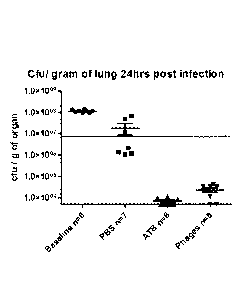
Efficacy of the three-phage mix in vivo was demonstrated by both fluorescence
imaging and by enumeration of bacteria in the lung (Antibiotic = ATB =
Ciprofloxacin
as stated) [Figures 1-5].
The efficacy of the three-phage mix derived using the method as presented was
confirmed in vivo.
The bacteriophage mix showed potent activity and no resistance in vitro at 24
hours.
in vivo, bacteriophage-treated mice showed a marked decrease in luminescence
after 6h with greater reduction overall compared with the ciprofloxacin group.
This
was particularly notable in the nasopharyngeal area, although reductions were
also
seen in luminescence with the abdominal area. Luminescence in the lungs was
broadly comparable, but was markedly reduced with both ciprofloxacin and the
bacteriophage mixture. By 24h all phage and antibiotic treated mice survived
with ¨3
log, reduction in lung CFU observed for both groups.
zo In conclusion:
The three-phage mix is highly effective in vitro.
It is also able to rapidly control bacteria in the oropharynx and lungs of
mice infected
by the PAK strain of P. aeruginosa in an acute phase model.
Its efficacy is equivalent or superior to a high dose of an antibiotic proven
to be
active against the infecting organism.
Its action appears to be faster than the antibiotic, and the dissemination of
the
infection is reduced.
Moving on from this acute model, both laboratory biofilm studies and clinical
trial
data from the chronically infected ear suggests that a heavily colonised,
biofilm-rich
environment can provide the optimal conditions for bacteriophage therapy.
The cystic fibrosis lung may provide such an environment.
CA 02871986 2014-10-29
WO 2013/164640
PCT/GB2013/051163
CLAUSES
Clause 1) A method of designing a panel of bacteriophages for use as a
therapeutic composition against a bacterial infection, the method comprising
a) assessing the activity of two or more individual bacteriophages in
separate liquid cultures, wherein each of said separate liquid cultures
consists or comprises a population of a target bacterial species/ strain
(causative of the bacterial infection) by monitoring one or more of a
change in bacterial growth rate, a development of bacterial resistance
to an individual bacteriophage, and a specificity of the development of
resistance to an individual bacteriophage;
b) determining the efficacy of bacteriophage combinations (e.g. of said
two or more individual bacteriophages) in a liquid culture that consists
or comprises a population of a target bacterial species/ strain
(causative of the bacterial infection) with the intention of identifying an
optimised mixture of bacteriophages, optionally by monitoring one or
more of a change in bacterial growth rate, a development of bacterial
resistance to the bacteriophage combination, and a specificity of the
development of resistance to the bacteriophage combination; and
c) selecting a panel of bacteriophages demonstrating anti-microbial
efficacy against the target bacterial species/ strain.
Clause 2) The method of clause 1, comprising identifying a panel of
bacteriophages having optimal anti-bacterial efficacy for therapeutic use
against the target bacterial species/ strain.
Clause 3)A method for detecting cross-resistance in the method of clause 1 or
clause 2 by culturing bacteriophages in liquid cultures of their bacterial
host by
extended incubation, harvest of resistant forms, and assay against other
bacteriophages specific for the same bacterial host in liquid culture or other
appropriate assay system.
13
CA 02871986 2014-10-29
WO 2013/164640
PCT/GB2013/051163
Clause 4) A method for detecting antagonistic activity of bacteriophages in
clause
1 or clause 2 by culturing bacteriophages singly and in specified combinations
in liquid cultures of their bacterial host.
Clause 5) The method of clause 1 or clause 2 where activity is separately
validated in an in vivo model to confirm anti-bacterial efficacy against the
target bacterial species/ strain and suppression of said bacterial infection.
Clause 6) The method of any preceding clause where the bacterial target is
Acinetobacter baumanii, Clostridium difficile, Escherichia coil, Klebsiella
pneumonia, Pseudomonas aeruginosa, Stenotrophomonas maltophilia,
bacterial species causative of body odour, Staphylococcus aureus or
Streptococcus mutans.
Clause 7) The method according to any preceding clause where the therapeutic
is for use in domestic or farm animals.
Clause 8) The method according to any preceding clause where the therapeutic
is for use in humans.
Clause 9) The method according to any preceding clause where the therapeutic
is for use in food hygiene.
Clause 10) The
method according to any preceding clause where the
therapeutic is for use in agriculture or crop protection.
Clause 11) The
method according to any preceding clause where the
therapeutic is for use in environmental hygiene applications.
Clause 12) A
bacteriophage panel obtainable by a method according to any
of one the preceding clauses.
14