Language selection

Search

Patent 2873074 Summary

Third-party information liability

Some of the information on this Web page has been provided by external sources. The Government of Canada is not responsible for the accuracy, reliability or currency of the information supplied by external sources. Users wishing to rely upon this information should consult directly with the source of the information. Content provided by external sources is not subject to official languages, privacy and accessibility requirements.

Claims and Abstract availability

Any discrepancies in the text and image of the Claims and Abstract are due to differing posting times. Text of the Claims and Abstract are posted:

  • At the time the application is open to public inspection;
  • At the time of issue of the patent (grant).
(12) Patent: (11) CA 2873074
(54) English Title: PATIENT-SPECIFIC INSTRUMENTATION FOR IMPLANT REVISION SURGERY
(54) French Title: INSTRUMENTS SPECIFIQUES AU PATIENT POUR UNE CHIRURGIE DE REVISION D'IMPLANT
Status: Expired and beyond the Period of Reversal
Bibliographic Data
(51) International Patent Classification (IPC):
  • A61B 17/56 (2006.01)
(72) Inventors :
  • ABIVEN, JEAN-GUILLAUME (Canada)
(73) Owners :
  • ORTHOSOFT ULC
(71) Applicants :
  • ORTHOSOFT INC. (Canada)
(74) Agent: NORTON ROSE FULBRIGHT CANADA LLP/S.E.N.C.R.L., S.R.L.
(74) Associate agent:
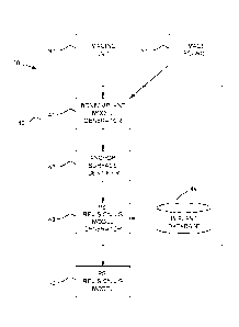
(45) Issued: 2020-09-22
(86) PCT Filing Date: 2013-07-23
(87) Open to Public Inspection: 2014-01-30
Examination requested: 2018-04-30
Availability of licence: N/A
Dedicated to the Public: N/A
(25) Language of filing: English

Patent Cooperation Treaty (PCT): Yes
(86) PCT Filing Number: PCT/CA2013/050572
(87) International Publication Number: WO 2014015432
(85) National Entry: 2014-11-10

(30) Application Priority Data:
Application No. Country/Territory Date
61/674,529 (United States of America) 2012-07-23

Abstracts

English Abstract

A system and method for generating a patient specific instrumentation jig model for implant revision comprises an anchor surface identifier to identify at least one anchor surface from a patient specific bone model of a bone requiring implant revision and from data related to an implanted implant on the bone. The anchor surface is in close proximity to the implanted implant. A PSI revision jig model generator to generate a jig model using at least the identified anchor surface and a model of a replacement implant, the PSI revision jig model generator outputting a jig model comprising at least one patient specific contact surface corresponding to the identified anchor surface, and at least one tool interface portion positioned and/or oriented relative to the at least one contact surface, the at least one tool interface portion adapted to be interfaced to a tool altering the bone for subsequently installing an implant on the bone.


French Abstract

L'invention concerne un système et un procédé pour générer un modèle de gabarit d'instruments spécifiques au patient pour une révision d'implant, qui comprennent un identificateur de surface d'ancrage pour identifier au moins une surface d'ancrage à partir d'un modèle d'os spécifique au patient d'un os nécessitant une révision d'implant et à partir de données associées à un implant implanté sur l'os. La surface d'ancrage est à proximité immédiate de l'implant implanté. L'invention concerne également un générateur de modèle de gabarit de révision d'instruments spécifiques au patient (PSI) pour générer un modèle de gabarit à l'aide au moins de la surface d'ancrage identifiée et d'un modèle d'un implant de remplacement, le générateur de modèle de gabarit de révision de PSI délivrant un modèle de gabarit comprenant au moins une surface de contact spécifique au patient correspondant à la surface d'ancrage identifiée, et au moins une partie interface d'outil positionnée et/ou orientée par rapport à la ou aux surfaces de contact, la ou les parties interface d'outil étant conçues pour servir d'interface avec un outil modifiant l'os de façon à installer par la suite un implant sur l'os.

Claims

Note: Claims are shown in the official language in which they were submitted.


CLAIMS:
1. A method for creating a patient specific
instrumentation (PSI) jig for implant revision, comprising:
obtaining a model of at least part of a bone
requiring removal of an implanted implant being mounted to
the bone, the model being physiologically patient specific
and being obtained from imaging of the implanted implant as
installed on the bone;
obtaining a model of a replacement implant used
to replace the implanted implant on the bone;
identifying alterations to the bone through the
removal of the implanted implant using the model of the
bone and data related to the implanted implant;
identifying at least one anchor surface for the
PSI jig on the bone , the anchor surface being in close
proximity to the implanted implant, the anchor surface
selected for being unaltered through the removal of the
implanted implant; and
generating a jig model using at least the
identified anchor surface and the model of the replacement
implant, the jig model comprising at least one patient
specific contact surface corresponding to the identified
anchor surface for complementary contact, and at least one
tool interface portion positioned and/or oriented relative
to the at least one contact surface, the at least one tool
interface portion adapted to be interfaced with a tool
altering the bone for subsequently installing the
replacement implant after removal of the implanted implant.
2. The method according to claim 1, wherein
generating the jig model comprises generating a cut slot
opening to the at least one contact surface.
- 11 -

3. The method according to claim 1, wherein
identifying at least one anchor surface from data related
to an implanted implant comprises obtaining a model of the
implanted implant on the bone.
4. The method according to claim 1, wherein
obtaining a model of at least part of a bone comprises
imaging the part of the bone and the implanted implant on
the bone, and generating the model of the part of the bone
with the implanted implant.
5. The method according to claim 1, wherein
obtaining a model of at least part of a bone comprises
obtaining images of a femur at a knee.
6. The method according to claim 5, wherein
identifying at least one anchor surface comprises
identifying at least one of surfaces of an epicondyle and
an interior cortex as the at least one anchor surface.
7. The method according to claim 6, wherein
generating the jig model comprises generating at least one
cut slot oriented and positioned for at least one
predetermined femoral cut plane.
8. The method according to claim 1, wherein
obtaining a model of at least part of a bone comprises
obtaining images of a tibia at a knee.
9. The method according to claim 8, wherein
identifying at least one anchor surface comprises
identifying at least one of surfaces of medial and lateral
aspects of the tibia and of a superior tubercle portion of
the tibia as the at least one anchor surface.
- 12 -

10. The method according to claim 9, wherein
generating the jig model comprises generating at least one
cut slot oriented and positioned with at least one
predetermined tibial cut plane.
11. A system for generating a patient specific
instrumentation (PSI) jig model for implant revision,
comprising:
an anchor surface identifier configured to
receive a patient specific bone model of a bone supporting
an implanted implant and requiring removal of the implanted
implant mounted thereon, the patient specific bone model of
the bone obtained from imaging of the implanted implant as
installed on the bone, the anchor surface identifier
identifying alterations to the bone through the
removal of the implanted implant using the patient
specific bone model and data related to the implanted
implant, and
identifying at least one anchor surface for the
PSI jig, the anchor surface being in close proximity to
the implanted implant, the anchor surface selected for
being unaltered through the removal of the implanted
implant; and
a PSI revision jig model generator to generate
the PSI jig model using at least the identified anchor
surface and a model of a replacement implant used to
replace the implanted implant on the bone, the PSI revision
jig model generator outputting a jig model comprising at
least one patient specific contact surface corresponding to
the identified anchor surface, and at least one tool
interface portion positioned and/or oriented relative to
the at least one contact surface, the at least one tool
- 13 -

interface portion adapted to be interfaced to a tool
altering the bone for subsequently installing the
replacement implant on the bone after removal of the
implanted implant.
12. The system according to claim 11, further
comprising a model generator for generating the model of
the part of the bone with the implanted implant from images
of the part of the bone and the implanted implant on the
bone.
13. The system according to claim 12, further
comprising an imaging unit for imaging the part of the bone
and the implanted implant on the bone.
14. The system according to claim 11, wherein said
data related to an implanted implant is a model of the
implanted implant on the bone.
15. The system according to claim 11, wherein the at
least one anchor surface is at least one surface of an
epicondyle and an interior cortex of a femur.
16. The system according to claim 15, wherein the jig
model has at least one cut slot oriented and positioned for
at least one predetermined femoral cut plane.
17. The system according to claim 11, wherein the at
least one anchor surface is at least one surface of medial
and lateral aspects of the tibia and of a superior tubercle
portion of a tibia.
- 14 -

18. The system according to claim 17, wherein the jig
model has at least one cut slot oriented and positioned for
at least one predetermined tibial cut plane.
19. A method for creating a patient specific
instrumentation (PSI) jig for implant revision, comprising:
obtaining a model of at least part of a bone
requiring removal of an implanted implant being mounted to
the bone, the model being physiologically patient specific
and being obtained from imaging of the implanted implant as
installed on the bone;
obtaining a model of a replacement implant used
to replace the implanted implant on the bone;
identifying alterations to the bone through the
removal of the implanted implant using the model and data
related to the implanted implant; and
identifying at least one anchor surface on the
bone for anchoring the PSI jig on the bone, the at last one
anchor surface being exposed on the model despite a
presence of the implanted implant, the anchor surface being
in close proximity to the implanted implant, the anchor
surface selected for being unaltered through the removal of
the implanted implant; and
- 15 -

generating a jig model using at least the
identified anchor surface and the model of the replacement
implant, the jig model comprising at least one patient
specific contact surface corresponding to the identified
anchor surface for complementary contact, and at least one
tool interface portion positioned and/or oriented relative
to the at least one contact surface, the at least one tool
interface portion adapted to be interfaced with a tool
altering the bone for subsequently installing the
replacement implant after removal of the implanted implant.
- 16 -

Description

Note: Descriptions are shown in the official language in which they were submitted.


CA 02873074 2014-11-10
WO 2014/015432 PC
T/CA2013/050572
PATIENT-SPECIFIC INSTRUMENTATION
FOR IMPLANT REVISION SURGERY
FIELD OF THE INVENT ION
[0001] The present disclosure pertains to patient
specific instrumentation (PSI) used in orthopaedic surgery
and, more particularly, to PSI used for implant revision.
BACKGROUND OF THE INVENT ION
[0002] An implant revision process is a process by which
an existing implant is removed to be replaced. However, due
to the bond between the implant to be removed and the bone,
the bone is often damaged during implant revision. As a
result, the subsequent positioning and installation of a
replacement implant may lack precision due to damaged bone
surfaces. For instance, in knee revision surgery, machining
of the bone surfaces using conventional cutting blocks may
lack precision as conventional bone landmarks used for
defining the orientation of the cutting block may be altered
or removed during the removal of the implant.
[0003] Patient specific instrumentation (hereinafter
"PSI") pertains to the creation of instruments that are made
specifically for the patient. PSI are typically manufactured
from data using imaging to model bone geometry. Therefore,
PSI have surfaces that may contact the bone in a predictable
way as such contact surfaces are specifically manufactured
to match the surface of a bone of a given patient. It would
therefore be desirable to use PSI technology in an implant
removal process.
-1-

SUMMARY OF THE DISCLOSURE
glom It is an aim of the present disclosure to provide
a method for creating a PSI jig for implant revision.
pomq It is a further aim of the present disclosure to
provide system for creating a PSI implant revision jig
model.
10006] Therefore, in accordance with the present
disclosure, there is provided a method for creating a
patient specific instrumentation jig for implant revision,
comprising: obtaining a model of at least part of a bone
requiring implant revision, the model being physiologically
patient specific; obtaining a model of a replacement
implant; identifying at least one anchor surface on the bone
from the model of the bone and from data related to an
implanted implant on the bone, the anchor surface being in
close proximity to the implanted implant; and generating a
jig model using at least the identified anchor surface and
the model of the replacement implant, the jig model
comprising at least one patient specific contact surface
corresponding to the identified anchor surface for
complementary contact, and at least one tool interface
portion positioned and/or oriented relative to the at least
one contact surface, the at least one tool interface portion
adapted to be interfaced with a tool altering the bone for
subsequently installing an implant.
(000/ Further in accordance with the present disclosure,
generating the jig model comprises generating a cut slot.
WM] Still further in accordance with the present
disclosure, identifying at least one anchor surface from
- 2 -
CA 2873074 2019-12-31

CA 02873074 2014-11-10
WO 2014/015432
PCT/CA2013/050572
data related to an implanted implant comprises obtaining a
model of the implanted implant on the bone.
[0009] Still further in accordance with the present
disclosure, obtaining a model of at least part of a bone
comprises imaging the part of the bone and the implanted
implant on the bone, and generating the model of the part of
the bone with the implanted implant.
[0010] Still further in accordance with the present
disclosure, obtaining a model of at least part of a bone
comprises obtaining images of a femur at a knee.
[0oll] Still further in accordance with the present
disclosure, identifying at least one anchor surface
comprises identifying at least one of surfaces of an
epicondyle and an interior cortex as the at least one anchor
surface.
[0012] Still further in accordance with the present
disclosure, generating the jig model comprises generating at
least one cut slot oriented and positioned for at least one
predetermined femoral cut plane.
[0013] Still further in accordance with the present
disclosure, obtaining a model of at least part of a bone
comprises obtaining images of a tibia at a knee.
[0014] Still further in accordance with the present
disclosure, identifying at least one anchor surface
comprises identifying at least one of surfaces of medial and
lateral aspects of the tibia and of a superior tubercle
portion of the tibia as the at least one anchor surface.
[0015] Still further in accordance with the present
disclosure, generating the jig model comprises generating at
- 3 -

CA 02873074 2014-11-10
WO 2014/015432
PCT/CA2013/050572
least one cut slot oriented and positioned with at least one
predetermined tibial cut plane.
[0016] Further in accordance with the present disclosure,
there is provided a system for generating a patient specific
instrumentation jig model for implant revision, comprising:
an anchor surface identifier to identify at least one anchor
surface from a patient specific bone model of a bone
requiring implant revision and from data related to an
implanted implant on the bone, the anchor surface being in
close proximity to the implanted implant; and a PSI revision
jig model generator to generate a jig model using at least
the identified anchor surface and a model of a replacement
implant, the PSI revision jig model generator outputting a
jig model comprising at least one patient specific contact
surface corresponding to the identified anchor surface, and
at least one tool interface portion positioned and/or
oriented relative to the at least one contact surface, the
at least one tool interface portion adapted to be interfaced
to a tool altering the bone for subsequently installing an
implant on the bone.
[0017] Further in accordance with the present disclosure,
a model generator generates the model of the part of the
bone with the implanted implant from images of the part of
the bone and the implanted implant on the bone.
[0018] Still further in accordance with the present
disclosure, an imaging unit images the part of the bone and
the implanted implant on the bone.
[0019] Still further in accordance with the present
disclosure, said data related to an implanted implant is a
model of the implanted implant on the bone.
- 4 -

CA 02873074 2014-11-10
WO 2014/015432
PCT/CA2013/050572
[0020] Still further in accordance with the present
disclosure, the at least one anchor surface is at least one
surface of an epicondyle and an interior cortex of a femur.
[0021] Still further in accordance with the present
disclosure, the jig model comprises at least one cut slot
oriented and positioned for at least one predetermined
femoral cut plane.
[0022] Still further in accordance with the present
disclosure, the at least one anchor surface is at least one
surface of medial and lateral aspects of the tibia and of a
superior tubercle portion of a tibia.
[0023] Still further in accordance with the present
disclosure, the jig model comprises at least one cut slot
oriented and positioned for at least one predetermined
tibial cut plane.
BRIEF DESCRIPTION OF THE FIGURES
[0024] Fig. 1 is a flow chart showing a method for
creating a PSI jig for implant revision in accordance with
an embodiment of the present disclosure; and
[0025] Fig. 2 is a block diagram showing a system for
creating a PSI implant revision jig model in accordance with
another embodiment of the present disclosure.
DESCRIPTION OF THE PREFERRED EMBODIMENTS
g026] Referring to the drawings, and more particularly
to Fig. 1, there is illustrated a method 10 for creating
patient specific instrumentation (hereinafter PSI) jig for
implant revision. For clarity, reference to patient specific
in the present application pertains to the creation of
- 5 -

CA 02873074 2014-11-10
WO 2014/015432
PCT/CA2013/050572
negative corresponding surfaces, i.e., a surface that is the
negative opposite of a patient bone/cartilage surface, such
that the patient specific surface conforms to the patient
bone/cartilage surface, by complementary confirming contact.
The method is particularly suited to be used in knee
revision in which the tibial knee Implant, the femoral knee
implant or both implants need to be replaced. The method
may also be used in other orthopedic implant revision
surgery, for instance in shoulder revision surgery.
[0027] According to
12, the bone and its implant are
modeled. The models may be obtained and/or generated using
imaging. The imaging
may be done by any appropriate
technology such as CT scanning (computerized tomography),
fluoroscopy, or like radiography methods, providing suitable
resolution of images. The model of
the bone comprises a
surface geometry of parts of the bone that are exposed
despite the presence of the implant and/or the limitations
of the imaging. The model of the bone may include a surface
geometry of the implant relative to adjacent bone surfaces,
and a 3D geometry of the implant, for instance using a 3D
model of implant (e.g., from the manufacturer, etc).
[0028] The bone
modeling may comprise generating a 3D
surface of the bone if the bone modeling is not directly
performed by the imaging equipment, or if not complete. In
the instance in which multiple implants must be replaced
(e.g., total knee revision), all bones supporting implants
are modeled. Additional structures may be modeled as well,
such as cartilage, etc.
[0029] According to
14, anchor surfaces are identified on
the bone from the model(s) of 12. The anchor surfaces are
selected as being sufficiently large to support a PSI jig,
- 6 -

CA 02873074 2014-11-10
WO 2014/015432
PCT/CA2013/050572
and as not being altered by the removal of the implant from
the bone. For example,
in the case of femoral knee
revision, the anchor surfaces may be the epicondyles and the
interior cortex. The epicondyles may be used to restore the
joint line to set the axial position of the replacement
implant. Other parts
of the femur may also be used as
anchor surfaces.
[0030] As another
example, in the case of tibial knee
implant replacement, the anchor surfaces may be that of the
medial and lateral aspects as well as the superior tubercle
portion of the tibia. In this case, the medial and lateral
aspects may be used to restore the joint line by setting the
axial position of the replacement implant. Other parts of
the tibia may also be used as anchor surfaces. Similar
considerations are taken into account in the case of
shoulder surgery. In both cases, the anchor surfaces are in
close proximity to the implanted implant as it is in the
vicinity of the removed implant that bone alterations will
be performed. Although the
anchor surface(s) is in close
proximity to the removed implant, the anchor surface will
not substantially damaged by the removal of the implant.
[0031] According to
16, using the anchor surfaces as
obtained from the bone model(s) and the geometry of the
replacement implant that is known (i.e., obtained from a
database, from the manufacturer, generated as a PSI implant,
etc), a PSI revision jig model is generated. The jig model
will have a contact surface(s) defined to abut against the
anchor surface(s) obtained in 14, in a predictable and
precise manner. Typically,
the PSI revision jig is a
cutting block or cutting guide that will allow to cut planes
upon which will be anchored the implant. The PSI revision
jig model of 16 therefore comprises cutting planes, guides,
- 7 -

CA 02873074 2014-11-10
WO 2014/015432
PCT/CA2013/050572
slots, or any other tooling interface or tool, oriented
and/or positioned to allow bone alterations to be formed in
a desired location of the bone, relative to the contact
surface(s). Thus, PSI revision jig model may also take into
consideration any revision planning done by the operator
(e.g., surgeon), to therefore allow the removal of
sufficient bone material to reproduce desired gaps between
out planes on adjacent bones, etc.
[0032] According to 18, once the PSI revision jig model
has been generated, the PSI jig may be created. When
installing the PSI jig on the bone, the contact surface(s)
on the PSI jig is(are) applied against the corresponding
anchor surface(s) of step 14. The PSI jig created in 18 may
then be used intra-operatively after the implant is removed
to allow alterations to be made on the bone. For instance,
in the case of total knee revision, jigs are used to perform
femoral distal and tibial cuts.
[0m] Now that a method for creating a PSI revision jig
for implant replacement has been defined, a system is set
forth.
[mu] A system for the creation of a PSI revision jig
model is generally shown at 20 in Fig. 2. The system 20 may
comprise an imaging unit 30, such as a CT scan or an X-ray
machine, so as to obtain images of the bone and implant. As
an alternative, images may be obtained from an image source
31. As an example, a CT scan may be operated remotely from
the system 20, whereby the system 20 may simply obtain
images and/or processed bone and implant models from the
image source 31.
[0035] The system 20 comprises a processor unit 40 (e.g.,
computer, laptop, etc.) that comprises different modules so
- 8 -

CA 02873074 2014-11-10
WO 2014/015432
PCT/CA2013/050572
as to ultimately produce a revision jig model. The
processing unit 40 of the system 20 may therefore comprise a
bone/implant model generator 41 receiving images from
sources 30 or 31 to generate a 3D model of the bone with the
implant, prior to implant revision. In accordance with the
method 10 of Fig. 1, the 3D model of the bone with implant
may comprise data pertaining to the surface geometry of a
relevant portion of a bone, including surfaces of the bone
that are exposed despite the presence of the implant.
[0036] The
bone/implant model generator 41 will create
the 3D model of the bone and implant that is then used by an
anchor surface identifier 42 of the processing unit 40.
Alternatively, the anchor surface identifier 42 may use a 3D
model provided by the image source 31, provided the model
obtained from the image source 31 comprises sufficient data.
The anchor surface identifier 42 identifies surfaces on the
bone that will substantially not be altered by the removal
of the damaged implant. The anchor surface(s) is(are)
selected as being sufficiently large to support a PSI jig,
and as not obstructing the removal of the implant. For
example, reference is made to step 14, in which examples are
provided for appropriate anchor surfaces on the femur and
the tibia in the case of total knee replacement.
[0037] Once the
anchor surface(s) is(are) identified, a
PSI revision jig model generator 43 will generate a jig
model. As in 16 of the method 10, the jig model will have a
contact surface(s) defined to abut against the anchor
surface(s) identified by the anchor surface identifier 42,
in a predictable and precise manner. As the PSI
revision
jig will support a tool to perform alterations on the bone,
the jig model comprises cutting planes, guides, slots, or
any other tooling interface or tool, trackers (oriented
- 9 -

CA 02873074 2014-11-10
WO 2014/015432
PCT/CA2013/050572
and/or positioned to allow bone alterations to be formed in
a desired location of the bone, relative to the contact
surface(s).
[0038] Thus, PSI
revision jig model generator 43 may also
take into consideration any revision planning done by the
operator (e.g., surgeon). The PSI
revision jig model
generator 43 may also take into consideration a geometry of
the existing damaged implant, the replacement implant (e.g.,
obtained from an implant database 44), in addition to the
anchor surface(s).
[0039] Accordingly,
the system 20 outputs a PSI revision
jig model 50 that will be used to create the PSI revision
jig. The PSI revision jig is then used intra-operatively to
resurface bone for subsequent implant installation, as
described for method 10 in Fig. 1.
[0040] While the
methods and systems described above have
been described and shown with reference to particular steps
performed in a particular order, these steps may be
combined, subdivided or reordered to form an equivalent
method without departing from the teachings of the present
disclosure. Accordingly,
the order and grouping of the
steps is not a limitation of the present disclosure.
- 10-
Representative Drawing
A single figure which represents the drawing illustrating the invention.
Administrative Status

2024-08-01:As part of the Next Generation Patents (NGP) transition, the Canadian Patents Database (CPD) now contains a more detailed Event History, which replicates the Event Log of our new back-office solution.

Please note that "Inactive:" events refers to events no longer in use in our new back-office solution.

For a clearer understanding of the status of the application/patent presented on this page, the site Disclaimer , as well as the definitions for Patent , Event History , Maintenance Fee  and Payment History  should be consulted.

Event History

Description Date
Time Limit for Reversal Expired 2023-01-24
Letter Sent 2022-07-25
Letter Sent 2022-01-24
Letter Sent 2021-07-23
Grant by Issuance 2020-09-22
Inactive: Cover page published 2020-09-21
Inactive: COVID 19 - Deadline extended 2020-08-06
Inactive: COVID 19 - Deadline extended 2020-07-16
Pre-grant 2020-07-15
Change of Address or Method of Correspondence Request Received 2020-07-15
Inactive: Final fee received 2020-07-15
Notice of Allowance is Issued 2020-04-01
Letter Sent 2020-04-01
Notice of Allowance is Issued 2020-04-01
Inactive: Q2 passed 2020-03-10
Inactive: Approved for allowance (AFA) 2020-03-10
Amendment Received - Voluntary Amendment 2019-12-20
Letter Sent 2019-11-21
Common Representative Appointed 2019-11-21
Common Representative Appointed 2019-10-30
Inactive: Multiple transfers 2019-10-23
Inactive: S.30(2) Rules - Examiner requisition 2019-06-21
Inactive: Report - No QC 2019-06-19
Letter Sent 2018-05-09
Request for Examination Received 2018-04-30
Request for Examination Requirements Determined Compliant 2018-04-30
All Requirements for Examination Determined Compliant 2018-04-30
Inactive: Cover page published 2015-01-16
Inactive: First IPC assigned 2014-12-05
Inactive: Notice - National entry - No RFE 2014-12-05
Inactive: IPC assigned 2014-12-05
Application Received - PCT 2014-12-05
National Entry Requirements Determined Compliant 2014-11-10
Application Published (Open to Public Inspection) 2014-01-30

Abandonment History

There is no abandonment history.

Maintenance Fee

The last payment was received on 2020-06-29

Note : If the full payment has not been received on or before the date indicated, a further fee may be required which may be one of the following

  • the reinstatement fee;
  • the late payment fee; or
  • additional fee to reverse deemed expiry.

Please refer to the CIPO Patent Fees web page to see all current fee amounts.

Fee History

Fee Type Anniversary Year Due Date Paid Date
Basic national fee - standard 2014-11-10
MF (application, 2nd anniv.) - standard 02 2015-07-23 2015-06-22
MF (application, 3rd anniv.) - standard 03 2016-07-25 2016-06-22
MF (application, 4th anniv.) - standard 04 2017-07-24 2017-06-23
Request for exam. (CIPO ISR) – standard 2018-04-30
MF (application, 5th anniv.) - standard 05 2018-07-23 2018-06-22
MF (application, 6th anniv.) - standard 06 2019-07-23 2019-06-20
Registration of a document 2019-10-23 2019-10-23
MF (application, 7th anniv.) - standard 07 2020-07-23 2020-06-29
Final fee - standard 2020-08-03 2020-07-15
Owners on Record

Note: Records showing the ownership history in alphabetical order.

Current Owners on Record
ORTHOSOFT ULC
Past Owners on Record
JEAN-GUILLAUME ABIVEN
Past Owners that do not appear in the "Owners on Record" listing will appear in other documentation within the application.
Documents

To view selected files, please enter reCAPTCHA code :


To view images, click a link in the Document Description column. To download the documents, select one or more checkboxes in the first column and then click the "Download Selected in PDF format (Zip Archive)" or the "Download Selected as Single PDF" button.

List of published and non-published patent-specific documents on the CPD .

If you have any difficulty accessing content, you can call the Client Service Centre at 1-866-997-1936 or send them an e-mail at CIPO Client Service Centre.


Document
Description 
Date
(yyyy-mm-dd) 
Number of pages   Size of Image (KB) 
Representative drawing 2020-08-24 1 4
Claims 2014-11-10 4 109
Drawings 2014-11-10 2 16
Description 2014-11-10 10 340
Abstract 2014-11-10 1 62
Representative drawing 2014-11-10 1 8
Cover Page 2015-01-16 2 45
Description 2019-12-31 10 365
Claims 2019-12-31 6 192
Cover Page 2020-08-24 1 39
Notice of National Entry 2014-12-05 1 193
Reminder of maintenance fee due 2015-03-24 1 110
Reminder - Request for Examination 2018-03-26 1 118
Acknowledgement of Request for Examination 2018-05-09 1 174
Commissioner's Notice - Application Found Allowable 2020-04-01 1 550
Commissioner's Notice - Maintenance Fee for a Patent Not Paid 2021-09-03 1 554
Courtesy - Patent Term Deemed Expired 2022-02-21 1 538
Commissioner's Notice - Maintenance Fee for a Patent Not Paid 2022-09-06 1 540
PCT 2014-11-10 2 103
Request for examination 2018-04-30 2 72
Examiner Requisition 2019-06-21 3 171
Amendment / response to report 2019-12-20 10 338
Final fee / Change to the Method of Correspondence 2020-07-15 5 154