Note: Descriptions are shown in the official language in which they were submitted.
- 1 -
MEMBRANE ENCAPSULATED NANOPARTICLES AND METHOD OF USE
CROSS-REFERENCE TO RELATED APPLICATIONS
moll This application claims priority to U.S. Provisional Application
Serial No.
61/492,626, filed June 2, 2011.
FIELD OF THE INVENTION
[0002] The present invention relates to methods and compositions for
delivery of
synthetic nanoparticle materials, including pharmaceutically active agents,
encapsulated
with cellular membranes.
BACKGROUND OF THE INVENTION
[0003] Long-circulating polymeric nanoparticles have significant
clinical impact as
they promise sustained systemic delivery and better targeting through both
passive and
active mechanisms (1-3). Different approaches including modifications on
particle size,
surface, shape, and flexibility have been explored to extend particle
residence time in vivo
(4-6). The current gold standard for nanoparticle stealth coating is
polyethylene glycol
(PEG). The adoption of PEG as a stealth moiety on nanoparticle surface has led
to great
success with several clinical products (2, 3), but recent observation of anti-
PEG
immunological response has triggered the interest of further investigation on
its biological
relevance (7).
[0004] Synthetic zwitterionic materials such as poly (carb oxy betaine)
and
poly(sulfobetaine) have been proposed as alternatives to PEG because of their
strong
hydration that is highly resistant to nonspecific protein adsorption (8, 9).
In addition, recent
advances in molecular and cellular biology have inspired scientists and
nanotechnologists
to model nanocarriers after red blood cells (RBCs), which are nature's long-
circulating
delivery vehicles. Properties of RBCs such as their structure and surface
proteins have been
taken as design cues to devise the next-generation delivery platforms (10-12).
Date Recue/Date Received 2020-07-02
-2-
10005J While significant efforts have been devoted to bridging the gap
between
synthetic nanomaterials and biological entities, an RBC-mimicking delivery
vehicle has
remained elusive to biomedical researchers. One major challenge lies in the
difficulty in
functionalizing nanoparticles with the complex surface chemistry of a
biological cell.
Despite the recent great progress in reducing macrophage engulfment of
polystyrene beads
following their conjugation with an immunosuppressive RBC membrane protein,
CD47
(11), current chemistry-based bioconjugation techniques often lead to protein
denaturation.
In addition, these bottom-up approaches are largely inadequate in duplicating
a complex
protein makeup on a nanoscale substrate.
100061 Therefore, what is needed are improved methods and compositions for
delivery
of synthetic nanoparticle materials. The present invention addresses these and
other related
needs in the art.
SUMMARY OF THE INVENTION
100071 The present invention provides novel nanoparticles, and methods
of using and
making thereof. More specifically, the inventive nanoparticle comprises a) an
inner core
comprising a non-cellular material; and b) an outer surface comprising a
cellular membrane
derived from a cell, wherein said inner core supports said outer surface. In
certain
embodiments, the inner core of the inventive nanoparticle comprises a
biocompatible and/or
a synthetic material including but not limited to, poly(lactic-co-glycolic
acid), polylactic
acid, polyglycolic acid, polycaprolactone, polylysine, polyglutamic acid, and
any other
suitable synthetic material or the like.
10008] In certain embodiments, the outer surface of the inventive
nanoparticle
comprises cellular membrane comprising plasma membrane or an intracellular
membrane
derived from a unicellular (e.g. a bacterium or fungus) or multicellular
organism (e.g., a
plant, an animal, a non-human mammal, vertebrate, or a human). In certain
embodiments,
the miter surface of the inventive nanoparticle comprises a naturally
occurring cellular or
viral membrane and/or further comprises a synthetic membrane.
CA 2873404 2018-09-19
CA 02873404 2014-11-12
WO 2013/052167
PCT/US2012/039411
-3-
100091 In certain embodiments, the cellular membrane of the outer surface
of the
inventive nanoparticle is derived from a blood cell (e.g., red blood cell
(RBC), white blood
cell (WBC), or platelet). In other embodiments, the cellular membrane of the
outer
surface is derived from an immune cell (e.g., macrophage, monocyte, B-cell, or
T-cell), a
tumor or cancer cell, and other cells, such as an epithelial cell, an
endothelial cell, or a
neural cell. In other embodiments, the cellular membrane of the outer surface
is derived
from a non-terminally differentiated cell, such as a stem cell, including a
hematopoietic
stem cell, a bone marrow stem cell, a mesenchymal stern cell, a cardiac stem
cell, a neural
stem cell. The non-terminally differentiated cell can be isolated in a
pluripotent state from
tissue or induced to become pluripotent. In yet other embodiments, the
cellular membrane
is derived from a cell component or cell organelle including, but not limited
to, an
exosome, a secretory vesicle, a synaptic vesicle, an endoplasmic reticulum
(ER), a Golgi
apparatus, a mitochondrion, a vacuole or a nucleus.
100101 In certain embodiments, the present invention further provides that the
inventive
nanoparticle comprises a releasable cargo that can be located in any place
inside or on the
surface of the nanoparticle. A trigger for releasing the releasable cargo from
the inventive
nanoparticle includes, but is not limited to, contact between, the
nanoparticle and a target
cell, tissue, organ or subject, or a change of an environmental parameter,
such as the pH,
ionic condition, temperature, pressure, and other physical or chemical
changes,
surrounding the nanoparticle. In certain embodiments, the releasable cargo
comprises one
or more therapeutic agent, prophylactic agent, diagnostic or marker agent,
prognostic
=
agent, e.g., an imaging marker, or a combination thereof. In yet certain other
embodiments, the releasable cargo is a metallic particle, a polymeric
particle, a dendrimer
particle, or an inorganic particle.
100111 The present nanoparticle can have any suitable shape. For example, the
present
nanoparticle and/or its inner core can have a shape of sphere, square,
rectangle, triangle,
circular disc, cube-like shape, cube, rectangular parallelepiped (cuboid),
cone, cylinder,
prism, pyramid, right-angled circular cylinder and other regular or irregular
shape. The
present nanoparticle can have any suitable size.
100121 The present invention further provides that in certain embodiments the
inventive
nanoparticle has a diameter from about 10 nm to about 10 i_un. In certain
embodiments,
CA 02873404 2014-11-12
WO 2013/052167
PCMJS2012/039411
-4-
the diameter of the invention nanoparticle is about 50 nm to about 500 nm. In
other
embodiments, the diameter of the nanoparticle can be about 10 nm, 20 nm, 30
urn, 40 nm,
50 nm, 60 urn, 70nm, 80 urn, 90 nm, 100 nm, 110 nm, 120 hm, 130 nm, 140 nm,
150 nm,
200 nm, 300 nm, 400 nm, 500 nm, 600 urn, 700 nm, 800 rim, 900 nm, 1 pm, 2 pm,
3 pm,
4 pm, 5 um, 6 pm, 7 pm, 8 m, 9 pm, and 10 pm, or any suitable sub-ranges
within the
about 10 nm to about 10 pm range, e.g., a diameter from about 50 nm to about
150 nm. In
certain embodiments, the inner core supports the outer surface.
100131 The present invention further provides that the invention nanoparticle
substantially
lacks constituents of the cell from which the cellular membrane is derived or
constituents
of the virus from which the viral membrane is derived. For example, the
present
nanoparticle can lack, in terms of types and/or quantities, at least 10%, 20%,
30%, 40%,
50%, 60%, 70%, 80%, 90%, 95%, 96%, 97%, 98%, 99% or 100% of the constituents
of
the cell from which the cellular membrane is derived or constituents of the
virus from
which the viral membrane is derived.
100141 In yet certain other embodiments, the nanoparticle of the present
invention
substantially maintains natural structural integrity or activity of the
cellular membrane, the
membrane derived from a virus or the constituents of the cellular membrane or
viral
membrane. The structural integrity of the cellular membrane includes.primary,
secondary,
tertiary or quaternary structure of the cellular membrane, the membrane
derived from a
virus or the constituents of the cellular membrane. or viral membrane, and the
activity of
the cellular membrane includes, but is not limited to, binding activity,
receptor activity,
signaling pathway activity, and any other activities a normal naturally
occurring cellular
membrane, the membrane derived from a virus or the constituents of the
cellular
membrane or viral membrane, would have. In certain embodiments, the
nanoparticle of
the present. invention is biocompatible and/or biodegradable. For example, the
present
nanoparticle can maintain, in terms of types and/or quantities, at least 10%,
20%, 30%,
40%, 50%, 60%, 70%, 80%, 90%, 95%, 96%, 97%, 98%, 99% or 100% of the natural
structural integrity or activity of the cellular membrane, the membrane
derived from a
virus or the constituents of the cellular membrane or viral membrane.
100151 In certain embodiments, the nanoparticle of the present invention
comprises the
cellular plasma membrane derived from a red blood cell and an inner core
comprising
CA 02873404 2014-11-12
WO 2013/052167 PCT/US2012/039411
-5-
poly(lactic-co-glycolic acid) (PLGA), wherein the nanoparticle substantially
lacks
hemoglobin. For example, the present nanoparticle can lack, in terms of types
and/or
quantities, at least 10%, 20%, 30%, 40%, 50%, 60%, 70%, 80%, 90%, 95%, 96%,
97%,
98%, 99% or 100% of the hemoglobin of the red blood cell from which the plasma
.. membrane is derived.
100161 Such inventive nanoparticle has a half-life in blood circulation in
vivo at least
about 2-5 times of a half-life of a polyethylene glycol (PEG)-coated,
comparable
nanoparticle. In certain embodiments, such inventive nanoparticle has a half-
life in blood
circulation in vivo for at least about 5 to abont 40 hours or longer.
100171 In certain embodiments, the invention nanoparticle substantially lacks
immunogenicity to a species or subject from which the cellular membrane is
derived. For
example, the present nanoparticle can lack, in terms of types and/or
quantities, at least
10%, 20%, 30%, 40%, 50%, 60%, 70%, 80.%, 90%, 95%, 96%, 97%, 98%, 99% tn t00%
of the immunogenicity to a species or subject from which the cellular membrane
is
derived.
100181 The present invention further provides a medicament delivery system,
and/or a
pharmaceutical composition comprising the inventive nanoparticle. In
certain
embodiments, the medicament delivery system and/or the pharmaceutical
composition of =
the present invention further comprises one or more additional active
ingredient and/or a
medically or pharmaceutically acceptable carrier or excipient, that can be
administered
along with or in combination with the nanoparticle of the present invention.
100191 The present invention further provides a method for treating and/or
preventing a
disease or condition in a subject in need using the inventive nanoparticles,
the medicament
delivery system, or the pharmaceutical composition comprising the same. In
certain
embodiments, the cellular membrane of the nanoparticle used for the inventive
method is
derived from a cell of the same Species of the subject or is derived from a
cell of the
subject. In certain embodiments, the cellular membrane of the nanoparticle
used for the
inventive method is derived from a red blood cell of the same species of the
subject and
the red blood cell has the same blood type of the subject. In certain
embodiments, the
nanoparticle, the medicament delivery system, or the pharmaceutical
composition is
-6-
administered via any suitable administration route. For example, the
nanoparticle, the
medicament delivery system, or the pharmaceutical composition can be
administered via an
oral, nasal, inhalational, parental, intravenous, intraperitoneal,
subcutaneous, intramuscular,
intradermal, topical, or rectal route.
100201 In other embodiments, the nanoparticle is administered via a medicament
delivery
system. In yet other embodiments, the inventive method further comprises
administering
another active ingredient, or a pharmaceutically acceptable carrier or
excipient, to the
subject in need. The inventive method further provides that the nanoparticle
of the present
invention can be administered systemically or to a target site of the subject
in need. Use of
an effective amount of nanoparticles of the present invention for the
manufacture of a
medicament for treating or preventing a disease or condition in a subject in
need is also
provided.
100211 Furthermore, the present invention provides an immunogenic composition
comprising an effective amount of nanoparticle that comprises an inner core
comprising a
non-cellular material, and an outer surface comprising a cellular or plasma
membrane
derived from a cell and an antigen or a hapten. A vaccine comprising the
immunogenic
composition of the present invention is also provided. The present invention
further
provides a method of use of the invention immunogenic composition for
eliciting an
immune response to the antigen or hapten in a subject in need of such
elicitation, and method
of use of the invention vaccine comprising the immunogenic composition for
protecting a
subject against the antigen or hapten. In certain embodiments, the immune
response is T-
cell or B-cell mediated immune response. Use of an effective amount of the
nanoparticle
of the present invention for the manufacture of the immunogenic composition
against an
antigen or hapten, and use of an effective amount of the immunogenic
composition for the
manufacture of a vaccine for protecting a subject against the antigen or
hapten, are also
provided.
10022] The present invention further provides a method for making the
nanoparticle of the
invention, comprising mixing a nanoparticle inner core comprising a non-
cellular material
with a cellular membrane derived from a cell while exerting exogenous energy
to form the
nanoparticle, wherein said inner core supports said outer surface. In certain
embodiments,
the exogenous energy is a mechanical energy, e.g., a mechanical energy exerted
by
CA 2873404 2018-09-19
=
-7-
extrusion. In other embodiments, the exogenous energy is an acoustical energy,
e.g., an
acoustical energy exerted by sonication. In yet other embodiment, the
exogenous energy is
a thermal energy, e.g., a thermal energy exerted by heating. In yet other
embodiments, the
inventive method further comprises mixing a nanoparticle inner core comprising
non-
cellular material with a naturally occurring cellular membrane derived from a
cell or a
naturally occurring membrane derived from a virus with a synthetic membrane
while
exerting exogenous energy to form the nanoparticle comprising the inner core
and an outer
surface comprising the cellular membrane or viral membrane and the synthetic
membrane.
100231 The present invention further provides a neoplasm specific immunogen
comprising
an effective amount of the nanoparticle that comprises an inner core
comprising a non-
cellular material, and an outer surface comprising a cellular membrane derived
from a
neoplasm cell, wherein the cellular membrane substantially retains its
structurally integrity
for eliciting an immune response to the neoplasm cell. For example, the
present nanoparticle
can maintain, in terms of types and/or quantities, at least 100/a, 20%, 300/,,
40%, 50%, 60%,
70%, 80%, 90%, 95%, 96%, 97%, 98%, 99% or 100% of its structurally integrity
for
eliciting an immune response to the neoplasm cell.
100241 In certain embodiments, the inner core supports the outer surface of
such
nanoparticles. In certain embodiments, the inner core of such nanoparticles
comprises
PLGA and the outer surface comprises a plasma membrane derived from a neoplasm
cell.
In other embodiments, the outer surface of such nanoparticles comprises
naturally occurring
cellular or viral membrane and further comprises a synthetic membrane.
100251 The nanoparticle contained in the inventive neoplasm specific
immunogenic
composition substantially lacks constituents of the neoplasm cell from which
the cellular
membrane is derived. For example, the present nanoparticle can lack, in terms
of types
and/or quantities, at least 10%, 20%, 30%, 40%, 50%, 60%, 70%, 80%, 90%, 95%,
96%,
97%, 98%, 99% or 100% the constituents of the neoplasm cell from which the
cellular
membrane is derived.
100261 In certain embodiments, the nanoparticle in the invention neoplasm
specific
immunogenic composition has a diameter from about 10 nm to about 10 um. In
certain
embodiments, such nanoparticle has a diameter from about 50 nm to about 500
nm. In
CA 2873404 2018-09-19
-8-
certain embodiments, the nanoparticle in the inventive neoplasm specific
immunogenic
composition further comprises another active ingredient, or a releasable
cargo. In yet other
embodiments, the inventive neoplasm specific immunogenic composition further
comprises
an immunogenic adjuvant or an immunopotentiator.
10027] The present invention further provides a vaccine comprising the
neoplasm specific
immunogenic composition. Methods for treating or preventing a neoplasm in
subject in
need using the invention neoplasm specific immunogenic composition or the
vaccine are
also provided. The present invention further provides the use of an effective
amount of the
nanoparticle of the present invention for the manufacture of a cancer or
neoplasm specific
immunogenic composition or vaccine for treating or preventing a subject
against a
neoplasm.
[00281 The present invention further provides a pharmaceutical composition
comprising the
nanoparticle of the invention for treating or preventing a disease or
condition associated
with a cell membrane inserting toxin, wherein the nanoparticle contained in
the
pharmaceutical composition comprises an inner core comprising a non-cellular
material and
an outer surface comprising a cellular or plasma membrane derived from a
target cell, e.g.,
a red blood cell, wherein said inner core supports said outer surface. In
certain
embodiments, the toxin inserted into the cellular or plasma membrane of the
target cells is
part of the natural pathological mechanism, or the cellular or plasma membrane
in the outer
surface of the nanoparticle substantially retains the toxin. In certain
embodiments, the toxin
is a bacterial (e.g., S. aureus), plant, fungal, or an animal toxin.
100291 In certain embodiments, the inner core supports the outer surface, and
the cellular
membrane in the outer surface of the nanoparticle substantially retains its
structural integrity
for substantially retaining the toxin. In yet certain other embodiments, the
outer surface of
the nanoparticle comprises a naturally occurring cellular or viral membrane
and further
comprises a synthetic membrane or synthetic or naturally occurring components
added to
the cellular membrane. In yet certain other embodiments, the nanoparticle
contained in such
pharmaceutical composition is biocompatible, biodegradable, or comprises a
synthetic
material. In yet certain other embodiments, the pharmaceutical composition of
the present
invention further comprises another active ingredient or a pharmaceutically
acceptable
carrier or excipient.
CA 2873404 2018-09-19
-9-
10030] Methods for treating or preventing a disease or condition associated
with a cell
membrane inserting toxin using the nanoparticle of the present invention, as
well as a
pharmaceutical composition comprising such nanoparticles, are also provided.
The present
invention further provides the use of an effective amount of the
pharmaceutical composition
comprising the nanoparticle for the manufacture of a medicament for treating
or
preventing a disease or condition associated with a cell membrane inserting
toxin in
subject in need.
[0031] Furthermore, the present invention provides an immunogen comprising an
effective amount of nanoparticle that comprises an inner core comprising a non-
cellular
material, and an outer surface comprising a cellular or plasma membrane
derived from a cell
and a cell membrane inserting toxin, wherein said inner core supports said
outer surface. A
vaccine comprising the immunogenic composition of the present invention is
also provided.
The present invention further provides a use of the inventive immunogen for
eliciting an
immune response to a cell membrane inserting toxin in a subject in need of
such elicitation,
.. and use of the inventive vaccine comprising the immunogen for protecting a
subject
against the cell membrane inserting toxin. In certain embodiments, the immune
response
is T-cell or B-cell mediated immune response. Use of an effective amount of
the
nanoparticle of the present invention for the manufacture of the immunogen
against a cell
membrane inserting toxin, and use of an effective amount of the immunogen for
the
manufacture of a vaccine for protecting a subject against cell membrane
inserting toxin, are
also provided.
[0032] The present invention contemplates treatments, prevention, diagnosis
and/or
prognosis of any diseases, disorders, or physiological or pathological
conditions,
including, but not limited to, an infectious disease, a parasitic disease, a
neoplasm, a disease
of the blood and blood-forming organs, a disorder involving the immune
mechanism,
endocrine, nutritional and metabolic diseases, a mental and behavioral
disorder, a
disease of the nervous system, a disease of the eye and adnexam, a disease of
the ear
and mastoid process, a disease of the circulatory system, a disease of the
respiratory system,
a disease of the digestive system, a disease of the skin and subcutaneous
tissue, a
disease of the musculoskeletal system and connective tissue, a disease of the
genitourinary system, pregnancy, childbirth and the puerperium, a condition
CA 2873404 2018-09-19
CA 02873404 2014-11-12
WO 2013/052167
PCT/US2012/039411
-10-
originating in the perinatal period, a congenital malformation, a deformation,
a
chromosomal abnormality, an injury, a poisoning, a consequence of external
causes, and
an external cause of morbidity and mortality.
100331 In some embodiments, the present nanoparticles, medicament delivery
systems,
pharmaceutical compositions and methods can be used to treat or prevent the
exemplary
cancers and tumors listed in Table I, to deliver the exemplary cancer
medications listed in
Table 2, to treat or prevent the exemplary ocular diseases or conditions
listed in Table 3, to
deliver the exemplary ocular medications listed in Table 4, to treat or
prevent the
exemplary diseases or conditions affecting the lungs listed in Table 5, to
deliver the
exemplary lungs/respiratory disease medications listed in Table 6, to treat or
prevent the
exemplary diseases or conditions affecting the heart listed in Table 7, or to
deliver the
exemplary heart medications listed in Table 8. In some embodiments, the
present
nanoparticles, medicament delivery systems, pharmaceutical compositions and
methods
can be used to treat or prevent the exemplary conditions listed in Table 9.
Tables 1-9 are
attached herewith at the end of the instant specification.
100341 In some embodiments, the present nanoparticles, medicament delivery
systems,
pharmaceutical compositions and methods, can be used to deliver the exemplary
medications listed in the Orange Book: Approved Drug Products with Therapeutic
Equivalence Evaluations (Current through March 2012) published by the U.S.
Food and
Drug Administration, the exemplary medications listed in The Merck Index (a
U.S.
publication, the printed 14th Edition, Whitehouse Station, N.J., USA) and its
online
version (The Merck Index Onlinesm, Last Loaded on Web: Tuesday, May 01, 2012),
and
the exemplary medications listed in Biologics Products & Establishments
published by the
U.S. Food and Drug Administration, and can be used to treat or prevent the
corresponding
diseases and disorders.
BRIEF DESCRIPTION OF THE DRAWINGS
100351 Those of skill in the art will understand that the drawings, described
below, are for
illustrative purposes only. The drawings are not intended to limit the scope
of the present
teachings in any way.
CA 02873404 2014-11-12
WO 2013/052167
PCT/US2012/039411
-11-
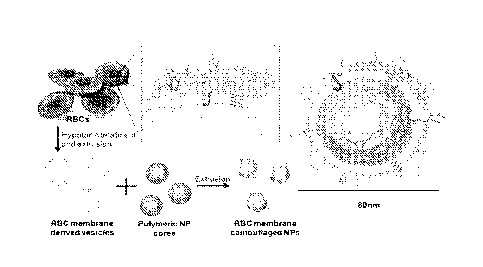
100361 Fig. I. Schematics of the preparation process of the RBC membrane-
coated PLGA
nanoparticles (NPs).
100371 Fig. 2. Structural characterization of the RBC membrane-coated PLGA
nanoparticles. Fig. 2A. The nanoparticles were negatively stained with uranyl
acetate and
subsequently visualized with TEM. Fig. 2B. DLS measurements of the size,
polydispersity index (PDI), and surface zeta potential of the nanoparticles
over 14 days.
Fig. 2C. Scanning fluorescence microscopy images demonstrated the co-
localization of the
RBC membranes (visualized with green rhodamine-DMPE dyes) and polymeric cores
(visualized with red DiD dyes) after being internalized by HeLa cells. The RBC
membrane-coated nanoparticles were incubated with HeLa cells for 6 hours. The
excess
nanoparticles were washed out and the cells were subsequently fixed for
imaging.
100381 Fig. 3. Membrane protein retention, particle stability in serum, and
the ill vivo
circulation time of the R13C membrane-coated nanopai tides (NI's). Fig. 3A.
Fiutehis in
emptied R.BCs, RBC membrane-derived vesicles, and purified RBC membrane-coated
IS PLGA nanoparticles were solubilized and resolved on a polyactylamide
gel. Fig. 3B. RBC
membrane-coated PLGA nanoparticles, PEG-coated lipid-PLGA hybrid
nanoparticles, and
bare PLGA nanoparticles were incubated in 100% fetal bovine serum and
monitored for
absorbance at 560 run for 4hours, Fig. 3C. DiD-loaded nanoparticles were
injected
intravenously through the tail vein of mice. At.various time points blood was
withdrawn
intraorbitally and measured for fluorescence at 670 rim to evaluate the
systemic circulation
lifetime of the nanoparticles (n=6 per group).
100391 Fig. 4. Biodistributions of the RBC membrane-coated polymeric
nanoparticles.
Fluorescently labeled nanoparticles were injected intravenously into the mice.
At each
time points (24, 48, and 72 hour respectively), the organs from a randomly
grouped subset
of mice were collected, homogenized and quantified for fluorescence. Fig.
4A.
Fluorescence intensity per gram of tissue (n---6 per group). Fie. 4B. Relative
signal per
organ.
100401 Fig. 5. Phase contrast microscopy images of inouse red blood cells
(RBCs) before
(left panel) and after (right panel) hemolytic treatment in hypotonic
solution_ Deprivation
CA 02873404 2014-11-12
WO 2013/052167 PCT/US2012/039411
-12-
of RBC interior contents (hemoglobins) was verified by the change in phase
contrast,
which indicates an alteration of the medium inside the RBCs.
100411 Fig. 6. The average diameter of the RBC membrane-derived vesicles
following
RBC ghosts derivation, 5 min of sonication, 400 rim extrusion, and 100 nm
extrusion as
measured by dynamic light scattering (DLS).
10042i Fig. 7. The fluorescence retention of DiD dye in PEGylated lipid-P.LGA
hybrid .
nanoparticles (NPs) over a period of 72 hours.
100431 Fig. 8. The mean particle diameter of PLGA nanoparticles (NPs) prior to
(left) and
following (right) RBC membrane coating as measured by DLS.
100441 Fig. 9. Schematic illustration of building materials and the
preparation process of
RBCm-cloaked NPs. The hydrodynamic size of RBC ghosts, RBCm-derived vesicles,
polymeric cores, and RBCm-cloaked NPs were measured by DLS.
10045) Fig. 10. Doxorubicin (DOX) loading yields in the RBCm-cloaked NPs at
various
initial drug inputs. Drug molecules were loaded into the NPs through two
distinct loading
mechanisms: physical encapsulation and chemical conjugation, respectively.
100461 Fig. I I. In vitro stability test of DOX-loaded RBCm-cloaked NPs. DOX
was
loaded into the NPs through either chemical conjugation or physical
encapsulation. Fig.
11(A) Long-term stability of DOX-loaded RBCm-cloaked NPs in terms of particle
size
(diameter, nm) and polydispersity index (PDI) in PBS buffer, which were
monitored for a
=
period of 7 days at room temperature. Fig. 11(B) Stability of DOX-loaded RBCm-
cloaked
NPs and bare NP cores (without RBC.in cloak) in 100% FBS was assessed by
measuring
the UV-absorbance at the wavelength of 560 nm.
100471 Fig. 12(A) DOX release profiles of RBCm-cloaked NPs and PEGylated NPs.
For
these release studies, initial DOX concentration inside the NPs was 5 wt% for
chemical
conjugation and 1.8 wt% for physical encapsulation, respectively. Fig. 12(B)
For the
physical encapsulation systems, the drug release percentage was plotted
against the square
root of time, which yielded linear fittings using a diffusion-dominant Higuchi
model.
CA 02873404 2014-11-12
WO 2013/052167
PCT/US2012/039411
-13-
100481 Fig. 13. A comparative cytotoxicity study against Kasumi-1 cell line
established
from the peripheral blood of an AML patient, where squares represent RBCm-
cloaked
NPs with chemically conjugated DOX, circles represent RBCm-cloaked 1\11's with
physically encapsulated DOX, and triangles represent free DOX. All samples
were
incubated with Kasumi-1 cells for 72 hours prior to MTT assay (n = 4).
100491 Fig. 14. Schematic illustration of cancer cell membrane cloaked
immunostimulatory nanoparticle as a cancer treatment vaccine.
100501 Fig. 15. Illustration of a three-step process to prepare cancer cell
membranes
cloaked polymeric nanoparticles: synthesizing adjuvant-loaded polymeric
nanoparticles,
making cancer cell membrane derived vesicles, and fusing the polymeric
nanoparticles
with the vesicles.
100511 Fig. 16. Schematic illustrating the working mechanism of the proposed
personalized cancer treatment vaccine: (i) cancer cells are collected from
individual
patient's tumor and the natural cancer cell membranes are used to wrap
adjuvant-loaded
nanoparticles; (ii) these immunostirnulatory nanoparticles are taken up by
immature
dendritic cells and thus trigger their maturation; (iii) the matured dendritic
cells present the
cancer antigens to cytotoxic T cells and activate an immune response against
the antigens;
(iv) the activated cytotoxic T cells destroy the tumor expressing the specific
cancer
antigens.
100521 Fig. 17a. TEM image show the core-shell structure of the cancer cell
membrane
cloaked PLGA nanoparticles. Fig. 17b. Nanoparticle diameter as measured by
DLS. Fig.
17c_ SDS-PAGE of protein and DNA contents of dialyzed cancer cell membrane
cloaked
nanoparticles in comparison to whole cancer cells. Fig. 17d. Decenvolution
fluorescence
microscopy images demonstrate co-delivery of membrane materials with PLGA
cores.
The cancer cell membrane is stained with NBD dye (green), the polymeric core
is loaded
with DiD dye (red), and the nucleus is stained with DAPI (blue).
100531 Fig. 18(A) Schematic of the toxin nanosponges in neutralizing PFTs. The
nanosponges consist of substrate-supported RBC bilayer membranes into which
PFTs can
incorporate. Fig. 18(B) TEM visualization of a single nanosponge in the
presence of a-
CA 02873404 2014-11-12
WO 2013/052167
PCMJS2012/039411
-14-
toxin. The sample was negatively stained in uranyl acetate (scale bar = 20
nm). Fig. I 8(C)
TEM visualization of nanosponges mixed with a-toxin (scale bar = 80 nm).
loa.54] Fig. 19(A) Centrifuged RBCs after 30 min incubation with a-toxin
prepared in
PBS, PEGylated PLGA nanoparticle, PEGylated liposome, RBC membrane vesicles,
and
toxin nanosponges solutions. Each tube contained 5% purified RBCs, 3 pg of a-
toxin, and
200 1.1g of the corresponding nanoformulation in a final volume of 2 mL PBS.
Fig. 19(B)
Quantification of the RBC hemolysis based on the absorbance at 540 nm. Fig.
I9(C) 200
pg of the nanoformulations mixed with 3 pg of a-toxin was filtered and
analyzed by SDS-
PAGE for toxin absorption. 3 pg of unfiltered a-toxin was prepared as a
reference. Fig.
19(D) A lipophilic dye, DMPE-rhodamine (red), was incorporated with the
nanoformulations to indicate the distributions of the membrane materials upon
incubation
with cells. Following 1 h of incubation with human umbilical vein endothelial
cells, the
broad distribution of the dye (left) suggested that the membrane vesicles
likely fused with
the cellular membrane, and the distinctive particulates (right) indicated that
the membrane
materials of the nanosponges were taken up intracellularly. Fig. I9(E)
Hemolytic activity
of varying amounts of a-toxin with or without prior mixture with nanosponges.
The
overall nanosponge content was fixed at 200 jig and hemolysis was examined in
2 mL of
PBS solution containing 5% of RBCs. Fig. 19(F) Inhibition of a-toxin hemolysis
with
varying amounts of nanosponges. The overall toxin content was fixed at 9 pg
and
hemolysis was examined in 2 mL of PBS solution containing 5% of RBCs.
100551 Fig. 20. 150 pL of 12 pg/mL a-toxin and the same fomiulation
neutralized by 100
pg of nanosponges were injected into the flank region of mice subcutaneously.
Fig. 20(A)
Representative skin lesions were observed on the toxin-injected mice 3 days
following the
injection. Fig. 20(B) Nanosponge-neutralized toxin injection showed no
observable effect
on the skin. Fig.20(C) Histological sectioning revealed that the toxin
inflicted
demonstrable inflammatory infiltrate, apoptosis, necrosis and edema in the
epidermis_
(Scale bar = 80 gm) Fig. 20(D) No abnormality was observed in the epidermis
following
the injection of nanosponge-neutralized toxin. (Scale bar = 80 pm) Fig. 20(E)
Tears on
muscle fibers, interfibril edema, and extravasation of neutrophils from
surrounding
vasculatures revealed the toxin damages on the muscles. (Scale bar = 20 gm)
Fig. 20(F)
CA 02873404 2014-11-12
WO 2013/052167
PCMJS2012/039411
-15-
Normal muscle fiber structures and the lack of inflammatory signs suggest
toxin
=
neutralization by the nanosponges. (Scale bar = 20 um)
100561 Fig. 21. Survival rates of mice over a 15-day period following
intravenous
injections of 75 jig/kg a-toxin (black); 80 mg/kg of nanosponges was
administered
intravenously 2 min either after (red) or before (blue) the 'toxin injection.
p values were
obtained using the log-rank test. The mice injected with toxin only had a 0%
survival rate;
the mice post-inoculated with the nanosponges had a 44% survival rate (p =
0.0091); the
mice pre-inoculated with the nanosponges had an 89% survival rate (p <
0.0001). All
injections were performed through the intravenous route via the tail vein
(n=9).
100571 Fig. 22. Schematics of the preparation process of the toxin
nanosponges.
100581 Fig. 23. Schematic illustration of membrane coated nanoparticles for
active
immunization of toxins.
100591 Fig. 24. Representative images of mice inoculated with either
staphylococcal
al pha -hemolysins, heat-denatured toxins, or
nanoparticle-neutralized toxins
subcutaneously in the neck region. 72 hours after the inoculation, the mice
were examined
and no skin lesions was observed on the particle/toxin inoculated mice.
100601 Fig. 25. Following 3 weekly inoculations of either the heat-denatured
toxins or the
nanoparticle-neutralized toxins, serum of inoculated mice were extracted and
examined for
antibody titres against alpha hemolysin using ELIZA. The nanoparticle/toxin
group
showed equivalent antibody titre to the heat-denatured toxin group.
100611 Fig. 26. Red blood cell hernolysis assay was conducted by first
incubating toxins
with dilutions of serum from the inoculated mice. The mixture was subsequently
mixed
with RBCs and examined for hemolytic activity. The serum from the
nanoparticle/toxin
inoculated mice showed significant inhibition of toxin activity.
100621 Fig. 27. Mice were inoculated with nanoparticle-neutralized alpha
hemolysin
weekly for 3 times prior to undergoing a toxin challenge in which a lethal
dose of alpha
hemolysins were injected intravenously. Non-immunized mice were injected with
the
same dose of toxin as a control. The particle/toxin immunized mice showed 100%
survival
- 1 6-
at the 72 hour mark whereas the none of the non-immunized mice survived past
the 6hr
mark (n = 10).
100631 Fig. 28. Schematic illustration of membrane coated nanoparticles for
toxin
neutralization.
DETAILED DESCRIPTION OF THE INVENTION
100641 The practice of the present invention will employ, unless otherwise
indicated,
conventional techniques of nanotechnology, nano-engineering, molecular biology
(including recombinant techniques), microbiology, cell biology, biochemistry,
immunology, and pharmacology, which are within the skill of the art. Such
techniques are
explained fully in the literature, such as, Molecular Cloning: A Laboratory
Manual, 2"d ed.
(Sambrook et al., 1989); Oligonucleotide Synthesis (M. J. Gait, ed., 1984);
Animal Cell
Culture (R. I. Freshney, ed., 1987); Methods in Enzymology (Academic Press,
Inc.); Current
Protocols in Molecular Biology (F. M. Ausubel et al., eds., 1987, and periodic
updates):
PCR: The Polymerase Chain Reaction (Mullis et al., eds., 1994); and Remington,
The
Science and Practice of Pharmacy, 20th ed., (Lippincott, Williams & Wilkins
2003).
100651 Unless defined otherwise, all technical and scientific terms used
herein have the
same meaning as is commonly understood by one of ordinary skill in the art to
which this
invention belongs.
100661 To facilitate understanding of the invention, a number of terms and
abbreviations as
used herein are defined below as follows:
100671 When introducing elements of the present invention or the preferred
embodiment(s) thereof, the articles "a", "an", "the" and "said" are intended
to mean that
there are one or more of the elements. The terms "comprising", "including" and
"having"
CA 2873404 2019-09-09
CA 02873404 2014-11-12
WO 2013/052167
PCT/US2012/039411
-17-
are intended to be inclusive and mean that there may be additional elements
other than the
listed elements.
100681 The term "and/or" when used in a list of two or more items, means that
any one of
the listed items can be employed by itself or in combination with any one or
more of the
.. listed items. For example, the expression "A and/or B" is intended to mean
either or both
of A and B, i.e. A alone, B alone or A and B in combination. The expression
"A, B and/or
C" is intended to mean A alone, B alone, C alone, A and B in combination, A
and C in
combination, B and C in combination or A, B, and C in combination.
10001 Cellular Membrane: The term "cellular membrane" as used herein refers to
a
.. biological membrane enclosing or separating structure acting as a selective
barrier, within
or around a cell or an emergent viral particle. The cellular membrane is
selectively
permeable to ions and organic molecules and controls the movement of
substances in and
out of cells. The cellular membrane comprises a phospholipid bilayet, and
optionally associated proteins and carbohydrates. As used herein, the cellular
membrane
.. refers to a membrane obtained from a naturally occurring biological
membrane of a cell or
cellular organelles, or one derived therefrom. As used herein, the term
"naturally
occurring" refers to one existing in nature. As used herein, the term "derived
therefrom"
refers to any subsequent modification of the natural membrane, such as
isolating the
cellular membrane, creating portions or fragments of the membrane, removing
and/or
.. adding certain components, such as lipid, protein or carbohydrates, from or
into the
membrane taken from a cell or a cellular organelle. A membrane can be derived
from a
naturally occurring membrane by any suitable methods. 'For example, a membrane
can be
prepared or isolated from a cell or a virus and the prepared or isolated
membrane can be
combined with other substances or materials to form a derived membrane. In
another
example, a cell or virus can be recombinantly engineered to produce "non-
natural"
substances that are incorporated into its membrane in viva, and the cellular
or viral
membrane can be prepared or isolated from the cell or the virus to form a
derived
membrane.
100701 In various embodiments, the cellular membrane covering either of the
unilamellar
or multilamellar nanoparticles can be further modified to be saturated or
unsaturated with
other lipid components, such as cholesterol, free fatty acids, and
phospholipids, also can
CA 02873404 2014-11-12
WO 2013/052167
PCMJS2012/039411
-18-
include endogenous or added proteins and carbohydrates, such as cellular
surface antigen.
In such cases, an excess amount of the other lipid components can be added to
the
membrane wall which will shed until the concentration in the membrane wall
reaches
equilibrium, which can be dependent upon the nanoparticle environment.
Membranes
may also comprise other agents that may or may not increase an activity of the
nanoparticle. In other examples, functional groups such as antibodies and
aptamers can be
added to the outer surface of the membrane to enhance site targeting, such as
to cell
surface epitopes found in cancer cells. The membrane of the nanoparticles can
also
comprise particles that can be biodegradable, cationic nanoparticles
including, but not
limited to, gold, silver, and synthetic nanoparticles.
100711 Synthetic or artificial membrane: As used herein, the term "synthetic
membrane"
or "artificial membrane" refers to a man-made membrane that is produced from
organic
material, such as polymers and liquids, as well as inorganic materials. A wide
variety of
synthetic membranes are well known in the art.
100721 Viral membrane: As used herein, the term " membrane derived from a
virus" refers
to viral envelopes that cover the nucleic acid or protein capsids of a virus,
and typically
contain cellular membrane proteins derived from portions of the host cell
membrane
(phospholipid and proteins) and include some viral glycoproteins. The viral
envelop fuses
with the host's membrane, allowing the capside and viral genome to enter and
infect the
host.
100731 Nanoparticle: The term "nanoparticle" as used herein refers to
nanostructure,
particles, vesicles, or fragments thereof having at least one dimension (e.g.,
height, length,
width, or diameter) of between about I nm and about 10 pm. For systemic use,
an average
diameter of about 50 nrn to about 500 nm, or 100 nm to 250 tun may be
preferred. The
terms "nanostructure" includes, but is not necessarily limited to, particles
and engineered
features. The particles and engineered features can have, for example, a
regular or
irregular shape. Such particles are also referred to as nanoparticles. The
nanoparticles can
be composed of organic materials or other materials, and can alternatively be
implemented
with porous particles. The layer of nanoparticles can be implemented with
nanoparticles in
a monolayer or with a layer having agglomerations of nanoparticles. As used
herein, the
nanoparticle consisting an inner core covered by an outer surface comprising
the
CA 02873404 2014-11-12
WO 2013/052167
PCMJS2012/039411
-19-
membrane as discussed herein. The invention contemplates any nanoparticles now
known
and later developed that can be coated with the membrane described herein.
p)0741 Pharmaceutically active: The terms "pharmaceutically active" as used
herein refer
to the beneficial biological activity of a substance on living matter and, in
particular, on
cells and tissues of the human body. A "pharmaceutically active agent" or
"dnig," is a
substance that is pharmaceutically active and a "pharmaceutically active
ingredient" (API)
is the pharmaceutically active substance in a drug.
100751 Pharmaceutically acceptable: The terms "pharmaceutically acceptable" as
used
herein means approved by a regulatory agency of the Federal or a state
government or
listed in the U.S. Pharmacopoeia, other generally recognized pharmacopoeia in
addition to
other formulations that are safe for use in animals, and more particularly in
humans and/or
non-human mammals.
100761 Pharmaceutically acceptable salt: The terms "pharmaceutically
acceptable salt" as
used herein refer to acid addition salts or base addition salts of the
compounds, such as the
multi-drug conjugates, in the present disclosure. A pharmaceutically
acceptable salt is any
salt which retains the activity of the parent compound and does not impart any
deleterious
or undesirable effect on a subject to whom it is administered and in the
context in which it
is administered. Pharmaceutically acceptable salts may be derived from amino
acids
including, but not limited to, cysteine. Methods for producing compounds as
salts are
known to those of skill in the art ( see, for example, Stahl et al., Handbook
of
Pharmaceutical Salts: Properties, Selection, and Use, Wiley-VCH., Verlag
Helvetica
Chimica Acta, Zurich, 2002; Berge et al., .1 Pharm. Sci. 66: 1, 1977). In some
embodiments, a "pharmaceutically acceptable salt" is intended to mean a salt
of a free acid
or base of a compound represented herein that is non-toxic, biologically
tolerable, or
otherwise biologically suitable for administration to the subject.
,S'eeõgenerally, Berge, et
al., ./. Pharm. Sci., 1977, 66, 1-19. Preferred pharmaceutically acceptable
salts are those
that are pharmacologically effective and suitable for contact with the tissues
of subjects
without undue toxicity, irritation, or allergic response. A compound described
herein may
possess a sufficiently acidic group, a sufficiently basic group, both types of
functional
groups, or more than one of each type, and accordingly react with a number of
inorganic
CA 02873404 2014-11-12
WO 2013/052167
PCT/US2012/039411
-20-
or organic bases, and inorganic and organic acids, to form a pharmaceutically
acceptable
salt.
. 100771 Examples of pharmaceutically acceptable salts include sulfates,
pyrosul fates,
bisulfates, sulfites, bisulfites, phosphates,
monohydrogen-phosphates,
dihydronenphosphates, metaphosphates, pyrophosphates, chlorides, bromides,
iodides,
acetates, propionates, decanoates, caprylates, acrylates, formates,
isobutyrates, caproates,
heptanoates, propiolates, oxalates, malonates, succinates, suberates,
sebacates, fumarates,
maleates, butyne-1,4-dioates, hexyne-1,6-dioates, benzoates, chlorobenzoaies,
methylbenzoates, dinitrobenzoates, hydroxybenzoates, methoxybenzoates,
phthalates,
sulfonates, tnethylsulfonates, propylsulfonates, besylates, xylenesulfonates,
naphthalene-
1-sullonates, naphthalene-2-sulfonates,
phenylacetates, phenylpropionates,
phenylbutyrates, citrates, lactates, y-hydroxybutyrates, glycolates,
tartrates, and
mandelates.
11)0781 Pharmaceutically acceptable carrier: The terms "pharmaceutically
acceptable
carrier" as used herein refers to an excipient, diluent, preservative,
solubilizer, emulsifier,
adjuvant, and/or vehicle with which a compound, such as a multi-drug
conjugate, is
administered. Such carriers may be sterile liquids, such as water and oils,
including those
of petroleum, animal, vegetable or synthetic origin, such as peanut oil,
soybean oil,
mineral oil, sesame oil and the like, polyethylene glycols, glycerine,
propylene glycol or
other synthetic solvents. Antibacterial agents such as benzyl alcohol or
methyl parabens;
antioxidants such as ascorbic acid or sodium bisulfite; chelating agents such
as
ethylenediaminetetraacetic acid; and agents for the adjustment of tonicity
such as sodium
chloride or dextrose may also be a carrier. Methods for producing compositions
in
combination with carriers are known to those of skill in the art. In some
embodiments, the
language "pharmaceutically acceptable carrier" is intended to include any and
all solvents,
dispersion media, coatings, isotonic and absorption delaying agents, and the
like,
compatible with pharmaceutical administration. The use of such media and
agents for
pharmaceutically active substances is well known in the art. See, e.g.,
Remington, The
Science and Practice of Pharmacy, 20th ed., (Lippincott, Williams & Wilkins
2003).
Except insofar as any conventional media or agent is incompatible with the
active
compound, such use in the compositions is cpntemplated.
CA 02873404 2014-11-12
WO 2013/052167
PCT/US2012/039411
-21-
100791 Phospholipid: The term "phospholipid", as used herein, refers to any of
numerous
lipids contain a diglyceride, a phosphate group, and a simple organic molecule
such as
choline. Examples of phospholipids include, but are not limited to,
Phosphatidic acid
(phosphatidate) (PA), Phosphatidylethanolamine (cephalin) (PE),
Phosphatidylcholine
(lecithin) (PC), Phosphatidylserine (PS), and Phosphoinositides which include,
but are not
limited to, Phosphatidylinositol (PI), Phosphatidylinositol phosphate (PIP),
Phosphatidylinositol bisphosphate (PIP2) and Phosphatidylinositol rriphosphate
(PIP3).
Additional examples of PC include DDPC, DLPC, DMPC, DPPC, DSPC, DOPC, POPC,
DRPC, and DEPC as defined in the art.
[ 0 100001 Therapeutically Effective Amount: As used herein, the term
"therapeutically
effective amount" refers to those amounts that, when administered to a
particular subject
in view of the nature and severity of that subject's disease or condition,
will have a desired
therapeutic effect, e.g., an amount which will cure, prevent, inhibit, or at
least partially
arrest or partially prevent a target disease or condition. More specific
embodiments are
[5 included in the Pharmaceutical Preparations and Methods of
Administration section
below. In some embodiments, the term "therapeutically effective amount" or
"effective
amount" refers to an amount of a therapeutic agent that when administered
alone or in
combination with an additional therapeutic agent to a cell, tissue, or subject
is effective to
prevent or ameliorate the disease or condition S.-Itch as an infection or the
progression of
20 the disease or condition. A therapeutically effective dose further
refers to that amount of
the therapeutic agent sufficient to result in amelioration of symptoms, e.g.,
treatment,
healing, prevention or amelioration of the relevant medical condition, or an
increase in rate
of treatment, healing, prevention or amelioration of such conditions. When
applied to an
individual active ingredient administered alone, a therapeutically effective
dose refers to
25 that ingredient alone. When applied to a combination, a therapeutically
effective dose
refers to combined amounts of the active ingredients that result in the
therapeutic effect,
whether administered in combination, serially or simultaneously.
ionsii Vaccine: a composition capable of eliciting in a patient a beneficial
active or
passive immune response to a specific antigen. While protective immunity may
be
30 desired, it is understood that various levels of temporal immune
response can be
beneficial.
CA 02873404 2014-11-12
WO 2013/052167
PCT/US2012/039411
-22-
100821 "Treating" or "treatment" or "alleviation" refers to therapeutic
treatment wherein
the object is to slow down (lessen) if not cure the targeted pathologic
condition or disorder
or prevent recurrence of the condition. A subject is successfully "treated"
if, after
receiving a therapeutic amount of a therapeutic agent, the subject shows
observable and/or
.. measurable reduction in or absence of one or more signs and symptoms of the
particular
disease. Reduction of the signs or symptoms of a disease may also be felt by
the patient.
A patient is also considered treated if the patient experiences stable
disease_ In some
embodiments, treatment with a therapeutic agent is effective to result in the
patients being
disease-free 3 months after treatment, preferably 6 months, more preferably
one year, even
more preferably 2 or more years post treatment. These parameters for assessing
successful
treatment and improvement in the disease are readily measurable by routine
procedures
familiar to a physician of appropriate skill in the art.
100831 The term "combination" refers to either a fixed combination in one
dosage unit
form, or a kit of parts for the combined administration where a compound and a
IS combination partner (e.g., another drug as explained below, also
referred to as "therapeutic
agent" or "co-agent") may be administered independently at the same time or
separately
within time intervals, especially where these time intervals allow that the
combination
partners show a cooperative, e.g., synergistic effect. The terms "co-
administration" or
"combined administration" or the like as utilized herein are meant to
encompass
administration of the selected combination partner to a single subject in need
thereof (e.g.,
a patient), and are intended to include treatment regimens in which the agents
are not
necessarily administered by the same route of administration or at the same
time. The
term "pharmaceutical combination" as used herein means a product that results
from the
mixing or combining of more than one active ingredient and includes both fixed
and non-
.. fixed combinations of the active ingredients. The term "fixed combination"
means that
the active ingredients, e.g., a compound and a combination partner, are both
administered
to a patient simultaneously in the form of a single entity or dosage. The term
"non-fixed
combination" means that the active ingredients, e.g., a compound and a
combination
partner, are both administered to a patient as separate entities either
simultaneously,
concurrently or sequentially with no specific time limits, wherein such
administration
provides therapeutically effective levels of the two compounds in the body of
the patient.
CA 02873404 2014-11-12
WO 2013/052167
PCT/US2012/039411
-23-
The latter also applies to cocktail therapy, e.g., the administration of three
or more active
ingredients.
itios41 It is understood that aspects and embodiments of the invention
described herein
include "consisting" and/or "consisting essentially of' aspects and
embodiments.
100851 Throughout this disclosure, various aspects of this invention are
presented in a
range format. It should be understood that the description in range format is
merely for
convenience and brevity and should not be construed as an inflexible
limitation on the
scope of the invention. Accordingly, the description of a range should be
considered to
have specifically disclosed all the possible sub-ranges as well as individual
numerical
values within that range. For example, description of a range such as from 1
to 6 should
be considered to have specifically disclosed sub-ranges such as from 1 to 3,
from 1 to 4,
from I to 5, from 2 to 4, from 2 to 6, from 3 to 6 etc., as well as individual
numbers within
that range, for example, I, 2, 3, 4, 3, and 6. This applies regardless of the
breadth of the
range.
100861 Other objects, advantages and features of the present invention will
become
apparent from the following specification taken in conjunction with the
accompanying
drawings.
100871 The present invention provides novel nanoparticles, method of using and
making
thereof. More specifically; the inventive nanoparticle comprises a) an inner
core
comprising a non-cellular material; and b) an outer surface comprising a
membrane
derived from a cell or a membrane derived from a virus.
100881 In certain embodiments, the inner core of the inventive nanoparticle
supports the
outer surface and can be of any shape, including but not limited to, sphere,
square,
rectangle, triangle, circular disc, cube-like shape, cube, rectangular
parallelepiped
(cuboid), cone, cylinder, prism, pyramid, right-angled circular cylinder, and
other regular
or irregular shape. In other embodiments, the non-cellular material of the
inner core
comprises a biocompatible synthetic material, including but not limited to,
poly(1actic-co-
glycolic acid), polylactic acid, polyglycolic acid, polycaprolactone,
polylysine,
polyglutamic acid, and any other suitable synthetic material or the like.
-24-
10089] In certain embodiments, the membrane of the outer surface of the
invention
nanoparticle comprises naturally occurring cellular membrane derived from
plasma
membrane of a cell from any unicellular (e.g. a bacterium or fungus) or
multicellular
organisms (e.g., a plant, an animal, a non-human mammal, or a human). The
naturally
occurring cellular plasma membrane maintains natural structural integrity and
activity of
the membrane. For instance, the lipid bilayer structure and at least some of
the associated
membrane proteins embedded therewith are intact, such that the membrane
encapsulation
substantially lacks immunogenicity to a species or subject from which the
membrane is
derived.
100901 In certain embodiments, the cell includes, but is not limited to, a
blood cell such as
a red blood cell (RBC), a white blood cell (WBC), and a platelet, an immune
cell, such as a
macrophage, a monocyte, a B-cell, and a T-cell, a tumor or cancer cell, and
other cells, such
as an epithelial cell, an endothelial cell, and a neural cell. In other
embodiments, the
membrane of the outer surface is derived from non-terminally differentiated or
pluripotent
.. stem cells, such as a hematopoietic stem cell, a bone marrow stem cell, a
mesenchymal stem
cell, a cardiac stem cell, or a neural stem cell. In yet other embodiments,
the cellular
membrane is derived from a cell component including, but not limited to, an
exosome, a
secretory vesicle or a synaptic vesicle. In certain embodiments, the outer
surface of the
nanoparticle of the present invention further comprises a synthetic membrane
or synthetic
components, along with the naturally derived membrane.
100911 The membranes according to the invention can be obtained and assembled
by
methods described herein and known in the art, for example, see Desilets et
al., Anticancer
Res. 21: 1741-47; Lund et al., J Proteome Res 2009, 8(6), 3078-3090; Graham,
Methods
Mal Biol 1993, /9, 97-108; Vayro etal., Biochem J1991, 279 ( Pt 3), 843-848;
Navas et al.,
.. Cancer Res 1989, 49 (8), 2147-2156; Henon et al., C R Acad Sci Hebd Seances
Acad Sci D
1977, 285 (1), 121-122; and Boone et al., J Cell Biol 1969, 41(2), 378-392).
[0092] The present invention further provides that the invention nanoparticle
comprises a
releasable cargo that can be located in any place inside or on the surface of
the nanoparticle.
In certain embodiments, the releaseable cargo is located within or on the
inner core of the
inventive nanoparticle. In other embodiments, the releasable cargo is
CA 2873404 2018-09-19
CA 02873404 2014-11-12
WO 2013/052167
PCMJS2012/039411
-25-
located between the inner core and the outer surface of the inventive
nanoparticle. In yet
other embodiments, the releasable cargo is located within or on the outer
surface of the
inventive nanoparticle. A trigger for releasing the releasable cargo from the
inventive
nanoparticle includes, but is not limited to, a contact between the
nanoparticle and a target
cell, tissue, organ or subject, or a change of an environmental parameter,
such as the pH,
ionic condition, temperature, pressure, and other physical or chemical
changes,
surrounding the nanoparticle.
100931 In certain embodiments, the releasable cargo comprises one or more
therapeutic
agent, prophylactic agent, diagnostic or marker agent, prognostic agent, or a
combination
thereof. Examples of therapeutic agents include, but are not limited to, an
antibiotic, an
antimicrobial, a growth factor, a chemotherapeutic agent, or a combination
thereof.
Exemplary diagnostic or prognostic agent can be an imaging marker. In yet
certain other
embodiments, the releasable cargo is a metallic particle comprising a gold
particle, a silver
particle, or an iron oxide particle. In other embodiments, the releasable
cargo is a
polymeric particle comprising a poly(lactic-co-glycolic acid) (PCL) particle,
a chitosan
particle, a hydroxypropyl methacrylamide copolymer (HPMA) particle. In other
embodiments, the releasable cargo is a dendrimer particle or an inorganic
particle
comprising a silica particle, a porous silica particle, a phosphate calcium
particle or a
quantum dot, or a metallic particle comprising a gold particle, a silver
particle, or an iron
oxide particle.
100941 The present invention further provides that the inventive nanoparticle
can be in any
suitable shape, including, but not limited to, sphere, square, rectangle,
triangle, circular
disc, cube-like shape, cube, rectangular parallelepiped (cuboid), cone,
cylinder, prism,
pyramid, right-angled circular cylinder, or other regular or irregular shape,
and has a
diameter from about 10 nm to about 10 um. In certain embodiments, the
invention
nanoparticle has a diameter from about 50 nm to about 500 mit.
100951 The present invention further provides that the nanoparticle can
substantially lack
constituents of the cell from which the cellular membrane is derived or
constituents of the
virus from which the viral membrane is derived. In certain embodiments, the
nanoparticle
of the present invention substantially lacks cytoplasm; nucleus and/or
cellular organelles
of the cell from which the cellular membrane is derived. In yet certain
embodiments, the
CA 02873404 2014-11-12
WO 2013/052167
PCMJS2012/039411
-26-
nanoparticle of the present invention substantially maintains natural
structural integrity or
activity of the cellular membrane, the membrane derived from a virus or the
constituents
of the cellular membrane or viral membrane. The structural integrity of the
cellular
membrane includes primary, secondary, tertiary or quaternary structure of the
cellular
membrane, the membrane derived from a virus or the constituents of the
cellular
membrane or viral membrane, and the activity of the cellular membrane
includes, but is
not limited to, binding activity, receptor activity, signaling pathway
activity, and any other
activities a normal naturally occurring cellular membrane, the membrane
derived from a
virus or the constituents of the cellular membrane or viral membrane, Would
have. In
certain embodiments, the nanoparticle of the present invention is
biocompatible and/or
biodegradable.
100961 In certain embodiments, the nanoparticle of the present invention
comprises the ,
cellular plasma membrane derived from a red blood cell and an inner core
comprising
poly(lactic-co-glycolic acid) (PLGA), wherein the nanoparticle substantially
lacks
hemoglobin and has a half-life in blood circulation in vivo for at least about
2-5 times of a
half-life of a nanoparticle having a poly(lactic-co-glycolic acid) (PLGA)
inner core coated
with polyethylene glycol (PEG). In certain embodiments, such nanoparticle has
a half' life
in blood circulation in vivo for at least about 5 to about 40 hours.
100971 The present invention also provides a pharmaceutical composition
comprising a
medicament delivery system comprising an effective amount of the nanoparticle
of the
present invention. In certain embodiments, the pharmaceutical composition of
the present
invention further comprises one or more additional active ingredient, with or
without a
medically or pharmaceutically acceptable carrier or excipient, that can be
administered
along with or in combination with the nanoparticle of the present invention.
10981 In certain embodiments, the pharmaceutical composition of the present
invention is
a neoplasm-specific immunogenic composition comprising nanoparticles coated
with a
cellular membrane derived from cancer cells, such as benign neoplasm cell, a
potentially
malignant neoplasm cell, a tumor or cancer cell of a subject or cell line,
with structural
integrity for eliciting an immune response to the neoplasm or cancer cell. In
other
embodiments, the pharmaceutical composition of the present invention is a
cancer vaccine
comprising the neoplasm-specific immunogenic composition.
-27-
[0099] In other embodiments, the pharmaceutical composition of the present
invention
comprising nanoparticles comprising a cell membrane-inserting toxin, wherein
the cellular
membrane of the outer surface of the nanoparticle is derived from a target
cell or a cellular
or intracellular component, and retains a toxin of a bacterial, fugal and an
animal source. In
certain embodiments, the target cells include, but are not limited to, a blood
cell such as a
red blood cell (RBC), a white blood cell (WBC), and a platelet, an immune
cell, such as a
macrophage, a monocyte, a B-cell, and a T-cell, a tumor or cancer cell, and
other cells, such
as an epithelial cell, an endothelial cell, and a neural cell, or non-
terminally differentiated
or pluripotent stem cells, such as a hematopoietic stem cell, a bone marrow
stem cell, a
mesenchymal stem cell, a cardiac stem cell, or a neural stem cell. In certain
embodiments,
the target cell is a red blood cell. In other embodiments, the intracellular
component
includes, but are not limited to, exosomes, secretory vesicles, or synaptic
vesicles. In certain
embodiments, the pharmaceutical composition is an immunogenic composition
comprising
nanoparticles coated cellular membrane on the outer surface that retains
structural integrity
for retaining the toxin, or for eliciting an immune response to a natural
toxin. In other
embodiments, the pharmaceutical composition of the present invention is a
vaccine
comprising the immunogenic composition.
1001001 The inventive pharmaceutical composition or the medicament delivery
system
comprising the nanoparticle of the present invention can be administered via
any suitable
administration route, including but not limited to, oral, nasal, inhalational,
parental,
intravenous, intraperitoneal, subcutaneous, intramuscular, intradermal,
topical, or rectal
route.
1001011 The present invention further provides a method or use for eliciting
an immune
response to a target cell of a subject in need. The inventive method
comprising administering
to the subject in need an effective amount of a pharmaceutical composition or
a medicament
delivery system comprising the nanoparticle of the present invention, wherein
the cellular
membrane of the nanoparticle administered substantially retains structural
integrity for
eliciting the immune response to the target cell. As used herein, the target
cell refers to any
suitable cells, including but not limited to, blood cells (e.g., RBCs, WBCs,
or platelets),
immune cells (e.g., B-cells, T-cells, macrophages, or monocytes), tumor or
cancer cells
(e.g., a benign neoplasm cell, a malignant neoplasm cell), or stem cells
(e.g., a
CA 2873404 2018-09-19
-28-
hemotopoietic stem cell, a bone marrow stem cell, a mesenchymal stem cell, a
cardiac stem
cell or a neural stem cell). In certain embodiments, the target cell is a red
blood cell. In
other embodiments, the target cell is a neoplasm or cancer cell. In certain
embodiments, the
immune response is an active immune response. In other embodiments, the immune
response is a passive immune response. In yet other embodiments, the immune
response is
protective vaccination. In certain embodiments, the vaccination is neoplasm or
cancer-
specific vaccination.
100102] The present invention further provides a method or use for eliciting
an immune
response against a cell membrane-inserting toxin in a subject in need. The
inventive
method/use comprises administering to the subject in need an effective amount
of a
pharmaceutical composition or a medicament delivery system comprising the
nanoparticle
of the present invention, wherein the cellular membrane of the nanoparticle
retains the toxin
and natural structural integrity of the toxin as bound for delivery to a
target cell to elicit the
immune response against the target cell. In certain embodiments, the target
cell is red blood
cell, and the toxin is a bacterial, fungal or an animal toxin. In certain
embodiments, the
immune response is an active immune response. In other embodiments, the immune
response is a passive immune response. In yet other embodiments, the immune
response is
protective vaccination.
[00103] The present invention further provides that the inventive composition
can be used
for treating or preventing a disease, disorder, or condition in a subject in
need, such disease
or condition includes, but is not limited to, an infectious disease, a
parasitic disease, a
neoplasm, a disease of the blood and blood-forming organs, a disorder
involving the
immune mechanism, endocrine, nutritional and metabolic diseases, a mental and
behavioral
disorder, a disease of the nervous system, a disease of the eye and adnexam, a
disease of the
ear and mastoid process, a disease of the circulatory system, a disease of the
respiratory
system, a disease of the digestive system, a disease of the skin and
subcutaneous tissue, a
disease of the musculoskeletal system and connective tissue, a disease of the
genitourinary
system, pregnancy, childbirth and the puerperium, a condition originating in
the perinatal
period, a congenital malformation, a deformation, a chromosomal abnormality,
an injury, a
poisoning, a consequence of external causes, and an external cause of
morbidity and
mortality.
CA 2873404 2018-09-19
-29-
100104] In certain embodiments, the inventive composition is used for treating
or preventing
infectious diseases caused by pathogenic microorganisms, such as bacteria,
viruses,
parasites or fungi. In other embodiments, the inventive composition is used
for treating or
preventing cancer or a neoplasm condition. As used herein, a subject in need
refers to an
animal, a non-human mammal or a human. As used herein, "animals" include a
pet, a farm
animal, an economic animal, a sport animal and an experimental animal, such as
a cat, a
dog, a horse, a cow, an ox, a pig, a donkey, a sheep, a lamb, a goat, a mouse,
a rabbit, a
chicken, a duck, a goose, a primate, including a monkey and a chimpanzee. In
certain
embodiments, the cellular membrane of the nanoparticle used for the inventive
method is
derived from a cell of the same species of the subject. In certain
embodiments, the cellular
membrane of the nanoparticle used for the inventive method is derived from a
red blood cell
of the same species of the subject and the red blood cell has the same blood
type of the
subject. In certain embodiments, the cellular membrane of the nanoparticle
used in the
inventive method is derived from a cell of the subject.
[001051 The present invention further provides that the inventive composition
for eliciting
an immune response to a target cell of a subject in need or to treat or
prevent a disease,
disorder, or condition further comprises administering the subject in need one
or more other
active ingredient with or without a pharmaceutically acceptable carrier,
adjuvant, or
excipient, along or in combination with the pharmaceutical composition or
medicament
delivery system comprising the nanoparticles of the present invention. The
inventive use
further provide that the nanoparticle of the present invention is administered
to a target site
of the subject in need, including but not limited to, a target dermal site,
blood or plasma, a
target organ, a target tumor site, or target cells, and further provides a
mechanism to trigger
the release of a releasable cargo at the target site. Mechanisms for
triggering the releasable
cargo include, but are not limited to, a contact between the nanoparticle of
the present
invention and a target cell, tissue, organ or subject, or a change of an
environmental
parameter, such as the pH, ionic condition, temperature, pressure, and other
physical or
chemical changes, surrounding the nanoparticle of the present invention.
1001061 The present invention further provides a method / use for making the
nanoparticle,
as well as the pharmaceutical composition or medicament delivery system
comprising the
nanoparticles thereof. Such inventive method / use of making the nanoparticle
comprises a)
CA 2873404 2018-09-19
CA 02873404 2014-11-12
WO 2013/052167 PCT/US2012/039411
-30-
combining an inner core comprising a non-cellular material, and an outer
surface
comprising a membrane derived from a cell or virus, and optionally, a
synthetic
membrane, and b) exerting exogenous energy on the combination to form a
nanoparticle,
wherein the inner core supports the outer surface. In certain embodiments, the
exogenous
energy is a mechanical energy exerted by extrusion. In other embodiments, the
exogenous
= energy is an acoustic energy exerted by sonication. In yet other
embodiment, the
exogenous energy is a thermal energy exerted by heating. The present inventive
method
contemplates any other suitable exogenous energy delivery system now existing
or later
developed being used in forming a nanoparticle.
i 0 Cancer specific immunoeenic composition or vaccine
1001.071 The present invention provides a neoplasm specific immunogenic
composition
comprising an effective amount of a nanoparticle, which comprises an inner
core
comptising a non-cellulat niatei ml, and ati outet sutface comptising a
cellulat ineinbiane
derived from a neoplasm cell, and optically, a synthetic membrane as well. In
certain
embodiments, the cellular membrane is derived from a benign neoplasm cell, a
potentially
malignant neoplasm cell or a cancer cell. In certain embodiments, the cellular
membrane
is derived from a cancer cell line. In other embodiments, the cellular
membrane is derived
from a cancer cell of a subject. The neoplasm specific immunogenic composition
of the
present invention can further provide that the cellular membrane in the outer
surface of the
nanoparticle substantially retains its structural integrity for eliciting an
immune response
to the neoplasm cell. As used herein, the structural integrity includes
primary, secondary,
tertiary, or quaternary structure of the cellular membrane or its
constituents.
1001081 In certain embodiments, the inner core comprises a biocompatible or a
synthetic
material, and supports the outer surface of the nanoparticle. Examples of the
inner core
material include, but are not limited to, poly(lactic-co-glycolic acid)
(PLGA), polylactic
acid (PLA), polyglycolic acid (PGA), polycaprolactone (PCL), polylysine,
polyglutamic
acid, and any other synthetic materials or like now known or later development
that can be
used for this purpose. In certain embodiments, the inner core comprises PLGA
and the
outer surface comprises a plasma membrane derived from a neoplasm cell.
CA 02873404 2014-11-12
WO 2013/052167
PCMJS2012/039411
=
-31-
[001091 In certain embodiments, the neoplasm specific immunogenic composition
of the
present invention comprises the nanoparticle that further comprises one or
more active
ingredient or a releaseable cargo, and can be in any shape, including but not
limited to,
sphere, square, rectangle, triangle, circular disc, cube-like shape, cube,
rectangular
parallelepiped (cuboid), cone, cylinder, prism, pyramid, right-angled circular
cylinder and
other regular or irregular shape. The diameter of the nanoparticle can be from
about 10
nm to about 10 pm. In certain embodiments, the diameter of the nanoparticle in
the
neoplasm specific immunogenic composition is about 10 rim, 20 nm, 30 urn, 40
nm, 50
nm, 60 mu, 70nm, 80 urn, 90 nun, 100 nm, 110 nm, 120 um, 130 nm, 140 nm, 150
nm, 200
urn, 300 nm, 400 nm, 500 tun, 600 nm, 700 nm, 800 urn, 900 nm, 1 pm, 2 pm, 3
pm,
4 pm, 5 pm, 6 pm, 7 pm, 8 pm, 9 pm, and 10 pm. In certain embodiments, the
nanoparticle in the neoplasm specific immunogenic composition substantially
lacks
constituents of the neoplasm cell from which the cellular membrane is derived.
1001101 The present invention further provides that the neoplasm specific
immunogenic
composition further comprises an immunogenic adjuvant or immunopotentiator. As
used
herein, the "immunogenic adjuvant" is a substance or composition which can
induce
and/or enhance an immune response against an antigen. As used the
"immunopotentiator" refers to an agent that on inoculation enhances the immune
response.
The present invention contemplates any suitable immunogenic adjuvant or
immunopotentiator now known or later developed, and the type of the
immunogenic
adjuvant or immunopotentiator used along with or in combination with the
nanoparticle of
the present invention is not particularly limited. Exemplary immunogenic
adjuvant can be
Freund's complete adjuvant which is a mixture of light mineral oil, Arlacel
detergent, and
inactivated mycobacterium tuberculosis bacilli. Exemplary immunopotentiator
includes
Bacille Calrnette-Guerin (BCG), Corynebacterium Parvum, Brucella abortus
extract,
glucan, levamisole, tilorone, an enzyme and a non-virulent virus. =
1001111 The present invention further provides a vaccine containing the
aforementioned
neoplasm specific immunogenic composition and an antigen. In certain
embodiments, the
antigen consists of one kind or two or more kinds of antigens selected from
the group
consisting of tumor tissues, tumor cells, tumor cell ingredients, tumor
antigen proteins,
and tumor antigen peptides, and which is for use in prophylactic and/or
therapeutic
CA 02873404 2014-11-12
WO 2013/052167
PCMJS2012/039411
-32-
treatment of a tumor. If a foreign protein is used as the antigen, antibodies
directed to the
antigen can be efficiently produced in a mammal other than human with the
aforementioned neoplasm specific immunogenic composition. Therefore, an
antibody-
producing animal and an antibody-producing cell or antibody gene derived from
the
antibody-producing animal are provided by the present invention. The present
invention
therefore provides a tumor vaccine comprising the aforementioned neoplasm
specific
immunogenic composition for administration into a tumor tissue of a subject
including
human to induce an antitumor immune response in the living body of the mammal.
1001121 The present invention further provides a method for inducing a
systemic or
antitumor immune response, thus resulting in treating or preventing a neoplasm
in a
subject, such method comprises the step of administrating an effective amount
of the
aforementioned neoplasm specific immunogenic composition or a vaccine
therefrom, to a
subject in need, wherein the cellular membrane of the outer surface of the
nanoparticle in
the aforementioned neoplasm specific immunogenic composition or vaccine
substantially
retains its structural integrity for eliciting an immune response to the
neoplasm cell. As
used herein, the immune response can be a T-cell mediated immune response,
and/or a B-
cell mediated immune response. As used herein, the neoplasm refers to a benign
neoplasm, a potentially malignant neoplasm or a cancer. In certain embodiment,
the
neoplasm is a cancer, and the type of the cancer that can be treated or
prevented by the
inventive method is not limited.
1001131 In certain embodiments, the cellular membrane of the outer surface of
the
nanoparticle in the aforementioned neoplasm specific immunogenic composition
or
vaccine is derived from a cancer cell line, or a cancer cell of the same or
different species
of the subject, or the same or different subject. As used herein, the
"subject" refers to non-
human mammal, an animal, or a human.
1001141 The present invention further provides administering to the subject in
need one or
more other active ingredient, with or without a phammceutically acceptable
carrier or
excipient, along or in combination with the aforementioned neoplasm specific
immunogenic composition or vaccine. The neoplasm specific immunogenic
composition
or the vaccine of the present invention, as well as the other active
ingredient, can be
administered, alone or in combination, via any suitable administration route,
including but
CA 02873404 2014-11-12
WO 2013/052167 PCT[US2012/039411
-33-
not limited to oral, nasal, inhalational, parental, intravenous,
intraperitoneal, subcutaneous,
intramuscular, intradennal, topical, or rectal. In certain embodiments, the
neoplasm
specific immunogenic composition or the vaccine of the present invention, as
well as the
other active ingredient, is administered via a medicament delivery system to
the subject in =
need. The type of administration route or the type of other active ingredient
used herein
are not particularly limited.
Treatment of disease or condition associated with cell membrane insertinu
toxins
loom] The present invention provides a pharmaceutical composition for treating
or
preventing a disease or condition associated with a cell membrane inserting
toxin, which
pharmaceutical composition comprises an effective amount of a nanoparticle
comprising
an inner core comprising a non-cellular material and an outer surface
comprising a cellular
membrane derived from a target cell, and optionally, a synthetic membrane as
well. In
certain embodiments, die inner cure supports the outer sulface and uumpriscs
ci
biocompatible or a synthetic material. Examples of the biocompatible or a
synthetic
material include, but are not limited to, poly(lactic-co-glycolic acid)
(PLGA), polylactic
acid (PLA), polyglycolic acid (PGA), polycaprolactone (PC L), poly lysine,
polyglutamic
acid, and any other biocompatible or synthetic material that are suitable. The
present
invention contemplates any biocompatible or synthetic material, now known or
later
developed, that can be used in the inner core of the nanoparticle, and the
type of such
io material is not particularly limited.
1001161 In certain embodiments, the cellular membrane is a plasma membrane
derived
from red blood cells, and wherein the cellular membrane or plasma membrane in
the outer
surface of the nanoparticle substantially retains its structural integrity for
substantially
retaining the toxin. In certain embodiments the toxin inserts into the
cellular membrane or
plasma membrane of the target cell as part of the natural pathological
mechanism.
1001171 As used herein, the "toxin" refers to a toxic material or product of
plants, animals,
microorganisms (including, but not limited to, bacteria, virus, fungi,
rickettsiae or
protozoa), or infectious substances, or a recombinant or synthesized molecule,
whatever
their origin and method of production. In certain embodiment, the "toxin"
includes a
bacterial, fungal, or animal toxin that produced within living cells or
organisms.
=
CA 02873404 2014-11-12
WO 2013/052167 PCT/US2012/039411
=
-34-
1001181 In certain embodiments, the bacterial toxin includes exotoxin and
endotoxin. As
used. herein, "exotoxins" are generated by the bacteria and actively secreted,
while
"endotoxins" are part of the bacteria itself (e.g., bacterial outer membrane),
and it is not
released until the bacteria is killed by the immune system. The present
invention
contemplates any exotoxin and endotoxin now known and later discovered. The
type of
bacterial toxin inserted in the cellular membrane is not particularly limited.
In certain
embodiments, the bacterial toxin is a cell membrane inserting toxin from S.
allrellS, such
as alpha-hemolysin.
1001191 The present invention further contemplates any fungal toxins now known
and later
.. discovered, including but net limited to, aflatoxin, eitrinin, ergotamine,
fumonisins,
ergovaline, ochratoxin, phomopsin, slaframine, sporidesmin, trichothecenes
(e.g.
satratoxin, deoxynivalenol), zearalenone. The type of fungal toxin inserted in
the cellular
membrane is not particularly limited.
1001201 The animal toxins contemplated in the present invention includes any
poison
substances produced by an animal. Examples of animal toxins include, but are
not limited
to, cardiovascular toxins, gastrointestinal toxins respiratory toxin,
neurological toxins,
kidney/organ failure toxins. The present invention contemplates any animal
toxins now =
known and later discovered, and the type of animal toxin inserted in the
cellular membrane
is not particularly limited. In certain embodiments, the animal toxin
inserting into the cell
membrane is from an arthropod such as the insects, arachnids and crustaceans
or a reptile
such as crocodilia, rhynchocephalia, squamata (including lizards and snakes)
and
testudines.
1001211 In certain embodiments, the pharmaceutical composition of the present
invention
for treating or preventing a disease or condition associated with a cell
membrane inserting
toxin comprises the nanoparticle that further comprises one or more other
active ingredient =
or a releaseable cargo, with or without a pharmaceutically acceptable carrier
or excipient.
The nanoparticles contained in such pharmaceutical composition is
biodegradable, and can
be in any shape, including but not limited to, sphere, square, rectangle,
triangle, circular
disc, cube-like shape, cube, rectangular parallelepiped (cuboid), cone,
cylinder, prism,
pyramid, right-angled circular cylinder and other regular or irregular shape.
The diameter
of the nanoparticle can be from about 10 nm to about 10 gm. In certain
embodiments, the
=
CA 02873404 2014-11-12
WO 2013/052167
PCMJS2012/039411
-35-
diameter of the nanoparticle in the neoplasm specific immunogenic composition
is about
nm, 20 nm, 30 um, 40 urn, 50 nm, 60 urn, 70nm, 80 nm, 90 urn, 100 um, 110 mn,
120
nm, 130 nm, 140 nm, 150 rim, 200 nm, 300 nm, 400 nm, 500 nm, 600 um, 700 nm,
800
urn, 900 nm, 1 pm, 2 pm, 3 pm, 4 pm, 5 pm, 6 pm, 7 pm, 8 pm, 9 pm, and 10 pm.
5 100122] The present invention further provides a method for treating or
preventing a
disease or condition associated with a cell membrane inserting toxin in a
subject, which
method comprises administering, to a subject in need of such treatment or
prevention, an
effective amount of the aforementioned pharmaceutical composition. As used
herein, .the
"subject" refers to non-human mammal, an animal, or a human. In certain
embodiments,
10 the cellular membrane of the outer surface of the nanoparticle in the
aforementioned
pharmaceutical composition is derived from a cell of the same species of the
subject. In
certain embodiments, the plasma membrane is derived from a red blood cell of
the same
species of the subject and the RBC has the same blood type of the subject. In
other
embodiments, the cellular membrane or plasma membrane is derived from a cell
of the
subject.
1001231 The present invention further provides administering to the subject in
need one or
more other active ingredient, with or without a pharmaceutically acceptable
carrier or
excipient, along or in combination with the aforementioned pharmaceutical
composition.
The aforementioned pharmaceutical composition of the present invention, as
well as the
other active ingredient, can be administered, alone or in combination, via any
suitable
administration route, including but not limited to, oral, nasal, inhalational,
parental,
intravenous, intraperitoneal, subcutaneous, intramuscular, intradennal,
topical, or rectal.
In certain embodiments, the aforementioned pharmaceutical composition of the
present
invention, as well as the other active ingredient, is administered via a
medicament delivery
system to the subject in need. The type of administration route or the type of
other active
= ingredient used herein are not particularly limited.
Vaccine for disease or condition associated with cell membrane inserting
toxins
1001241 The present invention provides an immunogenic composition, which
immunogenic
composition comprises an effective amount of a nanoparticle, said nanoparticle
comprising an inner core comprising a non-cellular material, and an outer
surface
-36-
comprising a cellular membrane derived from a cell and a cell membrane
inserting toxin,
wherein said inner core supports said outer surface and optionally, a
synthetic membrane as
well. In certain embodiments, the inner core supports the outer surface and
comprises a bio-
compatible or a synthetic material. Examples of the biocompatible or a
synthetic material
include, but are not limited to, poly(lactic-co-glycolic acid) (PLGA),
polylactic acid (PLA),
polyglycolic acid (PGA), polycaprolactone (PCL), polylysine, polyglutamic
acid, and any
other biocompatible or synthetic material that are suitable. The present
invention contem-
plates any biocompatible or synthetic material, now known or later developed,
that can be
used in the inner core of the nanoparticle, and the type of such material is
not particularly
limited.
[00125] In certain embodiments, the cellular membrane is a plasma membrane
derived from
a cell, such as red blood cells, and wherein the cellular membrane or plasma
membrane in
the outer surface of the nanoparticle substantially retains its structural
integrity for
substantially retaining the toxin or for eliciting an immune response to a
natural toxin. As
used herein, the structural integrity of the toxin includes primary,
secondary, tertiary and/or
quatemary structure of the toxin as bound to a target cell. In certain
embodiments the toxin
inserts into the cellular membrane or plasma membrane of the target cell as
part of the
natural pathological mechanism. The definition and types of"toxin"is fully
described above.
In certain embodiments, the nanoparticles in the aforementioned immunogenic
composition
is biodegradable.
1001261 In certain embodiments, the immunogenic composition of the present
invention
comprises the nanoparticle that further comprises one or more active
ingredient or a
releaseable cargo, and can be in any shape, including but not limited to,
sphere, square,
rectangle, triangle, circular disc, cube-like shape, cube, rectangular
parallelepiped (cuboid),
cone, cylinder, prism, pyramid, right-angled circular cylinder and other
regular or irregular
shape. The diameter of the nanoparticle is from about 10 nm to about 10 gm. In
certain
embodiments, the diameter of the nanoparticle in the neoplasm specific
immunogenic
composition is about 10 nm, 20 nm, 30 rim, 40 rim, 50 nm, 60 tun, 70nm, 80 nm,
90 nm,
100 nm, 110 nm, 120 nm, 130 nm, 140 nm, 150 nm, 200 nm, 300 nm, 400 nm, 500
nm, 600
nm, 700 nm, 800 nm, 900 nm, 1 gm, 2 gm, 3 gm, 4 gm, 5 gm, 6 gm, 7 gm, 8 gm, 9
gm,
and 10 gm. In certain embodiments, the nanoparticle in the immunogenic
CA 2873404 2018-09-19
CA 02873404 2014-11-12
WO 2013/052167
PCT/US2012/039411
-37-
composition substantially lacks constituents of the cell from which the
cellular membrane
is derived.
1001271 The present invention further provides that the immunogenic
composition further
comprises an immunogenic adjuvant or immunopotentiator. The definition and the
types
of immunogenic adjuvant or immunopotentiator is fully described above. The
present
invention contemplates any suitable immunogenic adjuvant or immunopotentiator
now
known or later developed, and the type of the immunogenic adjuvant or
immunopotentiator used along with or in combination with the nanoparticle of
the present
invention is not particularly limited.
1001281 The present invention further provides a vaccine containing the
aforementioned
immunogenic composition. In this embodiment, the cell membrane inserting toxin
is used
as the antigen, antibodies directed to the cell membrane inserting toxin can
be efficiently
produced in a mammal other than human with the aforementioned inunutiugenik;
composition. Therefore, an antibody-producing animal and an antibody-producing
cell or
antibody gene derived from the antibody-producing animal are provided by the
present
invention. The present invention therefore provides a vaccine comprising
the
aforementioned immunogenic composition for administration into a target tissue
of a
subject including human to induce an immune response in the living body of the
mammal.
1001291 The present invention further provides a method for inducing, a
systemic or anti-
disease immune response, thus resulting in treating or preventing the target
disease in a
subject, such method comprises the step of administrating an effective amount
of the
aforementioned immunogenic composition or a vaccine therefrom, to a subject in
need,
wherein the cellular membrane of the outer surface of the nanoparticle in the
aforementioned immunogenic composition or vaccine substantially retains its
structural
integrity for eliciting an immune response to the target disease cell. As used
herein, the
immune response is T-cell mediated immune response, B-cell mediated immune
response_
The present invention contemplates any diseases, disorders, or physiological
or
pathological conditions, including, but not limited to, an infectious disease,
a parasitic
disease, a neoplasm, a disease of the blood and blood-forming organs, a
disorder involving
the immune mechanism, endocrine, nutritional and metabolic diseases, a mental
and
behavioral disorder, a disease of the nervous system, a disease of the eye and
adnexam, a
=
-38-
disease of the ear and mastoid process, a disease of the circulatory system, a
disease of the
respiratory system, a disease of the digestive system, a disease of the skin
and subcutaneous
tissue, a disease of the musculoskeletal system and connective tissue, a
disease of the
genitourinary system, pregnancy, childbirth and the puerperium, a condition
originating in
the perinatal period, a congenital malformation, a deformation, a chromosomal
abnormality,
an injury, a poisoning, a consequence of external causes, and an external
cause of morbidity
and mortality.
[00130] In certain embodiments, the cellular membrane of the outer surface of
the
nanoparticle in the aforementioned immunogenic composition or vaccine is
derived from a
cell line, or a disease cell of the same or different species of the subject,
or the same or
different subject. As used herein, the "subject" refers to non-human mammal,
an animal, or
a human.
1001311 The present invention further provides administering to the subject in
need one or
more other active ingredient, with or without a pharmaceutically acceptable
carrier or
excipient, along or in combination with the aforementioned immunogenic
composition or
vaccine. The aforementioned immunogenic composition or the vaccine of the
present
invention, as well as the other active ingredient, can be administered, alone
or in
combination, via any suitable administration route, including but not limited
to oral, nasal,
inhalational, parental, intravenous, intraperitoneal, subcutaneous,
intramuscular,
intradermal, topical, or rectal. In certain embodiments, the immunogenic
composition or
the vaccine of the present invention, as well as the other active ingredient,
is administered
via a medicament delivery system to the subject in need. The type of
administration route
or the type of other active ingredient used herein are not particularly
limited.
EXAMPLES
001331 Aspects of the present teachings may be further understood in light
of the
following examples, which should not be construed as limiting the scope of the
present
teachings in any way. Some of the Examples described herein are also described
in Hu et
al., PNAS, 108(27):10980-10985 (2011).
CA 2873404 2018-09-19
CA 02873404 2014-11-12
WO 2013/052167
PCMJS2012/039411
=
-39-
Example 1
Erythrocyte Membrane-Camouflaged Polymeric Nanoparticles
as a Biomimetic Delivery Platform
=
1001341 By extruding
poly(lactic-co-glycolic acid) (PLGA) particles with
preformed RBC membrane-derived vesicles, inventors coat the sub-100nm
polymeric
particles with the bilayered RBC membranes including both lipids and the
corresponding
surface proteins. This approach aims to camouflage the nanoparticle surface
with the
erythrocyte exterior for long circulation while retaining the applicability of
the polymeric
core. The inventors report the physical characterizations, physicochemical
properties,
protein contents, pharrnacokinetics, and biodistributions of this biomimetic
nanoparticle
delivery platform.
1001351 The preparation
process of the RBC membrane-coated nanoparticles is
divided into two pans: membrane vesicle derivation front RBCs and vesicic-pai
ticle
IS fusion (Fig. I).
The derivation of RBC membrane vesicles follows a previously reported
method with slight modifications (13). Briefly, RBCs were first purified from
the fresh
blood of male ICR mice (6-8 wks) from Charles River Laboratories (Wilmington,
MA) by
centrifugation and PBS wash. The isolated RBCs then underwent membrane rupture
in a
hypotonic environment to remove its intracellular contents. Next, the emptied
RBCs were
washed and extruded through 100 ,nm porous membranes to create RBC membrane-
derived vesicles. To synthesize the RBC membrane-camouflaged polymeric
nanoparticles,
PLGA particles of approximately 70 nm in diameter were first prepared from
0.67dUg
carboxyl-terminated PLGA polymer using a solvent displacement method (14).
1001361 The resulting PLGA
nanoparticles were subsequently fused with the RBC
membrane-derived vesicles through mechanical extrusion. Based on calculatioas
from
PLGA polymer density, nanoparticle size, the erythrocyte lipid contents, and
the estimated
project area of a lipid molecule, each milligram of PLGA nanoparticles was
mixed with
vesicles derived from ImL of blood for complete particle coating. The mixture
was
physically extruded through an apparatus with 100 nm pores. The mechanical
force
facilitated the sub-100 nm PLGA nanoparticles to cross the lipid bilayers,
resulting in
vesicle-particle fusion. Repeated passing through the extruder overcomes
previously
reported issues with liposome-particle fusion, such as broad particle size
distribution,
CA 02873404 2014-11-12
WO 2013/052167
PCMJS2012/039411
-40-
incomplete particle coating, and inconsistent lipid shells (15). It should
also be noted that
the bilayer structure of the RBC membranes is retained throughout the entire
preparation
process to minimize the loss of and damages to the membrane proteins.
1001371 To characterize the RBC membrane-coated PLGA nanoparticles, the
particles were first negatively stained with uranyl acetate and then
visualized using
transmission electron microscopy (TEM) (Fig. 2A). The resulting image reveals
a core-
shell structure as expected in a lipid bilayer-coated polymeric particle. The
particle size is
¨80 ntn and matches the hydrodynamic diameter measured by dynamic light
scattering
(DLS). Closer examination reveals a polymeric core approximately 70 nm in
diameter and
an outer lipid shell 7-8 nm in thickness. The thickness of the lipid layer is
in agreement
with the reported membrane width of RBCs (16), suggesting a successful
membrane
translocation to the polymeric particle surface.
(001381 To examine the long-term stability of the resulting RBC-mimicking
nanoparticles, they were suspended in IX PBS at a concentration of Img/mL and
then .
monitored by DLS for the particle size, the polydispersity index (P131), and
the zeta
potential (Fig. 2B). Over a span of two weeks the particle size increased from
85 to 130
urn, the zeta potential decreased from -10.2 to -12.7 mV, and the PDI remained
relatively
the same at 0.26. The changes in size and zeta potential are likely caused by
the fusion of
small amount of excess vesicles in the particle solution. To verify the
integrity of the core-
shell particle structure, hydrophobic DiD fluorophore (excitation/emission =
644 nin/655
rim) and the lipophilie rhodamine-DMPE dye (excitation/emission = 557 nm/571
nm)
were loaded into the polymeric core and the RBC membrane-derived vesicles,
respectively, prior to the vesicle-particle fusion. The resulting dual-
fluorophore labeled
nanoparticles were incubated with HeLa cells for 6 hours and visualized using
fluorescence microscopy. In Fig. 2C, DiD (red) and rhodamine-DMPE (green),
each of
which corresponds to a different particle compartment, overlap in the same
locations. This
fluorescence co-localization indicates an intact core-shell structure of the
nanoparticles
after they are internalized by the cells.
1001391 Following the structural studies, the particles were examined for
their
protein contents. The RBC membrane-coated nanoparticles were dialyzed with 30
urn
porous membranes for 24 hours to remove unbound proteins and subsequently
treated with
CA 02873404 2014-11-12
WO 2013/052167
PCT/US2012/039411
-41-
sodium dodecyl sulfate (SDS) to solubilize the membrane proteins. Samples of
emptied
RBCs and RBC membrane-derived vesicles were prepared in parallel as a
comparison.
Protein separation by polyacrylamide gel electrophoresis (PAGE) indicates that
the
composition of membrane proteins were mostly retained throughout the particle
synthesis
and can be identified on the RBC membrane-coated PLGA nanoparticles (Fig. 3A).
This
finding suggests that the translocation of the bilayered cellular membranes
also transfers
the associated membrane proteins to the nanopartiele surface. Since the solid
PLGA core
'precludes protein entries and unbound proteins are filtered out by dialysis,
the detected
membrane proteins are most likely anchored in the bilayered lipid membranes
that
surround the nanoparticles. The resulting protein-containing lipid membrane-
coated
particles can be likened to a well-studied polymer-supported planer lipid
bilayer model,
which has been shown to retain the functionalitieS of membrane:associated
proteins (15).
Minor alteration in the protein makeup, however, was observed as a band near
5IkDa is
noticeably fainter. The faint band likely corresponds to peripheral membrane
proteins
associated with spectrin cytoskeletal proteins, which are lost during the
mechanical
extrusion for the vesicle-particle fusion as can be observed by the missing
band at
¨200kDa.
1001401 The inventors then determined the serum stability = and the in
vivo
circulation half-life of the RBC membrane-coated nanoparticles. To put the
results into
perspective, similarly sized bare PLGA nanoparticles (-75 nm) and structurally
analogous
PEG (Mw 2000)-functionalized lipid-polymer hybrid nanoparticles (-80 nm) were
used as
negative and positive controls respectively. For the serum stability test, a
previously cited
absorbance method was used to monitor the particle size change in the presence
of fetal
bovine serum (FBS) (17, 18). Since larger particles induce higher light
scattering,
.. aggregation of unstable particles can be observed by monitoring the
increase in the
absorbance value. Each type of the nanoparticles were suspended in 100% FBS
with a
final nanoparticle concentration of 1 mg/mL. All samples were incubated at 37
C and
shaked gently prior to each absorbance measurement. The absorbance values
measured at
560 nm suggest that the RBC-membrane coated nanoparticles have equivalent
serum
stability as the PEG-functionalized lipid-polymer hybrid nanoparticles as
neither sample
showed any observable change in absorbance within 4 hours (Fig. 3B). In
contrast, the
CA 02873404 2014-11-12
WO 2013/052167
PCT/US2012/039411
-42-
bare PLGA nanoparticles showed little stability as they immediately aggregated
upon
mixture with the serum solution.
1001411 To study the systemic circulation time of the each type of
nanoparticles, the
inventors loaded the hydrophobic DiD fluorescent dye to all three types of
nanoparticles.
The dye shows minimal release (<20% in 72hours) and has been widely cited as a
marker
for the circulation studies of nanoparticles (19, 20). For each particle type,
150 L., of' 3
ing/mL DiD-loaded nanoparticles were injected into a group of 6 mice through
tail-vein
injection. To avoid the immune responses associated with different blood
types, the mice
subject to the circulation studies are of the same strain from which the RBCs
are collected
to prepare the nanoparticles. At various time points following the injection,
20 1iL blood
were collected from the eye socket of the mice for fluorescence measurements.
1001421 Fig. 3C shows that the RBC membrane-coated nanoparticles had
superior
blood retention to the PEG-timetionalizeci nanoparticles. At 24 and 46 hour
marks, the
RBC membrane-coated nanoparticles exhibited 29% and 16% overall retention
respectively as compared to the 11% and 2% exhibited by the PEG-coated
nanoparticles.
The bare PLGA nanoparticles, on the other hand, showed negligible signal in
the first
= blood withdrawal at the 2 minute mark, which was expected based on their
rapid
aggregations in serum. The semi-log plot in the inset of Fig. 3C better
illustrates the
difference in the pharmacokinetic profiles as circulation half-life can be
derived from the
slope of the semi-log signals. Based on a two-compartment model that has been
applied in
previous studies to fit the circulation results of nanoparticles (21, 22), the
elimination half-
life was calculated as 39.6 hours for the RBC membrane-coated nanoparticles
and 15.8
hours for the PEG-coated nanoparticles.
1001431 Alternatively, the circulation data in Fig. 3C can be
interpreted through a
one-way non-linear clearance model, where the causes of nanoparticle clearance
(i.e.
availability of clearing sites and opsonin proteins) are continuously depleted
to give rise to
a slowing particle uptake. Simberg et al. have reported that by injecting
"decoy" particles
prior to the injection of primary particles, the circulation half-life of the
primary particles
can be prolonged by nearly 5-fold (23). It is reasonable to expect that the
saturation of the
RES system can retard additional particle uptake and account for a non-linear
particle
elimination rate. Based on this non-linear elimination model, the first
apparent half-life
CA 02873404 2014-11-12
WO 2013/052167
PCMJS2012/039411
-43-
(i.e., 50% of the particles are cleared) is 9.6 hours for the RBC membrane-
coated
nanoparticles and 6.5 hours for the PEG-coated nanoparticles. Regardless of
the
pharmacokinetic models, the RBC membrane-coated nanoparticles have Ionizer
elimination half-life, which suggests that the RBC membrane coating is
superior in
retarding in vivo clearance compared to the conventional PEG stealth coating.
This finding
further confirms that the nanoparticles were modified with the functional
components on
the RBC membranes, which contain immunosuppressive proteins that inhibit
macrophage
uptake (24). Since these membrane proteins are from the natural RBCs collected
from the
host blood, they are expected to stimulate negligible immune response after
they are
translocated to the surface of polymeric nanoparticles. With the TEM
visualization, the
SDS-PAGE results, and the circulation half-life study, the inventors
demonstrate the
transfer of cell membranes and the corresponding functional surface proteins
for
nanoparticle functionalization using the reported technique.
1001441 The inventors then determined the in vivo tissue distribution of
the RBC
membrane-coated nanoparticles to further evaluate their potential as a
delivery vehicle.
For the biodistribution study, 18 mice received an injection of 150 1_11_, of
3mg/mL
DiD-
loaded nanoparticles through the tail -vein. At each of the 24, 48, and 72
hour time points
following the particle injection, 6 mice were euthanized and their liver,
kidney, spleen,
brain, lung, heart and blood were collected. For fluorescence quantification,
the organs
collected at different time points were washed, weighed, homogenized in 1 mL
PBS, and
then measured by a fluorospectrometer. Fig. 4A shows the nanoparticle content
per gram
of tissue. The two primary organs of the RES system, liver and spleen,
contained the
highest amount of nanoparticles. However, significant fluorescent level was
also observed
in the blood at the 3 time points.
1001451 To better understand the overall particle distribution, the
fluorescence
signals were multiplied by the measured weight of the corresponding organs,
with the
weight of the blood being estimated as 6% of the total body weight. Fig. 4B
shows relative
signal in each organ normalized to the total fluorescence. After accounting
for the tissue
mass, it can be observed that the nanoparticles are distributed mainly in the
blood and the
liver. The fluorescence signals from the blood correlate well with the data
from the
circulation half-life study, with 21%, 15%, and 11% of nanoparticle retention
at 24, 48,
CA 02873404 2014-11-12
WO 2013/052167 PCT/US2012/039411
-44-
and 72 hour marks respectively. Also, as the blood fluorescence decreased, a
corresponding increase in signal was observed in the liver, which indicates
that the source
of the fluorescence in the blood was eventually taken up by the RES system.
This result
validates that the observed blood fluorescence came from the long-circulating
nanoparticles rather than leakage of the dye, which would be secreted by the
kidneys and =
= result in a reduction in the signal intensity from the liver. It is worth
noting that the RBC
membrane-coated polymeric nanoparticles have a significantly longer
circulation time
' compared to previously reported RBC-derived liposomes, which are cleared
from the
blood circulation in less than 30 minutes (13). This prolonged circulation
time by the R.BC
membrane-coated nanoparticles can be attributed to the higher structural
rigidity, better
particle stability, and the more reliable cargo/dye encapsulation. As compared
to other
published data on nanoparticle circulations in mice models (14, 25, 26), most
of which
show negligible blood retention after 24 hours, the RBC membrane-coated
nanoparticles
exhibit superior in vivo residence time and hold tremendous potentials for
biomedical
5 applications as a robust delivery platform.
100r46] The erythrocyte membrane-coated nanoparticles reported
herein ,are
structurally analogous to the commonly cited lipid polymer hybrid
nanoparticles, which
are quickly emerging as a promising multi-functional drug delivery platform
that contains
the desirable characteristics of both liposomes and polymeric nanoparticles
(27, 28).
Lipid-polymer hybrid nanoparticles have shown a more sustained drug release
profile
compared to polymeric nanoparticles with similar size owing to the diffusional
barrier
provided by the lipid rnonolayer coating.. The drug release kinetics from the
RBC
membrane-coated nanoparticles is expected to be even more gradual because the
RBC
membrane provides a more dense and bilayered lipid barrier against drug
diffusion. The
membrane coating approach in this study can also be extended to other
nanostructures as
the versatility of lipid coating has made its way to silica nanoparticles and
quantum dots
(29-31). Further particle functionalization can be achieved by inserting
modified lipids,
lipid derivatives, or transmembrane proteins to the lipid membranes prior to
the
preparation of the RBC membrane-coated nanostructures.
1001471 Regarding the translation of these RBC membrane-coated
nanoparticles as
a clinical drug delivery vehicle, many challenges and opportunities lie ahead.
Unlike in
=
CA 02873404 2014-11-12
WO 2013/052167
PCMJS2012/039411
-45-
animal studies human erythrocytes contain numerous surface antigens that can
be
classified to many different blood groups. To optimize the particles for long-
circulating
drug delivery, the particles need to be cross-matched to patients' blood as in
the case of
blood transfusion. For more versatile applications to broad populations of
patients, the
particles can be selectively depleted of those immunogenic proteins during the
synthesis
steps. Alternatively, this biomimetic delivery platform could be an elegant
method for
personalized medicine whereby the drug delivery nanocarrier is tailored to
individual
patients with little risk of immunogenicity by using their own RBC membranes
as the
particle coatings.
too 1,481 In conclusion, the inventors demonstrate the synthesis of an
erythrocyte
membrane-camouflaged polymeric nanoparticle for long-circulating cargo
delivery. The
adopted technique provides fabricate cell-mimicking nanoparticles through a
top-down
approach which bypasses the labor-intensive processes of protein
identifications,
purifications, and conjugations. The proposed method also provides a bilayered
medium
for transmembrane protein anchorage and avoids chemical modifications which
could
compromise the integrity and functionalities of target proteins. The inventors
demonstrate .
that the lipid layer can be derived directly from live cells. The
translocation of' natural
cellular membranes and their associated functionalities to the particle
surface represents a
unique and robust top-down approach in nanoparticle functionalization.
Materials and Methods
1001491 Red blood cell (RBC) ghost derivation. RBC ghosts devoid of
cytoplasmic
contents were prepared following previously published protocols with
modifications(32).
Whole blood was first withdrawn from male ICR mice (6-8wks) obtained from
Charles
River Laboratories (Wilmington, MA) through cardiac puncture using a syringe
containing
a drop of heparin solution (Cole-Parmer, Vernon Hills, IL). The whole blood
was then
centrifuged at 2000 rpm for 5 minutes at 4 C, following which the serum and
the buffy
coat were carefully removed. The resulting packed RBCs were washed in ice cold
IX PBS
prior to hypotonic medium treatment for hemolysis. The washed RBCs were
suspended in
0.25X PBS in an ice bath for 20 minutes and were centrifuged at 2000 rpm for 5
minutes.
The hemoglobin was removed whereas the pink pellet was collected. The
resulting RBC
CA 02873404 2014-11-12
WO 2013/052167
PCMJS2012/039411
-46-
ghosts were verified using phase contrast microscopy, which revealed an intact
cellular
structure with an altered cellular content (Fig. 5).
1001501 Preparation
of RBC membrane-derived vesicles. The collected RBC ghosts
were sonicated in a capped glass vial for 5 minutes using a FS30D bath
sonicator (Fisher
Scientific, Pittsburgh, PA) at a frequency of 42 kHz and power of 100W. The
resulting
vesicles were subsequently extruded serially through 400 nm and then 100 rim
polycarbonate porous membranes using an Avanti mini extruder (Avanti Polar
Lipids,
Alabaster, AL). To visualize the liposomal compartment in the RBC membrane-
derived
vesicles, 1 mL of whole blood was mixed with 20 ug of 1,2-Dimyristoyl-sn-
Glycero-3-
Phosphoethanolamine-N-(Lissamine Rhodamine B Sulfonyl) (Ammonium Salt) (DMPE-
Rh13) (Avanti Polar Lipids, Alabaster, AL) during the vesicle preparation
process. The size
of the RBC membrane-derived vesicles was measured by dynamic light scattering
(DLS)
after each preparation step (Fig. 6).
1001511 Preparation
of PLGA nanoparticles. The PLGA polymeric cores were
prepared using 0.67dL/g carboxy-terminated 50:50 poly(nt-lactide-co-glycolide)
(LACTEL Absorbable Polymers, Cupertino, CA) in a solvent displacement process.
The
PLGA polymer was first dissolved in acetone at a 1 mg/mL concentration. To
make I mg
of PLGA nanoparticles, 1 mL of the solution was added dropwise to 3 mL of
water. The
mixture was then stirred in open air for 2 hours. The resulting nanoparticle
solution was
filtered with 10K MWCO Amicon Ultra-4 Centrifugal Filters (Millipore,
'Billerica, MA)
and resuspended in 1 mL PBS (IX, pH=7.4). For fluorescence microscopy imaging
and iii
vivo particle tracking purposes, 21n .. of ..
1,11-dioctadecy1-3,3,3',3'-
tetramethylindodicarboeyanine, 4-chlorobenzenesulfonate salt (DiD) dye
(Invitrogen,
Carlsbad, CA) were added to the PLGA acetone solution prior to PLGA
nanoparticle
synthesis. The release of DiD dye from PLGA nanoparticles was examined using a
dialysis method in which 100 tiL of the prepared nanoparticle solutions were
loaded into a
Slide-A-Lyzer MINI dialysis microtube with a molecular weight cutoff of 3.5
kDa (Pierce,
Rockford, IL). The nanoparticles were dialyzed in PBS buffer at 37 C. The PBS
solution
was changed every 12 hours during the dialysis process. At each predetermined
time point,
nanoparticle solutions from three Mini dialysis units were collected
separately for dye
quantification using an Infinite M200 multiplate reader (TeCan, Switzerland)
(Fig. 7). As
CA 02873404 2014-11-12
W02013/052167
PCMJS2012/039411
-47-
=
a control particle, the PEG-coated lipid-PLGA hybrid nanoparticles were
prepared through
a nanoprecipitation method.
1001521 Tissue culture and nanoparticle endocytosis_ The human epithelial
carcinoma cell line (HeLa) was maintained in RPM1 (Gibco BRL, Grand Island,
NY)
supplemented with 10% fetal bovine albumin, penicillin/streptomycin (Gibco-
BRL), L-
glutamine (Gibco-BRL), MEM nonessential amino acids (Gibco-BRL), sodium
bicarbonate (Cellgro, Herndon, VA), and sodium pyruvate (Gibco-BRL). The cells
were
cultured at 37 C with 5% CO, and were plated in chamber slides (Cab-Tek II,
eight wells;
Nunc, Rochester, NY) with the aforementioned media. On the day of experiment,
cells
were washed with pre-warmed PBS and incubated with pre-warmed RPM! media for
30
minutes before adding 100 jig of DMPE-RhB and DID labeled RBC membrane-coated
PLGA nanoparticles. The nanoparticles were incubated with cells for 4 hours at
37 C.
The cells were then washed with PBS 3 times, fixed with tissue fixative
(Millipore,
Bel!erica, MA) for 30 minutes at room temperature, stained with 4',6-diamidino-
2-
phenylindole (DAPI, nucleus staining), mounted in ProLong Gold antifade
reagent
(Invitrogen), and imaged using a deconvolution scanning fluorescence
microscope
(DeltaVision System, Applied Precision, .Issaquah, WA). Digital images of
blue, green,
and red fluorescence were acquired under DAPI, FITC, and CY5 filters
respectively usinu.
a 100X oil immersion objective. Images were overlaid and deconvoluted using
softWoRx
software.
1001531 Fusion of RBC membrane-derived vesicles with PLGA nanoparticles.
To
fuse the RBC membrane-derived vesicles with the PLGA nanoparticles, I mg of
PLGA
nanoparticles was mixed with RBC membrane-derived vesicles prepared from 1 mL
of
whole blood and then extruded 7 times through a 100 nm polycarbonate porous
membrane
using an Avanti mini extruder. The mixture ratio was estimated based on the
membrane
volume of RBCs and the total membrane volume required to fully coat 1 mg of
PLGA
nanoparticles. Parameters used for the estimation include mean surface area of
mouse
RBCs (75p.m2) (34), membrane thickness of RBC (7nm), density of 50:50 PLGA
nanoparticles (1.34g/crn1) (35), red blood cell concentration in mouse blood
(7 billion per
mL) (36), and the mean particle size as measured by DLS before and after the
RBC
membrane coating (Fig. 8). An excess of blood was used to compensate for the
membrane.
CA 02873404 2014-11-12
WO 2013/052167
PCMJS2012/039411
-48-
loss during RBC ghost derivation and extrusion. The resulting RBC membrane-
coated
PLGA nanoparticles were dialyzed against 30 nin porous membranes (Avanti Polar
Lipids) for 24 hours and concentrated through nitrogen purging. The particle
size and
polydispersity remained identical following dialysis and concentration.
1001541 Characterization of RBC membrane-coated PLGA nanoparticles.
Nanoparticle size (diameter, nm), polydispersity, and surface charge (zeta
potential, mV)
were measured by DLS using Nano-ZS, model ZEN3600 (Malvern, U.K.).
Nanoparticles
(-500 gig) were suspended in IX PBS (-1 mL) and measurements were performed in
triplicate at room temperature for 2 weeks. Serum stability tests were
conducted by
suspending the nanoparticles in 100% fetal bovine serum (FBS) (Hyclone, Logan,
UT)
with a final nanoparticle concentration of 1 mg/mL. The particles were first
concentrated
to 2 mg/mL and a concentrated 2X FBS was then added at equal volume.
Absorbance
measurements were conducted using an Infinite M200 multiplate reader. Samples
were
incubated at 37 C with light shaking prior to each measurement. The absorbance
at 560
.. nrn was taken approximately every 30 minutes over a period of 4 hours.
1001551 Transmission electron microscopy imaging. The structure of the
RBC
membrane-coated nanoparticles was examined using a transmission electron
microscope.
A drop of the nanopanicle solution at a concentration of 4 pg/mL was deposited
onto a
glow-discharged carbon-coated grid. Five minutes after the sample was
deposited the grid
was rinsed with 10 drops of distilled water. A drop of 1% itrany1 acetate
stain was added
to the grid. The grid was subsequently dried and visualized using a FE! 200KV
Sphera
microscope.
1001561 Protein characterization using SDS-PAGE. The RBC ghosts, the RBC
membrane-derived vesicles, and the dialyzed RBC membrane coated PLGA
nanoparticles
were prepared in SDS sample buffer (lnyitrogen). The samples were then run on
a
NuPAGEO Novex 4-12% Bis-Tris 10-well minigel in 3-(N-morpholino)
propanesulfonic
acid (MOPS) running buffer using NovexSureLockXcell Electrophoresis System
(Invitrogen). The samples were run at 150 Y for 1 hour, and the resulting
polyacrylamide
gel was stained in SimplyBlue (Invitrogen) overnight for visualization.
CA 02873404 2014-11-12
WO 2013/052167
PCT/US2012/039411
-49-
1001571 Pharmacokinetics and biodistribution studies. All the animal
procedures
complied with the guidelines of University of California San Diego
Institutional Animal
Care and Use Committee. The experiments were performed on male ICR mice (6-8
wks)
from Charles River Laboratories (Wilmington, MA). To evaluate the circulation
half-life
of RBC membrane-coated nanoparticles, 150 AL of DiD-loaded nanoparticles were
injected into the tail vein of the mice. 20 AL blood was collected at 1, 5,
15, 30 minutes,
and I, 2, 4, 8, 24, 48, and 72 hours following the injection. The same dose of
DiD
containing PEG-coated lipid-PLGA hybrid nanoparticles and bare PLGA
nanoparticles
were also tested in parallel as controls. Each particle group contained 6
mice. The
collected blood samples were diluted with 30 pL PBS in a 96-well plate before
fluorescence measurement. Pharmacokinetics parameters were calculated to fit a
two-
compartment model.
1001581 To study the biodistribution of the nanoparticles in various
tissues, 18 mice
received an injection of 150 AL of 3mg/naL DiD-loaded nanoparticles through
the tail
vein. At each of the 24,48, and 72 hour time points following the particle
injection, 6 mice
were randomly selected and euthanized. Their liver, kidney, spleen, brain,
lung, heart and
blood were collected. The collected organs were carefully weighed and then
homogenized
in 1 mL PBS. Total weight of blood was estimated as 6% of mouse body weight.
The
fluorescence intensity of each sample was determined by an Infinite M200
multiplate
reader.
References
I. Moghimi SM, Hunter AC, Murray JC (2001) Long-circulating and target-
specific
nanoparticles: theory to practice. Phormacol Rev 53:283-318.
2. Davis ME, Chen ZG, Shin DM (2008) Nanoparticle therapeutics: an emerging
treatment modality for cancer. Nat Rev Drug Discov 7:771-782.
3. Peer D, et al. (2007) Nanocarriers as an emerging platform for cancer
therapy. Nal
Nanolechnol 2:751-760.
4. Yoo jW, Chambers E, Mitraeotri S (2010) Factors that control the
circulation time
of nanoparticles in blood: challenges, solutions and future prospects. 011T
PI7C71771
Des 16:2298-2307.
CA 02873404 2014-11-12
WO 2013/052167
PCT/US2012/039411
-50-
5. Geng Y, et al. (2007) Shape effects of filaments versus spherical
particles in flow
and drug delivery. Na! Nanotechnol 2:249-255.
6. Alexis F, Pridgen E, Molnar LK, Farokhzad OC (2008) Factors affecting
the
clearance and biodistribution of polymeric nanoparticles. Alol Pharr)? 5:505-
515.
7. Knop K, Hoogenboom R, Fischer D, Schubert US (2010) Poly(ethylene
glycol) in
drug delivery: pros and cons as well as potential alternatives. Angew Chem lin
Ed
49:6288-6308.
8. Jiang SY, Cao ZQ (2010) Ultralow-fouling, functionalizable, and
hydrolyzable
zwitterionic materials and their derivatives for biological applications. Adv
MOW
22:920-932.
9. Yang W, Zhang L, Wang S, White AD, Jiang S (2009) Functionalizable and
ultra
stable nanoparticles coated with zwitterionic poly(carboxybetaine) in
undiluted
bluud set um. Biumaieriu/s 30.5617-5621.
10. Doshi N, Zahr AS, Bhaskar S, Lahann J, Mitragotri S (2009) Red blood
cell-
mimicking synthetic biomaterial particles. Prix Nail Acad Sci USA 106:21495-
21499
11. Tsai RK, Rodriguez PL, Discher DE (2010) Self inhibition of
phagocytosis: the
affinity of 'marker of self CD47 for SIRPalpha dictates potency of inhibition
but
only at low expression levels. Blood Cells Mot Dis 45:67-74.
12. Merkel Ti, et al. (2011) Using mechanobiological mimicry of red blood
cells to
extend circulation times of hydrogel microparticles. Proc Nat! Acad Sci LI S A
108:586-591.
13. Desilets J, Lejeune A, Mercer J, Gicquaud C (2001) Nanoerythrosomes, a
new
derivative of erythrocyte ghost: IV. Fate of reinjected nanoerythrosomes.
Anticancer Res 21:1741-1747. =
14. Cheng J, et al. (2007) Formulation of functionalized PLGA-PEG
nanoparticles for
in vivo targeted drug delivery. Biontaterials 28:869-876. =
15. Tanaka M, Sackmann E (2005) Polymer-supported membranes as models of
the
cell surface. Nature 437:656-663.
CA 02873404 2014-11-12
WO 2013/052167 PCT/US2012/039411
=
-51-
16. Hochmuth RM, Evans CA, Wiles HC, McCown JT (1983) Mechanical
measurement of red cell membrane thickness. Science 220:101-102.
17. Fang RH, Aryal S. Hu CM, Zhang L (2010) Quick synthesis of lipid-
polymer =
hybrid nanoparticles with low polydispersity using a single-step sonication
= 5 method. Langmuir 26:16958-16962.
18. Popielarski SR, Pun SH, Davis ME (2005) A nanoparticle-based
model delivery
system to guide the rational design of gene delivery to the liver. 1.
Synthesis and
characterization. Bioconjug Chem I 6:1063-1070.
. 19. Goutayer M, et al. (2010) Tumor targeting of functionalized lipid
nanoparticles:
assessment by in vivo fluorescence imaging. Eur .1 Pharm Biophartn 75:137-147.
20. Xiao K, et al. (2009) A self-assembling- nanoparticle for paclitaxel
delivery in
ovarian cancer. Biomaterials 30:6006-6016.
21. Gratton SE, et al. (2007) Nanofabricated particles for engineered drug
therapies: a
preliminary biodistribution study of PRINT nanoparticles. J Control Release
121:10-18.
22. Peracchia MT, et al. (1999) Stealth PEGylated polycyanoacrylate
nanoparticles for
intravenous administration and splenic targeting. I (70ntrol Release 60:121-
128.
23. Simberg D, et al. (2007) Biomimetic amplification of nanoparticle
homing to
tumors. Proc Notl Aoad Sci II S A 104:932-036.
24. Oldenborg PA, et al. (2000) Role of CD47 as a marker of self on red
blood cells.
Science 288:2051-2054.
25. Cu F, et at. (2008) Precise engineering of targeted nanoparticles
by using self-
assembled biointegrated block copolymers. Proc Nail Acad S'ci USA 105:2586-
2591.
26. Aygoustakis K, et al. (2003) Effect of copolymer composition on the
physicochemical characteristics, in vitro stability, and biodistribution of
PLGA-
rnPEG nanoparticles. in!.! Pharm 259:115-127.
= 27. Zhang L. (2010) Lipid-polymer hybrid nanoparticles:
synthesis, characterization
and applications. Nana. LIFE 1:163-173.
=
=
CA 02873404 2014-11-12
WO 2013/052167
PCMJS2012/039411
-52-
28. Sengupta S, et at. (2005) Temporal targeting of tumour cells and
neovasculature
with a nanoscale delivery system. Nature 436:568-572.
29. Valencia PM, et al. (2010) Single-step assembly of homogenous lipid-
polymeric
and lipid-quantum dot nanoparticles enabled by microfluidic rapid mixing. ACS
Nano 4:1671-1679.
30. Liu J, Stace-Naughton A, Jiang X, Brinker CJ (2009) Porous nanoparticle
supported lipid bilayers (protocells) as delivery vehicles. J Am Chem Svc
131:1354-1355.
31. van Schooneveld MM, et at. (2010) Imaging and quantifying the
morphology of an
organic-inorganic nanoparticle at the sub-nanometre level. Nat Nanotechnol
5:538-
544.
32, Dodge JT, Mitchell C, Hanahan DJ (1963) The preparation and chemical
characteristics of hemoglobin-free vliosts of human el), thwcytes. Arai Diochc-
m
Biophys 100:119-130.
33. Zhang L, et al. (2008) Self-assembled lipid-polymer hybrid
nanoparticles: A robust
drug delivery platform. ACS Nano 2:1696-1702
34. Waugh RE, Sarelius IH (1996) Effects of lost surface area on red blood
cells and
red blood cell survival in mice. Am .1 Physiol 271:C1847-1852.
35. Arnold MM, Gorman EM, Schieber L.1, Munson EJ, Berkland C (2007)
NanoCipro
encapsulation in monodisperse large porous PLGA microparticles. J Control
Release 121:100-109.
36, Jacobs RL, Allin DW, Cantrell WF (1963) An evaluation of antimalarial
combinations against plasmodium berghei in the mouSe../ Parctsilol 49:920-925.
37. Lund, R.; Leth-Larsen, R.; Jensen, 0. N.; Ditzel, H. J., Efficient
isolation and
quantitative proteomic analysis of cancer cell plasma membrane proteins for
identification of metastasis-associated cell surface markers. J Pro/come Res
2009,
8(6), 3078-3090.
38. Graham, J. M., Isolation of membranes from tissue culture cells.
Methods 11/101131o1
1993, 19, 97-108.
CA 02873404 2014-11-12
WO 2013/052167
PCT/US2012/039411
-53-
39. Vayro, S.;
Kemp, R.; Beechey, R. B.; Shirazi-Beechey, S., Preparation and
characterization of basolateral plasma-membrane vesicles from sheep parotid
glands. Mechanisms of phosphate and D-glucose transport. Iliochern .1 1991,
279 (
Pt 3), 843-848.
40. Navas, P.; Nowack, D. D.; Morre, D. J., Isolation of purified plasma
membranes
from cultured cells and hepatomas by two-phase partition and preparative free-
flow electrophoresis. Cancer Res 1989, 49 (8), 2147-2156.
41. Henon, M.; Bedouin, A.; Polonovski, J., [Isolation, identification and
characterization of a plasma membrane preparation of guinea pig macrophages].
C
R Acrid Sc-i Hcbc1 S'eances Acad Sci D 1977, 285 (I), 121-122.
42. Boone, C. W.; Ford, L. E.; Bond, H. E.; Stuart, D. C.; Lorenz, D.,
Isolation of
plasma membrane fragments from HeLa cells. J cell Biol 1969, 4/(2), 378-392.
Example 2
Erythrocyte Membrane-Cloaked Polymeric Nanoparticles for Controlled
Drug Loading and Release
1001591 Polymeric
nanoparticles (NPs) cloaked by red blood cell membrane
(RBCm) confer combined advantages of long circulation lifetime and controlled
drug
retention and releases. Toward the development of this cell-mimicking NP
platform for
advanced drug delivery applications, herein, the inventor carried out studies
to gain better
understandings on its drug loading, drug release kinetics, and cell-based
efficacy.
Specifically, to study drug releases from RBCm-cloaked NPs, the inventor
compared two
strategies for loading doxortibicin (DOX), a model anti-cancer drug, into RBCm-
cloaked
NPs: physical encapsulation and chemical conjugation. In vitro efficacy was
examined by
using acute myeloid leukemia (AML) Kasumi-1 cell line.
1001601 The
inventors found that chemical Conjugation strategy resulted in a more
sustained drug release profile. Furthermore, by formulating PEGylated NPs of
the same
polymeric cores as RBCm-cloaked NPs but different surface coatings, the
inventors
demonstrated that the RBCm cloak provided a barrier retarding the outward
diffusion of
encapsulated drug molecules. Efficacy study on AML Kasumi-1 cell line, RBCm-
cloaked
CA 02873404 2014-11-12
WO 2013/052167
PCT/US2012/039411
-54-
NPs exhibited higher toxicity in comparison to free DOX. These results
indicate that the
RBCm-cloaked NPs are a valuable delivery platfomi for controlled and sustained
delivery
of therapeutic agents for the treatment of various diseases such as blood
cancers.
Introduction
[00161] In the past decades, advances in engineering materials at the
nanometer
scale have resulted in a myriad of nanoparticle (NP)-based drug delivery
systems in
clinical applications [I, 2]. The unique advantages of these nanomedicines,
particularly
their improvement on existing therapeutic agents through altered
pharmacokinetics and
biodistribution profiles, hinge on their ability to circulate in the blood
stream for a
prolonged period of time [3, 4]. As a result, considerable research interest
has been
focused on the search of novel materials, both naturally and synthetically
made, that allow
NPs to bypass macrophage uptake and systemic clearance [5, 6]. Meanwhile,
strategies
aimed at extending. particle recirlence time in vivn through modifying NP
physicochemical
properties including size, shape, deformity, and surface characteristics have
also been
extensively explored [7, 8].
1001621 In this perspective, the inventor recently developed a red blood
cell
membrane (RBCm)-cloaked NP drug delivery system with combined advantages of a
long
circulation lifetime from RBCs and controlled drug retention and releases from
polymeric
particles [9]. The top-down approach, based on the extrusion of polymeric
particles mixed
with preformed RBCm-derived vesicles, translocated the entire RBCm with
preserved
membrane proteins to the surface of sub-100-nm polymeric cores, resulting in
NPs
cloaked by the erythrocyte exterior for long systemic circulation. This cell-
mimicking
strategy provides a cellular membrane medium surrounding polymeric cores for
transmernbrane protein anchorage, hence avoiding chemical modifications in
conventional
NP surface fictionalizations that could compromise the integrity and
functionalities of the
proteins.
1001631 In the continuing efforts to further develop this cell-mimicking
NP platform
for advanced drug delivery applications, herein, the inventors report
formulation strategies
of loading small-molecule chemotherapy drugs such as doxorubicin (DOX), a
model anti-
cancer drug, into the NPs and study drug release kinetics with an emphasis on
the role
CA 02873404 2014-11-12
WO 2013/052167
PCT/US2012/039411
-55-
played by RBCin cloak in drug retention. Specifically, to load DOX molecules
into NP
core, the inventors explored two distinct strategies: physically encapsulating
drug
molecules into the polymer matrix and chemically conjugating drug molecules to
the
polymer backbones, and showed that they resulted in distinct drug loading
yields and
.. release kinetics. The inventors further formulated NPs with the same
polymer cores as
RBCm-cloaked NPs, but coated by poly (ethylene glycol) (PEG, PEGylated NPs)
rather
than RBCm. Comparison of drug release profiles of the two delivery systems
demonstrated that RBCm cloak provides a barrier retarding the outward
diffusion of
encapsulated drug molecules, and therefore can be potentially exploited to
better control
.. drug releases. Additionally, in examining the therapeutic potential of the
RBCin-cloaked
NPs, the inventors chose an acute myeloid leukemia (AML) Kasumi-1 cell line
and
showed that DOX-loaded RBCm-cloaked NPs exhibited higher toxicity in
comparison to
the same amount of free DOX.
Materials and Methods =
2.1. RBC ghost derivation
1001641 RBC ghosts devoid of' cytoplasmic cOntents were prepared
following
previously published protocols [9, 10]. Briefly, whole blood, withdrawn from
male 1CR
mice (6-8 weeks, Charles River Laboratories) through cardiac puncture with a
syringe
containing a drop of heparin solution (Cole-Parmer), was centrifuged (800 x g
for 5 min .at
4 C) to remove serum and buffy coat. The packed RBCs were washed in ice cold
Ix PBS,
treated by hypotonic medium for hemolysis, and then suspended in 0.25x PBS in
an ice
bath for 20 min. The hemoglobin was removed by centrifuging the suspension at
800 xt.t
for 5 min. RBC ghosts in the form of a pink pellet were collected.
2.2. Preparation of RBOn-derived vesicles
101651 The collected RBC ghosts were sonicated in a capped glass vial for 5
min
using a FS3OD bath sonicator (Fisher Scientific) at a frequency of 42 kHz and
power of
100 W. The resulting vesicles were subsequently extruded repeatedly through
400 nm and
then 200 run polycarbonate porous membranes by using an Avanti mini extruder
(Avanti
Polar Lipids). After each extrusion, the size of the RBCm-derived vesicles was
monitored
by dynamic light scattering (DLS, Nano-ZS, model ZEN3600).
=
CA 02873404 2014-11-12
WO 2013/052167
PCT/US2012/039411
-56-
2.3. Ring-opening polymerization of L-lactide
j001661 DOX-poly(lactide acid) (PLA) conjugates were synthesized based
on a
published protocol [11, 12]. Briefly, ring-opening polymerization of L-lactide
(Sigma-
Aldrich, USA) was catalyzed by an alkoxy complex (BDI)ZnN(SiMe3)2 in a glove-
box
filled with argon at room temperature. (BD1)ZnN(SiMe3)2 (6.4 mg, 0.01 mmol)
and DOX
(Jinan Wedo Co., Ltd., jinan, China) (5.4 mg, 0.01 mmol) were mixed in
anhydrous
tetrahydrofuran (THF, 0.5 mL), where L-lactide (101 mg, 0.7 mmol) dissolved in
2 mL of
anhydrous THF was added dropwise. After the L-lactide was completely constimed
as
indicated by 11-1 NMR (Varian Mercury 400 MHz spectrometer), the crude product
was
precipitated in cold diethyl ether and purified by multiple dissolution-
precipitation cycles.
The conjugation was confirmed by ill NMR and conjugates had a molecular weight
of
¨10,000 g/mol determined by gel permeation chromatography (GPC, Viscotek,
USA).
24 Preparation of NP core and loading, of DOX
[001671 The DOX-PLA conjugate was first dissolved in acetonitrile to
form 1
mg/mL solution and 1 inL of such solution was added dropwise to 3mL of water.
The
mixture was then stirred in open air for 2 hours, allowing acetonitrile to
evaporate. The
resulting solution of NP cores was filtered by Amicon Ultra-4 Centrifugal
Filters
(Millipore, 10 kDa cutoff) and then re-suspended in 1 mL distilled water. To
physically
encapsulate DOX, 1 mg poly(lactic-co-glycolic acid) (PLGA, 0.67 dlig, carboxy-
terminated, LACTEL Absorbable Polymers) was first dissolved into 1 nil_
acetonitrile,
followed by the addition of DOX pre-dissolved in 25 pL of dimethyl sulfoxide
(DMSO).
Similar procedures as described above were followed to generate suspensions
containing
NP cores.
2.5. Fusion qf RBC'tn-derived vesicles with NP cores
1001681 To fuse the RBCm-derived vesicles with the aforementioned NP cores,
suspensions containing I mg of NP cores was first mixed with RBCm-derived
vesicles
prepared from 1 mL of whole blood. The mixture was then extruded 11 times
through a
100-nm polycarbonate porous membrane with an Avanti mini extruder. To fully
coat 1 mg
of NP cores, an excess of blood was used to compensate for the membrane loss
during
RBC ghost derivation and extrusion [9].
CA 02873404 2014-11-12
WO 2013/052167
PCT/US2012/039411
-57-
2.6. Preparation of PEGylatecl NPs
1001691 The DOX-PLA conjugate and PLA-PEG-0001-1 (10 kDa, PDI=1.12) [13]
at a weight ratio of 1: I was first dissolved in acetonitrile at a
concentration of 1 in WmL,
followed by the same procedures as described above to produce NP suspensions.
To
physically encapsulate DOX into PEGylated NPs, I mg poly(lactic-co-glycolic
acid)
(PLGA, 0.67 dIig, carboxy-terminated, LACTEL Absorbable Polymers) was first
dissolved into 1 mL acetonitrile, followed by the addition of 100 jig DOX
dissolved in 25
of DMSO. Same procedures as described above were used to produce NP
suspensions.
2.7. NP stability studies
I 0 1001701 NP stability in PBS was assessed by monitoring particle
size using DLS.
Specifically, 500 jig NPs were suspended in 1 mL lx PBS and the sizes were
measured in
triplicate at room temperature every 24 hours over a period of one week.
Between
measurements, samples were incubated at 37 C with gentle shaking. NP serum
stability
was evaluated by monitoring the UV-absorbance at the wavelength of 560 nm.
Specifically, NPs were first concentrated to 2 mg/mL in PBS, followed by the
addition of
2g fetal bovine serum (FBS, Hyclone) of equal volume. The absorbance was
measured by
using an Infinite M200 multiplate reader at 37 C approximately every 1 minute
over a
period of 2 hours.
2.8 Meacitt:entent of drug loading yield and releases
1001711 The concentration of DOX in a solution was determined by measuring
florescence intensities at 580 rim with excitation wavelength of 480 nm. To
determine
DOX loading yield of NPs, the above fluorescent measurement was carried out
after
incubating 100 1.IL NP solution with 100 11.., 0.1 M HCI in acetonitrile for
24 hours. To
plot DOX release profiles, 200 1.tL NP solution (1 mg/mL) was loaded into a
Slide-A-
Lyzer MINI dialysis Microtube (Pierce, Rockford, IL, molecular weight cutoff=
3.5 kDa)
and then dialyzed against 2 L of PBS (pH = 7.4) at 37 C. PBS buffer was
changed every
12 hours during the whole dialysis process. At each predetermined time point,
NP
solutions from three mini dialysis units were collected and DOX concentration
was
measured.
CA 02873404 2014-11-12
WO 2013/052167
PCT/US2012/039411
-58-
2.9. Cell viability assay
[00172] Cytotoxicity of free DOX and DOX-loaded NPs was assessed against
Kasumi-1 cell line established from the peripheral blood of an acute myeloid
leukemia
(AML) patient using MIT assay (Promega Corporation, Madison,WI, USA). Cells
were
first seeded (¨ 5 x 10³ per well) in 96-well plates and then incubated for 24
hours. After
the addition of free DOX or DOX-loaded NPs, the cells were incubated for
additional 72
hours. Cell viability was then determined by using MTT assay following a
protocol
provided by the manufacturer.
Results and Discussion
3.1. Preparation of RDCm-cloaked
1001731 The preparation process of RBCm-cloaked NPs was based on the
previously published protocol and schematically illustrated in Fie. 9 [9].
Briefly, purified
RBCs first underwent membrane rupture in a hypotonic environment to remove its
intracellular contents. Next, the emptied RBCs (-2 pm in diameter) were washed
and
extruded through 100-nm porous membranes to create RBC-membrane derived
vesicles
(-200 ntu in diameter). Meanwhile, polymeric cores ( -70 nm in diameter), such
as those
made from PLA or PLGA, were prepared by using a solvent displacement method.
The
resulting polymeric cores were subsequently mixed with RBC-membrane derived
vesicles
and the mixture was physically extruded through 100-nm pores, where the two
components fused under the mechanical force and formed RBCin-cloaked NPs (-90
rim in
diameter).
3.2. Loading of doxontbicin (DOX) into 11BC1n-cloaked iVPs
[00174] In this study, the inventors examined two distinct methods to
load DOX as
a model drug into the RBCm-cloaked NPs: physical encapsulation and chemical
conjugation. Physical ,encapsulation is achieved by first mixing DOX and
polymers in
acetonitrile, followed by precipitation into water. In this case, drug loading
yield can be
varied through different formulation parameters. For example, when varying
initial DOX
to PLGA weight ratio from 5 % to 20 %, the loading yield increased from 0.9 %
to 1.8 %
(see Fig. 10),
CA 02873404 2014-11-12
WO 2013/052167
PCT/US2012/039411
-59-
1001751 Alternatively, DOX molecules can be loaded into NP cores by
covalently
conjugating drug molecules to polymer backbones. Intuitively, DOX molecules
can be
directly conjugated to carboxyl terminated PLA chains through hydroxyl groups;
however,
this approach causes heterogeneities for polymer-drug conjugates, owing
largely to the
polydispersity of the polymer chains, the lack of control over the regio- and
chemoselective conjugation of the DOX molecules containing multiple hydroxyl
groups,
and the lack of control over the conjugation efficiency. Therefore, the
inventors adopted
an alternative approach, where the hydroxyl group of the DOX, with the
presence of L-
lactide monomer and (BDI)ZnN(SiMe3)2 as a catalyst, were utilized to initiate
the ring
opening polymerization (ROP) and led to the formation of PLA-DOX conjugates
[11, 12].
In this approach, as the polymerization reaction is initiated by the drug
molecule itself, a
conjugation efficiency of near 100% can be achieved. In addition, the metal
amido catalyst
(BDI)ZnN(SiMe3)2 preferentially allows for PLA propagation at C14-0H position
of DOX
instead of its more sterically hindered C4' and C. 014 positions. After the
reaction was
terminated, products were purified by using repeated dissolution-precipitation
cycles and
then characterized by using 1H-NMR spectroscopy. Proton resonance peaks
corresponding
to both DOX molecules and PLA backbones are present, including the aromatic
protons of
DOX between 6 = 7.5 and 8.0 ppm, protons of -CH3 group of PLA at 6= 1.5 ppm,
and -
CH group of PLA at 8 = 5.2 ppm, hence confirming the formation of PLA-DOX
conjugates [II].
1001761 In contrast to physical encapsulation, where the drug loading
yield
primarily depends on formulation parameters, in chemical conjugation, drug
loading yield
is dictated by polymer chain length, which is in turn determined by
polymerization
conditions such as initiator (DOX)-to-monomer ratio. For example, the PLA-DOX
conjugates synthesized in our study were found to have a molecular weight of
10 kDa and
a narrow polydispersity index (PDI) of 1.16, corresponding to an approximately
5%
loading yield of DOX after the conjugates were formulated into the NPs (see
Fig. 10).
3.3. In vitro stabilily of DOX-Ioaded R.130n-cloaked ,VPs
1001771 Next, the inventor studied the stability of DOX-loaded RBCin-
cloaked NPs
in physiologically relevant buffer solutions. In PBS, NP stability is
monitored by
measuring NP sizes at different time points, as unstable particles tend to
aggregate and
CA 02873404 2014-11-12
WO 2013/052167 PCT/US2012/039411
-60-
their sizes increase. In this study (Fig. .1 IA), NPs loaded with DOX
molecules by using
both physical encapsulation and chemical conjugation showed similar initial
diameters of =
¨ 90 nm without significant size increase over the span of one week.
Similarly, only a
slight change in the PDIs of the NPs was observed over the same time span,
indicating a
high stability of DOX-loaded RBCm-cloaked NPs in PBS. NP stability was further
examined in serum by monitoring UV absorbance at 560 nm, a characteristic
wavelength
reflecting the extent of particle aggregation [14, 15] RBCm-cloaked NPs,
loaded with
DOX molecules by either physical encapsulation or chemical conjugation, showed
a
nearly constant absorbance at 560 nm over a time span of two hours (Fig. 1 I
B), suggesting
that the NPs are highly stable in 100% fetal bovine serum (FBS). In contrast,
absorbance
of bare polymeric cores made from PLGA or PLA-DOX conjugates without RBCm
cloaks
immediately increased upon addition into FBS. These results showed that the
RBCm cloak
played a significant in stabilizing NPs in both buffer solutions and serum.
From a practical
perspective, the fast aggregation of uncoated polymeric particles in buffer
solutions
provided a way of selective precipitation and removal of uncoated particles
from RBCm-
cloaked NPs after their preparation.
3.4. Release kinetics of DOXfront RBCin-eloakad NPs
100178i Following the formulation of stable DOX-loaded RBCm-cloaked NPs,
the
inventors proceeded to investigate their DOX release kinetics (Fig. 12). The
inventors
first examined how different drug loading mechanisms would affect DOX releases
from
RBCm-cloaked NPs. The results showed = that, when DOX molecules were
physically
encapsulated into the polymer matrix, the drug release rate was significantly
faster, as 20%
of DOX molecules were released within the first two hours from the RBCm-
cloaked NPs.
In contrast, when formulations of chemical conjugation were examined, within
the first
two hours, only 5% of DOX molecules were released. Such difference has been
attributed
to the fact that covalent bonding of DOX molecules to the polymer backbone
requires
drug molecules first be hydrolyzed from the polymer by bulk erosion before
they can
diffuse out of the polymeric matrix for release [11, 12, 16]. A more sustained
release
profile resulted from drug-polymer covalent conjugation also suggests that
chemical
linkers responsive to environmental triggers can achieve better-controlled
drug releases
when developing RBCm-cloaked NPs for advanced drug delivery applications [13,
17].
CA 02873404 2014-11-12
WO 2013/052167
PCT/US2012/039411
-61-
[001791 In order to gain a better understanding on the role played by
RBCin cloak
in drug retention, the inventors followed an established procedure to generate
NPs by
blending PLA-PEG di-block copolymers and resulted in PEGylated NPs, where NP
cores
were coated and stabilized by a surrounding PEG layer instead of RBCm cloak
[18]. If
two formulations have similar NP cores, the difference in drug releases is
primarily caused
by the different abilities of RBCm cloak and surface PEG coating in drug
retention. By
comparing DOX release from RRCm-cloaked NPs to that from PEGylated NPs, the
inventors found that the release rate of the RBCm-cloaked NPs was lower:
approximately
20% of DOX was released within of the first 72 hours in the RBCm-cloaked NPs,
whereas
40% of DOX was released from the PEGylated NPs over the same time span. In
fact, by
using NPs formulated by PLGA-PEG di-block copolymers, surface PEG molecules
have
been found to hinder drug release from NP cores [19].
1001801 Hence, the observation, where DOX is released at a higher rate
from PEG-
coated NPs compared to RBCm-cloaked NPs, indicates that RBCm indeed acts as a
diffusion barrier for DOX release. This observation also in accordance with
previous
studies showing that phospholipid coating can act as a barrier to drug
diffusion [20]. Such
a role played by RBCin cloak further suggests that strategics aimed at
engineering lipid
membrane coatings may allow for responsive drug releases from RBCm-cloaked NPs
under certain environmental cues in addition to those achieved by chemical
conjugations
embedded in polymer cores [21].
1001811 To gain a quantitative understanding on the membrane coating
effect on
drug retention, the drug release profiles were analyzed using mathematic
models
established in pervious particle drug release studies. Since the degradation
of PLGA is on
the order of weeks [22, 23], markedly slower than the observed drug release
for the
physically loaded systems, a diffusion-dominant Higuchi model was applied to
both
RBCm-coated and PEGylated NPs containing physically encapsulated DOX. Plotting
the
drug release percentage against the square root of time yielded linear
fittings with R2=0.98
and 0.96 for the RBCm-cloaked and the PEGylated NPs, respectively (Fig. I2B).
The
goodness of the fit implies a diffusion-controlled drug release mechanism and
further
.. allows for the derivation of the diffusion coefficient through the
following Higuchi
equations [24, 25]:
CA 02873404 2014-11-12
WO 2013/052167
PCT/US2012/039411
-62-
= Kti"
K= A(2C1111DC,0)112 (2)
where, M, is drug release at time t in hours, K is the Higuchi constant, Cm is
the initial
drug concentration, Cs is the drug solubility, A is the total surface area of
the particles, and
D is the diffusion coefficient. Given the particle dimensions, the drug
loading yield, the
solubility of DOX in water (1..18 g/L), and the drug release data, the
diffusion coefficients
were determined to be 6.6x1016 cm2/sec and 8.2 x10⁻¹⁶ cm2/sec for the RBCm-
clocked and
PEGylated NPs, respectively, which are also consistent with previously
reported drug
diffusivities from PLGA/PLA NPs [26]. In our study, the bilayered membrane
coating
reduced the drug diffusivity by 1.2 times. This retardation effect by the
1113Cm cloak
would likely vary with different particle sizes, polymer types, and
therapeutic cargoes.
1001821 On the other hand, applying zero order, first order, and Higuchi
models to
the drug release profiles of chemically conjugated DOX yielded poor fittings
(data not
shown), indicating complex release kinetics when additional drug cleavage is
coupled with
drug diffusion out of the polymer matrix. Precise modeling of retardation
effect imposed
by the RBCm cloak on the chemically conjugated DOX is beyond the scope of this
study.
1001831 Nevertheless, as identical particle cores are present in both
RBCm-cloaked
and PEGylated NPs, polymer matrix relaxation and hydrolytic cleavage of the
linkage are
not dominant factors contributing to the difference observed in DOX release
profiles.
ZO Instead, the inventors contribute the slower release rate of the RBCm-
eloaked NPs to two
diffusion-dominated components: the diffusion of water into the polymer matrix
and the
diffusion of the cleaved drugs outward across the polymer matrix [27]. As the
membrane
coating was shown to decrease the drug diffusivity in the physical entrapment
system, it
likely affected both the influx of water and the efflux of cleaved drugs in
the covalent
conjugate system, thereby resulting in a more sustained drug release profile.
3.5. Cyudoxicity of 1)0K-loaded 1U-3On-cloaked NPs
1001841 Lastly, the inventors examined the therapeutic potential of the
DOX-loaded
RBCm-cloaked NPs against an AML Kasumi-1 cell line. AML, an illness
characterized by
uncontrolled growth and accumulation of leukemia blasts in the blood stream,
was chosen
as a disease target because of the RBCm-cloaked NPs' long circulation lifetime
in the
CA 02873404 2014-11-12
WO 2013/052167
PCT/US2012/039411
-63-
blood stream and their sustained drug release profiles. The current standard
of care for
AML is high-dose anthracyclines, which raises serious concerns for cardiac
toxicity [28].
Long-circulating NPs releasing therapeutic compounds .in a sustained manner
offer the
opportunity to reduce the necessary dosing and improve on the treatment
efficacy. RBCin-
cloaked NPs, where DOX were either physically loaded or covalently conjugated,
exhibited higher toxicity in comparison to free DOX over a 72-hour incubation
period
(Fig. 13). This enhancement in efficacy can be likely attributed to endocytic
uptake of
NPs, which enables a high payload of drugs to enter the intracellular region
[29]. The free
DOX, in contrast, relies on passive membrane diffusion for cellular entry,
which is less
efficient and susceptible to membrane-bound drug efflux pumps [30-32]. This
study
suggests that RBCm-cloaked NPs, with a prolonged circulation lifetime,
sustained drug
release, and improved cell internalization, are a platform toward the
treatment of blood
cancer. Further studies are warranted to investigate the therapeutic potential
of these NPs
in vivo.
Conclusions
1001851 In summary, herein, the inventors examined two strategies for
loading .. '
drugs into an RBCm-cloaked NP delivery system: physical encapsulation and
chemical
conjugation. Release studies suggested that chemical conjugation strategy
resulted in a
more sustained drug release profile. The inventors further formulated
PEGylated NPs that
had the same NA cores but different surface coatings compared to RBCm-cloaked
NPs. By
comparing drug release profiles of these two delivery systems, the inventors
demonstrated
that RBCm cloak provided a barrier slowing down the outward diffusion of
encapsulated
drug molecules. These results provide that chemical modifications on drug-
polymer
linkage in the NP core and engineering on the NP surface coatings can gain
better controls
over drug releases of RBCm-cloaked NPs. In a following efficacy study by using
AML
Kasumi-I cell line, RBCm-cloaked NPs exhibited higher toxicity in comparison
to free
DOX. The previously observed long systemic circulation lifetime in the blood
stream and
the sustained drug release kinetics reported hereby indicate that this
biomimetic drug
delivery system provides a viable systemic delivery of payloads for the
treatment of
various diseases such as blood cancers. These RBCm-cloaked NPs provide a
robust drug
delivery system that combines the advantages of both synthetic polymers and
natural
cellular membranes.
CA 02873404 2014-11-12
WO 2013/052167
PCT/US2012/039411
=
-64-
1001861 RBCm-cloaked NPs represent a novel class of NP formulations
bringing
together both the long circulation lifetime of RBC and controlled drug
retention and
releases of synthetic polymers. This NP formulation can be further tailored by
engineering
both parts to improve systemic delivery of therapeutic payloads. This
formulation
provides a robust delivery platform and make significant impacts on both
biomedical
applications and nanotechnology research.
1001871 The executive summary of this example is provided as follows:
. To combine the advantages of a long circulation lifetime from RBCs and
controlled drug retention and releases from polymeric particles, the inventors
formulated RBCm-cloaked NPs in sub-100-nm sizes, which contained: Sub-100-
nm polymeric cores made from PLA or PLGA, and An erythrocyte exterior made
from RBCm with preserved membrane proteins.
The inventors examined two distinct methods to load DOX as a model drug to the
RBCm-cloaked NPs: Physical encapsulation, resulting loading yields ranging
from 0.9 % to 1.8 %; and Covalent conjugation, resulting an approximate
loading
yield of 5%.
Dy monitoring NP sizes and UV absorbance, the inventors found that RBCm-
cloaked NPs had a superior stability when compared to bare polymeric cores
without RBCm cloaks, implying that the RBCm cloak played a significant role in
stabilizing NPs in biological solutions.
Release studies showed drug-polymer covalent conjugation approach has a more
sustained release profile than physical encapsulation, demonstrating that the
chemical linkers responsive to environmental triggers could achieve better-
controlled drug releases when developing RBCm-cloaked NPs for advanced drug
delivery applications.
By comparing RBCm-cloaked NPs with PEGylated NPs, the inventors found that
RBCm acted as a diffusion barrier for DOX release. This observation was
consistent with quantitative analysis using Higuchi equations. Therefore,
strategies
aimed at engineering lipid membrane coatings can also enable responsive drug
releases from RBCm-cloaked NPs under certain environmental cues.
DOX-loaded RBCm-cloaked NPs enhanced the efficacy against AML Kasumi-1
cells when compared to free DOX. This enhancement in efficacy can be likely
=
CA 02873404 2014-11-12
WO 2013/052167 PCT/US2012/039411
= -65-
attributed to endocytic uptake of NPs, which enables a high payload of drugs
to
enter the intracellular region.
References
Davis ME, Chen Z, Shin DM. Nanoparticle therapeutics: an emerging treatment
modality for cancer. Nat. Rev. Drug Discov. 7(9), 771-782 (2008).
2. Petros RA, DeSimone JM. Strategies in the design of nanoparticles for
therapeutic
applications. Nat. Rev. Drug Discov. 9(8), 615-627 (2010).
3. Peer D, Karp JM, Hong S, FaroKhzad OC, Margalit R, Langer R.
Nanocarriers as
an emerging platform for cancer therapy. Na,. Nanotechnol. 2(12), 751-760
(2007). .
4. Farokhzad OC, Langer R. Impact of Nanotechnology on Drug Delivery. ACS
Nano
3(1), 16-20 (2009).
5. Alexis F, Pridgen E, Molnar LK, Farokhzad OC. Factors affecting the
clearance
and biodistribution of polymeric nanoparticles. Mot. Pharm. 5(4), 505-515
(2008).
6. Knop K, Hoogenboom R, Fischer D, Schubert US. Poly(ethylene glycol) in
Drug
15' = Delivery: Pros and Cons as Well as Potential Alternatives. Angew.
Chen?. Int. Edit.
49(36), 6288-6308 (2010).
7. Gem:, Y. Dalhaimer P. Cai S et al. Shape effects of filaments versus
spherical
particles in flow and drug delivery. Nat. Nanotechnol. 2(4), 249-255 (2007).
8. Yoo J-W, Chambers E, Mitragotri S. Factors that Control the Circulation
Time of
Nanoparticles in Blood: Challenges, Solutions and Future prospects. Curt%
Pharm.
Design 16(21), 2298-2307 (2010).
9. Hu CM, Zhang L, Aryal S, Cheung C, Fang RI-I, Zhang L. Erythrocyte
membrane-
camouflaged polymeric nanoparticles as a biomimetic delivery platform. Proc.
Natl. Acad. S'ci. LISA 108(27), 10980-10985 (201 I ).
10. Dodge JT, Mitchell C, Hanahan DJ. The preparation and chemical
characteristicS
of hemoglobin-free ghosts of human erythrocytes. Arch. Bioehem. Biophys. 100,
119-130 (1963).
11. Aryal S, Hu CM, Zhang L. Polymeric nanoparticles with precise
ratiometric
control over drug loading for combination therapy. Mol. Pharm. 8(4), 1401-1407
(2011).
12. Tong R, Cheng J. Ring-opening polymerization-mediated controlled
formulation
of polylactide-drug nanoparticles. J. Am. Chem. Soc. 131(13), 4744-4754
(2009).
CA 02873404 2014-11-12
WO 2013/052167 PCT/US2012/039411
-66-
13. Aryal S, Hu CM, Zhang L. Polymer--cisplatin conjugate nanoparticles for
acid-
responsive drug delivery. ACS Nano 4(1), 251-258 (2010).
14. Popielarski SR, Pun SH, Davis ME. A nanoparticle-based model delivery
system
to guide the rational design of gene delivery to the liver. I. Synthesis and
characterization. Bioconjug. Chem. 16(5), 1063-1070 (2005).
15. Fang RH, Aryal S. Hu CM, Zhang L. Quick synthesis of lipid-polymer
hybrid
nanoparticles with low polydispersity using a single-step sonication method_
Langmuir 26(22), 16958-16962 (2010).
16. Tong R, Cheng J. Controlled Synthesis of Camptothecin-Polylactide
Conjugates
and Nanoconjugates. Bioconjug. Chem. 21(1), 111-121(2010).
17. Gao W, Chan JM, Farokhzad OC. pH-Responsive Nanoparticles for Drug
Delivery. Mol. P1101'111. 7(6), 1913-1920 (2010).
=
18. Cu F, Zhang L, Teply BA et al. Precise engineering of targeted
nanoparticles by
using self assembled biointegrated block copolymers. Proc. Mail. Acad. Sei CNA
105(7), 2586-2591 (2008).
19. Takae S, Miyata K, Oba M at al. PEG-detachable polyplex micelles based
on
disulfide-linked block catiomers as bioresponsive nonviral gene vectors. ./.
Am.
Chem. Soc. 130(18), 6001-6009 (2008).
20. Zhang L, Chan JM, Cu FX el al. Self-assembled lipid-polymer hybrid
nanoparticles: A robust drug delivery platform. ACS Nano 2(8), 1696-1702
(2008).
21. Pornpattananangkul 0, Zhang L., Olson S el al. Racterial Toxin-
Triggered Drug
Release from Gold Nanoparticle-Stabilized Liposomes for the Treatment of
Bacterial Infection. J. Am. Chem. Soc. 133(11), 4132-4139 (2011).
22. Avgoustakis K, Beletsi A, Panagi Z, Klepetsanis P, Karydas AG,
lthakissios DS.
PLGA-mPEG nanoparticles of cisplatin: in vitro nanoparticle degradation, in
vitro
drug release and in Vivo drug residence in blood properties. .1. Control.
Release
79(1-3), 123-135 (2002).
23. Li J, Jiang G, Ding F. The effect of pH. on the polymer degradation and
drug
release from PLGA-mPEG microparticles. .1. Appl. Polym. Sci. 109(1), 475-482
(2008).
24. Higuchi T. Rate of release of medicaments from ointment bases
containing drugs
in suspension. J. Pharm. 5'ci. 50, 874-875 (1961).
CA 02873404 2014-11-12
WO 2013/052167
PCMJS2012/039411
-67-
25. Siepmann J, Peppas NA. Higuchi equation: derivation, applications, use
and
misuse. Pharm. 418(1), 6-12 (2011).
26. Budhian A, Siegel Si, Winey KI. Controlling the in vitro release
profiles for a
system of haloperidol-loaded PLGA nanoparticles. hit J. Phan?). 346(1-2), 151-
159 (2008).
27. Pitt CG, Schindler A. The kinetics of drug cleavage and release from
matrices
containing covalent polymer-drug conjugates. .1. C'onirol. Release 33(3), 391-
395
(1995).
28. Lowenberg B, Ossenkoppele GJ, van Putten W et al. Iligh-Dose
Daunorubicin in
Older Patients with Acute Myeloid Leukemia. New Engl. J. Med. 361(13), 1235-
1248 (2009).
29. Hu C-MJ, Zhang L. Therapeutic Nanoparticles to Combat Cancer Drug
Resistance.,
Curr. Drug Meta& 10(8), 836-841 (2009).
30. Huvvyler J, Cerletti A, Fricker C, Eberle AN, Drewe J. 13y-passing of P-
glycoprotein using immunoliposomes..1. Drug Target, 10(1), 73-79 (2002).
31. Rapoport N, Mann A, Luo V. Prestwich GD, IvIuninizzaman M.
Intracellular
uptake and trafficking of pluronic micelles in drug-sensitive and MDR cells:
Effect
on the intracellular drug localization.]. Phalli'. Sc!. 91(1), 157-170 (2002).
32. Sahoo SK, Labhasetwar V. Enhanced anti proliferative activity of
transferrin-
conjugated paclitaxel-loaded nanoparticles is mediated. via sustained
intracellular
drug retention. Mo/. Phorm. 2(5), 373-3g3 (200S).
Example 3
Nanoparticles with Cancer Cell Membranes for Personalized lmmunotherapy
1001881 The present example provides a immunotherapeutic system that has
several
advantages over existing approaches.
10(11891 I) Current strategies concentrate only on individual tumor
associated
antigens (TAAs) that are expressed by the general cancer type in question.
Cancer is a
heterogeneous disease, and one limitation of such an approach is that the
antigen
expression of one patient's cancer could be completely different from
another's. This
CA 02873404 2014-11-12
WO 2013/052167
PCT/US2012/039411
-68-
leads to a less than optimal percentage of patients who are actual candidates
for receiving
such treatments. Another concern is that targeting a single TAA leads to a
weak overall
immune response against the cancer, allowing it to ultimately mutate and
develop
resistance. The described invention solves these problems by tailoring the
treatment
towards each individual patient via the collection of membrane material from
their
autologous tumors. This approach allows the accurate recreation of the antigen
expression
profile onto the nanoparticle, which gives the immune system a chance to mount
a strong,
multi-pronged response against the cancer.
1001901 2) Another limitation of current strategies is that they for the
most part
require the chemical conjugation of the TAAs to the immunological adjuvant.
This is done
in order to co-localize the antigen with the adjuvant, which ultimately allows
the immune
system to mount a response against self-antigens that would otherwise ,have
low
immunogenicity. The problem with such an approach is that chemical
conjugations can
often distort the antigens, resulting in poor presentation by the APCs.
Additionally, the
random nature of chemical conjugations can lead to low yields and results in
the inability
to generalize such a system for use with different kinds of TAAs at the same
time. The
described invention addresses both of these aforementioned problems. By
translocating the
entire cell membrane onto the nanoparticle, all of the surface membrane TAAs
are in their
native environment and are therefore faithfully presented by APCs in their
native form.
The use of a nanoparticle core allows for the co-delivery of immunological
adjuvant with
the antigenic material at tunable adjuvant to antigen ratios, something that
cannot be done
with traditional chemically conjugated systems.
1001911 3) Most current cancer vaccines are small compounds with
unfavorable
pharmacokinetics and biodistributions. Once injected in vivo these compounds
can diffuse
away from the target site or be degraded before uptake by APCs. The described
invention
overcomes this in a variety of ways. First, because the membrane is supported
by a
nanoparticle surface, it is much less likely to fuse with unwanted targets. In
this way, the
antigens can be preserved and stabilized in their optimal form until uptake by
APCs.
Additionally, nanoparticle systems are orders of magnitude larger than small
compounds;
the size of the nanoparticles can also be fine-tuned over a large range. By
controlling the
nanoparticle core size to be around 200-300 mn, diffusion away from the target
site can be
=
=
CA 02873404 2014-11-12
WO 2013/052167 PCT/US2012/039411
-69-
prevented, allowing APCs to come in .and take up the particles efficiently. At
the same
time, the small size of the nanoparticles also allows for the maximization of
surface area
on which the membrane material can reside, leading to the delivery of more
antigenic
material per dosage.
1001921 . The present
example provides that cancer cell membrane material is
derived from a patient's tumor or from an established cell line and is used to
coat
nanoparticles with immunological adjuvant loaded inside in order to create a
potent cancer
vaccine (Fig. 14). Using this platform, it is possible to deliver all of the
antigenic material
from a cancer cell's surface to antigen presenting cells (APCs). Additionally,
the co-
l() delivery of
an immunological adjuvant will allow the immune system to mount a strong =
response against otherwise weakly immunogenic material. This strategy can be
used to
treat a wide array of cancer types including, but not limited to: bladder
cancer, bone
cancer, brain cancer, breast cancer, cervical cancer, colorectal cancer,
gastric cancer, liver
cancer, ovarian cancer, pancreatic cancer, lung cancer, skin cancer, and
prostate cancer.
1001931 The described
cancer vaccine can be used for both preventative and
therapeutic purposes. Using established cancer cell lines as the membrane
source, patients
can be vaccinated against cancers expressing common antigen motifs. On the
other hand,
using membrane material derived from an individual patient's tumor, a strong
immune
response can be mounted against the exact cancer type of the patient. This
would have
broad implications for the treatment of cancer given the heterogeneity of the
disease from
patient to patient.
Treatment preparation:
Cancer cells are derived from a patient's resected tumor or from a common
cancer cell line,
= 21))
The membrane material is derived from the cells using a method such as
fractionation. An example is as follows:
The cancer cells are mechanically homogenized to disrupt the
membrane,
The homogenate is spun down to pellet the intracellular contents
and the supernatant with membrane is collected.
CA 02873404 2014-11-12
WO 2013/052167
PCMJS2012/039411
-70-
3) Nanoparticles loaded with adjuvant are prepared using a method
such as
nanoprecipitation. An example is as follows:
Polymer (e.g. PLGA) and adjuvant (e.g. monophosphoryl lipid A)
are dissolved in an organic phase,
The organic phase is nanoprecipitated into an aqueous phase to
form nanoparticles of the desired size.
4) The final immunotherapeutic particles are made as follows:
The membrane material collected from cancer cells is physically
extruded to make smaller membrane vesicles,
The premade adjuvant-loaded cores are extruded along with the
vesicles to form the final particles (Fig. 15).
Treatment administration:
1) The nanoparticle formulation is administered subcutaneously
2) Alternatively, the treatment is administered intravenously
Mechanism of action (Fig. 16):
1) Upon injection into the patient, a primary immune response is triggered,
2) APCs migrate to the inflammation site and take up the particles,
3) Upon uptake, the particles are degraded, releasing the immunological
adjuvant,
4) Upon detecting the immunological adjuvant, the APCs mature,
5) The antisenic material that was on the nanoparticle surface is
now
presented on the exterior of the APCs,
6) The antigens on the mature APCs are presented to CD8+ T cells,
7) Upon interfacing with cancer-specific antigens, the CD8+ T cells
activate
and become cytotoxic T cells, and
8) Cytotoxic T cells against the cancer cell antigens proliferate
and attack the
tumor.
1001941 The present example provides a cancer cell-coated nanoparticle-
based
immunotherapeutic vaccine, and the feasibility of manufacturing such a vaccine
(Fig. 17).
The inventors have confirmed that it is possible to manufacture cancer cell
membrane
coated nanoparticles with encapsulated payloads. Membrane material derived
from the
cancer cells are devoid of large intracellular content and are efficiently
translocated to the
CA 02873404 2014-11-12
WO 2013/052167
PCMJS2012/039411
-71-
nanoparticle surface. Additionally, the inventors have confirmed that, upon
uptake by a
cell, the contents of the nanoparticle core and the outer membrane material
are co-
localized, which represents the most important data in verifying a successful
design.
Herein, the inventors conduct the in vitro and in vivo experiments that are
required to
prove the efficacy of the platform. These experiments include: 1) confirming
the
presentation of cancer cell antigenic material through the pulsing of immature
dendritic
cells with our nanoparticle formulation, 2) confirming the activation of CD8+
T cells upon
co-culture with mature dendritic cells from the first experiment, 3)
determination of
therapeutic efficacy in vivo using a C57BL/6 with B16 melanoma murine model
and
observing for a reduction in tumor size upon direct administration of the
treatment.
1001951 The described invention holds enormous potential for
commercialization.
Because it is easy to manufacture, is personalizable to each individual
patient, and can be
generalized to almost any form of cancer, such technology could eventually
reside on the
frontline of cancer treatment. On the therapeutic side, this immunotherapeutic
treatment
can be .used along with suruery. Material from resected tumors is used to make
the
vaccine, which is administered to the patient to destroy any tumor remains and
prevent
tumor recurrence. On the preventative side, the treatment can be generalized
to use
established cell lines of common cancers to vaccinate against many cancer
types.
References:
I. Cho, Nam-fiyuk et al. "A Multifunctional Core¨Shell Nanoparticle for
Dendritic
Cell-Based Cancer Immunotherapy." Nature Nanotechnolo,D:: 6, 675-82 (2011).
2. Li, Haiyan et al. "Alpha-Alumina Nanoparticles Induce Efficient
Autophagy-
= Dependent Cross-Presentation and Potent Antitumour Response." Nature
Nanotechnology 6, 645-650 (2011).
3. Moon, James J et al. "Interbilayer-Crosslinked Multilamellar Vesicles as
Synthetic
Vaccines for Potent Humoral and Cellular Immune Responses." Nature Materials
10.3 (2011): 243-251. .
4. Tongchusak, S et al. "Induction of Anti-Tumor Cytotoxic T Cell
Responses
Through PLGA-Nanoparticle Mediated Antigen Delivery." Biomaterials (2011),
32(14):3666-78.
CA 02873404 2014-11-12
WO 2013/052167
PCT/US2012/039411
-72-
Example 4
Biomimetic Toxin Nanosponges
1001961 Antitoxin treatments offer the potential to cleanse the body of
virulence
factors that underlie numerous health threats including bacterial infections,
venomous
injuries, and biological weaponry. Yet despite growing efforts in antitoxin
development,
safe and effective treatment options remain limited. Herein, the inventors
construct a
biomimetic nanosponge and demonstrate its ability to absorb and neutralize a-
toxin from
Staphylococcus aureus. Consisting of polymeric nanoparticle-supported RBC
membrane
bilayers, these nanosponges readily take in the membrane-damaging toxin and
divert them
away from their cellular targets. In a mouse model, the nanosponges markedly
reduce the
toxicity of the toxin. This biologically inspired nanofonnulation presents an
advance in
nanomedicine for antitoxin treatments.
1001971 The growing awareness of toxin-mediated diseases and injuries
has
motivated the search for safer and more effective antitoxin solutions.
Moreover, toxin-
targeted anti-virulence therapy is emerging as a compelling strategy against
infectious
diseases amidst the rising, threat of antibiotic-resistant bacteria (1).
Existing antitoxin
platforms, such as anti-sera (2), monoclonal antibodies (3 4), small-molecule
inhibitors
(,5 6), and molecularly imprinted polymers (7 8) neutralize toxins by
targeting their
molecular structures. However, factors including high i mmunogeni city, low
biocompatibility, poor pharmacokinetics, as well as the need for toxin-
specific custom
synthesis limit their clinical adoption. Using a biodegradable PLGA polymer
and the
membrane components of red blood cells, the inventors construct a biomimetic
nanosponge that targets the action mechanism of pore-forming toxins (PFTs).
100198i PFTs are the most common protein toxins found in nature (9 /O.
These
toxins disrupt cells by forming pores in cellular membranes and altering their
permeability. In bacterial infections, the attack by PFTs constitutes a major
virulence
mechanism by playing a key role in microbial defense and nourishment (JO). It
has been
found that, in Staphylococcus aureus, the level of the membrane-damaging a-
toxin
expression correlates directly with the virulence of the strain (1/). Studies
have
demonstrated that the inhibition of a-toxin can reduce the severity of S.
aureus infections
CA 02873404 2014-11-12
WO 2013/052167
PCMJS2012/039411
-73-
(U, 12), and similar PFT-targeted strategies have shown therapeutic potential
against
other pathogens including Clostridium petfringens, Escherichia coli (13);
Listeria
monocytogenes (Lit, /5), Bacillus anthracis (LD, and Streptococcus pneumonicie
(17 /8).
1001991 Aside from their *roles in bacterial pathogenesis, PFTs are
commonly
employed in venomous attacks by animals, including those of sea anemones,
sCorpions,
and snakes (19). It has become evident that effective treatments against these
widespread
cytolytic toxins would address a multitude of health issues. Over 80 families
of PFTs have
been identified, displaying diverse molecular structures and distinctive
epitopic targets.
Despite these differences, the functional similarity among these toxins in
perforating
cellular membranes prOvides the design cue for a mechanism-targeted antitoxin
platform
with a broad applicability. In general, PFTs disrupt cellular membranes
through
spontaneous incorporation into phospholipid bilayers. This propensity to
interact with
lipid membranes has inspired a number of applications based on bilayered
membrane
platforms (20-22). Herein, the inventors apply the use of nanoparticle-
stabilized RBC
membranes to absorb and arrest membrane-damaging proteins. Using
staphylococcal a-
toxin as the PET model, the inventors demonstrate that these nanosponges can
neutralize
the toxin's virulent pore-forming activity (Fig. ISA).
1002001 The toxin nanosponges were prepared by extruding red blood cell
membrane vesicles with 70 mu PLGA nanoparticles (Fig. 22), yielding a core-
shell
nanostructure approximately 85 nm in diameter (Fig. 18B, 18C). The RBC
membrane
vesicles were derived from RBCs purified from the whole blood of mice, and the
PLGA
particles were prepared from a nanoprecipitation process. This red-blood cell
membrane
coating technique was previously reported to camouflage nanoparticles,
improving their
serum stability and extending their in -OM circulation half-life (23). In the
present study,
the interaction between these particle-supported RBC membranes and hemolytic a-
toxin is
visited. Under transmission electron microscopy, the nanosponges revealed a
core-shell
structure, consisting of a polymeric core wrapped in RBC bilaycrs (Fig. 18C).
1002011 To establish the nanosponges' ability to neutralize a-toxin, 200
pg of
nanosponges was mixed with 3 pg of a-toxin in PBS for 30 min. The mixture was
subsequently mixed with 5% of purified mouse RBCs. As a comparison, art
equivalent
amount of PEGylated PLGA particles, PEGylated liposomes, and RBC membrane
vesicles
CA 02873404 2014-11-12
WO 2013/052167
PCT/US2012/039411
-74-
of comparable sizes were mixed with the toxin. Following 30 min of incubation,
the
solutions were centrifuged and the supernatant was observed for released
hemoglobin. As
shown in Fig. 19A, the nanosponge sample was noticeably different from the
other
samples, exhibiting a clear supernatant that indicated the RBCs were
undamaged, Using
toxin-treated and PBS-treated RBC solutions as positive and negative controls,
the degree
= of hemolysis was quantified by measuring the absorbance of the
supernatant at 540 mu.
While PEGylated PLGA nanoparticles, PEGylated liposomes, and RBC membrane
vesicles failed to deter the hemolytic activity of the toxin, the sample with
nanosponges
showed complete toxin inhibition.
1002021 TO better elucidate the mechanism behind the a-toxin inhibition,
the
nanoformulationsitoxin mixtures were filtered through a Sepharose CL-4B
column to
separate out free-floating, unbound toxin. Given a-toxin's tendency to
spontaneously
incorporate into erythrocyte membranes (..11) and to substrate-supported
membrane
bilayers (25), the RBC membrane vesicles and the nanosponges were expected to
retain
the toxin after being run through the filtration column. Following SDS-PAGE
analysis, it
was Tound that the both the RBC membrane vesicles and the nanosponges retained
the
majority of the a-toxin (95.3 and 90.2% respectively) as indicated by the 3/1
kDa protein
band of similar intensity to the toxin reference (Fig. 19C). On the other
hand, the toxin
protein band was almost nonexistent in the PEGylated PLGA NPs and the
PEGylated
liposome samples (3.4 and 4.7% respectively), which suggested that the
PEGylated
formulations had little interaction with the toxin. This lack of toxin
retention can be
attributed to the hydrophilic PEG coating, which precludes protein
interactions through
steric repulsions. The nanosponges, which are stabilized by a solid core and
camouflaged
by RBC membrane components, can interact directly with toxin targets.
1002031 Even though the RBC membrane vesicles also absorbed a-toxin, their
failure to reduce the toxin's hemolytic activity highlights the role of the
polymeric core in
the nanosponges. To better understand the disparity between the neutralization
capabilities
of the RBC membrane vesicles and the nanosponges, a cellular uptake study was
conducted using the two nanoformulations prepared with a membrane dye, DMPE-
rhodamine. Fluorescence microscopy tellingly revealed the different fates of
the two
nanoforrnulations upon incubation with cells (Fig. 19D). In the sample with
RBC
CA 02873404 2014-11-12
WO 2013/052167
PCT/US2012/039411
-75-
membrane vesicles, a broadly distributed red fluorescence was cast over the
entire cellular
area, which can be explained by the fusion of these unstabilized vesicles with
the cellular
membrane. This observation is consistent with previous studies on liposomal
RBC
membrane vesicles, which were readily absorbed onto cell membranes and did not
undergo cellular endocytosis (26). In contrast, the nanosponges showed up as
distinct
fluorescent particles within the intracellular region, demonstrating the
ability of the
= polymeric cores to stabilize the RRC membrane component and enable its
cellular uptake,
These findings help to justify the results from the hemolysis study, in which
case the RBC
vesicles with bound a-toxin likely fused with the RBCs and thus failed to
deter hemolysis.
The nanosponges, on the other hand, were able to arrest the toxin and keep
them away
from the other RBC membranes. In addition, Fig. I9D suggests that. the
nanosponges
could facilitate the endocytic uptake of membrane-bound toxin. This
nanoparticle-induced
entry mechanism would enhance the endolysosomal digestion of the absorbed
proteins,
preventing further damages that the toxin could inflict.
p)02041 To further characterize the nanosponges, their toxin absorption
capacity
was examined through titration studies. Different amounts of a-toxin were
incubated with
200 pl of I mg/mlnanospongcs in PBS for 30 min. As a control, the same
concentrations
of a-toxin were prepared in the absence of the nanosponges. The
toxin/nanosponge
mixtures were subsequently added to 1.8 mL of PBS solution containing 5% of
RBCs, and
hemolysis was monitored following 30 min of incubation (Fin. 19E). In the
absence of the
nanosponges, significant hemolysis was observed with L2 pg of a-toxin (42%)
and near
complete hemolysis was achieved with 3.6 pg of a-toxin. With nanosponge
treatment,
however, negligible hemolysis was observed with up to 9 pg of a-toxin and
complete
hemolysis was achieved with 30 pg of the toxin. This data indicates that the
nanosponges
significantly reduced the a-toxin activity but had a capacity limit. An
additional titration
study of the nanosponges with the overall toxin content fixed at 9 pg revealed
that the
inhibition of the hemolytic activity correlated directly with the amount of
nanosponges
(Fig. 19F). It was approximated that 9 pg of the toxin could be completely
neutralized by
200 pg of the nanosponges. Based on the titration data, the size of the
nanosponge, the
density, of PLGA, and the molecular weight of the toxin, the absorption
capacity of the
nanosponge was estimated to be 173 toxin monomers per particle. As a
comparison, the
theoretical capacity of approximately 2000 toxin monomers per particle was
estimated =
CA 02873404 2014-11-12
WO 2013/052167
PCT/US2012/039411
-76-
from the surface area of the nanosponges and the projection area of the toxin
proteins. 'The
lower experimental value can be attributed to steric hindrance among toxin
molecules and
= the presence of RBC membrane proteins on the surface of the nanosponges.
1002051 The ability of the nanosponges to neutralize a-toxin was
tested in vivo by
subcutaneous administration. Skin lesion formation in mice was compared 72
hours after
the injection of a-toxin or a-toxin/nanosponge mixture beneath the right flank
skin.
Following a 150 tiL injection at a concentration of 12 ug/mL, the a-toxin
alone induced
severe skin lesions with demonstrable edema and inflammation in the control
group (Fig.
20A). However, mixing with 100 11.8 of the nanosponges (-69:1 toxin to
particle ratio)
appeared to neutralize the toxin, as there was no observable damage on the
mice (Fig.
20B). Closer examination of the skin tissue harvested from the control group
showed
necrosis, apoptosis and inflammatory infiltrate of neutrophils with dermal
edema (Fig.
20C). Moreover, the toxin inflicted damages to the underlying muscle tissue as
evidenced
by interfibril edema, tears on muscles fibers, and significant number of
extravasating
neutrophils from the surrounding vasculature (Fig. 20E). This contrasted
strongly with
what was observed in the tissue samples of mice receiving the toxin/nanosponge
mixture
(Figs. 20D and 20F), which showed normal epithelial structures in skin
histology and
intact fibrous structures with no visible infiltrate in the muscle
histology(Figs. 20D, 20F).
1002061 The detoxification ability of the nanosponges was evaluated
through
systemic administration in mice. The safety of the nanosponges was first
verified by
injecting mice with SO mg/kg of the nanosponges intravenously. The dose was
well
tolerated, as the inoculated group exhibited no mortality over a 2-week period
(data not
shown). Upon confirming the safety of the formulation, treatments through both
pre- and
post-inoculation were examined. A bolus lethal dose of a-toxin (75 pg/kg),
known to
. 25 induce acute death in mice, was injected through the tail vein. In
the two experimental
groups, 80 mg/kg of the nanosponges was injected either 2 min before or 2 min
after the
toxin injection. A 100% mortality rate was observed within 6 h of the toxin
injection in the -
control group (n=9, Fig. 21). In the group that was post-inoculated with the
nanosponges,
the mortality rate was reduced markedly to 56% (p value is 0.0091; n=9). The
survival rate
was further improved in the pre-inoculation group, in which only an 11%
mortality rate
was observed (p value <0.0001; n=9). The results suggest that the nanosponges
confer
CA 02873404 2014-11-12
WO 2013/052167
PCT/US2012/039411
-77-
protection against the a-toxin in vivo. The benefit of the nanosponges was
found to be
higher when given prophylactically, which is unsurprising given the rapid
kinetics of a-
toxin hemolysis (27). In' both treatment groups, no additional death was
observed past the
6 h mark, suggesting that the absorbed toxin was detoxified rather than merely
having its
toxicity delayed. These results indicate the potential clinical applications
of these
nanosponges in both preventive and palliative settings.
1002071 In
conclusion, the nanosponge, which consists of a PLGA nanoparticle-
supported RBC membrane, was constructed as an antitoxin solution in light of
the
functional property of PFTs. The inventors demonstrated that membrane
accessibility and
structural stability are the key aspects that enable toxin neutralization via
this platform.
The nanosponges inhibited the hemolytic activity of a-toxin in vitro and
greatly reduced
the toxin's damage in mice. This toxin-absorbing platform presents a new
paradigm in
both therapeutic nanoparticles and antitoxin treatments. Unlike conventional
stealthy
strategies that preclude protein interactions through hydrophilic coatings,
the RBC
membrane-covered nanosponges can interact with toxic proteins and function as
a toxin
decoy in vivo. And unlike other structure-specific antitoxin platforms, the
nanosponges
address a common membrane disrupting mechanism and have the potential to treat
a
variety of PFT-induced injuries and diseases. More importantly, the platform
poses little
risk of complication upon topical or systemic administration, as it is
comprised entirely of
biocornpatible and biodegradable materials. The polymeric core could also be
substituted
with other therapeutic cargoes to create multimodal treatments against
infectious diseases.
As PFTs are the most common toxin, the nanosponge platform has tremendous
therapeutic
implications in clinics.
Experimental Absorption Capacity of Nanospmes
Density of PLGA: p = 1.2 g/mL
Radius of the polymer core: r = 35 nm
4
p ¨ ;'
Mass of nanosponges: = x 3Tr =
2.2 x10⁻¹⁶ g per particle = 1.30x10⁸ g per
mole
Mass of a-toxin: M1= 34,000 g per mole
Based on the observation that 9 ug of toxin can be fully absorbed by 200 pg of
NPs:
CA 02873404 2014-11-12
WO 2013/052167
PCT/US2012/039411
=
-78-
200 ug of nanosponges ¨1.5 x10⁻¹² mole
9 us of a-toxin ¨ 2.6 x101 mole
Toxin : NP = 173 : I
Theoretical Absorption Capacity of Nanosponges
Average diameter of the nanosponges: rn, = 42.5 nm
Nanosponge surface area: Ans= 47[1..2= 22697 nm2
Assume fully packed heptamerie rings of a-toxin on the nanosponges.
Based on 10 nm outer diameter of an oligomerized a-toxin ring CO, the
projection
area of the ring is: Atoxin 1101)10111er = 7Trr2ing = 78.5 nm2
Number of oligomerized rings per nanosponge = 22697/78.5 = 289 heptameric
rings
a-toxin monomers per nanosponge = 289 x 7 = 2024
Materials and Methods
Prepannion of Toxin Nanosponges
1002081 Nanosponee
particles were synthesized as previously reported (2). Whole
= blood collected from 6 week-old male ICR mice (Charles River
Laboratories) was
Centrifuged at 800 x g for 5 min in order to isolate the RBCs. The RBCs were
then
subjected to hypotonic treatment and the RBC ghosts were collected by
centrifuging at
800 x g for 5 min. The resulting ghosts were serially extruded through 400 nm
and 100 nm
polycarbonate porous membranes using a mini extruder (Avanti Polar Lipids).
PLGA
polymeric cores were concurrently prepared using 0.67 dL/g carboxy-terminated
50:50
poly(oL-lactide-co-glycolide) (LACTEL Absorbable Polymers) using a solvent
displacement process_ PLGA was dissolved in acetoaitrile at I mg/mL. To make I
mg of
particles, 1 mL of the PLGA solution was added dropwise into 3 triL of water.
The
resulting mixture was stirred in open air for 2 Ii and concentrated using 10
kDa molecular
weight cutoff Amicon Ultra-4 Centrifugal Filters (Millipore). The final RBC
nanosponges
were synthesized by extruding the PLGA nanoparticles with vesicles prepared
from fresh
blood (1 mg PLGA per 1 mL blood) through a 100 nun polycarbonate membrane.
Nitrogen
purging was used to concentrate the nanoparticles as necessary. The weight of
PLGA
polymer is used for all =subsequent mass values quoted for the nanosponges.
The size of
CA 02873404 2014-11-12
WO 2013/052167
PCMJS2012/039411
-79-
the nanosponges was obtained from three repeat dynamic light scattering
measurements
using a Malvern ZEN 3600 Zetasizer, which showed an average size of 85 nm.
Transmission Eleciron Microscopy of Toxin Nanosponges
1002091 100 ug of RBC nanosponges was incubated with 3 fig of
Staphylococcus
aureus a-toxin (Sigma Aldrich) for 15 mm. A drop of nanoparticle solution was
deposited
onto a glow-discharged carbon-coated grid at a nanosponge concentration of 4
fte/rnL. A
minute after deposition, the droplet was washed away with 10 drops of
distilled water and
stained with 1% uranyl acetate. The sample was imaged under an FE1 Sphera
Microscope
at 200kV.
Preparaiion of PEGylated PLGA Wanoparticles, PEGylated Liposomes, and RBC
Membrane Vesicles
1092101 The PEGylated nanoparticles were prepared following a
nanoprecipitaton
method. Briefly, 1 mg of PEG-PLGA diblock copolymer was dissolved in I mL of
acetonitrile and added to a vial containing 3 mL of water under constant
stirring. The
organic solvent was then evaporated in the hood for 2 h. The NP solutions were
then
washed three times using an Amicon Ultra-4 centrifugal filter (Millipore) with
a molecular
weight cutoff of 10 kDa. The PEGylated liopsomes. were prepared from
mechanical
extrusion. Briefly, 1 mg of Egg PC and 200 pg of DSPE-PEG-carboxy (Avanti
Polar
Lipids) were dissolved in 1 m.L of chloroform. The organic solvent was then
evaporated
by to form a dried lipid film. The lipid film was rehydrated with 1 mL of PBS,
followed by
vortexing for 1 min and sonicating for 3 min in an FS3OD bath sonicator
(Fisher
Scientific). The formulation was subsequently extruded through a 100 nm pore-
sized
polycarbonate membrane 11 times in order to form narrowly distributed
Liposomes. The
RBC membrane vesicles were prepared following the RBC purification and
membrane
extrusion protocols as described for the nanosponge preparation. The size of
the
nanofonnulations were obtained from three repeat measurements using dynamic
light
scattering, which showed an average size of 90, 105, and 120 nm for the
PEGylated PLGA
nanoparticles, the PEGylated liposomes, and the RBC membrane vesicles,
respectively.
CA 02873404 2014-11-12
WO 2013/052167
PCT/US2012/039411
-80-
RBC Natiosponges Competitive Neutralization Assay
1002111 3 pg of a-toxin was incubated for 30 min with 200 pL PBS
solutions
containing I mg/mL of RBC nanosponges, PEGylated PLGA nanoparticles, PEGylated
liposomes, and RBC membrane vesicles. A negative control was prepared with 9
jig of a-
toxin in PBS. Solutions were then incubated for an additional 30 min with 1.8
mL of 5%
purified mouse RBCs. Following the incubation, each sample was spun down at
14,000
rpm in a Beckman Coulter 1Vlicrofuge0 22R Centrifuge for 10 min. The
absorbance of the
hemoglobin in the supernatant was analyzed at 540 nm using a Tecan Infinite
M200
Multiplate Reader to assay for the degree of RBC lysis. Experiments were
performed in
triplicate.
RBC Nanosponges Binding Study
1002.121 9 pg of a-toxin was incubated for 30 min with 200 tiL PBS
solutions
containing 1 mg/mL of RBC nanosponges, PEGylated PLGA nanoparticles, PEGylated
liposomes, and RBC membrane vesicles. After incubation, the samples were
filtered
through a Sepharose CL-4B size-exclusion column to remove unbound toxin. The
samples were then lyophilized and prepared in SDS sample buffer (Invitrogen).
9 pg of
pure a-toxin was prepared alongside the filtered samples as a reference. The
prepared
samples were separated on a 4-12% Bis-Tris 10-well minigel in MOPS running
buffer
using a Novex XCell SureLock Electrophoresis System (Invitrogen). The samples
were
run at 200 V for 50 min, and the res'ulting polyacrylamide gel was stained in
SimplyBlue
(Invitrogen) overnight for visualization. To quantify the toxin retention, the
band intensity
at 34 kDa was analyzed using linageJ with toxin standard prepared from 0.3, 1,
3, and 9
pg of pure a-toxin.
a-toxin Titration Study
1002131 200 pt of 1 mg/mL nanosponges in PBS was incubated for 30 min with
30,
9,3.6, 1.2, 0.6, and 0.3 pg of a-toxin. As a control group, solutions
containing the same
concentrations of a-toxin were also prepared in the absence of the
nanosponges. The
sample and the control solutions were then incubated with 1.8 mL of 5% mouse
RBC in
PBS for 30 min. Each sample was spun down at 14,000 rpm for 10 min. The
absorbance
CA 02873404 2014-11-12
WO 2013/052167
PCT/US2012/039411
-81-
of the hemoglobin in the supernatant was analyzed at 540 nm to assay for the
degree of
RBC lysis. Experiments were performed in triplicate.
Nanosponges Titration S'Indy
1002141 PBS solutions were prepared to contain various amounts of toxin
nanosponges at 200, 60, 20, 6, and 2 tig. Each nanosponge solution was mixed
with 9 pg
of a-toxin in PBS and diluted to a final volume of 200 gL. Following 30 min of
incubation, the solutions were added to L8 mL of 5% mouse RBC in PBS and
incubated
for 30 min. The solution was then spun down at 14,000 rpm for 10 min. The
absorbance of
the hemoglobin in the supernatant was analyzed at 540 nm to assay for the
degree of RBC
lysis. Experiments were performed in triplicate.
Celhilar Uptake of RBC Nanosponges
1002151 To examine the membrane materials of the nanosponges and of the
RBC
membrane vesicles following cellular uptake, 10 pg of DMPE-rhodamine (Avanti
Polar
Lipids) was added to the RBC ghosts derived from I mL of whole blood prior to
mechanical extrusion into membrane vesicles. The resulting dye-loaded RBC
membrane
vesicles were used to prepare the nanosponges.. The fluorescent nanosponges
and
membrane vesicles were incubated for 1 hour with Human Umbilical Vein
Endothelial
Cells (HUVEC) (ATCC #CRL-1730) at a concentration of 300 pg/mL in Medium 199
with Hanks' BSS, with L-glutamine, HEPES, and 1.4 g/L NaHCO3 (Lonza)
supplemented
with 100 U/mL Penicillin with 100 gg/mL Streptomycin (Invitrogen) and 50 pg/mL
Endothelial Cell Growth Supplement (Biomedical Technologies, Inc.). The media
was
then aspirated and the cells were incubated in fresh media for 1 h. Following
the second
incubation period, the cells were washed with PBS, fixed with 10% formalin
(Millipore),
and mounted with DAP1-containing Vectashield (Invitrogen). The cells were
imaged
using a 60X oil immersion objective on an Applied Precision DeltaVision
Deconvolution
Scanning Fluorescence Microscope.
Toxin Neutralization Through Subcutaneous Route
1002161 RBC nanosponges were incubated with a-toxin at a final
concentration of
0.67 mg/mL nanosponge and 12 pg/mL a-toxin in PBS for 15 min. A volume of 150
pL of
CA 02873404 2014-11-12
WO 2013/052167
PCT/ITS2012/039411
-82-
the mixture was then injected subcutaneously into the flank region of 6 week-
old female
nu/nu nude mice (Charles River Laboratories). At day 3 after the injections
the mice were
imaged. Skin and muscles samples were cut at 5 gm and stained using H&E for
histology.
Toxin Neutralization Through Systentk Route
1002171 RBC nanosponges at a concentration 20 trig/mL and a-toxin at a
concentration of 60 g/m1., were prepared beforehand in distilled water. For
the pre-
inoculation studies, 6 week-old male 1CR mice were injected intravenously
through the
tail vein with 80 ing/ke, (dose per body weight) of the nanosponges followed
by a 75 mg/kg
injection of a-toxin 2 min later. For the post-inoculation studies, 6 week-old
male 1CR
mice were injected first with 75 g/kg of a-toxin followed by 80 mg/kg of
nanosponue 2
min later. The controls were injected with 75 gg/kg of a-toxin solution only.
The sample
size for each group was 9.
I. A. E. Clatworthy, E. Pierson, D. T. Hung, Targeting virulence: a new
paradigm for
antimicrobial therapy. Na! Chem Biol 3, 541 (Sep, 2007).
2. D. G. Beghini etal., Anti-sera raised in rabbits against crotoxin and
phospholipase
A2 from Crotalus durissus cascavella venom neutralize the neurotoxicity of the
venom and crotoxin. Toxicon 44, 141 (Aug, 2004).
3. Z. Chen etal., Potent neutralization of anthrax edema toxin by a
humanized
monoclonal antibody that competes with calmodulin for edema factor binding.
Proc lVailAcadSci USA 106,13487 (Aug 11,2009).
4. W. W. Kum, A. W. Chow, Inhibition of staphylococcal enterotoxin A-
induced
superantigenic and lethal activities by a monoclonal antibody to toxic shock
syndrome toxin-1.../ Thfeci Dis 183, 1739 (Jun 15, 2001). =
5. C. C. McCormick, A. R. Caballero, C. L. Balzli, A. Tang, R. J.
O'Callaghan,
Chemical inhibition of alpha-toxin, a key corneal virulence factor of
Staphylococcus aureus. Invest Ophihalmol Vis Sci 50, 2848 (Jun. 2009).
6. D. T. Hung, E. A. Shakhnovich, E. Pierson, J. J. Mekalanos, Small-
molecule
inhibitor of Vibrio cholerae virulence and intestinal colonization. Science
310, 670
(Oct 28, 2005).
7. Y. Hoshino etal., The rational design of a synthetic polymer
nanoparticle that
neutralizes a toxic peptide in vivo. ['roc Nall Acad Sci USA 109,33 (Jan
3,2012).
CA 02873404 2014-11-12
WO 2013/052167
PCT/US2012/039411
= -83-
8. Y. Hoshino etal., Recognition, neutralization, and clearance of target
peptides in
the bloodstream of living mice by molecularly imprinted polymer nanoparticles:
a
plastic antibody./ Am Chet Soc 132, 6644 (May 19, 2010).
9. R. J. Gilbert, Pore-forming toxins. Cell Mot Life Sci 59, 832 (May,
2002).
10. C. J. Rosado el al., The MACPF/CDC family of pore-forming toxins. Cell
Microbial 10, 1765 (Sep, 2008).
11. J. Bubeck Wardenburg, 0. Schneewind, Vaccine protection against
Staphylococcus aureus pneumonia. J Exp Med 205, 287 (Feb 18, 2008).
12. M. Shoham, Antivirulence agents against MRSA. Future Med Chem 3, 775
(May,
2011).
13. P. O'Hanley, G. Lalonde, G. Ji, Alpha-hernolysin contributes to the
pathogenicity
of piliated digalactoside-binding Escherichia coli in the kidney: efficacy of
an
alpha-hemolysin vaccine in preventing renal injury in the BA LB/c mouse model
of
pyelonephritis. 1112/M111 59, 1153 (Mar, 1991).
14. B. T. Edelson, E. R. Unanue, Intracellular antibody neutralizes
Listeria growth.
Immunity 14, 503 (May, 2001).
15. B. T. Edelson, P. Cossart, E. R. Unanue, Cutting edge: paradigm
revisited:
antibody provides resistance to Listefia infection. J Ininiunal 163, 4087 (Oct
15,
1999).
16. A. Nakouzi, J. Rivera, R. F. Rest, A. Casadevall, Passive
administration of
monoclonal antibodies to anthrolysin 0 prolong survival in mice lethally
infected
with Bacillus anthracis. BMC Microbial 8, 159 (2008).
=
17. J. E. Alexander et al., Immunization of mice with pneumolysin toxoid
confers a
significant degree of protection against at least nine serotypes of
Streptococcus
pneumoniae. Infect Immo, 62, 5683 (Dec, 1994).
18. L. A. Kirkham et al., Construction and immunological characterization
of a novel
nontoxic protective pneumolysin mutant for use in future pnetimococcal
vaccines.
MAC( 1117/171111 74, 586 (Jan, 2006).
19. I. Andreeva-Kovalevskaya Zh, A. S. Solonin, E. V. Sineva, V. I.
Ternovsky, Pore-
forming proteins and adaptation of living organisms to environmental
conditions.
Biochemistry (Masc.) 73, 1473 (Dec, 2008).
CA 02873404 2014-11-12
WO 2013/052167
PCT/US2012/039411
-84-
20. G. Ma, Q. Cheng, Vesicular polydiacetylene sensor for colorimetric
signaling of
bacterial pore-forming toxin. Langmuir 21, 6123 (Jul 5, 2005).
21. D. Pornpattananangkul el al., Bacterial toxin-triggered drug release
from gold
nanoparticle-stabilized liposomes for the treatment of bacterial infection.]
Am
Chem S'oc 133, 4132 (Mar 23, 2011).
22. D. Branton el al, The potential and challenges of nanopore sequencing.
Nat
Iliolechnol 26, 1146 (Oct, 2008).
23. C. M. Hu, L. Zhang, S. Aryal, C. Cheung, R. H. Fang, Erythrocyte
membrane-
camouflaged polymeric nanoparticles as a biomimetic delivery platform. hoc
Nall
Acad Sei USA 108, 10980 (Jul 5, 2011).
24. A. S. Klainer, M. A. Madoff, L. Z. Cooper, L. Weinstein, Staphylococcal
Alpha-
Hemolysin: Detection on the Erythrocyte Membrane by Immunofluorescence.
S'cience 145, 714 (Aug 14, 1964).
25. J. Chalmeau, NI. Monina, J. Shin, C. Vieu, V. Noireaux, alpha-l-
lemolysin pore
formation into a supported phospholipid bilayer using cell-free expression.
Biochim Biophys Aela 1808, 271 (Jan, 2011).
26. M. Moorjani et Nanoerythrosomes, a new derivative of erythrocyte
ghost II:
identification of the mechanism of action. Amicancer Res 16, 2831 (Sep-Oct,
1996).
27. S. Vandana, M. Raje, M. V. Krislmasastry, The role of the amino
terminus in the
kinetics and assembly of alpha-hemolysin of Staphylococcus aureus. Chem
272, 24858 (Oct 3, 1997).
28. A. Valeva ei al.,J Biol Chem 276, 14835 (May 4, 2001).
29. C. M. Hu, L. Zhang, S. Aryal, C. Cheung, R. H. Fang, Proc Nail Acad Sci
U S A
108, 10980 (Jul 5, 2011).
Example 5
Cell Membrane-Coated Nanoparticles for Active immunization of Toxins
1002181 Currently, the primary method in toxin vaccination is through
the use of
denatured toxins. This method, however, can be ineffective in neutralizing the
toxin
virulence and could disrupt the native structures that is imperative to the
antigenicity of
CA 02873404 2014-11-12
WO 2013/052167
PCMJS2012/039411
-85-
the toxin proteins. Genetically engineered toxins with reduced toxicity has
been
administered for toxin vaccination. These formulations, however, need to be
tailored made
toward specific toxin species and are be expensive to manufacture.
1002191 A toxin-
neutralizing particle for safe toxin immunization is unique and was
not previously described. Existing toxin immunization approaches entail
either
denaturation through heat or chemicals, which can influence the immunogenicity
of the
toxins, or engineered non-virulent proteins counterparts, which can be costly
and
cumbersome. The trade off between reducing toxin toxicity and preserving
vaccine
antigenicity has presented a significant challenge in toxin immunization. The
present
invention provides a major advantage over existing art as it is neutralizes
toxin virulence
without disrupting their native structures. It is easy to prepare and is
applicable to a large
number of toxin species.
[00220]
Nanoparticles coated in cell membranes are used to as a platfoim to deliver
antigens of interest for active immunization. It has been shown the membrane
bilayer
coated particle can absorb and detoxify bacterial toxins. These neutralized,
particle-bound
proteins are deprived of their virulence and yet retain their immtmogenicity
and are
delivered in vivo to induce an immune response. This strategy is used to
passivate toxins
for active immunization, upon which the subject acquires the defense against
the initial
toxin target (Fig. 23). The technique treats a wide array of infections and
diseases,
including, but not limited to Ow-iridium 11w:fringe-us, Eichcrichia
Listeria
inonocytugenes, Bacillus anthracis, Streptococcus pneuntoniae, Staphylococcus
aureus,
and Streptococcus pyogenes. The submicron size of the particles make the
platform
readily uptaken by antigen-presenting cells.
1002211 Pore-
forming toxins such as alpha-hemolysin from StaphylococcusUrellS
cause cellular damages by puncturing the cellular membranes. The toxin
nanosponge
described herein consisted of a polymeric core that is coated in the membrane
materials of
red blood cells. The membranes on the nanoparticles interact with toxins
similarly to real
cells. Once the toxins adhere to the nanosponges, they are locked in by the
stable structure
and therefore cannot inflict further damage (Fig. 24). The neutralize toxins
retains their
natural structure and conformation to induce adaptive immunity against the
toxin target.
CA 02873404 2014-11-12
WO 2013/052167
PCT/US2012/039411
-86-
100222] The present
example provides cell membrane-coated nanoparticles for
active immunization of toxins. Using nanoparticles prepared from red blood
cells of mice
and PLGA polymers, the inventors have successfully neutralized alpha
hemolysins of S.
aureus and delivered them subcutaneously in mice without inflicting any
observable
damages (Fig. 25). Upon 3 courses of inoculations with the particle/toxin
formulation, the
mice exhibit serum titre against the toxin target on the same level as those
inoculated with
heat-denatured toxin (Fig. 26). The inventors, thus, have demonstrated that
the serum of
particle/toxin immunized mice could inhibit the hemolytic activity of the
alpha-
hemolysins (Fig. 27). In a toxin challenge where a group of 10 mice were
injected
intravenously with a lethal dose of toxins, the particle/toxin immunized mice
showed
100% survival whereas the none of the non-immunized mice survived past the 6hr
mark
(Fig. 28).
1002231 Pore-forming
toxins are the key virulence factors in many major infectious
diseases, including but not limited to staph infection, pneumonia, anthrax,
gas gangrene,
and strep throat. The invention can be used as an toxin vaccination to treat
or prevent these
infections.
References:
Denatured toxins for immunization
1. Eaton M., "Chemical Modification of Purified Diphtheria Toxin." The
Journal of
Immunology. 1937 (33): 419-436.
2. Goshi K, Cluff L, Johnson J. "Studies on the Pathogenesis of
Staphylococcal
Infection." The Journal of Experimental Medicine. 1961, 113(2): 259-270.
Engineering inactive toxins for immunization
I. Heveker N,
Kiessig ST, Glaser R, Hunuerer KD, Von Baehr R., "Anti-Alpha-
/5 Hemolysin
Monoclonal Antibodies Mediate Protection against Staphylococcus
aureus Pneumonia." Infection and Immunity. 2009, 77(7):2712-2718.
2. Wardenburg B, Schneewind 0., "Vaccine protection against
Staphylococcus
aureus pneumonia." The Journal of Experimental Medicine. 2008, 205(2): 287-
294.
1002241 The detailed description set-forth above is provided to aid those
skilled in
the art in practicing the present invention. However, the invention described
and claimed
- 87 -
herein is not to be limited in scope by the specific embodiments herein
disclosed because
these embodiments are intended as illustration of several aspects of the
invention. Any
equivalent embodiments are intended to be within the scope of this invention.
Indeed,
various modifications of the invention in addition to those shown and
described herein will
become apparent to those skilled in the art from the foregoing description
which do not
depart from the spirit or scope of the present inventive discovery. Such
modifications are
also intended to fall within the scope of the appended claims.
[00225] Citation of a reference herein shall not be construed as an
admission that
such is prior art to the present invention.
***
In some aspects, embodiments of the present invention as described herein
include the following items:
1. A nanoparticle comprising:
a) an inner core comprising a biocompatible or a synthetic material selected
from
the group consisting of poly(lactic-co-glycolic acid) (PLGA), polylactic acid
(PLA), polyglycolic acid (PGA), polycaprolactone (PCL), polylysine, and
polyglutamic acid; and
b) an outer surface comprising a plasma membrane derived from a cell,
wherein said inner core supports said outer surface, and
said nanoparticle substantially lacks constituents of a virus.
2. The nanoparticle of item 1, wherein the inner core comprises a
biocompatible or a
synthetic material selected from the group consisting of poly (lactic-co-gly
colic acid)
(PLGA), polylactic acid (PLA), polyglycolic acid (PGA), polycaprolactone
(PCL),
and polyglutamic acid.
3. The nanoparticle of item 1 or 2, wherein the inner core comprises PLGA.
4. The nanoparticle of any one of items 1 to 3, wherein the plasma membrane
is derived
from a blood cell.
5. The nanoparticle of any one of items 1 to 3, wherein the plasma membrane
is derived
from a unicellular organism selected from a bacterium and a fungus, or a
Date Recue/Date Received 2021-07-15
- 87a -
multicellular organism selected from a plant, a vertebrate, a non-human
mammal,
and a human.
6. The nanoparticle of any one of items 1 to 3, wherein the plasma
membrane is derived
from a blood cell, a tumor cell, a cancer cell or a bacterial cell.
7. The nanoparticle of item 6, wherein the plasma membrane is derived from
a red
blood cell.
8. The nanoparticle of any one of items 1 to 7, which further comprises a
releasable
cargo.
9. The nanoparticle of item 8, wherein the releasable cargo is located
within or on the
inner core, between the inner core and the outer surface, or within or on the
outer
surface.
10. The nanoparticle of item 8 or 9, wherein the release of the releasable
cargo is
triggered by a contact between the nanoparticle and a target cell, tissue,
organ or
subject, or by a change of a physical parameter surrounding the nanoparticle.
11. The nanoparticle of any one of items 8 to 10, wherein the releasable
cargo is a
therapeutic agent, a prophylactic agent, a diagnostic or marker agent, a
prognostic
agent, or a combination thereof
12. The nanoparticle of any one of items 8 to 11, wherein the
releasable cargo is a
metallic particle, a polymeric particle, a dendrimer particle, or an inorganic
particle.
13. The nanoparticle of any one of items 1 to 12, wherein the nanoparticle
has a diameter
from about 10 nm to about 10 p.m.
14. The nanoparticle of any one of items 1 to 13, wherein the nanoparticle
lacks at least
50% constituents of the cell from which the plasma membrane is derived.
15. The nanoparticle of item 14, wherein the plasma membrane is derived
from a red
blood cell and the nanoparticle lacks at least 50% hemoglobin of the red blood
cell
from which plasma membrane is derived.
Date Recue/Date Received 2021-07-15
- 87b -
16. The nanoparticle of any one of items 1 to 15, wherein the nanoparticle
maintains at
least 50% natural structural integrity or activity of the plasma membrane, or
the
constituents of the plasma membrane.
17. The nanoparticle of any one of items 1 to 16, wherein the nanoparticle
is
biocompatible or biodegradable.
18. The nanoparticle of item 1, wherein the inner core comprises PLGA and
the plasma
membrane is derived from a red blood cell.
19. The nanoparticle of item 18, wherein the nanoparticle has a half-life
in blood
circulation in vivo for at least about 2-5 times the half-life of a PEG-
coated,
comparable nanoparticle, or has a half-life in blood circulation in vivo for
at least
about 5 to about 40 hours.
20. The nanoparticle of any one of items 1 to 19, wherein the nanoparticle
lacks at least
50% immunogenicity to a species or subject from which the plasma membrane is
derived.
21. The nanoparticle of any one of items 1 to 20, wherein the outer surface
comprises a
naturally occurring plasma membrane as the plasma membrane derived from the
cell, and further comprises a synthetic membrane.
22. A medicament delivery system, which comprises the nanoparticle of any
one of
items 1 to 21.
23. The medicament delivery system of item 22, further comprising an active
ingredient,
or a medically or pharmaceutically acceptable carrier or excipient.
24. A pharmaceutical composition comprising the nanoparticle of any one of
items 1 to
21 and a pharmaceutically acceptable carrier or excipient.
25. The pharmaceutical composition of item 24 further comprising an active
ingredient.
26. Use of the nanoparticle of any one of items 1 to 21 for the manufacture
of a
medicament for treating or preventing a disease or condition in a subject in
need.
Date Recue/Date Received 2021-07-15
- 87c -
27. The use of item 26, wherein the subject is a human or a non-human
mammal.
28. The use of item 26 or 27, wherein the plasma membrane in the
nanoparticle is
derived from a cell of the same species of the subject or is derived from a
cell of the
subject.
29. The use of item 28, wherein the plasma membrane in the nanoparticle is
derived
from a red blood cell of the same species of the subject and the red blood
cell has
the same blood type of the subject.
30. The use of any one of items 26 to 29 further comprising using an active
ingredient,
or a pharmaceutically acceptable carrier or excipient for the manufacture of
the
medicament for treating or preventing the disease or condition in a subject in
need.
31. A process for making a nanoparticle comprising:
a) combining an inner core comprising a non-cellular material, and an outer
surface comprising a plasma membrane derived from a cell; and
b) exerting exogenous energy on the combination to form a nanoparticle
comprising said inner core and said outer surface, wherein said inner core
supports said outer surface;
wherein said inner core supports said outer surface and said inner core
comprises a
biocompatible or a synthetic material selected from the group consisting of
poly(lactic-co-glycolic acid) (PLGA), polylactic acid (PLA), polyglycolic
acid (PGA), polycaprolactone (PCL), polylysine, and polyglutamic acid, and
said nanoparticle substantially lacks constituents of a virus.
32. The process of item 31, wherein the exogenous energy is a mechanical
energy, an
acoustic energy, or a thermal energy.
33. The process of item 31, wherein said plasma membrane is a naturally
occurring
plasma membrane derived from a cell.
34. The process of item 32, wherein said outer surface further comprises a
synthetic
membrane, and the nanoparticle produced comprises said inner core and an outer
surface comprising said plasma membrane and the synthetic membrane.
Date Recue/Date Received 2021-07-15
- 87d -
35. A neoplasm-specific immunogen comprising an effective amount of a
nanoparticle
comprising an inner core comprising a non-cellular material, and an outer
surface
comprising a plasma membrane derived from a neoplasm cell.
36. The neoplasm specific immunogen of item 35, wherein the plasma membrane
is
derived from a benign neoplasm cell, a potentially malignant neoplasm cell, a
cancer
cell, a cancer cell line, or a cancer cell of a subject.
37. The neoplasm specific immunogen of item 35 or 36, wherein the plasma
membrane
in the outer surface of the nanoparticle retains at least 50% of its
structural integrity
for eliciting an immune response to the neoplasm cell.
38. The neoplasm specific immunogen of any one of items 35 to 37, wherein
the inner
core supports the outer surface.
39. The neoplasm specific immunogen of any one of items 35 to 38, wherein
the inner
core comprises PLGA.
40. The neoplasm specific immunogen of any one of items 36 to 39, wherein
the
nanoparticle further comprises an active ingredient, or a releasable cargo.
41. The neoplasm specific immunogen of any one of items 36 to 40, wherein
the
nanoparticle has a diameter from about 10 nm to about 10 p.m.
42. The neoplasm specific immunogen of any one of items 36 to 41, wherein
the
nanoparticle lacks at least 50% constituents of the neoplasm cell from which
the
plasma membrane is derived.
43. The neoplasm specific immunogen of any one of items 36 to 42 further
comprising
an immunogenic adjuvant or an immunopotentiator.
44. The neoplasm specific immunogen of any one of items 36 to 43, wherein
the outer
surface of the nanoparticle comprises a naturally occurring plasma membrane as
the
plasma membrane derived from a neoplasm cell, and further comprises a
synthetic
membrane.
Date Recue/Date Received 2021-07-15
- 87e -
45. A vaccine comprising the neoplasm specific immunogen of any one of
items 35 to
44.
46. Use of an effective amount of the nanoparticle of any one of items 1 to
21 for the
manufacture of a neoplasm or cancer specific immunogen, or an effective amount
of
the neoplasm specific immunogen of any one of items 36 to 44 for the
manufacture
of a vaccine for treating or protecting a subject against a neoplasm.
47. The use of item 46, wherein the subject is a human or a non-human
mammal.
48. The use of item 46 or 47, wherein the plasma membrane is derived from a
neoplasm
cell of the same species of the subject, or a neoplasm cell of the subject.
49. The use of any one of items 46 to 48 further comprising using an active
ingredient
or a pharmaceutically acceptable carrier or excipient for the manufacture of
the
neoplasm or cancer specific immunogen.
50. The use of any one of items 46 to 49 for the manufacture of a neoplasm
or cancer
specific immunogen, or a vaccine for treating a subject against a neoplasm.
51. A pharmaceutical composition for treating or preventing a disease or
condition
associated with a pore-forming toxin, wherein said pharmaceutical composition
comprises an effective amount of a nanoparticle comprising an inner core
comprising a non-cellular material and an outer surface comprising a plasma
membrane derived from a target cell, wherein said inner core supports said
outer
surface, and a pharmaceutically acceptable carrier or excipient.
52. The pharmaceutical composition of item 51, wherein the plasma membrane
in the
outer surface of the nanoparticle is configured to retain a pore-forming
toxin.
53. The pharmaceutical composition of item 52, wherein the pore-forming
toxin is a
bacterial, fungal, or animal pore-forming toxin.
54. The pharmaceutical composition of item 52 or 53, wherein the inner core
supports
the outer surface, and the plasma membrane in the outer surface of the
nanoparticle
retains its structural integrity for retaining the pore-forming toxin.
Date Recue/Date Received 2021-07-15
- 87f -
55. The pharmaceutical composition of any one of items 51 to 54, wherein
the outer
surface of the nanoparticle comprises a naturally occurring plasma membrane as
the
plasma membrane derived from a target cell, and further comprises a synthetic
membrane.
56. The pharmaceutical composition of any one of items 51 to 55, wherein
the
nanoparticle is biocompatible, biodegradable, or comprises a synthetic
material.
57. The pharmaceutical composition of any one of items 51 to 56, wherein
the plasma
membrane is derived from a red blood cell.
58. The pharmaceutical composition of any one of items 51 to 57 further
comprising an
active ingredient, or a pharmaceutically acceptable carrier or excipient.
59. Use of an effective amount of the pharmaceutical composition of any one
of items
51 to 58 for the manufacture of a medicament for treating or preventing the
disease
or condition associated with a pore-forming toxin in a subject.
60. The use of item 59, wherein the subject is a human.
61. The use of item 59, wherein the subject is anon-human mammal.
62. The use of any one of items 59 to 61, wherein the plasma membrane is
derived from
a cell of the same species of the subject or a cell of the subject.
63. The use of item 62, wherein the plasma membrane is derived from a red
blood cell
of the same species of the subject and the red blood cell has the same blood
type of
the subject.
64. The use of any one of items 59 to 63 further comprising using an active
ingredient
or a pharmaceutically acceptable carrier or excipient for the manufacture of
the
medicament for treating or preventing the disease or condition associated with
a
pore-forming toxin in a subject.
65. An immunogenic composition comprising an effective amount of a
nanoparticle
comprising an inner core comprising a non-cellular material, and an outer
surface
Date Recue/Date Received 2021-07-15
- 87g -
comprising a plasma membrane derived from a cell and a pore-forming toxin,
wherein said inner core supports said outer surface.
66. The immunogenic composition of item 65, wherein the plasma membrane is
derived
from a blood cell.
67. The immunogenic composition of item 65 or 66, wherein the pore-forming
toxin
inserts into the plasma membrane of a target cell as part of the natural
pathological
mechanism, or the plasma membrane in the outer surface of the nanoparticle
retains
the pore-forming toxin.
68. The immunogenic composition of item 67, wherein the pore-forming toxin
in the
outer surface of the nanoparticle retains its natural structural integrity for
eliciting
an immune response to a natural pore-forming toxin.
69. The immunogenic composition of any one of items 65 to 68, wherein the
pore-
forming toxin is a bacterial, fungal, or animal pore-forming toxin.
70. The immunogenic composition of any one of items 65 to 69, wherein the
outer
surface of the nanoparticle comprises a naturally occurring plasma membrane as
the
plasma membrane derived from a cell, and further comprises a synthetic
membrane.
71. The immunogenic composition of any one of items 65 to 70, wherein the
nanoparticle is biocompatible, biodegradable, or comprises a synthetic
material.
72. The immunogenic composition of any one of items 65 to 71, wherein the
pore-
forming toxin is a bacterial pore-forming toxin.
73. The immunogenic composition of any one of items 65 to 72, wherein the
plasma
membrane is derived from a red blood cell.
74. The immunogenic composition of any one of items 65 to 73 further
comprising an
active ingredient or an immunogenic adjuvant or immunopotentiator.
75. A vaccine comprising the immunogenic composition of any one of items 65
to 74.
Date Recue/Date Received 2021-07-15
- 87h -
76. Use of an effective amount of the immunogenic composition of any one of
items 65
to 74 for the manufacture of a medicament for eliciting an immune response to
the
pore-forming toxin in a subject.
77. Use of an effective amount of the vaccine of item 75 for the
manufacture of a
medicament for protecting a subject against the pore-forming toxin.
78. The use of item 76 or 77, wherein the pore-forming toxin is a
bacterial, fungal, or
animal pore-forming toxin.
79. The use of any one of items 76 to 78, wherein the subject is a human or
a non-human
mammal.
80. The use of any one of items 76 to 79, wherein the plasma membrane is
derived from
a cell of the same species of the subject or a cell of the subject.
81. The use of item 80, wherein the plasma membrane is derived from a red
blood cell
of the same species of the subject and the red blood cell has the same blood
type of
the subject.
82. The use of any one of items 76 to 81 further comprising using an active
ingredient
or a pharmaceutically acceptable carrier or excipient for the manufacture of
the
medicament for eliciting an immune response to the pore-forming toxin in a
subject.
83. The use of item 76, wherein the immune response is a T-cell mediated
immune
response, or a B-cell mediated immune response.
84. Use of an effective amount of a nanoparticle for the manufacture of an
immunogenic
composition against a pore-forming toxin, wherein said nanoparticle comprises
an
inner core comprising a non-cellular material, and an outer surface comprising
a
plasma membrane derived from a cell and said pore-forming toxin, wherein said
inner core supports said outer surface.
85. Use of an effective amount of the immunogenic composition of any one of
items
65 to 74 for the manufacture of a vaccine for protecting a subject against the
pore-
forming toxin.
Date Recue/Date Received 2021-07-15
0
t.)
=
...
r4.)
--,
=
TABLE 1. - Exemplary Cancers and Tumors adcnocarcinoma, papillary,
bladder ...
t.)
adenocarcinoma, pleomorphic
¨'
c"
ackerman tumor adenocarcinoma, polymorphous low-
grade -...I
adenocarcinoid, malignant, appendiceal adenocarcinoma, proximal jejunum
adenocarcinoma variant, gastric cancer adenocarcinoma, rete testis .
adenocarcinoma, alpha-fetoprotein-producing, esophageal adenocarcinoma,
small bowel
adenocarcinoma, apocrine adenocarcinoma, thymus
adenocarcinoma, appendiceal adenocarcinoma, unknown primary
site
* adenocarcinoma, batiholin gland adenocarcinoma, urachal
adenocarcinoma, bladder adenocarcinoma, urethral
adenocarcinoma, clear cell adenocarcinoma, vaginal
P
adenocarcinoma, colloid adenomyoepithelioma, malignant,
breast 2
adenocarcinoma, ductal type adenosarcoma, MOIlerian
'
...i
adenocarcinoma, eccrine adrenogenital syndrome
/testicular tumor
adenocarcinoma, endometrioid primary, in colorectal anneloblastoma,
desmoplastic
endometriosis arreloblastoma, malignant
.
i
adenocarcinoma, esophagus arryloid
,
,
adcnocarcinoma, fallopian tube angioblastoma, giant cell
17,'
adenocarcinoma, fetal pulmonary angioendothelioma, inalignant,
endovascular papillary
adenocarcinoma, gall bladder angjoendotheliomatosis,
malignant
adenocarcinoma, hepatoid angiontyxoma, malignant,
aggressive, scrotum
adenocarcinoma, in situ, cervix angiontyxoma, malignant,
aggressive, scrotum
adenocarcinoma, intra-extraliepatic, bile ducts angiosarcoma
adenocarcinoma, lacrimal gland angiosarcoma, cardiac
adenocarcinoma, large bowel angiosarcoma, pulmonary artery
-0
adenocarcinoma, low-grade, extraosseous endolymphatic sac
angiosarcoma, Wilson-Jones n
adenocarcinoma, mucinous askin tumor
adenocarcinoma, mucinous, prostate astrohlastorna
ci)
t..)
adcnocarcinoma, mucinous, stomach astrocytic neoplasm
adenocarcinoma, oncocytic astrocytoma, anaplastic
I.)
...._
=
adenocarcinoma, pancreatic astrocytoma, getnistocytic
f..4
.
sz
.1
0
r.)
=
...,
cAi
---
=
TABLE I ¨Continued carcinoma,
adenosquamous, liver r.r.
r.)
carcinoma, adenosquamous, pancreatic
¨,
.:"
= astrocytoma,
pilocytic carcinoma, adrenocortical -...I
astrocytoma, thalamic glioma carcinoma,
ameloblastic
blastorna, pleuropulmonary (PP B) carcinoma, anal
blastoma, pulmonary carcinoma,
anaplastic
borderline tumor, malignant, ovary . carcinoma,
anaplastic, thymic
Buschke-Lowenstein tumor giant condyloma carcinoma,
anaplastic, thyroid
calcifying epithelial odontogenic tumor (CEOT) carcinoma,
apocrine
carcinamitosis, peritoneal carcinoma, basal
cell, perianal
carcinoid, malignant carcinoma, basal
cell, vulva P
carcinoid, malignant, atypical carcinoma,
basaloid squamous cell, esophageal 2
carcinoid, malignant, bronchopulmonary,,, atypical carcinoma,
basaloid squamous cell, NOS ...i'
carcinoid, malignant, bronchopuhnonary, typical carcinoma,
basaloid, lung 8o .
..
`P
..
carcinoid, malignant, colorectal carcinoma, bile
duct
carcinoid, malignant, gastric carcinoma, biliary
tract .
i
carcinoid, malignant, gastrointestinal, appendix carcinoma,
bronchioalveolar (BAC) ,
,
carcinoid, malignant, goblet cell carcinoma,
bronchogcnic small cell undifferentiated
carcinoid, malignant, lung carcinoma, choroid
plexus
carcinoid, malignant, pulmonary . carcinoma,
ciliated cell
carcinoid, malignant, rectal - carcinoma, clear
cell, bladder
carcinoid, malignant, renal carcinoma, clear
cell, eccrine
carcinoid, malignant, small bowel carcinoma, clear
cell, odontorienic
carcinoid, malignant, thymic carcinoma, clear
cell, thymic
carcinoma, acinar cell (ACC) carcinoma,
collecting duct (CDC)
-0
carcinoma, acinic cell carcinoma,
collecting duct, kidney n
carcinoma, adenoid basal, uterine cervix carcinoma,
cribriforna
carcinoma, adenoid adenoid cystic (AdCC) carcinoma,
cribriform, breast ci)
t,..)
carcinoma, adenoid cystic, breast (ACCB) = carcinoma, cystic
carcinoma, adenoid cystic, breast, metastatic (ACC-M) carcinoma,
duodenal I.)
=
carcinoma, adenosquamous . catinoma,
epithelial-myoepithelial (EMC) f..4
sz
.1
r4.)
TABLE 1 ¨ Continued carcinoma, parathyroid
carcinoma, parietal cell
carcinoma, gall bladder carcinoma, penile
carcinoma, giant cell carcinoma, pi lomatrix
carcinoma, hepatocellular carcinoma, pituitary
carcinoma, Hurthle cell carcinoma, plasmacytoid
urothelial, bladder
carcinoma, Hurthle cell, thyroid carcinoma, poorly
differentiated, neuroendocrine (PDNEC)
carcinoma, insular carcinoma, primary intraosseous
carcinoma, insular, thyroid carcinoma, primary peritoneal,
extra-ovarian (EOPPC)
carcinoma, islet cell carcinoma, renal cell (RCC),
poorly differentiated
carcinoma, large cell, neuroendocrine (LCNEC) carcinoma, renal cell (RCC),
chromophobic (ChC)
carcinoma, lymphoepithelionta-like, thymic carcinoma, renal cell (RCC),
clear cell (CCC)
carcinoma, male breast carcinoma, renal cell (RCC),
collecting duct (CDC)
carcinoma, medullary thyroid carcinoma, renal cell (RCC),
papillary (PC)
`
carcinoma, me ibornian carcinoma, renal cell (RCC),
sarcomatoid P
carcinoma, merkel cell (MCC) carcinoma, sarcomatoid, colon
carcinoma, mctaplastic, breast carcinoma, sarcomatoid, thymic
carcinoma, microcystic adnexal carcinoma, sebaceous
carcinoma, mixed acinar, endocrine carcinoma, serous ovarian,
papillaty(Ps0C)
carcinoma, moderately differentiated, neuroendocrine carcinoma, signet-ring
cell
carcinoma, inucinous, bronchioloalveolar, lung carcinoma, small cell
carcinoma, mucinous, eccrine ca-cinoma, small cell
undifferentiated, prostate
carcinoma, mucoepidermoid carcinoma, small cell
undifferentiated, prostrate (SCUUP)
carcinoma, mucoepidermoid, bronchus carcinoma, small cell, anorectal
neuroendocrine
carcinoma, nasopharyngealicaucasians (N PC) carcinoma, small cell,
colorectal
-o
carcinoma, neuroendocrine carcinoma, small cell,
esophageal
carcinoma, neuroendocrine, lung carcinoma, small cell,
extrapulmonary
carcinoma, non-small cell w/neuroendocrine features, lung
carcinoma, small cell, gastrointestinal tract ci)
carcinoma, odontosIenic carcinoma, small cell,
neuroendocrine (oat cell) (SCNC)
carcinoma, papillary carcinoma, small cell,
pancreatic
carcinoma, papillary, breast carcinoma, small cell, renal
0
r.)
=
...,
r4.)
---
=
TABLE 1-Continued carcinoma, tubular, breast
...
t.)
carcinoma, undifferentiated, nasopharyngeal type (UCNT)
c"
--.1
carcinoma, small cell, stomach carcinoma, undifferentiated,
primary sinonasal nasopharyngea
carcinoma, small cell, thymic carcinoma, undifferentiated,
sinonasal (SNUC)
carcinoma, small intestine carcinoma, undifferentiated,
thymic
carcinoma, squamous cell, adnexal ductal cyst carcinoma, undifferentiated,
wilymphoid stroma
carcinoma, squamous cell, atypical carcinoma, vaginal
carcinoma, squamous cell, breast carcinoma, verrucous
carcinoma, squamous cell, diffuse pagetoid, esophagus carcinoma, w/ spindle
cell metaplasia, breast
carcinoma, squamous cell, esophageal carcinoma, w/metaplasia, osteo-
chondroid variant, breast
carcinoma, squamous cell, keratinizing, thymic (KTSC)
carcinoma, w/sarcomatous metaplasia, breast P
carcinoma, squamous cell, laryngeal carcinoma, well differentiated,
neuroendocrine (WDNEC) 2
carcinoma, squamous cell, lymphoepithelioma-like
carcinoma, well differentiated, thymic (WDTC) .
,
carcinoma, squamous cell, nasopharynx carcinosarcoma
¨
..
carcinoma, squamous cell, nonkeratinizing carcinosarcoma, uterine
carcinoma, squamous=cell, oral cavity cartilage tumor
.
,
,
carcinoma, squamous cell, ovarian cartilaginous tumor, larynx
,
carcinoma, squamous cell, stomach chemodectoma, malignant
17,'
carcinoma, squamous cell, subungual (SCC) chloroma .
carcinoma, squamous cell, thymic cholangio-carcinoma
carcinoma, squamous cell, thyroglossal duct cyst (TGDC) cholangitis,
primary sclerosing
carcinoma, squamous cell, thyroid chondroblastoma
carcinoma, squamous cell, urethra chondroid syringoma, malignant
(MCS)
carcinoma, squamous cell, vagina chondroma, malignant, pulmonary
(in Camcy's triad)
carcinoma, squamous cell, vulvar chondrosarcoma
-0
carcinoma, terminal duct = chondrosarcoma, acral synovial
n
carcinoma, testicular chondrosarcoma, classic, primary
intradural carcinoma, transitional transitional cell chondrosarcoma, clear
cell ci)
i..)
carcinoma, transitional cell, prostate chondrosarcoma, clear cell,
larynx
carcinoma, trichi lemma' chondrosarcoma, dural-based
I.)
--,
=
carcinoma, tubal chondrosarcoma, intracranial
i..4
sz
.1
IN)
c4.)
TABLE 1 - Continued dc-matofibrosarcoma protuberans,
NOS CE5
IN)
de-matofibrosarcoma protuberans, pigmented
c,
chondrosareoma, mesenchymal demoplastic, small round cell
(DSRCT)
chondrosarcoma, mysoid, extraskeletal dysembryoplastic reuroepithelial
tumor (DNT)
chordoma dysgermi noma
chordoma, clivus dysgermi norna, ovarian
chordoma, familial eccrine poroma, malignant
chordoma, intracranial cavity eccrine spiradenoma, malignant
chordoma, NOS ectomesenchymorna, malignant
chordoma, perifericum errlanoma, malignant, placenta
chordoma, sacrum endocrine tumor, pancreatic
0
chordoma, skull base eniodermal sinus tumor
chordoma, vertebrae endometrioid tumor, ovary
choriocarcinoma cpmdymoma
choriocarcinoma, esophagus epthelial cancer, ovarian (EOC)
(-;-)
choriocarcinoma, gastric = ep'thelial tumor, append iceal
choriocarcinoma, ovary cp thelial tumor, oral cavity
choriocarcinoma, stomach cp.theliorna cuniculatum
choriocarcinoma/male, primary, pulmonary erythroleukemia
cutaneous malignant tumor esthesioneuroblastoma
cylindroma, malignant fibrosarcoma
cylindroma, malignant, apocrine fibrous histiocytorna, malignant
cystadenocarci noma, acinar cell fihrous histiocytoma, malignant
(MFH)
cystadenocarcinoma, mucinous fibrous histiocytoma, malignant,
anaiomatoid
cystadenocarcinoina, pancreatic fibrous histiocytoma, malignant,
intraccrebral
1-0
cystadenocarcinoma, serous fibrous histiocytoma, malignant,
renal
cystic-pseudopapillary tumor/ pancreas fibrous tissue tumor, malignant
cystosarcoma phyllodes, malignant, breast fibrous tumor, solitary,
malignant ci)
cystosarcoma phylloides fihroxanthoma, atypical
= dermatofibrosarcoma protuberans
(DFSP) follicular tumor
dermatofibrosarcoma protuberans, fibrosarcomatous variant gan al
ioneuroblastoma
IN)
= c.=.)
CE5
TABLE 1 ¨Continued heaatoblastoma
IN)
hereditary non-polyposis colorectal cancer (11N PCC)
c,
gastrointestinal autonomic nerve tumor hidradenoma papilliferum,
malignant
germ cell tumor hiEtiocytoina
germ cell tumor, intracranial (GCTs) histiocytosis, malignant
germ cell tumor, ovarian Hodgkin's disease
germ cell tumor, testicular (GCTS) Hodgkin's disease, bladder
germinoma (seminoma) Hodgkin's disease, blood
germi noma, pi nea I Hodgkin's disease, bone
gestational trophoblastic tumor Hodgkin's disease, bone marrow
giant cell tumor. nonendocrine Hodgkin's disease, breast
glioblastoma multiforme, spinal chord Hodgkin's disease,
cardiovascular system
glioblastoma, giant cell Hodgkin's disease, central
nervous system
glioma Hodgkin's disease, connective
tissue disease
glioma, optic nerve Hodgkin's disease, endocrine
system
glomangiosarcoma Hodgkin's disease,
gastrointestinal tract
glomus tumor, malignant Hodgkin's disease, genitourinary
glucagonoma syndrome Hodgkin's disease, head & neck
granular cell tumor, malignant Hodgkin's disease, kidney
granular cell tumor, malignant, larynx Hodgkin's disease, lung
granulosa cell tumor, ovary Hodgkin's disease, muscle
granulosa tumor, stromal cell Hodgkin's disease, neurological
system
gynandroblastoma Hodgkin's disease, prostate
hamartoma, mesenchyrnal, liver ( M H L) H odgkin's disease, reproductive
system
hemangioendothelioma Hodgkin's disease, respiratory
system
1-0
hemangioendothelioma, epithelioid Hodgkin's disease, skin
hemangioendothelioma, spindle cell Hodgkin's disease, testis
hemangioendothelioma, thyroid Hodgkin's disease, thymus
ci)
hemangioendotheliomas, epithelioid, pulmonary (PER) Hodgkin's disease,
thyroid
hemangiopericytoma (H EPC) hypokzilemia & achlorhydria
syndrome, well differentiated
hemangiosarcoma inflammatory myoribroblastie
tumor (1 MT)
=
IN)
tA)
TABLE I ¨Continued leukemia, adult T-cell
CE5
IN)
leukemia, basophilic
c,
inflammatory myofibroblastic tumor (1MT), pulmonary leukemia, central
nervous system
insular papillary cancer, thyroid leukemia, chronic lymphocytic
(CLL)
insulinoma, malignant leukemia, chronic myelogenous
(CM L)
islet cell tumor, nonfunctioning leukemia, cutis
islet cell, pancreatic leukemia, eosinophilic
Krukenberg leukemia, ex tramedu I lary
Langerhans Cell Histiocytosis (LCH) leukemia, hairy cell (HCL)
leiomyoblastoma leukemia, Hodgkins cell
leiotnyomatosis, intravenous leukemia, lymphoblastie, t-cell,
acute (ALL)
0
leiomyosarcoma leukemia, prolymphocytic, t-cell
leiomyosarcoma, adrenal leukemia, promyelocytic
leiomyosarcoma, epithelioid, gastric leiomyosarcoma, gastric Leydig cell
tumor (LCT)
epithel io id lipoastrocytoma
leiomyosarcoma, esophagus lipoblastoma
leiomyosarcoma, lung liposarcoma
leiomyosarcoma, oral cavity liposarcoma, larynx
leiomyosarcoma, pancreas liposarcoma, myxoid
leiomyosarcoma, primary bone (PLMSB) liposarcoma, plcomorphic
leiomyosarcoma, renal liposarcoma, primary mesenteric
leiomyosarcoma, superficial perineal liposarcoma, renal
leiomyosarcoma, uterine liposarco ma, well-d i
fferentiated
leiomyosarcoma, vulva low malignant potential tumor,
ovary (LMP)
leukemia, acute erythroblastic (FAB M6) lyinphoepithelionta, parotid
gland
1-0
leukemia, acute lymphocytic (ALL) lymphoma, adrenal
leukemia, acute monocytic lymphoma, angiocentric
leukemia, acute myeloid (AML) lymphoma, angiotropic large cell
ci)
leukemia, acute nonlymphocytic (AN LL) lyinphoma, B-cell
leukemia, acute non lyinphoblastic lymphoma, B-cell, low grade,
liver k=-)
leukemia, acute undifferentiated (AUL) lymphoma, B-cell, salivary gland
=-k
r4.)
TABLE 1-Continued lymphoma, larynx
lymphoma, lung
lymphoma, bladder lymphoma, lymphoblastic (LBL)
lymphoma, bone lymphoma, MALT
lymphoma, breast lymphoma, mantle cell
lymphoma, breast, MALT-type lymphoma, mediterranean
lymphoma, Burkitt's lymphoma, muscle
lymphoma, cardiovascular system lymphoma, nasal
lymphoma, central nervous system lymphoma, neurological system
lymphoma, cervix lymphoma, non-Hodgkin's (NHL)
lymphoma, chest wall lymphoma, non-Hodgkin's, breast
lymphoma, colorectal mucosa associated lymphoid tumor lymphoma, non-
Hodgkin's, extranodal localization
lymphoma, cutaneous B cell lymphoma, non-Hodgkin's, larynx
lymphoma, cutaneous T cell (CTCL) lymphoma, non-Hodgkin's,
pulmonary
lymphoma, diffuse large cell lymphoma, non-Hodgkin's, testis
lymphoma, duodenal lymphoma, ocular
lymphoma, endocrine lymphoma, oral
lymphoma, esophageal lymphoma, orbital
lymphoma, follicular lymphoma, ovary
lymphoma, gall bladder lymphoma, pancreatic lymphoma,
pancreas
lymphoma, gastrointestinal tract lymphoma, panmasal sinus
lymphoma, genital tract lymphoma, penile
lymphoma, head & neck lymphoma, peripheral nervous
system
lymphoma, heart lymphoma, pharynx
lymphoma, hepatobilliary lymphoma, pituitary
-0
lymphoma, HI V-associated lymphoma, primary breast =
lymphoma, intravascular lymphoma, primary central
nervous system
lymphoma, Ki- I positive, anaplastic, large cell
lymphoma, primary lung ci)
lymphoma, kidney lymphoma, prostate =
lymphoma, large bowel lymphoma, pulmonary
lymphoma, large cell, anaplastic lymphoma, renal
TABLE 1¨Continued melanoma, cervix
CE5
t.)
= melanoma, choroidal
lymphoma, respiratory system melanoma, conjunctival
lymphoma, scrotum melanoma, desmoplastic
lymphoma, skin melanoma, endocrine
lymphoma, small bowel melanoma, esophageal
lymphoma, small intestine melanoma, gall bladder
lymphoma, soft tissue melanoma, aastrointcstinal
tract
lymphoma, spermatic cord melanoma, genitourinary tract
lymphoma, stomach melanoma, head & neck
lymphoma, t-cell (CTCL) melanoma, heart
lymphoma, testicular melanoma, intraocular
lymphoma, thyroid melanoma, intraoral
lymphoma, trachea melanoma, kidney
9.`
lymphoma, ureter melanoma, larynx
lymphoma, urethra melanoma, leptomeningeal
lymphoma, uroloeical system melanoma, lung
lymphoma, uterus melanoma, nasal mucosa
lymphomatosis, intravascular melanoma, oral cavity
MALT tumor melanoma, osteoid forming/
osteogenic
medulloblastoma melanoma, ovary
melanoma, adrenal melanoma, pancreas
melanoma, amelanotic melanoma, paranasal sinuses
melanoma, anal melanoma, parathyroid
melanoma, anorectal melanoma, penis
1-0
melanoma, biliary tree melanoma, pericardium
melanoma, bladder melanoma, pituitary
melanoma, brain melanoma, placenta
ci)
melanoma, breast melanoma, prostate
melanoma, cardiopulmonary system melanoma, pulmonary
melanoma, central nervous system melanoma, rectum
t.)
TABLE I -Continued mucosa-associated lymphoid
tissue (MALT)
Mallerian tumor, malignant mixed, fallopian tube
melanoma, renal pelvis M1lerian tumor, malignant mixed,
uterine cervix
melanoma, si n nasal myeloma, IgM
melanoma, skeletal system myoepithelioma
melanoma, small bowel myoepithelioma, malignant,
salivary gland
melanoma, small intestine = neahroblastoma
melanoma, spinal cord neiroblastoma
melanoma, spleen nearoectodermal tumor, renal =
melanoma, stomach ne.troendocrine tumor, prostate
melanoma, testis neirolibrosarcoma
melanoma, thyroid nodular hidradenoina, malignant
melanoma, ureter oligodendroglioma
,so
melanoma, urethra oligodendrogl lama, anaplas tic
melanoma, uterus o I igod endrogl lama, low-grade
melanoma, vagina ostosarcoma
melanoma, vulva Paget's disease, extramammary
(EM PD)
meningioma, malignant, anaplastie Paget's disease, mammary
meningioma, malignant, angioblastie paricreatoblastoma
meningioma, malignant, atypical paragang I ioma, malignant
meningioma, malignant; papillary pa-aganalioma, malignant, extra-
adrenal
mesenchymal neoplasm, stromal pa-aganglioma, malignant,
gangliocytic
mesenchymoma pwaganglioma, malignant,
laryngeal
mesoblastic nephrorna penipherial nerve sheath tumor,
malignant (MPNST)
mesothelioma, malignant pheoehromocytoma, malignant
190
mesothelioina, malignant, pleura phyllodes tumor, malignant,
breast
mesothelioina, papillary pi lomatri xoma, malignant
1-3
mesothelioma/tunica vaginal is, malignant (MMTV)
plasmacytoma, extramedullary (EMP) ci)
m icroad en aca rei norna, pancreatic phismacytoma, laryngeal
mixed cell tumor, pancreatic plasmacytoma, solitary
mixed mesodermal tumor (MMT) = plcomorphic adenoma,
malignant
c=.)
TABLE I -Continued sarcoma, botryoides
sarcoma, central nervous system
pleomorphic xanthoastrocytorna (PXA) sarcoma, clear cell, kidney
plexiform fibrohistiocytic tumor sarcoma, clear cell, soft parts
polyembryoma sarcoma, dendritic cell,
follicular
polypoid glottic tumor sarcoma, endometrial stromal
(ESS)
primary lesions, malignant, diaphragm sarcoma, epithel ioid
primary malignant lesions, chest wall sarcoma, Ewing's (EWS)
primary malignant lesions, pleura sarcoma, Ewing's, extraosseus
(EOE)
primary sinonasal nasopharyngeal undifferentiated (PSNPC) sarcoma, Ewing's,
primitive neuroectodermal tumor
primitive neuroectodermal tumor (PNET) sarcoma, fallopian tube
proliferating trichilemmal tumor, malignant sarcoma, fibromyxaid
pseudomyxoma peritonei, malignant (PMP) sarcoma, granulocytic
raniopharyngioma sarcoma, interdigitating
reticulum cell
reticuloendothelial tumor sarcoma, intracerebral
retiforme hemangioendothelioma sarcoma, intracranial
rctinoblastoma sarcoma, Kaposi's
reti nob lastoma, tri lateral sarcoma, Kaposi's, intraoral
rhabdoid teratoma, atypical teratoid AT/RI sarcoma, kidney
rhabdoid tumor, malignant sarcoma, mediastinum
rhabdomyosarcoma (R MS) sarcoma, meningeal
rhabdomyosarcoma, orbital sarcoma, neurogenic
rhabdomyosarcoma, alveolar sarcoma, ovarian
rhabdomyosarcoma, botryoid sarcoma, pituitary
rhabdomyosarcoma, central nervous system sarcoma, pleomorphic soft tissue
rhabdomyosarcoma, chest wall sarcoma, primary, lung
-3
rhabdomyosarcoma, paratesticu la r ( PT R) sarcoma, primary, pulmonar (PPS)
sarcoma, adult prostate gland sarcoma, prostate
ci)
sarcoma, adult sort tissue sarcoma, pulmonary arterial tree
sarcoma, alveolar soft part (ASPS) sarcoma, renal
sarcoma, bladder sarcoma, respiratory tree
IN)
c4.)
CE5
TABLE I ¨Continued stramal cell, testicular
IN)
stromal luteoma
c,
sarcoma, soft tissue stromal myosis, endolymphatic
(ESM)
sarcoma, stromal, gastrointestinal (GIST) stromal tumor, colorectal
sarcoma, stromal, ovarian stromal tumor, gastrointestinal
(GIST)
sarcoma, synovial stromal tumor, gonadal (sex
cord) (GSTS)
sarcoma, synovial, intraarticular stromal tumor, ovary
sarcoma, synovial, lung stromal tumor, small bowel
sarcoma, true struma ovarii
sarcoma, uterine tentocareinosarcoma, sinonasal
(SNTCS)
sarcoma, vaginal tentoma, immature
sarcoma, vulvar ternoma, intramedullary spine
sarcomatosis, men ingeal teratoma, mature
sarcomatous metaplasia tetatom a , pericardium
0
schwannoma, malignant teratotna, thyroid gland
schwannorna, malignant, cellular, skin thecoma stronial luteoma
schwannoma, malignant, epithelioid thymoma, malignant
schwannoma, malignant, esophagus diploma, malignant, medullary
r;
schwannoma, malignant, nos thyroid/brain, anaplastic
Sertoli cell tumor, large cell, calcifying trichoblastoma, skin
sertoli-Leydig cell tumor (sLcr) triton tumor, malignant, nasal
cavity
small cell cancer, lungsmall cell lung cancer (SCLC) trophoblastic tumor,
fallopian tube
solid-pseudopapi Ilary tumor, pancreas trophoblastie tumor, placental
site
somatostinoma urethral cancer
spindle cell tumor vipoma (islet cell)
1-0
spindle epithelial tumour w/thymus-like element vulvar cancer
spiradenocylindroma, kidney Waldenstrom's macroglobullinemia
squamous neoplasm, papillary W Ims tumor Nephrohlastoma
ci)
steroid cell tumor Wilms' tumor, lung
Stewart-Treves syndrome
stromal cell tumor, sex cord
IN)
r.A)
TABLE 2 ¨ Exemplary Cancer Medications Campath (Alcmtuzumab)
IN)
Camptosar (Irinotecan Hydrochloride)
c,
Abiraterone Acetate Capecitabine
Abitrexate (Methotrexate) Carboplatin
Adriatnycin (Doxorubicin Hydrochloride) Cerubidine (Daunorubicin
Hydrochloride)
Adrucil (Fluorouracil) Cervarix (Recombinant HPV
Bivalent Vaccine)
A fin itor (Everolitnus) Cetuximab
Aldara (Innquimod) CIlorambucil
Aldesleukin Ci3platin
Alemtuzumab Chfen (Cyclophosphamide)
Alimta (Pemetrexed Disodium) Chfarabine
Aloxi (Palonosetron Hydrochloride) Clafarex (Clofarabine)
Ambochlorin (Chlorambucil) Chlar (Clofarabine)
Amboclorin (Chlorambucil) Cyclophosphamidc
Aminolevulinic Acid Cyfos ( I fosfamide)
Anastrozole Cytarabine
Aprepitant Cytarabine, Liposomal
Arimidex (Anastrozole) Cytosar-U (Cytarabine)
Aromasin (Exemestane) Cytoxan (Cyclophosphamide)
Arranon (INIelarabine) DEcarbazine
Arsenic Trioxide Mcogen (Decitabine)
Arzerra (0 fatu mumab) DEsatinib
Avastin (Bevacizumab) Munorubicin Hydrochloride
Azacitid ine Decitabine
Bendamustine Hydrochloride Deoarelix
1-0
Bevacizumab Denileukin Di ftitox
Bexarotene Denosumab
Bexxar (Tositumornab and 1 131 Iodine Tostumomab)
DepoCyt (Liposomal Cytarabinc) ci)
Bleotnycin DepoPoam (Liposomal Cytarabine)
Bortezomib Dexrazoxane Hydrochloride
Cabazitaxel Docetaxel
0
r.)
.
=
...,
r4.)
---..
TABLE 2 -Continued Gardasil (Recombinant
HPV Quadrivalent Vaccine) =
t...
r.)
Gefitinib
¨,
e"
.
Doxorubicin Hydrochloride Gemcitabine
Hydrochloride --.1
Efudex (Fluorouracil) Gemtuzumab Ozogamicin
Elitek (Rasburicase) Genizar (Gemcitabine
Hydrochloride)
Ellence (Epirubicin Hydrochloride) Gleevec (Imatinib
Mesylate)
Eloxatin (Oxaliplatin) Hal aven (Eribulin
Mesylate)
Eltrombopag Olamine Herceptin
(Trastuzumab)
Emend (Aprepitant) HPV Bivalent Vaccine,
Recombinant
Epirubicin Hydrochloride HPV Quadrivalent
Vaccine, Recombinant
Erbitux (Cetuximab) liNcamtin (Topotecan
Hydrochloride)
P
Eribulin Mesylate lbritumomab Tiuxetan
2
Erlotinib Hydrochloride Ilex (Ifosfamide)
'
..,
.
.
Etopophos (Etoposide Phosphate) tfcsfamide
-
. .
Etoposide I fcsfarnidum
(Ifosfamide)
Etoposide Phosphate . I inatinib Mesylate
.
,
Everolimus Irniquimod
,
,
Evista (Raloxifene Hydrochloride) foil imumab
z
Exemestane I ressa (Gefitinib)
Fareston (Toremifene) I rinotecan
Hydrochloride
Faslodex (Fulvestrant) I stodax (Romidepsin)
Femara (Letrozole) Ixabepilone
. Filgrastim lxempra (Ixabepilene)
Fludara (Fludarabine Phosphate) Jevtana (Cabazitaxel)
Fludarabine Phosphate Keox ifene (Raloxifene
Hydrochloride)
-0
Fluoroplex (Fluorouracil) Kepivance (Palifermin)
n
. Fluorouracil Lapatinib Ditosylate
-,1-
Folex (Methotrexate) Lenalidomide
ci)
t..)
Folex PFS (Methotrexate) Letrozole
.
Folotyn (Pralatrexate) Leucovorin Calcium
I.)
...._
=
Fulvestrant Leukeran
(Chlorambucil) f..4
sz
.1
,.
-
IN)
c4.)
TABLE 2 ¨Continued Orcaspar (Pcgaspargase)
CE5
(J.
IN)
Ortak (Denileukin Diftitox)
c),
Leuprolide Acetate Omliplatin
LevuIan (Aminolevulinic Acid) Pnlitaxel
Linfolizin (Chlorambucil) Pal ifenni n
LipoDox (Doxorubicin Hydrochloride Liposome) Palonosetron Hydrochloride
Liposomal Cytarabine Paaitumumab
Lupron (Leuprolide Acetate) Pa-aplat (Carboplatin)
Lupron Depot (Lcuprolide Acetate) Pamplatin (Carboplatin)
Lupron Depot-Ped (Leuprolide Acetate) Pazopanib Hydrochloride
Lupron Depot-3 Month (Leuprolide Acetate) Pezaspargase
Lupron Depot-4 Month (Leuprolide Acetate) Pernetrexed Disodium
Matulane (Procarbazine Hydrochloride) Platinol (Cisplatin)
8'
Methazolastone (Temozoloinide) Platinol-AQ (Cisplatin)
Methotrexate Plerixafor
Methotrexate LPF (Methotrexate) Pralatrexate
Mexate (Methotrexate) ' Prednisone
Mexate-AQ (Methotrexate) Procarbazine Hydrochloride
Mozobil (Plerixafor) Proleukin (Aldesleukin)
Mylosar (Azacitidinc) Prolia (Dcnosumab)
Mylotarg (Gemtuzumab Ozogamicin) Promacta (Eltrornbopag Olamine)
Nanoparticle Paclitaxel (Paclitaxel Albumin-stabilized Provenge (Sipuleucel-
T)
Nanoparticle Formulation) Raloxifene Hydrochloride
Nelarabine Rosburicase
Neosar (Cyclophosphamide) Recombinant Fl PV Bivalent
Vaccine
1-0
Neupogen (Filin-astim) Recombinant HPV Quadrivalent
Vaccine
Nexavar (Soralenib Tosylate) Revli m id (Lenalidomide)
))-3
Nilotinib neurnatrex (Methotrexate)
ci)
Nolvadex (Tainoxi fen Citrate) Rituxan (Rituximab)
Nplate (Romiplostim) Rituximab
C:)")
Ofatumumab Rornidepsin
=
TABLE 2 ¨Continued Totect (Dcxrazoxane
Hydrochloride)
Trastuzumab
Romiplostim Treanda (Bendamustine
Hydrochloride)
Rubidomycin (Daunorubicin Hydrochloride) Trisenox (Arsenic Trioxide)
Sclerosol Intrapleural Aerosol (Talc) Tykerb (Lapatinib Ditosylate)
S ipuleucel-T Vandetanib
Sorafenib Tosylate Vectibix (Panitumumab)
Sprycel (Dasatinib) Velban (Vinblastinc Sulfate)
Sterile Talc Powder (Talc) V el cade ( Bortezorni b)
Steritalc (Talc) Velsar (Vinblastine Sulfate)
Sunitinib Malatc VePesid (Etoposide)
Sutent (Sunitinib Malate) V iadur (Leuprolide Acetate)
Synovir (Thalidomide) V idaza (Azacitidine)
Talc Viablastine Sulfate
8
Ta moxi fen Citrate Vileasar PFS (Vincristine
Sulfate)
Tarabine PFS (Cytarabine) Viacristine Sulfate
Tarccva (Erlotinib Hydrochloride) Verinostat
Targretin (Bcxarotene) Vctrient (Pazopanib
Hydrochloride)
Tasigna (Nilotinib) Wellcovorin (Leucovorin Calcium)
Taxol (Paclitaxel) Xeloda (Capecitabine)
Taxotere (Docctaxel) Xgeva (Dcnosurnab)
Temodar (Temozolomide) Yervoy ( I pu imumab)
Temozolonaide Zevalin (Ibritumoniab Tiuxetan)
Temsirolimus Zinccard (Dcxrazosane
Hydrochloride)
Thalidomide .Zolcdronic Acid
-0
Thalomid (Thalidomide) Zolinza (Vorinostat)
Toposar (Etoposide) Zometa (Zoledronic Acid)
Topotecan Hydrochloride Zytiga (Abiraterone Acetate)
ci)
Toren, i fene
Torisel (Ternsirolimus)
Tositumomab and 1 131 Iodine Tositumomab
0
TABLE 3¨ Exemplary Ocular Diseases and Conditions = kcratoconjunetivitis
sicca (dry eye s.yndrome)
= iridocyclitis
Examples of "hack of the eye" diseases include = iritis
= macular edema such as
angiographic cystoid = scleritis
macular edema = episcleritis
= retinal ischemia and choroidal
neovascularization. = corneal edema
ti macular degeneration = scleral disease
= retinal diseases (e.g.,
diabetic retinopathy, dittoetic = ocular cicatrcial pernphigoid
retinal edema, retinal detachment); inflammatory = pars planitis
diseases such as uveitis (including panuveitis) or - = Posner Schlossman
syndrome
choroiditis (including multifocal choroiditis) of
= Bchcet's disease
unknown cause (idiopathic) or associated with a
=
Vogt-Koyanagi-Harada syndrome ot,
systemic (e.g., autoimmune) disease; episcleritis or
= hypersensitivity reactions
scleritis
= conjunctival edema
,s,
= Birdshot retinochoroidopathy
= conjunctival venous congestion
= vascular diseases (retinal ischemia, retinal
= periorbital cellulitis; acute dacryocystitis
vasculitis, choroidal vascular insufficiency,
=
non-specific vascu hits r;
choroidal thrombosis)
= sarcoidosis
= neovascularization of the optic nerve
= optic neuritis
Examples of "front-of--eye" diseases include:
= blepharitis
= keratitis
= rubeosis iritis
e")
= Fuchs' heterechromic iridocyclitis
= chronic uveitis or anterior uveitis
= conjunctivitis
= allergic conjunctivitis (including seasonal or
perennial, vernal, atonic, and giant papillary)
r4.)
TABLE 4¨ Exemplary Ocular Medications TABLE 5- Exemplary Diseases and
Conditions
affecting the Lungs
Atropine
Brimondine (A 1phagan) Acute Bronchitis
Ci loxan Acute Respiratory Distress
Syndrome (ARDS)
Erythromycin Asbestosis
Gentamicin Asthma
Levobunolol (Betagan) Bronchiectasis
Metipranolol (Optipranolol) Bronchiolitis
Optivar Bronchopulmonary Dysplasia
Patanol Byssinosis
PredForte Chronic Bronchitis
Proparacaine Coccidioidomycosis (Cocci)
Timoptic COPD
Trusopt Cystic Fibrosis
Visudyne (Verteportin) Emphysema
Voltaren Hantavirus Pulmonary Syndrome
Xalatan Histoplasmosis
Human Metapneumovirus
Hypersensitivity Pneumonitis
Influenza
Lung Cancer
Ly mp ha ngio m atos i s
Mcsothel icona
Non tu bercolos s Mycobacterium
-0
Pertuss is
Pneumoconiosis
Pneumonia
ci)
Primary Ciliary Dyskinesia
Primary Pulmonary Hypertension
Pulmonary Arterial Hypertension
0
TABLE 5 - Continued TABLE 6¨ Exemplary Lung/Respiratory
disease medications:
c=
Pulmonary Fibrosis Accolate
Pulmonary Vascular Disease Accolate
Respiratory Syncytial Virus Adcirca (tadalafil)
Sarcoidosis Aldurazyme (laronidase)
Severe Acute Respiratory Syndrome Allegra (fexofenadine hydrochloride)
Silicosis A Ilegra-D
Sleep Apnea A tiesco (eiclesonide)
Sudden Infant Death Syndrome Astelin nasal spray
Tuberculosis Atrovent (ipratropium bromide)
A ugmentin (amoxici I lin/elav ulanate)
A elox I.V. (noxifloxacin hydrochloride)
A2macort (triamcinolone acetonide) Inhalation Aerosol
Biaxin XL (clarithromycin extended-release tablets)
Breathe Right
Bovana (arformoterol tartrate)
Cafcit Injection
Cayston (aztreonam for inhalation solution)
Cedax (ceftibuten)
Cefazolin and Dextrose USP
Ceftin (cefuroxime axctil)
Cipro (ciprotloxacin NCI)
Clarinex
Claritin RediTabs (10 mg loratadine rapidly-disintegrating tablet)
Claritin Syrup (loratadine)
e")
1-3
Claritin-D 24 Hour Extended Release Tablets ( I 0 rag loratadine,
24) me pseudoephedrine sulfate)
Ctemastine fumaratc syrup
Ccvera-HS (verapamil)
Curosurf
0
t.)
=
...,
r4.)
= ---
=
TABLE 6¨Continued Sc'erosol Intrapleural
Aerosol t...
t.)
Serevent
¨`
Daliresp (roflumilast) Singulair
--.I
Dulera (rnometasone furoate + formoterol fumarate dihydrate) Spiriva
HandiHaler (tiotropiutn bromide)
DuoNeb (albuterol sulfate and ipratropium bromide) Synagis
Dynabac Tavist (clemastine
fumarate)
= Flonase Nasal Spray
Tavist (clemastine fumarate)
Flovent Rotadisk Tellaro (ceftaroline
fosamil)
Foradil Aerolizer (formoterol fumarate inhalation powder) Tequin
Infasurf Tikosyn Capsules
Invanz Tilade (nedocromil sodium)
P
Iressa (gefitinib) Tilade (nedocromil sodium)
2
Ketek (telithromycin) Tilade (nedocromil sodium)
...,
Letairis (ambrisentan) Tobi
o ..
0
-..1
..
Metaprotereol Sulfate Inhalation Solution, 5% Tracker (bosentan)
Nasacort AQ (triamcinolone acetonide) Nasal Spray Tr-Nasal Spray
(triamcinolone acetonide spray) .
,.
,
Nasacort AQ (triamcinolone acetonide) Nasal Spray Trpedia (Dipthcria and
Tetanus Toxoids and Acellular Pertussis ,
,
NasalCrom Nasal Spray Vaccine Absorbed)
z
OcuHist Tygacil (tiaecycline)
Omnicef Tyvaso (treprostinil)
Patanase (olopatadine hydrochloride) Vmeenase AQ 84 meg Double
Strength
Priftin Vinceril 84 mcg Double
Strength (beclomethasone dipmpionate,
Proventil HFA Inhalation Aerosol 84 mcg) inhalation Aerosol
Pulmozyme (domase al fa) Ventol in HFA (albuterol
sulfate inhalation aerosol)
Pulmozyme (domase alfa) V isipaque (iodixanol)
-0
Qvar (beclomethasone dipropionate) Xolair (omalizumab)
n
Raxar (arepailoxacin) Xopenex
-,1-
Remodulin (treprostini I) Xyzal (levocetirizine
dihydrochloride) ci)
t,..)
RespiGam (Respiratory Syncitial Virus Immune Globulin Zagam (spartloxacin)
tablets
Intravenous) Zeinaira (alphal-
proteinase inhibitor) I.)
...._
.
=
Rhinocort Aqua Nasal Spray Zcsyn (sterile
piperacillin sodium/tazobactam sodium) i..4
sz
.1
.
.
tA)
TABLE 6 -Continued TABLE 7¨ Exemplary Diseases and
Conditions affecting the
Heart:
Zytlo (Zileuton)
Zyrtec (cetirizine HCI) Heart attack
Atherosclerosis
H411 blood pressure
Ischemic heart disease
Heart rhythm disorders
Tachycardia
Heart murmurs
Rheumatic heart disease
Pulmonary heart disease
Hypertensive heart disease
Valvular heart disease
Infective endocarditis
Ccngenital heart diseases
Ccronary heart disease
Atrial myxoma
HOCM
Long QT syndrome
NA/01ff Parkinson White syndrome
Supraventricular tachycardia
Atrial flutter
Ccnstrictive pericarditis
Atrial myxoma
-0
Long QT syndrome
Wolff Parkinson White syndrome
Supraventricular tachycardia
ci)
Atrial flutter
t.4
IN)
tA)
TABLE 8¨ Exemplary Heart Medications niacin and lovastatin, Advicor
CE5
IN)
niacin, Niacor, Niaspan, Sic-Niacin
cs
ACE Inhibitors nitroglycerin, Nitro-Bid, Nitro-
Dur, Nitrostat, Transderm-
acetylsalicylic acid, Aspirin, Ecotrin Nitro, Minitran, Deponit, Nitro(
alteplase, Activase, TPA oxprenolol-oral
anistreplase-injectioa, Eminase pravastatin, Pravachol
Aspirin and Antiplatelet Medications pravastatinbuffered aspirin-
oral, Pravigard PAC
atenolol, Tenormin propranolol, Inderal, Inderal LA
atorvastatin, Lipitor quinapril
hcl/hydrochlorothiazide-oral, Accuretic
benazepril, Lotensin quinapril, Accupril
Beta Blockers ramipril, Altace
0
Bile Acid Sequestrants reteplase-injection, Retavase
Calcium Channel Blockers si invastatin, Zocor
captopril and hydrochlorothiazide, Capozide Statins
captopril, Capoten streptokinase-injection,
Kahikinase, Streptase
clopidogrel bisulfate, Plavix torsemide-oral, Demadex
colesevelam, Welchol trandolapril, Mavik
dipyridamole-oral, Persantine
enalapril and hydrochlorothiazide, Vaseretic
enalapril, Vasotec
czetimibe and simvastatin, Vytorin
librates
fluvastatin, Lescol
fosinopril sodium, Monopril
lisinopril and hydrochlorothiazide, Zestoretic, Prinzide
1-0
lisinopril, Zestril, Prinivil
lovastatin, Mevacor. Altocor
magnesium sulfate-injection
ci)
metoprolol, Loprcssor, Toprol XL
moexipri I-oral, Univasc
nadolol, Corgard
=-k
IN)
tA)
TABLE 9¨ Exemplary Bacterial, Viral, Fungal and Parasitic = Acanthamoeba
species
Conditions = Giardia laniblia
= Septata species
Bacterial Infections caused by: = Dirofilaria immitis
= Borrel i a species
= Streptococcus pneumoniae
= Staphylococcus aureus
= Mycobacterium tuberculosis
= Mycobacterium leprae
= Neisseria gonorrheae
=
Chlamydia trachomatis 0
= Pseudomonas aeruginosa
0
Viral Infedions caused by:
=
Herpes simplex 0
= Herpes zoster
= cytomegalovirus
Fungal infections caused hy:
= Aspergillus fumigatus
= Candida albicans
= Histoplasrnosis capsulatum
= Cryptococcus species
= Pneumocystis carinii
1-0
Parasitic Infections caused hy:
= Toxoplasmosis gondii
= Trypanosome cruzi
= Leish amnia species