Note: Descriptions are shown in the official language in which they were submitted.
- 1 -
PRE-ASSEMBLED BIOPROSTHETIC VALVE AND SEALED CONDUIT
Field of the Invention
[0001] The present invention generally relates to prosthetic
heart valves
assembled with a flow conduit and, more particularly, to a pre-assembled
bioprosthetic
heart valve and sealed conduit.
Background of the Invention
[0002] Heart valve disease continues to be a significant cause
of morbidity and
mortality, resulting from a number of ailments including rheumatic fever and
birth defects.
Cardiovascular disease is the number one cause of death, killing more than
600,000
Americans each year. According to the American Heart Association, more than
five
million Americans are diagnosed with heart valve disease each year. Heart
valve disease
can occur in any single valve or a combination of the four valves, but
diseases of the aortic
and mitral valves are the most common, affecting more than five percent of the
population. An estimated 85,000 aortic valve replacement procedures are
performed every
year in the U.S. Worldwide, approximately 300,000 heart valve replacement
surgeries are
performed annually. About one-half of these patients receive bioprosthetic
heart valve
replacements, which utilize biologically derived tissues for flexible fluid
occluding
leaflets.
[0003] The most successful bioprosthetic materials for flexible
leaflets are
whole porcine valves and separate leaflets made from bovine pericardium
stitched together
to form a tri-leaflet valve. The most common flexible leaflet valve
construction includes
three leaflets mounted to commissure posts around a peripheral support
structure with free
edges that project toward an outflow direction and meet or coapt in the middle
of the
flowstream. A suture-permeable sewing ring is provided around the inflow end.
Various
tissue treatments extend the life of the heart valve, such as by reducing
calcification, thus
deferring the need for a second surgery to replace the first implanted valve.
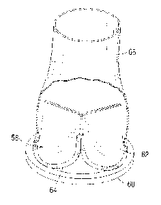
The use of
glutaradehyde in such tissue treatments has been proven effective in avoiding
resorption of
the treated tissue after implantation.
[0004] Bioprosthetic heart valves are conventionally packaged
in jars filled
with preserving solution for shipping and storage prior to use in the
operating theater. The
CA 2873478 2018-09-07
- 2 -
preserving solution maintains the functionality of the bioprosthetic tissue
within the heart
valve. Glutaraldehyde and formaldehyde are widely used as storage solutions
due to their
sterilant properties.
[0005] Prosthetic heart valves may be implanted independently
in one of the
orifices or annuluses of the heart, or may be coupled to a flow conduit which
extends in
line with the valve a predetermined distance. For example, the Carpentier-
Edwards
Bioprosthetic Valved Conduit available from Edwards Lifesciences of Irvine,
California
features a porcine bioprosthetic heart valve to which are coupled both and
inflow and
outflow woven polyester extensions. The Edwards valved conduit is particularly
well-
suited for treatment of a malfunctioning pulmonic valve. Other valved conduits
are
designed for reconstruction of portions of the flow passage above and below
the aortic
valve, such as the ascending aorta, in addition to replacing the function of
the valve itself.
There are also other applications for valved conduits, such as to provide a
bypass flow
path connecting the apex of the heart directly to the descending aorta. Prior
bioprosthetic
valved conduits, as with bioprosthetic heart valves, are stored in a liquid
preserving
solution, and thus the conduits are formed of woven polyester without a
bioresorbable
sealant. Although such conduits are suitable in certain situations, and tend
to seal
relatively quickly in the body from tissue ingrowth, too much blood can
initially seep
through their walls after implant which may be detrimental. Uncoated fabric
such as
polyethylene terephthalate (PET) has a high leakage rate, and thus the surgeon
needs to
pre-clot the graft with patient's blood before use. Nevertheless, such grafts
still produce
unacceptable leaking. Others have proposed using a non-bioresorbable sealant
layer, such
as silicone in U.S. Patent Publication No. 2008/0147171 to Ashton, et al.,
published June
19, 2008, but such layered conduits tend to be relatively thick walled and not
very flexible,
and so are not preferred.
[0006] Consequently, some surgeons prefer conduits or grafts in
which porous
tubular structures such as woven polyester (e.g., DacronTM) are impregnated
with
bioresorbable materials such as gelatin, collagen or albumin. These conduits
are not
porous initially, and thus prevent blood loss, but the sealant medium
eventually degrades
by hydrolysis when exposed to water after implant and are replaced by natural
tissue
ingrowth. Gelatin in the graft can also be treated in such a way as to cause
cross links to
form between the amino groups present in the gelatin molecules, which renders
the gelatin
CA 2873478 2018-09-07
- 3 -
more resistant to hydrolysis. Methods of forming such grafts are seen in U.S.
Patent No.
4,747,848 to Maini, issued May 31, 1988.
[0007] Unfortunately, it is not possible to pre-assemble
conduits or grafts sealed
using bioresorbable materials with bioprosthetic heart valves because of
storage
complications. That is, the liquid sterilant in which tissue valves are stored
will eventually
wash the bioresorbable sealing medium (gelatin, collagen, albumin, etc.) out
of the
permeable conduit material. Because of the benefits of using sealed conduits
or grafts and
the positive attributes of bioprosthetic heart valves, some surgeons couple
the two
components together at the time of surgery - post-storage. That is,
technicians in the
operating theater connect the sealed conduit which has been stored dry to the
bioprosthetic
heart valve which has been stored wet. Such assemblies can be seen in U.S.
Patent
Publication No. 2010/0274351 to Rolando, et al., published October 28, 2010,
and in U.S.
Patent No. 7,575,592 to Woo, et al., issued August 18, 2009. The sealed
conduit may be
sewn to the sewing ring of the bioprosthetic heart valve, or some other form
of quick-
connect coupling can be provided, such as seen in U.S. Patent Publication No.
2006/0085060 to Campbell, published April 20, 2006. Although these assemblies
are in
theory the best of both worlds, the time and effort required to connect a
sealed conduit
with a bioprosthetic heart valve creates problems in the high-pressure
environment of the
cardiac operating theater. Adding to the complexity of the connection
procedure, the
biological valve must be kept damp to avoid degradation of the tissue, while
the graft must
be kept dry to avoid initiating hydrolysis. Further, prolonged exposure of the
valve and
conduit in the operating room prior to implantation increases the chance of
infection. For
aortic conduits, even a small leak in this connection can be fatal because the
pressure is so
high. So the implanting surgeon has to sew these components together quickly
without
any leaks.
[0008] Accordingly, there is a need for a valved conduit having
a bioprosthetic
tissue valve and a conduit or graft preferably sealed using a bioresorbable
material which
is simpler to prepare and deploy in the operating room.
Summary of the Invention
[0009] The present application discloses a valved conduit
including a
bioprosthetic heart valve and a tubular conduit sealed with a bioresorbable
material. The
CA 2873478 2018-09-07
- 4 -
bioprosthetic heart valve includes prosthetic tissue that has been treated
such that the
tissue may be stored dry for extended periods without degradation of
functionality of the
valve. For example, the tissue may have been cross-linked using glutaraldehyde
or other
aldehyde containing agents, treated with a capping agent, and dehydrated with
a glycerol
solution. The bioprosthetic heart valve may have separate bovine pericardial
leaflets or a
whole porcine valve. The sealed conduit includes a tubular matrix impregnated
with a
bioresorbable medium such as gelatin or collagen. The valved conduit is stored
dry in
packaging in which a desiccant pouch is supplied having a capacity for
absorbing moisture
within the packaging limited to avoid drying the bioprosthetic tissue out
beyond a point
where its ability to function in the bioprosthetic heart valve is compromised.
The heart
valve may be sewn within the sealed conduit, sewn to the end of the conduit or
coupled
thereto with a snap-fit connection to limit handling of the two treated
components and
provide a hemostatic seal with minimal assembly complexity. In one embodiment,
the
bioprosthetic valve couples within the conduit such that the conduit extends
on both ends
of the valve to provide both inflow and outflow extensions.
[0010] Another aspect of the present application is a packaged
valved conduit,
including a bioprosthetic valve such as a heart valve having bioprosthetic
tissue, the valve
having been treated such that the tissue may be stored dry for extended
periods without
degradation of functionality of the valve. The bioprosthetic valve is coupled
to a conduit
sealed with a bioresorbable medium to provide the valved conduit. Packaging
for the
valved conduit has at least one sterile container in which the valved conduit
is stored
without a preserving solution. The packaging may comprise a dual layer
packaging with
the valved conduit sealed within an inner gas-permeable sterile barrier to
enable gas
sterilization and an outer gas-impermeable barrier to prevent long term
oxidation of the
bioprosthetic tissue. Desirably, the conduit comprises a tubular matrix
impregnated with
gelatin, and further including a desiccant pouch provided within the packaging
having a
capacity for absorbing moisture within the packaging limited to avoid drying
the
bioprosthetic tissue out beyond a point where its ability to function as a
bioprosthetic
valve is compromised.
[0011] A method of preparing and delivering a valved conduit is
also disclosed
which includes first procuring a pre-assembled valved conduit including a
bioprosthetic
valve having bioprosthetic tissue coupled to a conduit sealed with a
bioresorbable
CA 2873478 2018-09-07
- 5 -
medium, the valved conduit being stored in a dry package. The method then
requires
opening the dry package and removing the valved conduit, and delivering the
valved
conduit to an implantation site. The conduit is preferably secured to the
bioprosthetic
valve using sutures, but may alternatively be secured using a snap-fit
connection. The
bioprosthetic valve may be a heart valve with bovine pericardial leaflets, and
the conduit is
desirably a tubular matrix impregnated with gelatin or collagen. In one
embodiment, the
bioprosthetic valve couples within the conduit such that the conduit extends
on both ends
of the valve to provide both inflow and outflow extensions. The bioprosthetic
tissue has
preferably been cross-linked using glutaraldehyde or other aldehyde containing
agents,
treated with a capping agent, and dehydrated with a glycerol solution.
[0012] A further understanding of the nature and advantages of
the present
invention are set forth in the following description and claims, particularly
when
considered in conjunction with the accompanying drawings in which like parts
bear like
reference numerals.
Brief Description of the Drawings
[0013] The invention will now be explained and other advantages
and features
will appear with reference to the accompanying schematic drawings wherein:
[0014] Figure 1 is an exploded view of the combination of a
bioprosthetic heart
valve coupled to a sealed conduit of the present application;
[0015] Figure IA is a view of a human heart showing implant of
a valved
conduit to replace the aortic valve and a portion of the ascending aorta;
[0016] Figures 2A and 2B are side elevational views of an
exemplary valved
conduit of the present application wherein a bioprosthetic heart valve couples
to two
segments of sealed conduit extending from both ends thereof;
[0017] Figure 3 is a still further view of a valved conduit
with a bioprosthetic
heart valve coupled within a sealed outflow conduit;
[0018] Figures 4A and 4B are enlarged cross-sectional views
illustrating
alternative techniques for attaching the outflow conduit to the bioprosthetic
heart valve of
Figure 3;
[0019] Figure 5 is an exploded perspective view illustrating an
additional
application of the valved conduit the present application;
CA 2873478 2018-09-07
-6-
100201 Figure 6 is an exploded view of an exemplary snap-fit
connection between
a bioprosthetic heart valve (shown in partial section) and a sealed conduit;
[0021] Figure 7 is a sectional view of an exemplary sealed
conduit connected with
sutures to a sewing ring of a bioprosthetic heart valve;
[0022] Figure 8 is an elevational view of a valved conduit
similar to that shown
in Figure 5 with a sealed conduit attached to a stent structure of a
bioprosthetic heart
valve;
[0023] Figure 9A is an elevational view of a valved conduit
with a sealed
conduit attached to a sewing ring of a bioprosthetic heart valve, and Figure
9B is a
perspective view of the valved conduit looking into an outflow end of the
conduit;
[0024] Figure 9C is a sectional view through a cusp region of
the heart valve in
Figure 9A showing one possible connection between the sealed conduit and the
sewing
ring;
[0025] Figure 10 is an elevational view of a valved conduit
similar to that
shown in Figure 9A with a longer sealed conduit having a sinus region attached
to a
sewing ring of a bioprosthetic heart valve;
[0026] Figures 11A-11C are sectional views through a cusp
region of the heart
valve in Figure 10 showing possible connections between the sealed conduit and
the
sewing ring;
[0027] Figure 12 is an exploded plan view of a valved conduit
and holder
therefore mounted in a primary storage container in the form of a tray; and
[0028] Figure 13 is a perspective view of the valved conduit in
the tray as in
Figure 12 contained within a secondary storage container in the form of a
pouch.
[0029] Figure 14 is an exploded plan view of the valved conduit
of Figure 10
mounted in an exemplary primary storage container in the form of a tray; and
[0030] Figure 15 is an exploded plan view of the valved conduit
of Figure 10 in
the tray as in Figure 14 contained within a secondary storage container in the
form of an
outer tray.
Detailed Description of the Preferred Embodiments
CA 2873478 2018-09-07
-7-
100311 The present application provides techniques for coupling
implantable
valves with sealed conduits, and in particular bioprosthetic heart valves that
have been
dried and are not stored immersed in a preservative solution. The term "dried"
or "dry"
bioprosthetic heart valves refers in general to the ability to store those
heart valves without
immersion in solution (e.g., a preservative like glutaraldehyde), and in
particular to dry
storage for extended periods without degradation of functionality of the
bioprosthetic
valve. There are a number of proposed methods for drying bioprosthetic heart
valves, and
for drying tissue implants in general, and the present application provides
packaging for
bioprosthetic heart valves that are processed by any of these methods.
[0032] One strategy for drying tissue is to dehydrate the
bioprosthetic tissue in a
glycerol/ethanol mixture, sterilize with ethylene oxide, and package the final
product
"dry." This process eliminates the potential toxicity of glutaraldehyde as a
sterilant and
storage solution. There have been several methods proposed to use sugar
alcohols (i.e.,
glycerine), alcohols, and combinations thereof as post-glutaraldehyde
processing methods
so that the resulting tissue is in a "dry" state rather than a wet state with
excess
glutaraldehyde. Glycerol-based methods can be used for such storage, such as
described
in Parker et al. "Storage of heart valve allografts in glycerol with
subsequent antibiotic
sterilization" (Thorax 1978 33:638-645). A particularly preferred method of
drying
bioprosthetic heart valves is disclosed in U.S. Patent No. 8,007,992 to Tian,
et al. wherein
fixed tissue is treated with a non-aqueous mixture of glycerol and C1-C3
alcohol selected
from the group consisting of methanol, ethanol, n-propanol, 2-propanol.
Likewise, U.S.
Pat. No. 6,534,004 (Chen et al.) describes the storage of bioprosthetic tissue
in polyhydric
alcohols such as glycerol. In processes where the tissue is dehydrated in an
ethanol/glycerol solution, the tissue may be sterilized by ethylene oxide
(ETC)), gamma
irradiation, or electron beam irradiation.
100331 More recently, Dove, et al. in U.S. Patent No.
7,972,376, issued July 5,
2011, propose solutions for certain detrimental changes within dehydrated
tissue that can
occur as a result of oxidation. Dove, et al. propose permanent capping of the
aldehyde
groups in the tissue (reductive amination). One preferred anticalcification
tissue treatment
includes applying a calcification mitigant such as a capping agent or an
antioxidant to the
tissue to specifically inhibit oxidation in dehydrated tissue and reduce in
vivo calcification.
The treatment specifically caps aldehyde groups in crosslinked (e.g., with
glutaraldehyde)
CA 2873478 2018-09-07
- 8 -
bovine, porcine, or equine pericardial tissue or a porcine valve. In one
method, tissue
leaflets in assembled bioprosthetic heart valves are pretreated with an
aldehyde capping
agent prior to dehydration and sterilization. Dove, et al. also describe the
addition of
chemicals (e.g. antioxidants) to the dehydration solution (e.g.,
ethanol/glycerol) to prevent
oxidation of the tissue during sterilization (ethylene oxide, gamma
irradiation, electron
beam irradiation, etc.) and storage. The capping process uses an amine, for
example
ethanolamine or lysine, and a reducing agent, followed by final processing
with glycerol
and an alcohol. The capping agent may be selected from the group consisting
of: an
amine, an amino acid, and an amino sulfonate. The reducing agent may be a
borohydride,
for example sodium borohydride or cyanoborohydyride. Other reducing agents
include:
sodium bisulfite + acetylacetone, and formic acid + formaldehyde.
[0034] These and other methods for drying bioprosthetic heart
valves are used
prior to coupling of the valve with the sealed conduit. The removal of a
percentage of
water from the valve and replacement with glycerol and ethanol allows the
device to be
stored "dry" (i.e. glycerolized). The "dry" valve may then be sewn into the
polyester
conduit or graft and be ready for implantation. This process allows making a
valved
conduit that is ready for implantation without the need for a clinical rinse
in saline, thereby
shortening implant time. For purpose of definition, a "dry" bioprosthetic
tissue is one with
less than 70% water content. In terms of practical rehydration, functional
valves have at
least 70% water content. The most important distinction of "dry" valves (or
tissue
therein), however, is that they may be stored dry for extended periods
(sometimes years)
without degradation of functionality of the valve.
[0035] A number of exemplary bioprosthetic heart valves and
conduits are
shown and described in the present application. Each of these different types
of heart
valves may be processed so that they are stored dry. The reader will
understand that the
present methodologies apply to any and all bioprosthetic valves that are
stored dry, and are
not limited to those exemplary valves shown herein. In particular, prosthetic
heart valves
for implant at any of the four native valve annuluses - aortic, mitral,
pulmonary, and
tricuspid - may be dried and stored in accordance with the principles
described herein.
Alternatively, valved conduits produced in accordance with the principles
disclosed herein
may be used in locations other than heart valve replacement, such as venous
valves by
connecting a small bileaflet valve to or within a small diameter conduit.
CA 2873478 2018-09-07
-9-
100361 Additionally, a number of techniques for packaging the
dry bioprosthetic
heart valves and their delivery systems are illustrated and described herein,
though these
techniques can also apply to other packaging configurations. In general, a
bioprosthetic
heart valve must be stored in sterile conditions, which requires at least one
sterile
container. Preferably, however, a dual-barrier packaging system is used to
reduce the
chance of contamination of the implant at the time of surgery. For instance,
U.S. Patent
Publication No. 2011/0147251 to Hodson, et al. discloses exemplary packaging
systems
which can be utilized.
[0037] The present application describes systems and methods
for pre-
assembling and storing a bioprosthetic heart valve and sealed conduit to form
the valved
conduit. The term "pre-assembling" or "pre-assembled" refers to connection of
the heart
valve and sealed conduit prior to the operating room technicians opening the
sterile
packaging. In other words, the valved conduit emerges mechanically assembled
from the
packaging, substantially ready for delivery (after any pre-surgery washing or
other such
preparation).
[0038] Figure 1 is an exploded view of an exemplary combination
of a
bioprosthetic heart valve 20 coupled to a sealed conduit 22. As suggested
schematically,
the prosthetic heart valve 20 is positioned within one end of the sealed
conduit 22. Such a
valved conduit may be used for replacing a native heart valve and an
associated blood
vessel in a patient. The aortic valve and the ascending aorta are one non-
limiting example
of such a valve and an associated blood vessel. The pulmonary valve and the
pulmonary
artery are another such example.
[0039] The heart valve 20 may include a rigid or semi-rigid
stent or be a so-
called "stentless" type. In the illustrated embodiment, the heart valve 20
comprises a
plurality of flexible leaflets 24 (typically three) that are mounted to a
peripheral stent
structure 26 and form fluid occluding surfaces within the valve orifice to
form a one-way
valve. The stent structure 26 includes a plurality of generally axially
extending
commissures 28 circumferentially distributed around the valve between and in
the same
number as the number of leaflets 24. Although not shown, additional components
of the
heart valve 20 typically include an inner stent and/or wireform support
structure that
provide a structural skeleton surrounding an inflow orifice and extending up
the
commissures 28. The inner components of the heart valve 20 may be made of
suitable
CA 2873478 2018-09-07
- 10 -
metal or plastic. As is well known, adjacent flexible leaflets 24 connect to
and extend
upward to meet along each of the commissures 28. In the illustrated
embodiment, the
structural components of the heart valve 20 support each flexible leaflet 24
along a valve
cusp 30 and along two commissure 28 edges. A free edge 25 of each leaflet 24
extends
inward toward a central flow orifice and coapts, or mates, with the free edges
of the other
leaflets, as shown. The valve orifice is oriented around an axis along an
inflow-outflow
direction through the valve. The valve commissures 28 project in the outflow
direction,
with the convex valve cusps 30 extending in the inflow direction between
adjacent
commissures. The bioprosthetic heart valves further includes a sewing ring 32
on the
inflow end that conforms to the undulating contours of the valve cusps, or
defines a
generally circular, planar ring. The present application should not be
considered limited to
a particular valve construction unless explicitly stated herein.
[0040] The sealed conduit 22 defines a generally tubular
structure that extends
from an inflow end 42 to an outflow end 44. In the embodiment shown, the valve
20 is
associated with the conduit 22 in such a way that the valve leaflets 24
control flow of
blood through the conduit by permitting blood flow into the conduit (e.g.,
blood flow into
the aorta, when the conduit is used for aortic replacement) while preventing
flow of blood
out of the conduit in the opposite direction (i.e., back into the left
ventricle of the patient
when used for aortic replacement).
[0041] The illustrated conduit 22 is particularly suited for
attachment within the
aortic annulus and ascending aorta, and as such closely matches the aortic
root anatomy
and includes an enlarged region or bulge 46 close to the inflow end 42 that
conforms to the
sinuses of valsalva just above the aortic annulus. In the preferred
embodiment, the conduit
22 comprises a tubular textile structure, such as Dacron, sealed with a
bioresorbable
medium. A majority of the conduit 22 includes a corrugated (i.e., grooved)
structure
providing longitudinal flexibility and radial compressibility while ensuring
that the
conduit will not unduly radially expand under the pressure of blood flowing
therethrough.
The conduit 22 desirably has a length of from a few centimeters to 10-12
centimeters.
[0042] Figure IA illustrates a human heart H showing the valved
conduit from
Figure I implanted above the left ventricle LV to replace the aortic valve and
a portion of
the ascending aorta AA. The surgeon sews the inflow end 42 to the aortic
annulus, and the
outflow end 44 to the remainder of the ascending aorta AA. Because the entire
valve and
CA 2873478 2018-09-07
- 11 -
a portion of the ascending aorta AA including the sinuses are removed, the two
coronary
arteries CA (one shown) attach at anastomoses AN to the enlarged region 46 of
the
conduit 22 between two of the three adjacent pairs of the commissures 28 of
the valve 20.
The bulged region 46 mimics the native sinuses and helps improve blood flow
into the
coronary arteries CA.
[0043] In one embodiment, the conduit 22 may be a Vascutek
Gelweave
ValsalvaTM Grafts gelatin sealed, aortic root graft that is indicated for
aortic root
replacement using valve sparing or replacement techniques, and available from
the
Vascutek business of Terumo Cardiovascular Systems Corporation of Ann Arbor,
MI. As
explained below, the use of a bioresorbable medium to provide a temporary seal
to the
implanted graft is preferred and may be preassembled with the exemplary
bioprosthetic
heart valves disclosed herein. However, the exemplary bioprosthetic heart
valves may
also be pre-assembled with other sealed grafts or conduits, such as those that
utilize non-
bioresorbable material. It should be understood that unless excluded by claim
language, a
variety of sealed conduits are contemplated.
[0044] In the preferred embodiment, the sealed graft or conduit
22 is relatively
impermeable in a dry state and immediately after implantation begins to become
permeable. Such a response can be obtained by impregnating porous tubular
structures
with such materials as gelatin, collagen or albumin. A gelatin impregnated
graft is not
porous but when exposed to water the gelatin degrades by hydrolysis the rate
at which
hydrolysis proceeds being higher at the body temperature of 37 C than it is
at normal
temperature. If the graft becomes porous at a rate too fast to keep pace with
clotting and
tissue growth, the gelatin may be treated in such a way as to cause cross
links to form
between the amino groups present in the gelatin molecules. Such cross linking
renders the
gelatin more resistant to hydrolysis and thus reduces considerably the rate at
which the
permeability of the graft increases. One method of initiating cross linking
comprises
exposing the gelatin to formaldehyde. The present application contemplates a
conduit or
graft which requires no pre-impregnation with blood and which after
implantation starts to
degrade and become permeable at an accurately known rate. It is to be
understood that
according to the medical circumstances of different implantations the porosity
of the
implanted grafts should increase at a rate only sufficient to avoid hemorrhage
occurring. A
method of producing a vascular graft according to these principles by
impregnating a tube
CA 2873478 2018-09-07
- 12 -
of flexible porous material with gelatinous material then treating the
impregnated tube to
cause only the amino groups always present in the molecules of gelatinous
material to
form cross links is disclosed in U.S. Patent No. 4,747,848 to Maini, issued
May 31, 1988.
[0045] Figures 2A and 2B illustrate an alternative valved
conduit 50 of the
present application wherein a bioprosthetic valve 52 couples to two segments
54, 56 of
sealed conduit extending from both ends thereof. The segments 54, 56 of sealed
conduit
comprise woven polyester sealed by techniques described above. The valved
conduit 50
extends between an inflow end at the free end of the inflow segment 54 and an
outflow
end at the free end of the outflow segment 56. This construction can be
utilized in a
variety of locations within the body, including in the venous vasculature, as
a bypass graft
from the left ventricle to the descending aorta, or in the aortic annulus as
described above.
As such, the diametric sizes A of the valved conduit 50 can be as small as the
smallest
venous valves (2-5 mm) or as large as the largest heart valves (30 mm). A
preferred
diametric size ratio is between about 12-36 mm. An exemplary combination is a
29 mm
valve connected to or within a 34 mm sealed conduit. Bioprosthetic heart
valves with
conduits are also used in the pulmonic position and in apical conduits. It
should be noted
that the bioprosthetic valve 52 illustrated is a bioprosthetic heart valve
with three leaflets.
Alternatively, the bioprosthetic valve 52 may be a bileaflet type, typical
with venous
valves.
[0046] The inflow and outflow segments 54, 56 are variable in
length, and can
be trimmed to size. Indeed, the two segments 54, 56 are typically trimmed
close to the
bioprosthetic valve 52. In the embodiment for use as a pulmonic valve
replacement, the
outflow segment 56 is approximately 50% longer than the inflow segment 54,
such as 9
cm versus 6 cm. In one embodiment, the valved conduit 50 is constructed in the
same
manner as the Carpentier-Edwards41) Bioprosthetic Valved Conduit available
from
Edwards Lifesciences of Irvine, California, though the conduit segments 54, 56
are
preferably sealed with a bioresorbable medium such as gelatin or collagen. For
an aortic
valve replacement, the inflow extension segment 54 is not typically used,
though a very
short (<1cm) inflow segment 54 can be provided, and the outflow segment 56 is
preferably
bulged, rather than being a straight tube as in Figures 2A/2B.
[0047] Figure 3 is a still further view of a valved conduit
with a bioprosthetic
heart valve 58 coupled within a sealed outflow conduit 66. In some patients
requiring
CA 2873478 2018-09-07
- 13 -
replacement of the aortic valve, a portion of the aorta itself may be damaged
or diseased such
that it needs replacement as well, and the outflow conduit 66 functions to
replace the
damaged aorta. The bioprosthetic heart valve 58 is similar to that described
above and
includes flexible leaflets 62 supported by an undulating stent 64 having
commissures. In
contrast to the earlier valved conduit 20, the heart valve 58 couples to the
outflow conduit 66
in a manner that the leaflets 62 are within the lumen of the conduit, while
the sewing ring 60
of the valve is outside the conduit. This enables conventional attachment of
the sewing ring
60 to the aortic annulus.
[0048] For example, Figures 4A and 4B illustrate techniques for
attaching the
outflow conduit 66 to the bioprosthetic heart valve 58. In both techniques,
the outflow
conduit 66 attaches to the cloth covering on a wireform 63 that forms a part
of the stent 64 at
the time that the tissue leaflets 62 are being secured. Referring to Figure
4A, the conduit 66
may be secured on a side of wireform 54 opposite to tissue leaflets 62 by, for
example,
stitching. Alternatively, as shown in Figure 4B, the conduit 66 may be
stitched and secured
to wireform 54 on the same side as tissue leaflets 62, or sandwiched
therebetween. A third
option is to simply secure conduit 66 to the periphery of the finished valve
(not shown) as a
subsequent sewing step. The valve 58 may be attached to an outflow conduit
either with or
without a sinus as shown. It should be noted that a short portion of the
outflow conduit 66
that comes into direct contact with the leaflets 62 may be left free of a
bioresorbable sealing
medium to prevent any long-term reaction during storage between the medium and
the
bioprosthetic leaflets. Although Figures 4A and 48 are associated with the
valved conduit 50
for aortic implant, the methods of construction are not preferred for a
pericardial valve aortic
conduit since the fabric of the conduit connected in abutment with the
pericardial leaflets may
cause abrasion of the leaflets and ultimately failure. However, such
constructions may be
suitable for porcine valve pulmonic conduits. Alternative construction
techniques where the
conduit attaches to the valve sewing ring are more desirable for aortic
conduits with
pericardial leaflet valves.
[0049] Figure 5 is an exploded perspective view illustrating an
additional
application of a valved conduit including a modified bioprosthetic heart valve
58. Namely, in
applications such as artificial hearts or left ventricular assist devices
(LVADs), suture ring 60
is not necessarily required; hence, the lower end of stent 64 may be attached
to a flange 68
for use in mounting the valve in an artificial heart or LVAD. Yet a further
alternative
CA 2873478 2018-09-07
- 14 -
adaptation involves those applications where an inflow conduit 70 is desired.
In such
applications, inflow conduit 70 may be attached directly to stent 64 of valve
58. More
specifically, inflow conduit 70 may be configured to have a stepped
circumference 72 that
snugly mates with the outer periphery (or, alternatively, the inner periphery)
of stent 64 and
which can be sewn thereto. In this configuration, for example, in an
artificial heart or an
LVAD application, suture ring 60 could be attached to inflow conduit 70 rather
than to valve
58.
[0050] The pre-assembled valved conduits disclosed herein each
includes a
bioprosthetic valve connected to a sealed conduit. The connection can be sewn
within or to
the end of the sealed conduit via sutures, as described above, or by a less
time-consuming
technique to limit handling of the two treated components and provide a
hemostatic seal
with minimal assembly complexity. For example, the bioprosthetic valve and
conduit could
be snapped together to minimize handling. Various potential snap mechanisms
are disclosed
in the art, including in U.S. Patent Publication No. 2006/0085060 to Campbell.
Snap
together mechanisms could employ a wire, metal or plastic rings sewn into a
proximal end
of the conduit which would be captured by a metal or plastic ring surrounding
the tissue
valves. Alternatively, the conduit could be placed between an external ring
and the
bioprosthetic valve, or it could be snapped around the outside of the ring as
long as the
conduit did not block suture needles from being passed through the valve
sewing ring.
[0051] Figure 6 is an exploded view of an exemplary snap-fit
connection between
a bioprosthetic heart valve 80 and a sealed conduit 82. In particular, a
coupling ring 84
attached to a sewing ring 86 of the valve 80 connects to an inflow end 90 of
the sealed
conduit 82. An inner periphery of the coupling ring 84 may be sewn or attached
with barbs
or the like to the sewing ring 86, and includes an open end surrounding the
commissures 92
of the valve 80 with an inwardly projecting rim 94. The rim 94 mates with an
outwardly
projecting rim 96 on the inflow end 90 of the sealed conduit 82. It should be
understood that
the illustrated engagement between the coupling ring 84 and the inflow end of
the conduit 82
is exemplary only, and representative of a variety of different such
structures.
[0052] In an exemplary embodiment, the sealed conduit 82 has a
tubular structure
with a body portion 100 having a corrugated or pleated sidewall extending
between the
inflow end 90 and outflow and (not shown). The conduit 82 is desirably formed
of a
biocompatible fabric impregnated with a bioresorbable sealing medium such as
gelatin or
CA 2873478 2018-09-07
- 15 -
collagen. The corrugated or pleated sidewall provides longitudinal flexibility
and radial
compressibility while ensuring that the graft does not unduly radially expand
under the
pressure of blood flowing there through. The conduit 82 further includes an
expandable
portion 102 located between the body portion 100 and the inflow end 90. The
expandable
portion 102 may be formed of a material that is more radially expandable than
the
corrugated body portion 100 to allow expansion at that location into the
Valsalva sinuses.
[0053] Figure 7 illustrates an exemplary sealed conduit 110
connected with
sutures 112 to a sewing ring 114 of a bioprosthetic heart valve 116. A portion
of the sealed
conduit 110 wraps around and captures a resilient biocompatible band 118. In
particular, the
sutures 112 enclose the resilient band 118 in a pocket of the sealed conduit
110. The band
118 provides an added structural connection between the conduit 110 and valve
116 to
prevent separation thereof, and as such may be made of a variety of
biocompatible materials,
including Elgiloy, titanium, or other such metals, or suitable polymers such
as polypropylene.
[0054] Figure 8 shows a valved conduit 130 similar to that
shown in Figure 5
with a sealed conduit 132 attached to a stent structure 134 of a bioprosthetic
heart valve
136. In particular, an inflow end of the conduit 132 is sewn along an
undulating path that
follows the cusps and commissures of the stent structure 134. Axial marker
lines 138 are
provided on the conduit 132 to denote the three commmisure post locations.
This
connection provides the advantage that a surgeon can easily see the commissure
locations,
but because the conduit is within the commissures the anastomotic attachment
of coronary
arteries is somewhat more difficult. Furthermore, as mentioned above for
aortic pericardial
conduits there is a greater possibility of leaflet abrasion.
[0055] Figures 9A and 9B on the other hand show a valved
conduit 140 with a
sealed conduit 142 attached to a sewing ring 144 of a bioprosthetic heart
valve 146. The
valve 146 is within the conduit 142 and axial marker lines 147 are provided on
the conduit
142 to denote the three commissure post locations. Figure 9C is a sectional
view through
a cusp region of the heart valve 146 schematically showing a suture loop 148
passing
through the sewing ring 144 to connect the sealed conduit 142 to the sewing
ring. The
valve 146 has a cloth-covered wireform 150 that rests above a cloth-covered
metallic or
polymer stent 152, with a valve leaflet 154 sandwiched therebetween. A portion
of the
leaflet edge 156 and a flap 158 of the cloth around the wireform 150 extend
outward onto
a ledge 160 formed by a cloth-covered silicone waffle ring 162 that forms the
sewing ring.
CA 2873478 2018-09-07
- 16 -
The suture loop 148 passes through the entire waffle ring 162 and secures the
inflow end
of the sealed conduit 142 (not shown in Figure 9C for clarity). This leaves an
outer rim of
the sewing ring 144 outside the conduit 142, as seen in Figure 9A, through
which
anchoring sutures can be pre-threaded for parachuting the valved conduit 140
down to the
target annulus.
[0056] Figure 10 shows a valved conduit 170 similar to that
shown in Figure
9A with a longer sealed conduit 172 having a sinus region 174 attached to a
sewing ring
176 of a bioprosthetic heart valve (not visible). Figures 11A-11C are
sectional views
through a cusp region of the heart valve in Figure 10 showing possible
connections
between the sealed conduit 172 (not shown for clarity) and the sewing ring
176. Figure
11A shows a suture loop 178 passing just through an upper cloth layer 180 of
the sewing
ring 176, or through a supplemental upper ring washer 182 formed of a polymer
such as
Nylon, which is sometimes used in sewing rings. Figure 11B shows a suture loop
184
extending the entire axial height of the sewing ring 176, much like in Figure
9C. Finally,
Figure 11C shows two loops 186, 188 passing through the entire sewing ring 176
as well
as just through an upper cloth layer 180 for added security.
[0057] Figure 12 illustrates an exemplary primary storage
container for a valved
conduit 200 of the present application. The primary storage container
comprises a molded
storage tray 210 and a sheet-like gas-permeable lid 212. In particular, the
assembled
valved conduit 200 is placed within a cavity of the storage tray 210,
whereupon the lid 212
is adhered to an upper rim 214 of the tray. The upper rim 214 defines the tray
upper
surface, and the process of adhering the lid 212 to the rim 214 can be
performed easily
using automated equipment. The adhesive may be provided on the upper rim 214,
or on
the underside of the lid 212.
[0058] In a preferred embodiment, features provided in the
cavity of the tray
210 secure the valved conduit 200 from movement therein, and prevent a sealed
conduit
218 from touching any inner surfaces of the tray. The bioresorbable sealing
medium in
the sealed conduit 218 can abrade if permitted to touch the inside of the tray
during
handling, potentially degrading its sealing capability. For example, a valve
holder 220
may be attached to commissure posts of the heart valve 222, and an elongated
delivery
handle 224 extends from the holder out of an outflow end 226 of the sealed
conduit 218.
A series of cooperating brackets 226 firmly holds the handle 224 in position
in the tray
CA 2873478 2018-09-07
-17-
210, while the valved conduit 200, and in particular the sealed conduit 218,
is suspended
within an enlarged cavity (not shown) of the tray. Because the tray 210
secures the
components in this manner, the valved conduit 200 is stably suspended within
the cavity
without touching the sides of the tray 210.
[0059] Preferably, the lid 212 is closely dimensioned to the
perimeter of the
upper rim 214, and the band of adhesive is a pressure-seal or a heat seal
adhesive to
facilitate sealing under pressure and/or temperature. The material of the lid
212 is
breathable, or gas-permeable, to permit gas sterilization of the contents
sealed within the
tray 210, in particular the dry tissue heart valve of the valved conduit 200.
One suitable
gas-permeable material is a sheet of high-density polyethylene fibers, which
is difficult to
tear but can easily be cut with scissors. The material is highly breathable
and water vapor
and gasses can pass through the fibers, but not liquid water. For instance,
various
TyvekTm materials from DuPontTM may be used. Also, exemplary hot-melt
adhesives used
to secure the lid 212 to the tray 210 may be obtained from Perfecseal or
Oliver-TolasTm,
for example. Such a material permits sterilization of the tray contents using
Ethylene
Oxide (ETO), which gradually passes through the lid 212 to the interior tray.
The lid 212
presents a sterile barrier and prevents ingress of microorganisms. The tray
210 is a gas-
impermeable molded material, such as a polyethylene terephthalate copolymer
(PETG).
Various medical storage materials and packaging suitable for assembly of
components of
the present application are available from companies such as Dupont,
Perfecseal, Oliver-
Tolas, and Mangar. Other means of sterilization include gamma irradiation or
electron
beam irradiation.
[0060] Ethylene oxide (ETO), also called oxirane, is the
organic compound
with the formula C2I-140. It is commonly handled and shipped as a refrigerated
liquid.
ETO is often used as sterilant because it kills bacteria (and their
endospores), mold, and
fungi. It is used to sterilize substances that would be damaged by high
temperature
techniques such as pasteurization or autoclaving. Ethylene oxide is widely
used to sterilize
the majority of medical supplies such as bandages, sutures, and surgical
implements in a
traditional chamber sterilization method, where a chamber has most of the
oxygen
removed (to prevent an explosion) and then is flooded with a mixture of
ethylene oxide
and other gases that are later aerated.
CA 2873478 2018-09-07
- 18 -
[0061] Certain features of the tray 210 facilitate gas
sterilization, such as with
ETO, though other means such as gamma irradiation or electron beam irradiation
could be
used. Specifically, the tray 210 retains the valved conduit 200 securely
therein but
provides adequate channeling in and around the components to eliminate any
enclosed
spaces. The sterilization gas can therefore flow evenly throughout the entire
enclosure.
[0062] One advantage of the packaging solutions described
herein is a double
sterile barrier, wherein the inner and outer sterile containers allow for gas
sterilization,
such as with ETO, and with a second seal the outer sterile container also
provides an
oxygen barrier to the product after sterilization. The inner sterile container
has been
described above with reference to Figure 12 in the form of a storage tray 210
sealed with
the lid 212. The sealed storage tray 210 is received within a secondary or
outer container
and the dual barrier assembly is then sterilized, so that there are redundant
sterile barriers.
Subsequently, the dual barrier assembly is sealed to prevent oxygen from
reaching the
valved conduit 200, thus preventing oxygenation and potentially reducing
calcification
after implant. In the exemplary packaging sequence, the primary and secondary
containers are first assembled together and each closed with a gas-permeable
barrier to
form a dual barrier assembly which is gas-sterilized. Subsequently, the oxygen
barrier is
added, such as by converting the secondary container from being gas-permeable
to being
gas-impermeable. However, if the entire process is done in sterile conditions,
such as in a
clean room environment, the primary container may be closed and sterilized
before being
placed within the secondary container, which is then closed and sterilized. In
other words,
there may be one or two sterilization steps prior to sealing the entire
assembly against
oxygen ingress.
[0063] Desirably, a dessicant is used within the inner and/or
the outer
packaging layers. For instance, a dessicant pouch may be inserted with the
valved conduit
200 into the inner package, to absorb any residual water vapor trapped therein
when the
gas-permeable tray lid 212 is closed. A second dessicant pouch may be inserted
between
the inner and outer barriers to absorb any residual water vapor therein, or it
may be the
only dessicant pouch used.
[0064] The present application describes two different
secondary barriers - one
a storage tray described below, and the other a flexible pouch. The secondary
barrier
protects and preserves the primary sterile barrier package in a sterile
environment, and
CA 2873478 2018-09-07
- 19 -
prevents oxygen from reaching the heart valve of the valved conduit 200
within. A further
outer shelf box may be used to facilitate temperature monitoring during
distribution and
storage, and protect the delicate implant from distribution hazards such as
shock, impact
and extreme temperatures.
[0065] Figure 13 is a perspective view of the valved conduit
200 in the primary
storage tray 210 with the lid 212 attached (all not shown), as in Figure 12,
and then
contained within a secondary storage container in the form of a pouch 230.
Desirably, the
storage pouch 230 includes a dual seal system on its open end which provides
both a gas-
permeable portion and a gas-impermeable portion, depending on which seal is
closed.
[0066] Figure 14 illustrates the valved conduit 220 mounted in
an exemplary
primary storage container in the form of a tray 240 and a sheet-like gas-
permeable lid 242.
The tray 240 features cavities for the valved conduit 220 which retain and
stabilize the
components therein. The lid 242 adheres to an upper rim 244 of the tray 240.
[0067] Figure 15 illustrates the valved conduit 220 in the tray
(of which the lid
242 is seen) as in Figure 14, and then placed within a secondary storage
container in the
form of an outer or secondary tray 250 for a dual-barrier packaging system.
The
secondary storage tray 250 desirably mimics the shape of the primary storage
tray 240
such that the latter can be easily nest within a cavity formed therein. The
secondary
storage tray 250 comprises an upper surface including a peripheral flange 252.
[0068] The outer storage tray 250 provides a rigid secondary
sterile barrier that
protects and preserves the inner sterile barrier formed by the inner storage
tray 240 and its
lid 242. The outer storage tray 250 may be constructed of a gas-impermeable
molded
material, such as a polyethylene terephthalate copolymer (PETG). Once the
sealed inner
tray 240 is placed within the outer storage tray 250, a gas-permeable lid 254
seals against
the flange 252 and permits sterilization gas (e.g., ETO) to reach the spaces
within both
trays.
[0069] Subsequently, a gas-impermeable label 262 sized to cover
the secondary
storage tray 250 is shown. The label 262 is applied over the sterilized tray
250, and sealed
on top of the lid 254. Once pressure adhered or heat sealed against the lid,
the label 262
provides a complete barrier to gas transfer. The label 262 preferably includes
a layer of
metal foil laminated to a layer of a gas-permeable material such as DuPont
1073B Tyvek,
or more preferably is a single layer of foil. The label 262 may have
information printed
CA 2873478 2018-09-07
- 20 -
thereon about the contents of the packaging, such as implant type, model,
manufacturer,
serial number, date of packaging, etc. A layer of pressure sensitive adhesive
is provided to
seal on top of the previously attached lid 254.
[0070] Alternatively, the secondary storage tray 250 features a
double flange
(not shown) around its upper edge. An inner flange may first be sealed with a
die-cut and
heat seal adhesive coated gas-permeable lid (e.g., Tyvek), such as lid 254,
after placement
of the inner sterile barrier package, enabling subsequent ETO sterilization of
the entire
package, and in particular the space between the two sterile barriers. A gas-
impermeable
label such as the foil label 262 is then sealed to an outer flange.
[0071] The packaging solutions disclosed herein facilitate
access to the valved
conduits at the time of implantation. The process for removing the valved
conduit 220
from its packaging will be described, though similar steps can be used to
remove the other
valved conduits. The first step is removal of the outer or secondary sterile
barrier (pouch
or tray). This description will assume a secondary storage tray 250. One or
both sealed
labels over the outer tray 250 are first detached, and the inner tray 240
sealed by a sterile
lid 242 removed therefrom (alternatively, the technician tears open a sterile
pouch). At
this stage, the inner sterile packaging may be transported to the immediate
vicinity of the
operation site without undue concern for the integrity of the package because
of the
relatively rigid inner tray 240 and sterile seal 242.
[0072] Subsequently, the technician detaches the lid 242,
exposing the valved
conduit 220. The valved conduit 220 is then removed from the packaging and
implanted
according to various procedures.
[0073] The preferred dual-barrier packaging system provides a
number of
distinctive advantages to manufacturers of the valved conduits of the present
application.
Due to presence of a gas-permeable sterile barrier such as a Tyvek Header
(breathable
vent) the product can easily be ETO sterilized and aerated for acceptable
levels of
residuals. After appropriate aeration time, the outer container, or second
barrier, can be
sealed (e.g., foil to foil) to prevent long term oxidation of the dry tissue
valve. The ETO
sterilization obviates traditional oven sterilization, therefore reducing the
amount of
energy spent in heating the packaged product in an oven for multiple days.
Similarly,
elimination of autoclaving before packaging will reduce the energy consumption
required
in the sterilization process.
CA 2873478 2018-09-07
-21-
100741 As mentioned, the double sterile barrier allows for gas
sterilization, such
as with ETO, but also provides an oxygen barrier to the product after
sterilization.
Consequently, the entire assembly can be reliably stored in oxygen-free
conditions for
extended periods of time, even years, yet the outer sterile container can be
removed at the
time of use without exposing the contents of the inner sterile container to
contaminants.
The double layer of packaging enables sterile transfer of the inner package to
the sterile
operating field, and the inner package can even be temporarily stored for
significant
periods before the product is used. The new package design will be lighter in
weight due
to the choice of materials (PETG/Tyvek and air vs. Polypropylene with
glutaraldehyde),
which will reduce the shipping costs for single unit shipments.
[0075] Indeed, the biggest advantage over existing "wet" heart
valve package
designs is the elimination of storage and handling of liquid glutaraldehyde
during the
packaging and storage process, as well as the absence of glutaraldehyde at the
time of use.
This reduces hazards to the health of employees, customers, and patients, as
well as the
environment. Additionally, disposal of glutaraldehyde is bio-hazardous and
therefore
OSHA requires neutralization of the chemical before disposal or placement of
appropriate
controls for disposal. Due to decreased handling and critical storage
requirements
described herein, the packaging process is rendered less complex. The
elimination of
glutaraldehyde will not require an increased level of insulation from higher
temperatures
as the dry tissue valve already has the capability to withstand temperatures
as high as 55
C. The replacement of water with glycerin also provides protection from
freezing down to
a temperature of -18 C. Therefore this will likely reduce the bulkiness of the
design by
reducing the size and insulation used for shipping the valve during summers
and winters.
[0076] The packaging provided for the various valved conduits
desirably
incorporate some means within to maintain a predetermined low humidity level.
The
valved conduits which include bioresorbable materials, in particular gelatin,
may
hydrolyze faster in the body if they are stored in excessively moist
conditions. Typically,
sealed conduits with a bioresorbable sealing medium are stored with desiccant
pouches to
maintain extremely dry conditions. However, the bioprosthetic valves disclosed
herein,
although stored dry, must retain a certain amount of moisture to remain
functional. In one
embodiment, a desiccant pouch is provided within the packaging having a
capacity for
absorbing moisture within the packaging limited to avoid drying the
bioprosthetic tissue
CA 2873478 2018-09-07
- 22 -
out beyond a point where its ability to function in the bioprosthetic heart
valve is
compromised. In short, the two components of the valved conduits described
herein have
slightly different needs when it comes to the moisture content of the shipping
package.
100771
While the invention has been described in its preferred embodiments, it
is to be understood that the words which have been used are words of
description and not
of limitation. Therefore, changes may be made within the appended claims
without
departing from the true scope of the invention.
CA 2873478 2018-09-07