Note: Descriptions are shown in the official language in which they were submitted.
METHODS AND COMPOSITIONS FOR TREATING AMYLOID DEPOSITS
RELATED APPLICATIONS
This patent application claims the benefit of priority of U.S. Application
Serial No.
61/648,801, filed May 18, 2012 .
15
BACKGROUND
Gene transfer is now widely recognized as a powerful tool for analysis of
biological
events and disease processes at both the cellular and molecular level. More
recently, the
application of gene therapy for the treatment of human diseases, either
inherited (e.g., ADA
deficiency) or acquired (e.g., cancer or infectious disease), has received
considerable
attention. With the advent of improved gene transfer techniques and the
identification of an
ever expanding library of defective gene-related diseases, gene therapy has
rapidly evolved
from a treatment theory to a practical reality.
Traditionally, gene therapy has been defined as a procedure in which an
exogenous
gene is introduced into the cells of a patient in order to correct an inborn
genetic error. More
recently, gene therapy has been broadly defined as the correction of a disease
phenotype
through the introduction of new genetic information into the affected
organism. In in vivo
gene therapy, a transferred gene is introduced into cells of the recipient
organism in situ that
is, within the recipient. In vivo gene therapy has been examined in animal
models. The
feasibility of direct gene transfer in situ into organs and tissues such as
muscle, hematopoietic
stem cells, the arterial wall, the nervous system, and lung has been reported.
Direct injection
of DNA into skeletal muscle, heart muscle and injection of DNA-lipid complexes
into the
vasculature also has been reported to yield a detectable expression level of
the inserted gene
product(s) in vivo.
1
CA 2873890 2019-07-15
CA 02873890 2014-11-17
WO 2013/172964 PCT/US2013/031725
Treatment of diseases of the central nervous system (CNS), e.g., genetic
diseases of
the brain such as Alzheimer's disease, remains an intractable problem. A major
problem with
treating brain diseases is that therapeutic proteins when delivered
intravenously do not cross
the blood-brain barrier, or when delivered directly to the brain, are not
widely distributed.
Thus, therapies for treating Alzheimer's disease need to be developed.
SUMMARY
In certain embodiments, the present invention provides a method of treating
Alzheimer's disease in a mammal comprising administering to the cerebrospinal
fluid (CSF)
of the mammal an rAAV particle comprising an AAV capsid protein and a vector
comprising
a nucleic acid encoding a protective ApoE isoform protein inserted between a
pair of AAV
inverted terminal repeats in a manner effective to infect an ependymal cell in
the non-rodent
mammal, wherein the ependymal cell secretes the ApoE so as to treat the
disease. As used
herein, the term "protective ApoE isoform" is used to distinguish ApoE
isoforms that
decrease the risk of Alzheimer's disease by at least 5%, such as 10%, 20%,
30%, 40%, 50%,
60%, 70%, 80%, 90%, 100% or more.
In certain embodiments, the present invention provides a method of delivering
a
protective ApoE isoform to the central nervous system of a non-rodent mammal,
comprising
administering to the cerebrospinal fluid (CSF) of the non-rodent mammal an
rAAV particle
comprising an AAV capsid protein and a vector comprising a nucleic acid
encoding the
protective ApoE isoform inserted between a pair of AAV inverted terminal
repeats in a
manner effective to infect ependymal cells in the non-rodent mammal such that
the
ependymal cells secrete the ApoE into the CSF of the mammal. In certain
embodiments, the
rAAV particle is an rAAV2 particle that infects the non-rodent ependymal cell
at an rate of
more than 20% than the infectivity rate of AAV4, such as at a rate of more
than 50% or
100%, 1000% or 2000% than the infectivity rate of AAV4.
In certain embodiments, the present invention provides a method of treating a
disease
in a non-rodent mammal comprising administering to ependymal cells of the
mammal an
rAAV particle comprising an AAV capsid protein and a vector comprising a
nucleic acid
encoding a protective ApoE isoform protein inserted between a pair of AAV
inverted
terminal repeats, thereby delivering the nucleic acid to the ependymal cell,
wherein the
ependymal cell secretes the ApoE protein so as to treat the disease. The
present invention
provides a method of delivering a nucleic acid to an ependymal cell in a
mammal comprising
administering to the mammal an AAV particle comprising the nucleic acid
inserted between a
2
CA 02873890 2014-11-17
WO 2013/172964 PCT/US2013/031725
pair of AAV inverted terminal repeats, thereby delivering the nucleic acid to
an ependymal
cell in the mammal.
In certain embodiments, the present invention provides method of delivering a
nucleic
acid encoding a protective ApoE isoform to an ependymal cell of a mammal
comprising
administering to the ependymal cell an rAAV particle comprising an AAV capsid
protein and
a vector comprising the nucleic acid inserted between a pair of AAV inverted
terminal
repeats, thereby delivering the nucleic acid to the ependymal cell.
In certain embodiments, the present invention provides a method of delivering
a
nucleic acid encoding a protective ApoE isoform to a mammal comprising
administering to
an ependymal cell from the mammal an rAAV particle comprising an AAV capsid
protein
and a vector comprising the nucleic acid inserted between a pair of AAV
inverted terminal
repeats, and returning the ependymal cell to the mammal, thereby delivering
the nucleic acid
to the mammal.
In certain embodiments, the present invention provides a method of delivering
a
nucleic acid encoding a protective ApoE isoform to an ependymal cell in a
mammal
comprising administering to the mammal an rAAV particle comprising an AAV
capsid
protein and a vector comprising the nucleic acid inserted between a pair of
AAV inverted
terminal repeats, thereby delivering the nucleic acid to an ependymal cell in
the mammal.
In certain embodiments, the present invention provides a method of
transfecting an
ependymal cell a mammalian brain comprising administering to the cerebrospinal
fluid (CSF)
of the mammal an rAAV particle comprising an AAV capsid protein and a vector
comprising
a nucleic acid encoding a protective ApoE isoform inserted between a pair of
AAV inverted
terminal repeats in a manner effective to infect ependymal cells in the mammal
such that the
ependymal cells secrete the agent into the CSF of the mammal.
In certain embodiments, the mammal is a non-rodent mammal. In certain
embodiments, the non-rodent mammal is a primate, horse, sheep, goat, pig, or
dog. In certain
embodiments, the primate is a human.
In certain embodiments, the protective ApoE isoform has at least about 80%
homology to ApoE E2. In certain embodiments, the protective ApoE isoform has
100%
homology to ApoE E2.
In certain embodiments, the AAV particle is an rAAV4 particle. In certain
embodiments, the AAV particle is an rAAV2 particle. In certain embodiments,
the rAAV2
capsid has at least 80% homology to AAV2 capsid protein VP1, VP2, and/or VP3.
In certain
3
CA 02873890 2014-11-17
WO 2013/172964
PCT/US2013/031725
embodiments, the rAAV2 capsid has 100% homology to AAV2 capsid VP1, VP2,
and/or
VP3.
In certain embodiments, the present invention provides an rAAV particle
containing a
vector comprising a nucleic acid encoding a protective ApoE isoform inserted
between a pair
.. of AAV inverted terminal repeats for use in the transfection of ependymal
cells in a mammal
to generate a therapeutic result.
In certain embodiments, the present invention provides a use of an rAAV
particle
containing a vector comprising a nucleic acid encoding a protective ApoE
isoform inserted
between a pair of AAV inverted terminal repeats for the manufacture of a
medicament useful
for the treatment of or prevention of Alzheimer's disease in an animal, such
as a human.
The present invention provides a cell as described hereinabove for use in
medical
treatment or diagnosis.
The present invention provides a use of the cell as described hereinabove to
prepare a
medicament useful for treating Alzheimer's disease in a mammal.
In certain embodiments, the present invention provides a kit comprising a
compound
of rAAV particle containing a vector comprising a nucleic acid encoding a
protective ApoE
isoform inserted between a pair of AAV inverted terminal repeats, a container,
and a package
insert or label indicating the administration of the AAV particle to the CSF
for treating
Alzheimer's disease in an animal.
BRIEF DESCRIPTION OF THE DRAWINGS
Figures 1A-1B. Intraventricular injection of AAV4-ApoE leads to a stable
expression of huAPOE and a sustained detection of recombinant huApoE protein
in the brain.
Figure 2. Overexpression of each isoform of ApoE differentially affects the
progression of the arnyloidosis.
Figure 3. The sizes of amyloid plaques vary according to each ApoE isoform.
Figures 4A-4B. Post-mortem evaluation of amyloid load confirms the effects of
ApoE2 and ApoE4 on amyloid deposition.
Figure 5A-51). Each ApoE isform differentially affects synaptic density around
amyloid deposits.
Figure 6A is an alignment of AAV2 (SEQ ID NO:1) and AAV4 (SEQ ID NO:2)
proteins and Figure 6B is and alignment of AAV2 (SEQ ID NO:3) and AAV4 (SEQ ID
NO:4) nucleotides based on the sequence from AAV2 (NC 001401) and AAV4
(NC 001829).
4
CA 02873890 2014-11-17
WO 2013/172964
PCT/US2013/031725
Figure 7 shows elevated TPP1 activity in various brain regions.
Figure 8 shows the results of T-maze performance of control and treated dogs.
Light
circles are for affected dogs; dark squares are for normal dogs, and dark
circles are for a TPP-
/- dog treated with AAV2-CLN2.
Figures 9A-9B. Figure 9A-9B. Validation of the APOE gene transfer approach
by intraventricular injection of an AAV serotype 4. Immunohistological
labeling of GFP
or ApoE revealed the presence of GFP or the human ApoE protein in the ependyma
and in
the choroid plexus. (A) Use of a species-specific HASA assay to quantify the
concentrations
of recombinant human ApoE protein within the cerebral homogenates of injected
mice. (B)
Evaluation of the percentage of human ApoE protein compared with endogenous
apoE per
mouse. The ratio of human ApoE and murine endogenous apoE was calculated for
each
animal. Using the specific anti-human ApoE antibody 3111, the presence of
recombinant
protein could be detected around some amyloid deposits where it tends to
accumulate, within
the cortical parenchyma of APP/PSI injected mice. Detection of ApoE by Western
Blot in
the ISF sample of apoE KO mice injected with an AAV4-APOE4 vector. The highly
sensitive (but non-species specific) Goat anti-apoE antibody from Millipore
(AB947) was
used as a detection antibody. Albumin was used as a control. n= 4-6 animals
per group.
*p<0.05.
Figures 10A-10D. The levels of AP peptides and the density of amyloid deposits
are
.. modulated by the overexpression of different APOE alleles. (A) Analysis of
the density of amyloid
deposits in the cortex (left panel) and hippocampus (right panel) of injected
transgenic mice. A similar
trend could be observed between both cerebral areas, but the data only reached
statistical significance
in the cortex. (B) Determination by ELISA of the concentrations of A340 and
Af142 peptides in the
formic acid (FA) fraction. (C) Quantification by ELISA of the levels of A1340
and A1342 peptides in the
TBS soluble fraction 5 months after intraventricular injection of each AAV.
(D) Quantification of the
plasma levels of A[340 peptides, 5 months after intraventricular injection of
APP/PS I mice with AAV-
GFP and AAV-APOE2/3/4 vectors. n= 4-7 animals per group. *p<0.05
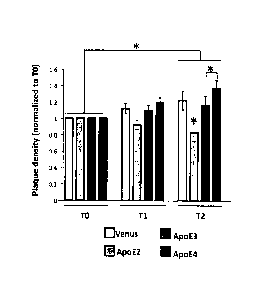
Figures 11A-11B. Overexpression of each APOE variant differentially modulates
the
progression of amyloidosis in vivo. In vivo two-photon images were developed
of amyloid
deposition in APP/PS I mice one week (TO), one month (Ti) and two months (T2)
after
intracerebroventricular injection with AAV-GFP, -APOE2, -3 or -4 vectors. An
intravenous injection
of Dextran, Texas red (70,000 Da) was performed prior imaging so that the same
Fields of view could
be followed over time. Within a two month period of time, few new amyloid
plaques could be
detected, whereas occasional deposits initially visible were not detectable
anymore after one or two
.. months. (A) Evaluation of the volumetric cortical density of amyloid
deposits over a two-month
5
CA 02873890 2014-11-17
WO 2013/172964 PCT/US2013/031725
period of time after intraventricular injection of an AAV-GFP, -APOE2, -APOE3
or APOE4 in 7
month-old APP/PSI mice. Six to eight fields of view were longitudinally imaged
for each animal and
the density of plaques was calculated per volume of cortex and reported to the
initial value for each
animal at baseline (TO). An overall progression of 0.23 of the density of
amyloid deposits was
.. observed over time (T2/TI, p<0.011). In addition, ApoE2 significantly
reduces the density relative to
GFP by 0.66 (se=0.21, p=0.002), relative to ApoE3 by 0.67 (se=0.17, p<0.0001)
and relative to
ApoE4 by 0.74 (se=0.17, p<0.0001). (B) Linear regression fit of amyloidosis
progression over 2
months after gene transfer in APP/PS1 mice shows that only AAV-APOE4 induces a
significant
positive slope during this period of time. n= 4-6 animals per group. *p<0.05.
Figure 12. Evolution of the size of amyloid deposits one and two month(s)
after infusion
with ApoE2, -3 and -4. Scatter dot plots representing the ratio of plaque
sizes between TI and TO
showed that ApoE4 was associated with increased plaque growth compared with
both ApoE2 and
ApoE3 after one month. This effect is not sustained after 2 months. n> 50
plaques measured per group
within 3 to 4 animals. *p<0.05.
Figures 13A-13C. The neuropathological changes associated with the amyloid
deposits
are differentially affected by each APOE variant. Images of array tomography
sections
immunostained for PSD95 (post-synaptic element) and amyloid deposits in APP/PS
I mice 2 months
after intraventricular injection of AAV-GFP, -APOE2, -APOE3 and -APOE4 were
prepared. Amyloid
deposits were labeled using the antibody NAB61 that was previously shown to
preferentially label
toxic Af3 oligomeric species (A) A significantly higher loss of the synapsin-1
marker was observed in
the vicinity of amyloid plaques when both APOE3 and APOE4 were expressed
compared with GFP
or APOE2. (B) A similar effect was observed when post-synaptic elements were
quantified, so that
the density of PSD95 surrounding the deposits was decreased 2 months after an
intraventricular
injection of AAV4-APOE4. As an additional parameter of neuropathological
change, the number of
neuritic dystrophies per amyloid plaque was evaluated in the brain of injected
APP/PSI mice, after
immunostaining for ThioS and the axonal marker SMI312. (C) A significant shift
toward a higher
number of dystrophies was observed when mice were infused with ApoE4 was
expressed compared
with ApoE3 and ApoE2 groups, thus suggesting that ApoE4 may have deleterious
effects beyond
amyloid plaques formation and may modulate the neurotoxic potential of smaller
oligomeric amyloid
aggregates. n= 4-6 animals per group. *p<0.05.
Figure 14. Early changes in the content of oligomeric AD species are observed
in the ISF
after intracerebroventricular injection of AAV4-APOE2, -3, -4 in Tg2576 mice.
Quantification of
the ISF content in oA13 using the 82E1/82E1 ELISA assay shows that there is a
higher concentration
of oligomeric amyloid 13 species after injection of AAV4-APOE4 compared with
AAV4-APOE2 and
--GFP, whereas AAV4-APOE3 injected mice reached an intermediate level. n= 3-6
animals per
group. *p<0.05.
6
CA 02873890 2014-11-17
WO 2013/172964 PCT/US2013/031725
Figure 15A-15B. Detection of human and endogenous murine APOE mRNA and protein
after intraventricular injection of an AAV4 in APP/PS1 mice. (A) Box blot
graphs representing
the amounts of endogenous murine apoE protein in the brains of injected mice.
(B) Comparison of the
levels of ApoE protein 2 and 5 months after intracerebroventricular injection
of AAV4 in APP/PS1
mice (samples from all ApoE injected mice were pooled together at 2 and 5
months, without
discrimination for the APOE variant). n= 4-6 animals per group. *p<0.05.
Fig. 16A-16B. Effects on A13 are associated with each ApoE isoform after a
short (2
month) exposure. Images were prepared of amyloid deposition in APP/PS I mice 2
months after
injection. Both immunostaining using the Bam10 antibody and ThioS were used to
stain all amyloid
deposits or dense-core plaques respectively. (A) Stereological analysis of the
density of amyloid
deposits in the cortex revealed that overexpression of APOE4 led to an
increased number of plaques
as early as 2 months after injection, whereas no difference could be observed
between the other
experimental groups. (B) The ratio between Bam10 and rfhioS staining, on the
other hand, remain
unchanged among all the different groups. (C) Determination of the
concentrations of A1340 (left
panels) and A1342 (right panels) peptides in the insoluble formic acid
extracts after a short exposure
with the different ApoE variants. n= 3-5 animals per group. *p<0.05.
Figures 17A-17B. Changes in soluble and insoluble A13 species detected 3
months after
injection in Tg2576 mice. (A) Quantification by ELISA of the 'SF content in
A1340 and Af342 (B)
shows that there is a tendency towards higher concentration of soluble amyloid
3 peptides after
injection of AAV4-APOE4 compared with AAV4-APOE2, -APOE3 and -GFP. (B) As
previously
observed in APP/PS1 mice, the stronger effect was observed with ApoE4, which
causes significantly
higher amounts of AB42 in the formic acid fraction of Tg2576 mice. n= 3-5
animals per group.
*p<0.05.
DETAILED DESCRIPTION
There are several different human apolipoprotein F (ApoE) isoforms, the
presence of
some of these isoforms in the brain increase the risk for Alzheimer's disease
(AD), whereas
the presence of other isoforms decreases the risk for AD. The presence of the
ApoE c4
isoform is a strong genetic risk factor for late-onset, sporadic AD.
(Casellano et al., Sci
Transl Med, 3(89):89ra57 (29 June 20I1).) The ApoE c4 allele strongly
increases AD risk
and decreases age of onset. On the other hand, the presence of the ApoE c2
allele appears to
decrease AD risk. It is suggested that human ApoE isoforms differentially
affect the
clearance or synthesis of amyloid-13 (Afl) in vivo.
Adeno associated virus (AAV) is a small nonpathogenic virus of the
parvoviridae
family. AAV is distinct from the other members of this family by its
dependence upon a
7
CA 02873890 2014-11-17
WO 2013/172964 PCT/US2013/031725
helper virus for replication. In the absence of a helper virus, AAV may
integrate in a locus
specific manner into the q arm of chromosome 19. The approximately 5 kb genome
of AAV
consists of one segment of single stranded DNA of either plus or minus
polarity. The ends of
the genome are short inverted terminal repeats which can fold into hairpin
structures and
serve as the origin of viral DNA replication. Physically, the parvovirus
virion is non-
enveloped and its icosohedral capsid is approximately 20 nm in diameter.
To-date eight serologically distinct AAVs have been identified and five have
been
isolated from humans or primates and are referred to as AAV types 1-5.
Govindasamy et al.,
"Structurally Mapping the Diverse Phenotype of Adeno-Associated Virus Serotype
4," 1
Vir., 80 (23):11556-11570 (2006). The genome of AAV2 is 4680 nucleotides in
length and
contains two open reading frames (ORFs). The left ORF encodes the non-
structural Rep
proteins, Rep 40, Rep 52, Rep 68 and Rep 78, which are involved in regulation
of replication
and transcription in addition to the production of single-stranded progeny
genomes.
Furthermore, two of the Rep proteins have been associated with the
preferential integration of
AAV genomes into a region of the q arm of human chromosome 19. Rep68/78 has
also been
shown to possess NTP binding activity as well as DNA and RNA helicase
activities. The
Rep proteins possess a nuclear localization signal as well as several
potential phosphorylation
sites. Mutation of one of these kinase sites resulted in a loss of replication
activity.
The ends of the genome are short inverted terminal repeats (ITR) which have
the
potential to fold into T-shaped hairpin structures that serve as the origin of
viral DNA
replication. Within the ITR region two elements have been described which are
central to the
function of the ITR, a GAGC repeat motif and the terminal resolution site
(trs). The repeat
motif has been shown to bind Rep when the ITR is in either a linear or hairpin
conformation.
This binding serves to position Rep68/78 for cleavage at the trs which occurs
in a site- and
strand-specific manner. In addition to their role in replication, these two
elements appear to
be central to viral integration. Contained within the chromosome 19
integration locus is a
Rep binding site with an adjacent trs. These elements have been shown to be
functional and
necessary for locus specific integration.
The AAV2 virion is a non-enveloped, icosohedral particle approximately 25 nrn
in
diameter, consisting of three related proteins referred to as VP1, VP2 and
VP3. The right
ORF encodes the capsid proteins VP1. VP2, and VP3. These proteins are found in
a ratio of
1:1:10 respectively and are all derived from the right-hand ORF. The capsid
proteins differ
from each other by the use of alternative splicing and an unusual start codon.
Deletion
analysis has shown that removal or alteration of VP1 which is translated from
an alternatively
8
CA 02873890 2014-11-17
WO 2013/172964
PCT/US2013/031725
spliced message results in a reduced yield of infections particles. Mutations
within the VP3
coding region result in the failure to produce any single-stranded progeny DNA
or infectious
particles. An AAV2 particle is a viral particle comprising an AAV2 capsid
protein. An
AAV2 capsid polypeptide can encode the entire VP1, VP2 and VP3 polypeptide.
The
particle can be a particle comprising AAV2 and other AAV capsid proteins
(i.e., a chimeric
protein, such as AAV4 and AAV2). Variations in the amino acid sequence of the
AAV2
capsid protein are contemplated herein, as long as the resulting viral
particle comprises the
AAV2 capsid remains antigenically or immunologically distinct from AAV4, as
can be
routinely determined by standard methods. Specifically, for example, ELISA and
Western
blots can be used to determine whether a viral particle is antigenically or
immunologically
distinct from AAV4. Furthermore, the AAV2 viral particle preferably retains
tissue tropism
distinct from AAV4.
An AAV2 particle is a viral particle comprising an AAV2 capsid protein. An
AAV2
capsid polypeptide encoding the entire VP1, VP2, and VP3 polypeptide can
overall have at
least about 63% homology (or identity) to the polypeptide having the amino
acid sequence
encoded by nucleotides set forth in SEQ ID NO:1 (AAV2 capsid protein). The
capsid protein
can have about 70% homology, about 75% homology, 80% homology, 85% homology,
90%
homology, 95% homology, 98% homology, 99% homology, or even 100% homology to
the
protein set forth in SEQ ID NO: 1. The capsid protein can have about 70%
identity, about
75% identity, 80% identity, 85% identity, 90% identity, 95% identity, 98%
identity. 99%
identity, or even 100% identity to the protein set forth in SEQ ID NO: 1. The
particle can be a
particle comprising both AAV4 and AAV2 capsid protein, i.e., a chimeric
protein. Variations
in the amino acid sequence of the AAV2 capsid protein are contemplated herein,
as long as
the resulting viral particle comprising the AAV2 capsid remains antigenically
or
immunologically distinct from AAV4, as can be routinely determined by standard
methods.
Specifically, for example, ELISA and Western blots can be used to determine
whether a viral
particle is antigenically or immunologically distinct from AAV4. Furthermore,
the AAV2
viral particle preferably retains tissue tropism distinction from AAV4, such
as that
exemplified in the examples herein, though an AAV2 chimeric particle
comprising at least
one AAV2 coat protein may have a different tissue tropism from that of an AAV2
particle
consisting only of AAV2 coat proteins.
As indicated in Figures 6A and 6B, AAV2 capsid sequence and AAV4 capsid
sequence are about 60% homologous. In certain embodiments, the AAV2 capsid
comprises
9
CA 02873890 2014-11-17
WO 2013/172964
PCT/US2013/031725
(or consists of) a sequence that is at least 65% homologous to the amino acid
sequence set
forth in SEQ ID NO:l.
In certain embodiments, the invention further provides an AAV2 particle
containing,
i.e., encapsidating, a vector comprising a pair of AAV2 inverted terminal
repeats. The
nucleotide sequence of AAV2 ITRs is known in the art. Furthermore, the
particle can be a
particle comprising both AAV4 and AAV2 capsid protein, i.e., a chimeric
protein. Moreover,
the particle can be a particle encapsidating a vector comprising a pair of AAV
inverted
terminal repeats from other AAVs (e.g., AAV1-AAV8). The vector encapsidated in
the
particle can further comprise an exogenous nucleic acid inserted between the
inverted
terminal repeats.
The following features of AAV have made it an attractive vector for gene
transfer.
AAV vectors have been shown in vitro to stably integrate into the cellular
genome; possess a
broad host range; transduce both dividing and non dividing cells in vitro and
in vivo and
maintain high levels of expression of the transduced genes. Viral particles
are heat stable,
resistant to solvents, detergents, changes in pIl, temperature, and can be
concentrated on
CsC1 gradients. Integration of AAV provirus is not associated with any long
term negative
effects on cell growth or differentiation. The ITRs have been shown to be the
only cis
elements required for replication, packaging and integration and may contain
some promoter
activities.
The present invention provides methods of administering AAV particles,
recombinant
AAV vectors, and recombinant AAV virions. For example, an AAV2 particle is a
viral
particle comprising an AAV2 capsid protein, or an AAV4 particle is a viral
particle
comprising an AAV4 capsid protein. A recombinant AAV2 vector is a nucleic acid
construct
that comprises at least one unique nucleic acid of AAV2. A recombinant AAV2
virion is a
particle containing a recombinant AAV2 vector. To be considered within the
term "AAV2
ITRs" the nucleotide sequence must retain one or both features described
herein that
distinguish the AAV2 ITR from the AAV4 ITR: (1) three (rather than four as in
AAV4)
"GAGC" repeats and (2) in the AAV2 ITR Rep binding site the fourth nucleotide
in the first
two "GAGC" repeats is a C rather than a T.
The promoter can be any desired promoter, selected by known considerations,
such as
the level of expression of a nucleic acid functionally linked to the promoter
and the cell type
in which the vector is to be used. Promoters can be an exogenous or an
endogenous
promoter. Promoters can include, for example, known strong promoters such as
SV40 or the
inducible metallothionein promoter, or an AAV promoter, such as an AAV p5
promoter.
Additional examples of promoters include promoters derived from actin genes,
immunoglobulin genes, cytomegalovirus (CMV), adenovirus, bovine papilloma
virus,
adenoviral promoters, such as the adenoviral major late promoter, an inducible
heat shock
promoter, respiratory syncytial virus, Rous sarcomas virus (RSV), etc.
Specifically, the
promoter can be AAV2 p5 promoter or AAV4 p5 promoter. Furthermore, smaller
fragments
of p5 promoter that retain promoter activity can readily be determined by
standard procedures
including, for example, constructing a series of deletions in the p5 promoter,
linking the
deletion to a reporter gene, and determining whether the reporter gene is
expressed, i.e.,
transcribed and/or translated.
The AAV vector can further comprise an exogenous (heterologous) nucleic acid
functionally linked to the promoter. By "heterologous nucleic acid" is meant
that any
heterologous or exogenous nucleic acid can be inserted into the vector for
transfer into a cell,
tissue or organism. For example, in certain embodiments, the heterologous
nucleic acid
encodes a protective ApoE isoform. By "functionally linked" is meant such that
the promoter
can promote expression of the hetcrologous nucleic acid, as is known in the
art, such as
appropriate orientation of the promoter relative to the heterologous nucleic
acid.
Furthermore, the heterologous nucleic acid preferably has all appropriate
sequences for
expression of the nucleic acid, as known in the art, to functionally encode,
i.e., allow the
nucleic acid to be expressed. The nucleic acid can include, for example,
expression control
sequences, such as an enhancer, and necessary information processing sites,
such as ribosome
binding sites, RNA splice sites, polyadenylation sites, and transcriptional
terminator
sequences. The nucleic acid can encode more than one gene product, limited
only by the size
of nucleic acid that can be packaged.
An AAV2 particle is a viral particle comprising an AAV2 capsid protein.
Variations
in the amino acid sequence of the AAV2 capsid protein are contemplated herein,
as long as
the resulting viral particle comprising the AAV2 capsid remains antigenically
or
immunologically distinct from AAV4, as can be routinely determined by standard
methods.
Specifically, for example, ELISA and Western blots can be used to determine
whether a viral
particle is antigenically or immunologically distinct from other AAV
scrotypes.
AAV4 is a unique member of the AAV family. A discussion of AAV4 is provided in
US Patent No. 6,468,524. DNA
hybridization data
indicated a similar level of homology for AAV1-4. However, in contrast to the
other AAVs,
only one ORF corresponding to the capsid proteins was identified in AAV4 and
no ORF was
detected for the Rep proteins. The present invention provides a vector
comprising the AAV4
11
CA 2873890 2019-07-15
virus as well as AAV4 viral particles. While AAV4 is similar to AAV2, the two
viruses are
found herein to be physically and genetically distinct. These differences
endow AAV4 with
some unique advantages which better suit it as a vector for gene therapy. For
example, the
wildtype AAV4 genome is larger than AAV2, allowing for efficient encapsidation
of a larger
recombinant genome. Furthermore, wildtype AAV4 particles have a greater
buoyant density
than AAV2 particles and therefore are more easily separated from contaminating
helper virus
and empty AAV particles than AAV2-based particles. Additionally, in contrast
to AAV1, 2,
and 3, AAV4 is able to hemagglutinate human, guinea pig, and sheep
erythrocytes.
In certain embodiments, the present invention provides a vector comprising the
AAV5
virus or a vector comprising subparts of the virus, as well as AAV5 viral
particles. A
discussion of AAV5 is provided in US Patent No. 6,855,314 .
While AAV5 is similar to AAV2, the two viruses are found herein to be
physically and genetically distinct. These differences endow AAV5 with some
unique
properties and advantages which better suit it as a vector for gene therapy.
For example, one
of the limiting features of using AAV2 as a vector for gene therapy is
production of large
amounts of virus. Using standard production techniques, AAV5 is produced at a
10-50 fold
higher level compared to AAV2. Because of its unique TRS site and rep
proteins, AAV5
should also have a distinct integration locus compared to AAV2.
Furthermore, AAV5 capsid protein, again surprisingly, is distinct from AAV2
capsid
protein and exhibits different tissue tropism, thus making AAV5 capsid-
containing particles
suitable tbr transducing cell types for which AAV2 is unsuited or less well-
suited. AAV2
and AAV5 have been shown to be serologically distinct and thus, in a gene
therapy
application, AAV5, and AAV5-derived vectors, would allow for transduction of a
patient
who already possess neutralizing antibodies to AAV2 either as a result of
natural
immunological defense or from prior exposure to AAV2 vectors. Another
advantage of
AAV5 is that AAV5 cannot be rescued by other serotypes. Only AAV5 can rescue
the
integrated AAV5 genome and effect replication, thus avoiding unintended
replication of
AAV5 caused by other AAV serotypes.
The term "polypeptide" as used herein refers to a polymer of amino acids and
includes
full-length proteins and fragments thereof. Thus, "protein," polypeptide," and
"peptide" are
often used interchangeably herein. Substitutions can be selected by known
parameters to be
neutral. As will be appreciated by those skilled in the art, the invention
also includes those
polypeptides having slight variations in amino acid sequences or other
properties. Such
variations may arise naturally as allelic variations (e.g. due to genetic
polymorphism) or may
12
CA 2873890 2019-07-15
CA 02873890 2014-11-17
WO 2013/172964
PCT/US2013/031725
be produced by human intervention (e.g., by mutagenesis of cloned DNA
sequences), such as
induced point, deletion, insertion and substitution mutants. Minor changes in
amino acid
sequence are generally preferred, such as conservative amino acid
replacements, small
internal deletions or insertions, and additions or deletions at the ends of
the molecules. These
modifications can result in changes in the amino acid sequence, provide silent
mutations,
modify a restriction site, or provide other specific mutations.
The present method provides a method of delivering a nucleic acid to a cell
comprising administering to the cell an AAV particle containing a vector
comprising the
nucleic acid inserted between a pair of AAV inverted terminal repeats, thereby
delivering the
.. nucleic acid to the cell. Administration to the cell can be accomplished by
any means,
including simply contacting the particle, optionally contained in a desired
liquid such as
tissue culture medium, or a buffered saline solution, with the cells. The
particle can be
allowed to remain in contact with the cells for any desired length of time,
and typically the
particle is administered and allowed to remain indefinitely. For such in vitro
methods, the
virus can be administered to the cell by standard viral transduction methods,
as known in the
art and as exemplified herein. Titers of virus to administer can vary,
particularly depending
upon the cell type, but will be typical of that used for AAV transduction in
general.
Additionally the titers used to transduce the particular cells in the present
examples can be
utilized. The cells can include any desired cell in humans as well as other
large (non-rodent)
mammals, such as primates, horse, sheep, goat, pig, and dog.
More specifically, the present invention provides a method of delivering a
nucleic
acid to an ependymal cell, comprising administering to the ependymal cell an
AAV particle
containing a vector comprising the nucleic acid inserted between a pair of AAV
inverted
terminal repeats, thereby delivering the nucleic acid to the ependymal cell.
The present invention also includes a method of delivering a nucleic acid to a
subject
comprising administering to a cell from the subject an AAV particle comprising
the nucleic
acid inserted between a pair of AAV inverted terminal repeats, and returning
the cell to the
subject, thereby delivering the nucleic acid to the subject. In certain
embodiments, the AAV
ITRs can be AAV2 ITRs. For such an ex vivo administration, cells are isolated
from a
subject by standard means according to the cell type and placed in appropriate
culture
medium, again according to cell type. Viral particles are then contacted with
the cells as
described above, and the virus is allowed to transfect the cells. Cells can
then be transplanted
back into the subject's body, again by means standard for the cell type and
tissue. If desired,
13
CA 02873890 2014-11-17
WO 2013/172964
PCMJS2013/031725
prior to transplantation, the cells can be studied for degree of transfection
by the virus, by
known detection means and as described herein.
The present invention further provides a method of delivering a nucleic acid
to a cell
in a subject comprising administering to the subject an AAV particle
comprising the nucleic
acid inserted between a pair of AAV inverted terminal repeats, thereby
delivering the nucleic
acid to a cell in the subject. Administration can be an ex vivo administration
directly to a cell
removed from a subject, such as any of the cells listed above, followed by
replacement of the
cell back into the subject, or administration can be in vivo administration to
a cell in the
subject. For ex vivo administration, cells are isolated from a subject by
standard means
according to the cell type and placed in appropriate culture medium, again
according to cell
type. Viral particles are then contacted with the cells as described above,
and the virus is
allowed to transfect the cells. Cells can then be transplanted back into the
subject's body,
again by means standard for the cell type and tissue. If desired, prior to
transplantation, the
cells can be studied for degree of transfection by the virus, by known
detection means and as
described herein.
Also provided is a method of delivering a nucleic acid to an ependymal cell in
a
subject comprising administering to the subject an AAV particle comprising the
nucleic acid
inserted between a pair of AAV inverted terminal repeats, thereby delivering
the nucleic acid
to an ependymal cell in the subject.
In certain embodiments, the amino acid sequence that targets brain vascular
endothelium targets brain vascular endothelium in a subject that has a
disease, e.g.,
Alzheimer's disease.
In certain embodiments, the amino acid sequence that targets brain vascular
endothelium targets brain vascular endothelium in a subject that does not have
Alzheimer's
disease.
In certain embodiments, the viral vector comprises a nucleic acid sequence
encoding a
therapeutic agent. In certain embodiments, the therapeutic agent is a
protective ApoE
isoform.
Certain embodiments of the present disclosure provide a cell comprising a
viral vector
as described herein.
In certain embodiments, the cell is a mammalian cell of a non-rodent mammal.
In
certain embodiments, the cell is a primate cell. In certain embodiments, the
cell is a human
cell. In certain embodiments, the cell is a non-human cell. In certain
embodiments, the cell
14
CA 02873890 2014-11-17
WO 2013/172964
PCT/US2013/031725
is in vitro. In certain embodiments, the cell is in vivo. In certain
embodiments, the cell is an
ependymal cell.
Certain embodiments of the present disclosure provide a method of treating a
disease
in a mammal comprising administering a viral vector or the cell as described
herein to the
mammal.
In certain embodiments, the mammal is human.
Certain embodiments of the present disclosure provide a method to deliver an
agent to
the central nervous system of a subject, comprising administering to the CSF
with a viral
vector described herein so that the transduced ependymal cells express the
therapeutic agent
and deliver the agent to the central nervous system of the subject. In certain
embodiments,
the viral vector transduces ependymal cells.
Certain embodiments of the present disclosure provide a viral vector or cell
as
described herein for use in medical treatments.
Certain embodiments of the present disclosure provide a use of a viral vector
or cell
as described herein to prepare a medicament useful for treating a disease,
e.g, Alzheimer's
disease, in a mammal.
The vector may further comprise a protective ApoE isoform protein. As used
herein,
the term "secreted protein" includes any secreted protein, whether naturally
secreted or
modified to contain a signal sequence so that it can be secreted.
Nucleic acid is "operably linked" when it is placed into a functional
relationship with
another nucleic acid sequence. Generally, ''operably linked" means that the
DNA sequences
being linked are contiguous. However, enhancers do not have to be contiguous.
Linking is
accomplished by ligation at convenient restriction sites. If such sites do not
exist, the
synthetic oligonucleotide adaptors or linkers are used in accordance with
conventional
practice. Additionally, multiple copies of the nucleic acid encoding enzymes
may be linked
together in the expression vector. Such multiple nucleic acids may be
separated by linkers.
The present disclosure also provides a mammalian cell containing a vector
described
herein. The cell may be human, and may be from brain. The cell type may be a
stem or
progenitor cell population.
The present disclosure provides a method of treating a disease such as a
genetic
disease or cancer in a mammal by administering a polynucleotide, polypeptide,
expression
vector, or cell described herein. The genetic disease may be a
neurodegenerative disease,
such as Alzheimer's disease.
CA 02873890 2014-11-17
WO 2013/172964
PCMJS2013/031725
Certain aspects of the disclosure relate to polynucleotides, polypeptides,
vectors, and
genetically engineered cells (modified in vivo), and the use of them. In
particular, the
disclosure relates to a method for gene or protein therapy that is capable of
both systemic
delivery of a therapeutically effective dose of the therapeutic agent.
According to one aspect, a cell expression system for expressing a therapeutic
agent
in a mammalian recipient is provided. The expression system (also referred to
herein as a
"genetically modified cell") comprises a cell and an expression vector for
expressing the
therapeutic agent. Expression vectors include, but are not limited to,
viruses, plasmids, and
other vehicles for delivering heterologous genetic material to cells.
Accordingly, the term
.. "expression vector" as used herein refers to a vehicle for delivering
heterologous genetic
material to a cell. In particular, the expression vector is a recombinant
adenoviral, adeno-
associated virus, or lentivirus or retrovirus vector.
The expression vector further includes a promoter for controlling
transcription of the
heterologous gene. The promoter may be an inducible promoter (described
below). The
expression system is suitable for administration to the mammalian recipient.
The expression
system may comprise a plurality of non-immortalized genetically modified
cells, each cell
containing at least one recombinant gene encoding at least one therapeutic
agent.
The cell expression system can be formed in vivo. According to yet another
aspect, a
method for treating a mammalian recipient in vivo is provided. The method
includes
.. introducing an expression vector for expressing a heterologous gene product
into a cell of the
patient in situ, such as via intravenous administration. To form the
expression system in vivo,
an expression vector for expressing the therapeutic agent is introduced in
vivo into the
mammalian recipient i.v., where the vector migrates via the vasculature to the
brain.
According to yet another aspect, a method for treating a mammalian recipient
in vivo
is provided. The method includes introducing the target protein into the
patient in vivo.
The expression vector for expressing the heterologous gene may include an
inducible
promoter for controlling transcription of the heterologous gene product.
Accordingly,
delivery of the therapeutic agent in situ is controlled by exposing the cell
in situ to conditions,
which induce transcription of the heterologous gene.
The mammalian recipient may have a condition that is amenable to gene
replacement
therapy. As used herein, "gene replacement therapy" refers to administration
to the recipient
of exogenous genetic material encoding a therapeutic agent and subsequent
expression of the
administered genetic material in situ. Thus, the phrase "condition amenable to
gene
rprdapp.mt.nt thi,ranx," isrnbraces conditions such as genetic diseases (i.e.,
a disease condition
17023.122W01/2012-060 16
CA 02873890 2014-11-17
WO 2013/172964
PCT/US2013/031725
that is attributable to one or more gene defects), acquired pathologies (i.e.,
a pathological
condition which is not attributable to an inborn defect), cancers and
prophylactic processes
(i.e., prevention of a disease or of an undesired medical condition).
Accordingly, as used
herein, the term "therapeutic agent" refers to any agent or material, which
has a beneficial
-- effect on the mammalian recipient. Thus, "therapeutic agent" embraces both
therapeutic and
prophylactic molecules having nucleic acid or protein components.
According to one embodiment, the mammalian recipient has a genetic disease and
the
exogenous genetic material comprises a heterologous gene encoding a
therapeutic agent for
treating the disease. In yet another embodiment, the mammalian recipient has
an acquired
-- pathology and the exogenous genetic material comprises a heterologous gene
encoding a
therapeutic agent for treating the pathology. According to another embodiment,
the patient
has a cancer and the exogenous genetic material comprises a heterologous gene
encoding an
anti-neoplastic agent. In yet another embodiment the patient has an undesired
medical
condition and the exogenous genetic material comprises a heterologous gene
encoding a
-- therapeutic agent for treating the condition.
As used herein, the term "a protective ApoE isoform" includes variants or
biologically active or inactive fragments of this polypeptide. A "variant" of
one of the
polypeptides is a polypeptide that is not completely identical to a native
protein. Such variant
protein can be obtained by altering the amino acid sequence by insertion,
deletion or
substitution of one or more amino acid. The amino acid sequence of the protein
is modified,
for example by substitution, to create a polypeptide having substantially the
same or
improved qualities as compared to the native polypeptide. The substitution may
be a
conserved substitution. A "conserved substitution" is a substitution of an
amino acid with
another amino acid having a similar side chain. A conserved substitution would
be a
substitution with an amino acid that makes the smallest change possible in the
charge of the
amino acid or size of the side chain of the amino acid (alternatively, in the
size, charge or
kind of chemical group within the side chain) such that the overall peptide
retains its spacial
conformation but has altered biological activity. For example, common
conserved changes
might be Asp to Glu, Asn or Gln; His to Lys, Arg or Phe; Asn to Gln, Asp or
Glu and Ser to
-- Cys, Thr or Gly. Alanine is commonly used to substitute for other amino
acids. The 20
essential amino acids can be grouped as follows: alanine, valine, leucine,
isoleucine, proline,
phenylalanine, tryptophan and methionine having nonpolar side chains; glycine,
serine,
threonine, cystine, tyrosine, asparagine and glutamine having uncharged polar
side chains;
17
CA 02873890 2014-11-17
WO 2013/172964
PCMJS2013/031725
aspartate and glutamate having acidic side chains; and lysine, arginine, and
histidine having
basic side chains.
The amino acid changes are achieved by changing the codons of the
corresponding
nucleic acid sequence. It is known that such polypeptides can be obtained
based on
substituting certain amino acids for other amino acids in the polypeptide
structure in order to
modify or improve biological activity. For example, through substitution of
alternative
amino acids, small conformational changes may be conferred upon a polypeptide
that results
in increased activity. Alternatively, amino acid substitutions in certain
polypeptides may be
used to provide residues, which may then be linked to other molecules to
provide peptide-
molecule conjugates which, retain sufficient properties of the starting
polypeptide to be
useful for other purposes.
One can use the hydropathic index of amino acids in conferring interactive
biological
function on a polypeptide, wherein it is found that certain amino acids may be
substituted for
other amino acids having similar hydropathic indices and still retain a
similar biological
.. activity. Alternatively, substitution of like amino acids may be made on
the basis of
hydrophilicity, particularly where the biological function desired in the
polypeptide to be
generated in intended for use in immunological embodiments. The greatest local
average
hydrophilicity of a "protein", as governed by the hydrophilicity of its
adjacent amino acids,
correlates with its immunogenicity. Accordingly, it is noted that
substitutions can be made
based on the hydrophilicity assigned to each amino acid.
In using either the hydrophilicity index or hydropathic index, which assigns
values to
each amino acid, it is preferred to conduct substitutions of amino acids where
these values are
2. with 1 being particularly preferred, and those with in 0.5 being the most
preferred
substitutions.
The variant protein has at least 50%, at least about 80%, or even at least
about 90%
but less than 100%, contiguous amino acid sequence homology or identity to the
amino acid
sequence of a corresponding native protein.
The amino acid sequence of the variant polypeptide corresponds essentially to
the
native polypeptide's amino acid sequence. As used herein "correspond
essentially to" refers
to a polypeptide sequence that will elicit a biological response substantially
the same as the
response generated by the native protein. Such a response may be at least 60%
of the level
generated by the native protein, and may even be at least 80% of the level
generated by native
protein.
18
CA 02873890 2014-11-17
WO 2013/172964
PCT/US2013/031725
A variant may include amino acid residues not present in the corresponding
native
protein or deletions relative to the corresponding native protein. A variant
may also be a
truncated "fragment" as compared to the corresponding native protein, i.e.,
only a portion of a
full-length protein. Protein variants also include peptides having at least
one D-amino acid.
The variant protein may be expressed from an isolated DNA sequence encoding
the
variant protein. "Recombinant- is defined as a peptide or nucleic acid
produced by the
processes of genetic engineering. It should be noted that it is well-known in
the art that, due
to the redundancy in the genetic code, individual nucleotides can be readily
exchanged in a
codon and still result in an identical amino acid sequence. The terms
"protein," "peptide"
and "polypeptide" are used interchangeably herein.
The present disclosure provides methods of treating a disease in a mammal by
administering an expression vector to a cell or patient. For the gene therapy
methods, a
person having ordinary skill in the art of molecular biology and gene therapy
would be able
to determine, without undue experimentation, the appropriate dosages and
routes of
administration of the expression vector used in the novel methods of the
present disclosure.
According to one embodiment, the cells are transformed or otherwise
genetically
modified in vivo. The cells from the mammalian recipient are transformed
(i.e., transduced or
transfected) in vivo with a vector containing exogenous genetic material for
expressing a
heterologous (e.g., recombinant) gene encoding a therapeutic agent and the
therapeutic agent
is delivered in situ.
As used herein, "exogenous genetic material" refers to a nucleic acid or an
oligonucleotide, either natural or synthetic, that is not naturally found in
the cells; or if it is
naturally found in the cells, it is not transcribed or expressed at
biologically significant levels
by the cells. Thus, "exogenous genetic material" includes, for example, a non-
naturally
occurring nucleic acid that can be transcribed into anti-sense RNA, as well as
a "heterologous
gene" (i.e., a gene encoding a protein which is not expressed or is expressed
at biologically
insignificant levels in a naturally-occurring cell of the same type).
In the certain embodiments, the mammalian recipient has a condition that is
amenable
to gene replacement therapy. As used herein, "gene replacement therapy" refers
to
administration to the recipient of exogenous genetic material encoding a
therapeutic agent
and subsequent expression of the administered genetic material in situ. Thus,
the phrase
"condition amenable to gene replacement therapy" embraces conditions such as
genetic
diseases (i.e., a disease condition that is attributable to one or more gene
defects), acquired
pathologies (i.e., a pathological condition which is not attributable to an
inborn defect),
19
CA 02873890 2014-11-17
WO 2013/172964 PCT/US2013/031725
cancers and prophylactic processes (i.e., prevention of a disease or of an
undesired medical
condition). Accordingly, as used herein, the term "therapeutic agent' refers
to any agent or
material, which has a beneficial effect on the mammalian recipient. Thus,
''therapeutic agent"
embraces both therapeutic and prophylactic molecules having nucleic acid
(e.g., antisense
RNA) and/or protein components.
Alternatively, the condition amenable to gene replacement therapy is a
prophylactic
process, i.e., a process for preventing disease or an undesired medical
condition. Thus, the
instant disclosure embraces a cell expression system for delivering a
therapeutic agent that
has a prophylactic function (i.e., a prophylactic agent) to the mammalian
recipient.
In summary, the term "therapeutic agent" includes, but is not limited to,
agents
associated with the conditions listed above, as well as their functional
equivalents. As used
herein, the term "functional equivalent" refers to a molecule (e.g., a peptide
or protein) that
has the same or an improved beneficial effect on the mammalian recipient as
the therapeutic
agent of which is it deemed a functional equivalent.
The above-disclosed therapeutic agents and conditions amenable to gene
replacement
therapy are merely illustrative and are not intended to limit the scope of the
instant disclosure.
The selection of a suitable therapeutic agent for treating a known condition
is deemed to be
within the scope of one of ordinary skill of the art without undue
experimentation.
AAV Vectors
In one embodiment, a viral vector of the disclosure is an AAV vector. An "AAV"
vector refers to an adeno-associated virus, and may be used to refer to the
naturally occurring
wild-type virus itself or derivatives thereof. The term covers all subtypes,
serotypes and
pseudotypes, and both naturally occurring and recombinant forms, except where
required
otherwise. As used herein, the term "serotype" refers to an AAV which is
identified by and
distinguished from other AAVs based on capsid protein reactivity with defined
antisera, e.g.,
there are eight known serotypes of primate AAVs, AAV-1 to AAV-8. For example,
serotype
AAV2 is used to refer to an AAV which contains capsid proteins encoded from
the cap gene
of AAV2 and a genome containing 5' and 3' ITR sequences from the same AAV2
serotype.
As used herein, for example, rAAV may be used to refer an AAV having both
capsid proteins
and 5'-3' ITRs from the same serotype or it may refer to an AAV having capsid
proteins from
one serotype and 5'-3' ITRs from a different AAV serotype, e.g, capsid from
AAV serotype 2
and ITRs from AAV serotype 5. For each example illustrated herein the
description of the
vector design and production describes the serotype of the capsid and 5'-3'
ITR sequences.
CA 02873890 2014-11-17
WO 2013/172964
PCT/US2013/031725
The abbreviation "rAAV" refers to recombinant adeno-associated virus, also
referred to as a
recombinant AAV vector (or "rAAV vector").
An "AAV virus" or "AAV viral particle" refers to a viral particle composed of
at least
one AAV capsid protein (preferably by all of the capsid proteins of a wild-
type AAV) and an
encapsidated polynucleotide. If the particle comprises heterologous
polynucleotide (i.e., a
polynucleotide other than a wild-type AAV genome such as a transgene to be
delivered to a
mammalian cell), it is typically referred to as "rAAV".
In one embodiment, the AAV expression vectors are constructed using known
techniques to at least provide as operatively linked components in the
direction of
transcription, control elements including a transcriptional initiation region,
the DNA of
interest and a transcriptional termination region. The control elements are
selected to be
functional in a mammalian cell. The resulting construct which contains the
operatively linked
components is flanked (5' and 3') with functional AAV !FR sequences.
By "adeno-associated virus inverted terminal repeats" or "AAV ITRs" is meant
the
art-recognized regions found at each end of the AAV genome which function
together in cis
as origins of DNA replication and as packaging signals for the virus. AAV
ITRs, together
with the AAV rep coding region, provide for the efficient excision and rescue
from, and
integration of a nucleotide sequence interposed between two flanking ITRs into
a mammalian
cell genome.
The nucleotide sequences of AAV ITR regions are known. As used herein, an
''AAV
ITR" need not have the wild-type nucleotide sequence depicted, but may be
altered, e.g by
the insertion, deletion or substitution of nucleotides. Additionally, the AAV
ITR may be
derived from any of several AAV serotypes, including without limitation, AAV1,
AAV2,
AAV3, AAV4, AAV5, AAV7, etc. Furthermore, 5' and 3' ITRs which flank a
selected
nucleotide sequence in an AAV vector need not necessarily be identical or
derived from the
same AAV serotype or isolate, so long as they function as intended, L e., to
allow for excision
and rescue of the sequence of interest from a host cell genome or vector, and
to allow
integration of the heterologous sequence into the recipient cell genome when
AAV Rep gene
products are present in the cell.
In one embodiment, AAV ITRs can be derived from any of several AAV serotypes,
including without limitation, AAV1, AAV2, AAV3, AAV4, AAV5, AAV7, etc.
Furthermore, 5' and 3' ITRs which flank a selected nucleotide sequence in an
AAV
expression vector need not necessarily be identical or derived from the same
AAV serotype
or isolate, so long as they function as intended, i.e., to allow for excision
and rescue of the
21
CA 02873890 2014-11-17
WO 2013/172964
PCT/US2013/031725
sequence of interest from a host cell genome or vector, and to allow
integration of the DNA
molecule into the recipient cell genome when AAV Rep gene products are present
in the cell.
In one embodiment, AAV capsids can be derived from AAV2. Suitable DNA
molecules for use in AAV vectors will be less than about 5 kilobases (kb),
less than about 4.5
kb, less than about 4kb, less than about 3.5 kb, less than about 3 kb, less
than about 2.5 kb in
size and are known in the art.
In one embodiment, the selected nucleotide sequence is operably linked to
control
elements that direct the transcription or expression thereof in the subject in
vivo. Such control
elements can comprise control sequences normally associated with the selected
gene.
Alternatively, heterologous control sequences can be employed. Useful
heterologous control
sequences generally include those derived from sequences encoding mammalian or
viral
genes. Examples include, but are not limited to, the SV40 early promoter,
mouse mammary
tumor virus LTR promoter; adenovirus major late promoter (Ad MLP); a herpes
simplex
virus (I-ISV) promoter, a cytomegalovirus (CMV) promoter such as the CMV
immediate
early promoter region (CM VIE), a rous sarcoma virus (RSV) promoter, pol II
promoters, pol
III promoters, synthetic promoters, hybrid promoters, and the like. In
addition, sequences
derived from nonviral genes, such as the murine metallothionein gene, will
also find use
herein. Such promoter sequences are commercially available from, e.g,
Stratagene (San
Diego, Calif.).
In one embodiment, both heterologous promoters and other control elements,
such as
CNS-specific and inducible promoters, enhancers and the like, will be of
particular use.
Examples of heterologous promoters include the CMV promoter. Examples of CNS-
specific
promoters include those isolated from the genes from myelin basic protein
(MBP), glial
fibrillary acid protein (GFAP), and neuron specific eno1ase (NSE). Examples of
inducible
promoters include DNA responsive elements for ecdysone, tetracycline, hypoxia
and aufin.
In one embodiment, the AAV expression vector which harbors the DNA molecule of
interest bounded by AAV ITRs, can be constructed by directly inserting the
selected
sequence(s) into an AAV genome which has had the major AAV open reading frames
("ORFs'') excised therefrom. Other portions of the AAV genome can also be
deleted, so long
as a sufficient portion of the ITRs remain to allow for replication and
packaging functions.
Such constructs can be designed using techniques well known in the art.
Alternatively, AAV ITRs can be excised from the viral genome or from an AAV
vector containing the same and fused 5' and 3' of a selected nucleic acid
construct that is
present in another vector using standard ligation techniques. For example,
ligations can be
22
CA 02873890 2014-11-17
WO 2013/172964
PCT/US2013/031725
accomplished in 20 mM Tris-Cl pH 7.5, 10 mM MgCl2, 10 mM DTI, 33 1..tg/m1 BSA,
10
mM-50 mM NaCl, and either 40 uM ATP, 0.01-0.02 (Weiss) units T4 DNA ligase at
0 C (for
"sticky end" ligation) or I mM ATP, 0.3-0.6 (Weiss) units T4 DNA ligase at 14
C (for "blunt
end" ligation). Intermolecular "sticky end" ligations are usually performed at
30-100 [tg/m1
total DNA concentrations (5-100 nM total end concentration). AAV vectors which
contain
ITRs.
Additionally, chimeric genes can be produced synthetically to include AAV ITR
sequences arranged 5' and 3' of one or more selected nucleic acid sequences.
Preferred
codons for expression of the chimeric gene sequence in mammalian CNS cells can
be used.
The complete chimeric sequence is assembled from overlapping oligonucleotides
prepared by
standard methods.
In order to produce rAAV virions, an AAV expression vector is introduced into
a
suitable host cell using known techniques, such as by transfection. A number
of transfection
techniques are generally known in the art. See, e.g., Sambrook et al. (1989)
Molecular
Cloning, a laboratory manual, Cold Spring Harbor Laboratories, New York.
Particularly
suitable transfection methods include calcium phosphate co-precipitation,
direct micro-
injection into cultured cells, electroporation, liposome mediated gene
transfer, lipid-mediated
transduction, and nucleic acid delivery using high-velocity microprojectiles.
In one embodiment, suitable host cells for producing rAAV virions include
microorganisms, yeast cells, insect cells, and mammalian cells, that can be,
or have been,
used as recipients of a heterologous DNA molecule. The term includes the
progeny of the
original cell which has been transfected. Thus, a "host cell" as used herein
generally refers to
a cell which has been transfected with an exogenous DNA sequence. Cells from
the stable
human cell line, 293 (readily available through, e g., the American Type
Culture Collection
under Accession Number ATCC CRL1573) can be used in the practice of the
present
disclosure. Particularly, the human cell line 293 is a human embryonic kidney
cell line that
has been transformed with adenovirus type-5 DNA fragments, and expresses the
adenoviral
Ela and El b genes. The 293 cell line is readily transfected, and provides a
particularly
convenient platform in which to produce rAAV virions.
By "AAV rep coding region" is meant the art-recognized region of the AAV
genome
which encodes the replication proteins Rep 78, Rep 68, Rep 52 and Rep 40.
These Rep
expression products have been shown to possess many functions, including
recognition,
binding and nicking of the AAV origin of DNA replication, DNA helicase
activity and
modulation of transcription from AAV (or other heterologous) promoters. The
Rep
23
CA 02873890 2014-11-17
WO 2013/172964 PCT/US2013/031725
expression products are collectively required for replicating the AAV genome.
Suitable
homologues of the AAV rep coding region include the human herpesvirus 6 (HHV-
6) rep
gene, which is also known to mediate AAV2 DNA replication.
By "AAV cap coding region" is meant the art-recognized region of the AAV
genome
which encodes the capsid proteins VP1, VP2, and VP3, or functional homologues
thereof.
These Cap expression products supply the packaging functions which are
collectively
required for packaging the viral genome.
In one embodiment, AAV helper functions are introduced into the host cell by
transfecting the host cell with an AAV helper construct either prior to, or
concurrently with,
the transfection of the AAV expression vector. AAV helper constructs are thus
used to
provide at least transient expression of AAV rep and/or cap genes to
complement missing
AAV functions that are necessary for productive AAV infection. AAV helper
constructs lack
AAV IIRs and can neither replicate nor package themselves. These constructs
can be in the
form of a plasmid, phage, transposon, cosmid, virus, or virion. A number of
AAV helper
constructs have been described, such as the commonly used plasmids pAAV/Ad and
pIM29+45 which encode both Rep and Cap expression products. A number of other
vectors
have been described which encode Rep and/or Cap expression products.
Methods of delivery of viral vectors include injecting the AAV into the CSF.
Generally, rAAV virions may be introduced into cells of the CNS using either
in vivo or in
vitro transduction techniques. If transduced in vitro, the desired recipient
cell will be removed
from the subject, transduced with rAAV virions and reintroduced into the
subject.
Alternatively, syngeneic or xenogeneic cells can be used where those cells
will not generate
an inappropriate immune response in the subject.
Suitable methods for the delivery and introduction of transduced cells into a
subject
have been described. For example, cells can be transduced in vitro by
combining recombinant
AAV virions with CNS cells e.g., in appropriate media, and screening for those
cells
harboring the DNA of interest can be screened using conventional techniques
such as
Southern blots and/or PCR, or by using selectable markers. Transduced cells
can then be
formulated into pharmaceutical compositions, described more fully below, and
the
composition introduced into the subject by various techniques, such as by
grafting,
intramuscular, intravenous, subcutaneous and intraperitoneal injection.
In one embodiment, pharmaceutical compositions will comprise sufficient
genetic
material to produce a therapeutically effective amount of the nucleic acid of
interest, i.e., an
amount sufficient to reduce or ameliorate symptoms of the disease state in
question or an
24
CA 02873890 2014-11-17
WO 2013/172964 PCT/US2013/031725
amount sufficient to confer the desired benefit. The pharmaceutical
compositions will also
contain a pharmaceutically acceptable excipient. Such excipients include any
pharmaceutical
agent that does not itself induce the production of antibodies harmful to the
individual
receiving the composition, and which may be administered without undue
toxicity.
Pharmaceutically acceptable excipients include, but are not limited to,
sorbitol, Tween80, and
liquids such as water, saline, glycerol and ethanol. Pharmaceutically
acceptable salts can be
included therein, for example, mineral acid salts such as hydrochlorides,
hydrobromides,
phosphates, sulfates, and the like; and the salts of organic acids such as
acetates, propionates,
malonates, benzoates, and the like. Additionally, auxiliary substances, such
as wetting or
emulsifying agents, pH buffering substances, and the like, may be present in
such vehicles. A
thorough discussion of pharmaceutically acceptable excipients is available in
Remington's
Pharmaceutical Sciences (Mack Pub. Co., N.J. 1991).
As is apparent to those skilled in the art in view of the teachings of this
specification,
an effective amount of viral vector which must be added can be empirically
determined.
Administration can be effected in one dose, continuously or intermittently
throughout the
course of treatment. Methods of determining the most effective means and
dosages of
administration are well known to those of skill in the art and will vary with
the viral vector,
the composition of the therapy, the target cells, and the subject being
treated. Single and
multiple administrations can be carried out with the dose level and pattern
being selected by
the treating physician.
It should be understood that more than one transgene could be expressed by the
delivered viral vector. Alternatively, separate vectors, each expressing one
or more different
transgenes, can also be delivered to the CNS as described herein. Furthermore,
it is also
intended that the viral vectors delivered by the methods of the present
disclosure be combined
with other suitable compositions and therapies.
Methods for Introducing Genetic Material into Cells
The exogenous genetic material (e.g., a cDNA encoding one or more therapeutic
proteins) is introduced into the cell ex vivo or in vivo by genetic transfer
methods, such as
transfection or transduction, to provide a genetically modified cell. Various
expression
vectors (i.e., vehicles for facilitating delivery of exogenous genetic
material into a target cell)
are known to one of ordinary skill in the art.
As used herein, "transfection of cells" refers to the acquisition by a cell of
new genetic
material by incorporation of added DNA. Thus, transfection refers to the
insertion of nucleic
acid into a cell using physical or chemical methods. Several transfection
techniques are
CA 02873890 2014-11-17
WO 2013/172964 PCMJS2013/031725
known to those of ordinary skill in the art including: calcium phosphate DNA
co-
precipitation; DEAE-dextran; electroporation; cationic liposome-mediated
transfection; and
tungsten particle-faciliated microparticle bombardment. Strontium phosphate
DNA co-
precipitation is another possible transfection method.
In contrast, "transduction of cells" refers to the process of transferring
nucleic acid
into a cell using a DNA or RNA virus. A RNA virus (i.e., a retrovirus) for
transferring a
nucleic acid into a cell is referred to herein as a transducing chimeric
retrovirus. Exogenous
genetic material contained within the retrovirus is incorporated into the
genome of the
transduced cell. A cell that has been transduced with a chimeric DNA virus
(e.g., an
.. adenovirus carrying a cDNA encoding a therapeutic agent), will not have the
exogenous
genetic material incorporated into its genome but will be capable of
expressing the exogenous
genetic material that is retained extrachromosomally within the cell.
Typically, the exogenous genetic material includes the heterologous gene
(usually in
the form of a cDNA comprising the exons coding for the therapeutic protein)
together with a
promoter to control transcription of the new gene. The promoter
characteristically has a
specific nucleotide sequence necessary to initiate transcription. Optionally,
the exogenous
genetic material further includes additional sequences (i.e., enhancers)
required to obtain the
desired gene transcription activity. For the purpose of this discussion an
"enhancer" is simply
any non-translated DNA sequence which works contiguous with the coding
sequence (in cis)
.. to change the basal transcription level dictated by the promoter. The
exogenous genetic
material may introduced into the cell genome immediately downstream from the
promoter so
that the promoter and coding sequence are operatively linked so as to permit
transcription of
the coding sequence. A retroviral expression vector may include an exogenous
promoter
element to control transcription of the inserted exogenous gene. Such
exogenous promoters
include both constitutive and inducible promoters.
Naturally-occurring constitutive promoters control the expression of essential
cell
functions. As a result, a gene under the control of a constitutive promoter is
expressed under
all conditions of cell growth. Exemplary constitutive promoters include the
promoters for the
following genes which encode certain constitutive or "housekeeping" functions:
hypoxanthine phosphoribosyl transferase (HPRT), dihydrofolate reductase
(DHFR),
adenosine deaminase, phosphoglycerol kinase (PGK), pyruvate kinase,
phosphoglycerol
mutase, the actin promoter, and other constitutive promoters known to those of
skill in the art.
In addition, many viral promoters function constitutively in eucaryotic cells.
These include:
the early and late promoters of SV40; the long terminal repeats (LTRs) of
Moloney Leukemia
26
CA 02873890 2014-11-17
WO 2013/172964
PCT/US2013/031725
Virus and other retroviruses; and the thymidine kinase promoter of Herpes
Simplex Virus,
among many others. Accordingly, any of the above-referenced constitutive
promoters can be
used to control transcription of a heterologous gene insert.
Genes that are under the control of inducible promoters are expressed only or
to a
greater degree, in the presence of an inducing agent, (e.g.. transcription
under control of the
metallothionein promoter is greatly increased in presence of certain metal
ions). Inducible
promoters include responsive elements (REs) which stimulate transcription when
their
inducing factors are bound. For example, there are REs for serum factors,
steroid hormones,
retinoic acid and cyclic AMP. Promoters containing a particular RE can be
chosen in order
to obtain an inducible response and in some cases, the RE itself may be
attached to a different
promoter, thereby conferring inducibility to the recombinant gene. Thus, by
selecting the
appropriate promoter (constitutive versus inducible; strong versus weak), it
is possible to
control both the existence and level of expression of a therapeutic agent in
the genetically
modified cell. If the gene encoding the therapeutic agent is under the control
of an inducible
promoter, delivery of the therapeutic agent in situ is triggered by exposing
the genetically
modified cell in situ to conditions for permitting transcription of the
therapeutic agent, e.g.,
by intraperitoneal injection of specific inducers of the inducible promoters
which control
transcription of the agent. For example, in situ expression by genetically
modified cells of a
therapeutic agent encoded by a gene under the control of the metallothionein
promoter, is
enhanced by contacting the genetically modified cells with a solution
containing the
appropriate (i.e., inducing) metal ions in situ.
Accordingly, the amount of therapeutic agent that is delivered in situ is
regulated by
controlling such factors as: (1) the nature of the promoter used to direct
transcription of the
inserted gene, (i.e., whether the promoter is constitutive or inducible,
strong or weak); (2) the
number of copies of the exogenous gene that are inserted into the cell; (3)
the number of
transduced/transfected cells that are administered (e.g, implanted) to the
patient; (4) the size
of the implant (e.g., graft or encapsulated expression system); (5) the number
of implants; (6)
the length of time the transduced/transfected cells or implants are left in
place; and (7) the
-
production rate of the therapeutic agent by the genetically modified cell.
Selection and
optimization of these factors for delivery of a therapeutically effective dose
of a particular
therapeutic agent is deemed to be within the scope of one of ordinary skill in
the art without
undue experimentation, taking into account the above-disclosed factors and the
clinical
profile of the patient.
In addition to at least one promoter and at least one heterologous nucleic
acid
27
CA 02873890 2014-11-17
WO 2013/172964
PCT/US2013/031725
encoding the therapeutic agent, the expression vector may include a selection
gene, for
example, a neomycin resistance gene, for facilitating selection of cells that
have been
transfected or transduced with the expression vector. Alternatively, the cells
are transfected
with two or more expression vectors, at least one vector containing the
gene(s) encoding the
therapeutic agent(s), the other vector containing a selection gene. The
selection of a suitable
promoter, enhancer, selection gene and/or signal sequence (described below) is
deemed to be
within the scope of one of ordinary skill in the art without undue
experimentation.
The therapeutic agent can be targeted for delivery to an extracellular,
intracellular or
membrane location. If it is desirable for the gene product to be secreted from
the cells, the
expression vector is designed to include an appropriate secretion ''signal"
sequence for
secreting the therapeutic gene product from the cell to the extracellular
milieu. If it is
desirable for the gene product to be retained within the cell, this secretion
signal sequence is
omitted. In a similar manner, the expression vector can be constructed to
include "retention"
signal sequences for anchoring the therapeutic agent within the cell plasma
membrane. For
.. example, all membrane proteins have hydrophobic transmembrane regions,
which stop
translocation of the protein in the membrane and do not allow the protein to
be secreted. The
construction of an expression vector including signal sequences for targeting
a gene product
to a particular location is deemed to be within the scope of one of ordinary
skill in the art
without the need for undue experimentation.
Example 1
Changes in the Progression of Amyloid Deposition
This Example studied changes in the progression of amyloid deposition in
app/ps
mice after overexpression of different ApoE isoforms through intraventricular
injection of an
adeno-associated virus serotype 4 (AAV4).
The epsilon 4 allele of ApoE (ApoE c4) is the first genetic risk factor for
Alzheimer
disease (AD), whereas inheritance of the rare epsilon 2 allele of ApoE (ApoE
c2) reduces this
risk by about half. However, despite the discovery of these strong genetic
clues almost 17
years ago, the mechanisms whereby ApoE confer risk remains uncertain.
In order to decipher how the different ApoE isoforms (ApoE c2, e3 and c4)
impact the
formation and stability of fibrillar amyloid plaques, AAV4 vectors coding for
each ApoE
isoform were injected into the ventricle of 7 month-old APP/PS mice. Using in
vivo
multiphoton imaging, populations of amyloid deposits were tracked at baseline
and after
exposure to ApoE over a two-month interval, thus allowing a dynamic view of
amyloidosis
28
CA 02873890 2014-11-17
WO 2013/172964
PCT/US2013/031725
progression in a living animal.
The kinetic of amyloid plaque deposition was observed to be variable according
to
each isoform, so that ApoE c4 injected mice had a 38% increase of senile
plaques whereas
mice treated with ApoE c2 presented a 15% decrease of the number of amyloid
deposits
compared to ApoE c3 after 2 months. The post-mortem analysis confirmed these
results and
revealed the presence of human ApoE proteins decorating plaques in the cortex,
reflecting a
large diffusion of the protein throughout the parenchyma and its focal
accumulation where
AB peptides are deposited. It is important to note that this increased content
of ApoE
protein was also associated with a more severe synapse loss around the amyloid
deposits.
Overall, the present data demonstrated that over-production of different ApoE
isoforms was able to influence the progression of the disease and could
modulate the extent
of synapse loss, one of the parameters that correlates best with cognitive
impairment in AD
patients
1. Intraventricular injection of AAV4-ApoE led to a stable
expression of huApoE
.. and a sustained detection of recombinant human ApoE (huApoE) protein in the
brain
Briefly, GFP and huApoE were immunodetected in APP/PS mice injected with AAV4
vectors. GFP signal could be observed into the entire ventricle area (upper
panel) and in the
cells lining the ventricle, as well as human APOE.
In order to evaluate the present approach, AAV4-Venus (control), -ApoE2, -
ApoE3
and ¨ApoE4 were injected in the ventricle of wild-type mice. Two months after
injection,
human ApoE proteins could be detected in the cortical parenchyma around
amyloid deposits
(note the 3H1 antibody; only nonspecific background was observed in AAV4-GFP
injected
mice). Thus, a significant level of human ApoE was detected by ELISA in the
brain, and
immunohistological stainings for Venus and ApoE confirmed the expression of
the different
.. transgenes by the cells lining the ventricle.
qRT-PCR experiments were performed in order to evaluate the mRNA levels of
the transgene. A standard curve lowed us to determine the concentrations of
huApoE
mRNA according to the level of endogenous GAPDH. Samples from mice that were
exposed for 2 or 5 months were included. An ELISA assay designed to
specifically
detect human APOE was performed on brain homogenates (Fig. 1A). Low levels of
recombinant proteins could be detected in AAV4-APOE injected mice compared
with
AAV4-GFP treated animals, as quantified by ELISA specific for human APOE (Fig.
1B)
and confirmed by Western blot.
29
CA 02873890 2014-11-17
WO 2013/172964
PCT/US2013/031725
2. Overexpression of each isoform of APOE differentially affects the
progression of the amyloidosis.
In vivo two-photon imaging was used to follow amyloid deposition over time in
a
living animal. Briefly, APP/PS mice (7 month old) were stereotactically
injected with
AAV4 vectors coding for ApoE2, ApoE3, ApoE4 and Venus. After 1 week, a cranial
window was implanted and amyloid deposits were imaged over time after
craniotomy.
After 2 months, the animals were sacrificed and post-mortem analyses were
performed.
2-photon images of APP/PS mice injected with AAV4-ApoE2, AAV4-ApoE3 or
AAV4-ApoE4 were prepared. Amyloid plaques could be detected after
intraperitoneal
injection of Methoxy-X04 (5mg/kg) and Texas Red dextran (70,000 Da molecular
weight; 12.5 mg/ml in sterile PBS) was injected into a lateral tail vein to
provide a
fluorescent angiogram. Images were taken one week (=TO), one month and two
months
after injection. The same fields were captured over time to follow the
progression of the
lesions. Few new amyloid deposits appeared whereas few of them could not be
detected
anymore over a two-month period of time.
A complete analysis of in vivo images shows that the number of amyloid
deposits
increases significantly more rapidly in AAV4-ApoE4 injected APP/PS mice
compared
with both AAV4-ApoE3 and AAV4-Venus treated animals. By contrast, a small but
significant decrease density of plaques is measured when AAV4-ApoE2 was used
(Fig.
2). A trend toward bigger plaques is observed in APP/PS mice injected with
AAV4-
ApoE4 (p<0.06), but overall the size of the plaques remains constant. The
summarized
data of in vivo imaging shows that overexpression of each APOE isoform
differentially
affect the progression of amyloid deposition in vivo. Injection of AAV4-ApoE2
leads to
a slight decrease of amyloid density over times, whereas injection of AAV4-
ApoE4
aggravates the amyloidosis.
3. The size of amyloid plaques vary according to each ApoE isoform.
In vivo two-photon imaging allowed the following of the changes of the size of
each amyloid deposit over a period of 2 months. The size of plaques may remain
stable,
increase or decrease over time. The distributions of the size ratios between
T1/f0 and
12/11 show that there is a shift towards bigger amyloid plaques in mice
injected with an
AAV4-ApoE4 compared with the other groups (Fig. 3).
4. Post-mortem evaluation of amyloid load confirms the effects of ApoE2 and
ApoE4 on amyloid deposition.
CA 02873890 2014-11-17
WO 2013/172964
PCMJS2013/031725
Two months after AAV4 injection, the post-mortem stereological evaluation
revealed that AAV4-ApoE4 injected animals have a higher density of amyloid
plaques in
the cortex, whereas no difference could be detected between the other groups
(Fig. 4A).
This increased number of amyloid deposits is observed when plaques were
labeled with
ThioS or Barn10. However, no change in the ratio between Bam10 and ThioS was
detected. Five months after injection, the effects of each ApoE isoform are
more
pronounced compared with two months (Fig. 4B). A significant increased density
of
deposits was observed when mice were injected with AAV4-ApoE4 whereas an
inverse
effect was detected with ApoE2. Again, no change in the ratio between Bam10
and
.. ThioS awes detected.
5. Each ApoE isform differentially ctlfects synaptic density
around amyloid
deposits.
Array tomography was used to precisely determine the density of pre- and post-
synaptic elements around amyloid deposits. This new imaging method offers
capabilities
for high-resolution imaging of tissue molecular architecture. Array tomography
is based
on ultrathin sectioning of the specimen (70nm), immunostaining and 3D
reconstruction.
Representative images of array tomography samples stained for amyloid plaque
and the
post-synaptic maker PSD95. Array tomography images show that a decreased
number of
the post-synaptic marker PSD95 is observed around the amyloid deposits, but
this effect
is abolished far from plaques. The quantification of pre- (synapsin-1) and
post-synaptic
markers in the vicinity or far from plaques was determined in each group of
mice injected
with an AAV4 (Fig. 5A-D). The extensive quantification of pre- and post-
synaptic
elements confirmed that a decreased density of synapsin 1 and PSD95 was
associated
with amyloid plaques, this effect being dramatically amplified when ApoE4 was
overexpressed in the brain of APP/PS1 mice (Fig. 5C, Fig. 5D). Overexpression
of
ApoE4 is associated with an increased spine loss compared with the other
groups in the
vicinity of amyloid deposits. On the contrary, the density of synapsin puncta
is higher in
ApoE2 treated animals around plaques.
Conclusion
Intraventricular injections of AAV virus serotype 4 led to a sustained and
chronic
over-production of soluble recombinant proteins throughout the cerebral
parenchyma.
Overexpression of ApoE2, ApoE3 and ApoE4 differentially affected the course of
the
pathology in APP/PS mice, so that the progression of amyloid load was
significantly
increased when ApoE4 is injected compared with ApoE3. Conversely, ApoE2 was
17023.122W01/2012-060 31
CA 02873890 2014-11-17
WO 2013/172964 PCMJS2013/031725
associated with protective effects and few amyloid deposits are not detectable
anymore
two months after injection. The post-mortem immunohistological analysis
confirmed the
adverse effect of ApoE4. Sustained over-production of ApoE4 exacerbated the
synapse
loss observed around amyloid deposits compared with ApoE3, whereas ApoE2 had a
mild effect. The present study demonstrated a direct connection between ApoE
isoforms,
amyloidosis progression and synapse loss in vivo.
Example 2
Treating Central Nervous System Disorders via Cerebral Spinal Fluid (CSF) in
Large
Mammals
In order to achieve gene therapy for brain disorders, such as Alzheimer's
disease, it
needed to be determined whether long-term, steady-state levels of therapeutic
enzymes could
be achieved in a mammal. It was discovered that ependymal cells (cells that
lie the ventricles
in the brain) can be transduced and secrete a targeted enzyme into the
cerebral spinal fluid
(CSF). It was determined that adeno-associated virus (AAV4) can transduce the
ependyma in
a mouse model with high efficiency. (Davidson et al. PNAS, 28:3428-3432,
2000.) In mice
there was a normalization of stored substrate levels in disease brain after
AAV4 treatment.
It was investigated whether global delivery of a vector could be effectively
performed
in order to achieve steady-state levels of enzyme in the CSF. First, a vector
needed to be
found that could transduce ependymal cells (cells that line the ventricles) in
the brain of
larger mammals. Studies were performed in a dog model of LINCL and a non-human
primate model of LINCL. The LINCL dogs are normal at birth, but develop
neurological
signs around 7 months, testable cognitive deficits at ¨ 5-6 months, seizures
at 10-11 months,
and progressive visual loss.
An adeno-associated virus (AAV) was selected as the vector because of its
small size
(20 nm), most of its genetic material can be removed ("gutted") so that no
viral genes are
present, and so that it is replication incompetent. It was previously tested
whether adeno-
associated virus type 4 (AAV4) vectors could mediate global functional and
pathological
improvements in a murine model of mucopolysaccharidosis type VII (MPS VII)
caused by
beta-glucuronidase deficiency (Liu et al., I Neuroscience, 25(41):9321-9327,
2005).
Recombinant AAV4 vectors encoding beta-glucuronidase were injected
unilaterally into the
lateral ventricle of MPS VII mice with established disease. Transduced
ependyma expressed
high levels of recombinant enzyme, with secreted enzyme penetrating cerebral
and cerebellar
structures, as well as the brainstem. Immunohistochemical studies revealed
close association
32
CA 02873890 2014-11-17
WO 2013/172964 PCMJS2013/031725
of recombinant enzyme and brain microvasculature, indicating that beta-
glucuronidase
reached brain parenchyma via the perivascular spaces lining blood vessels.
Aversive
associative learning was tested by context fear conditioning. Compared with
age-matched
heterozygous controls, affected mice showed impaired conditioned fear response
and context
discrimination. This behavioral deficit was reversed 6 weeks after gene
transfer in AAV4
beta-glucuronidase-treated MPS VII mice. The data show that ependymal cells
can serve as a
source of enzyme secretion into the surrounding brain parenchyma and CSF.
Surprisingly, however, when these studies were extended to large mammals
(i.e., dogs
and non-human primates), the AAV4 vectors were not effective in targeting the
ependyma in
.. these animals. Instead, an AAV2 vector needed to be used. Briefly, rAAV2
was generated
encoding TPP1 (AAV2-CLN2), and injected intraventricularly to transduce
ependyma (Liu et
al., J. Neuroscience, 25(41):9321-9327, 2005). TPP1 is the enzyme deficient in
LINCL. The
data indicated that ependymal transduction in NHP brain resulted in a
significant increase of
enzyme in CSF. The results indicated elevated levels of TPP1 activity in
various brain
.. regions, where the vertical axis show % control of activity (Figure 7).
In the first dog that was treated, the delivery of vector was suboptimal, but
still
exhibited CLN2 activity in the brain. Subsequent dogs underwent ICV delivery
with
stereotaxy. It was found that the cognitive abilities of the treated dogs were
significantly
improved over a non-treated dog, as measured by T-maze performance (Figure 8).
Further,
the effects of ICV delivery of AAV2-CLN2 in the dog model of LINCL were very
pronounced. In the untreated (-/-) animal, large ventricles are present,
whereas the brains of
the untreated control and the treated animals did not exhibit ventricles.
Following delivery of
AAV.TPP1 to ventricles of LINCL dogs, detectable enzyme activity was noted in
various
brain regions, including the cerebellum and upper spinal cord. In two living
additional
affected dogs, brain atrophy was significantly attenuated, longevity was
increased and
cognitive function was improved. Finally, in NHP, we show that this method can
achieve
TPP1 activity levels 2-5 fold above wildtype.
Several AAV vectors were generated and tested to determine the optimal
combination
of ITR and capsid. Five different combinations were produced, once it was
determined that
the AAV2 ITR was most effective: AAV2/1 (i.e., AAV2 ITR and AAV1 capsid),
AAV2/2,
AAV2/4, AAV2/5, and AAV2/8. It was discovered that AAV2/2 worked much better
in the
large mammals (dogs and NHP), followed by AAV2/8, AAV2/5, AAV2/1 and AAV2/4.
This was quite surprising because the order of effectiveness of the viral
vectors is the
opposite of what was observed in mice.
33
CA 02873890 2014-11-17
WO 2013/172964 PCT/US2013/031725
Thus, the present work has shown that ventricular lining cells can be a source
of
recombinant enzyme in CSF for distribution throughout the brain, and that
AAV2/2 is an
effective vehicle for administering therapeutic agents, such as the gene
encoding CLN2
(TPP1) in dogs and nonhuman primates.
Example 3
Human APOE Isoforms Delivered Via Gene Transfer Differentially Modulate
Alzheimer's Disease
By Affecting Amyloid Deposition, Clearance, And Neurotoxicity
Alzheimer's disease (AD) is the most frequent age-related neurodegenerative
disorder
and has become a major public health concern. Among the susceptibility genes
associated with
the late onset sporadic form of AD, the apolipoprotein E ELI (APOE - gene;
ApoE - protein) allele
is by far the most significant genetic risk factor. The presence of one APOE
s4 copy substantially
increases the risk to develop the disease by a factor of 3 compared with the
most common APOE
g3 allele, whereas two copies lead to a 12-fold increase. Intriguingly, APOE a
has an opposite
impact and is a protective factor, so that inheritance of this specific allele
decreases the age-
adjusted risk of AD by about a half compared to APOE3/3. The average age of
onset of dementia
also corresponds to these risk profiles, with APOE4/4 carriers having an onset
in their mid-60's
and APOE2/3 carriers in their early 90's, a shift of almost 3 decades, whereas
APOE3/3
individuals have an age of onset in between - in the mid 1970's.
The mechanism whereby ApoE impacts AD is controversial. The accumulation of
Ail
containing senile plaques in the hippocampus and cortex of patients is
believed to play a central
role in AD, because all the known genes responsible for the rare autosomal
dominant forms of
the disease participate in the production of Ali peptides. Interestingly, APOE
genotype was
shown to strongly affect the extent of amyloid deposition in patients with AD
as well as the
amount of neurotoxic soluble oligomeric Af3 detected in autopsy samples. ApoE
isoforms have
been suggested to differentially influence cerebrovascular integrity and
affect the efflux of A13
peptides through the blood brain barrier, thus modulating the buildup of
amyloid aggregates
around blood vessels (cerebral amyloid angiopathy or CAA). In addition, ApoE
has also been
implicated directly in neurodegeneration and in neuronal plasticity. The
effects of ApoE2 have
been relatively understudied in these contexts.
Genetically engineered animals expressing human APOE2, -E3 and -E4 have a
similar
rank order of amyloid burden as humans, consistent with the hypothesis that
different ApoE
isoforms impact plaque initiation and/or growth. However, further studies are
needed to dissect
mechanisms of ApoE mediated effects on existing amyloid deposits and on extant
34
CA 02873890 2014-11-17
WO 2013/172964
PCT/US2013/031725
neurodegeneration. To overcome this gap in knowledge, we used a gene transfer
approach in
which adeno-associated virus vectors expressing the various APOE alleles (or
GFP control) are
injected into the lateral ventricle to primarily transduce the ependyma, which
then act as a
biological factory to deliver ApoE within the cerebrospinal and interstitial
fluids. We then used
intravital multiphoton microscopy to track the effects of various ApoE
isoforms on plaque
formation, growth, and in the case of ApoE2, dissolution, as well as in vivo
microdialysis
approaches to monitor ApoE and AP biochemical variables in the ISF, and array
tomography to
evaluate changes in AP-associated neurotoxicity. We found that ApoE isoforms
impact the
levels of soluble oligomeric AP in the ISF, the pace of Ap fibrillization and
deposition, the
stability of amyloid deposits once formed, their clearance, and the extent of
pen-plaque
neurotoxic effects. Indeed, AD mice treated with ApoE4 show an enhanced amount
of soluble
AP, a higher density of fibrillar plaques, an exacerbation of synaptic element
loss and an
increased number of neuritic dystrophies around each deposit, whereas a
relative protective
effect was observed with ApoE2. These data support the hypothesis that APOE
alleles mediate
their effect on AD primarily through A13, and highlight ApoE as a therapeutic
target.
RESULTS
Intro ventricular injection of AAV4-APOE leads to stable APOE expression and
to
sustained production of human ApoE in the brain
Apolipoprotein E is a naturally secreted protein, produced mainly by
astrocytes and
microglial cells and can diffuse throughout the cerebral parenchyma. We took
advantage of this
property by injecting an AAV serotype 4 coding for GFP (control) or each APOE
allele into the
lateral cerebral ventricles of 7 month-old APP/PS1 mice. Considering the large
cerebral areas
affected by the characteristic lesions of AD, this strategy offered a great
advantage compared
with multiple intraparenchymal injections.
Two months after injection, transduced cells were detected in the choroid
plexus and
ependyma lining the ventricle, thus confirming the functionality of the AAV4
vectors. Using
antibodies specific for each species, both human and murine ApoE proteins were
also detected
by ELISA (Fig. 9A, 9B and 15A) and Western Blot. We observed that the
concentration of
human apolipoprotein E reached 20 lag/mg of total protein on average (Fig.
9A), representing
about 10% of the endogenous murinc apoE (Fig. 9B). The presence of this modest
additional
amount of human ApoE did not detectably alter the levels of endogenous murine
apoE protein
(Fig. 15A). A small but statistically significant decrease was observed
between 2 and 5 months
after the AAV4 injection (Fig. 15B). Nonetheless, the levels of human protein
remained
detectable compared with the control group, suggesting that AAV4-mediated
transduction
CA 02873890 2014-11-17
WO 2013/172964 PCT/US2013/031725
provided a platform for sustained production of the secreted recombinant
protein throughout the
parenchyma. Indeed, human ApoE proteins could be detected around amyloid
deposits of
APP/PSI mice throughout the cortical mantle, where endogenous murine apoE
protein is known
to accumulate.
Next, we assessed the presence of human ApoE in the interstitial fluid (ISF),
an
extracellular compartment that also contains highly biologically active AP
soluble species.
Because of the relatively small amount of ApoE detected in the entire brain
lysate, we injected
several apoE KO mice with each AAV4-APOE vector, and tracked the presence of
the human
protein using highly sensitive but non-species specific antibodies. Using a
microdialysis
technique, we confirmed the presence of ApoE in the ISF of apoE KO injected
animals.
Overall, these data confirm that a single intracerebroventricular injection of
AAV4 was
sufficient to lead to sustained production of a protein of interest throughout
the entire brain
parenchyma and within the ISF, and that the ependyma/choroid plexus can be
used as a
"biological pump" to deliver potentially therapeutic proteins to the brain.
Infusion of the ApoE isoforms differentially affect amyloid peptides and
plaque
deposition
APP/PS I mice were transduced with vectors expressing GFP or the various ApoE
isoforrns for 5 months before euthanasia. An analysis of the amyloid plaque
load revealed that,
after 5 months, a significant increase in the density of amyloid deposits was
observed in the
cortex of animals injected with the AAV4-APOE4 compared with those expressing
APOE2.
Plaque density in AAV4-GFP and AAV4-APOE3 treated mice were not different from
one
another at an intermediate level (Fig. 16A).
The concentrations of A1340 and A1342 peptides measured from the formic acid
extracts
mimicked the changes observed in the amyloid plaques content, so that an
increased
concentration of amyloid peptides was found in mice expressing the APOE4
allele (Fig. 1613),
and an opposite effect was detected with APOE2 after 5 months. The content of
A1340 and MI42
peptides in the TBS-soluble fraction was similarly affected by the injection
of each AAV-APOE
(Fig. 16C). In addition, the ratio between aggregated and soluble A3 peptides
remained
unchanged by ApoE exposure, thus suggesting that overexpression of each
distinct human ApoE
isoform concomitantly modulates both the fibrillar and soluble amyloid
species.
Overexpression of each ApoE isoform for only 2 months leads to smaller effects
than
observed in the 5 month study. Nevertheless, a significant increase in amyloid
plaque density
within the cortical area of AAV4-APOE4 injected mice was observed compared
with the other
experimental groups (Fig. 16A). This was paralleled by the amount of AP
contained in the
36
CA 02873890 2014-11-17
WO 2013/172964
PCT/US2013/031725
formic acid fraction (Fig. 16C), demonstrating a predominant effect of this
specific variant. TBS-
soluble A1340142 species only showed a tendency to be lower or higher when
AAV4-APOE2 or
AAV4-APOE4, respectively, was expressed for 2 months (data not shown).
To determine if the presence of human ApoE isoforms might reflect an early
change in
the degree of fibrillization of AP, we also measured the ratio between robust
immunostaining for
AP using Bam10 (that labels all amyloid deposits) and Thio-S (that only stains
the dense core) 2
months after injection. No change was detected among the 3 isoforms,
suggesting that there was
no differential effect on the distribution of the dense and diffuse amyloid
deposits populations
across the experimental groups in this time frame (Fig. 16B). These data
indicate that a longer
exposure to ApoE variants has stronger effects on amyloid deposition than
shorter exposure.
It has been suggested that ApoE plays a role in AP transport across the blood-
brain
barrier. To test if exposure to the ApoE isoforms might modulate the efflux of
AP peptides
through the blood brain barrier, the concentration of Ap40 was measured in the
plasma of each
injected animal. We observed that the plasma content of human Af3 in both
intracerebroventricularly injected AAV4-APOE3 and AAV4-APOE4 mice was lower
compared
with AAV4-APOE2 and AAV4-GFP (Fig. 10D). This suggests that both E3 and E4
variants help
retain AP in the central nervous system compartment, consistent with the
relative increased
concentrations of Ap in cerebral parenchyma observed and with previous data
suggesting
enhanced half-life of AP due to ApoE.
APOE4 carriers are more susceptible to neurovascular dysfunction, and blood
brain
barrier breakdown was recently shown to be favored in APOE4 transgenic mice
even in the
absence of amyloid deposition. In order to assess if an intraventricular
injection of an AAV4-
APOE in APP/PS might compromise the integrity of the BBB, post-mortem staining
with
Prussian blue was performed. Despite the presence of few hemosiderin positive
focal areas
sparsely spread across the brain in all groups, no obvious differences were
observed between any
of the experimental groups of animals.
Expression of ApoE isoforms modulates the kinetics of the progression of
amyloidosis
ApoE4 was associated with an increased density of amyloid deposits, whereas
the
opposite effect was observed with ApoE2 after 5 months. This could reflect
changes in the rates
of amyloid P deposition, clearance, or both. To assess how the ApoE variants
affect the dynamic
progression of amyloidosis, we used in vivo two-photon imaging and followed
the kinetics of
amyloid plaque formation and clearance. Mice received an intraventricular
injection with an
AAV4 vectors at 7 months of age and a cranial window was implanted one week
after injection
37
CA 02873890 2014-11-17
WO 2013/172964 PCT/US2013/031725
in order to perform the first imaging session (TO). After 1 (T1) and 2
month(s) (T2), amyloid
deposits were imaged in the same fields of view. Mice were euthanized for post-
mortem analysis
after the second imaging session.
The vast majority of amyloid deposits remained stable, although occasional new
plaques
could be detected in the small viewing volume over the two-month time period.
Moreover, on
rare occasion, a Methoxy-positive plaque that was imaged at the beginning of
the experiment
could not be detected after one or two month(s), suggesting that some plaques
could be cleared.
Overtime, we observed an overall increase in the volumetric density of amyloid
deposits, with
the density at T2 on average 23% greater than of TI. The rate of amyloid
progression was faster
in ApoE4 treated APP/PS1 mice, whereas ApoE2 exposed animals had a
significantly reduced
amyloid deposit density relative to GFP (0.66), ApoE3 (0.67) and ApoE4 (0.74)
after 2 months
(Figs. 11A,11B). Importantly, the ApoE2 changes reflect a decrease from
baseline, showing
directly, and for the first time, non-immune mediated active clearance of
plaques. In contrast to
data obtained from APOE transgenic animals, these results demonstrate that
induction of a
modest increase of the amount of ApoE can affect the ongoing amyloidogenic
process even after
amyloid deposition has already started.
We next assessed single amyloid plaque growth by measuring the ratio of the
cross-
sectional area of individual deposits between Ti/TO and T2/T1. Differences
were detected
among groups at T1 (ratio Ti/TO), but not at T2 (ratio T2/T1, Fig. 12),
suggesting that the
presence of human ApoE variants mainly affects the plaque growth during the
first month after
exposure, but this parameter does not differ afterwards. In particular, the
size of amyloid deposits
grew significantly more in ApoE4 treated mice compared with both ApoE2 and
ApoE3,
suggesting that not only the number of plaques as well as their size was
exacerbated by this
allele. ApoE4 therefore affects both the seeding of AP peptides as well as the
size of pre-existing
plaques.
Synaptic density around amyloid deposits is worsened by ApoE3 and ApoE4
isoforms
compared to ApoE2
Synapse loss is a parameter that correlates best with cognitive impairment. We
recently
showed that the presence of ApoE4 is associated with higher levels of synaptic
oligomeric Af3 in
.. the brains of human AD patients and leads to significantly decreased
synapse density around
amyloid plaques compared to ApoE3 (R. M. Koffie et al., Apolipoprotein E4
effects in
Alzheimer's disease are mediated by synaptotoxic oligomeric amyloid-beta.
Brain 135, 2155
(Jul, 2012); T. Hashimoto et al., Apolipoprotein E, Especially Apolipoprotein
E4, Increases the
Oligomerization of Amyloid beta Peptide.] Neurosci 32, 15181 (Oct 24, 2012)).
In addition,
= 38
CA 02873890 2014-11-17
WO 2013/172964 PCMJS2013/031725
recent in vitro evidence demonstrated that ApoE4 failed to protect against A13
induced synapse
loss (M. Buttini et al., Modulation of Alzheimer-like synaptic and cholinergic
deficits in
transgenic mice by human apolipoprotein E depends on isoform, aging, and
overexpression of
amyloid beta peptides but not on plaque formation. J Neurosci 22, 10539 (Dec
15, 2002); A.
Sen, D. L. Alkon, T. J. Nelson, Apolipoprotein E3 (ApoE3) but not ApoE4
protects against
synaptic loss through increased expression of protein kinase C epsilon. J Biol
Chem 287, 15947
(May 4, 2012)). We therefore hypothesized that a continuous and diffuse
distribution of each
ApoE isoform may not only differentially affect the kinetics of Af3 deposition
and clearance in
the brain of APP/PS mice, but also the integrity of synapses surrounding
amyloid deposits.
The densities of pre- and post-synaptic elements (respectively synapsin-1 and
PSD95)
were determined using array tomography, a high-resolution technique based on
immunofluorescence staining of ultrathin tissue sections (K. D. Micheva, S. J.
Smith, Array
tomography: a new tool for imaging the molecular architecture and
ultrastructure of neural
circuits. Neuron 55, 25 (Jul 5, 2007); R. M. Koffie et al., Oligomeric amyloid
beta associates
with postsynaptic densities and correlates with excitatory synapse loss near
senile plaques. Proc
Nati Acad Sci USA 106, 4012 (Mar 10, 2009)). As amyloid oligomeric species
were shown to
be highly concentrated in the close vicinity of amyloid deposits, synapsin-1
and PSD95 puncta
were quantified either far (> 501.1m) or close (< 501,im) from plaques using
previously established
protocols (R. M. Koffie et al., Oligomeric amyloid beta associates with
postsynaptic densities
and correlates with excitatory synapse loss near senile plaques. Proc Nail
Acad Sci USA 106,
4012 (Mar 10, 2009)). We observed that the loss of pre-synaptic elements near
plaques was
exacerbated when either APOE3 or APOE4 was expressed, which was not the case
after injection
of AAV4-APOE2 or AAV4-GFP (Fig. 13A). By contrast, the density of post-
synaptic puncta
remained unchanged between GFP, ApoE2 and ApoE3 injected mice, whereas ApoE4
treated
animals showed a significant loss of PSD95 around amyloid deposits, thus
reinforcing the
deleterious effect of ApoE4 on the neurotoxic effects of A f3 (Fig. 13C). When
the density of
synaptic elements was evaluated in areas located far from amyloid deposits (>
501.tm), no
difference could be detected between the groups, suggesting that there is no
effect of the human
ApoE variants per se on synaptic density, but an important effect of ApoE
isoforms on Al3
.. induced neurotoxicity. The relative synaptic loss observed with ApoE3 and
ApoE4 is therefore
directly related to the presence of Af3 peptides surrounding each plaque (at a
distance <50 vim
from its edge).
As an additional neuropathological parameter, we also evaluated the number of
neuritic
dystrophies associated with amyloid deposits in AAV4 injected APP/PS1 mice. In
addition to a
39
CA 02873890 2014-11-17
WO 2013/172964 PCT/US2013/031725
decreased spine density around them, senile plaques also cause a more general
alteration of the
neuropil with an increase of neurite curvature and the appearance of swollen
dystrophies. These
pathological changes are likely attributable to soluble oligomeric Af3 species
that are enriched in
a region within 50um of the plaque surface. We observed that overexpression of
ApoE4
exacerbates the formation of SMI312-positive neuritic dystrophies associated
with amyloid
deposits compared with GFP, ApoE2 and ApoE3 (Fig. 13C). This result confirms
the
observation that the ApoE4 isoform has the strongest effect and not only
modulates plaque
formation but also affects amyloid associated neurotoxicity.
Human ApoE proteins modifj, the amount oligomeric Ail species contained in the
interstitial fluid in another mouse model of AD
We next addressed the question of whether the presence of different ApoE
isoforms
within the ISF may alter the amount of soluble amyloid species in that same
extracellular
compartment. We chose to inject another model of AD, the Tg2576 mice, in order
to validate our
previous findings in a different transgenic mouse line. Tg2576 mice
overexpress the mutated
form of APP containing the Swedish mutation and present a much milder
phenotype than
APP/PS1 mice at a given age. We injected cohorts of 16 to 18 month old
animals, so that
amyloid deposits were already present at the time of AAV4-APOE transduction.
Three months
after gene transfer, a microdialysis probe was inserted into the hippocampus
and samples were
collected to characterize early changes associated with each APOE variant
within the ISF.
We observed that the concentration of A13 oligomeric species measured using
the specific
82E1/82E1 ELISA assay was significantly higher (by 42 7%) after injection of
the AAV4-
APOE4 compared with AAV4-APOE2 (Fig. 14), suggesting that the presence of ApoE
may
modulate the nature of amyloid aggregates in this extracellular compartment.
Moreover, when
the total A1340 and A1342 were assessed in the ISF, the same trends were
observed but did not reach
significance (Fig. 17A), suggesting that the presence of different ApoE
isoforms in the ISF
influences the aggregation state of amyloid peptides somewhat more than the
total amount.
As expected, post-mortem biochemical analyses of brains from 1g2576 mice
exposed to
the various ApoE isoforms showed that the concentration of Af342 in the formic
acid fraction was
significantly increased in ApoE4 treated animals (Fig. 17B), confirming in a
second transgenic
model our observations in APP/PSI mice.
Taken together, these biochemical measures suggest that ApoE expression in
Tg2576
mice induce similar changes in amyloid biology as observed in APP/PSI mice.
Importantly, an
early change is observed in the content of oA13 within the ISF, where these
neurotoxic species
can directly interact with the synaptic terminals.
CA 02873890 2014-11-17
WO 2013/172964 PCT/US2013/031725
DISCUSSION - -
The striking connection between inheritance of APOE4 alleles increasing risk,
and
APOE2 alleles having a dramatic opposite effect for the development of AD has
led to multiple
suggestions as to how this risk is mediated. ApoE has been implicated as an AP
binding protein
involved in AP clearance. However, studies in apoE knockout mice surprisingly
reported that Ap
deposits were substantially lower in the absence of apoE. Replacement with
human APOE2,
APOE3, or APOE4 led to increasing amyloid deposits in the same order as in AD
patients, which
was postulated to occur via an effect on plaque initiation or fibril
formation. Alternative
hypotheses focus on differential effects on neuritic outgrowth, or even
propose that the effect of
APOE genotype on Alzheimer disease phenotype is a consequence of another gene
in genetic
disequilibrium with APOE on chromosome 19.
Our data, derived from study of 2 different mouse models using an approach
previously
tested in the setting of lysosomal storage disease and Huntington disease,
directly addressed
these issues by using a combination of in vivo multiphoton imaging, standard
quantitative
immunohistopathology, array tomography studies of synaptic structure, and
novel high molecular
weight microdialysis approaches that allow for examination of oligomeric A. We
showed that
changing the ISF ApoE microenvironment in animals with established disease has
striking and
rapid allele specific effects on Af3 economy. Our study demonstrated that even
a modest (-10%)
increase in ApoE4 levels, delivered to the ISF, markedly impacts the Ap
phenotype and
clearance kinetics, with ApoE4 associated retention of increased soluble AP as
well as fibrillar
and formic acid extractable forms, and increases neurotoxicity around plaques
marked by
synaptic loss and increased neuritic dystrophies. Conversely, apoE2 decreases
AP, and has a
marked neuroprotective effect.
Since modest changes in the levels of ISF ApoE have such dramatic
consequences, these
results may lead to insight into the effects of a wide variety of
environmental and genetic factors
that might alter risk for AD or progression of AD by influencing APOE
expression. Increases in
ApoE of substantially more than the magnitude we demonstrated can occur after
trauma,
epilepsy, ischemia and high cholesterol diets, all of which have been
associated with elevated
cerebral AP. Moreover, promoter polymorphisms that have been previously found
to be in
genetic dysequilibrium with the APOE4 allele impact APOE expression.
Other manipulations that impact ApoE or ApoE-lipoproteins homeostasis in the
CNS
clearly change AP deposition. For example, in experiments with focal gene
transfer with APOE
lentiviruses (primarily in hippocampal neurons), APOE4 overexpression exerts a
stronger effect
on amyloid relative to APOE3. Previous studies also showed that RXR agonists,
that have
41
CA 02873890 2014-11-17
WO 2013/172964 PCMJS2013/031725
multiple effects including enhancing endogenous apoE synthesis, lead to
clearance of AP from
the brain perhaps by an effect on clearance across the blood brain barrier. In
addition, brain
transduction of CYP46A, which metabolizes cholesterol in the CNS and lowers
its levels,
reduces AP deposition as does increasing LDL-R in the brain, which is known to
decrease apoE
levels. Finally, genetic manipulations suggest that changing apoE expression
by half can impact
AP phenotype. Our results interestingly suggest that more modest changes can
also have
dramatic effects.
Our data directly address four other important areas of controversy in the
APOE-
Alzheimer literature. 1) We demonstrate a clear effect of ApoE isoform on
neurotoxicity
assessed by synapse loss and neuritic dystrophies, both likely related to
impairments of neuronal
system function. Since these effects were evident in the immediate vicinity of
plaques, but not in
areas distant from plaques, the synapse protective nature of ApoE2 compared to
ApoE4 is likely
mediated by effects on pen-plaque AP rather than due to a direct effect of
ApoE on synaptic
stability. 2) Direct observation of the kinetics of plaque deposition and
growth using
longitudinal multiphoton in vivo imaging show that ApoE4 enhances plaque
deposition and
growth, whereas ApoE2 is actually associated with resolution of plaques ¨
arguing that the ApoE
isoforms have a powerful impact on the pace and progression of disease beyond
an initial effect
on fibrillar plaque formation. The results reinforce the idea that ApoE4 may
accelerate the
disease process in terms of both amyloid deposition and neurotoxicity (and
hence lead to an
earlier age of onset) while ApoE2 does the opposite, which raises the
possibility that introduction
of ApoE2 (or an ApoE2 mimetic) into the CNS might have therapeutic value even
after the
disease is well established. 3) ApoE has variously been suggested as a
mechanism to clear AP
from the brain or as a retention molecule that increases clearance half-life;
our current results
show that introduction of modest amounts of ApoE into the ISF is sufficient to
enhance retention
of AP in the CNS, unless it is ApoE2. 4) The mechanisms of APOE2's remarkable
protective
action in AD have long been unclear, in part since ApoE2 binds ApoE receptors
relatively
poorly. Our current data suggest that ApoE2 has a gain of function ¨ able to
actually reverse
established Ap deposits, as well as support synaptic and neuritic plasticity ¨
in addition to a
likely neutral or null effect on AP clearance into the plasma. This suggests
that the decades long
difference in age of onset between patients who inherit APOE-I and APOE2
alleles may reflect
both a different initiation point and continuous differences in the kinetics
of AP deposition and
clearance as well as allele specific differences in the extent of
neurotoxicity associated with the
deposits. This dual function of ApoE2 may lead to therapeutic approaches aimed
at mimicking
its plaque clearing and synaptic restoration capacity.
42
CA 02873890 2014-11-17
WO 2013/172964 PCT/US2013/031725
These results are consistent with a model in which apoE acts as a scaffold for
A13
oligomerization, with efficiency of the formation and stabilization of
oligomeric AfI
ApoE4>ApoE3>ApoE2, and with our recent observation that oligomeric A[3 is
elevated in the
CNS of ApoE4 >ApoE3 human patients with AD (even when plaque burden is
normalized across
cases). If ApoE, especially ApoE4, mediates formation of neurotoxic oligomeric
A13, we
predicted that enhanced ApoE4 would lead to increased synaptic and neuritic
alterations, as
appears to be the case in the current data. Based on these results, caution
should be exercised
with regard to agents that would increase ApoE levels in the brain in patients
with AD who have
inherited the APOE4 allele.
Finally, our data confirm the power of AAV mediated transduction of ependyma
to
deliver secreted proteins to the brain and here, to the entire cortical
mantle. Gene transfer or
other approaches that decrease apoE4, or increase apoE2, are a powerful means
of impacting AD
disease progression.
MATERIALS AND Methods
Animals. Experiments were performed using both APPswe/PS I dE9 (APP/PS1)
double transgenic mice (D. R. Borchelt et al., Accelerated amyloid deposition
in the brains of
transgenic mice coexpressing mutant presenilin 1 and amyloid precursor
proteins. Neuron 19,
939 (Oct, 1997)) (obtained from Jackson laboratory, Bar Harbor, Maine) and
Tg2576 mice
(K. Hsiao et al., Correlative memory deficits, Abeta elevation, and amyloid
plaques in
transgenic mice. Science 274, 99 (Oct 4, 1996)). A human mutant amyloid
precursor protein
gene containing the Swedish double mutation K594N/M595L was inserted in the
genome of
these two mouse lines, under the control of the prion protein promoter. In
addition, the
APP/PS1 mouse model overexpresses a variant of the Presenilin 1 gene deleted
for the exon 9
(driven by the same promoter). The concomitant overexpression of APPswe and
PSEN1 in
APP/PS1 mice leads to a more severe phenotype, with substantial amyloid
deposition visible
as soon as 6 months of age. On the other hand, the Tg2576 mouse line is a much
milder
model that only develops amyloid plaques around one year of age. To determine
if the
introduction of different ApoE isoforms would affect the progression of the
disease, we
respectively injected 7 months old and 16 months old APP/PS1 (between 4 and 7
animals per
condition) and Tg2576 (between 3 to 5 animals per condition) mice. APOE-
deficient mice
(ApoE-KO, the Jackson Laboratory, Bar Harbor, Maine) were also used.
Experiments were
performed in accordance with NIH and institutional guidelines.
Viral vectors construction and production
APOE-2, -3 and -4 cDNA were generously provided by Dr. LaDu at the University
of
43
CA 02873890 2014-11-17
WO 2013/172964
PCT/US2013/031725
Illinois (Chicago). After amplification by PCR, each of them was digested by
BamHI and
inserted into an AAV2-pCMV-hrGFP backbone. High titers of AAV serotype 4
vectors
(AAV4-APOE2, AAV4-APOE3, AAV4-APOE4 and AAV4-GFP) were produced using the
baculovirus system by the Gene Transfer Vector Core at the University of Iowa,
Iowa City.
Viruses were titered using quantitative PCR.
Stereotactic Intraventricular injections. Stereotactic intraventricular
injections of
AAV serotype 4 vectors were performed as described previously (T. L. Spires et
al.,
Dendritic spine abnormalities in amyloid precursor protein transgenic mice
demonstrated by
gene transfer and intravital multiphoton microscopy. J Neurosci 25, 7278 (Aug
3, 2005); G.
Liu, I. H. Martins, J. A. Chiorini, B. L. Davidson, Adeno-associated virus
type 4 (AAV4)
targets ependyma and astrocytes in the subventricular zone and RMS. Gene Ther
12, 1503
(Oct, 2005)). Animals were anesthetized by intraperitoneal injection of
ketamine/xylazine
(100mg/kg and 50mg/kg body weight, respectively) and positioned on a
stereotactic frame
(David Kopf Instruments, Tujunga, CA). Injections of vectors were performed in
each lateral
ventricle with 5[11 of viral preparation (titer 2 10 12 vg/ml) using a 33-
gauge sharp
micropipette attached to a 10111 Hamilton syringe (Hamilton Medical, Reno, NV)
at a rate of
0.25 [11/minute. Stereotactic coordinates of injection sites were calculated
from bregma
(anteroposterior +0.3 mm, mediolateral lmm and dorsoventral -2mm).
Cranial window implantation and multiphoton imaging. One week after
intraventricular injection, mice were anesthesized with isoflurane (1.5%) and
a cranial
window was implanted by removing a piece of skull and replacing it with a
glass coverslip of
8mm diameter (as described previously, T. L. Spires et al., Dendritic spine
abnormalities in
amyloid precursor protein transgenic mice demonstrated by gene transfer and
intravital
multiphoton microscopy. J Neurosci 25, 7278 (Aug 3, 2005)). For imaging, a wax
ring was
built along the border of the window to create a well of water for the
objective (20
objective, numerical aperture of 0.95, Olympus). In order to visualize the
amyloid deposits,
transgenic animals received an intraperitoneal injection of methoxy-X04
(5mg/kg) 24hrs
prior to surgery, a fluorescent compound that crosses the blood¨brain barrier
and binds to
amyloid deposits (B. J. Bacskai, W. E. Klunk, C. A. Mathis, B. T. Hyman,
Imaging amyloid-
beta deposits in vivo. J Cereb Blood Flow Metab 22, 1035 (Sep, 2002)). Prior
to imaging,
Texas Red dextran (70,000 Da molecular weight; 12.5 mg/ml in sterile PBS;
Molecular
Probes, Eugene, OR) was injected into a lateral tail vein to provide a
fluorescent angiogram,
so that the shape of the vasculature would be used as a landmark to follow the
exact same
fields of view over time. Mice were imaged one week after AAV injection in
order to
44
CA 02873890 2014-11-17
WO 2013/172964 PCMJS2013/031725
evaluate the baseline level of amyloid deposits, then one and two month(s)
after injection.
A mode-locked Ti:Sapphire laser (MaiTai, Spectra-Physics, Mountain View, CA)
mounted on a multiphoton imaging system (Bio-Rad 1024ES, Bio-Rad, Hercules,
CA)
generated 860 nm two-photon fluorescence excitation light. Emitted light was
collected
through a custom-built external detector containing three photomultiplier
tubes (lIamamatsu
Photonics, Bridgewater, NJ), in the range of 380-480, 500-540 and 560-650 nm.
2-color
images were acquired for plaques and angiography simultaneously. Low
magnification in
vivo images (615 615 i.tm; z-step, 2 mn, depth, ¨2001..tm) were acquired and 6
to 8 fields of
view were imaged to cover a large cortical area.
Image processing and analysis. The density of plaque in each field of view was
quantified using Image J by reporting the total number of amyloid deposits per
volume of
cortex imaged. We considered the cortical volume starting from the first slice
of the z-stack at
the surface to the last slice where an amyloid deposit could be detected. The
size of amyloid
deposits was evaluated over time by measuring their cross-sectional area from
the maximal
intensity after two-dimensional projection. For each plaque, the ratio of the
area between the
initial time point and the first month (II/TO), or between the second and
first months (T2/T1)
was calculated.
The settings of the multiphoton microscope (laser power and PMI's) were
maintained
unchanged throughout the different imaging sessions during the whole time of
the
experiment.
In vivo microdialysis sampling. In vivo microdialysis sampling of brain
interstitial A13
and ApoE was performed on Tg2576 mice, 3 months after intracerebroventricular
injection of
each AAV4 (S. Takeda et al., Novel microdialysis method to assess
neuropeptides and large
molecules in free-moving mouse. Neuroscience 186, 110 (Jul 14, 2011)). The
microdialysis
probe had a 4 mm shaft with a 3.0 mm, 1000 kDa molecular weight cutoff (MWCO)
polyethylene (PE) membrane (PEP-4-03, Eicom, Kyoto, Japan). Before use, the
probe was
conditioned by briefly dipping it in ethanol, and then washed with artificial
cerebrospinal
fluid (aCSF) perfusion buffer (in mM: 122 NaCl, 1.3 CaCl2, 1.2 MgCl2, 3.0
KH2PO4, 25.0
NaHCO3) that was filtered through a 0.2-1.tm pore size membrane. The
preconditioned
probe's outlet and inlet were connected to a peristaltic pump (ERP-10, Eicom,
Kyoto, Japan)
and a microsyringe pump (ESP-32. Eicom, Kyoto, Japan), respectively, using
fluorinated
ethylene propylene (FEP) tubing (cp 250[1m i.d.).
Probe implantation was performed as previously described (S. 'f akeda et al.,
Novel
microdialysis method to assess neuropeptidcs and large molecules in free-
moving mouse.
Neuroscience 186, 110 (Jul 14, 2011; J. R. Cirrito et al., In vivo assessment
of brain
interstitial fluid with microdialysis reveals plaque-associated changes in
amyloid-beta
metabolism and half-life. J Neurosci 23, 8844 (Oct 1, 2003)), with slight
modifications.
Briefly, anesthetized animals (1.5% isoflurane) were stereotactically
implanted whit a guide
cannula (PEG-4, Eicom, Kyoto, Japan) in the hippocampus (bregma -3.1 mm, -2.5
mm lateral
to midline, -1.2 mm ventral to dura). The guide was then fixed to the skull
using binary dental
cement.
Four days after guide cannula implantation, the mice were placed in a standard
microdialysis cage and a probe was inserted through the guide. After insertion
of the probe,
in order to obtain stable recordings, the probe and connecting tubes were
perfused with aCSF
for 240 min at a flow rate of 10 ttl/min before sample collection. Samples
were collected a
flow rate of 0.25 (for AP quantification) and 0.1 pl/min (for ApoE detection).
Samples were
stored at 4 C in polypropylene tubes. During microdialysis sample collection,
mice were
awake and free-moving in the microdialysis cage designed to allow unrestricted
movement of
the animals without applying pressure on the probe assembly (AtmosLM
microdialysis
system, Eicom, Kyoto, Japan).
Immunohistological analysis. APP/PS1 mice were euthanized by CO2 inhalation 2
or
5 months after intraventricular injection (short and long term exposure),
whereas Tg2576
animals were sacrificed after 3 months. One entire cerebral hemisphere was
fixed in 4%
paraformaldehyde in phosphate buffer saline for immunohistological analysis
and embedded
in paraffin wax. A lmm coronal section through the frontal cortex was
processed for the
array tomography assay, whereas the rest of the hemibrain was snap frozen to
perform
biochemical and biomolecular analyses.
To detect amyloid deposits, ApoE and GFP, paraffin-embedded sections (10tim)
were
sequentially deparaffinized in xylene, rehydrated in ethanol, treated in
citrate buffer (10mM
Sodium Citrate, 0.05% TweenTm 20, pll 6.0), permeabilized in PBS with 0.5%
Triton and
blocked in PBS with 3% BSA for 2 hours at room temperature. Incubation with
primary
antibodies was done overnight at 4 C: Barn10 (SIGMA 1:1000) and R1282 (1:500,
provided
by Dr Dennis Selkoe) for amyloid plaques, mouse monoclonal antibody 3H1
(Ottawa Heart
Institute) for human ApoE, Chicken anti-GFP (1:500, Ayes) and SM1-312
(Covance) for
neuritic dystrophies. Incubation with the secondary antibody was done for 2hrs
at room
temperature the next day. Amyloid dense core plaques were labeled by
incubating the slices
for 8 minutes in a solution of Thio-S (Sigma, St Louis, MO) 0.05% in 50%
ethanol before
mounting.
46
CA 2873890 2019-07-15
Sample preparation, immunostaining and image analysis for array tomography
Array tomography analyses for pre- and post-synaptic elements were performed
as
previously described (R. M. Koffie et al., Oligomeric amyloid beta associates
with
postsynaptic densities and correlates with excitatory synapse loss near senile
plaques. Proc
Nall Acad Sci USA 106, 4012 (Mar 10, 2009)). Briefly, a piece of cortical
tissue (1 mm3)
adjacent to the ventricular region was dissected and fixed for 3h in 4%
paraformaldehyde,
2.5% sucrose in 0.01M PBS. After dehydratation in ethanol, samples were
incubated in LR
White resin (Electron Microscopy Sciences) overnight at 4 C before
polymerization at 53 C.
Ribbons of sections (70nm) were then cut on an ultracut microtome (Leica) by
using a Jumbo
Histo Diamond Knife (Diatome).
After rehydration in 50 mM glycine in TBS for 5 minutes, sections were blocked
in
0.05% TweenT" and 0.1% BSA in Tris for 5 minutes, and primary antibodies
applied 1:50 in
blocking buffer for 2 hours (PSD95 Abcam Ab12093, synapsin I Millipore AB1543
and
NAB61 from Dr Virginia Lee that preferentially stains oligomeric A13 species,
E. B. Lee et
.. al., Targeting amyloid-beta peptide (Abeta) oligomers by passive
immunization with a
conformation-selective monoclonal antibody improves learning and memory in
Abeta
precursor protein (APP) transgenic mice. J Biol Chem 281, 4292 (Feb 17,
2006)). Slides
were washed with TBS and secondary antibodies applied (anti goat- Alexa Fluor
488, anti-
mouse Cy3, or anti mouse Alexa Fluor 488 Invitrogen). Images were obtained on
7-30 serial
sections through the frontal cortex and were acquired by using a Zeiss
AxioplanTM LSM510
confocal/multiphoton microscope (63x numerical aperture Plan Apochromatic oil
objective).
Images were analyzed as previously described using Image J (National
Institutes of
Health open software) and MATLAB (Mathworks) (R. M. Koffie et al., Oligomeric
amyloid
beta associates with postsynaptic densities and correlates with excitatory
synapse loss near
senile plaques. Proc Natl Acad Sc! USA 106, 4012 (Mar 10, 2009)). Each set of
images was
converted to stacks, and aligned by using the Image J MultiStackReg and
StackReg plug-ins
(courtesy of Brad Busse and P. Thevenaz, Stanford University). Known volumes
were
selected and an automated, threshold-based detection program was used to count
both PSD95
and synapsin puncta that appeared in more than one consecutive section
(WaterShed
program, provided by Brad Busse, Stephen Smith, and Kristina Micheva, Stanford
University). Watershed exported a thresholded image stack (separate for each
channel)
showing puncta that were present in more than one slice of the array. Several
sites in the
cortex were sampled per mouse and their distance from the edge of a plaque was
measured.
47
CA 2873890 2019-07-15
AP quantification
The concentrations of AN and Af342 in the TBS soluble fraction, formic acid
fraction
as well as in the microdialysate were determined by BNT-77/13A-27 (for Af340)
and BNI-
77/BC-05 (for A1342) sandwich ELISA (Wako Pure Chemical Industries, Osaka,
Japan),
according to the manufacturer's instructions. The amount of oligomer A13 in
the sample was
determined by 82E1/82E1 sandwich ELISA (1mmuno-Biological Laboratories, Inc,
Hamburg, Germany), in which the same N-terminal (residues 1-16) antibodies
were used for
both capture and detection (W. Xia etal., A specific enzyme-linked
immunosorbent assay for
measuring beta-amyloid protein oligomers in human plasma and brain tissue of
patients with
Alzheimer disease. Arch Neurol 66, 190 (Feb, 2009)).
/mmunoblot analysis
Brain TBS-soluble fractions and microdialysates (20 ug protein) were
electrophoresed
on 4-12% NovexTM Bis-Tris gels (lnvitrogen) in MOPS running buffer for SDS-
PAGE
(Invitrogen). Gels were transferred to PVDF membrane, and blocked for 60 min
at RT in 5%
Milk / TBS-T. Membranes were probed with goat anti-ApoE antibody (1:1000,
Millipore,
AB947) to detect small amount of APOE in the ISF of APOE null animals, whereas
albumin
was detected as a control. Blots for human and mouse ApoE were respectively
probed with
EP1373Y antibody (1:1000, Novus Biologicals, NB110-55467) and with Rabbit
polyclonal
apoE antibody (1:1000, Abcam, ab20874). Incubation with HRP-conjugated goat
IgG
antibodies (Vector) was done for 2 hours. Immunoreactive proteins were
developed using
ECL kit (Western Lightning, PerkinElmer) and detected on Hyperfilm ECL (GE
healthcare).
qRT-PR
Total RNA from brain samples were extracted using TRIzolt Reagent (Life
technologies; 15596-026) and cDNA were then synthesized according to the
SuperScript III
One-Step RT-PCR System (Life technologies; 12574-018) manufacturer
instructions. PCR
primers were specifically designed to amplify the recombinant human APOE mRNA
and the
endogenous Apoe and Gapdh mRNAs (Apoe Forward: 5'-AGCTCCCAAGTCACACAAGA ;
Apoe Reverse : 5'- GTTGCGTAGATCCTCCATGT ; APOE Forward: 5'-
CCAGCGACAATCACTGAAC ; APOE Reverse: 5'- GCGCGTAATACGACTCACTA;
Gapdh Forward: 5'- ATGACATCAAGAAGGTGGTG and Gapdh Reverse: 5'-
CATACCAGGAAATGAGCTTG).
APOE ELISA
Specific ELISA assays were used to detected both human and endogenous murine
APOE proteins. Briefly, ELISA plates were coated overnight with 1.5ug/m1 of
Goat anti-
48
CA 2873890 2019-07-15
=
APOE antibody (to detect Murine APOE) or 1.5ugiml WUE4 antibody (to detect
Human
APOE) and blocked with 1% non-fat milk diluted in PBS for 1.5h at 37 C. FIuman
recombinant apoE proteins were used as standards (for human-specific assay,
Biovision) or
in-house mouse standards from brain extract (for the murine specific assay)
and samples were
.. diluted in ELISA buffer (0.5% BSA and 0.025% Tween-20 in PBS) and incubated
overnight.
After washing, detection antibodies specific for human (goat-apoe Millipore;
1:10,000) or
mouse (Abeam ab20874 ; 1 :2,000) were respectively used, followed by 1.5h
incubation with
an appropriate HRP-conjugated secondary. Revelation of the signal was done
using the TMB
substrate before stopping the solution using 113PO4. The colorimetrie results
were measured
at 450nrn.
Statistical analyses
Statistical analyses were performed using the Prism software. Because of the
small
size of the samples, normality could not be assumed for most of the analyses.
For all the post-
mortem analysis, a nonparametric Kruskal-Wallis test followed by a Dunn's
Multiple
Comparison Test was performed to evaluate the effect of each vector injected.
In vivo
imaging data of amyloid progression were analyzed using a mixed effects model,
with a
random effect for mouse, and fixed effects for vector, time and baseline
volumetric density.
An interaction between time and vector was considered in this analysis, but
was not
significant. For the analysis of the plaque size over time, two mixed effects
models were
fitted thr log of the ratio of two consecutive time points, with random
effects for mouse and
fixed effects for log baseline size (t0 in the first analysis, ti in the
second analysis).
While in the foregoing specification this invention has been described in
relation to certain
preferred embodiments thereof, and many details have been set forth for
purposes of
illustration, it will be apparent to those skilled in the art that the
invention is susceptible to
additional embodiments and that certain of the details described herein may be
varied
considerably without departing from the basic principles of the invention.
The use of the terms "a" and "an" and "the" and similar referents in the
context of
.. describing the invention are to be construed to cover both the singular and
the plural, unless
otherwise indicated herein or clearly contradicted by context. The terms
"comprising,"
"having," "including," and "containing" are to be construed as open-ended
terms (i.e.,
meaning "including, but not limited to") unless otherwise noted. Recitation of
ranges of
values herein are merely intended to serve as a shorthand method of referring
individually to
49
CA 2873890 2019-07-15
CA 02873890 2014-11-17
WO 2013/172964 PCT/US2013/031725
each separate value falling within the range, unless otherwise indicated
herein, and each
separate value is incorporated into the specification as if it were
individually recited herein.
All methods described herein can be performed in any suitable order unless
otherwise
indicated herein or otherwise clearly contradicted by context. The use of any
and all
examples, or exemplary language (e.g., "such as") provided herein, is intended
merely to
better illuminate the invention and does not pose a limitation on the scope of
the invention
unless otherwise claimed. No language in the specification should be construed
as indicating
any non-claimed element as essential to the practice of the invention.
Embodiments of this invention are described herein, including the best mode
known
to the inventors for carrying out the invention. Variations of those
embodiments may become
apparent to those of ordinary skill in the art upon reading the foregoing
description. The
inventors expect skilled artisans to employ such variations as appropriate,
and the inventors
intend for the invention to be practiced otherwise than as specifically
described herein.
Accordingly, this invention includes all modifications and equivalents of the
subject matter
recited in the claims appended hereto as permitted by applicable law.
Moreover, any
combination of the above-described elements in all possible variations thereof
is
encompassed by the invention unless otherwise indicated herein or otherwise
clearly
contradicted by context.