Note: Descriptions are shown in the official language in which they were submitted.
CA 02874319 2015-02-12
SAMPLE ANALYSIS
Field
[0001] This invention generally relates to analyzing a sample, for example
a
mineral sample.
Background
[0002] In many applications it is desirable to obtain an analysis of a
sample of
interest. The analysis desired may be qualitative, quantitative, or both, and
may be of only
one or of multiple elements or compounds in a sample. For example, in mining
applications, such as oil and gas exploration, it is desirable to analyze
multiple samples for
the presence of one or more minerals. This type of analysis can directly
provide analytical
information on a mineral which is sought or can provide analytical information
on an
indicator mineral which may suggest nearby locations of a mineral or other
deposit which
is sought. For example, by understanding the mineral makeup of a sample, one
can
identify the possibility that an area being explored is more or less likely to
contain oil, gas
or oil/gas bearing formations.
[0003] X-ray fluorescence ("XRF") is a technique which has been used for
elemental analysis of various samples, including minerals. An XRF analyzer
determines
the chemistry of a sample by illuminating a spot on the sample with x-rays and
measuring
the spectrum of characteristic x-rays emitted by the different elements in the
sample. The
primary source of x-rays may be an x-ray tube or a radioactive material, such
as a
radioisotope. The term "x-rays" as used herein, includes photons of energy
between about
1 keV and about 150 keV and will, therefore, include: the characteristic x-
rays emitted by
an excited atom when it deexcites; bremsstrahlung x-rays emitted when an
electron is
scattered by an atom; elastic and inelastically scattered photons generally
referred to as
Rayleigh and Compton scattered radiation, respectively.
[0004] When exposed to high energy primary x-rays from a source, each
atomic
element present in a sample produces a unique set of characteristic
fluorescence x-rays
that are essentially a fingerprint for the specific element. An x-ray
fluorescence analyzer
1
CA 02874319 2015-02-12
determines the chemistry of a sample by illuminating a spot on the sample with
x-rays and
measuring the spectrum of characteristic x-rays emitted by the various
elements in the
sample. The primary source of x-rays may be an x-ray tube or a radioactive
material, such
as a radioisotope. At the atomic level, a characteristic fluorescent x-ray is
created when a
photon of sufficient energy strikes an atom in the sample, dislodging an
electron from one
of the atom's inner orbital shells. The atom then nearly instantaneously
regains stability,
filling the vacancy left in the inner orbital shell with an electron from one
of the atom's
higher energy (outer) orbital shells. Excess energy may be released in the
form of a
fluorescent x-ray, of an energy characterizing the difference between two
quantum states
of the atom. By inducing and measuring a wide range of different
characteristic
fluorescent x-rays emitted by the different elements in the sample, XRF
analyzers are able
to determine the elements present in the sample, as well as to calculate their
relative
concentrations based on the number of fluorescent x-rays occurring at specific
energies.
However, except in special circumstances, low concentrations of light elements
(those
with low atomic number, Z, typically below 20) cannot typically be measured
directly
with portable XRF analyzers because fluorescent x-rays with energies below
about 2.5
kiloelectron volts (keV) are absorbed within short path lengths of air. For
this reason, light
element XRF analysis requires either a helium gas purge or the evacuation of
the volumes
through which the relevant x-rays pass, which can be inconvenient for a
portable or hand-
held instrument.
[0005] XRF analyzers are well known, and include those described in U.S.
patents
1JS7875847, US7916834, and US7791027.
Summary
[0006] The present invention realizes that the complete elemental analysis
provided by XRF is often not feasible for analyzing samples. For example, in
the above
mentioned mining applications the identified minerals to be analyzed are often
inorganic
compounds containing one or more lighter elements such as magnesium, oxygen,
sulfur,
or the like. However, detection of elements lighter than magnesium by XRF is
problematic as already described. Therefore, limited information is gained via
XRF for
2
many minerals because the majority of them consist of mixture of oxides of
aluminum,
and silicon. A similar situation occurs with plastics/polymers. composed
principally of
carbon and hydrogen. with some nitrogen and oxygen. The present invention also
realizes
that in such situations the inability of XRF to directly measure light
elements such as
ox..,gen. nitrogen and carbon negatively affects the accuracy of analysis for
other
elements/constituents of sample which can be analyzed by XRF.
[0007] Accordingly. the present invention provides in some embodiments
an
apparatus and a method for analyzing a composition of a sample wherein the
sample is
illuminated with x-rays to obtain x-ray spectral data, and also illuminated
with light to
obtain Raman spectral data. An analysis of the sample is provided based on
both the x-ray
spectral data and the Raman spectral data. In some embodiments a first element
is
identified based on the x-ray spectral data and a molecule is identified based
on the Raman
spectral data, with a compound being identified based on the identified
element and
molecule. In other embodiments, techniques are provided to reduce the effect
of optical
fluorescence which may be interfering with the Raman spectral data. For
example. such
techniques may use UV illuminating light such as UV light of less than 300 nm
in
wavelength, or may use anti-Stokes Raman spectral data to identify the
molecule when the
Stokes Raman spectral data is obscured by interfering optical fluorescence, or
may select
the Raman spectral data on the basis of time following a pulse of the light
(for example,
based on time following the beginning of the light pulse). In some
embodiments. the
apparatus and method may omit the x-ray illumination and x-ray spectrometer.
In still
other embodiments a quantitative analysis of a first element of a compound in
the sample
is provided based on the x-ray spectral data. and the presence of a second
element is
identified from the Raman spectral data.
[00081 In accordance with an aspect of at least one embodiment of the
invention.
there is provided an analyzer for analyzing a composition of a sample.
comprising: an x-
ray illuminator to illuminate the sample with x-rays; an x-ray spectrometer to
produce x-
ray spectral data representative of fluorescence radiation emitted from the
sample in
response to the illuminating x-rays; an optical illuminator to illuminate the
sample with
light: a Raman spectrometer to produce Raman spectral data that includes
Stokes spectral
3
CA 2874319 2017-07-12
data and anti-Stokes spectral data representative of Raman radiation emitted
from the
sample in response to the light; and a processor to receive the x-ray spectral
data and
Raman spectral data and to determine if the Stokes spectral data is obscured
by an
interfering optical fluorescence signal and, when the Stokes spectral data is
determined to
be obscured, to use the anti-Stokes spectral data and not the Stokes spectral
data, to
identify a molecule and provide an analysis of a compound in the sample based
on both.
[0009] In accordance with an aspect of at least one embodiment of the
invention,
there is provided an analyzer for analyzing a composition of a sample,
comprising: an
optical illuminator to illuminate the sample with light; a Raman spectrometer
to produce
Raman spectral data representative of Raman radiation emitted from the sample
in
response to the light, wherein the Raman spectral data includes both Stokes
spectral data
and anti-Stokes spectral data; and a processor to identify a molecule in the
sample based
on the Raman spectral data; wherein: the processor determines if the Stokes
spectral data
is obscured by an interfering optical fluorescence signal; and when the Stokes
spectral data
is determined to be obscured uses the anti-Stokes spectral data and not the
Stokes spectral
data to identify the molecule.
[0010] In accordance with an aspect of at least one embodiment of the
invention,
there is provided an analyzer for analyzing a composition of a sample,
comprising: an
optical illuminator to illuminate the sample with light; a Raman spectrometer
to produce
Raman spectral data representative of Raman radiation emitted from the sample
in
response to the light; and a processor to identify a molecule in the sample
based on the
Raman spectral data; wherein: the optical illuminator illuminates the sample
with a light
pulse; and the processor selects as the Raman spectral data, that data from
the Raman
spectrometer produced in response to radiation emitted from the sample within
a
preselected time following a beginning of the light pulse so as to separate
the Raman
signal from an interfering optical fluorescence signal when present.
[0011] In accordance with an aspect of at least one embodiment of the
invention,
there is provided a method of analyzing a mineral sample, comprising:
illuminating the
sample with x-rays; producing x-ray spectral data representative of
fluorescence radiation
emitted from the sample in response to the x-rays; illuminating the sample
with light;
4
CA 2874319 2018-05-28
producing Raman spectral data that includes Stokes spectral data and anti-
Stokes spectral
data in response to Raman radiation emitted from the sample in response to the
light;
determining if the Stokes spectral data is obscured by an interfering optical
fluorescence
signal, and when the Stokes spectral data is determined to be obscured, using
the anti-
Stokes spectral data and not the Stokes spectral data to identify a molecule
of a compound;
and providing an analysis of the compound in the sample based on the x-ray
spectral data
and the Raman spectral data.
[0012] In accordance with an aspect of at least one embodiment of the
invention,
there is provided a method of analyzing a mineral sample, comprising:
illuminating the
sample with light; producing Raman spectral data representative of Raman
radiation
emitted from the sample in response to the light, wherein the Raman spectral
data
comprises Stokes spectral data and anti-Stokes spectral data; identifying a
molecule in the
sample based on the Raman spectral data; wherein the method additionally
comprises:
determining if the Stokes spectral data is obscured by an interfering optical
fluorescence
signal; and when the Stokes signal is determined to be obscured, identifying
the molecule
based on the anti-Stokes spectral data and not the Stokes spectral data.
[0013] In accordance with an aspect of at least one embodiment of the
invention,
there is provided a computer program product carrying a computer program
which, when
loaded into a programmable processor, executes the method of: controlling an x-
ray
illuminator to illuminate a sample with x-rays; receiving x-ray spectral data
representative
of x-ray fluorescence emitted from the sample in response to the x-rays;
controlling an
optical illuminator to illuminate the sample with light; receiving Raman
spectral data that
includes Stokes spectral data and anti-Stokes spectral data representative of
Raman
radiation emitted from the sample in response to the light; determining if the
Stokes
spectral data is obscured by an interfering optical fluorescence signal, and
when the Stokes
spectral data is determined to be obscured, using the anti-Stokes spectral
data and not the
Stokes spectral data; and providing an analysis of a compound in the sample
based on both
the x-ray spectral data and the Raman spectral data.
[0014] Computer program products carrying a computer program which can
execute a method of the present invention when loaded into a computer, are
also provided.
CA 2874319 2018-05-28
Drawing.s
[0015] Embodiments of the invention will now be described in which:
[0016] Fig. 1 is a schematic view of an analyzer of the present
invention;
100171 Fig. 2A is a top view of a hand-held analyzer of the present
invention.
while Fig. 2B is a side cross-section;
[0018] Fig. 3 is a perspective view of a hand-held analyzer of the
present
invention:
Sa
CA 2874319 2017-07-12
CA 02874319 2015-02-12
[0019] Figs. 4A and 4B are flowcharts illustrating a method of the present
invention; and
[0020] Figs. 5A and 5B illustrate another example of applying the present
invention to mineral analysis.
[0021] In the figures, the same reference numerals are used to represent
the same
or similar components.
Detailed Description of Embodiments
[0022] As mentioned above, embodiments of the present invention make use of
a
Raman spectral data obtained from a sample in response to illuminating the
sample with
light. Raman spectroscopy is an effective tool for identifying and
characterizing a vast
array of molecules. In Raman spectroscopy, a sample is illuminated with light
typically
from a laser and of a known wavelength (typically visible, or near infrared,
but also
ultraviolet). The laser light (also sometimes referred to as the Raman pump)
interacts with
the electron clouds in the molecules of the specimen and, as a result of this
interaction,
experiences selected wavelength shifting representing differences between the
vibrational
and/or rotational energy levels of the molecule. The precise nature of this
wavelength
shifting depends upon the molecules present in the specimen and can include
both a
Stokes shift (where the emitted photon is of longer wavelength than the
incident or
illuminating photon) and an anti-Stokes shift (where the emitted photon is of
shorter
wavelength that the incident photon). However, because they arise from
molecules in
excited vibration states, anti-Stokes spectra are lower in intensity than
Stokes spectra, and
also diminish in intensity with greater anti-Stokes shifts. A unique
wavelength signature
(typically called the Raman signature, or Raman spectrum) is produced by each
molecule.
This unique Raman signature permits the molecule to be identified and
characterized.
More specifically, the spectrum of light returning from the specimen is
analyzed with an
optical spectrometer so as to identify the Raman-induced wavelength shifting
in the
Raman pump light, and then this resulting Raman spectrum is compared (for
example, by
a processor) with a library of known Raman signatures so as to identify a
molecule in the
sample. Raman theory, including the Stokes/anti-Stokes ratio is described, for
example, in
D. A. Long, "Raman Spectroscopy", McGraw-Hill, 1977, particularly at pages 82-
84.
6
CA 02874319 2015-02-12
[0023] Raman spectroscopy is widely used in scientific, commercial and
public
safety areas. Recent technological advances have made it possible to
significantly reduce
the size and cost of Raman spectroscopy systems. This has in turn increased
the range of
practical applications for Raman spectroscopy. For example, portable units
have recently
become available for various field uses, such as the on-site identification of
potentially
hazardous substances. Details of analyzers using Raman spectroscopy and
spectra
interpretation can be found, for example, in U.S. patents US8107069, US
8081305,
US7928391, US7701571, US7636157, US8107069, and U.S. patent publications
US2009/0213361, US2010/0191493, US2010/0315629, and elsewhere. The design of
Raman spectrometers, including discussions of lasers and detectors, is also
described in
Richard L. McCreery, "Raman Spectroscopy for Chemical Analysis", Wiley-
Interscience,
2000. Raman spectra of some minerals have been reported, for example in
"Handbook of Infrared and Raman Spectra of Inorganic Compounds and Organic
Salts,
Four-Volume Set", Richard A. Nyquist, Curtis L. Putzig and M. Anne Leugers,
Academic
Press, San Diego, 1997. However, the present inventors recognize that in
practice Raman
spectroscopy can encounter difficulty in analyzing many minerals. In
particular, it has
been found that many minerals contain materials which are strongly optically
fluorescent
in response to the typical illuminating wavelengths used in Raman
spectroscopy, for
example in "Luminescence Spectroscopy of Minerals and Materials", Michael
Gaft,
Renata Reisfeld and Gerard Panczer, Springer-Verlag, Berlin Heidelberg, 2005.
Optical
fluorescence (which term is used to include optical luminescence) arises from
trace
quantities of transition element and rare earth ions. For instance, Mn2+ and
other divalent
cations can substitute for Ca2+, while Fe3+ and Cr3+ can substitute for A13+.
Fluorescence and luminescence are much more efficient processes than Raman
scattering
with the quantum yield for fluorescence approaching unity, whereas
approximately only 1
in 106 photons are Raman scattered. Therefore very low levels of fluorescent
impurities
(parts per billion, parts per million) can produce optical signals equal to or
much greater
than Raman scattering and often obscure Raman spectra. This appears to result
from the
noise in the fluorescence signal being larger than the Raman signal, such that
the Raman
signal cannot be observed. In particular, a study by the present inventors of
approximately
100 minerals and certified reference materials, has found that useful Raman
spectra could
only be obtained from only ¨20% of these, despite using different exciting
wavelengths of
785 nm, 532 nm and 1064 nm.
7
CA 02874319 2015-02-12
[0024] Embodiments of the present invention then, provide an analyzer for
analyzing a composition of a sample, which analyzer includes an x-ray
illuminator to
illuminate the sample with x-rays and an x-ray spectrometer to produce x-ray
spectral data
representative of fluorescence radiation emitted from the sample in response
to the
illuminating x-rays. The analyzer also includes an optical illuminator to
illuminate the
sample with light, and a Raman spectrometer to produce Raman spectral data
representative of the Raman radiation emitted from the sample in response to
the
illuminating light. A processor receives the x-ray fluorescence spectral data
and the
Raman spectral data and provides an analysis of a compound in the sample based
on both
signals.
[0025] In some embodiments the processor controls the x-ray and optical
illuminators so as to turn them on sequentially. This sequential turning on
may be done
such that either one is turned on immediately after the other is turned off,
or gaps in time
may be provided when either one is turned off and before the other is turned
on. Of
course, the x-ray and optical illuminators may make simultaneous measurements.
[0026] In other embodiments the analyzer may optionally omit the x-ray
illuminator and x-ray spectrometer, in which case the processor may provide an
analysis
of a molecule (which may be a compound) based only on the Raman spectral data.
Again,
in any embodiment an "analysis" may simply be an identification of a molecule
(that is, a
qualitative analysis that a molecule is present), though quantitative
information may also
be provided.
[0027] In any embodiment the processor may identify a first element of the
compound based on the x-ray spectral data (when an x-ray illuminator and x-ray
spectrometer are present), and also identify a molecule of the compound based
on the
Raman spectral data. The processor may then identify the compound based on any
identified element and the identified molecule. For example, if the first
element has been
identified as strontium and the molecule identified as sulfate, then depending
on anything
else that may be identified as present, the compound might be identified as
strontium
sulfate. Optionally, the processor may identify the presence of a second
element based on
the Raman spectral data, typically simply from the identified molecule. For
example, if a
sulfate molecule was identified then the presence of sulfur and oxygen (in a
ratio of 1 to 4)
8
CA 02874319 2015-02-12
can be inferred. When one or more second elements are identified in this
manner, the
processor may provide a quantitative analysis of the first element based on
the
fluorescence signal and the presence of the second element. For example,
information on
the presence of a second element (such as concentration present or approximate
concentration present) can be used in a fundamental parameters type
calculation applied to
x-ray spectra to more accurately analyze the concentration of a first element.
The use of
fundamental parameter calculations has been described, for example, in
"FUNDAMENTAL PARAMETER METHODS IN XRF SPECTROSCOPY", Advances
in X-ray Analysis, Vol.42 by Hans A. van Sprang. Any of the foregoing methods
may be
performed for multiple elements or multiple molecule types and compounds.
[0028] Any of the
embodiments of the present invention may include one or more
methods of reducing or eliminating the effect of optical fluorescence which
might
otherwise interfere with the Raman spectral data. In a first one of such
methods, an
optical illuminator is used which illuminates the sample with UV light of less
than 300 nm
wavelength, or even less than 280 nm, less than 270 nm, or less than 260 nm
(for example
in the 200-250 nm range). Raman spectral data substantially free of
interfering optical
fluorescence can often be obtained in this manner. UV Raman spectrometery and
its
practice have been previously described by Sanford A. Asher "Ultraviolet Raman
Spectrometry" appearing in The Handbook of Vibrations Spectroscopy", John
Wiley &
Sons, 2002. Of course, longer wavelength light (for example, visible or infra-
red, such as
up to 1500 nm or up to 1600 nm in wavelength) could be used in situations
where
fluorescence is not anticipated to be a problem for samples of interest, or
where other
methods of countering fluorescence is employed (such as those described
following). In a
second method wherein the Raman spectral data includes both Stokes and anti-
Stokes
spectral data, the processor may determine if the Stokes spectral data is
obscured by an
interfering optical fluorescence signal. That is, whether an interfering
optical fluorescence
overlaps the Stokes spectra in location and intensity to a degree that the
processor
determines, for example based on preselected criteria, that the Stokes
spectral data or some
part of it is unreliable. Where the processor determines that the Stokes
spectral data is
obscured by an interfering optical fluorescence, it uses the anti-Stokes
spectral data to
identify the molecule and does not use the Stokes spectral data. Where the
processor
determines that the Stokes spectral data is not obscured by an interfering
optical
9
CA 02874319 2015-02-12
fluorescence, it uses the Stokes spectral data and not the anti-Stokes
spectral data for the
identification (since, as pointed out above, the Stokes spectra are typically
of higher
= intensity). Of course, the processor may use both Stokes and anti-Stokes
spectral data for
the foregoing purpose, particularly where the Stokes signal is not obscured by
an
interfering optical fluorescence, or use part of one along with part, or all,
of the other. In a
third method the optical illuminator illuminates the sample with a light pulse
(for example,
under the control of the processor), and the processor selects the Raman
spectral data from
the Raman spectrometer on the basis of a preselected time following the light
pulse. This
is done so as to separate the Raman spectral data from an interfering optical
fluorescence
signal when present. Of course, this separation in time may not be perfect.
Light pulse
duration may be 1000 picoseconds or less, for example 500 picoseconds or less,
300
picoseconds or less, 200 picoseconds or less, or 100 picoseconds or less. The
processor
may select as the Raman spectral data, that data from the Raman spectrometer
produced in
response to radiation emitted from the sample within a preselected time
following the
beginning of the light pulse (for example, such as within 1 microsecond, 100
nanoseconds,
or even with 50 nanoseconds). For example the preselected time following the
beginning
of the light pulse may be equal to the duration of the light pulse or may be
less or longer.
Since Raman radiation is typically produced almost instantaneously and
fluorescence
radiation occurs within nanoseconds to milliseconds after exciting
illumination, this third
method can substantially reduce or eliminate interference of the optical
fluorescence with
the Raman spectral data. This third method may be referenced as "time-gating".
Methods
and apparatus for implementing time-gating are described, for example, in
"Time-resolved
Raman spectroscopy for in situ planetary mineralogy" APPLIED OPTICS, Vol. 49,
No.
26 (September 2010).
100291 The present invention contemplates that multiple light
pulses may be
generated as a series of light pulses and multiple sets of Raman spectral data
obtained in
the foregoing manner following the beginning of each pulse. In this case the
Raman
spectral data may be summed to reduce signal/noise ratio. Sufficient time may
be allowed
between light pulses to allow most, or substantially all, interfering optical
fluorescence to
decay. For example, at least 100 milliseconds, at least 10 milliseconds, or at
least 1
millisecond, or as little as 100 microseconds, 10 microseconds, or 1
microsecond might be
provided between the end of one pulse and the start of the next in a pulse
sequence.
CA 02874319 2015-02-12
[0030] Rather than using preselected times for the purposes of time-gating,
the
present invention alternatively provides an adaptive time-gating technique.
This technique
recognizes that different sample types may have widely different fluorescing
components.
In this adaptive time-gating the optical illuminator illuminates the sample
with a light
pulse. The processor identifies as a cut-off an elapsed time after the
beginning of a light
pulse at which optical fluorescence interferes with Raman spectral data, then
selects as
Raman spectral data that data from the Raman spectrometer produced in response
to
radiation emitted from the sample within the cut-off after the beginning a
light pulse.
"Interferes" or "interference" in this context references substantial
interference, and an
amount of interference can have been pre-selected by the instrument or
software designer,
or by the user, as being unacceptable (for example, where the signal/noise
ratio for the
Raman spectral data resulting from a single pulse would be decreased, or
decreased by
more than 10%, 20%, or 40%). The pulse (which as mentioned below, may be one
or
more pulses) following which the cut-off is identified in this manner can be
the same or
different from the pulse (again, including one or more pulses) following which
the Raman
spectral data is selected. Also, the samples may be different between these
pulses, such
that the cut-off is identified for one type of sample (for example, from a
mineral sample
collected from a location) and Raman spectral data is collected from multiple
samples of
the same type (for example, from mineral samples collected from the same
location) using
that identified cut-off. A different cut-off may be identified for different
sample types (for
example, different mineral samples from different locations, or sample types
which are
known or suspected to contain different types or amounts of fluorescing
materials).
Alternatively, the analyzer may be set to automatically determine a cut-off
each time any
new sample is placed in the analyzer for analysis.
[0031] In the described adaptive time-gating, the processor may also
control the
optical illuminator to produce a series of light pulses which are spaced
(beginning to
beginning) by at least the cut-off time (for example, the cut-off time, at
least 10% or 50%
more than the cut-off, or at least 2, 3 or 10 times the cut-off time or some
greater multiple
of the cut-off). Also, a variation of the adaptive time-gating could be used
in a same
manner to alternatively or additionally adjust pulse intensity and length
between samples
or sample types so as to reduce the interference of fluorescence with Raman
spectral data
which might otherwise occur when samples or sample types are changed.
CA 02874319 2015-02-12
[0032] In one embodiment of the invention, an analyzer is provided
according to
any of the constructions herein, wherein the optical illuminator comprises a
scanning
optical illuminator. In a method of using such an analyzer, the scanning
optical
illuminator illuminates the sample with a beam of light scanned across an area
on the
sample. This allows one to obtain a spectra at different regions across the
sample which
may be useful in identifying different compositions at different regions of
inhomogeneous
samples. In another embodiment the optical illuminator could simply have a
beam (such
as a laser beam) which covers the same area as the X-ray illumination (for
example,
coextensive with the area of X-ray illumination). In this case, when a narrow
beam, such
as a laser beam, is used, then the beam can be expanded (such as by suitable
optics) to
have the same area as the x-ray illumination on the sample or scanned to cover
the same
area as the x-ray illumination of the sample. Of course, the areas can be
matched in other
manners, such as by suitable means or optics to shrink the x-ray beam so that
it covers an
area on the sample which is the same as that of the optical illumination.
[0033] Various constructions of any of the analyzer embodiments described
herein
are possible. In one embodiment the analyzer includes a housing having an
optical port.
For example, the x-ray illuminator, the optical illuminator, and the
spectrometers may be
positioned within the housing such that the x-ray illuminator and optical
illuminator
illuminate the sample out through the optical port and the spectrometers
receive radiation
from the sample passing back through the optical port. In this event, the x-
ray illuminator
may be positioned with a shorter path length to the optical port than the
optical
illuminator, and the x-ray spectrometer may be positioned with a shorter path
length from
the optical port than the Raman spectrometer. Since x-rays tend to become more
attenuated over distance, particularly in the absence of a vacuum or special
gases whereas
light is not as sensitive to attenuation, the foregoing arrangements assists
in keeping x-ray
attenuation low while allowing components to be packed in a compact manner for
hand-
held or portable analysis devices. By "path length" in the foregoing context
is meant the
length of a path the X-rays or light take from the last optical component in
their respective
illuminators to the optical port, or the length of a path the x-ray
fluorescence or Raman
radiation take from the optical port to the first optical component in their
respective
spectrometers. In any embodiment, the x-rays and light may overlap (including
being
coextensive) at the optical port. Typically, the optical port may be covered
by a suitable
12
CA 02874319 2015-02-12
window allowing the x-rays and light to pass out of the housing to the sample
and
resulting x-ray fluorescence and Raman radiation to pass back from the sample
through
the window and into the housing, while keeping dust and other contaminants out
of the
housing. In use, this window may be placed adjacent or against a sample so
that the x-rays
and light overlap (including being coextensive) on the sample.
100341 In another construction, the analyzer additionally comprises a
housing
having an optical port and an x-ray port. In this construction the x-ray
illuminator
illuminates the sample out through the x-ray port and the x-ray spectrometer
receives
radiation from the sample passing back through the x-ray port. Also, the
optical
illuminator illuminates the sample out through the optical port and the
optical
spectrometer receives radiation from the sample passing back through the
optical port. A
transport moves a sample between a position in which it is illuminated by x-
rays from the
optical port, and a position in which it is illuminated by light from the
optical port. Such a
transport may be under control of the processor. This embodiment is not as
well suited for
a hand-held analyzer, but may be more suited for a portable analyzer. Of
course, the
transport may not be present and instead an operator could just manually
reposition the
sample as needed.
100351 Any embodiment of an analyzer of the present invention may be hand-
held
or portable. By "hand-held" is referenced that the analyzer weighs less than 5
kg, 2, 1, or
even less than 0.5 or 0.2 kg, and may have dimensions of less than 50cm or
even 30cm in
each dimension, and one of the dimensions (the thickness) may even be less
than 10cm or
or 3 cm. A "hand-held" analyzer will often be battery powered with the battery
typically
fitting within the foregoing dimensions and included in the foregoing weights,
although a
separate power supply could be provided and connected to the spectrometer. A
"portable"
analyzer may be somewhat larger in size, for example less than 50 kg, 20 kg or
10 kg,
such as 10 to 50 kg or 20 to 50 kg, and have dimensions somewhat larger (such
as up to
500, 200 or up to100 cm in any one dimension) and typically includes a power
input
which connects to an external power supply (though a battery may be provided).
[0036] As mentioned above, methods of the present invention include any
method
which can be executed by any apparatus described in this application. Computer
program
products of the present invention include any computer program product
carrying a
13
CA 02874319 2015-02-12
computer program which can execute any method of the present invention. A
computer
program "product" is a tangible, non-transitory medium, which may carry a
computer
program of the present invention (for example, a magnetic, optical, or solid-
state memory)
in a non-transitory, but potentially temporary, form.
100371 Throughout the
present application the following terms have the described
meaning unless a usage is clearly to the contrary. Words such as "first" and
"second" do
not indicate any particular relationship, and are used just to distinguish
similarly named
elements. It will be appreciated that while different elements of embodiments
of the
present invention have been described separately, they could in practice use
some or all of
the same components. For example, the x-ray and Raman spectrometers may use
one or
more of the same components (such as a same detector) though in practice, they
will
typically use completely separate components. "Analysis", "analyze", or
similar words,
reference identifying one or more of an element, molecule, or compound. This
identification can be either or both, qualitative (for example, an element is
or is not
present) or quantitative (for example, the presence of a compound is "high" or
likely
exceeds a predetermined amount, or is present in a stated amount or
concentration).
"Identification" references the information presented, and need not be
absolutely correct.
For example, a processor may determine that an element, molecule, or compound
is likely
to be present and presents that result as an "identification" with or without
additional
information that the result is uncertain or has a specified degree of
certainty (for example,
"molecule X is present with 60% certainty"). A "molecule" is composed of two
or more
atoms, which may be the same or different, and may carry a charge or not
(therefore, a
molecule includes cations or anions with multiple atoms). A "compound" is
composed of
two or more different atoms, so a molecule may be a compound where the atoms
of the
molecule are different. In analyzing a sample, the analyzed compound may be an
analyzed molecule (when made of different elements) or an analyzed molecule
which
itself is part of a larger compound which optionally includes a further
identified element
(such as from the x-ray spectral data). For example, sulfate ion is a molecule
which may
be identified from a Raman signal and iron is an element which may be
identified by XRF.
If both are found in a sample analysis then, depending upon what else was
found by the
analysis, an analysis might provide iron sulfate as a likely compound in the
sample. A
"processor" is any hardware, or hardware and software combination, that can
accomplish
14
CA 02874319 2015-02-12
the tasks required of it. For example, a processor could include a suitably
programmed
general purpose microprocessor, or an application specific integrated circuit
("ASIC"). In
the case where the processor is programmable, it may not yet be programmed but
only
capable of being loaded with the program required so the processor can then
accomplish
the tasks required. "Light" reference any electromagnetic radiation in the
ultraviolet (100
to 400nm), visible (400-700nm), or infra-red (700-2000nm) ranges. "A" means a
single
one of a thing and includes more than one. For example, "identifying a first
element"
means identifying one or more first elements. Similarly, when the processor
identifies as a
cut-off an elapsed time after the beginning of "a" light pulse, this
identification can be
accomplished based on data following multiple light pulses (and, for example,
an average
suitable cut-off may then be identified. Likewise when Raman spectral data is
selected
from data from the Raman spectrometer produced in response to radiation
emitted from
the sample within the cut-off after the beginning "a" light pulse, one or more
such pulses
may be used. "Or" means any one or more of the specified items. For example,
"identifies multiple elements or multiple molecule types" includes identifying
both
multiple elements and multiple molecule types, as well as just only elements
or only
molecule types. -May" means optionally. For example, if any embodiment of the
invention "may have feature X" then that embodiment can actually include
feature X or
not include feature X. When a range of any quantity is mentioned, that range
specifically
describes every included whole unit value within that range (for example, "up
to 100
picoseconds" specifically describes values which include 1, 2, 3, 4, ... , 100
picoseconds
and the like). The order of any sequence of events in any method recited in
the present
application, is not limited to the order recited. Instead, the events may
occur in any order,
including simultaneously, which is logically possible.
[0038] Referring now to Fig. 1, the analyzer shown includes a housing 100
typically made of metal or high impact plastic, and which is substantially
closed and light-
tight except for an optical port defined by a window 104. Window 104 is made
of any
suitable material transparent to x-rays, UV, and visible light (for example, a
suitable
polymer film such as polypropylene, polyester (MylarO) , or polyimide (Kapton
)).
While some materials for window 104 may also produce a Raman spectrum, the
analyzer
can store such spectrum in memory and the processor can mathematically
eliminate such
spectrum from the analysis. An x-ray section 2 within housing 100 includes an
x-ray
CA 02874319 2015-02-12
illuminator in the form of an x-ray source 110, and an x-ray spectrometer 116
which
includes an x-ray detector 118 and a digital signal processor 124. X-ray
source 110 may
be a suitable x-ray tube, such as having anode target made of an appropriate
metal such as
silver, tungsten, molybdenum, rhodium, palladium, tantalum, copper, chromium,
gold or
titanium. X-ray source 110 is arranged to illuminate a sample 200 with an x-
ray beam 112
when the sample is positioned adjacent window 104 and the x-ray source is
activated. The
x-ray spectrometer 116 is arranged to receive x-ray fluorescence 114 emitted
from the
sample 200 in response to the illuminating x-ray beam 112, and produce in
digital signal
processor 124 x-ray fluorescence spectral data representative of the x-ray
fluorescence
114. As previously mentioned, in some embodiments the x-ray section 2 may be
omitted.
[0039] An optical
section 4 within housing 100 includes an optical illuminator 50.
Optical illuminator 50 includes a light source 52 and various optics 54 (shown
schematically as a single lens in Fig. 1) so as to direct a light beam 56
through window
104 to illuminate sample 200 with light. Light source 52 provides a light beam
56 of
sufficient intensity for Raman spectroscopy in the UV range. For example,
light source 52
may be a UV laser such as a laser of wavelength less than 300 nm, for example
260 nm or
less. Note that x-ray beam 112 and light beam 56 overlap at port 104, and
could even be
coextensive (that is, they both cover the same area on sample 200). In the
case of a laser
beam, suitable optics could be used to expand it so that it covers the desired
area on
sample 200. When sample 200 is particularly inhomogeneous, this overlapping
increases
the chances that x-ray spectral data and Raman spectral data are being
collected from a
same composition in the sample. Also, the x-ray spectral data will typically
represent an
average over a surface region (for example, 1 cm2). However, the Raman
spectral data
could be collected as a scan over the same area as described below, to provide
useful data
on mineral mapping with the sample, as described below. Optical section 4
further
includes a Raman spectrometer 120 which includes detector 122 and various
optics (not
shown). Raman spectrometer 120 is arranged to receive Raman radiation 58
emitted from
a sample 200 in response to illumination by light beam 56, and produce Raman
spectral
data representative of the Raman radiation so emitted. As illustrated in Fig.
1, the x-ray
illuminator 110 is positioned with a path length to optical port 104 which is
shorter than
the path length from the optical illuminator 50 to optical port 104. That is,
the length of x-
ray beam 114 to window 104 is shorter than the length of light beam 56 to
window 104.
16
CA 02874319 2015-02-12
Similarly, x-ray spectrometer 116 is positioned with path length from optical
port 104
which is shorter than the path length from optical port 104 to Raman
spectrometer 120.
That is, the path of x-ray fluorescence 114 is shorter than the path of Raman
radiation 58.
[0040] A processor 124 communicates with x-ray spectrometer 116 and Raman
spectrometer 120 to receive x-ray and Raman spectral data (both Stokes and
anti-Stokes
spectral data) from them, and communicates with x-ray source 110 and optical
illuminator
50 to control their operation (for example, controlling their ON and OFF
states as
described herein). Processor 124 may also control operating parameters of
either or both
spectrometers 116, 120. Processor further communicates with a memory 121 (such
as an
optical, magnetic, or solid state memory), a location module 123 which can
provide a
geographic location of the analyzer (such as a Global Position System chip, or
"GPS"
chip), and a wireless communication module 125 (such as a cellular, satellite,
or Wi-Fi
communication module) which allows processor 124 to communicate with remote
devices.
Programming for processor 124 to execute any of the methods described herein,
any pre-
selected parameters for any controlling functions, and any other needed data,
may be
provided by any one or more of memory 121, communication module 125, or an
operator
interface 128 located on the outside of housing 100. Similarly, memory 121 can
store any
spectral data produced by either spectrometer or analysis information or data
intermediate
to an analysis. Operator interface 128 may include navigating buttons which
can bring up
a virtual keyboard on a display 126 also located to be visible from the
outside of housing
100.
[0041] While the analyzer of Fig. 1 may be constructed as a portable
analyzer, it
may in particular be constructed as a hand-held analyzer such as shown in
Figs. 2 and 3.
The hand-held analyzer of Figs. 2 and 3 has the same components as shown in
Fig. 1 with
some additional features as now described. In particular, housing 100 is
constructed in the
shape of a gun for easy holding by a user. The hand-held analyzer also
includes a battery
130 (which may be rechargeable) to power all of the analyzer components, a
safety
interlock switch 154, and a trigger 156. Safety interlock switch 154 is
designed to be open
and prevent activation of the analyzer until port 104 is immediately adjacent
to a sample
200 such that sample 200 will then push switch 154 inward to a closed
position. Trigger
17
CA 02874319 2015-02-12
156 can be pressed by a user to begin an analysis of a sample when safety
interlock switch
154 is closed.
[0042] Note that while in the embodiments of Figs. 1-3 have both the x-ray
and
optical illumination directed through a same window 104, it is possible to
have an analyzer
with separate ports for each, as described above. These two separate ports
could be
adjacent each other, or could be separated by a portion of the housing 100. In
such a
situation a transport 300 may be provided to move the sample 200 from one
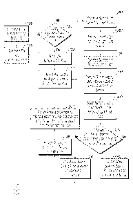
window to the
next. Transport 300 can include a sample support 310 and motor 320 linked to
translate
support 310, with motor 320 being controlled by processor 124. In this
variation the
window for the optical section could then be visible and UV-transparent
material such as
glass, quartz, silica, sapphire, calcium fluoride, barium fluoride, or the
like. Such an
embodiment would be particularly useful in portable analyzers which may not be
hand-
held.
[0043] Operation of the hand-held analyzer of Figs. 2 and 3 will now be
described
with reference to Fig. 4 although it will be appreciated that such operation
is the same as
the analyzer shown in Fig. 1 except the safety interlock switch 154 and
trigger 156 are
present in the hand-held embodiment. It will be assumed that processor 124 has
already
been suitably programmed to carry out the required tasks, in any of the
manners already
described. First, a user will grip housing 100 in one hand, then enter (300)
information on
elements or minerals to be searched using user interface 128 and screen 126.
Alternatively, this action can be omitted either because the user wishes to
search for all
elements or minerals which the analyzer is capable of identifying, or because
this
information was previously stored in memory 121 (for example, by delivery from
communication module 123). Processor 124 then generates (320) any needed
preselected
parameters for controlling any components of x-ray section 2 or optical
section 4, if such
parameters were not previously provided from memory 121. Such preselected
parameters
might include any one or more of: the duration of an x-ray beam 114 delivered
from x-ray
source 110; light pulse duration, frequency, and total elapsed time for light
beam 56
delivered from optical illuminator 50; parameters for evaluating when a Stokes
spectra are
obscured by interfering optical fluorescence; the time period over which
processor 124
18
CA 02874319 2015-02-12
will select the Raman spectral data from the Raman spectrometer so as to
separate the
Raman spectra data from an interfering optical fluorescence signal when
present.
[0044] Processor 124 then checks (350) if safety interlock switch 154 is
closed,
which would only normally happen once the user has placed optical port 104
adjacent and
against sample 200, and if the user has pressed trigger 156. Once both events
are satisfied
then processor 124 activates x-ray source 110 to cause it to illuminate the
sample 200 with
x-ray beam 112 for a preselected period of time. During this time X-ray
spectrometer 116
receives any x-ray fluorescence 114 emitted from sample 200 in response to
illuminating
x-ray beam 112, and produces (370) x-ray spectral data representative of that
x-ray
fluorescence. Processor 124 then activates optical illuminator 50 (by
controlling light
source 52) to illuminate (380) sample 200 with one or more light pulses 380.
Processor
124 then identifies as a cut-off an elapsed time after the beginning of a
light pulse at which
optical fluorescence interferes with Raman spectral data, as well as light
pulse spacing,
and sets (382) these values for a sample or sample type in a manner described
above. For
example, interfering fluorescence will cause an apparent rise in the signal
baseline which
eventually may go above any peak values from Raman spectral data. When such a
rise has
reached a value which has been predetermined to be unacceptable, the time
duration from
the beginning of the light pulse can be set as the cut-off (or an average or
mean used if
multiple pulses are used to identify a cut-off). Spacing between light pulses
can then also
be set (382) as a time which is at least equal to the cut-off time (and
preferably somewhat
greater than the cut-off time). The same, or a different sample (preferably of
the same
sample type), can then be illuminated (385) with a series of light pulses
using the set light
pulse spacing.
[0045] During the time each light pulse is ON, Raman spectrometer 120
receives
any Raman radiation 58 emitted from sample 200 in response to illuminating
pulses of
light beam 56 and produces (390) Raman spectral data representative of that
Raman
radiation (with both Stokes and anti-Stokes components). Processor 124 selects
(400)
Raman spectral data based on time following the beginning of each light pulse
so as to
separate Raman spectral data from any interfering optical fluorescence signal
if present.
In particular, in FIG. 4 processor 124 selects as Raman spectral data that
data from the
19
CA 02874319 2015-02-12
Raman spectrometer produced in response to radiation emitted from the sample
within the
cut-off after the beginning of one or more light pulses.
[0046] Processor 124 also determines (420) if the Stokes spectral data is
obscured
by interfering optical fluorescence. This determining (420) can be based on a
check for
one or more clear peaks within typical expected Stokes shifts from the
wavelength of
illuminating light beam 56 (Stokes shifts being to longer wavelengths than the
illuminating light). An intensity check can also be performed since Raman
radiation is far
weaker than optical fluorescence (so a broad high intensity band in a region
of expected
Stokes shift would indicate interfering fluorescence). The selecting (400) or
determining
(420) methods for reducing the effect of interfering optical fluorescence can
be used
together, as illustrate, or either one can be used without the other.
Alternatively, for many
samples interfering optical fluorescence will likely be sufficiently low when
a short
wavelength UV light source 52 is used (for example, about 260 nm or shorter
wavelengths) so that both the selecting (400) and determining (420) could be
eliminated,
and light source 52 need not then provide a series of light pulses.
[0047] If processor 124 determines (420) that the Stokes spectral data is
obscured
by interfering optical fluorescence it uses the anti-Stokes spectral data, and
not the Stokes
spectral data, to provide (440) an analysis of a molecule in sample 200. On
the other
hand, if processor 124 determines (420) that the Stokes spectral data is not
obscured by an
interfering fluorescence then it uses the Stokes spectral data to provide
(430) an analysis
of a molecule in sample 200. It is typically better to use Stokes spectral
data when it is
not obscured since Stokes radiation is of higher intensity than anti-Stokes
radiation. Of
course, if the Stokes spectral data (including any part of that spectral data)
is determined
(420) not to be obscured then processer 124 could use both the anti-Stokes
spectral data,
and any Stokes spectral data which is not obscured, in the foregoing molecule
analysis.
[0048] Steps 380-400 represent an adaptive time gating, method (with steps
380-
382 representing the adaptive aspect). However, in some embodiments the
adaptive
aspect of steps 380-382 could be omitted and step 385 could use a series of
light pulses of
preselected characteristics as previously described. As previously mentioned,
interfering
optical fluorescence generally occurs at a later time after the beginning of a
light pulse,
than does the Raman radiation (which occurs almost instantaneously after the
light pulse
CA 02874319 2015-02-12
begins). So processor 124 would identify as Raman spectral data that data from
Raman
spectrometer 120 which occurs in a first preselected time period following
initiation of the
light beam, and identify as interfering optical fluorescence that data from
Raman
spectrometer that occurs after the preselected time period, and select the
former as Raman
spectral data.
[0049] In any event, the analysis of a molecule may be made by comparing
the
Stokes and/or anti-Stokes spectral data with a database of spectral data in a
known
manner. Even when there are mixtures of molecules present, known techniques
can be
used to resolve the different molecule types. The database may either be held
in memory
121 or accessed at a remote location using communication module 125. An
analysis of
one or more elements in sample 200 can then be provided (480) based on the
Raman
spectral data. This analysis may simply be an identification of the presence
of one or more
elements, with or without their concentration, based on the analysis of one or
more
molecules being present in sample 200. This information on an analysis of
elements from
the Raman spectral data, can then be used together with x-ray spectral data to
provide
(500) a more accurate quantitative analysis of one or more elements for which
an analysis
could be provided (500) based on the x-ray spectral data. For example, an
initial
quantitative analysis on the presence of strontium may have been provided
(500) based
only on the x-ray spectral data. However, an analysis of the presence of
sulfur and oxygen
(as sulfate) may have been provided (480) from the Raman spectral data. In
this event,
processor 124 may then provide (500) a more accurate quantitative analysis of
strontium
using the sulfur and oxygen analysis and the x-ray spectral data in a
fundamental
parameter type calculation for strontium.
[0050] From the one or more different elements analyzed, and from the
molecules
analyzed, processor 124 may then provide (550) an analysis of one or more
minerals
present in sample 200. Again, this can be accomplished by comparing the
results with a
database of minerals available in memory 121 or accessible at a remote site
using wireless
communication module 125. For example, if strontium and sulfate have been
identified,
processor 124 can ascertain from the database that strontium sulfate is a
known mineral
and therefore likely to be present. Processor 124 can then save (600) the
results on the
mineral analysis into a database either in memory 121 or a remote location
along with the
21
CA 02874319 2015-02-12
geographical location obtained from location module 123. After multiple
samples from
different locations have been collected by the analyzer, or by one or more
different
analyzers which can exchange information either directly or indirectly (such
as through a
remote database using their communication module 125), multiple locations of
different
analyzed minerals can be extracted. This can be done by processor 124
accessing memory
121 or accessing a remote memory using communication module 125, or can be
done by a
remote processor and the result communicated to processor 124 using
communication
module 125. In any event, processor 124 can then generate and present (680) a
satellite,
topological, or map image or other geographical information obtained (640)
from a
suitable database, overlaid with the exctracted mineral analysis information
on display
126. Using the foregoing information, processor 124 or a remote processor, may
identify
(700) further sites for mineral sampling and present those site locations or
other
instructions to for collecting further mineral samples, on display 126. For
example, a
preselected grid of an area to be explored may have obvious missing locations
which
processor 124 can identify for further sample analysis.
100511 As mentioned
above, the x-ray spectrum is typically collected from larger
area, for example 1 square centimeter, and therefore represents an average
over that area
while the Raman signal comes from a much smaller area, typically about 1
square
millimeter. Therefore, in a variation of the embodiment described, optical
illuminator 50
can be constructed with suitable optics to allow light beam 56 to be scanned
across sample
200 under control of processor 124 (such as by a raster scan). In operation
the
illumination (380) with light will then be by scanning the light beam 56
(including
scanning pulses of beam 56, when used) across an area on the sample (for
example, in a
raster scan). That is, the Raman spectrometer is run in a scanning mode, with
a laser beam
of 1 square millimeter scanned across the area from which the x-ray spectrum
is collected
(for example, about 1 square centimeter). This feature allows mineral mapping
across an
area on sample 200 within the field of view of the x-ray spectrometer. A Raman
scan of
such an area could be easily accomplished during a typical x-ray exposure time
of 30 sec.
Further Examples
22
CA 02874319 2015-02-12
100521 In one example, if the elements Fe and S are both identified from
the x-ray
spectral data, the sample could be Iron Sulfide (FeS), Magnetite (Fe304) with
free S, Pyrite
(FeS2), or Pyrrhotite (Fe7S8). The Raman spectral data can be used to identify
the
molecule types present and hence the compounds of the mineral composition, in
the
manner previously described. Once the mineral composition is analyzed, for
example that
the sample is evaluated to be FeS, FeS2 or Fe304, the analyzer can properly
account in
XRF calculations for the presences of oxygen in the sample (which is not seen
by the
XRF), and obtain a more accurate quantitative result for iron and sulfur.
Similarly, if XRF
spectral data analysis identifies the presence of calcium, sulfur and iron, an
analysis of
Raman spectra data may determine whether sample contains calcium sulfate or
calcium
carbonate or both (carbon being another element not analyzed directly by XRF).
[0053] Another example is analysis of molybdenum rock or ore. Molybdenum
metal is recovered from its most abundant ore, mineral molybdenite, which is
molybdenum disulfide, MoS2. However, in such ore there are usually other
compounds
which may also contain sulfur such as pyrite (FeS2) or calcium sulfate. X-ray
analysis of
such material is complicated by the fact that molybdenum atoms when excited
produce not
only their main characteristic x-rays at 17.4 keV but also characteristic x-
rays at energy
identical to that of the sulfur x-rays, that is at 2.3 keV. Presence of x-ray
signals from iron
and calcium also implies possibility of sulfur presence. Therefore, the
intensity of x-rays
measured by the XRF section of the instrument at 2.3 keV energy is a potential
composite
of sulfur x-rays from pyrite, calcium sulfate, molybdenum disulfide and of 2.3
keV x-rays
from molybdenum itself. Using Raman spectral data it is possible to identify
which of the
compounds containing sulfur is present in the sample. Specifically, it is
possible to
determine whether the sample contains molybdenum disulfide or molybdenum oxide
(both compounds would produce x-rays at 2.3 keV, one from sulfur and
molybdenum the
other from just molybdenum). Such information when fed to XRF analytical
software
would allow for much more accurate elemental analysis of the sample. This
example is
illustrated by low energy range x-ray spectra of molybdenite pure, molybdenum
and pure
sulfur, shown FIGS. 5A and the Raman spectra of 5B. FIG. 5A shows the low
energy
range X-ray spectra of Mo ore, pure Mo and pure S. The Mo ore spectrum shows
the
presence of Calcium and Iron so that peak at 2.3 keV may represent composite
of sulfur
K-a and Mo-La lines, both at 2.3 keV. FIG. 5B shows the Raman spectra of
minerals
23
CA 02874319 2015-02-12
molybdenite, gypsum (calcium sulphite), pyrite and sulfur. As is seen all
minerals
produce Raman spectra with different peak features (non-overlaping, at
different wave
numbers) that clearly distinguish them. Based on such information the X-ray
intensity at
2.3 keV may be properly apportioned between sulfur Ka and Molybdenum La lines
and
thus improve overall accuracy of elemental analysis of the sample.
[0054] Particular embodiments of the present invention have been described
in
detail above. However, it will be apparent that variations and modifications
of the
described embodiments are possible. For example, it will be appreciated that
operations in
the methods described can be performed in the order described or in any other
order, or
simultaneously, that is logically possible. In one such variation steps 500,
520 in Fig. 4
could be performed, for example, before step 400 or at some other time before
step 530.
Accordingly, the present invention is not limited by the embodiments
described.
24