Note: Descriptions are shown in the official language in which they were submitted.
WO 2013/181107
PCT/US2013/042710
BIOACTIVE AGENT DELIVERY DEVICES AND METHODS OF MAKING
AND USING THE SAME
INTRODUCTION
Among the various conventional modes of drug administration, oral delivery of
pharmaceuticals is a preferred route as it offers several advantages. It is
less invasive,
provides higher patient compliance, rapid availability, and low cost of
manufacturing.
however, a unique set of intestinal harriers including the stomach's acidic
environment,
poor permeation of active therapeutics across the thick mucus and epithelial
interface, an
array of drug degrading intestinal enzymes, and limited retention time due to
peristalsis
and shear flow conditions limit the overall drug efficacy. There are also
instances where
a combination therapy is needed, where multiple drugs are to be delivered at
the same
time to achieve a synergistic effect. In addition, the absence of targeting
strategies for
intestinal diseases such as, irritable bowel syndrome (IBS), inflammatory
bowel disease
(IBD), and Crohn's disease results in an increased risk of side effects.
Although various oral delivery systems including enteric-coated capsules,
tablets,
particles, liposomes, bioadhesive agents, and permeation enhancers have been
developed
in an effort to improve oral bioavailability of drugs, many of these systems
suffer from
poor intestinal localization and low therapeutic efficacy due to the various
physiological
conditions inside the intestine and high shear fluid flow. As such, these
systems require
administration with increased frequency and over an extended time period,
which is not
practical for expensive and/or toxic drugs.
Microfabricated drug delivery vehicles, such as, micropoarticles have been
developed by techniques such as, emulsification, droplet extrusion, solvent
evaporation,
or nanoprecipitation. however, these microparticles tend to aggregate leading
to
polydispersity (B. Bugarski, et al., AIChE J. 40 (1994), 1026-1031; G.H.J.
Wolters, et
al., J. Appl. Biomater. 3 (1992), 281-286; W.T. Leach, et al., AAPS Pharm.
Sci. Tech. 6
1
CA 2875146 2019-07-29
CA 02875146 2014-11-27
WO 2013/181107
PCT/US2013/042710
(2005), E605-E617). The polydispersity of these microparticles can lead to non-
uniform
drug loading and release (S.K. Lai, et al., Biomaterials. 28 (2007), 2876-
2884; J.
Rejman, et al., Biochem. J. 377 (2004), 159-169; C.L. Randall, et al., Adv.
Drug Deliv.
Rev. 59 (2007), 1547-1561). Moreover, the symmetry of spherical particles can
result in
a loss of drug into the lumen caused by the omni-directional drug release at
the mucus-
particle interface (K.M. Ainslie and T.A. Desai, Lab Chip. 8 (2008), 1864-
1878).
As such, there is a need for devices for delivery of a bioactive agent(s) to a
target
tissue. The invention described herein fulfills this need, as well as other.
SUMMARY OF THE INVENTION
A method of preparing a substantially planar microdevice comprising a
plurality
of reservoirs is provided. In general, the method comprises forming a
plurality of
microdevices comprising a plurality of reservoirs from a planar layer of a
biocompatible
polymer. The method also comprises depositing one or more bioactive agents
into the
reservoirs. The microdevice is configured to attach to a target tissue and
release the
bioactive agent in close proximity to the tissue. Accordingly, the microdevice
is
configured to release the bioactive agent unidirectionally.
In certain embodiments, the method of preparing a substantially planar
microdevice comprising a plurality of reservoirs includes fabricating a planar
layer of a
biocompatible polymer on a substrate, the planar layer comprising a first
surface and a
second surface opposite to the first surface; defining a microdevice structure
in the planar
layer using successive deposition of photoresist layer, light exposure, and
etching; and
introducing a plurality of reservoirs in the microdevice structure using
successive
deposition of photoresist layer, light exposure, and partial etching, wherein
the plurality
of reservoirs are open only at a first surface of the microdevice and are
closed at the
second surface of the microdevice, thereby producing a planar microdevice
comprising a
plurality of reservoirs.
In certain cases, the method further comprises depositing a bioactive agent
into
the plurality of reservoirs. The bioactive agent may be deposited in the form
of solution
comprising the bioactive agent and a photopolymer, wherein the method further
comprises exposing the reservoirs to light, thereby polymerizing the solution.
In certain cases, the method includes depositing a first solution comprising a
first
bioactive agent into the plurality of reservoirs; polymerizing the first
solution only in the
first reservoir of the plurality of reservoirs; removing unpolymerized first
solution;
CA 02875146 2014-11-27
WO 2013/181107
PCT/US2013/042710
depositing a second solution comprising a second bioactive agent into the
plurality of
reservoirs and polymerizing the second solution only in a second reservoir of
the
plurality of reservoirs. In certain embodiments, the first solution may also
include a
prepolymer, for example, a photopolymer, and polymerizing the first solution
only in the
first reservoir may include exposing only the first reservoir to light. In
certain
embodiments, the second solution may also include a prepolymer, for example, a
photopolymer, and polymerizing the second solution only in the second
reservoir may
include exposing only the second reservoir to light.
In certain embodiments, the first solution may comprise a first prepolymer and
the second solution may comprise a second prepolymer, wherein the first
bioactive agent
is released from the first reservoir at a different rate compared to release
of the second
bioactive agent from the second reservoir.
In certain embodiments, the method includes, after the microdevice structure
has
been defined, a step of attaching an adhesion molecule to the first surface to
facilitate
attachment of the first surface of the microdevice to cells of a target
tissue.
In certain embodiments, the method includes a step of attaching an adhesion
molecule to the first surface to facilitate attachment of the first surface of
the
microdevice to cells of a target tissue, after the plurality of reservoirs in
the microdevice
structure has been introduced.
In some cases, the biocompatible polymer may be poly(DI,-lactide-co-glycolide)
(PLGA), poly(DL-lactide-co-e-caprolactone) (DLPLCL), poly(e-caprolactone)
(PCL),
collogen, gelatin, agarose, poly(methyl methacrylate),galatin/e-caprolactone,
collagen-
GAG, collagen, fibrin, PLA, PGA, PLA-PGA co-polymers, poly(anhydrides),
poly(hydroxy acids), poly(ortho esters), poly(propylfumerates),
poly(caprolactones),
poly(hydroxyvalerate), polyamides, polyamino acids, polyacetals, biodegradable
polycyanoacrylates, biodegradable polyurethanes and polysaccharides,
polypyrrole,
polyanilines, polythiophene, polystyrene, polyesters, non-biodegradable
polyurethanes,
polyureas, poly(ethylene vinyl acetate), polypropylene, polymethacrylate,
polyethylene,
polycarbonates, poly(ethylene oxide), co-polymers of the above, mixtures of
the above,
and adducts of the above, or combinations thereof.
In certain cases, the biocompatible polymer may be poly(methyl methacrylate)
or
a derivative thereof. In other embodiments, the biocompatible polymer may be
poly(e-
caprolactone) (PCL) or a derivative thereof.
3
CA 02875146 2014-11-27
WO 2013/181107
PCT/US2013/042710
In certain embodiments, fabricating the substantially planar layer includes
depositing the biocompatible polymer at an average thickness of about 5 p.m to
about
100 [mt.
In certain examples, the microdevice may have an average thickness of about 5
im to about 100 pm.
In certain embodiments, the microdevice may be disc-shaped, which may be
circular or oval in shape. In some embodiments, the microdevice has an average
diameter
of about 50 pm -1000 i.tm.
In some cases, the plurality of reservoirs may have different depths. In other
cases, the plurality of reservoirs may have the same or similar depths. In
some
embodiments, the plurality of reservoirs may have different volumes. In some
embodiments, the plurality of reservoirs may have different diameters.
In exemplary embodiments, the method may further comprise removing the
microdevice from the substrate.
In certain cases, the cell adhesion molecule is lectin, chitosan, laminin,
fibrin,
fibronectin, proteoglycans, glycoproteins, glycosaminoglycan, or a combination
thereof.
The microdevice may be useful as medical implants, including gastrointestinal
implants, dental implants, cardiovascular implants, neurological implants,
neurovascular
implants, muscular implants, and ocular implants. The present invention also
provides
methods of treating a patient in need of such an implant.
As noted above, the microdevice includes a bioactive agent(s) for elution of
the
bioactive agent from a single surface of the microdevice to the adjacent
tissue upon
placement in a subject. In some embodiments, the microdevice attaches to a
mucosal
surface and provides a localized delivery of the bioactive agent to the
mucosal surface.
In some embodiments, the bioactive agent is selected from a polypeptide,
growth
factor, a steroid, an antibody, an antibody fragment, a DNA, an RNA, and
siRNA, an
antimicrobial agent, an antibiotic, an antiretroviral drug, an anti-
inflammatory
compound, an antitumor agent, anti-angiogeneic agent, and a chemotherapeutic
agent.
A microdevice produced by the process outlined above is also described herein.
These and other objects, advantages, and features of the invention will become
apparent to those persons skilled in the art upon reading the details of the
invention as
more fully described below.
4
CA 02875146 2014-11-27
WO 2013/181107
PCT/US2013/042710
BRIEF DESCRIPTION OF THE DRAWINGS
A detailed description of various embodiments of the present disclosure is
provided herein with reference to the accompanying drawings, which are briefly
described below. The drawings are illustrative. It is emphasized that,
according to
common practice, the various features of the drawings are not to-scale. On the
contrary,
the dimensions of the various features are arbitrarily expanded or reduced for
clarity. The
drawings illustrate various embodiments of the present disclosure and may
illustrate one
or more embodiment(s) or example(s) of the present disclosure in whole or in
part.
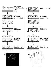
FIG 1, Panel A provides a schematic of fabrication process of a microdevice.
FIG 1, Panel B is a scanning electron microscopic (SEM) image of the
fabricated
microdevice. FIG 1, Panel C depicts the dimensions of the microdevice.
FIG 2, Panel A, shows a schematic of process for fabricating single or multi-
drug
loaded microdevices. FIG 2, Panel B, shows a fluorescent micrograph showing
the
presence of a single model drug in all three reservoirs of the same
microdevice. FIG 2,
Panel C, shows a fluorescent micrograph composite of multi-drug loaded
microdevices.
FIG 3 shows fluorescent micrograph composite confirming conjugation of model
fluorophore-lectin to the surface of poly(methyl methacrylate) (PMMA) and
showing the
loading of model drug.
FIG 4 illustrates petineation of drug loaded in microdevices or hydrogel bolus
through Caco-2 epithelial monolayer.
FIG 5 shows the permeation across Caco-2 epithelial monolayer of different
model drugs loaded in the same microdevice.
FIG 6 depicts the controlled release and permeation of different model drugs
loaded in the same microdevice but with different crosslinking ratio/amounts
of
crosslinker.
FIG 7 shows the effect of particle shape and surface functionality on the in
vivo
bioadhesion of microdevices. Flat microdevices show enhanced bioadhesion than
that of
spherical particles of same surface area. Further enhancement is provided by
the
presence of GI epithelia targeting lectin.
FIG 8 shows the cumulative release profile of model FITC-BSA from
PEGDMA-MMA hydrogel discs at different pH.
FIG 9 shows the plasma vs. time curve for Acyclovir released from microdevices
compared to Acyclovir solution at same and 5X concentrations.
5
WO 2013/181107
PCT/US2013/042710
DETAILED DESCRIPTION OF THE INVENTION
A method of preparing a substantially planar microdevice comprising a
plurality
of reservoirs is provided. In general, the method comprises forming a
plurality of
microdevices containing a plurality of reservoirs from a planar layer of a
biocompatible
polymer. The method also comprises depositing one or more bioactive agents
into the
reservoirs. The microdevice is configured to attach to a target tissue and
release the
bioactive agent into the tissue. Accordingly, the microdevice is configured to
release the
bioactive agent unidirectionally.
Before the present invention described, it is to be understood that this
invention is
not limited to particular embodiments described, as such may, of course, vary.
It is also
to be understood that the tel minology used herein is for the purpose of
describing
particular embodiments only, and is not intended to be limiting, since the
scope of the
present invention will be limited only by the appended claims.
Where a range of values is provided, it is understood that each intervening
value,
to the tenth of the unit of the lower limit unless the context clearly
dictates otherwise,
between the upper and lower limits of that range is also specifically
disclosed. Each
smaller range between any stated value or intervening value in a stated range
and any
other stated or intervening value in that stated range is encompassed within
the
invention. The upper and lower limits of these smaller ranges may
independently be
included or excluded in the range, and each range where either, neither or
both limits are
included in the smaller ranges is also encompassed within the invention,
subject to any
specifically excluded limit in the stated range. Where the stated range
includes one or
both of the limits, ranges excluding either or both of those included limits
are also
included in the invention.
Unless defined otherwise, all technical and scientific terms used herein have
the
same meaning as commonly understood by one of ordinary skill in the art to
which this
invention belongs. Although any methods and materials similar or equivalent to
those
described herein can be used in the practice or testing of the present
invention, some
potential and preferred methods and materials are now described.
6
Date Recue/Date Received 2021-02-04
CA 02875146 2014-11-27
WO 2013/181107
PCT/US2013/042710
It must be noted that as used herein and in the appended claims, the singular
forms "a", "an", and "the" include plural referents unless the context clearly
dictates
otherwise. Thus, for example, reference to "a microdevice" includes a
plurality of such
microdevices and reference to "the bioactive agent" includes reference to one
or more
bioactive agents and equivalents thereof known to those skilled in the art,
and so forth.
The publications discussed herein are provided solely for their disclosure
prior to
the filing date of the present application. Nothing herein is to he construed
as an
admission that the present invention is not entitled to antedate such
publication by virtue
of prior invention. Further, the dates of publication provided may be
different from the
actual publication dates which may need to be independently confirmed.
Microdevices and Methods for Making the Same
As noted above, the present invention provides microdevices that are generally
planar and include a plurality of reservoirs in which a bioactive agent may be
placed.
These microdevices can contain a single bioactive agent in the plurality of
reservoirs, a
mixture of two or more bioactive agents in the plurality of reservoirs, or
different
bioactive agents in separate reservoirs. In addition, the microdevices may be
configured
to release bioactive agents present in different reservoirs at different
rates. The
microdevices may further include an adhesion molecule on a first surface of
the
microdevice. The adhesion molecule may facilitate attachment of the first
surface of the
microdevice to cells of a target tissue resulting in release of the bioactive
agent from the
reservoirs towards the cells.
The substantially planar microdevice comprising a plurality of reservoirs may
be
prepared by depositing a planar layer of a biocompatible polymer on a
substrate. The
planar layer is substantially flat and includes a first surface and a second
surface opposite
to the first surface, where the second surface is in contact with the
substrate. A plurality
of microdevice structures may be defined in the planar layer using
photolithography and
etching. In general, the method may include depositing a layer of a
photoresist on the
first surface of the planar layer, exposing a defined region of the
photoresist to light, and
etching areas of the polymer layer from which the photoresist has been removed
to
remove the polymer, thereby providing a plurality of microdevice structures.
As used
herein, the phrase "microdevice structure" refers to an unfinished
microdevice, wherein
the unfinished microdevices do not yet have reservoirs defined in the
microdevice
structure.
7
CA 02875146 2014-11-27
WO 2013/181107
PCT/US2013/042710
A plurality of reservoirs may be introduced in the microdevice structures
using
photolithography and partial etching. In general, the method may include
depositing a
layer of a photoresist on a first surface of the microdevice structures. The
first surface of
the microdevice structure corresponds to the first surface of the planar
polymer layer.
Defined regions of the photoresist may then be removed by exposure to light.
The
regions of polymer from which the photoresist has been removed may then be
partially
etched to remove the polymer. As used herein, "partial etching" as contrasted
with
"etching" or "complete etching" refers to removing the polymer partially, for
example, in
embodiments where the microdevice structure is, for example, 10 pm thick,
partial
etching removes the polymer to a depth of less than 10 pm, such as, a depth of
1 m, 2
p.m, 3 Jim, 4 p.m, 5 p.m, 6 p.m, 7 j.tm, 8 p.m, or 9 p.m. In contrast,
"complete etching" or
"etching" as used herein refers to removing the polymer completely or
substantially
completely, for example, in embodiments where the planar layer of
biocompatible
material is, for example, 10 p.m thick, "etching" or "complete etching"
removes the
polymer to a depth of about 10 p.m, such as, a depth of 9.999 j.tm, 9.5 pm,
9.2 p.m. In
general, "etching" or "complete etching" removes the polymer to an extent such
that the
individual microdevices fabricated on a substrate are no longer connected to
each other
as a result of the polymer present in between the microdevices not being
completely
removed. As such, "etching" or "complete etching" provides for microdevices
that when
removed from the substrate are released as individual microdevices instead of
being
connected by residual polymer layer.
The plurality of microdevices with the plurality of reservoirs may then be
loaded
with bioactive agent(s). In general, the depositing of bioactive agent(s) in
the
microdevices is carried out while the microdevices are attached to the
substrate. In
general, the bioactive agent is loaded into the reservoirs in conjunction with
a
prepolymer. As used herein, the phrase "in conjunction with" in the context of
a
prepolymer refers to filling of the bioactive agent mixed with a prepolymer
into the
reservoirs, or loading the bioactive agent into reservoirs which already
contain a
prepolymer, or filling the bioactive agent into reservoirs followed by filling
the
reservoirs with a prepolymer. In certain instances, the bioactive agent may be
in a
solution containing a prepolymer and the solution may then be deposited into
the
reservoirs.
Following deposition of the bioactive agent into the reservoirs in conjunction
with a prepolymer, the prepolymer may be polymerized in one or more of the
reservoirs.
8
CA 02875146 2014-11-27
WO 2013/181107
PCT/US2013/042710
In certain embodiments, one or more bioactive agents may be deposited in the
reservoirs.
In other embodiments, a first bioactive agent may be deposited into a first
reservoir or a
plurality of first reservoirs and second bioactive agent may be deposited into
a second
reservoir or a plurality of second reservoirs of the microdevices. In other
embodiments, a
first bioactive agent may be deposited into a first reservoir, a second
bioactive agent may
be deposited into a second reservoir, a third bioactive agent may be deposited
into a third
reservoir, and so on.
As noted above, the bioactive agent(s) may be deposited into the reservoirs in
conjunction with a prepolymer. The preolymer may be polymerized by a variety
of
techniques, such as, exposure to light, heating, drying, and the like.
In certain embodiments, a first solution comprising a first bioactive agent
and a
first prepolymer may be deposited into a first reservoir of the plurality of
microdevices.
In certain embodiments, the depositing of the first solution comprises
depositing the first
solution onto the first surface of the microdevice resulting in filling of the
plurality of
reservoirs with the first solution. The first solution may then be polymerized
only in the
first reservoir in the plurality of microdevices. Any unpolymerized first
solution
deposited on the microdevice and/or in the reservoirs may then be removed. The
method
may further include depositing a second solution comprising a second bioactive
agent
and a second prepolymer into a second reservoir of the plurality of
microdevices. In
certain instances, the depositing of the second solution comprises depositing
the second
solution onto the first surface of the microdevice resulting in filling of any
empty
reservoirs with the second solution. The second solution may then be
polymerized only
in the second reservoir in the plurality of microdevices. The process may be
repeated to
deposit a third bioactive agent, a fourth bioactive agent, and so forth.
In certain embodiments, the first, second, third, fourth bioactive agents may
be
different bioactive agents, where the different bioactive agents are released
simultaneously or sequentially. In certain embodiments, the same prepolymer
may be
used for loading the different bioactive agents. In other cases, different
prepolymers may
be used for loading the different bioactive agents. Accordingly, the first,
second, third
prepolymer may be the same prepolymer or different prepolymers.
A variety of prepolymers known in the art may be used. In certain embodiments,
the prepolymer may be mixed with a photoinitiator, wherein exposure of the
photoinitiator to light results in polymerization of the prepolymer. Useful
photoinitiators
can be those known in the art, such as, those disclosed in US 5,410,016. For
example, the
photoinitiator may be acetophenone derivatives, e.g., dimethyl acetophenone
(DMPA),
9
CA 02875146 2014-11-27
WO 2013/181107
PCT/US2013/042710
2-methoxy-2-phenylacetophenone, 2,2-dimethoxy-2-phenyl acetophenone; ethyl
eosin;
camphorquinone. Initiation of polymerization may be accomplished by
irradiation with
light at a wavelength of between about 200-700 nm, for example, 100 nm-440 nm.
In other embodiments, thermal polymerization initiator systems may also be
used
to selectively polymerize a bioactive agent containing solution in a
particular reservoir.
Such systems include, for example, potassium persulfate, with or without
tetraamethyl
ethylenedi amine; benzoylperoxide, with or without triethanolamine; and
ammonium
persulfate with sodium bisulfite.
In certain cases, one or more adhesion molecules may be deposited on the first
surface of the microdevice structure before the defining of reservoirs in the
microdevice
structures. In other cases, one or more adhesion molecules may be deposited on
the first
surface of the microdevice after the defining of reservoirs in the microdevice
structures.
In certain cases, one or more adhesion molecules may be deposited on the first
surface of
the microdevice after depositing a bioactive agent in the reservoir(s) of the
microdevices.
The finished microdevice may be removed from the substrate using standard
procedures to provide a plurality of individual microdevices. In general, the
microdevices released from the substrate are released as individual
microdevices such
that the microdevices are not interconnected by any residual polymer layer. In
certain
cases, removing the microdevice from the substrate results in release of
microdevices
wherein more than 50% of the microdevices are released as single microdevices,
for
example, more than 60%, 70%, 80%, 90%, or more of the microdevices are
released as
single microdevices.
Any substrate suitable for carrying out the subsequent steps of the method may
be
used for depositing a layer of biocompatible polymer. In certain examples, the
substrate
is a silicon wafer, a glass chip, a plastic chip, or another suitable
material. The substrate
may be of any size, shape, and dimension. The size of the substrate may be
selected
based on, for example, the number of microdevices to be manufactured. In
certain cases,
the substrate is a silicon wafer. In certain cases, the silicon wafer is a 1-
inch silicon
wafer, or a 2-inch silicon wafer, or a 3-inch silicon wafer, or a 4-inch
silicon wafer, or a
6-inch silicon wafer, or a 12-inch silicon wafer.
The planar layer of a biocompatible polymer may be deposited on the substrate
using a variety of deposition techniques. In certain cases, the biocompatible
polymer
may be deposited by coating the polymer in form of a solution onto the
substrate.
Coating may be carried out by dipping the substrate in the polymer solution,
by pipetting
the polymer solution onto the substrate, or by spin coating, for example. The
solvent in
CA 02875146 2014-11-27
WO 2013/181107
PCT/US2013/042710
the polymer solution may be subsequently dried to obtain the planar layer of
the
biocompatible polymer. Drying may include air drying, forced air drying,
heating, such
as, baking, a combination thereof, and the like.
The planar layer biocompatible polymer is substantially uniform in thickness
and
the average thickness may range from 5 p.m to about 100 p.m. For example, the
planar
layer may have an average thickness of about 5 p.m, 8 p.m, 101.tm, 12 p.m, 15
j.tm,
30 p.m, 40 p.m, 50 p.m, 60 p.m, 70 pm, 80 m, 90 p.m, or 100 p.m.
The biocompatible polymer may be poly(DL-lactide-co-glycolide) (PLGA),
poly(DL-lactide-co-e-caprolactone) (DLPLCL), poly(e-caprolactone) (PCL),
collogen,
1() gelatin, agarose, poly(methyl methacrylate),galatink-caprolactone,
collagen-GAG,
collagen, fibrin, PEA, PGA, PT A-PGA co-polymers, poly(anhydrides),
poly(hydroxy
acids), poly(ortho esters), poly(propylfumerates), poly(caprolactones),
poly(hydroxyvalerate), polyamides, polyamino acids, polyacetals, biodegradable
polycyanoacrylates, biodegradable polyurethanes and polysaccharides,
polypyrrole,
polyanilines, polythiophene, polystyrene, polyesters, non-biodegradable
polyurethanes,
polyureas, poly(ethylene vinyl acetate), polypropylene, polymethacrylate,
polyethylene,
polycarbonates, poly(ethylene oxide), co-polymers of the above, mixtures of
the above,
and adducts of the above, or combinations thereof.
In certain cases, the biocompatible polymer may be poly(methyl methacrylate)
or
a derivative thereof. In other embodiments, the biocompatible polymer may be
poly(e-
caprolactone) (PCL) or a derivative thereof.
Either a positive or a negative photoresist may be used to define the
dimensions
and shape of the microdevice structures. The photoresist may be deposited by
dipping
the substrate with the polymer layer in a solution containing the photoresist,
by pipetting
the photoresist solution onto the substrate, or by spin coating, for example.
In certain
cases, a positive photoresist may be used. A mask that defines the shape and
surface area
of the microdevice structures may be positioned over the photoresist. In
certain
embodiments, the mask may allow light to pass through a ring shaped region in
the
mask, thereby exposing a ring shaped region of the positive photoresist to
light and
making the photoresist in the ring shaped region soluble to the photoresist
developer.
Accordingly, upon development of the photoresist, ring shaped region of the
photoresist
is removed.
In other embodiments, the photoresist may be a negative photoresist. In these
embodiments, the mask may be designed to allow light to pass through a
circular region
11
CA 02875146 2014-11-27
WO 2013/181107
PCT/US2013/042710
in the mask, thereby exposing a circular region of the negative photoresist to
light and
making the photoresist in the ring shaped region surrounding the circular
region soluble
to the photoresist developer. Accordingly, upon development of the
photoresist, a ring
shaped region of the photoresist is removed.
A variety of positive and negative photoresists may be used in the methods
disclosed herein. As used herein, the phrase "positive photoresist" refers to
a type of
photoresist in which the portion of the photoresist that is exposed to light
becomes
soluble to the photoresist developer. While, the portion of the photoresist
that is
unexposed remains insoluble to the photoresist developer. As used herein, the
phrase
"negative photoresist- refers to a type of photoresist in which the portion of
the
photoresist that is exposed to light becomes insoluble to the photoresist
developer.
While, the unexposed portion of the photoresist is dissolved by the
photoresist developer.
For example, the photoresist may be Hoechst AZ 4620, Hoechst AZ 4562, AZ 1500,
e.g.,
AZ 1514 H, Shipley 1400-17, Shipley 1400-27, Shipley 1400-37, etc.
Other shapes of the microdevice structures, such as triangular, oval, diamond,
etc., may also be defined by using an appropriately designed mask. The surface
area of
the microdevice may be determined by the surface area of the area in the
photomask
through which the light passes. As such, the surface area of the microdevice
may be in
the range of 1,900 gm2-790,000 m2, such as 3,000 pm2-500,000 m2, or about
10,000
pm2-1 00,000 gm2, or about 15,000 gm2-50,000 gm2, or about 20,000 gm2-40,000
gm2,
e.g., 18,000 gm2-35,000 pm2, for example, about 15,000 gm2, 17,000 gm2, 19,000
pm2,
20,000 pm2, or about 23,000 pm2. In certain cases, the microdevice may be
circular in
shape and have an average diameter in the range of about 50 gm -1000 gm, for
example,
70 gm -500 gm, 80 gm -300 gm, 90 pm -250 gm, 100 gm -200 pm, e.g., 50 gm, 60
pm,
70 gm, 80 gm, 90 pm, 100 gm, 130 gm, 150 gm, 180 gm, 200 pm, 250 gm, 300 gm,
400
gm, or 500 gm.
The photomask may be generated by standard procedure based on the desired
pattern of the microdevices to be manufactured. As described above, the image
for the
photomask defines the shape and dimension of the microdevices.
Light may be used to expose a defined region of the photoresist layer via the
mask. In certain cases, light may be a short wavelength light (for example, a
wavelength
of about 100 nm-440 nm), such as, ultra violet (UV) light, deep UV light, H
and I lines
of a mercury-vapor lamp. The step of exposing the photoresist to light may he
followed
with a step of photoresist development where the photoresist is contacted with
a
photoresist developer. In embodiments, where a positive photoresist is used,
the regions
12
WO 2013/181107
PCT/US2013/042710
of the positive photoresist layer exposed to light are washed away in the
photoresist
developer. In embodiments, where a negative photoresist is used, the regions
of the
negative photoresist layer not exposed to light are washed away in the
photoresist
developer.
Any standard photoresist developer compatible with the photoresist deposited
may be used in the methods described herein. As such, a positive developer may
be used
to remove any positive photoresist exposed to light. In certain cases, a
negative
developer may be used to remove any negative photoresist not exposed to light.
The regions of the polymer layer from which the photoresist has been removed
are then etched to remove the biocompatible polymer layer. The portion or
portions of
the biocompatible polymer layer that are covered by the photoresist form the
microdevice. A dry or wet etching process as is standard in the art may be
used to
remove the exposed biocompatible polymer layer. In certain cases, the etching
process is
reactive ion etching. Standard procedures and apparatus for etching may be
used. For
example, reactive ion etching methods and apparatus are described in US
6,669,807, US
5,567,271. The etching is carried out for a
length of time sufficient to remove all of the polymer material not covered
with the
photoresist such that the plurality of microdevice structures are not
connected together
via any residual polymer material.
Following the etching step, the photoresist may be removed using any standard
photoresist remover or photoresist stripper compatible with the photoresist
used.
Exemplary photoresist removers include 1-methyl-2-pyrrolidon, dimethyl
sulfoxide,
AZ 100 Remover, and the like.
The plurality of microdevice structures generated by the foregoing method may
be 2, 5, 10, 20, 50, 100, 500, 1000, 1500, 2000, 2500, 3000, 3500, 4000, 4500,
5000,
5500, 6000, 6500, or more, for example, 1000-10,000 microdevices may be
generated,
such as 2000-8000, or about 3000-7000.
Defining a plurality of reservoirs in the microdevice structures includes
depositing a layer of photoresist onto the first surface of the microdevice
structure.
Depositing of the photoresist may be carried out in the same manner as
described above.
The photoresist may be the same photoresist used for fabricating the
microdevice
structures or a different photoresist. A mask may be positioned over the
photoresist layer.
The pattern in the mask defined the regions through which light may pass
through to the
photoresist layer. The pattern in the mask may be any desired pattern
depending upon the
number, shape and dimensions of the reservoirs to be defined in a microdevice
structure.
13
Date Recue/Date Received 2021-02-04
CA 02875146 2014-11-27
WO 2013/181107
PCT/US2013/042710
In certain cases, the plurality of reservoirs present per microdevice include
2 or
more, 3 or more, 4 or more, 5 or more, 6 or more, 7 or more, 8 or more, 9 or
more, 10 or
more, or more. In certain cases, 1-10 reservoirs, or 2-8 reservoirs, or 3-7
reservoirs may
be defined in a microdevice structure.
The reservoirs may have any shape, such as, cylindrical, conical,
frustoconical,
cubical, cuboidal, etc. The volume of the reservoirs may be determined by
dimension of
the mask region allowing light to pass through to expose the photoresist
layer. In
addition, the volume of the reservoirs may be determined by the depth to which
the
polymer layer is removed. The reservoirs may be have a circular shaped
opening, the
average diameter of the opening may be about 10 m, 20 pm, 30 Jim, 40 pm, 50
m,
60 pin, 80 lam, 100 m, 120 m, 150 pm, 200 pm, 250 pm, or 300 pm. For
example, the
average diameter of reservoirs with a circular opening may be in the range of
30 pm -
100 pm, or about 40 m -80 j.tm.
Following positioning of a photomask over the microdevice structure, the
photoresist may be exposed to light, followed by removal of the exposed
photoresist. The
regions of the microdevice structures from which the photoresist has been
removed may
be partially etched to remove a portion of the biocompatible polymer layer.
The duration
and the intensity of the etching step may be varied to define reservoirs of
different
depths. For example, the etching process may be carried out for a shorter
duration or
with a low ion flow rate to define shallow reservoirs while the etching
process may be
carried out for a longer duration or with a high ion flow rate to define deep
reservoirs. In
addition, the thickness of the planar layer of biocompatible polymer affects
the depth of
the reservoir. In general, the average depth of the reservoirs may range from
1 pm-
80 pin, 1.5 pm -70 pm, 2 pm -50 m, 2 pm -30 m, 3 pm -30 pm, 3.5 pm -20 pm,
such
as about 1 pm, 2 pm, 3 p,m, 4 m, 5 p,m, 6 p,m, 7 pm, 8 pm, 9 m, or 10 p,m.
The
volume of the reservoirs may be range from about lx l 0-3 nI, to about I pt,
such as
about, 5x 10⁻³ nL to about 0.1 L, or about lx 10-2 nL to about 50 nL, or
about lx 10-2
ni, to about 5x 10⁻¹ nT., for example. Accordingly, the microdevices may
include a
plurality of reservoirs where the reservoirs are open on the first surface due
to the
removal of the polymer layer by etching and are closed on the second surface
due to the
presence of the polymer layer. As such, a bioactive agent deposited into the
reservoirs
may exit through the opening on the first surface of the microdevice.
In certain cases, an adhesion molecule may be attached to the first surface of
the
microdevice to facilitate the attachment of the first surface of the
microdevice to the cells
14
CA 02875146 2014-11-27
WO 2013/181107
PCT/US2013/042710
of a target tissue. Accordingly, the microdevices include reservoirs
comprising a single
opening, wherein the opening is located on the first surface of the
microdevices, which
first surface may be the cell contacting surface. Exemplary adhesion
molecules, that
facilitate adhesion of the microdevice to the cells of a target tissue where
the bioactive
agents loaded into the microdevice need to be delivered, include lectin (e.g.,
wheat germ
agglutinin), polycations (e.g., chitosan, polylysine, and the like), laminin,
fibrin,
fibronectin, integrin, vitronectin, hyaluronic acid, elastin, vitronectin,
proteoglycans,
glycoproteins, glycosaminoglycans, collagen, gelatin, and the like. The
adhesion
molecule may be attached covalently or non-covalently to the first surface of
the
microdevice. The method may include attaching an adhesion molecule to the
first surface
of the microdevice after defining the microdevice structure and before
defining the
reservoirs. In certain cases, the method may include attaching an adhesion
molecule to
the first surface of the microdevice after introducing the plurality of
reservoirs in the
microdevice structure. The cell adhesion molecule may be attached covalently
to the first
surface of the microdevice using a standard chemistry, which does not affect
the integrity
or stability of the polymer layer.
In certain embodiments, the target tissue may be a mucosal tissue of a
patient.
For example, the target tissue may be gastrointestinal tissue, for example,
esophagus,
stomach, small intestine, large intestine. In other embodiments, the target
tissue may be
mucosa] tissue in mouth, such as, epithelial cell lining of the mouth. The
microdevices
described herein may be administered to a patient in need thereof by a number
of routes
of administration, including but not limited to, oral, sublingual, ocular,
intra-vaginal,
intra-rectal.
As described above, the bioactive agent(s) may be deposited into the
reservoirs in
conjunction with a prepolymer. The preolymer may be polymerized by a variety
of
techniques, such as. exposure to light, heating, drying, and the like. In
certain
embodiments, the prepolymer polymerizes upon exposure to light, such as, UV
light. In
these embodiments, a solution of a bioactive agent and a prepolymer deposited
into a
plurality of reservoirs of a microdevice(s) may be polymerized by exposure to
light. In
certain embodiments, only the reservoirs are exposed to light by using an
appropriately
patterned mask. In certain cases, a mask similar to the mask used for creating
the
reservoirs may be used for exposing the reservoirs to light.
In certain cases, a first solution containing a first bioactive agent and a
polymer
present in a first reservoir may be polymerized by exposing only the first
reservoir to
light using a mask patterned to allow light to pass through to only the first
reservoir. Any
CA 02875146 2014-11-27
WO 2013/181107
PCT/US2013/042710
unpolymerized first solution may be removed. The method may further comprise,
depositing a second bioactive agent into the microdevice. The depositing step
may result
in filling of any empty reservoirs with the second bioactive agent. As noted
above, the
second bioactive agent may be deposited in fouli of a second solution
comprising the
second bioactive agent and the same polymer used with the first bioactive
agent or a
different prepolymer. The method further comprises exposing only the second
reservoir
to light thereby polymerizing the second bioactive agent in the second
reservoir. Any
unpolymerized second solution may then be removed. The steps of depositing a
solution
containing a bioactive agent and a prepolymer, polymerizing the solution in a
particular
to reservoir by using a mask and light, and removing unpolymerized solution
may be
repeated to fill different reservoirs with different bioactive agents.
In further embodiments, the release kinetics of the bioactive agents that is
eluted
from the microdevice may be modulated by using an appropriate prepolymer or a
combination of prepolymers and cross-linkers, modulating the concentration of
the
prepolymers and/or cross-linkers. As used herein, the term "prepolymer" refers
to a
polymer that is not yet polymerized into a semi-solid or solid state. A
synthetic or natural
polymer can be used as a polymer and may be combined with the bioactive agent
prior to
or at the same time microdevices are loaded with the bioactive agent. Suitable
synthetic
and natural polymers include, but are not limited to, biodegradable or
bioerodible
polymers, such as poly(DL-lactide-co-glycolide) (PI,GA), poly(DL-lactide-co-r.-
caprolactone) (DLPLCL), or poly(e-caprolactone) (PCL), collagen, gelatin,
agarose, and
other natural biodegradable materials. In certain embodiments, the
concentration of the
polymer may be decreased or increased to achieve a higher or lower release
kinetic for a
bioactive agent. In certain cases, the release kinetics may be modulated by
controlling
the ratio of a cross linker to a monomer that react to font' a polymerized
gel. For
example, poly(ethylene glycol)dimethacrylate (PECTDMA) may be used to cross-
link a
monomer such as monomethyl methacyrlate. The ratio of the crosslinker to
monomer
may be decreased resulting in a less dense polymer through which the bioactive
agent is
released at a higher rate upon swelling of the polymer. Increasing the ratio
of the
crosslinker to monomer may result in a dense polymer through which the
bioactive agent
is released at a slower rate upon swelling of the polymer. A similar effect
can also be
obtained with the use of different molecular weight (length of the chain)
monomers or
crosslinkers. In certain embodiments, a first bioactive agent may be
polymerized with a
first polymer and a second bioactive agent may be polymerized with a second
polymer,
where the first and second polymers release the bioactive agents at different
rates.
16
CA 02875146 2014-11-27
WO 2013/181107
PCT/US2013/042710
In general, the microdevice will elute the bioactive agent to the surrounding
tissue upon placement of the microdevice in a patient for a period ranging
from about 2
minutes to about 3 months or more, including 5 minutes to about 14 weeks, such
as 10
minutes, 30 minutes, 60 minutes, 100 minutes, 130 minutes, 200 minutes, about
6 hours,
about 12 hours, about 24 hours, 72 hours, about 3 days, about 7 days, about 2
weeks,
about 3 weeks, about 4 weeks, about 6 weeks, about 8 weeks, about 12 weeks, or
more.
As noted above, a first bioactive agent may he released from the first
reservoir and a
second bioactive agent may be released from a second reservoir over a similar
period of
time or over different periods of time.
In general, the subject method produces microdevices that are substantially
planar, and provide for release of the bioactive agent(s) deposited in the
reservoirs of the
microdevice from the first surface of the microdevice. As such, the release of
the
bioactive agents is substantially in a single direction in contrast to
bioactive agents
release from a capsule, tablet, or microsphere. In addition, in certain
embodiments, the
microdevice includes a cell adhesion molecule that mediates attachment of the
first
surface of the microdevice to the surface of a target tissue, such as, to
epithelial cells of a
mucosal lining of the gastrointestinal tract. The combination of attachment of
the first
surface of the microdevice to the target tissue and release of the bioactive
agent from the
first surface of the microdevice provides a localized release of the bioactive
agent in
close proximity to the target tissue, thereby providing a higher effective
concentration of
bioactive agent available for uptake by the cells. As such, the microdevice
lowers the
amount of bioactive agent that may be required to treat a condition. In
addition, the
attachment of the microdevice to the target tissue may increase the residence
time of the
microdevice near the target tissue. For example, attachment of the microdevice
to the
epithelial lining of the gastrointestinal tract increases its residence time
in the
gastrointestinal tract as the attached microdevice may be better able to
resistant
peristaltic motion of the gastrointestinal tract. Moreover, the microdevice
may be sized
to increase the surface area available to attach to the cells of the target
tissue while
simultaneously being resistant to the shear stress that may be present in the
target tissue.
In general, the openings of the reservoirs of the microdevice structures are
located on the first surface of the microdevice facilitating simultaneous
release of the
bioactive agents present in the reservoirs. This feature of the microdevices
may be
especially useful for simultaneous release of different bioactive agents.
In certain embodiments, the planar geometry of the microdevice leads to an
improvement in the delivery of a bioactive agent, included in a reservoir of
the
17
CA 02875146 2014-11-27
WO 2013/181107
PCT/US2013/042710
microdevice, to the target tissue. In certain embodiments, the size of the
microdevice
leads to an improvement in the delivery of a bioactive agent, included in a
reservoir of
the microdevice, to the target tissue. In certain embodiments, the planar
geometry and the
size of the microdevice leads to an improvement in the delivery of a bioactive
agent,
included in a reservoir of the microdevice, to the target tissue. Without
being bound to a
particular theory, it is hypothesized that the microdevice described herein is
capable of
binding to the target tissue, for example, epithelial cell lining of
intestinal wall, and
mechanically restructure cell to cell adhesion of the cells. This
restructuring of cell to
cell adhesion by, for example, modulation of the tight junctions between the
epithelial
to cells of intestinal wall, may result in increased peimeability of the
epithelial cell lining
and thus may result increased delivery of the bioactive agent to the target
tissue.
The bioactive agents may be in a purified form, partially purified form,
recombinant fomi, or any other form appropriate for inclusion in the
microdevices. In
general, the bioactive agents are free of impurities and contaminants.
Exemplary
bioactive agents that may be incorporated in the microdevices are sugars,
carbohydrates,
peptides, nucleic acids, aptamers, small molecules, large molecules, vitamins;
inorganic
molecules, organic molecules, proteins, co-factors for protein synthesis,
antibody
therapies, such as Herceptin , RituxanO, MyllotargO, and ErbituxO; hormones,
enzymes such as collagenase, peptidases, and oxidases; antitumor agents and
chemotherapeutics such as cis-platinum, ifosfamide, methotrexate, and
doxorubicin
hydrochloride; immuno-suppressants; permeation enhancers such as fatty acid
esters
including laureate, myristate, and stearate monoesters of polyethylene glycol;
bisphosphonates such as alendronate, clodronate, etidronate, ibandronate, (3-
amino-l-
hydroxypropylidene)-1,1-bisphosphonate (APD), dichloromethylene
bisphosphonate,
aminobisphosphonatezolendronate, and pamidronate; pain killers and anti-
inflammatories such as non-steroidal anti-inflammatory drugs (NSA1D) like
ketorolac
tromethamine, lidocaine hydrochloride, bipivacaine hydrochloride, and
ibuprofen;
antibiotics and antiretroviral drugs such as tetracycline, vancomycin,
cephalosporin,
erythromycin, bacitracin, neomycin, penicillin, polymycin B, biomycin,
chloromycetin,
streptomycin, cefazolin, ampicillin, azactam, tobramycin, clindamycin,
gentamicin, and
aminoglycocides such as tobramycin and gentamicin; and salts such as strontium
salt,
fluoride salt, magnesium salt, and sodium salt.
Examples of antimicrobial agents include, but are not limited to, tobramycin,
amoxicillin, amoxicillin/clavulanate, amphotericin B, ampicillin,
ampicillin/sulbactam,
atovaquone, azithromycin, cefazolin, cefepime, cefotaxime, cefotetan,
cefpodoxime,
18
CA 02875146 2014-11-27
WO 2013/181107
PCT/US2013/042710
ceftazidime, ceftizoxime, ceftriaxone, cefuroxime, cefuroxime axetil,
cephalexin,
chloramphenicol, clotrimazole, ciprofloxacin, clarithromycin, clindamycin,
dapsone,
dicloxacillin, doxycycline, erythromycin, fluconazole, foscarnet, ganciclovir,
atifloxacin,
imipenem/cilastatin, isoniazid, itraconazole, ketoconazole, metronidazole,
nafcillin,
nafcillin, nystatin, penicillin, penicillin G, pentamidine,
piperacillin/tazobactam,
rifampin, quinupristin-dalfopristin, ticarcillin/clavulanate,
trimethoprim/sulfamethoxazole, valacyclovir, vancomycin, mafenide, silver
sulfadiazine,
mupirocin, nystatin, triamcinolone/nystatin, clotrimazole/betamethasone,
clotrimazole,
ketoconazole, butoconazole, miconazole, and tioconazole.
Antiangiogenic agents include, but are not limited to, interferon-a, COX-2
inhibitors, integrin antagonists, angiostatin, endostatin, thrombospondin-1,
vitaxin,
celecoxib, rofecoxib, JTE-522, EMD-121974, and D-2163, FGFR kinase inhibitors,
EGFR kinase inhibitors, VEGFR kinase inhibitors, matrix metalloproteinase
inhibitors,
mattniastat, prinomastat, BMS275291, BAY12-9566, neovastat, rhuMAb VEGF,
SU5416, SU6668, ZD6474, CP-547, CP-632, ZD4190, thalidomide and thalidomide
analoges, sqalamine, celecoxib, ZD6126, TNP-470, and other angiogenesis
inhibitor
drugs.
In some embodiments, the bioactive agent is a small molecule, such as but not
limited to an anti-inflammatory drug, an immunosuppressant drug, a vitamin,
micronutrient or antioxidant, an antibacterial drug (e.g., vancomycin or
cephazolin), an
anti-viral drug (e.g., gancyclovir, acyclovir or foscarnet), an anti-fungal
drug (e.g.,
amphotericin B, fluconazole or voriconazole) or an anti-cancer drug (e.g.,
cyclophosphamide or melphalan). In certain embodiments, the small molecule is
a
vitamin, micronutrient or antioxidant, such as but not limited to, vitamin A,
vitamin C,
vitamin E, zinc, copper, lutein or zeaxanthin. In certain embodiments, the
small molecule
is an immunosuppressant drug, such as but not limited to, cyclosporine,
methotrexate or
azathioprine. In certain embodiments, the small molecule is an anti-
inflammatory drug,
such as but not limited to, a corticosteroid (e.g., triamcinolone acetonide or
dexamethasone) or a non-steroidal drug (e.g., ketorolac or diclofenac).
In certain embodiments, the large molecule drug is an immunosuppressant drug,
such as but not limited to, etanercept, infliximab or daclizumab. In certain
embodiments,
the large molecule drug is a neuromuscular blocker drug, such as but not
limited to,
botulinum toxin A. In certain embodiments, the large molecule drug is a
complement
inhibitor, such as but not limited to, an anti-C3 compound.
19
CA 02875146 2014-11-27
WO 2013/181107
PCT/US2013/042710
In certain embodiments, the bioactive agent may be Mesalazine, also known as
Mesalamine, or 5-aminosalicylic acid (5-ASA), prednisone, TNF inhibitor,
azathioprine
(Imuran), methotrexate, or 6-mercaptopurine, aminosalicylate anti-inflammatory
drugs,
corticosteroids, azathioprine, mercaptopurine, methotrexate, infliximab,
adalimumab,
certolizumab, natalizumab, and hydrocortisone, statins, e.g., atorvastatin,
such as
atorvastatin calcium, anti-psychotic drugs, e.g., olanzapine.
In certain cases, the bioactive agent may be combined with a pharmaceutically
acceptable additive before or after placement of the bioactive agent on a
layer of the
subject device. The term "pharmaceutically acceptable additive" refers to
preservatives,
antioxidants, emulsifiers, dyes and excipients known or used in the field of
drug
formulation and that do not unduly interfere with the effectiveness of the
biological
activity of the active agent, and that is sufficiently non-toxic to the
patient. For example,
the bioactive agent may be formulated with inert fillers, anti-irritants,
gelling agents,
stabilizers, surfactant, emollients, coloring agents, preservatives, or
buffering agents, as
are known in the art. The term "excipients" is conventionally known to mean
carriers,
diluents and/or vehicles used in foimulating drug compositions effective for
the desired
use.
The microdevice may be configured to deliver any therapeutic of choice. For
example, the microdevice may be configured to deliver therapeutics that are
delivered
orally, such as, in the fauna of pills, tablets, capsules, solutions,
emulsions, and the like.
The microdevice may be suitable for treatment for a variety of conditions. For
example,
the microdevice may be administered to patients diagnosed with inflammatory
bowel
disorder, irritable bowel syndrome, Crohn's disease, cancer, such as,
intestinal cancer,
Ulcerative colitis, etc.
The methods and devices disclosed herein can be used for both human clinical
medicine and veterinary applications. Thus, the subject or patient to whom the
device is
administered can be a human or, in the case of veterinary applications, can be
a
laboratory, agricultural, domestic, or wild animal. The subject devices and
methods can
he applied to animals including, hut not limited to, humans, laboratory
animals such as
monkeys and chimpanzees, domestic animals such as dogs and cats, agricultural
animals
such as cows, horses, pigs, sheep, goats, and wild animals in captivity such
as bears,
pandas, lions, tigers, leopards, elephants, zebras, giraffes, gorillas,
dolphins, and whales.
The dosage of the microdevices required to treat a condition may be determined
empirically or experimentally by a trained physician, and may depend on a
number of
factors, such as, route of administration, severity of the condition, amount
of bioactive
CA 02875146 2014-11-27
WO 2013/181107
PCT/US2013/042710
agent loaded per microdevice, etc. Utilizing ordinary skill, the competent
clinician will
be able to optimize the dosage of a particular therapeutic in the course of
routine clinical
trials.
Microdevices
As noted above, a substantially planar microdevice comprising a plurality of
reservoirs, wherein the planar device is provided. The substantially planar
microdevice
comprising a plurality of reservoirs is prepared by a method comprising
fabricating a
planar layer of a biocompatible polymer on a substrate; defining a microdevice
structure
in the planar layer using successive deposition of photoresist layer, light
exposure, and
etching; an introducing a plurality of reservoirs in the microdevice structure
using
successive deposition of photoresist layer, light exposure, and partial
etching, thereby
producing a planar microdevice comprising a plurality of reservoirs, wherein
the
plurality of reservoirs are open at a first surface of the microdevice and are
closed at the
second surface of the microdevice.
In certain embodiments, the plurality of reservoirs comprise a bioactive
agent, the
method further comprising depositing a solution comprising the bioactive agent
and a
prepolymer into the plurality of reservoirs and polymerizing the solution.
In some cases, a first reservoir of the plurality of reservoirs comprises a
first
bioactive agent and second reservoir of the plurality of reservoirs comprises
a second
bioactive agent, the method further comprising depositing a first solution
comprising the
first bioactive agent into the plurality of reservoirs; polymerizing the first
solution only
in the first reservoir; removing unpolymerized first solution; depositing a
second solution
comprising the second bioactive agent into the plurality of reservoirs;
polymerizing the
second solution only in the second reservoir.
In certain cases, the microdevice comprises an adhesion molecule attached to
the
first surface to facilitate adhesion of the first surface of the microdevice
to cells of a
target tissue.
The biocompatible polymer may be poly(DL-lactide-co-glycolide) (PLGA),
poly(DL-lactide-co-c-caprolactone) (DLPLCL), poly(c-caprolactone) (PCL),
collogen,
gelatin, agarose, poly(methyl methacrylate),galatink-caprolactone, collagen-
GAG,
collagen, fibrin, PLA, PGA. PLA-PGA co-polymers, poly(anhydrides),
poly(hydroxy
acids), poly(ortho esters), poly(propylfumerates), poly(caprolactones),
poly(hydroxyvalerate), polyamides, polyamino acids, polyacetals, biodegradable
21
WO 2013/181107
PCT/US2013/042710
polycyanoacrylates, biodegradable polyurethanes and polysaccharides,
polypyrrole,
polyanilines, polythiophene, polystyrene, polyesters, non-biodegradable
polyurethanes,
polyureas, poly(ethylene vinyl acetate), polypropylene, polymethacrylate,
polyethylene,
polycarbonates, poly(ethylene oxide), co-polymers of the above, mixtures of
the above,
and adducts of the above, or combinations thereof.
In certain cases, the biocompatible polymer may be poly(methyl methacrylate)
or
a derivative thereof. In other cases, the biocompatible polymer may be poly(8-
caprolactone) (PCL) or a derivative thereof.
The microdevice may have an average thickness of about 51.tm to about 100 p.m
and wherein fabricating the substantially planar layer comprises depositing
the
biocompatible polymer at an average thickness of about 5 p.m to about 100
1..tm.
In certain cases, the microdevice may be disc-shaped. The microdevice may have
an average diameter of about 50 p.m -1000 Jim.
In certain embodiments, the plurality of reservoirs may different depths,
and/or
different volumes, and/or different diameters.
In certain embodiments, the cell adhesion molecule may be lectin, chitosan,
laminin, fibrin, fibronectin, proteoglycans, glycoproteins, glycosaminoglycan,
or a
combination thereof.
Microdevices having features similar to the microdevices disclosed herein are
described in USSN 12/530,015 filed on November 16, 2010.
Kits
Kits for use in connection with the subject invention are also provided. The
above
described microdevice comprising a plurality of reservoirs may be provided in
kits, with
suitable instructions in order to conduct the methods, such as, depositing
bioactive agents
into the reservoirs, as described above. Instructions (e.g., written, tape,
VCR, CD-ROM,
etc.) for carrying out the methods may be included in the kit. The kit can
also contain,
depending on the particular method, other packaged reagents and materials
(i.e. buffers
and the like).
The instructions are generally recorded on a suitable recording medium. For
example, the instructions may be printed on a substrate, such as paper or
plastic, etc. As
such, the instructions may be present in the kits as a package insert, in the
labeling of the
container of the kit or components thereof (e.g., associated with the
packaging or
CA 2875146 2019-07-29
CA 02875146 2014-11-27
WO 2013/181107
PCT/US2013/042710
subpackaging), etc. In other embodiments, the instructions are present as an
electronic
storage data file present on a suitable computer readable storage medium,
e.g., CD-
ROM, diskette, etc, including the same medium on which the program is
presented.
In yet other embodiments, the instructions are not themselves present in the
kit,
but means for obtaining the instructions from a remote source, e.g. via the
Internet, are
provided. An example of this embodiment is a kit that includes a web address
where the
instructions can be viewed from or from where the instructions can he
downloaded.
Still further, the kit may be one in which the instructions are obtained are
downloaded from a remote source, as in the Internet or world wide web. Some
form of
access security or identification protocol may be used to limit access to
those entitled to
use the subject invention. As with the instructions, the means for obtaining
the
instructions and/or programming is generally recorded on a suitable recording
medium.
EXAMPLES
The following examples are put forth so as to provide those of ordinary skill
in
the art with a complete disclosure and description of how to make and use the
present
invention, and are not intended to limit the scope of what the inventors
regard as their
invention nor are they intended to represent that the experiments below are
all or the only
experiments performed. Efforts have been made to ensure accuracy with respect
to
numbers used (e.g. amounts, temperature, etc.) but some experimental errors
and
deviations should be accounted for. Unless indicated otherwise, parts are
parts by
weight, molecular weight is weight average molecular weight, temperature is in
degrees
Centigrade, and pressure is at or near atmospheric.
Methods and Materials
The following methods and materials were used in the Examples below.
Fabrication of PMMA microdevices
Materials for microdevice fabrication. All chemicals were purchased from
Sigma Aldrich and used as received, unless noted otherwise. Concentrated
sulfuric acid,
30% hydrogen peroxide, acetone, methanol, and isopropanol were used for
standard
RCA pre-cleaning of the wafers. The device material poly(methyl methacrylate);
(PMMA) of molecular mass 950,000 suspended in 11% anisole, Shipley 1818
positive
23
CA 02875146 2014-11-27
WO 2013/181107
PCT/US2013/042710
photoresist, microposit 351 developer, and 1112A photoresist remover were
purchased
from Microchem. Positive masks for fabricating the device body (200 pm
circles) and its
reservoirs (three 60 pm circles inside the 200 tim bigger body circle) were
obtained from
CAD art services (Badon, OR). The three 60 tim circles were placed on the
corners of an
equilateral triangle equidistant from the center of the 200 vim circle.
Microfabrication process. Photolithography and reactive ion etching were used
to
create 200 x 8 pm cylindrical PMMA microdevices with three 60 x 5 pm
cylindrical
reservoirs over 3-inch silicon wafers. Each wafer was cleaned in piranha
solution
(3:1::H2SO4:11202) for 20 mm, and rinsed with deionized water thrice. Wafers
were then
rinsed with acetone, methanol, isopropanol, and baked at 100 C for 2 mm to
remove all
impurities. FIG 1, panel A shows the scheme of steps involved in the
microfabrication
process. The wafers were spin coated twice with PMMA (1400 rpm, 30 s) using a
Headway Research PW101 spinner (Garland) to get the microdevice body layer.
Baking
was done before and after the second coat (110 C, 1 min) on a vented hot
plate to
remove solvents from the PMMA layer. After 2 mm of cooling, the wafers were
spin
coated with positive photoresist (5000 rpm, 30 s) and pre-baked (110 'C. 1
min). The
cooled wafers were then exposed to a 405 nm UV light of a mercury lamp using a
Karl
Suss MJB3 mask aligner holding the positive photomask that defines the 200 lam
microdevice body at 16 mW/cm2 for 20 s. The photoresist was developed for 75 s
in a
1:3 solution of 351 microposit developer to de-ionized (DI) water. The wafers
were then
rinsed in a DI water cascade, blown dry with nitrogen, and post-baked (110 C,
1 min).
The exposed PMMA was dry etched away using a Surface Technology Systems PE1000
AC Plasma Source Reactive ion etcher (RIE; PETS Inc.) at 20% oxygen flow, 30
mTorr
pressure. and 450 W power (75%) for 10 minutes. After etching any residual
photoresist
was removed using a 1112A photoresist remover for 1 mm, followed by water,
isopropanol rinse, and blown dry with nitrogen.
Once the device body was defined, a second photolithography step was
performed to define the microdevice reservoirs. The wafers were spin coated
with
positive photoresist (5000 rpm, 30 s), pm-baked (110 C, 1 min), and again
exposed to
UV light using the mask aligner through the second photomask designed to
define the
three 60 i.tm microdevice reservoirs. The reservoirs were aligned to the
microdevice body
using front side alignment techniques on the same mask aligner. Following
exposure, the
wafers were developed as before using the 351 developer-DI water mixture,
rinsed in a
DI cascade, nitrogen dried, and post-baked. The unmasked reservoir defining
areas were
reactive ion etched as before for 8 minutes. The depth of the reservoirs can
be controlled
CA 02875146 2014-11-27
WO 2013/181107
PCT/US2013/042710
by the etching time, but for this work, 5 gm deep reservoirs were obtained.
Any residual
resist was removed by using 1112A resist remover solution. Characterization of
the
microdevice dimensions was done using an Ambios Technology XP-2 Stylus
Profiler at
a scan speed of 0.05 mm/s, a length of 500 gm, and a stylus force of 0.8 mg,
while a
Novel X my-SEM (Lafayette, CA) scanning electron microscope was used to
visualize
the microdevices.
PMMA-protein binding chemistry
Surface aminolysis. The bioadhesive property to the PMMA microdevices is
provided by binding targeting proteins to their surface. Amine groups were
introduced to
the PMMA microdevices using N-lithioethylenedi
amine. Briefly, N-
lithioethylenediamine was synthesized by purging 19.8 mL of ethylenediamine
with
nitrogen for 30 mM. 400 jil of butyllithium in 2 M cyclohexane was then added
to
ethylenediamine and the reaction was allowed to proceed under nitrogen
atmosphere for
3 hr under constant stirring. The PMMA microdevice containing wafers were
surface
modified to include amines only on the sides containing the reservoirs. The
wafers were
rinsed in DI water, blown dry with nitrogen, and placed on a petri dish that
was supplied
with nitrogen. After 2 mM of nitrogen purging, 500 gl of N-
lithioethylenediamine was
added to the wafers and evenly applied to coat all microdevices. After 3 min,
the wafers
were taken out and immersed in DI water to stop aminolysis and eventual
release of pH
responsive PMMA microdevices from the wafer. After gentle washing in DI water,
the
wafers were blown dry with nitrogen.
Surface immobilization of protein. The amines were conjugated to model protein
tomato lectin (Fluorescein isothiocyanate (EITC)-labeled) using 1-Ethyl-3 -(3-
dimethylaminopropyl)carbodiimide (EDC; Invitrogen) and N-Ifydroxisuccinimide
(NHS; lnvitrogen). Briefly, to 600 gl of 1 mg/mL model protein in MPS buffer
(pH 5.5),
13 gl of 100 mM EDC and 13 gl of 200 mM NHS were added and allowed to react
for
20 min. Once, the carboxylic acid groups of the proteins were modified into a
stable
EDC-NHS ester, the reaction was stopped by adding 0.8 pi of 14 M P-
mercaptoethanol.
After a mM, the pH of the protein mixture was raised to 7.4 by adding sodium
bicarbonate and immediately applied to the amine functionalized PMMA
microdevice
wafers. The binding of the amine groups of PMMA with the modified carboxylic
acid
groups of the protein was allowed to take place for 4 hr, after which, the
wafers were
extensively rinsed with DI water to remove any non-covalently bound protein.
25
CA 02875146 2014-11-27
WO 2013/181107
PCT/US2013/042710
Drug loading of microdevices. Single or multiple drugs are loaded to the
microdevice reservoirs using photolithography. Briefly, hydrogel-drug
prepolymer
solutions were prepared by mixing 2 mL of crosslinker poly(ethylene glycol)
dimethacrylate (PEGDMA; 750 mol wt) with 300 gl of 60 mg/mL photoinitiator
dimethyl acetophenone (DMPA) in monomer monomethyl methacrylate (MMA), and
200 gl of 3 mg/inL model fluorophore-drug in PBS. The model fluorophore-drug
was
dissolved in PBS via sonication prior to mixing with the crosslinker-monomer
solution.
The different fluorophore-drugs used were fluorescently labeled bovine serum
albumins
(BSA) ¨ fluorescein isothiocyanate-BSA (FITC-BSA; excitation (ex): 494 nm;
emission
(em): 520 nm), Texas red-BSA (ex: 596 nm; em: 615 nm), and 2,4-
dinitrophenylated-
BSA (DNP-BS A; ex: 360 nm; em: 385 nm). Upon mixing all ingredients for
hydrogel
prepolymer solution, the mixture was sonicated for 30 min to ensure equal
distribution of
initiator and drug.
For single drug loaded microdevices, the prefabricated wafers were spin coated
(3000 rpm, 30 s) with 300 gl of the respective single drug prepolymer solution
and
exposed to UV light for 90 s using the mask aligner (FIG 2, Panel A). The
photomask
used for single drug loading in all three reservoirs is a negative photomask
designed to
allow light to pass through all three 60 gm reservoirs for photopolymerization
of the
prepolymer solution into a drug encompassing hydrogel matrix. Development was
done
using DI water for 30 s and blown dry using nitrogen. For loading of multiple
drugs
individually in their respective reservoirs, a series of spin coating,
alignment, exposure,
development, and drying was done using three different negative masks, each
allowing
light to pass through only one of the reservoirs for photopolymerization (FIG
2, Panel
A). Also, a similar multi-drug loaded wafer was obtained by varying the
crosslinking
ratio of the prepolymer solution. The cros slinking ratios (PEGDMA:MMA) were
15:85,
30:70, and 45:55 for Texas red-BSA, FITC-B SA, and DNP-BS A, respectively.
Fluorescent microscopy of the protein conjugated and drug loaded devices was
done
using an Olympus BX60 microscope (Mellville, NY).
In vitro drug permeation studies. The drug loaded microdevices were released
within 2 min from the wafers using 8 M potassium hydroxide (KOH) solution that
was
preheated to 40 C. The released microdevices being less dense than water was
ultracentrifuged (30 kDa Amicon ultra centrifugal filters) and washed with PBS
twice for
release and permeation studies. Human colorectal adenocarcinoma epithelial
cells (caco-
2 ATCC) were grown to confluency (transepithelial electrical resistance
plateau at 900-
1000 n) on 50% collagen-ethanol (Type 1, Becton Dickinson, Franklin Lakes, NJ)
26
CA 02875146 2014-11-27
WO 2013/181107
PCT/US2013/042710
treated 24-well Transwell inserts. The caco-2 cells were maintained in
Modified
Eagle's Media (MEM) with 20% fetal bovine serum (Invitrogen), 2 mM L-
glutamine,
1.5 g/L sodium bicarbonate, 2.5 g/L glucose, 10 mM HEPES buffer, 1.0 mM sodium
pyruvate, and 1 mg/mL penicillin/streptomycin for 5 days or more prior to
seeding. 500
gl of the microdevice solution containing about 1800 devices was added to the
Transwell insert. Discrete time (20 min) samples were taken from both the
upper and
lower chamber of the Transwell for the different drug loaded systems and
observed for
fluorescence using a Packard Fluorocount Microplate Fluorometer (Meriden, CT).
Bioadhesive displacement studies. Caco-2 monolayers were grown to confluency
m on 6 well plates under standard conditions. About 100 microdevices per
sample (with
and without lectin and/or hydrogel) were incubated on the monolayer surface
using PBS
for 30 min at 37 oC. The wells were then visualized using the microscope. The
wells
were then displaced five times in a controlled vertical fashion with PBS. The
initial view
field that was imaged before displacement was imaged again and the
microdevices were
traced in a MS Word grid. Devices that were within 80% of their initial area
were
considered still stationary and bioadhesive. Those microdevices that were
outside the
initial 80% area were considered to be displaced.
EXAMPLE 1
Fabrication of Drug Loaded Microdevices
A series of photolithographic steps and reactive ion etching was used to
fabricate
5600 microdevices per silicon wafer. Herein, circular shaped microdevices with
three
drug reservoirs were fabricated from PMMA with dimensions that would allow for
in
vivo transit through the mammalian gastrointestinal wall (thickness of about
ten microns,
and the length of the maximum dimension being 200 gm). Though it is possible
to
fabricate a multitude of devices of varying dimensions and shapes, herein as
shown in
the SEM image (FIG 1, Panel B), a circular 200 gm device with three 60 gm
reservoirs
was maintained as the prototype. FIG 1, Panel C shows the thickness profile of
the
device along the dotted line using a profilometer. The prototype device had a
body
thickness of about 7.5 gm, while the reservoirs were 5 gm deep. The thickness
of the
devices is chosen small enough to reduce the shear forces, per mass,
experienced by the
microdevice sides to flow conditions that can dislodge the device and disrupt
therapeutic
release and eventual peimeation. The device body thickness limits the depth to
which the
reservoirs can be etched. In other words, the device thickness governs the
volume of
27
CA 02875146 2014-11-27
WO 2013/181107
PCT/US2013/042710
drug that can be loaded in a reservoir. By varying the spin speed of PMMA
(1000 ¨ 5000
rpm), the number of PMMA layers (1 ¨ 3 layers), and baking (with or without a
bake
step in between layers), the thickness of the devices were varied from 3 ¨ 12
gm. Any
further layering had no significant effect on the thickness, while spin rates
slower than
1400 rpm produced an edge bead of PMMA on the wafer that varied the aspect
ratio
significantly. The depth of the reservoirs (3.5 ¨ 5 gm), and so the drug
loading volume
was easily adjusted by controlling the etch time or the ion flow rate (R1E
power). This
control over the drug volume is useful for instances of using expensive and
toxic drugs
for gastrointestinal (GI) delivery.
FIG 1, Panel A. Schematic representation of the process of fabricating PMMA
microdevices. FIG 1, Panel B. A scanning electron microscopic image of the
fabricated
microdevice prototype (200 gm circular device with three 60 gm circular
reservoirs).
FIG 1, Panel C. The dimensions of the microdevice, as measured using a
profilometer
(dotted line). The scale bar represents 100 gm.
The drugs were loaded into the reservoirs as a drug encompassing hydrogel
matrix using photolithography (FIG 2. Panel A). The concentration of the
photoinitiator
DMPA was optimized to 6% and used to polymerize the entire of the reservoir
volume
(K.M. Ainslie, T.A. Desai, Lab Chip. 8 (2008), 1864-1878). To confirm the
stability of
the drug-hydrogel matrix from staying in the reservoirs during flow
conditions, the
microdevice wafers were agitated (250 rpm) in PBS for three days. No
significant loss or
removal of the hydrogel from the reservoirs was observed (98.6 0.9% devices
remained occupied with hydrogel). From FIG 2, Panel C, it is observed that a
series of
spinning and UV exposure in the presence of respective individual reservoir
masks leads
to the filling of three different drugs to the three reservoirs with ease.
FIG 2, Panel A. Schematic process overview for fabricating single or multi-
drug
loaded microdevices using photolithography. FIG 2, Panel B. A fluorescent
micrograph
showing the presence of a single model drug (Texas red-BSA) loaded in all
three
reservoirs of the same microdevice. The drug uniformly filled all three
reservoirs. FIG 2,
Panel C. A fluorescent micrograph composite of a multi-drug (Texas red-BSA;
red,
FITC-BSA; green, DNP-BSA; blue) loaded microdevice as individual drug in
separate
reservoirs. The white circle highlights the microdevice area.
28
CA 02875146 2014-11-27
WO 2013/181107
PCT/US2013/042710
EXAMPLE 2
Conjugation of bioadhesive proteins to microdevices
The high surface area of the microdevice (23,000 tim2) can be harnessed to
facilitate multi-cell and multi-site attachment of the gastrointestinal mucosa
to overcome
issues associated with peristalsis and shear flow conditions experienced by
current oral
delivery systems. Tomato lectin is known to bind specifically to the N-
acetylglucosamine moieties present on the epithelial cell lining of the
intestinal wall, as
modeled in vitro with caco-2 cells (J. Rocca, K Shah, Drug Delivery Technology
2004,
4). Therefore, by introducing bioadhesive tomato lectin the microdevice
transit time can
be enhanced leading to increased drug retention, permeation, and eventual
delivery.
Tomato lectin was conjugated to the PMMA microdevice surface using two major
steps:
(a) the functionalization of PMMA to include amine groups via N-lithioethylene
diamine
aminolysis, and (b) the formation of amide bonds between the PMMA amines and
the
protein carboxylic acids using carbodiimide chemistry. The presence of amine
functional
groups and the ability of carbodiimide chemistry to bind the protein to PMMA
surface
were indirectly confirmed by probing the surface with fluorophore tagged
tomato lectin.
FIG 3 shows the fluorescent image of a microdevice that was initially tagged
with
FITC-tomato lectin and then used to introduce Texas red-BSA to the reservoirs.
Since
protein conjugation to the microdevices takes place for a time of 4 hr, it is
done first
prior to drug loading to avoid any drug loss associated with the swelling of
hydrogel in
the protein solution and eventual release of the drug. It is clear from FIG 3
that the
protein is mostly available on the surface of the PMMA microdevices and is
readily
available to recognize and bind with intestinal epithelia.
FIG 3. A fluorescent micrograph composite confirming the conjugation of model
fluorophore (FITC)-lectin to the surface of PMMA microdevice (bigger circle)
and
showing the loading of model drug (three reservoirs within the bigger circle).
The effect of using tomato lectin to introduce bioadhesive properties was
confirmed using displacement studies (Table 1). In these displacement studies,
microdevices were incubated with and without caco-2 cell monolayer in 6 well
plates.
The wells were displaced five times in a vertical fashion and device location
was
observed by comparing the before and after micrographs.
29
CA 02875146 2014-11-27
WO 2013/181107
PCT/US2013/042710
Table 1
Caeo-2 nwnolayer 3.11(7 de1rice Dritg-hydrogel Tomato
lean, 00 Binding
0+0
2+1
71+8
2_ 59 6
Clearly the presence of tomato lectin on the surface of PMMA microdevices
enhances
the bioadhesive property of the inicrodevices. Although it may seem that the
filling of
reservoirs with drug-hydrogel matrix results in a reduction of overall
bioadhesive
property (59 %) as compared that of empty microdevices (71 %), this difference
may just
be from the number of asymmetric devices (conjugated to lectin on one side)
that are not
facing towards the caco-2 monolayer. It is also observed that a slight
percentage of
devices as such show binding to the caco-2 monolayer. This number can be
increased by
using a mucoadhesive material such as chitosan for fabricating the microdevice
for
enhanced oral drug delivery applications (C. M. Lehr et al., Int. J. Phalli'.
78 (1992), 43-
48). The bioadhesive property of lectin coated microdevices may prove useful
for the
targeted treatment of various intestinal diseases such as IBD, IBS, and
Crohn's disease.
EXAMPLE 3
Controlled in vitro drug release from microdevices
To measure the drug elution kinetics of the various microdevices, release of
the
different fluorophore tagged BSAs were monitored, in vitro, from hydrogel
laden
microdevices. BSA has a molecular weight of about 66 kDa (14 x 4 x 4 nm3) and
is
above the gastrointestinal limit of epithelial absorption (20 kDa) (R. Goldie,
Ed. C. Page,
Elsevier 1994). The volume of a single reservoir is approximately 1.4 x 10⁻²
nL and
therefore a single drug loaded wafer (all three reservoirs loaded with same
drug; FIG 2,
Panel B) or a multi-drug loaded wafer (different drug in different reservoir;
FIG 2, Panel
C) holds approximately 85 ng of a single drug or 27 ng of each drug
respectively. Similar
drug loaded hydrogel boluses (hydrogel pellets with no microdevices) were
polymerized
as control samples and used for in vitro drug release studies. In the presence
of a fluid,
the hydrogel swells and allows the drug to diffuse out of the polymer matrix.
The
microdevices were added to the apical side of a caco-2 monolayer that
possesses in vivo-
CA 02875146 2014-11-27
WO 2013/181107
PCT/US2013/042710
like tight-junctions (1-3 nm) and drug concentration was measured in the basal
side (K.
Kitchens, et al., Pharm. Res. 33 (2006), 2818-2826).
FIG 4 shows the in vitro release profile of the single drug loaded wafers.
Relative
to the control (hydrogel bolus) sample, the microdevices show an enhanced
peimeation
of drug across the caco-2 monolayer. This may be attributed to the fact that
asymmetric
microdevices release drug in a unidirectional way as compared to the hydrogel
bolus to
provide an increased concentration of drug across the device-cell interface.
Similar
results have been predicted by others, wherein the transport of high molecular
weight
proteins is attributed to the increased paracellular transport across the
intestinal
epithelium in the presence of a bioadhesive microparticle (V. Uskokovic et
al.,
Biomaterials 33 (2012), 1663-1672; K.E. Fischer et al., Nano Lett. 9 (2009),
716-720).
This increase in drug permeation caused by the presence of a microdevice is
important in
the context of being able to improve the oral bioavailability of large
molecules.
FIG 4 shows the enhanced peimeation of different single drug loaded
microdevices as compared to their respective drug loaded hydrogel bolus
(control;
without devices) through a caco-2 epithelial monolayer on collagen treated
Transwells .
The concentration was noimalized with respect to total drug loaded in each
microdevice
wafer (N=3).
The effect of using multi-reservoir devices loaded with different individual
drug
in each reservoir as compared to a previously used single reservoir systems
loaded with
layers of different drugs was also studied (FIG 5). In the case of the single
reservoir
system loaded with multiple drugs loaded in layers of hydrogels, the release
of the
different drugs depended on the swelling kinetics of the overlaying hydrogel
layers
(K.M. Ainslie and T.A. Desai, Lab Chip. 8 (2008), 1864-1878). This dependency
on the
swelling of other hydrogel layers acts as additional barriers for the
different drugs to
release from the microdevices. It is observed from FIG 5 that unlike the
layered single
reservoir systems, the release of all three model fluorophore BSAs from the
three
reservoir prototype device is independent from each other. This independent
release
behavior proves useful for combination therapies, wherein multiple drugs are
to be
delivered at the same time at the same place. All three drugs show linear
release up to
three hours, after which steady state is reached, which is consistent with the
amount of
drug loaded in each microdevice per wafer. In addition to the molecular weight
and
amount of drug loaded, the properties of the drug encompassing polymer matrix
can also
31
CA 02875146 2014-11-27
WO 2013/181107
PCT/US2013/042710
be modified to control the release kinetics. The polymer can be chosen
specifically to
release the drug via degradation or in response to external stimuli (pH,
temperature, etc.).
FIG 5 shows the independent permeation of different model drugs from their
respective reservoirs of the same microdevice across the caco-2 epithelial
monolayer on
collagen treated Transwells (N=3).
The swelling property of the hydrogel matrix in each reservoir was modified to
provide different release kinetics by modifying the crosslinking ratio of the
hydrogel.
Increasing crosslinking ratio of PEGDMA from 15 % for Texas red-BSA loaded
reservoir to 30 % for FITC-BSA reservoir to 45 % for DNP-BSA reservoir was
used for
this study. It is observed from FIG 6 that Texas red-BSA released faster than
FITC-BSA
that released faster than DNP-BSA. In other words, the controlled release of
different
drugs is dependent on the crosslinking ratio of the hydrogel system. This is
due to the
fact that lower crosslinking ratio (15 %) results in the foimation of a less
tighter/loose
mesh network leading to an increased diffusion of the drug, while higher
crosslinking
ratio (45 %) results in the formation of a highly tighter mesh network leading
to a
decreased diffusion of the drug. A similar effect can also be obtained with
the use of
different molecular weight (length of the chain) monomers or crosslinkers (H.
D. China
and J. Z. Hilt, Langmuir 26 (2010), 11249-11257). The use of different polymer
systems
of varying release and degradation kinetics in each reservoir enables the use
of
microdevices for timed release of different drugs for effective therapy
(A.C.R. Grayson
et al., Nat. Mater. 2 (2003), 767-772; A.C.R. Grayson et al., J. Biomed.
Mater. Res. Part
A. 69A (2004), 502-512). Timed release of drugs from microdevices may enable
more
effect delivery of therapeutics to different regions of the gut as the device
transits
through the intestinal tract.
FIG 6 shows controlled release and permeation of different model drugs loaded
into their respective reservoirs of the same device using different
crosslinking
ratio/amounts of crosslinker (PEGDMA). Increasing or decreasing the amount of
PEG
resulted in a slower or faster release of similar molecular weight drug
respectively. This
proves useful for timed release therapy of various intestinal diseases (N=3).
EXAMPLE 4
Effect of Microdevices on Gastrointestinal (GI) Bioadhesion
Wild-type C57BL/6 mice (JAX, Bar Harbor, MA), aged 8-12 weeks were used in
this study. Prior to oral gavage of microdevices, mice were fasted for 24
hours. Sterile
32
CA 02875146 2014-11-27
WO 2013/181107
PCT/US2013/042710
18ga x 38mm plastic feeding tubes (Instech Solomon, Plymouth Meeting, PA) were
used
to instill 400u1 of PBS solution containing the microdevices (1 wafer of empty
devices =
5625 devices) with and without CI targeting lectin or a control solution. Mice
were then
euthanized at the appropriate time points (0, 20, 45, 90, 120 mm) according to
IACUC
guidelines, using intraperitioneal injection of 150-400mg/Kg of 2,2,2
Tribromoethanol
(Sigma, St. Louis, MO) at a concentration of 2.25% followed by cervical
dislocation.
The study protocol (ANS# 1692) was approved by and all animal studies were
conducted
in accordance with the University of California, San Francisco Institutional
Animal Care
and Use Committee.
Intestines were dissected and divided into glass scintillation vials (Sigma,
St.
Louis MO). Proteinase K (Roche, Indianapolis, IN) at a concentration of
lmg/mI, was
added and the samples were incubated with gentle rocking at 56 C overnight.
Lysates
were passed though 40uM cell strainers (BD Falcon, Franklin Lakes, NJ), rinsed
with DI
water, and collected in a 5 mL water solution prior to quantitation of
microdevices.
Multiple random 100 [IL samples of the washed lysates were added to a glass
slide and
counted using an optical microscope. The average number of microdevices in the
respective sections of the intestine at different time points after gavaging
is shown in
FIG. 7. A control sample of spherical PMMA microparticles having same surface
area as
that of flat microdevices was also used for comparison purposes.
The shear experienced by the thin walls of the flat microdevices is less than
that
of the spherical microparticles of same total surface area. Also, there is an
increased
contact area in the case of flat microdevices than spherical microparticles.
These reasons
enhance the chances of a microdevice to stay longer in a given section of the
gastrointestine as compared to spherical particles, thereby potentially
increasing the
residence time of a drug encompassed in microdevices. This should subsequently
increase the drugs absorption in the GI and its overall therapeutic
bioavailability.
EXAMPLE 5
Stability of Drug Encompassing Hydrogel Matrix to pH
The stability of the drug encompassing hydrogel from releasing the drug in the
various regions of the intestine was studied using different pH solutions.
Briefly, 100 tiL
of FITC-BSA (37 [tg) loaded PEG-MMA solution (used as before in loading
microdevice reservoirs) was photopolymerized as hydrogel discs. The 100 jil,
discs were
then individually placed in different pH solutions and the release of _HIV-BSA
was
33
CA 02875146 2014-11-27
WO 2013/181107
PCT/US2013/042710
measured over time using a fluorimeter. FIG 8 shows the release profile of the
drug
across different pHs.
Clearly the methacrylate group of the MMA in the hydrogel responds to changes
in pH. At pH below pKa, the hydrogel behaves hydrophobic and therefore remains
in a
compressed state. At this state the diffusion of drug outside of the hydrogel
is hindered.
At pH above pKa of the hydrogel, they behave hydrophilic and result in
swelling of the
hydrogel. This opens up the mesh size of the hydrogel, thereby releasing out
the model
drug at a faster rate via diffusion. Therefore, the chosen PEGDMA-MMA hydrogel
system would prove useful in not releasing the drug in the harsh stomach
environment
to (where pH is around 2) but releases the drug faster in the near
neutral intestinal and
colonic pH.
EXAMPLE 6
In Vivo Pharmacokinetic Analysis of Delivered Drug
11,000 microdevices/mice (two wafers/mice) were loaded with 17 ug of
Acyclovir (Sigma) for this study. Wild-type C57BL/6 mice (JAX, Bar harbor,
MA),
aged 8-12 weeks, fasted for 24 hours were orally gavaged with Acyclovir loaded
microdevices in 500 tiL of PBS using sterile 18ga x 38mm plastic feeding tubes
(Instech
Solomon, Plymouth Meeting, PA). Mice were then euthanized at the appropriate
time
points (20, 45, 90, 150, 240, and 360 mm) according to 'ACTT guidelines, using
intraperitioneal injection of 150-400mg/Kg of 2,2,2 Tribromoethanol (Sigma,
St. Louis,
MO) at a concentration of 2.25% followed by cervical dislocation. The study
protocol
(ANS# 1692) was approved by and all animal studies were conducted in
accordance with
the University of California, San Francisco Institutional Animal Care and Use
Committee. For serum isolation, blood was obtained by right heart puncture and
placed
in Z-gel microtubes (Sarstedt, Germany). Samples were then centrifuged at
10000 x g in
a tabletop centrifuge for 5 minutes at 40 C. Plasma was collected and frozen
at -20 C
until further analysis.
For IIPLC analysis plasma samples were thawed to room temperature. 7%
Perchloric acid was added to an equal volume of plasma and samples were vortex
mixed.
Precipitated plasma proteins were separated via centrifugation. The
supernatant was
filtered using a 0.22pm filter and 100 I_ was injected into the column.
Analysis was
carried out using an Agilent 1260 HPLC equipped with a multiple wavelength
detector.
Separation was performed on a Macherey-Nagel Nucleosil C18 HPLC column
equipped
34
CA 02875146 2014-11-27
WO 2013/181107
PCMJS2013/042710
with a Nucleosil C18 guard column using a mobile phase comprised of 92% 50 inM
octane sulfonate; pH 2.6 and 8% methanol with a flow rate of l mUmin.
Acyclovir was
then detected at 254 nm.
FIG 9 shows the pharmacokinetic data of gavaged Acyclovir at various time
points. It was observed that the plasma concentration of Acyclovir from
microdevices at
respective time points is more than that of orally gavaged Acyclovir solution
of same
concentration. This enhanced bioavailability of Acyclovir in plasma may be
attributed to
the increased bioadhesion of microdevices (FIG 7) leading to an increased
residency time
of drug available for absorption in the small intestinal section of GI. Also,
unidirectional
release of drug from devices results in an increased local concentration of
drug at the
device-epithelia interface, thereby resulting in an increased absorption of
the drug by the
GI. From FIG 9 it was observed that the bioavailabity of drug achieved from 17
14 of
Acyclovir is equivalent to 5X (or 83 lag) of orally gavaged Acyclovir
solution. This
enhanced bioavailability of drug in spite of low administered dosage of a drug
proves
useful to alleviate/eliminate systemic side effects associated with
administering toxic
dosages of drugs.