Note: Descriptions are shown in the official language in which they were submitted.
CA 02876032 2014-12-08
WO 2013/182852 PCT/GB2013/051496
-1-
A METHOD OF INHIBITING SCALE IN A GEOLOGICAL FORMATION
The present invention relates to a method of inhibiting scale in a geological
formation such
as a hydrocarbon reservoir and a kit of parts for performing the method.
The production (or extraction) of hydrocarbons such as oil and natural gas
from the earth is
achieved by drilling a well into a geological formation known as a reservoir.
It is desirable
to ensure that the flow of hydrocarbon from the reservoir is not impeded
during
production. The flow of hydrocarbon from the reservoir is affected by the
permeability of
the geological formation, If the pores of the geological formation are
narrowed or blocked,
the flow of hydrocarbon will be reduced. Mineral scale may form in a
geological
formation during hydrocarbon production. Scale formation is undesirable
because it may
constrict the pores of the geological formation and therefore reduce the flow
of
hydrocarbon from the reservoir.
To prevent scale formation during production of hydrocarbons, scale inhibitors
are pumped
periodically into the near-wellbore region of hydrocarbon reservoirs in an
operation known
as a squeeze treatment. The squeeze treatment lifetime is the period after
which a squeeze
treatment must be repeated and is dependent on how quickly the scale
inhibitors are
removed from the reservoir during production.
Various methods of delivering scale inhibitors into geological formations are
known.
US2008/0269083 discloses the injection of nano-particles comprising an anti-
scale
polymer into a geological formation. The nano-particles are retained
temporarily in the
geological formation by mechanical retention (i.e. the nano-particles are of a
size where
they will be retained in the pores of the geological formation) or physical
adsorption.
During production of the hydrocarbon, the anti-scale polymer diffuses out of
the nano-
particle to inhibit the formation of scale in the geological formation.
W02009/144566 discloses the use of an organosilane as a scale inhibition
promoter to
enhance the adsorption of a scale inhibitor in a geological formation. It is
proposed that
CA 02876032 2014-12-08
WO 2013/182852 PCT/GB2013/051496
-2-
the organosilane enhances the adsorption of the scale inhibitor by binding
together small
mineral particles such as kaolinite particles in the geological formation.
The present invention is based on the recognition that adhering a nano-
material to a surface
(e.g. an internal surface) of a geological formation by inducing a chemical
interaction
between the nano-material and a binder deposited on the surface of the
geological
formation allows adsorption sites on the nano-material to be retained within
the geological
formation over a sustained period. These adsorption sites are then able to
adsorb a
treatment chemical such as a scale inhibitor and provide sustained release of
the treatment
chemical into the geological formation.
Thus viewed from a first aspect the present invention provides a method of
inhibiting scale
in a geological formation comprising:
(a) depositing a binder on a surface of the geological
formation;
(b) delivering a nano-material to the surface of the geological
formation whereby to cause adherence by a chemical interaction between the
nano-
material and the binder, wherein the nano-material provides one or more
adsorption sites
for a scale inhibitor;
(c) emplacing a quantity of the scale inhibitor in the geological
formation so that an amount of the scale inhibitor is adsorbed by the nano-
material; and
(d) inhibiting scale in the geological formation by sustained release
of the amount of the scale inhibitor from the nano-material into the
geological formation.
An advantage of the method of the present invention is that the chemical
interaction
between the binder and the nano-material prolongs the retention of the
adsorption sites on
the nano-particles within the geological formation. This means that a
treatment chemical
emplaced in the geological formation which is adsorbed by the nano-material
will exhibit
sustained release into the geological formation. This may improve the
effectiveness of the
scale inhibitor by prolonging its presence in the geological formation. This
serves to
enhance, restore or maintain the productivity of the hydrocarbon reservoir
(e.g. by
maintaining or improving the flow of produced hydrocarbon from the reservoir).
CA 02876032 2014-12-08
WO 2013/182852 PCT/GB2013/051496
-3-
Once the amount of the scale inhibitor has been released into the geological
formation, a
second quantity of the scale inhibitor may be emplaced in the geological
formation to
advantageously re-use the adsorption sites provided by the nano-material.
The geological formation may be porous. The geological formation may include
internal
capillaries or pores. The surface of the geological formation may be an
internal surface. A
pore may vary in diameter along its length. Typically a pore may vary in
diameter from
about 511m to about 20 m along its length.
The geological formation may include mica, quartz or doddington rock. The
surface of the
geological formation may include hydroxyl groups.
The geological formation may be a part of a hydrocarbon reservoir. Typically
the
geological formation is a near-wellbore region of a hydrocarbon reservoir.
The fluid environment within the pores of the geological formation may include
hydrocarbon and/or brine.
Typically the binder is pumped into the geological formation. The binder may
be pumped
into the geological formation via a well. The well may be a production well or
an injection
well.
The binder may be carried (for example, dissolved, dispersed or suspended) in
a binder
carrier fluid. The binder carrier fluid may comprise ethanol and water. The
binder carrier
fluid may comprise toluene. The binder carrier fluid may be a solution of 95%
ethanol to
5% water by volume. The concentration of the binder in the binder carrier
fluid may be up
to lOwt%, preferably up to 2wt%. The concentration of the binder in the binder
carrier
fluid may be from 0.1wt% to 2wt%.
The surface of the geological formation may be porous. The binder may interact
with the
surface. Typically the binder will chemically interact with the surface.
CA 02876032 2014-12-08
WO 2013/182852 PCT/GB2013/051496
-4-
The binder may bond to the surface of the geological formation. The bond may
be a
chemical bond. The bond may be an ionic bond, a hydrogen bond or a Van der
Waals
bond. Typically the bond is a covalent bond. The bond may be a silicon-oxygen
(Si-0)
bond. The binder may bond to a hydroxyl group on the surface. The binder may
bond to
the surface via a silicon-oxygen-silicon (Si-O-Si) bond. An Si-O-Si bond may
be
advantageously resistant to the fluid environment within the pores of a
geological
formation.
The binder may include a silicon atom. The binder may be silicon based. The
binder may
be a silane. Typically the binder is an organo-silane. The binder may comprise
at least
one carbon-silicon bond.
The binder may be an alkoxy-silane. The binder may comprise at least one
alkoxy group.
The alkoxy group may be a methoxy group or an ethoxy group. The alkoxy group
may
react with the surface of the geological formation to covalently bond the
binder to the
surface. The alkoxy group may react with a hydroxyl group on the surface of
the
geological formation. Use of an alkoxy-silane binder may advantageously allow
the binder
to form Si-O-Si bonds with the surfaces of geological formations which have a
range of
mineral compositions.
The binder may include a nitrogen atom. The binder may be an amino-silane. The
binder
may comprise at least one amino group. The amino group may chemically interact
with
the nano-material. The binder may be a primary or secondary amine. Preferably
the
binder is a primary amine.
The binder may comprise at least one diazo group. The binder may be a
diazonium salt.
The binder may comprise at least one carboxyl group. The carboxyl group may
chemically
interact with the nano-material.
The binder may be an alkoxy-amino-silane. The binder may comprise at least one
alkoxy
group and at least one amino group. The binder may be selected from the group
consisting
CA 02876032 2014-12-08
WO 2013/182852 PCT/GB2013/051496
-5-
of 3-aminopropyltriethoxysilane, p-aminophenyltrimethoxysilane, 3-
aminopropylmethoxysilane, 4-aminobutyltriethoxysilane, m-
aminophenyltrimethoxysilane,
aminophenyltrimethoxysilane, 11-aminoundecyltriethoxysilane and 2-(4-
pyridylethyl)triethoxysilane.
Preferably the binder is (or includes) 3-aminopropyltriethoxysilane. The
binder may be (or
include) p-aminophenyltrimethoxysilane.
The binder may include ethyl undecylenate.
The deposition of the binder on the surface of the geological formation may
functionalise
the surface. For example, when the binder is an alkoxy-amino-silane, the
alkoxy group
may bond to the surface and the amino group may amino-functionalise the
surface. This
may allow a chemical interaction with the nano-material and causes the nano-
material to
adhere to the surface of the geological formation.
If the binder comprises a carboxyl group the carboxyl group may carboxyl-
functionalise
the surface of the geological formation. This may allow the surface to
chemically interact
with the nano-material and adhere the nano-material to the surface of the
geological
formation.
Typically the nano-material is carbon based. The nano-material may comprise at
least
50% carbon atoms by number, preferably at least 60% carbon atoms by number,
more
preferably at least 70% carbon atoms by number, particularly preferably at
least 80%
carbon atoms by number. The nano-material may comprise at least 90% carbon
atoms by
number or at least 95% carbon atoms by number.
The nano-material may comprise nano-particles. The nano-particles may provide
the one
or more adsorption sites.
The nano-particles may have a linear dimension in the range 0.5run to 2000nm.
The linear
dimension may be measured in any linear direction. Preferably the nano-
particles have a
CA 02876032 2014-12-08
WO 2013/182852 PCT/GB2013/051496
-6-
linear dimension in the range 0.5nm to 1000nm, more preferably in the range
0.5nm to
500nm, particularly preferably in the range 0.5nm to 250nm, especially
preferably in the
range 0.5nm to 100nm.
The longest linear dimension of a nano-particle may be up to 2000nm. A nano-
particle
longer than 2000nm may begin to impede flow in a pore of the geological
formation.
Typically the longest linear dimension of a nano-particle is up to 1500nm,
preferably up to
1000nm, more preferably up to 500nm.
The nano-particles may vary in size. The nano-particles may have a particle
size
distribution. Typically, 50% or more of the nano-particles by number have a
linear
dimension in the range from 0.5nm to 2000nm. Preferably, 75% or more of the
nano-
particles by number have a linear dimension in the range from 0.5nm to 2000nm.
90% or
more of the nano-particles by number may have a linear dimension in the range
from
0.5nm to 2000nm
The shape of the nano-particles may be selected from the gaup consisting of a
tube, a
wire, a sheet or a spheroid. The nano-particles may be carbon nano-tubes
(CNTs), carbon
nano-wires, fullerenes or graphene. Preferably the nano-material includes one
or more of
the group consisting of nanotubes, nanowires, fullerenes and graphene.
Preferably the nano-particles are carbon nano-tubes. The carbon nano-tubes may
be
single-walled carbon nano-tubes. The carbon nanotubes may be multi-walled
carbon nano-
tubes. The carbon nano-tubes may be a mixture of single-walled carbon nano-
tubes and
multi-walled carbon nano-tubes. Preferably the nano-particles are multi-walled
carbon
nano-tubes
The nano-material may include chemical functional groups. Each nano-particle
may
include one or more functional groups. The functional groups may chemically
interact
with the binder. The functional groups may form a chemical bond with the
binder. The
bond may be an ionic bond, a hydrogen bond or a Van der Waals bond. Typically
the bond
is a covalent bond. The functional groups may be carboxyl groups. The nano-
particles
CA 02876032 2014-12-08
WO 2013/182852 PCT/G82013/051496
-7-
may be carboxyl-functionalised nano-particles. The fimetional groups may be
amino
groups. The nano-particles may be amino-fitnetionalised nano-particles. The
functional
groups may be hydroxyl functional groups. The nano-particles may be hydroxyl-
functionalised nano-particles.
Typically the nano-material is pumped into the geological formation. The nano-
material
may be pumped into the geological formation via a production well or an
injection well.
The nano-material may be carried (for example, dissolved, dispersed or
suspended) in a
nano-material carrier fluid which is emplaced in the geological formation. The
concentration of the nano-material in the nano-material carrier fluid may be
up to 20wt%,
preferably up to lOwt%, more preferably up to 5wt%. The concentration of the
nano-
material in the nano-material carrier fluid may be up to lwt% or up to 0.2wt%.
The nano-material carrier fluid may include a dispersant to disperse the nano-
material.
The dispersant may be dimethylforrnamide (DMF ¨ see Figure 1). The dispersant
may be
sodium dodecyl sulfate (SDS ¨ see Figure 2). The dispersant may prevent the
nano-
particles from agglomerating. SDS may advantageously be less damaging to the
fluid
environment within the geological formation than DMF. The dispersant may be
Bt0H
(1H-benzotriazol- 1 -ol), Sodium dodecylbenzenesulfonate (SDBS), dimethyl
sulfoxide
(DMSO) or dodecyltrimethylammonium bromide (DATB).
The nano-material carrier fluid may include a linking agent. The linking agent
may
promote the chemical interaction between the nano-material and the binder. The
linking
agent may be a carbodiimide. The linking agent may be N,1\11-
dicyclohexylcarbodiimide
(DCC) or 1-ethyl-3-(3-dimethylaminopropy1)-carbodiimide (EDC). The linking
agent may
be acetonitrile or isoamyl nitrite.
In an embodiment, DMF is the dispersant and DCC and EDC are the linking
agents. In
another embodiment, SDS is the dispersant and EDC is the linking agent.
CA 02876032 2014-12-08
WO 2013/182852 PCT/GB2013/051496
-8-
The nano-material carrier fluid may include a cross-linking promoter. The
cross-linking
promoter may be hydroxysulfosuccinimide (sulfo-NHS).
Typically the chemical interaction between the nano-material and the binder is
a chemical
bond. Preferably the chemical bond between the nano-material and the binder is
an amide
bond. An amide bond may be advantageously resistant to the fluid environment
within the
pores of a geological formation.
An amide bond between the nano-material and the binder may be formed by the
reaction of
a carboxyl group on one of the nano-material or binder and an amino group on
the other of
the nano-material or binder. Preferably an amide bond between the nano-
material and the
binder is foimed by the reaction of a carboxyl group on the nano-material and
an amino
group on the binder.
The linking agent may promote the formation of a chemical bond between the
nano-
material and the binder. Preferably the linking agent promotes the formation
of an amide
bond between the nano-material and the binder.
The cross-linking promoter may promote cross-linking between the nano-material
and the
binder.
The chemical interaction between the nano-material and the binder may occur in
2 days or
less. Preferably the chemical interaction between the nano-material and the
binder occurs
in 1 day or less, more preferably in 12 hours or less, particularly preferably
in 6 hours or
less.
The scale inhibitor may be emplaced in the geological formation via a
production well or
an injection well. Typically the scale inhibitor is pumped into the geological
formation
The scale inhibitor may be part of a treatment fluid which is emplaced in the
geological
formation. The concentration of the scale inhibitor in the treatment fluid may
be up to
CA 02876032 2014-12-08
WO 2013/182852
PCT/GB2013/051496
-9-
20wt%, preferably up to 10wt%. The concentration of the scale inhibitor in the
treatment
fluid may be up to 5wt% or up to 1 wt%.
The scale inhibitor may be chosen from those known by a person skilled in the
art to be
.. suitable for use in a hydrocarbon reservoir. The scale inhibitor may be a
chemical which is
capable of delaying, inhibiting or preventing scale formation in the
geological formation.
The scale inhibitor may be emplaced in the geological formation as part of a
scale squeeze
treatment.
The scale inhibitor may include a phosphorous atom. The scale inhibitor may
include one
or more phosphorous-oxygen double bonds. The scale inhibitor may include one
or more
phosphono groups.
The scale inhibitor may be based on phosphonic acid, carboxylic acid, succinic
acid,
aspartic acid, acrylic acid, maleic acid or mellitic acid. Preferably the
scale inhibitor is
based on phosphonic acid.
The scale inhibitor may include polyphosphinocarboxylic acid (PPCA),
diethylenetriamine-penta-methylene phosphonic acid (DETPMP), amino-
tri(methylene-
phosphonic)acid (ATIVIP), 2-phosphonobutane-1,2,4-tricarboxylic acid (PBTC),
polyepoxysuccinic acid, poly-alpha, beta-D, L-aspartate, an acrylic acid ¨
vinyl sulphonate
co-polymer, polymaleic acid, a maleic acid ¨ vinyl sulfonate co-polymer or
mellitie acid.
Preferably the scale inhibitor is PPCA or DETPMP.
When a quantity of the scale inhibitor is emplaced in the geological
formation, an amount
of the scale inhibitor is adsorbed by the nano-material. The adsorption may be
physisorption. The adsorption may be chemisorption.
The nano-particles provide one or more adsorption sites for the scale
inhibitor. Typically
the adsorption sites are located on the surfaces of the nano-particles. Each
nano-particle
may provide one or more adsorption sites. A defect in the surface of a nano-
particle may
CA 02876032 2014-12-08
WO 2013/182852 PCT/GB2013/051496
-10-
provide an adsorption site. The nano-particles may include one or more
defects. A defect
may be a dislocation or a kink in the surface of the nano-particle. A defect
may be caused
by an impurity in the nano-particle or by a lower quality of production of the
nano-particle.
The use of defective nano-particles is advantageous because they provide more
adsorption
sites than higher quality nano-particles and are generally cheaper.
It may be desirable that the scale inhibitor is adsorbed quickly by the nano-
material. This
is because emplacing the scale inhibitor in the geological formation may
require shutting
down a well in the geological formation to allow the scale inhibitor to be
'shut in' the
geological formation. Costs are incurred in shutting down a well due to the
cost of the
operation and the loss of production during the shut down.
Preferably, step (c) of the method comprises the sub-steps:
(c)(i) pumping the quantity of the scale inhibitor into the geological
formation via
a well;
(c)(ii) shutting in the well until the amount of the scale inhibitor is
adsorbed by
the nano-material; and
(c)(iii) re-opening the well.
Adsorption of the amount of the scale inhibitor on the nano-material may take
less than 1
day. Typically the adsorption of the amount scale inhibitor on the nano-
material takes up
to 12 hours, preferably up to 6 hours, more preferably up to 2 hours.
An equilibrium may be reached when the amount of the scale inhibitor is
adsorbed on the
nano-material. When the nano-material comprises carbon nano-tubes and the
scale
inhibitor is PPCA, adsorption of the PPCA may have reached equilibrium within
1 hour.
Once adsorption of the scale inhibitor on the nano-material has reached
equilibrium, the
well may be re-opened. The concentration of the scale inhibitor in the fluid
environment
within the pores of the geological formation may reduce due to fluid flow
through the
geological formation and the scale inhibitor may begin to desorb from the nano-
material
due to this change in concentration. Due to the adsorption forces between the
scale
CA 02876032 2014-12-08
WO 2013/182852 PCT/GB2013/051496
-11-
inhibitor and the nano-material, the scale inhibitor will exhibit sustained
release into the
geological formation.
Core Flooding is a standard test to measure rock permeability and the flow of
fluid through
a rock core sample. The results of a coreflood test provide an indication of
the behaviour
of a geological formation in situ in a hydrocarbon reservoir. Coreflood
results in relation
to fluid flow through the core sample may be expressed in terms of multiples
of the total
volume of the pores in the core sample. For example, a fluid flow of 1 pore
volume may
be equivalent to the fluid in the core sample being replaced once.
The scale inhibitor may exhibit sustained release into the geological
formation during the
flow of a volume of fluid through the geological formation. Scale inhibitor
adsorbed onto
the nano-material may be released into the geological formation for more pore
volumes of
fluid flow through the geological formation than scale inhibitor adsorbed
directly onto the
surface of the geological formation (i.e. scale inhibitor adsorbed onto the
nano-material
may exhibit sustained release when compared with scale inhibitor adsorbed
directly onto
the surface of the geological formation).
The scale inhibitor may exhibit sustained release into the geological
formation for more
than 20, 40, 60, 80 or 100 pore volumes of fluid flow through the geological
formation.
Typically, the scale inhibitor exhibits sustained release into the geological
formation for
more than 200 pore volumes of fluid flow through the geological formation,
preferably for
more than 500 pore volumes of fluid flow through the geological formation,
more
preferably for more than 1000 pore volumes of fluid flow through the
geological
formation. The scale inhibitor may exhibit sustained release into the
geological formation
for more than 2000 or 5000 pore volumes of fluid flow through the geological
formation.
The calculation of the number of pore volumes of fluid flow through the
geological
formation may be based on an average pore volume per unit volume of the
geological
formation.
CA 02876032 2014-12-08
WO 2013/182852 PCT/GB2013/051496
-12-
The chemical interaction between the nano-material and the binder may allow
the nano-
material to remain in the geological formation on a more than temporary basis.
An amount
of the nano-material may remain in the geological formation on a permanent
basis.
After step (d) of the method has been performed, the concentration of scale
inhibitor in the
geological formation may drop below an acceptable level. Advantageously, an
amount of
the nano-material may remain in the geological formation. A second quantity of
the scale
inhibitor may then be emplaced in the geological formation so that a second
amount of the
scale inhibitor is adsorbed by the nano-material.
The method may further comprise the step of:
(e) emplacing a further quantity of the scale inhibitor in
the
geological formation so that a further amount of the scale inhibitor is
adsorbed by the
nano-material.
The further quantity of the scale inhibitor may vary. The further quantity of
the scale
inhibitor may be a second quantity. The further quantity of the scale
inhibitor may be the
same as the quantity defined in step (c) or it may be different.
The further amount of the scale inhibitor may vary. The further amount of the
scale
inhibitor may be a second amount. The further amount of the scale inhibitor
may be the
same as the amount defined in step (c) or it may be different.
The method may further comprise the step of:
(1) inhibiting scale in the geological formation by sustained release
of the further amount of the scale inhibitor from the nano-material into the
geological
formation.
Multiple emplacements of the scale inhibitor may be performed. Steps (e) and
(f) may be
repeated at least once.
CA 02876032 2014-12-08
WO 2013/182852 PCT/GB2013/051496
-13-
Viewed from a second aspect the present invention provides a kit of parts for
performing a
method according to the invention as described herein, the kit of parts
comprising:
a binder;
a nano-material capable of adhering to the binder by a chemical
.. interaction between the nano-material and the binder wherein the nano-
material provides
one or more adsorption sites; and
a scale inhibitor capable of adsorbing to the adsorption sites on the nano-
material .
The components of the kit of parts may incorporate any of the general or
specific features
described herein with reference to the method of the present invention.
For example, the binder may be carried (e.g. dissolved, dispersed or
suspended) in a binder
carrier fluid. The concentration of the binder in the binder carrier fluid may
be up to
.. 1 Owt%, preferably up to 2wt%. The concentration of the binder in the
binder carrier fluid
may be from 0.1wt% to 2wt%.
As a further example, the nano-material may carried (e.g. dissolved, dispersed
or
suspended) in a nano-material carrier fluid, The concentration of the nano-
material in the
nano-material carrier fluid may be up to 20wt%, preferably up to lOwt%, more
preferably
up to 5wt%. The concentration of the nano-material in the nano-material
carrier fluid may
be up to 1 wt% or up to 0.2wt%.
An embodiment of the invention will now be described by way of example only
with
reference to the accompanying drawings in which:
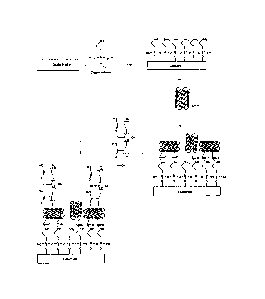
Figure 1 shows the chemical formula of dimethylformamide (DMF);
Figure 2 shows the chemical formula of sodium dodecyl sulfate (SDS);
Figure 3 shows the chemical interaction between a fiinctionalised nano-
material
comprising carbon nano-tubes (CNTs) and an organo-silane binder deposited on a
substrate surface;
CA 02876032 2014-12-08
WO 2013/182852 PCT/GB2013/051496
-14-
Figure 4 shows the chemical interaction between a carboxyl functional ised
nano-
material comprising carbon nano-tubes and an organo-silane binder deposited on
a
substrate;
Figure 5 shows the use of 1-ethyl-3-(3-dimethylaminopropy1)-carbodiimide
(EDC) and hydroxysulfosuccinimide (sulfo-NHS) to promote the chemical
interaction
between a nano-material and a binder deposited on a substrate;
Figure 6a is a scanning electron microscope (SEM) image of a cleaned quartz
surface;
Figures 6b and 6c are SEM images of the surface of Figure 6a with an organo-
silane binder deposited on the surface;
Figure 6d is an SEM image of the surface of Figure 6b with CNTs adhered to the
surface via the binder after the surface has been exposed to a solution
containing the CNTs
for 1 day;
Figure 6e is an SEM image of the surface of Figure 6b with CNTs adhered to the
surface via the binder after the surface has been exposed to a solution
containing the CNTs
for 7 days;
Figure 6f is an SEM image of carbon nano-tubes adhered to the surface using
SDS
as a dispersant;
Figure 7a is an SEM image of CNTs adhered to a doddington rock surface by
using DMF as a dispersant and DCC as a linking agent;
Figure 7b is an SEM image of CNTs adhered to a doddington rock surface by
using SDS as a dispersant and EDC-Sulfo-NHS as a linking agent;
Figure 8 shows the chemical formula of the scale inhibitors DETPMP and PPCA;
Figure 9 is a comparison of the adsorption of PPCA on CNTs and three different
sands;
Figure 10 is a comparison of the adsorption of PPCA on CNTs and rock;
Figure 11 is a comparison of the adsorption of DETPMP on CNTs and silica
powder;
Figure 12 is a comparison of the adsorption of DETPMP on CNTs and rock;
Figure 13 shows the adsorption of PPCA on CNTs over a 24 hour period;
Figure 14 is a comparison of the adsorption of PPCA on two different types of
CNT;
CA 02876032 2014-12-08
WO 2013/182852 PCT/GB2013/051496
-15-
Figure 15 shows the normalised concentration of PPCA in solution with CNTs,
carboxyl functionalised CNTs and hydroxyl funetionalised CNTs;
Figure 16 shows the composition of brine 1 used in experiments;
Figure 17 shows the composition of brine 2 used in experiments;
Figure 18 is a comparison of the adsorption of PPCA in CNTs in distilled water
(DW) and brine 1;
Figure 19 is a comparison of the adsorption of PPCA in CNTs in distilled water
(DW) and brine 2;
Figure 20 shows a thennogravimetric analysis (TGA) of functionalised CNTs and
pristine CNTs;
Figure 21 shows the results of Raman spectroscopy of functionalised CNTs and
pristine CNTs;
Figure 22 shows a coreflood test in distilled water;
Figure 23 shows a coreflood test in brine; and
Figure 24 is a schematic representation of an embodiment of the method of the
invention.
The embodiment of the method of inhibiting scale in a geological formation
which is
illustrated schematically in Figure 24 and will be described below comprises
the steps of:
(a) depositing a 3-aminopropyltrietboxysilane binder on a rock surface of
the
geological formation;
(b) delivering a nano-material comprising nano-particles which are carboxyl
functionalised carbon nano-tubes (CNTs) to the surface of the geological
formation
whereby to cause adherence by an amide bond between the carboxyl groups on the
CNTs
and amino groups on the binder, wherein the CNTs provide one or more
adsorption sites
for a scale inhibitor which is PPCA or DETPMP;
(c) emplacing a quantity of the PPCA or DETPMP in the geological
formation so that an amount of the PPCA or DETPMP is adsorbed by the CNTs; and
(d) inhibiting scale in the geological formation by sustained release of
the
amount of PPCA or DETPMP from the nano-material into the geological formation.
CA 02876032 2014-12-08
WO 2013/182852 PCT/GB2013/051496
-16-
Experiments which relate to the individual steps of this embodiment of the
method are
described in detail below.
Experiments relating to Step (a) - depositing a 3-aminopropvItriethoxysilane
binder on a
rock surface
The rock surface of the geological formation (labelled as substrate in the
Figures) is
functionalized by a 3-aminopropyltriethoxysilane binder to modify the rock
surface in
order to be reactive with the CNTs. 2wt% of 3-aminopropyltriethoxysilane is
added to a
.. binder carrier fluid which is a solution of 95% ethanol to 5% water by
volume. The binder
and binder carrier fluid mixture is pumped into the geological formation for a
desired time
varied from 1 hour to 4 hours. The rock surface is flushed by ethanol followed
by water to
remove the un-reacted organosilane.
.. A Field-emission gun scanning electron microscope (FEG-SEM) image of a
cleaned quartz
surface is shown in Figure 6a. FEG-SEM images of a quartz surface on which 3-
aminopropyltriethoxysilane binder has been deposited are shown in Figures 6b
and 6c.
Experiments relating to Step (b) ¨ adhering carboxyl functionalised CNTs to
the rock
surface
The CNTs used in this embodiment are carboxyl functionalised CNTs (COOH-CNTs)
with
a length less than 2p.m and a diameter less than 8nm. The COOH-CNTs bond with
the
amino group on 3-aminopropyltriethoxysilane binder.
Before pumping CNTs into the rock surface, they are dispersed in a nano-
material carrier
fluid. Two dispersants, DMF and SDS, were tested.
i. Dispersion of CNTs in ditnethylformamide (DMF)
The formula of DMF is shown in Figure 1. DMF is miscible in water and most
organic
liquids. DMF is adsorbed on to the wall of the CNTs and unwraps the
agglomerated CNTs
CA 02876032 2014-12-08
WO 2013/182852 PCT/GB2013/051496
-17-
with the assistance of an ultrasonic bath and the physisorption interaction
between DMF
and the CNTs.
The COOH-CNTs were added to the DMF solution in a ratios of 1:5 (wt./v),
followed by
putting the beaker in an ultrasonic bath for 2h. The solution was then left
static for 24h to
precipitate the un-dispersed CNTs to the bottom of the beaker. The upper
suspension was
used in experiments.
Dispersion of CNTs in Sodium Dodecyl Sulfate (SDS)
Another dispersant chosen for this work was SDS. The formula of SDS is shown
in Figure
2. SDS is more environmentally friendly than DMF. The amount of dispersion of
CNTs
changes depending on the dispersant used. SDS disperses CNTs by the
hydrophobic tail of
the SDS interacting with the CNTs, and the hydrophilic head of the SDS bonding
simultaneously with water. An ultrasonic bath was used to unbundle the
agglomerated
CNTs when SDS is targeting the CNTs.
Two varying ratios of COOH-CNTs/SDS (1:10 and 1:50, wt./wt.) were applied to
obtain
the best dispersion. The solution was put in an ultrasonic bath for 2h,
followed by stirring
for 24h. The solution was left in order to separate the non-dispersed COOH-
CNTs from
the dispersed ones.
For these two different methods of dispersing CNTs (using DMF or SDS), a
suitable
linking agent was chosen to facilitate the reaction of the carboxyl group of
the COOH-
CNTs with the amino group of the 3-aminopropyltriethoxysilane treated rock
surface.
N,N'-Dieyelohexylearbodiimide (DCC) was chosen as a linking agent for the DMF
dispersed COOH-CNTs. 1-Ethyl-3-(3-dimethylaminopropypearbodiimide (EDC) was
chosen for the SDS dispersed COOH-CNTs.
Experiments were carried out on quartz and doddington rock surfaces. An
organosilane-
functionalised quartz or doddington rock sample was embedded in a 25m1 beaker,
followed
by adding 5m1 of DMF or SDS dispersed COOH-CNTs. Samples were left in the
solutions
CA 02876032 2014-12-08
WO 2013/182852
PCT/GB2013/051496
-18-
for desired times. Following that, the sample was rinsed with distilled water
(DW) and
then dried at room temperature. After drying, the samples were taken for
characterization
by a field-emission gun scanning electron microscope (FEG-SEM).
iii. Adhering DMF dispersed COOH-CNTs to organosilane treated Quartz/Rock
Surface using DCC as a linking agent
Figure 3 shows the chemical interaction between DMF dispersed COOH-CNTs and
the
organosilane treated substrate using DCC as a linking agent.
First, 5m1 of DMF was added to a beaker with lmg COOH-CNTs. The solution was
shaken by hand to disperse the COOH-CNTs. For better and uniform dispersion,
an
ultrasonication bath was applied for 2h to disperse the COOH-CNTs into the DMF
solution. After that, 3mg DCC was added to the solution, followed by embedding
an
organosilane functionalised quartz coupon into the solution for fixed
durations. Two
different durations (1 and 7 days) were used in this work. The results were
imaged by
FEG-SEM after drying the surface and are shown in Figures 6d (after 1 day) and
6e (after
7 days).
iv. Adhering SDS dispersed COOH-CNTs on organosilane treated Quartz/Rock
Surface without using a linking agent
Figure 4 shows the chemical interaction of SDS dispersed COOH-CNTs with the
amine
group of organosilane-functionalised quartz without the presence of a linking
agent.
The treated surface was embedded in the 5m1 solution of SDS/CNTs for desired
times
followed by rinsing with distilled water. It was found that the likelihood of
reaction
between COOFI-CNTs and amines on a functionalised surface is low in the
absence of a
linking agent
v. Adhering SDS dispersed COOH-CNTs to organosilane treated Quartz/Rock
Surface using EDC as a linking agent and sulfo-NHS as a cross-linking promoter
CA 02876032 2014-12-08
WO 2013/182852 PCT/GB2013/051496
-19-
Figure 5 shows the chemical interaction of SDS dispersed COOH-CNTs with the
amine
group of organosilane-funetionalised quartz using EDC as a linking agent. The
EDC acts
as a conjugation compound to increase reactivity and Solfu-NHS is used as a
cross-linking
promoter.
To use EDC to erosslink the carboxylic groups of CNTs with amine on the
surface, the
following protocol was applied:
1. A solution with 10mg COOH-CNTs in SDS was prepared in an
ultrasonic bath for 2h.
2. After that, 1.644mg EDC was added to the above solution.
3. After mixing the solution, 1.086g Sulfo-NHS was added, and the solution
was shaken by hand before being used in experiments.
Figure 6f shows an SEM image of SDS dispersed COOH-CNTs adhered to
organosilane
treated quartz using EDC as a linking agent and sulfo-NHS as a cross-linking
promoter.
vi. Adhering COOH-CNTs to organosilane treated doddington rock surface
Using the methods above, COOH-CNTs were adhered to samples of doddington rock.
Figure 7a shows COOH-CNTs adhered to doddington rock using DMF and DCC. Figure
7b shows COOH-CNTs adhered to doddington rock using SDS, EDC and Sulfo-NHS.
Experiments relating to Step (c) ¨ adsorbing scale inhibitor to the CNTs
Static adsorption tests have been performed to assess the efficiency of CNTs
to adsorb
Poly Phosphino Carboxylic Acid (PPCA) and Diethylenetriamine Penta Methylene
Phosphonic Acid (DETPMP). The chemical structure of PPCA and DETPMP is shown
in
Figure 8.
It is assumed that the carbon atoms of PPCA are able to adsorb to the carbon
atoms in
CNTs.
-20-
The static adsorption tests were performed as follows. 50m1 solutions of
various
concentrations of the scale inhibitors (SI) and distilled water (DW) or brine
were prepared
every time for the experiments. Three samples were sent for Inductively
Coupled Plasma
(ICP) measurement to determine the concentration of SI in the solution as a
control. After
that, a desired amount of CNTs (or silica or rock for comparison) were added
to the solution
which was stirred for 24h. Alternatively, the solution was put in an
ultrasonic bath for 2h
and then stirred by magnetic bar for 22h. Then 3m1 of solution were filtered
by 0.45 m
syringe filter (Millex-HV 0.451.1m Millipore(TM)) and measured by ICP (Horiba
Jobin
Yvon, Instruments S.A.). Therefore, whole solution is filtered by vacuum pump
and CNTs
were collected on the top of membrane. At the end, functionalised CNTs were
dried in
vacuum oven at 70 C for 4h and analysed by Thermogravimetric Analysis (TGA)
and
Raman spectroscopy.
i. Adsorption of PPCA on CNTs compared to sands
Figure 9 shows the normalized adsorption of PPCA on the CNTs and various types
of sands
(labelled as A) when compared with a control (labelled as C). The y-axis of
Figure 9 shows
the concentration of PPCA remaining in solution. Figure 9 shows a higher
adsorption of
PPCA on CNTs than the adsorption on rock, silica powder or porous silica
particles because
less PPCA remains in solution after 24h of exposure to the CNTs when compared
with the
results for rock, silica powder and porous silica particles. Less PPCA in
solution means that
more PPCA has been adsorbed to the sample. These results illustrate that a
high adsorption
is achievable by CNTs. Previously, less than 1 mg/g PPCA adsorption has been
obtained in
a condition of a calcium containing brine and using kaolinite as an adsorbent
at 95 C.
Adsorption of PPCA on CNTs compared to crushed rock
Figure 10 indicates that less than lmg adsorption of PPCA per gram of crushed
rock has
been achieved previously with a calcium containing brine. In comparison, at
least 70 mg
of PPCA is adsorbed per gram of CNTs. This shows that CNTs have a far greater
capacity
to adsorb PPCA than crushed rock.
LEGAL_35398951.1
Date recue / Date received 2021-11-03
CA 02876032 2014-12-08
WO 2013/182852 PCT/GB2013/051496
-21-
Figure 10 indicates that less than lmg adsorption of PPCA per gram of crushed
rock has
been achieved previously with a calcium containing brine. In comparison, at
least 70 mg
of PPCA is adsorbed per gram of CNTs. This shows that CNTs have a far greater
capacity
.. to adsorb PPCA than crushed rock.
Adsorption of DETPMP on CNTs
Another scale inhibitor used in the oil and gas industry is DETPMP. The
adsorption of
DETPMP on CNTs was investigated. The experimental set-up for DETPMP used the
same protocol as the PPCA experiments.
Figure 11 shows a comparison result of DETPMP adsorption on CNTs and silica
powder
(labelled as A) when compared with a control (labelled as C). Figure 11 shows
no
adsorption of DETPMP on silica powder after 24h. 7.1mg out of 76 mg DETPMP
dissolved in 50m1DW adsorbed on the CNTs' surface which is not as much as
adsorption
of PPCA on CNTs.
Figure 12 shows a comparison between the adsorption of DETPMP on CNTs and
adsorption on rock from previous experiments. Again it is noteworthy that the
experiment
on the rock was carried out in a solution of brine containing calcium and
other divalent
ions. This result indicates that the affinity of DETPMP being adsorbed on the
surface of
CNTs is much higher than on the rock due to higher specific surface area of
CNTs (in
m2/g) and also the tendency of carbon atom of CNTs to adsorb DETPMP.
iv. Rate of adsorption of PPCA on CNTs
Figure 13 shows the rate with which PPCA is adsorbed on to CNTs. Samples for
ICP
measurement were taken at various times to observe the trend of adsorption.
300mg CNTs
.. were added to a solution of 1000ppm PPCA. Samples were taken before adding
CNTs and
at various times after adding the CNTs. Figure 13 illustrates the normalized
adsorption of
PPCA on CNTs over time by showing the normalised concentration of PPCA
remaining in
CA 02876032 2014-12-08
WO 2013/182852 PCT/GB2013/051496
-22-
solution. Samples were taken after 0.5, 1, 2 and 24h. Figure 13 indicates that
after 0.5h,
82% of reachable adsorption was obtained and after lb adsorption of PPCA on
CNTs
reached an equilibrium. This is significantly faster than the 24h well shut-in
time required
for some current scale inhibitor squeeze treatments and shows that step (c) of
the present
invention may advantageously be faster than known methods.
v. Influence of defects in CNTs on adsorption of PPCA
CNTs are available in the market with different qualities and structures. Two
different
types of CNTs with different qualities were compared in order to understand
the influence
of the quality of CNTs to adsorb PPCA as a scale inhibitor. 300mg of each type
of CNTs
were added into two solutions of 1000ppm PPCA in DW and were sampled at
various
times.
Figure 14 shows the normalized concentration of PPCA in the solution over the
sample
times. The left-hand columns in Figure 14 represent a second type of CNTs
(labelled as
CNTs (2)) which have a higher purity (a more perfect wall) and the right-hand
column
represents the first type of CNTs (labelled as CNTs (1)) which were used in
the previous
experiments. CNTs (1) have more defects in the wall of the carbon nano tubes
than CNTs
(2). CNTs (1) and CNTs (2) have 95% and 99% purity, respectively.
Figure 14 indicates that the adsorption of PPCA on both types of CNTs reaches
to
equilibrium after lh. It also shows higher adsorption by CNTs (1) at 2h, 5h,
7h and 24h.
This may be due to the lower quality CNTs (1) having more defects on the wall
of the
CNTs which provide more active adsorption sites for PPCA.
vi. Adsorption of PPCA on CNTs with different functional groups
Figure 15 shows PPCA adsorption on different functionalized CNTs (f-CNTs)
after 24h
(labelled as A) when compared with a control (labelled as C). Two different f-
CNTs were
compared with unfunctionalised CNTs. The f-CNTs were COOH-CNTs and hydroxyl
functionalized CNTs (OH-CNTs). Figure 15 shows similar adsorptions of PPCA on
all
CA 02876032 2014-12-08
WO 2013/182852 PCT/GB2013/051496
-23-
three types of CNT. This indicates that adhering f-CNTs such as COOH-CNTs to
the
surface of a geological formation would not decrease the adsorption of PPCA on
the
COOH-CNTs when compared with unfunctionalised CNTs.
vii. Effect of Brine on adsorption of PPCA
All the above experiments were performed in DW to eliminate the effects of
ions in
solution. Since the method may be performed in a geological formation in which
brine
may be present in the fluid environment within the pores of the geological
formation,
experiments were run in two brine solutions.
Two basic brines were selected. The composition of Brine 1 is shown in Figure
16 (in
grams per litre of water). The composition of Brine 2 is shown in Figure 17. A
comparison of the compositions of Brine 1 and Brine 2 is given in Table 1
below.
NaC1 (g/l) NaHCO3 (g/1) CaC12.61120 (g/l)
Brine 1 76.26 0.76 0
Brine 2 0 0 3.826
Table 1 ¨ Comparison of composition of Brines 1 and 2
300mg of CNTs were added into 1000ppm PPCA in DW and brine solutions. The
result is
shown in Figures 18 and 19.
Figure 18 shows the concentration of PPCA after 1 day (labelled as A) when
compared
with a control (labelled as C). Figure 18 shows a 47% decrease in adsorption
of PPCA on
CNTs in brine (1) compared with DW. The decrease in adsorption can be
explained by the
increased salinity of the solution which decreases the affinity of the PPCA to
adsorb to the
CNTs. Brine (1) contains no calcium.
Figure 19 shows the concentration of PPCA after 1 day (labelled as A) when
compared
with a control (labelled as C). Figure 19 shows the difference in adsorption
of PPCA on
CNTs in DW compared with brine (2). Brine (2) contains calcium in order to
observe the
CA 02876032 2014-12-08
WO 2013/182852 PCT/GB2013/051496
-24-
effect of calcium ions on adsorption. Unfunctionalised CNTs were used in this
test
because Figure 15 shows the adsorption behaviour of different types of CNTs is
similar.
Figure 19 indicates a 30% increase on adsorption of PPCA on CNTs in the
presence of
calcium.
Thennogravimetric Analysis of CNTs
Figure 20 shows the mass loss of CNTs which have adsorbed PPCA (labelled as
PPCA-
CNTs) compared with pristine CNTs which have not adsorbed PPCA (labelled as p-
CNTs)
under TGA in a N2 atmosphere at ambient pressure. The mass loss was measured
from 0
to 1000 C. PPCA-CNTs and p-CNTs were heated up to 1000 C with a temperature
rate of
10 C/min in the TGA. Samples were dried in a vacuum oven at 70 C for 4h before
use in
the TGA.
At the beginning in Figure 20, both samples follow the same trend of losing
weight as it is
believed that it would be mostly moisture evaporating. From 50 C to almost 700
C the
development of weight loss is faster in the PPCA-CNTs which may be due to the
PPCA
being removed from the CNTs. From 700 C onwards the trend is similar again as
the
CNTs themselves start to decompose.
It is notable that the mass loss versus the temperature is different for PPCA-
CNTs and p-
CNTs. Mass loss occurs at lower temperatures for the PPCA-CNTs. Although TGA
is not
able to give us an accurate mass loss, it was attempted to compare the TGA
results with
IC?. The difference in mass loss between p-CNTs and PPCA-CNTs up to 700 C was
used
for the calculation because above 700 C the CNTs begin to decompose. The
normalized
weight loss difference between p-CNTs and PPCA-CNTs for up to 700 C is 0.0572
from
Figure 20. This is equivalent to 0.62118mg for 10.8598mg of PPCA-CNTs used in
the pan
of TGA. Moreover, 300mg CNTs were used in the experiment which makes the
amount of
weight loss to 17.44mg. With converting this value to (mg/I), 348.9 (mg/1) of
adsorption
of PPCA on the PPCA-CNTs is calculated by TGA which is in a good agreement
with the
ICP result with 346 (mg/1).
CA 02876032 2014-12-08
WO 2013/182852 PCT/GB2013/051496
-25-
ix. Raman spectroscopy of CNTs
Raman spectroscopy is used to study and to characterize graphite materials
such as carbon
nanotubes and fullerenes. Different features of CNTs are characterized by
Raman
spectroscopy including the G-band which is the common sp2 carbon forms and
corresponds
to the tangential vibration of carbon atoms. D and G' bands correspond to
disorder and
dispersive carbon atoms, respectively. The radial breathing mode (RBM), where
carbon
atoms move in the radial direction, is more sensitive to the carbon nanotubes
diameter.
Raman spectroscopy was performed to analyse the vibrational, rotational and
other low-
frequency modes of PPCA-CNTs and p-CNTs.
Figure 21 shows the normalized intensity of Raman spectroscopy for PPCA-CNTs
and p-
CNTs. All samples were emplaced on a metal surface. The dashed spectrum
represents
CNTs which have not adsorbed PPCA (p-CNTs). The solid spectrum represents CNTs
which have adsorbed PPCA (PPCA-CNTs).
In Figure 21, the values of 2600, 1590, 1310 and 266 & 160 cm-1 correspond to
the G', G,
D-band and RBM in p-CNTs and PPCA-CNTs, respectively. The intensity ration
'WIG has
changed from 0.62 to 0.96 from p-CNTs to PPCA-CNTs which shows a 1.55
increment of
ratio of D-band to G-band. This illustrates more defects on the CNTs after
being
functionalized and after adsorption of PPCA which indicates that a covalent
bonding
occurred during functionalisation. Both p-CNTs and PPCA-CNTs showed a small
peak at
2600 cm-I which represents G'-band.
Since RBM is believed to be a unique characteristic of Single-Walled Carbon
Nanotubes
(SWCNTs), RBM peaks may not show on Raman spectroscopy of Multi-Walled Carbon
Nanotubes (MWCNTs). But it is understood that with a good resonance condition,
RBM
of MWCNTs can be observed by Raman spectroscopy if the CNTs have a small
diameter
inner tube (less than 2nm diameter). Usually the RBM signal of the outer wall
diameter is
too weak to be peaked and the signal of inner wall diameter is often
scattered. As the
diameter of MWCNTs being used in this research is small and less than 8nm the
chance of
characterizing of the RBM signal is high. Hence Figure 21 shows two peaks at
266 and
CA 02876032 2014-12-08
WO 2013/182852 PCT/GB2013/051496
-26-
160 cm-I for both samples which correspond to RBM. It can be seen that the
second peak
of RBM (160 em-1) is a little shorter for PPCA-CNTs. After adsorption of PPCA
on CNTs
the diameter of CNTs may increase slightly and this might affect the signal of
RBM.
Experiments relating to Step (d) ¨ sustained release of the scale inhibitor
into the
geological formation
Coreflood tests were carried out to evaluate the method and combine all the
steps together.
In the oil and gas industry, coreflood tests are performed before squeezing a
well to predict
the behaviour of the well. Therefore the results of the coreflood tests may
indicate the
suitability of the method.
Doddington rock was cored and used in the coreflood tests. The coreflood tests
were
carried out in both DW and brine. In both the DW and brine tests, a method
relating to the
.. present invention was compared against a simple squeeze treatment method
which is
available commercially.
i. Commercial method for coreflood test
The procedure for the commercial method was as follows:
1. 10 pore volumes (PV) of solution of PPCA and DW or Brine was
injected into the core and samples were taken at effluent for ICP to quantify
the outcome
concentrations.
2. The core was shut for 24h in order to enable PPCA to be adsorbed
directly on the surface of the rock.
3. Background solution (DW or Brine) was pumped into the core and
effluent samples were taken for ICP measurement of PPCA concentration at
desired times.
The flow rate for injecting the solutions was lml/min.
Method relating to the present invention for coreflood test
CA 02876032 2014-12-08
WO 2013/182852 PCT/GB2013/051496
-27-
The procedure for the method relating to the present invention was as follows:
1. 10 PV of 2wt% 3-aminopropyltriethoxy silane in a solution of
ethanol/DW (95%/5% v/v) was injected into the core followed by shutting the
core for 2h.
2. The core was flushed with 5 PV ethanol to remove un-reacted
organosilane form the core followed by rinsing with 5 PV DW.
3. 10 PV of dispersed COOH-CNTs in DMF with DCC (or in SDS with
EDC/Sulfo-NHS) was subsequently injected into the core followed by shutting
the core for
24h.
4. The core was afterwards rinsed with 10 PV DW.
5. Solution of PPCA and DW was then injected into the core to be adsorbed
by the COOH-CNTs and samples were taken at effluent for ICP measurements
followed
by 24h shut-in.
6. Post-flush was carried out with background solution (DW or Brine) and
effluent samples were taken for ICP measurement of PPCA concentration at
desired times.
The flow rate for injecting the solutions was lml/min.
Results of the Coreflood tests
The results of the coreflood tests are illustrated in Figure 22 and Figure 23.
Figure 22 compares the performance in distilled water (DW) of the method
relating to the
present invention described at ii. above (labelled NAST-DW) with the
commercial method
described at i. above (labelled Commercial-DW). In step 3 of the method
relating to the
present invention the COOH-CNTs were dispersed in DMF with DCC.
The y-axis of Figure 22 shows the mass of PPCA left in the core. The x-axis
shows the
fluid flow through the core in terms of multiples of the pore volume of the
core. The
injection of PPCA into the core is from 0 to 10 pore volumes. After 10 pore
volumes no
more PPCA is injected and DW is flushed through the core.
CA 02876032 2014-12-08
WO 2013/182852 PCT/GB2013/051496
-28-
It can be observed that after 15 pore volumes of fluid flow the amount of PPCA
remaining
in the core approaches zero for the known commercial method. In a hydrocarbon
reservoir
this would indicate that a further squeeze treatment would be required once 5
pore volumes
of flow had occurred after the well had been re-opened after the first squeeze
treatment. In
comparison even after 100 pore volumes of fluid flow the amount of PPCA
remaining in
the core is high with the method relating to the present invention. This
demonstrates the
sustained release of the amount of the scale inhibitor into the geological
formation.
Figure 23 compares the performance in brine of the method relating to the
present
invention described at ii. above (labelled NAST-Brine) with the commercial
method
described at i. above (labelled Commercial-Brine). In step 3 of the method
relating to the
present invention the COOH-CNTs were dispersed in SDS with EDC/Sulfo-NHS.
Figure 23 shows the amount of PPCA remaining in the core by the method
relating to the
present invention is higher over a fluid flow of greater than 80 pore volumes
when
compared with the Commercial-Brine method. It also shows that the amount of
remained
PPCA in the core is decrease compared with Figure 22. This may be due to the
different
dispersant and linking agent used when compared with Figure 22.