Note: Descriptions are shown in the official language in which they were submitted.
CA 02876226 2014-12-08
WO 2014/089399 PCT/US2013/073514
SYSTEM AND METHOD FOR EVALUATING INTRAOCULAR LENS
PERFORMANCE
RELATED APPLICATION
[0001] This application claims priority to U.S. application No. 61/734,240,
filed on
December 6, 2012 under the same title, which is incorporated herein by
reference in its entirety.
Full Paris Convention priority is hereby expressly reserved.
TECHNICAL FIELD
[0001] Embodiments of the present invention relate to vision treatment
techniques and in
particular, to ophthalmic lenses such as intraocular lenses (IOLs).
BACKGROUND OF THE INVENTION
[0002] Intraocular Lenses (IOLs) may be used for restoring visual performance
after a cataract
surgery or other ophthalmic procedure in which the natural crystalline lens is
replaced with or
supplemented by implantation of an IOL. A variety of different types of IOLs
are currently
available, including monofocal and multifocal IOLs, phakic IOLs and piggyback
IOLs (i.e. IOLs
implanted in an eye already having an IOL). In general, monofocal IOLs are
intended to provide
vision correction at one distance only, usually the far focus. In contrast,
multifocal IOLs use two
foci, one near and one far, optionally with some degree of intermediate focus.
Such multifocal,
or bifocal, IOLs are intended to provide good vision at two distances, and
include both refractive
and diffractive multifocal IOLs.
[0003] One significant issue is the cost and/or time needed to develop and
evaluate new IOL
designs. Visual performance of IOLs, including multifocal IOLs, is usually
evaluated through
human clinical trials with surgical implantation. Development of a new type of
IOL may need
multiple design iterations requiring multiple clinical trials which may prove
costly and time
consuming.
[0004] Therefore, what is needed are improved techniques for evaluating the
performance of
intraocular lenses (IOLs) to reduce the need for invasive and time consuming
clinical trials.
1
CA 02876226 2014-12-08
WO 2014/089399 PCT/US2013/073514
BRIEF SUMMARY OF THE INVENTION
[0005] Embodiments of the present invention generally provide improved
techniques for
evaluating performance of intraocular lenses. Such techniques can be used to
evaluate lens
designs and can help reduce the need for multiple clinical trials that may
otherwise be needed to
evaluate multiple design iterations. In one embodiment, a method is provided
for evaluating
performance of an intraocular lens, where the method comprises capturing a
plurality of images
through the intraocular lens at different focus positions; displaying at least
one selected image
from the plurality of images to a test subject; receiving input from the test
subject indicative of
perceived acuity of the at least one selected image; and determining a measure
of intraocular lens
performance from the received input. In another embodiment, a system is
provided for
evaluating performance of an intraocular lens, where the system comprises an
image capture
mechanism, a display screen, and a processing system. In this embodiment the
image capture
mechanism is configured to capture a plurality of images through the
intraocular lens at different
focus positions. The display screen is configured to display at least one
selected image from the
plurality of images to a test subject. Finally, the processing system is
configured to receive input
from the test subject indicative of perceived acuity of the at least one
selected image determine a
measure of intraocular lens performance from the received input.
[0006] For a fuller understanding of the nature and advantages of the present
invention,
reference should be had to the ensuing detailed description taken in
conjunction with the
accompanying drawings.
BRIEF DESCRIPTION OF THE DRAWINGS
[0007] FIGS. 1, 2 and 3 are cross-sectional side views of an image capturing
system for
capturing images though an intraocular lens in accordance with an embodiment
of the invention;
[0008] FIG. 4 is a flow diagram illustrating a method for capturing images
through an
intraocular lens in accordance with an embodiment of the invention;
[0009] FIG. 5 is a flow diagram illustrating a first method for determining a
measure of
intraocular lens performance in accordance with an embodiment of the
invention;
[0010] FIG. 6 is a flow diagram illustrating a second method for determining a
measure of
intraocular lens performance in accordance with an embodiment of the
invention;
2
CA 02876226 2014-12-08
WO 2014/089399 PCT/US2013/073514
[0011] FIGS. 7 and 8 are cross-sectional side views of an image capturing
system for capturing
images though an intraocular lens in accordance with another embodiment of the
invention; and
[0012] FIG. 9 is a cross-sectional view of an eye with a multifocal refractive
intraocular lens.
[0013] It should be noted that the geometries shown in certain aforementioned
figures were not
drawn exactly to scale. For example, the heights of the profiles shown in the
figures may vary
depending on factors such as the amount of correction needed by the patient,
the refractive index
of the lens material and surrounding medium, and the desired phase shift/
delay.
DETAILED DESCRIPTION OF THE INVENTION
[0014] It is to be understood that the figures and descriptions of the present
invention have
been simplified to illustrate elements that are relevant for a clear
understanding of the present
invention, while eliminating, for the purpose of clarity and brevity, many
other elements found in
typical ophthalmic lenses, implantable optic apparatuses, systems and methods.
Those of
ordinary skill in the art may thus recognize that other elements and/or steps
are desirable and/or
required in implementing the present invention. However, because such elements
and steps are
well known in the art, and because they do not facilitate a better
understanding of the present
invention, a discussion of such elements and steps is not provided herein. The
disclosure herein
is directed to all such variations and modifications to the disclosed elements
and methods known
to those skilled in the art.
[0015] Embodiments of the present invention encompass systems and methods that
provide
improved techniques for evaluating lens performance over an extended range of
focal points or
foci. Systems and methods disclosed herein can be applied to various types of
ophthalmic lenses
such as, for example, contact lenses, intraocular lenses, spectacle lenses,
and corneal inlays or
onlays. Exemplary embodiments include various types of intraocular lenses,
including
monofocal lenses, lenses having an extended depth of focus, multifocal
intraocular lenses, etc.
Furthermore, embodiments of the present invention may be used with monofocal
diffractive or
refractive lenses, bifocal diffractive or refractive lenses, and multifocal
diffractive or refractive
lenses.
3
CA 02876226 2014-12-08
WO 2014/089399 PCT/US2013/073514
[0016] Turning now to FIG. 9 a cross-sectional view of an eye E fit with a
multifocal IOL 11 is
illustrated. As shown, multifocal IOL 11 may, for example, comprise a bifocal
IOL. Multifocal
IOL 11 receives light from at least a portion of cornea 12 at the front of eye
E and is generally
centered about the optical axis of eye E. For ease of reference, FIG. 9 does
not disclose the
refractive properties of other parts of the eye, such as the corneal surfaces.
Only the refractive
and/or diffractive properties of the multifocal IOL 11 are illustrated.
[0017] Each major face of lens 11, including the anterior (front) surface and
posterior (back)
surface, generally has a refractive profile, e.g. biconvex, plano-convex,
plano-concave, meniscus,
etc.. The two surfaces together, in relation to the properties of the
surrounding aqueous humor,
cornea, and other optical components of the overall optical system, define the
effects of the lens
11 on the imaging performance by eye E. Conventional, monofocal IOLs have a
refractive
power based on the refractive index of the material from which the lens is
made, and also on the
curvature or shape of the front and rear surfaces or faces of the lens.
[0018] In a young healthy eye, contraction and relaxation of ciliary muscles
17 surrounding the
capsular bag 14 contribute to accommodation of the eye, the process by which
the eye increases
optical power to maintain focus on objects as they move closer. As a person
ages, the degree of
accommodation decreases and presbyopia, the diminished ability to focus on
near objects, often
results. A patient may therefore conventionally use corrective optics having
two optical powers,
one for near vision and one for far vision, as provided by multifocal IOL 11.
[0019] Multifocal lenses may optionally also make special use of the
refractive properties of
the lens. Such lenses generally include different powers in different regions
of the lens so as to
mitigate the effects of presbyopia. For example, as shown in FIG. 9 a
perimeter region of
refractive multifocal lens 11 may have a power which is suitable for viewing
at far viewing
distances. The same refractive multifocal lens 11 may also include an inner
region having a
higher surface curvature and a generally higher overall power (sometimes
referred to as a
positive add power) suitable for viewing at near distances.
[0020] It should be noted that the multifocal IOL 11 illustrated in FIG. 9 is
just one example of
the type of lens that can be evaluated using the systems and methods described
herein. For
example, the systems and methods can be applied to lenses with spherical
aberration to improve
optical image quality, extended depth of focus (i.e. increased range of
functional focus without
discrete far and near focal points), accommodating IOLs where at least one
aspect of the IOL is
4
CA 02876226 2014-12-08
WO 2014/089399 PCT/US2013/073514
capable of responding to ciliary muscle movements during near viewing,
spatially varying
optical properties to generally improve optical quality of visual function.
[0021] The embodiments described herein generally provide improved techniques
for
evaluating performance of intraocular lenses, such as the multifocal IOL 11
illustrated in FIG. 9.
Such techniques can be used to evaluate lens designs and can help reduce the
need for multiple
clinical trials that may otherwise be needed to evaluate multiple design
iterations. In one
embodiment, a method is provided for method for evaluating performance of an
intraocular lens,
where the method comprises capturing a plurality of images through the
intraocular lens at
different focus positions; displaying at least one selected image from the
plurality of images to a
test subject; receiving input from the test subject indicative of perceived
acuity of the at least one
selected image; and determining a measure of intraocular lens performance from
the received
input. In another embodiment, a system is provided for evaluating performance
of an
intraocular lens, where the system comprises an image capture mechanism, a
display screen, and
a processing system. In this embodiment the image capture mechanism is
configured to capture
a plurality of images through the intraocular lens at different focus
positions. The display screen
is configured to display at least one selected image from the plurality of
images to a test subject.
Finally, the processing system is configured to receive input from the test
subject indicative of
perceived acuity of the at least one selected image determine a measure of
intraocular lens
performance from the received input.
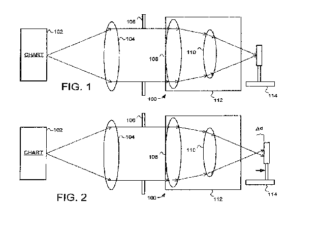
[0022] Turning now to FIG. 1, a cross-sectional side view of an image
capturing system 100
for capturing images though an intraocular lens in accordance with an
embodiment of the
invention is illustrated. The image capturing system 100 includes a
collimating lens 104, an
aperture 106, a cornea lens 108, an intraocular lens 110, an aqueous solution
112, and a sensor
114. The imaging capturing system 110 is configured to capture an image of the
chart 102 using
the sensor 114. It is important to note that image of the chart 102 is
captured through the
intraocular lens 110 that is under evaluation. It is also important to note
that the system 110
captures these images in a way that models the performance of the intraocular
lens 110 when it is
implanted in the human eye. Specifically, the collimating lens 104, aperture
106, cornea lens
108 and aqueous solution 112 are all preferably selected to provide a test
environment which
mimics the human eye in which the intraocular lens 110 is designed to be
implanted.
CA 02876226 2014-12-08
WO 2014/089399 PCT/US2013/073514
[0023] As one example, the image capturing system can be designed to use the
Average
Cornea Eye (ACE) Model. The ACE Model is based on studies and wavefront
measurements of
the human eye, with the aperture 106, cornea lens 108 and aqueous solution 112
chosen to model
the corresponding features of the human eye. For example, the cornea lens 108
is chosen to have
spherical aberration and chromatic aberration that mimics the human cornea.
Likewise, the
aqueous solution 112 is chosen to have refractive index difference similar to
that found in the
human eye. Finally, the aperture 106 can be configured to provide different
sizes to mimic
different entrance pupil diameters. For example, the aperture 106 can be
configured to provide
the equivalent of 3mm and 5mm pupil diameters. As another example, different
aperture sizes
from 2mm to 6 mm diameter can be used to evaluate performance at different
pupil sizes (e.g., to
simulate day and night conditions). For more information on the ACE model see:
Norrby, S.,
Piers, P., Campbell, C., & van der Mooren, M. (2007) Model eyes for evaluation
of intraocular
lenses. Appl Opt, 46 (26), 6595-6605, the content of which is incorporated
herein by reference.
[0024] It should be noted that the ACE model is just one type of model that
could be
implemented in the image capturing system, and that other models could be
used. For example,
the physiological model eye, also described in the above referenced paper can
also be used.
[0025] As was noted above, the image capturing system 100 is configured to
capture images
through the intraocular lens 110. The sensor 114 is preferably selected to
provide high resolution
images. For example, the sensor 114 is preferably selected to capture at
least12-bit grayscale
images of the chart 102. A variety of different types of sensors can thus be
utilized, including
charge coupled device (CCD) based sensors. For example, a PL-H9611A camera
available from
PixeLINK, Ontario, Canada can be used.
[0026] As was noted above, the image capturing system 100 is configured to
capture images at
different focus positions. In the embodiment of FIGS 1-3, the system 100 can
accomplish this by
moving the sensor 114 relative to the intraocular lens 110. This motion of the
sensor 114 allows
for images to be taken at different levels of defocus. For example, FIG. 2
shows the system 100
with the sensor 114 moved further away from the intraocular lens 110, while
FIG. 3 shows the
system 100 with the sensor 114 moved closer to the intraocular lens 110. In
both cases moving
the sensor allows images to be taken at different levels of focus. As one
specific example, the
system 100 can be configured to take images at levels of defocus from
approximately +0.75 to -
3.50D in 0.25D steps.
6
CA 02876226 2014-12-08
WO 2014/089399 PCT/US2013/073514
[0027] As was noted above, the image capturing system 100 is configured to
capture images
through the intraocular lens 110. Specifically, the image capturing system 100
is configured to
capture images of all or part of the chart 102. The chart 102 can comprise any
combination of
shapes and symbols. As one example, the chart 102 comprises all or part of
characters from an
Early Treatment of Diabetic Retinopathy Study (ETDRS) chart. The captured
images can then
be used to determine the performance of the intraocular lens based on feedback
from a test
subject. For example, by displaying one or more characters from the captured
chart to a test
subject and measuring the ability of the test subject to distinguish the
orientation or content of
the characters.
[0028] Turning now to FIG. 4, a flow diagram illustrates a method 400 for
capturing images
through an intraocular lens. The first step 402 is to capture a chart image at
a first focus position.
As described above, a variety of different devices and models can be used to
capture a chart
image at a first focus position, including the system 100 illustrated in FIGS.
1-3.
[0029] The next step 404 is to change focus position. In one embodiment, this
can be
accomplished by changing the position of the sensor relative to the
intraocular lens. An example
of this was illustrated in the system of FIGS. 1, 2 and 3. In another
embodiment, the change in
focus position is accomplished by changing the position of the source chart
relative to the image
capturing system. An example of this system is illustrated in FIGS. 7 and 8.
In either case it
will generally be desirable to change focus position in uniform steps of
defocus. For example,
by changing focus in 0.25D steps.
[0030] With the focus position changed, the next step 406 is to capture
another chart image.
The method then returns to step 404 where the focus is changed again, and then
another image
captured in step 406. This process is continued until images are captured at
each of the desired
levels of focus. Typically, the range and focus distance between images will
be determined
based on the intraocular lens being evaluated. For example, a typical
multifocal lens may have
images captured at different defocus levels from +0.75 to -3.50D, in 0.25D
steps and an extended
depth of focus lens may have images captured at different defocus levels from
+0.75 to -2.00D,
in 0.25D steps and a monofocal lens to improve optical quality at best focus
may have images
captured only at the 0.0D
[0031] Turning now to FIG. 5, a flow diagram illustrates a method 500 for
determining a
measure of intraocular lens performance. In general, the method 500 displays
all or part of a
7
CA 02876226 2014-12-08
WO 2014/089399 PCT/US2013/073514
captured images to a test subject, receives a series of inputs from the test
subject indicative of
whether or not the images can be visually distinguished, and determines a
measure of the
intraocular lens performance from the inputs. The first step 502 is to select
a chart image
corresponding to a focus position. The next step 504 is to display the chart
image to the test
subject at different sizes. In generally, the test image should be displayed
to a user in a way that
accurately represents the visual characteristics of the captured image. For
example, by
displaying the image using the same format in which it was captured. As one
specific example,
by displaying the image using 12 bit gray scale image processing when then
image was
originally captured to 12 bit gray scale.
[0032] The next step 506 is to receive input from the test subject for the
different sizes. The
next step 508 is to record the results. The next step 510 is to determine if
there is a chart image
for another focus position. If there is another chart the method returns to
step 502 and proceeds
again. This process is continued until chart images have been displayed at
different sizes and
user input has been received for each focus position. Then the method proceeds
to step 512 and
a measure of the intraocular lens performance is determined.
[0033] As one example of how method 500 can be implemented, a single character
from each
chart image can be cropped and scaled to different sizes using a tool such as
MatLab (The
Mathworks, Natick, MA). The character can then be randomly oriented and
displayed to the test
subject, and the test subject prompted to indicate the perceived orientation
of the letter as input
(for example, using the psychophysics toolbox and a 4-alternative forced
choice psychophysical
procedure, e.g., see Brainard, D. H. (1997) The Psychophysics Toolbox, Spatial
Vision 10:433-
436 and Pelli, D. G. (1997) The VideoToolbox software for visual
psychophysics: Transforming
numbers into movies, Spatial Vision 10:437-442.). For example the letter "E"
can be displayed
and the user prompted to indicate the one of four alternative orientations of
"E" they perceive .
If the perceived orientation is correct, the same or different letter is
randomly oriented and
displayed to the user at a smaller size. This process can be continued until
the smallest size letter
that can be distinguished by the user is determined. This process can be then
repeated for the
images captured at each focal position. Thus, the visual acuity of the images
for each focal
position can be determined. And from this, a measure of the intraocular lens
performance can
be determined as the visual acuity at different levels of focus. Typically
with a multifocal IOL
one can except good visual acuity and far and near focal planes with reduced
visual acuity in the
intermediate focal planes.
8
CA 02876226 2014-12-08
WO 2014/089399 PCT/US2013/073514
[0034] As another example a word or phrase from each chart image can be
cropped and scaled
to different sizes to judge reading comprehension ability through the
intraocular lens.
Specifically, complex charts words, sentences can be used to measure more
functional
performance as opposed to a simple acuity tests. Functional tests may include
reading speed (i.e.
maximum speed with which a sentence can be read), critical print size (i.e.
print or image size
smaller than which reading speed begins to decline). As another example, the
images can be
displayed at different contrast levels to better simulate intraocular
performance under different
visual conditions. For example, the chart can be made with different contrast
(high 100%
contrast or low 10% or 5% contrast) to evaluate IOL performance under
impoverished conditions
(i.e. low contrast).
[0035] Turning now to FIG. 6, a flow diagram illustrates a second method 600
for determining
a measure of intraocular lens performance. In general, the method 600 displays
all or part of a
chart image while dynamically changing the focus position, receives a series
of inputs from the
test subject indicative of whether or not the images can be visually
distinguished, and determines
a measure of the intraocular lens performance from the inputs. The first step
602 is to select a
chart image. The next step 604 is to display the chart image to the test
subject while dynamically
changing the focus position.
[0036] The next step 606 is to receive input from the test subject for the
different focus
positions. The next step 608 is to record the results. The next step 610 is to
determine a
measure of the intraocular lens performance.
[0037] As one example of how method 600 can be implemented, a chart image
displayed to
the test subject while the focus position is dynamically changed, and the test
subject prompted to
indicate when the image is not distinguishable by the user. This process can
be continued for
different size characters until the smallest size character that can be
distinguished by the user is
determined for each focus position. Thus, the visual acuity of the images for
each focal position
can be determined. And from this, a measure of the intraocular lens
performance can be
determined.
[0038] Example: Photographic images of a miniature ETDRS chart in a bench-top
eye model
(Average Cornea Eye (ACE) model) that has a cornea lens with the same
spherical aberration as
the average human cornea were obtained for a multifocal IOL (ZM900, Abbott
Medical Optics,
USA) and a monofocal control lens (Cee0n, Abbott Medical Optics, USA). Images
were
9
CA 02876226 2014-12-08
WO 2014/089399 PCT/US2013/073514
obtained in 12-bit grayscale at different defocus levels from +0.75 to -3.50D,
in 0.25D steps, by
adjusting the camera position, and for two different entrance pupil diameters
of the ACE model
(3mm and 5mm). A central `S' letter from the ETDRS chart was cropped and
scaled to different
sizes for visual acuity testing using Matlab (The Mathworks, USA),
psychophysics toolbox and
the QUEST procedure with 4-alternative forced choice. The letter was presented
on a CRT
monitor (NEC MultiSync FP2141SB, Mitsubishi Electronics, Illinois) through a
BITS# device
(Cambridge Research Systems, UK). Visual acuity testing was performed from OD
to 3D
defocus (in 0.50D steps) binocularly in two observers with no prior ocular
surgery and 20/20
visual acuity. The results were compared to the data from a FDA clinical trial
on the two IOLs.
[0039] Results: Visual acuity for different defocus levels of the `S' letter
was 20/20 at OD for
both IOLs (mean SD; 911A: -0.03 0.0 logMAR, ZM900: 0.01 0.01 logMAR) and
declined
with defocus for both IOLs, but returned to 20/20 at 3D with the multifocal
IOL (ZM900: 0.0
0.02 logMAR). The through focus visual acuities with the multifocal IOL were
similar for 3mm
and 5mm apertures (all differences were within 1-line or 0.1 logMAR). When
compared to
clinical trial data, visual acuities with the multifocal IOL were within 1-
line (or 0.1 logMAR) for
all defocus levels.
[0040] Thus, the embodiments described herein provide improved techniques for
evaluating
performance of intraocular lenses. Such techniques can be used to evaluate
lens designs and can
help reduce the need for multiple clinical trials that may otherwise be needed
to evaluate
multiple design iterations.
[0041] While the exemplary embodiments have been described in some detail, by
way of
example and for clarity of understanding, those of skill in the art will
recognize that a variety of
modification, adaptations, and changes may be employed. Hence, the scope of
the claims should
not be limited to the description of the preferred versions contained herein.