Note: Descriptions are shown in the official language in which they were submitted.
CA 02876277 2014-12-10
WO 2014/001501 1 PCT/EP2013/063623
ENDOSSEOUS ASSEMBLY FOR PERCUTANEOUS CONNECTOR
Field of the Invention
The present invention relates to the implantation of medical devices in the
body of
an animal, in particular in the human body, especially the implantation of
connection
devices, more particularly to set up a connection transfer energy and/or of
matter between
an external element and a medical apparatus implanted in the body.
Technical background
The substantial development made in electrical equipment designed to be
installed
inside the body of a patient to rectify failure of a natural organ already
implies a capability
for transmitting electric power required by this equipment, from a source of
external power
to the interior of the body.
Contactless power-supply techniques using power transmission via transformer
already exist. Power-supply techniques via percutaneous cranial connectors are
also
known.
Patent US 5 904 646 discloses in particular a percutaneous socket enabling an
electrical connection between an apparatus implanted in the body of a patient,
and an
external apparatus such as power supply. This percutaneous socket is fixed
onto the
surface of an osseous wall by means of an osteosynthesis screw, with all the
elements
making up the socket and the cables being therefore subcutaneous, that is,
essentially in
the zone of the dermis. However, such an arrangement of the socket is not
sufficiently
reliable and vulnerable to infections propagated from percutaneous passage.
Patent FR 03-04063 and patent application US 12/631,161 disclose a permanent
percutaneous connector and associated method to set up an epithelial seal,
which is
advantageous to prevent propagation of infections from the percutaneous
passage.
However, such arrangement requires a bone augmentation around the percutaneous
passage which might increase the duration of the surgical procedure.
An aim of the present invention is to propose a permanent percutaneous
electrical
connection device that can address at least one of the above identified
drawbacks.
In particular, an aim of the present invention is to propose a permanent
percutaneous electrical connection device which is very reliable, that limit
the risks of
infection, and that might be positioned easily and quickly by a practitioner.
CA 02876277 2014-12-10
WO 2014/001501 2 PCT/EP2013/063623
Summary of the Invention
To this end, there is provided a percutaneous connection device as defined in
the
appended claims.
In particular, there is provided a percutaneous connection device, preferably
intended to transfer energy or matter, intended to be fixed in an osseous
structure of a
patient to connect an internal entity located inside the body of the patient
to an entity
external to said body, wherein the device comprises:
- a percutaneous socket having a first end comprising a percutaneous
abutment and
a second end opposite to the first end;
- an elongated extension member designed to be inserted within a hole
created into
the osseous structure, said extension member having a first end comprising
means to be removably coupled to the second end of the socket, and a second
end opposite to the first end, the removable coupling of the extension member
relative to the percutaneous socket being designed for angular shifting of the
first
end of the percutaneous socket relative to the second end of the extension
member;
- anchoring means provided for anchoring the device to the osseous
structure by
osseointegration; and
- separate connection means running through the device from the first end of
the
percutaneous socket to the second end of the extension member, said connection
means comprising at least a first connector arranged within the percutaneous
abutment.
Preferable but not limited aspects of such device, taken alone or in
combination,
are the following:
- the angular shifting of the first end of the percutaneous socket relative
to the
second end of the extension member is at least of 70 , preferably lower than
110 ,
even more preferably comprised between 90 and 100 , and most preferably of
950.
- the extension member is removably fastened to the percutaneous socket,
and the
anchoring means are arranged at the second end of the percutaneous socket,
said
second end of the percutaneous socket being designed for osseous burial in the
osseous structure so that the percutaneous abutment protrudes relative to the
surface of the osseous structure.
CA 02876277 2014-12-10
WO 2014/001501 3 PCT/EP2013/063623
- the percutaneous socket is removably fastened to the extension member,
and the
anchoring means are arranged at the first end of the extension member, close
to
the removable coupling of the extension member relative to the percutaneous
socket.
- the
extension member and the percutaneous socket are removably coupled via an
anchoring base comprising the anchoring means, said anchoring base being
designed for osseous burial in the osseous structure and comprising first
coupling
means for removable fastening of the percutaneous abutment intended to
protrude
relative to the surface of the osseous structure, and second coupling means
for
removable fastening of the extension member intended to be fully buried in the
osseous structure. Such device may preferably have the following features:
o the anchoring base comprises an implant having a cylindrical or truncated-
conical shape, said implant having at least one hole provided in a wall of
the implant;
o the percutaneous abutment has a shape to be at least partially inserted in
the implant;
o the first end of the elongated extension member comprises means to be
removably inserted within the hole of the implant.
- the implant comprises a threaded portion for implantation into the
osseous
structure, and a ring portion at one end of the implant for tightening and
adjusting
the position of the implant into the osseous structure.
- the ring portion comprises at least one lateral flat portion.
- the implant comprises a plurality of anchoring holes provided in a
lateral wall of the
implant, each of said anchoring holes being intended to receive an
osteosynthesis
screw for anchoring the implant into the osseous structure.
- the abutment comprises a through hole for reception of the first
connector, said
through hole having a shape designed for guiding positioning of the first
connector
within the abutment.
- the anchoring means comprises a plurality of osteosynthesis screws
intended to
protrude relative to the surface the device in order to mesh with a lateral
wall of a
cavity of the osseous structure.
- the anchoring means comprises at least one anchoring element arranged so
as to
be able to protrude relative to the surface the device in order to mesh with a
lateral
wall of a cavity of the osseous structure.
- the anchoring means comprises a threaded surface, said threaded surface
easing
primary anchoring of the device in the osseous structure.
CA 02876277 2014-12-10
WO 2014/001501 4 PCT/EP2013/063623
- the anchoring element comprises projecting portions, said projecting
portions
being designed to penetrate the lateral wall of the cavity in a depth between
20 micrometres and 2000 micrometres, and preferably in a depth of
400 micrometres.
- the projecting portions have a geometric shape to provide a retention
effect, said
shape being preferably a symmetric shape chosen among a cone shape, a
pyramid shape, and/or a polyhedron shape.
- the percutaneous socket, the percutaneous abutment, the implant, and/or
the
extension member are made of titanium, polyether ether ketone, zirconia and/or
any biocompatible material.
- the implant is made of titanium using machining and/or additive
manufacturing
processes.
- the implant and/or the extension member is coated with a coating for
promoting
osseointegration of the device into the osseous structure.
- the percutaneous abutment can be connected mechanically, magnetically,
and/or
physically to one or multiple external parts.
- the first connector is clipped within the percutaneous abutment with a
non-return
system.
- the first connector is maintained in position within the percutaneous
abutment with
a maintaining element inserted within the percutaneous abutment.
- the maintaining element is a ring screwed or pushed in the abutment, said
ring
preferably comprising a cutting on the inside in order to place an 0-ring to
maintain
the first connector in compression.
- the extension member has a tubular lumen geometry, said tubular lumen
geometry
being chosen among parallelepipedal, regular polygonal, irregular polygonal
circular, ovaloid, round or a combination thereof.
- the extension member comprises a plurality of tubes.
- the tubes are arranged parallel to each other.
- the first connector is connected to an intermediate connector by a ribbon
cable
made of biocompatible electrical wires, encapsulated with silicon or any other
material that is both flexible and biocompatible.
- the device comprises an intermediate connector intended to be connected
to the
internal entity, wherein said intermediate connector comprises a screw or pin
system to lock and seal the intermediate connector.
- the device comprises an intermediate connector intended to be connected to
the
internal entity, wherein said intermediate connector comprises at least one
eyelet
CA 02876277 2014-12-10
WO 2014/001501 5 PCT/EP2013/063623
on each side, said eyelets being used to attach the implant to the bone with
screw
and/or suture the implant to the fascia.
- the abutment is partly and/or totally cylindrical, triangular and/or
polygonal.
- the connection means comprises a second connector arranged at the
opposite
end of the connection means relative to the first connector, said second
connector
having a shape designed to pass through the percutaneous socket and the
extension member.
- the connection means are electrical connection means, and the first
connector
and/or the second connector are jack connectors, preferably having a cross-
section being circular or in cross arrangement.
According to another aspect, there is provided a percutaneous connection
device,
preferably intended to transfer energy or matter, intended to be fixed in an
osseous
structure of a patient to connect an internal entity located inside the body
of the patient to
an entity external to said body, wherein the device comprises:
- an implant having a cylindrical or truncated-conical shape, said implant
forming an
anchoring base with anchoring means for anchoring of the device in the osseous
structure, and said implant having at least one lateral hole provided in a
lateral wall
of the implant;
- a percutaneous abutment having a shape to be at least partially inserted in
the
implant, preferably in a removable manner;
- an elongated extension member intended to be inserted within a hole
created into
the osseous structure, said extension member having a first end comprising
means to be removably inserted within the lateral hole of the implant, and a
second end opposite to the first end; and
connection means running through the device from the percutaneous abutment to
the second end of extension member, said connection means comprising at least
a first
connector arranged within the percutaneous abutment.
There is also provided a percutaneous endosseous connection assembly (100)
intended to be fixed in an osseous structure of a patient to electrically
connect an internal
entity (150) located inside the body of the patient to an entity external to
said body,
characterized in that the device comprises an endosseous implant (131) in
which is
inserted a percutaneous abutment (111) adapted to be connected to the external
entity,
and an electrical connection element (130) adapted to be coupled to the
internal entity
(150), the abutment (111) coupled to an extension member (115), buried in the
osseous
CA 02876277 2014-12-10
WO 2014/001501 6 PCT/EP2013/063623
structure, which extremity emerging under the skin is separated from the
abutment by a
non-zero distance, the whole implant, abutment (111), extension member (115)
and
emerging extremity ensuring the gateway of the electrical connections means
between the
percutaneous connector (100) and the intermediate connector (138, 140).
There is further provided a percutaneous endosseous connection device (100)
intended to be fixed in an osseous structure of a patient to transfer liquids
to an internal
entity (150) to an entity external to said body and/or extract liquids from an
internal entity
(150) located inside the body of the patient to an entity external to said
body. The said
connection is characterized in that the device comprises a percutaneous
connector (100)
adapted to be connected to the external entity, and tube elements adapted to
be coupled
to the internal entity (150), the abutment (111) coupled to an extension
member (115),
buried in the osseous structure, which extremity emerging under the skin is
separated
from the abutment by a non-zero distance, that extremity (120) of the
extension member,
the whole connector extension (115) and emerging extremity ensuring the
gateway of the
tube connections means between the percutaneous connector (100) and the
intermediate
connector or directly the internal entity (150).
There is also provided a percutaneous endosseous connection device (100)
intended to be fixed in an osseous structure of a patient to transfer light
(fiber optics
and/or other means) to an internal entity (150) located inside the body of the
patient to an
entity external to said body, characterized in that the device comprises a
percutaneous
connector (100) adapted to be connected to the external entity, and tube
elements
adapted to be coupled to the internal entity (150), the abutment (111) coupled
to an
extension member (115), buried in the osseous structure (1), which extremity
emerging
under the skin is separated from the abutment by a non-zero distance, that
extremity
(125) of the extension member, the whole connector extension (115) and
emerging
extremity ensuring the gateway of the light connections means between the
percutaneous
connector (100) and the intermediate connector or directly the internal entity
(150).
Preferable but not limited aspects of such percutaneous endosseous connection
devices, taken alone or in combination, are the following:
- the implant is made of titanium using machining and/or additive
manufacturing
processes.
- the surface of the implant received a coating.
- the implant is made of polyether ether ketone, zirconia and/or any
biocompatible
material.
- the socket (110) can be connected mechanically, magnetically, and/or
physically to
one or multiple external parts.
CA 02876277 2014-12-10
WO 2014/001501 7 PCT/EP2013/063623
- the implanted cable(s) (110) can be maintained in the intermediate
connector by a
mechanical element (clipped, screwed, impacted).
- the abutment (111) and the tubular extension (115) form a single-piece
element.
- the abutment (111) and the tubular extension (115) do not form a single-
piece
element.
- the abutment (111) and the implant (131) form a single-piece element.
- the abutment (111) and the implant (131) do not form a single-piece
element.
- the abutment (111) and the electrical connection (130) form a single-
piece
element.
- the abutment (111) and the electrical connection (130) do not form a single-
piece
element.
- the tubular extension can be characterized by different lumen geometries,
including in particular: parallelepipedal, regular polygonal, irregular
polygonal
circular, ovaloid, round and/or any combination thereof to form a multiple
lumen
tubular extension.
- the tubular extension can consist of several tubes.
- a connector (130), that can be subcutaneous, is connected to the
percutaneous
connector (100) by a ribbon cable made of biocompatible electrical wires,
encapsulated with silicon or any other material that is both flexible and
biocompatible.
- the connector would comprise a screw or pin system to lock and seal the
subconnector.
- the connector would comprise one eyelet on each side. The said eyelets
being
used to attach the implant to the bone with screw and/or suture the implant to
the
fascia.
- the abutment (111) is partly and/or totally cylindrical, triangular
and/or polygonal.
Brief description of the drawings
Other characteristics and advantages of the invention will become clear from
the
following description which is only given for illustrative purposes and is in
no way !imitative
and should be read with reference to the attached drawings on which:
- Fig. 1A to 1J schematically illustrate one example of the positioning of
the different
elements of a percutaneous connection device in a calvaria, i.e. in the dome
of the
skull of a patient.
CA 02876277 2014-12-10
WO 2014/001501 8 PCT/EP2013/063623
- Fig. 2A to 2H are cross-sectional views of the calvaria, illustrating in
more details
the first step of preparing the bone structure, and the further steps of
positioning
the elements forming the percutaneous connection device.
- Fig.3A is an isometric projection of the percutaneous connector assembly
according to the first embodiment of the invention, showing all the elements
composing the assembly.
- Fig.3B is an isometric projection of the percutaneous connector assembly
of
Fig.3A, showing the external connector unplugged. Fig.4A is an isometric
projection of the percutaneous connector assembly according to another
embodiment of the invention.
- Fig.4B is an isometric projection of an exploded view of the percutaneous
connector assembly of Fig.4A.
- Fig.5A is an isometric projection of an endosseous percutaneous connector
assembly according to a second embodiment of the invention, wherein there are
three screw maintaining elements and wherein there are three implants.
- Fig.5B is an isometric projection of an endosseous percutaneous connector
assembly according to a third embodiment of the invention, wherein the
extension
is a quadrilateral housing
- Fig.5C is an isometric projection of an exploded view of an endosseous
percutaneous connector assembly according to a fourth embodiment of the
invention, wherein the maintaining element is a screw, the implant is a self-
taped
threaded system and the extension is made of two separate tubes.
- Fig.5D is an isometric projection of an exploded view of an endosseous
percutaneous connector assembly according to a fifth embodiment of the
invention, wherein the maintaining element is a screw, the implant is an
impacted
system associated with lateral screws, and the extension is a single-piece
element
created from the intersection of the two tubes.
- Fig.5E is an isometric projection of an exploded view of the device of
Fig.5C, with
a complementary stabilization shaft.
- Fig.5F is a cross-sectional view of the device of Fig.5D, illustrating
the angular
shifting.
- Fig.6A to Fig.6C are isometric projections of anchoring means provided on
the
endosseous implant designed to be impacted in the bone.
- Fig. 6D is an isometric projection of an alternative anchoring means for
the
endosseous implant, wherein the implant is designed to be impacted in the bone
and fixed by stabilization screws.
CA 02876277 2014-12-10
WO 2014/001501 9 PCT/EP2013/063623
- Fig 6E is an isometric projection of an alternative embodiment of the
endosseous
implant its surface is threaded with a self-tapping system.
- Fig. 7 is an isometric view of the first end of the intermediate
connector in its
circular connector embodiment.
- Fig. 8A
and Fig. 8B are global views of the permanent percutaneous connection
device of the invention comprising a cranial connection assembly and an
intermediate thoracic connection assembly.
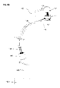
- FIG.9A illustrates a schematic front view of a patient being implanted
with an
internal entity and equipped with a percutaneous connector and external
components.
- FIG.9B illustrates a schematic front view of a patient being implanted
with a fully
implanted system comprising two internal entities, one of which hosting a
controller
and a battery and being connected to a percutaneous connector, and wherein the
implanted battery and implanted controller can be hosted in the same housing.
- FIG.9C illustrates a schematic front view of a patient being implanted with
two
internal entities, one of which hosting a controller and/or a battery
connected to a
percutaneous connector with a lid.
Detailed Description
The disclosed system is intended to transfer energy from an external source to
one
or several implanted device. The invention concerns an endosseous implant
assembly for
percutaneous connection. Most parts of the assembly are assembled in the
operating
room in order to preserve the living bone surrounding the percutaneous
passage.
The proposed percutaneous connection device is preferably intended to transfer
energy or matter (such as fluids) is designed to be fixed in an osseous
structure of a
patient to connect an internal entity (150) located inside the body of the
patient to an entity
external to said body.
Such percutaneous connection device comprises a percutaneous socket (110)
having a first end comprising a percutaneous abutment (111) and a second end
opposite
to the first end.
It further comprises an elongated extension member (115) intended to be
inserted
within a hole created into the osseous structure, said extension member (115)
having a
first end comprising means to be removably coupled to the second end of the
socket
(110), and a second end opposite to the first end. The extension member is
thus designed
and adapted for insertion within a bone hole, the corresponding shape and
material of
CA 02876277 2014-12-10
WO 2014/001501 1 0 PCT/EP2013/063623
such extension member being thus specifically provided for easing the
cooperation with
the bone structure of the bone hole.
Preferably, the removable coupling of the elongated extension member (115)
relative to the percutaneous socket (110) is designed for angular shifting of
the first end of
the percutaneous socket (110) relative to the second end of the extension
member (115).
Such angular shifting (a) is illustrated in Fig.5F for instance. Preferably,
the angular
shifting of the first end of the percutaneous socket (110) relative to the
second end of the
extension member (115) is at least of 70 . Most preferably the angular
shifting is lower
than 110 . The angular shifting may for instance be an angle a of a value
comprised
between 90 and 100 , and preferably an angle a of 95 .
The device also comprises anchoring means provided for anchoring the device to
the osseous structure, such anchoring means being preferably designed for
enabling the
device to be osseointegrated.
Finally, there are provided specific separate connection means running through
the
device from the first end of the percutaneous socket (110) to the second end
of the
extension member (115), said connection means comprising at least a first
connector
(130) arranged within the percutaneous abutment (111).
The specific arrangement which is proposed, in particular the angular shifting
provided with the fact that the extension member is elongated, enables moving
an end of
the connection elements away from the percutaneous passage, which reduces the
risks of
infections at this passage.
The endosseous positioning of the extension member, coupled with the anchoring
of the whole device, in particular at the second end of the percutaneous
socket (110)
enables having a firmly anchored percutaneous connection device, limiting the
risks of
movement of the connection elements.
Further, as it will be apparent from the description below, the device is
designed
for easing a quick implantation in the bone cavity of the patient,
consequently reducing the
duration of the surgical procedure which is of great advantage.
Contrary to some prior developed devices for percutaneous connection, this
device
enables a full implantation in a single surgical step, all the while
significantly reducing the
risks of post-surgery infections.
In particular, the proposed percutaneous connection device does not require a
bone augmentation around the percutaneous passage, which is very advantageous
as it
is mostly operational as soon as it has been implanted in the patient, without
requiring a
long time for bone healing for instance.
CA 02876277 2014-12-10
WO 2014/001501 11 PCT/EP2013/063623
In one embodiment, the extension member (115) is removably fastened to the
percutaneous socket (110), and the anchoring means are arranged at the second
end of
the percutaneous socket (110), said second end of the percutaneous socket
(110) being
designed for osseous burial in the osseous structure so that the percutaneous
abutment
(111) protrudes relative to the surface of the osseous structure.
In another embodiment, the percutaneous socket (110) is removably fastened to
the extension member (115), and the anchoring means are arranged at the first
end of the
extension member (115), close to the removable coupling of the extension
member (115)
relative to the percutaneous socket (110).
In still another embodiment, the extension member (115) and the percutaneous
socket (110) are removably coupled via an anchoring base comprising the
anchoring
means, said anchoring base being designed for osseous burial in the osseous
structure
and comprising first coupling means for removable fastening of the
percutaneous
abutment (111) intended to protrude relative to the surface of the osseous
structure, and
second coupling means for removable fastening of the extension member (115)
intended
to be fully buried in the osseous structure.
In this latter embodiment, the anchoring base may comprise an implant (131)
having a cylindrical or truncated-conical shape. The implant has preferably at
least a
partial hollow shape, being for instance provided with a blind-hole adapted to
receive the
percutaneous abutment (111). Such implant is further designed with at least
one hole
provided in one of the walls of the implant (131), preferably a lateral wall
of the implant, in
order to receive the extension member.
Most preferably, the percutaneous abutment (111) has thus a shape to be at
least
partially inserted in the implant (131). The first end of the elongated
extension member
(115) may also comprise means to be removably inserted within the hole of the
implant
(131).
In a specific embodiment, the percutaneous connection device comprises:
- an implant (131) having a cylindrical or truncated-conical shape, said
implant (131)
forming an anchoring base with anchoring means for anchoring of the device in
the
osseous structure, and said implant having at least one lateral hole provided
in a
lateral wall of the implant (131);
- a percutaneous abutment (111) having a shape to be at least partially
inserted in
the implant (131), preferably in a removable manner;
- an elongated extension member (115) intended to be inserted within a hole
created into the osseous structure, said extension member (115) having a first
end
CA 02876277 2014-12-10
WO 2014/001501 12 PCT/EP2013/063623
comprising means to be removably inserted within the lateral hole of the
implant
(131), and a second end opposite to the first end; and
- connection means running through the device from the percutaneous
abutment
(111) to the second end of extension member (115), said connection means
comprising at least a first connector (130) arranged within the percutaneous
abutment (111).
As is illustrated in figures 4A, 4B, 5A, 50 the implant (131) may comprise a
threaded portion for implantation into the osseous structure. It may further
comprise a ring
portion at one end of the implant for tightening and adjusting the position of
the implant
(131) into the osseous structure.
The ring portion may comprise at least one lateral flat portion. The ring
portion has
preferably a polygonal external shape, preferably an octagonal or hexagonal
external
shape. The ring portion can also be of a substantially cylindrical shape.
In another embodiment as illustrated in Fig.5D, the implant (131) may comprise
a
plurality of anchoring holes provided in a lateral wall of the implant (131),
each of said
anchoring holes being intended to receive an osteosynthesis screw (132b) for
anchoring
the implant into the osseous structure.
Preferably, the abutment (111) of the percutaneous connection device according
to
any embodiment comprises a through hole for reception of the first connector
(130), said
through hole having a shape designed for guiding positioning of the first
connector (130)
within the abutment (111). Such shape is a "mistake-proofing" shape, also
called "poka-
yoke" shape, which prevents any wrong positioning of the connector (130)
within the
abutment (111).
Preferably the abutment (111) is partly and/or totally cylindrical, triangular
and/or
polygonal.
The anchoring means of the device may have several alternative or
complementary features.
The anchoring means may for instance comprise a plurality of osteosynthesis
screws (132b) as illustrated in Fig.5D or Fig.6D. The implant is designed so
that the
osteosynthesis screws protrude relative to the surface the device in order to
mesh with a
lateral wall of a cavity of the osseous structure.
The anchoring means may also comprise any other anchoring element arranged
so as to be able to protrude relative to the surface the device in order to
mesh with a
lateral wall of a cavity of the osseous structure. For instance, the anchoring
means may
comprise a threaded surface as illustrated in Fig.4B, Fig.50, Fig.5E, or
Fig.6E. Such
CA 02876277 2014-12-10
WO 2014/001501 13 PCT/EP2013/063623
threaded surface is adapted for easing primary anchoring of the device in the
osseous
structure.
The anchoring element may also comprise projecting portions as illustrated in
Fig.6A to 60, said projecting portions being designed to penetrate the lateral
wall of the
cavity in a depth between 20 micrometres and 2000 micrometres, and preferably
in a
depth of 400 micrometres.
Preferably, the projecting portions have a geometric shape to provide a
retention
effect, said shape being preferably a symmetric shape chosen among a cone
shape, a
pyramid shape, and/or a polyhedron shape.
Preferably, the percutaneous socket (110), the percutaneous abutment (111),
the
implant (131), and/or the extension member (115) are made of titanium,
polyether ether
ketone, zirconia and/or any biocompatible material.
For instance, the implant can be made of titanium using machining and/or
additive
manufacturing processes.
The implant (131) and/or the extension member (115) may also be coated with a
coating for promoting osseointegration of the device into the osseous
structure.
There are several solutions for connecting the percutaneous abutment (111) to
elements external to the patient. For instance, the percutaneous abutment
(111) can be
connected mechanically, magnetically, and/or physically to one or multiple
external parts.
Preferably the first connector (130) is clipped within the percutaneous
abutment
(111) with a non-return system.
Additionally or alternatively, the first connector (130) is maintained in
position
within the percutaneous abutment (111) with a maintaining element (136)
inserted within
the percutaneous abutment (111).
Such maintaining element may for instance be a ring (136) as illustrated in
Fig.4B,
screwed or pushed in the abutment (111), said ring preferably comprising a
cutting on the
inside in order to place an 0-ring to maintain the first connector (130) in
compression.
For all embodiments of the percutaneous connection device, the shape of the
elongated extension member (115) may differ. For instance, the extension
member (115)
can have a tubular lumen geometry, said tubular lumen geometry being chosen
among
parallelepipedal, regular polygonal, irregular polygonal, ovaloid, round or a
combination
thereof.
Preferably, the extension member (115) comprises a plurality of tubes, such
tubes
being for instance arranged parallel to each other.
CA 02876277 2014-12-10
WO 2014/001501 14 PCT/EP2013/063623
The first connector (130) can be connected to an intermediate connector by a
ribbon cable made of biocompatible electrical wires, encapsulated with silicon
or any other
material that is both flexible and biocompatible.
Further, the percutaneous connection device may comprise an intermediate
connector intended to be connected to the internal entity, wherein said
intermediate
connector comprises a screw or pin system to lock and seal the intermediate
connector.
Additionally or alternatively, the intermediate connector comprises at least
one eyelet on
each side, said eyelets being used to attach the implant to the bone with
screw and/or
suture the implant to the fascia. Preferably, the intermediate connector is a
subcutaneous
connector.
The connection means also comprises a second connector (138) arranged at the
opposite end of the connection means relative to the first connector (130),
said second
connector (138) having a shape designed to pass through the percutaneous
socket (110)
and the extension member (115).
Preferably, the connection means are electrical connection means, and the
first
connector (130) and/or the second connector (138) are jack connectors,
preferably having
a cross-section being circular or in cross arrangement.
As is apparent from above and from the figures, the percutaneous connection
assembly may include some of the following elements:
- An external connector (101);
- An external cable (105);
- An abutment (111);
- A connection element (130);
- One or several maintaining element (136);
- An endosseous implant (131);
- An extension (115);
- A stabilization shaft (117);
- One or several implanted cables (135);
- An intermediate connector (138 and 140);
During the surgical procedure for implanting the percutaneous connection
device,
the surgeon drills with a template a first hole in the bone that will receive
the endosseous
implant (131). Then, a template is used to drill a second hole, and one or
several tunnels
are drilled between the two said holes (Fig.1A). Then, the different elements
of the device
are positioned in the bone structure (Fig. 1B to Fig.1F.)
CA 02876277 2014-12-10
WO 2014/001501 15 PCT/EP2013/063623
Fig. 1A to 1J schematically illustrate one example of the positioning of the
different
elements of a percutaneous connection device in a calvaria, i.e. in the dome
of the skull of
a patient.
- In a first step illustrated on Fig.1A, several holes are drilled in the
calvaria of the
patient, with one or several templates.
- In a second step illustrated on Fig.1B, the implant (131) of the
percutaneous
connection device is implanted in the surgically prepared bone cavity of
Fig.1A. On
Fig.1C, is illustrated the implant (131) inserted in the surgically prepared
bone
cavity of Fig.1A.
- In a third step illustrated on Fig.1D the elongated extension member (115)
of the
percutaneous connection device according to one embodiment of the invention is
inserted in bone channel holes. On Fig.1E, is illustrated the elongated
extension
member (115) inserted in bone channel holes.
- In a fourth step illustrated on Fig.1F, the abutment (111), the
electrical connection
element (130) and the corresponding electric cables (135) of the percutaneous
connection device are inserted through the implant (131) and the extension
(115).
On Fig.1G is illustrated the abutment of Fig.1F fixed to the implant (131),
and on
Fig.1H is illustrated the electrical connection element (130) completely
inserted in
the abutment (111).
- Finally,
in a fifth step illustrated on Fig.1I the maintaining element (136) is
inserted
on the electrical connection element (130). On Fig.1J is illustrated the
maintaining
element (136) inserted and fixed on the electrical connection element (130).
On Fig. 2A to 2H are illustrated in more details ¨ on cross-sectional views ¨
the
first step of preparing the bone structure, and the further steps of
positioning the elements
forming the percutaneous connection device.
On these figures, Fig.2A represents a simplified partial calvaria before any
drilling
of holes.
Fig.2B illustrates the first step of the drilling, wherein the bone hole
receiving the
implant of the percutaneous connection device has been drilled.
Fig.2C illustrates a second step of the drilling, wherein the bone hole
letting the
exit driveline pass has been drilled, in addition to the bone hole receiving
the implant.
Fig.2D illustrates a third step of the drilling, wherein the bone holes
channel under
the outer table for the elongated extension member have been drilled, in
addition to the
bone hole receiving the implant and the bone hole letting the exit driveline
pass.
CA 02876277 2014-12-10
WO 2014/001501 16 PCT/EP2013/063623
Fig.2E is a view of the skull with the implant (131) of the percutaneous
connection
device having been inserted in the bone cavity.
Fig.2F is a view of the skull with the extension member (115) of the
percutaneous
connection device inserted in the corresponding bone holes, and coupled with
the implant
(131).
Fig.2G is a view of the skull with the abutment (111) comprising the
electrical
connection element (130) coupled to the implant (131).
Fig.2H is a view of the skull with the connector (130) and the corresponding
connection wires (135) running throught the implant (131) and extension member
(115),
and with the external connector (101) positioned on the abutment (111).
The following description is made more specifically in reference to the
appended
figures.
The implant (131) preferably comprises a main portion (131a), which is
preferably
a cylindrical or truncated conical shaped element, and that can be pieced by
one or
several holes on its lateral side. Such main element of the implant can also
be
surmounted by a substantially cylindrical ring, or a polygonal ring preferably
a hexagonal
ring. This ring permits to tighten and to adjust the implant in the bone with
a torque
wrench.
The main element (131a) is preferably tapped in order to place a threaded ring
to
maintain the connection.
The main element (131a) comprises anchoring means. In a first embodiment, the
main element (131a) is threaded on its outside surface in order to screw the
implant in the
bone. To reduce the duration of implantation a self-tapping system (132a) can
be
considered (FIG 50 and FIG 6E).
To optimize the osseointegration and minimize the risks of bone depression the
height and width range of the thread could be shorter at the top of the main
element than
at the middle and the bottom of the main element (not shown in the figures).
In a first alternative embodiment, the anchoring means consist of projected
and/or
indent elements at the surface of the main element (FIG. 6A to 60). These
anchoring
means can maintain the implant in an osseous structure after an impaction of
said implant
(131) thanks to a harpoon effect or similar physical phenomenon.
In a second alternative embodiment, a cam system such as described in patent
application FR 11-50639 and one or several screws connected to the main
element
(131a) can be used as anchoring means.
CA 02876277 2014-12-10
WO 2014/001501 17 PCT/EP2013/063623
In a third alternative embodiment (FIG. 6D) the implant is designed to be
impacted
in the bone and fixed by stabilization screws (132b) to guarantee the primary
stability.
Having a strong primary stability promotes the osseointegration of the whole
device which
is advantageous. Moreover, the implant may have a soft conical shape or
tapered shape
which promotes impaction of the implant in the bone structure when the screws
are
tightened up.
In a fourth alternative embodiment, one or several threaded implants (131) are
screwed in the bone. These implants may be maintained together by a plate (not
shown in
the figures).
In the embodiment illustrated in Fig.5A, there are several threaded implants
(131)
that are to be screwed in the bone, and form all together the anchoring base
of the device.
In this particular embodiment there are three implants but there can be more
implants
used. An abutment is then fixed by one or several screws (136b) on the
implants (FIG
5A). An extension (not shown in the figures) can be place between two
implants.
Alternatively, a narrow bone trench could be used to place directly the
electrical wires.
The main element (131a) aims to generate a mechanical torque when the implant
is tightened.
The implant (131) can be made of titanium or other biocompatible materials
which
can favor osseointegration.
The implant (131) may have a coating on the surface to ensure
osseointegration.
The implant (131) may have a groove above the main element in order to obtain
a
larger contact surface for the skin.
The implant (131) may have a retaining element to get a depth stop. This
retaining
element is above the main element and under the said groove. A flange can top
the
implant (131) and protrude to the surface of the bone.
An extension member (115) is fixed to the implant through the holes located on
the
lateral side of the implant (Fig.1D). Alternatively the holes can be located
at the bottom-
end of the implant. The extension member (115) may be made of titanium or
other
biocompatible materials which can favor osseointegration.
Said extension member (115) prevents a direct contact between the bone and the
overmolded cable. It also prevents a bone growth in the bore.
The extension member (115) may have a coating on the surface to ensure
osseointegration.
The extension is preferably solid to preserve the cranium mechanical
properties
and protect the cable in case of trauma.
CA 02876277 2014-12-10
WO 2014/001501 18 PCT/EP2013/063623
In a first embodiment, the extension member (115) is made of several tubes.
These tubes may have a joint extremity (116) to facilitate their insertion.
For instance, the
tubes (115) may be flexible.
The tubes may have mechanical means to be clipped-in in the implant.
In a second embodiment, the extension member (115) is made of quadrilateral
housing (115b) as illustrated on Fig.5B. This housing may include an opening
on its top
side and one or several opening on its lateral extremity. The extremity side
being inserted
in the implant is preferably concave. The housing (115b) may have corners that
are
rounded shaped (not shown in the figures). Alternatively this housing shape
can be a
tube. The housing (115b) may have anchoring means to maximize its stability
preferably
on its lateral sides. The surface of the housing (115b) may include coatings
or any other
structure to favor bone growth into its surface.
In a third embodiment, the extension member (115) is a flexible structure.
Technical solutions considered include sheathing or titanium mesh.
An abutment (111) is mechanically fixed. In a first embodiment, it can be
secured
by different means: screwed (Fig 1F to 1H) or pushed in the implant (131).
Said abutment (111) is preferably composed by two parts: a threaded cylinder
surmounted by a tapped cylinder with a bigger diameter.
According to one embodiment, the upper cylinder contains two flat spots on the
outside. This two flat spots are used for tightening the abutment on the
implant and for
mechanical coding (poka yoke) for the external connector. The abutment (111)
may have
more than two flat spots in order to improve the tightening of the mechanical
torque
transmission.
The abutment (111) may also present one or several different geometrical
recess
in order to insert an instrument with the negative shape. The abutment (111)
may also
present one or several identical geometrical recess in order to insert an
instrument with
the negative shape.
The abutment (111) may have a coating to favor epithelial seal. The abutment
(111) may have a mistake-proofing geometry in the intern part. This element
prevents the
rotation of the connection element (130) inside the abutment (111). The
abutment (111)
also may have an anti-loosening system based on a deformable part or on a
blocking part.
A stabilization shaft (117) may be used to maintain the tubular extension
fixed to
the implant and prevent any implant rotation. The shaft is preferably threaded
at the
extremity in order to be screwed in the implant.
A connection element (130) is preferably inserted in said abutment (111). Two
support rings, monobloc with the abutment (111), are maintaining the
connection element
CA 02876277 2014-12-10
WO 2014/001501 19 PCT/EP2013/063623
(130) inside the abutment (111). The connection element (130) is composed of a
cylindrical or polygonal connector from which come out one or several cables.
Those
cables pass through the tubular extension osseointegrated in the bone (Fig. 1F
to 1H).
At the end of each cable is provided the first end of the intermediate
connector
(138).
In a first embodiment, these connectors may be in-line connectors, flexible
and
designed to facilitate their passage through the apertures of the implant
(131). These in-
line contacts connectors can embed various ranges of contacts. The number of
in-line
contacts connectors and the number of contacts considered varies depending on
the
internal entity.
Two combinations are particularly considered: the first combination is three
cables
with two contacts each; the second combination is two cables with 3 contacts
and one
cable with two contacts.
Each in-line contacts connectors preferably comprise a geometrical mistake-
proofing (poka yoke) in order to avoid a connection in the wrong connector.
Any other combinations can be considered depending on the number of contacts
required by the internal entity (150).
In a first embodiment, the in-line contacts might be annular as shown in the
figures
3A, 3B, 4A, 4B, 8A and 8B. In order to minimize heat dissipation due to energy
transfer, in
a second embodiment, the in-line contacts might not be annular hence
maximizing the
contact area (not shown in the figures). Cross-headed pins or other pins made
of one or
several blades are geometries that can be considered.
In a second embodiment (FIG. 7), the first end of the intermediate connector
may
be circular connectors (138b).
The connection element (130) is maintained in the abutment (111) by one or
several maintaining element.
In a first embodiment, the maintaining element could be a ring maintaining
element
(136a) screwed or pushed in the abutment (111), or the connector could be
clipped with a
non-return system (111a) provided on the abutment. The ring (136a) may contain
a
cutting on the inside in order to place an 0-ring to maintain the connector in
compression
(Fig 11¨ 1J).
In a second embodiment, the maintaining element could be one or several screw
maintaining elements (136b) screwed or pushed in the abutment (111) (FIG. 5A,
5C and
5D).
One or several implanted cables (135) connect the connection element (130) to
the internal entity (150) or internal entities (146 and 150).
CA 02876277 2014-12-10
WO 2014/001501 20 PCT/EP2013/063623
In a first embodiment shown in the figures, implanted cables (135) start with
the
connection (130) and end with the first end of the intermediate connector
(138). The
connection (130), the implanted cables (135) and the in-line contacts are
monobloc. The
connection (130) is connected to the external connector (101) and hosted in
the abutment
(111) and the implant (131). The first end of the intermediate connector (138)
is connected
with the second end of the intermediate connector (140) which extends from the
internal
entity (150).
In a second embodiment not shown in the figures, the connection (130) is made
of
in-line contacts. The implanted cables (135) connect directly the connection
(130) to the
internal entity (150), there is no intermediate connector (138 and 140).
The second end of the intermediate connector (140) is protected by a housing
made of titanium or other biocompatible materials comprising multiple contacts
and
electrical wires and/or optical fibers.
The connector (140) is preferably implanted under the skin in thoracic
subclavian
region. Alternatively it can be implanted anywhere close to the first internal
entity. For
example it can be placed in the pericardial area if the internal entity is
placed there.
An external connector (101) is magnetically and/or mechanically (including
clipped,
screwed or pushed in) fixed to the abutment (111) and rely the percutaneous
system
(100) to the external batteries or controller via an external cable.
The external connector (101) is a cylindrical or a polygonal part comprising
multiple contacts and electrical wires.
Several configurations are possible to power the internal entity (150) through
a
percutaneous connector (100).
In a first embodiment illustrated in Fig.9A, the internal entity (150) is
directly
connected to the external controller (311) and the external batteries (312)
through the
percutaneous connector (100) with external cables (313). An implanted electric
connection element (130), and an intermediate connector (138 and 140) may
facilitate the
change of the cervical electric cable (135) or the implanted entity (150).
In a second embodiment illustrated in Fig.9B, the percutaneous connector (100)
can be used with a fully implanted system wherein the implanted battery and
the
implanted controller are both hosted in a second internal entity (146). In
this configuration
the patient can remove the external components (105, 311, 312), and rely only
on the
implanted battery and controller (146) for a limited period of time. External
batteries (312)
can be used as a backup supply in order to charge the implanted battery via
the
percutaneous connector (100). An external controller (311) can be used as a
back-up
controller or used to add additional functions to the system (FIG. 9B).
CA 02876277 2014-12-10
WO 2014/001501 21 PCT/EP2013/063623
In another embodiment illustrated in Fig.9C, the first internal entity (150)
is
powered by an implanted battery hosted in the second internal entity (146) and
the
percutaneous connector (100) can host the controller and or the battery. The
cranial
housing (314) can be a lid on the percutaneous connector or alternatively it
can be hosted
in a headband or in a helmet (not shown in the pictures).
REFERENCES:
- US 5 904 646
- FR 03-04063
- US 12/631,161
- FR 11-50639