Note: Descriptions are shown in the official language in which they were submitted.
CA 02876659 2014-12-12
WO 2013/192290 PCT/US2013/046519
ENGINEERED THREE-DIMENSIONAL CONNECTIVE TISSUE CONSTRUCTS AND
METHODS OF MAKING THE SAME
CROSS-REFERENCE TO RELATED APPLICATIONS
[0001] This application claims the benefit of U.S. Application Serial No.
61/661,768, filed June 19,
2012, and is a continuation of U.S. Application Serial No. 13/801,780, filed
March 13, 2013, each
of which is hereby incorporated by reference in its entirety.
BACKGROUND OF THE INVENTION
[0002] A number of pressing problems confront the healthcare industry. As of
June 2012 there
were 114,636 patients registered by United Network for Organ Sharing (UNOS) as
needing an
organ transplant. According to UNOS, between January and March 2012 only 6,838
transplants
were performed. Each year more patients are added to the UNOS list than
transplants are
performed, resulting in a net increase in the number of patients waiting for a
transplant.
[0003] Additionally, the research and development cost of a new pharmaceutical
compound is
approximately $1.8 billion. See Paul, et al. (2010). How to improve R&D
productivity: the
pharmaceutical industry's grand challenge. Nature Reviews Drug Discovery
9(3):203-214. Drug
discovery is the process by which drugs are discovered and/or designed. The
process of drug
discovery generally involves at least the steps of: identification of
candidates, synthesis,
characterization, screening, and assays for therapeutic efficacy. Despite
advances in technology and
understanding of biological systems, drug discovery is still a lengthy,
expensive, and inefficient
process with low rate of new therapeutic discovery.
SUMMARY OF THE INVENTION
[0004] In one aspect, disclosed herein are engineered, living, three-
dimensional connective tissue
constructs comprising: connective tissue cells cohered to one another to
provide a living, three-
dimensional connective tissue construct; wherein the construct is
substantially free of pre-formed
scaffold. In some embodiments, the construct is substantially free of any pre-
formed scaffold at the
time of use. In some embodiments, the construct is non-innervated. In some
embodiments, the
connective tissue cells comprise connective tissue cells derived in vitro from
multi-potent cells. In
some embodiments, the multi-potent cells comprise one or more of: tissue-
specific progenitors,
mesenchymal stem/stromal cells, induced pluripotent stem cells, and embryonic
stem cells. In some
embodiments, the multi-potent cells are derived from mammalian adipose tissue.
In other
embodiments, the multi-potent cells are derived from mammalian bone marrow. In
yet other
embodiments, the multi-potent cells are derived from a non-adipose, non-bone
marrow tissue
-1-
CA 02876659 2014-12-12
WO 2013/192290 PCT/US2013/046519
source. In some embodiments, the multi-potent cells were exposed to one or
more differentiation
signals before fabrication of the construct. In some embodiments, the multi-
potent cells were
exposed to one or more differentiation signals during fabrication of the
construct. In some
embodiments, the multi-potent cells were exposed to one or more
differentiation signals after
fabrication of the construct. In some embodiments, the construct was
bioprinted. In further
embodiments, the construct further comprises an extrusion compound, the
extrusion compound
improving the suitability of the cells for bioprinting. In some embodiments,
the connective tissue is
selected from the group consisting of: bone, cartilage, tendon, and ligament.
In some embodiments,
the construct further comprises one or more of the following cell types:
vascular, endothelial,
fibroblasts, pericytes, stem/progenitor cells, immune cells. In some
embodiments, the construct is
substantially in the form of a sheet, patch, ring, tube, cube, polyhedron, or
sphere. In some
embodiments, the construct is substantially in the form of a shape that mimics
the shape or
architecture of a native human connective tissue in vivo. In some embodiments,
the construct is for
implantation in a subject at a site of injury, disease, or degeneration. In
some embodiments, the
construct further comprises one or more of discrete filler bodies, each filler
body comprising a
biocompatible material, wherein the one or more filler body creates a gap or
space in the cohered
cells. In further embodiments, each filler body substantially resists
migration and ingrowth of cells.
[0005] In another aspect, disclosed herein are arrays of engineered, living,
three-dimensional
connective tissue constructs, each construct fabricated by a process
comprising: exposing multi-
potent cells to one or more differentiation signals to provide a living, three-
dimensional connective
tissue construct; wherein each connective tissue construct is substantially
free of pre-formed
scaffold; wherein each connective tissue construct is maintained in culture.
In some embodiments,
each construct is substantially free of any pre-formed scaffold at the time of
use. In some
embodiments, each construct is non-innervated. In some embodiments, the multi-
potent cells
comprise one or more of: tissue-specific progenitors, mesenchymal stem/stromal
cells, induced
pluripotent stem cells, and embryonic stem cells. In some embodiments, the
multi-potent cells are
derived from mammalian adipose tissue. In other embodiments, the multi-potent
cells are derived
from mammalian bone marrow. In yet other embodiments, the multi-potent cells
are derived from a
non-adipose, non-bone marrow tissue source. In some embodiments, the multi-
potent cells were
exposed to the one or more differentiation signals before fabrication of the
construct. In some
embodiments, the multi-potent cells were exposed to the one or more
differentiation signals during
fabrication of the construct. In some embodiments, the multi-potent cells were
exposed to the one
or more differentiation signals after fabrication of the construct. In some
embodiments, each
construct was bioprinted. In some embodiments, the connective tissue is
selected from the group
-2-
CA 02876659 2014-12-12
WO 2013/192290 PCT/US2013/046519
consisting of: bone, cartilage, tendon, and ligament. In some embodiments, one
or more connective
tissue constructs further comprises one or more of the following cell types:
endothelial cells,
fibroblasts, stem/progenitor cells, pericytes, satellite cells, or vascular
cells. In some embodiments,
one or more connective tissue constructs are compound tissue constructs
comprising one or more
connective tissues. In further embodiments, one or more connective tissue
constructs are compound
tissue constructs comprising connective tissue and a non-connective tissue. In
still further
embodiments, one or more connective tissue constructs are compound tissue
constructs comprising
bone tissue and a non-connective tissue. In some embodiments, the arrays are
for use in in vitro
assays. In further embodiments, the arrays are for use in one or more of: drug
discovery, drug
testing, toxicology testing, disease modeling, three-dimensional biology
studies, and cell screening.
In some embodiments, the one or more differentiation signals comprise
mechanical, biomechanical,
soluble, or physical signals, or combinations thereof In some embodiments, one
or more constructs
further comprises one or more discrete filler bodies, each filler body
comprising a biocompatible
material, wherein the one or more filler body creates a gap or space in the
cohered cells. In further
embodiments, each filler body substantially resists migration and ingrowth of
cells.
[0006] In another aspect, disclosed herein are methods of fabricating a
living, three-dimensional
connective tissue construct comprising: incubating a bio-ink, comprising multi-
potent cells that
have been deposited on a support and exposed to one or more differentiation
signals, to allow the
bio-ink to cohere and to form a living, three-dimensional connective tissue
construct, wherein said
incubation has a duration of about 1 hour to about 30 days. In some
embodiments, the multi-potent
cells comprise one or more of: mesenchymal stem/stromal cells, induced
pluripotent stem cells, and
embryonic stem cells. In some embodiments, the multi-potent cells are derived
from mammalian
adipose tissue. In other embodiments, the multi-potent cells are derived from
mammalian bone
marrow. In yet other embodiments, the multi-potent cells are derived from a
non-adipose, non-bone
marrow tissue source. In some embodiments, the connective tissue cells are
exposed to one or more
differentiation signals at one or more time intervals between about 1-21 days
before depositing the
bio-ink onto the support to about 1-21 days after depositing the bio-ink onto
the support. In some
embodiments, the bio-ink is deposited by bioprinting. In some embodiments, the
construct is
substantially free of any pre-formed scaffold at the time of use. In some
embodiments, the construct
is non-innervated. In some embodiments, the connective tissue is selected from
the group
consisting of: bone, cartilage, tendon, and ligament. In some embodiments, the
bio-ink further
comprises one or more of the following cell types: vascular, endothelial,
fibroblasts, pericytes,
stem/progenitor cells, immune cells. In some embodiments, the bio-ink further
comprises an
extrusion compound. In some embodiments, the one or more differentiation
signals comprise
-3-
CA 02876659 2014-12-12
WO 2013/192290 PCT/US2013/046519
mechanical, biomechanical, soluble, or physical signals, or combinations
thereof In some
embodiments, the method further comprises the step of depositing one or more
discrete filler
bodies, each filler body comprising a biocompatible material, wherein the one
or more filler body
creates a gap or space in the cohered cells. In further embodiments, each
filler body substantially
resists migration and ingrowth of cells. In some embodiments, the method
further comprises the
step of assembling a plurality of living, three-dimensional connective tissue
constructs into an array
by spatially confining the constructs onto or within a biocompatible surface.
In some embodiments,
the construct is suitable for implantation in a subject at a site of injury,
disease, or degeneration.
[0007] In another aspect, disclosed herein are methods of fabricating a
living, three-dimensional
connective tissue construct comprising the steps of: preparing bio-ink
comprising multi-potent
cells; depositing the bio-ink onto a support; and incubating the bio-ink to
allow the bio-ink to
cohere and to form a living, three-dimensional connective tissue construct,
wherein said incubation
has a duration of about 1 hour to about 30 days; with the proviso that the
multi-potent cells are
exposed to one or more differentiation signals. In some embodiments, the multi-
potent cells
comprise one or more of: mesenchymal stem/stromal cells, induced pluripotent
stem cells, and
embryonic stem cells. In some embodiments, the multi-potent cells are derived
from mammalian
adipose tissue. In other embodiments, the multi-potent cells are derived from
mammalian bone
marrow. In yet other embodiments, the multi-potent cells are derived from a
non-adipose, non-bone
marrow tissue source. In some embodiments, the connective tissue cells are
exposed to one or more
differentiation signals at one or more time intervals between about 1-21 days
before depositing the
bio-ink onto the support to about 1-21 days after depositing the bio-ink onto
the support. In some
embodiments, the bio-ink is deposited by bioprinting. In some embodiments, the
construct is
substantially free of any pre-formed scaffold at the time of use. In some
embodiments, the construct
is non-innervated. In some embodiments, the connective tissue is selected from
the group
consisting of: bone, cartilage, tendon, and ligament. In some embodiments, the
bio-ink further
comprises one or more of the following cell types: vascular, endothelial,
fibroblasts, pericytes,
stem/progenitor cells, immune cells. In some embodiments, the bio-ink further
comprises an
extrusion compound. In some embodiments, the one or more differentiation
signals comprise
mechanical, biomechanical, soluble, or physical signals, or combinations
thereof In some
embodiments, the method further comprises the step of depositing one or more
discrete filler
bodies, each filler body comprising a biocompatible material, wherein the one
or more filler body
creates a gap or space in the cohered cells. In further embodiments, each
filler body substantially
resists migration and ingrowth of cells. In some embodiments, the method
further comprises the
step of assembling a plurality of living, three-dimensional connective tissue
constructs into an array
-4-
CA 02876659 2014-12-12
WO 2013/192290 PCT/US2013/046519
by spatially confining the constructs onto or within a biocompatible surface.
In some embodiments,
the construct is suitable for implantation in a subject at a site of injury,
disease, or degeneration.
BRIEF DESCRIPTION OF THE DRAWINGS
[0008] The novel features of the invention are set forth with particularity in
the appended claims. A
better understanding of the features and advantages of the present invention
will be obtained by
reference to the following detailed description that sets forth illustrative
embodiments, in which the
principles of the invention are utilized, and the accompanying drawings of
which:
[0009] Fig. 1 depicts a non-limiting exemplary timeline of stem cell
differentiation; in this case, a
timeline of differentiation demonstrating pre-differentiation, pen-
differentiation, and post-
differentiation periods wherein stem cells are incubated in contact with
osteogenic differentiation
media.
[0010] Fig. 2A is an image depicting a non-limiting example of bioprinted MSC
constructs; in this
case, in situ alkaline phosphatase staining of bioprinted MSC constructs
cultured in differentiation
media. This figure demonstrates expression of alkaline phosphatase in
constructs exposed to
differentiation media.
[0011] Fig. 2B is an image depicting a non-limiting example of bioprinted MSC
constructs; in this
case, in situ alkaline phosphatase staining of bioprinted MSC constructs
cultured in basal MSC
culture media. No expression of alkaline phosphatase was observed in
constructs exposed to basal
MSC culture media.
[0012] Fig. 2C is a photomicrograph at 20x depicting a non-limiting example of
bioprinted MSC
constructs; in this case, bioprinted MSC constructs cultured in
differentiation media immediately
post-printing and stained with Alizarin Red S to identify calcium deposits.
[0013] Fig. 2D is a photomicrograph at 20x depicting a non-limiting example of
bioprinted MSC
constructs; in this case, bioprinted MSC constructs cultured in basal MSC
culture media
immediately post-printing and stained with Alizarin Red S. No calcium deposits
were observed in
constructs exposed to basal MSC culture media.
[0014] Fig. 3 is a non-limiting photomicrograph of immunofluorescence staining
of tissue sections
of formalin-fixed paraffin-embedded MSC constructs after 5d of post-bioprint
incubation in
differentiation media detecting the expression of osteopontin, indicative of
MSC differentiation and
osteogenesis.
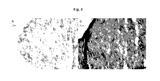
[0015] Figs. 4A and 4B are photomicrographs at 20x depicting mesenchymal stem
cell-containing
constructs that were bioprinted and cultured in either osteogenic
differentiation medium or only
basal mesenchymal stem cell culture media. Histological alkaline phosphatase
staining of
-5-
CA 02876659 2014-12-12
WO 2013/192290 PCT/US2013/046519
bioprinted constructs was utilized to detect osteoblast activity. Fig. 4A
illustrates little or no
expression of alkaline phosphatase in constructs exposed only to basal
mesenchymal stem cell
culture media. Whereas Fig. 4B illustrates expression of alkaline phosphatase
in constructs exposed
to osteogenic differentiation medium.
DETAILED DESCRIPTION OF THE INVENTION
[0016] At the beginning of 2008, 75,834 people were registered as needing a
kidney; at the end of
that year, the number had grown to 80,972. 16,546 kidney transplants were
performed that year, but
33,005 new patients were added to the list. The 2008 transplant rate for a
patient registered by
UNOS as needing a kidney was 20%. The mortality rate of waitlist patients was
7%. Furthermore,
many individuals suffer from chronic degenerative diseases for which
transplantation is not a
current healthcare paradigm. Thus, living, functional connective tissues
(bone, tendon, ligament,
etc.) would be of great clinical value. There is a need for materials, tools,
and techniques that
facilitate application of regenerative medicine and tissue engineering
technologies to relieving the
urgent need for implantable tissues and organs. More specifically, there is a
need for implantable
tissues and organs that are suitable for wound repair, tissue repair, tissue
augmentation, organ
repair, and organ replacement. Just as important, there is a need for
materials, tools, and techniques
that substantially increase the number and quality of innovative, cost-
effective new medicines,
without incurring unsustainable research and development costs.
[0017] Previous models have been focused on providing engineered tissue
constructs by seeding
cells onto a three-dimensional scaffold material that is pre-formed and shaped
to accommodate the
intended application. Cells seeded onto scaffold materials have been primary
cells, cell lines,
engineered cells, and/or stem/progenitor cells. When multipotential stem or
progenitor cells are
utilized, they have either undergone a differentiation program in two-
dimensional monolayer
culture prior to seeding on a three-dimensional scaffold material, or they
have first been seeded
onto a scaffold material and then been subjected to a differentiation program,
in situ or in vitro, to
generate the desired tissue. The traditional approach is both laborious and
inefficient in terms of
cell yield, the time required for terminal differentiation of the cells within
the construct, and the
overall cellularity of the resulting three-dimensional structure.
[0018] The invention relates to the field of regenerative medicine and tissue
engineering. More
particularly, the invention relates to living, three-dimensional connective
tissue constructs, arrays
thereof, and methods of fabrication. The connective tissue constructs are
useful as
implantable/therapeutic devices or as arrayed tissue constructs for in vitro
experimentation (i.e.,
drug development, compound screening, toxicology and disease modeling).
-6-
CA 02876659 2014-12-12
WO 2013/192290 PCT/US2013/046519
[0019] Disclosed herein, in certain embodiments, are engineered, living, three-
dimensional
connective tissue constructs comprising: connective tissue cells cohered to
one another to provide a
living, three-dimensional connective tissue construct; wherein the construct
is substantially free of
pre-formed scaffold.
[0020] Also disclosed herein, in certain embodiments, are arrays of
engineered, living, three-
dimensional connective tissue constructs, each construct fabricated by a
process comprising:
exposing multi-potent cells to one or more differentiation signals to provide
a living, three-
dimensional connective tissue construct; wherein each connective tissue
construct is substantially
free of pre-formed scaffold; wherein each connective tissue construct is
maintained in culture.
[0021] Also disclosed herein, in certain embodiments, are methods of
fabricating a living, three-
dimensional connective tissue construct comprising: incubating a bio-ink,
comprising multi-potent
cells that have been deposited on a support and exposed to one or more
differentiation signals, to
allow the bio-ink to cohere and to form a living, three-dimensional connective
tissue construct,
wherein said incubation has a duration of about 1 hour to about 30 days.
[0022] Also disclosed herein, in certain embodiments, are methods of
fabricating a living, three-
dimensional connective tissue construct comprising the steps of: preparing bio-
ink comprising
multi-potent cells; depositing the bio-ink onto a support; and incubating the
bio-ink to allow the
bio-ink to cohere and to form a living, three-dimensional connective tissue
construct, wherein said
incubation has a duration of about 1 hour to about 30 days; with the proviso
that the multi-potent
cells are exposed to one or more differentiation signals.
Certain Definitions
[0023] Unless otherwise defined, all technical and scientific terms used
herein have the same
meaning as commonly understood by one of ordinary skill in the art to which
this invention
belongs. As used in this specification and the appended claims, the singular
forms "a," "an," and
"the" include plural references unless the context clearly dictates otherwise.
Any reference to "or"
herein is intended to encompass "and/or" unless otherwise stated.
[0024] As used herein, "array" means a scientific tool including an
association of multiple elements
spatially arranged to allow a plurality of tests to be performed on a sample,
one or more tests to be
performed on a plurality of samples, or both.
[0025] As used herein, "assay" means a procedure for testing or measuring the
presence or activity
of a substance (e.g., a chemical, molecule, biochemical, protein, hormone, or
drug, etc.) in an
organic or biological sample (e.g., cell aggregate, tissue, organ, organism,
etc.).
-7-
CA 02876659 2014-12-12
WO 2013/192290 PCT/US2013/046519
[0026] As used herein, "biocompatible" means posing limited risk of injury or
toxicity to cells. As
presented in the specification and claims, "biocompatible multi-well
containers" and
"biocompatible membranes" pose limited risk of injury or toxicity to mammalian
cells, but the
definition does not extend to imply that these biocompatible elements could be
implanted in vivo
into a mammal.
[0027] As used herein, "bioprinting" means utilizing three-dimensional,
precise deposition of cells
(e.g., cell solutions, cell-containing gels, cell suspensions, cell
concentrations, multicellular
aggregates, multicellular bodies, etc.) via methodology that is compatible
with an automated,
computer-aided, three-dimensional prototyping device (e.g., a bioprinter).
[0028] As used herein, "cohere," "cohered," and "cohesion" refer to cell-cell
adhesion properties
that bind cells, multicellular aggregates, multicellular bodies, and layers
thereof The terms are used
interchangeably with "fuse," "fused," and "fusion."
[0029] As used herein, "multi-potent cells" refers to cells that are capable
of undergoing
differentiation to two or more cell types. Multi-potent cells include, for
example, mesenchymal
stem/stromal cells, induced pluripotent stem cells, and embryonic stem cells.
[0030] As used herein, "mesenchymal stem/stromal cells" refers to a specific
type of multi-potent
cells that potentially differentiate into a variety of cell types and exhibit
the properties and
characteristics described further herein. In some embodiments, the terms
"mesenchymal stem cells"
and "mesenchymal stromal cells" are used interchangeably with "mesenchymal
stem/stromal
cells."
[0031] As used herein, "scaffold" refers to synthetic scaffolds such as
polymer scaffolds and
porous hydrogels, non-synthetic scaffolds such as pre-formed extracellular
matrix layers and
decellularized tissues, and any other type of pre-formed scaffold that is
integral to the physical
structure of the engineered tissue and/or organ and not able to be removed
from the tissue and/or
organ without damage/destruction of the tissue and/or organ. The term
"scaffoldless," therefore, is
intended to imply that scaffold is not an integral part of the engineered
tissue at the time of use,
either having been removed or remaining as an inert component of the
engineered tissue.
"Scaffoldless" is used interchangeably with "scaffold-free" and "free of pre-
formed scaffold."
[0032] As used herein, "subject" means any individual, which may be a human, a
non-human
animal, any mammal, or any vertebrate. The term is interchangeable with
"patient," "recipient" and
"donor."
[0033] As used herein, "tissue" means an aggregate of cells. Examples of
tissues include, but are
not limited to, connective tissue (e.g., areolar connective tissue, dense
connective tissue, elastic
tissue, reticular connective tissue, and adipose tissue), muscle tissue (e.g.,
skeletal muscle, smooth
-8-
CA 02876659 2014-12-12
WO 2013/192290 PCT/US2013/046519
muscle and cardiac muscle), genitourinary tissue, gastrointestinal tissue,
pulmonary tissue, bone
tissue, nervous tissue, and epithelial tissue (e.g., simple epithelium and
stratified epithelium),
ectodermal tissue, endodermal tissue, or mesodermal tissue.
Tissue Engineering
[0034] Tissue engineering is an interdisciplinary field that applies and
combines the principles of
engineering and life sciences toward the development of biological substitutes
that restore,
maintain, or improve tissue function through augmentation, repair, or
replacement of an organ. The
basic approach to classical tissue engineering is to seed living cells into a
biocompatible and
eventually biodegradable environment (e.g., a scaffold), and then culture this
construct in a
bioreactor so that the initial cell population can expand further and mature
to generate the target
tissue upon implantation. With an appropriate scaffold that mimics the
biological extracellular
matrix (ECM), the developing tissue may adopt both the form and function of
the desired organ
after in vitro and in vivo maturation. However, achieving high enough cell
density with a native
tissue-like architecture is challenging due to the limited ability to control
the distribution and spatial
arrangement of the cells throughout the scaffold. These limitations may result
in tissues or organs
with poor mechanical properties and/or insufficient function. Additional
challenges exist with
regard to biodegradation of the scaffold, entrapment of residual polymer, and
industrial scale-up of
manufacturing processes. Scaffoldless approaches have been attempted. Current
scaffoldless
approaches are subject to several limitations:
= Complex geometries, such as multi-layered structures wherein each layer
comprises a
different cell type, or comprises specific cellular compartments that are
spatially confined,
may require definitive, high-resolution placement of cell types within a
specific architecture
to reproducibly achieve a native tissue-like outcome.
= Scale and geometry are limited by diffusion and/or the requirement for
functional vascular
networks for nutrient supply.
= The viability of the tissues may be compromised by confinement material
that limits
diffusion and restricts the cells' access to nutrients.
[0035] Disclosed herein, in certain embodiments, are engineered tissues,
engineered connective
tissue constructs, arrays thereof, and methods of fabrication. The tissue
engineering methods
disclosed herein have the following advantages:
= They are capable of producing cell-comprising tissues and/or organs with
a broad array of
complex, three-dimensional topologies.
-9-
CA 02876659 2014-12-12
WO 2013/192290 PCT/US2013/046519
= They mimic the environmental conditions of the natural tissue-forming
processes by
exploiting principles of developmental biology.
= They are compatible with automated means of manufacturing and are
scalable.
[0036] Bioprinting enables improved methods of generating cell-comprising
implantable tissues
that are useful in tissue repair, tissue augmentation, and tissue replacement.
Bioprinting further
enables improved methods of generating micro-scale tissue analogs including
those useful for in
vitro assays.
Bioprinting
[0037] In some embodiments, at least one component of the engineered tissues,
including
connective tissue constructs, and arrays thereof were bioprinted. In further
embodiments, the
engineered tissues were entirely bioprinted. In still further embodiments,
bioprinted constructs are
made with a method that utilizes a rapid prototyping technology based on three-
dimensional,
automated, computer-aided deposition of cells, including cell solutions, cell
suspensions, cell-
comprising gels or pastes, cell concentrations, multicellular bodies (e.g.,
cylinders, spheroids,
ribbons, etc.) (collectively "bio-ink"), and, optionally, confinement material
onto a biocompatible
surface (e.g., composed of hydrogel and/or a porous membrane) by a three-
dimensional delivery
device (e.g., a bioprinter). As used herein, in some embodiments, the term
"engineered," when used
to refer to tissues and/or organs means that cells, cell solutions, cell
suspensions, cell-comprising
gels or pastes, cell concentrates, multicellular aggregates (e.g., bio-ink),
and layers thereof are
positioned to form three-dimensional structures by a computer-aided device
(e.g., a bioprinter)
according to a computer script. In further embodiments, the computer script
is, for example, one or
more computer programs, computer applications, or computer modules. In still
further
embodiments, three-dimensional tissue structures form through the post-
printing fusion of cells or
bio-ink similar to self-assembly phenomena in early morphogenesis.
[0038] While a number of methods are available to arrange cells, bio-ink
(e.g., multicellular
bodies), and/or layers thereof on a biocompatible surface to produce a three-
dimensional structure
including manual placement, positioning by an automated, computer-aided
instrument such as a
bioprinter is advantageous. Advantages of delivery of cells or multicellular
bodies with this
technology include rapid, accurate, and reproducible placement of cells or bio-
ink (e.g.,
multicellular bodies) to produce constructs exhibiting planned or pre-
determined orientations or
patterns of cells, bio-ink (e.g., multicellular bodies), and/or layers thereof
with various
compositions. Advantages also include assured high cell density, while
minimizing cell damage.
-10-
CA 02876659 2014-12-12
WO 2013/192290 PCT/US2013/046519
[0039] In some embodiments, the method of bioprinting is continuous and/or
substantially
continuous. A non-limiting example of a continuous bioprinting method is to
dispense bio -ink from
a bioprinter via a dispense tip (e.g., a syringe, capillary tube, etc.)
connected to a reservoir of bio-
ink. In further non-limiting embodiments, a continuous bioprinting method is
to dispense bio-ink in
a repeating pattern of functional units. In various embodiments, a repeating
functional unit has any
suitable geometry, including, for example, circles, squares, rectangles,
triangles, polygons, and
irregular geometries. In further embodiments, a repeating pattern of
bioprinted function units
comprises a layer and a plurality of layers are bioprinted adjacently (e.g.,
stacked) to form an
engineered tissue or organ. In various embodiments, 2, 3, 4, 5, 6, 7, 8, 9,
10, 11, 12, 13, 14, 15, or
more layers are bioprinted adjacently (e.g., stacked) to form an engineered
tissue or organ.
[0040] In some embodiments, a bioprinted functional unit repeats in a
tessellated pattern. A
"tessellated pattern" is a plane of figures that fills the plane with no
overlaps and no gaps. An
advantage of continuous and/or tessellated bioprinting can include an
increased productivity of
bioprinted tissue. Another non-limiting potential advantage can be eliminating
the need to align the
bioprinter with previously deposited elements of bio-ink. Continuous
bioprinting may also facilitate
printing larger tissues from a large reservoir of bio-ink, optionally using a
syringe mechanism.
[0041] Methods in continuous bioprinting may involve optimizing and/or
balancing parameters
such as print height, pump speed, robot speed, or combinations thereof
independently or relative to
each other. In one example, the bioprinter head speed for deposition was 3
mm/s, with a dispense
height of 0.5 mm for the first layer and dispense height was increased 0.4 mm
for each subsequent
layer. In some embodiments, the dispense height is approximately equal to the
diameter of the
bioprinter dispense tip. Without limitation a suitable and/or optimal dispense
distance does not
result in material flattening or adhering to the dispensing needle. In various
embodiments, the
bioprinter dispense tip has an inner diameter of about, 20, 50, 100, 150, 200,
250, 300, 350, 400,
450, 500, 550, 600, 650, 700, 750, 800, 850, 900, 950, 1000 gm, or more,
including increments
therein. In various embodiments, the bio-ink reservoir of the bioprinter has a
volume of about .5, 1,
2, 3, 4, 5, 6, 7, 8, 9, 10, 15, 20, 25, 30, 35, 40, 45, 50, 55, 60, 65, 70,
75, 80, 85, 90, 95, 100 cubic
centimeters, or more, including increments therein. The pump speed may be
suitable and/or optimal
when the residual pressure build-up in the system is low. Favorable pump
speeds may depend on
the ratio between the cross-sectional areas of the reservoir and dispense
needle with larger ratios
requiring lower pump speeds. In some embodiments, a suitable and/or optimal
print speed enables
the deposition of a uniform line without affecting the mechanical integrity of
the material.
[0042] The inventions disclosed herein include business methods. In some
embodiments, the speed
and scalability of the techniques and methods disclosed herein are utilized to
design, build, and
-11-
CA 02876659 2014-12-12
WO 2013/192290 PCT/US2013/046519
operate industrial and/or commercial facilities for production of engineered
tissues and/or organs
for implantation or use in generation of cell-based tools for research and
development, such as in
vitro assays. In further embodiments, the engineered tissues and/or organs and
arrays thereof are
produced, stored, distributed, marketed, advertised, and sold as, for example,
implantable tissues
for wound repair, tissue repair, tissue augmentation, organ repair, and organ
replacement. In still
further embodiments, the engineered tissues and/or organs and arrays thereof
are produced, stored,
distributed, marketed, advertised, and sold as, for example, cellular arrays
(e.g., microarrays or
chips), tissue arrays (e.g., microarrays or chips), and kits for biological
assays and high-throughput
drug screening. In other embodiments, the engineered tissues and/or organs and
arrays thereof are
produced and utilized to conduct biological assays and/or drug screening as a
service.
Engineered tissues including connective tissue constructs
[0043] Disclosed herein, in some embodiments, are living, three-dimensional
tissue constructs
comprising: connective tissue cells cohered to one another; wherein the
construct is substantially
free of pre-formed scaffold. In further embodiments the construct is
substantially free of pre-
formed scaffold at the time of fabrication and/or the time of use. In some
embodiments, the tissues
are connective tissue constructs. Therefore, also disclosed herein, in some
embodiments, are living,
three-dimensional connective tissue constructs comprising: connective tissue
cells cohered to one
another to provide a living, three-dimensional connective tissue construct;
wherein the construct is
substantially free of pre-formed scaffold at the time of use. In some
embodiments, the connective
tissue cells are derived from multi-potent cells such as mesenchymal
stem/stromal cells, induced
pluripotent stem cells, and/or embryonic stem cells.
[0044] In some embodiments, the engineered tissues, including connective
tissues, are bioprinted, a
methodology described herein. In further embodiments, the tissues are
substantially free of any pre-
formed scaffold as described further herein at the time of printing and/or the
time of use. In some
embodiments, as a result of being fabricated by tissue engineering techniques,
including
bioprinting, the tissues of the present invention are further distinguished
from tissues developed in
vivo, as part of an organism. In some embodiments, the engineered tissues
described herein are
characterized by structural and architectural differences from tissues
developed in vivo, as part of
an organism. By way of non-limiting example, in some embodiments, the
engineered tissues
described herein are non-innervated or lack a functional nervous system. By
way of further non-
limiting example, in some embodiments, the engineered tissues described herein
lack a functional
immune system. By way of further non-limiting example, in some embodiments,
the engineered
tissues described herein lack blood components.
-12-
CA 02876659 2014-12-12
WO 2013/192290 PCT/US2013/046519
[0045] In some embodiments, the engineered tissues, including connective
tissues, include any type
of mammalian cell. In various further embodiments, the tissues, including
connective tissues,
include 1,2, 3,4, 5, 6, 7, 8, 9, 10, 11, 12, 13, 14, 15, 16, 17, 18, 19,20 or
more cell types. In some
embodiments, the tissues include stem cells. In further embodiments, the
tissues include multi-
potent cells such as mesenchymal stem/stromal cells, induced pluripotent stem
cells, and/or
embryonic stem cells.
[0046] In some embodiments, some or all of the multi-potent cells (e.g.,
mesenchymal
stem/stromal cells, induced pluripotent stem cells, embryonic stem cells,
etc.) are undifferentiated
and multi-potent at the time of fabrication of the tissue. In further
embodiments, some or all of the
multi-potent cells are partially differentiated, to some degree, toward one or
more tissue-specific
phenotypes consistent with, for example, osteocytes, chondrocytes, or adipose
cells at the time of
fabrication of the tissue. In further embodiments, some or all of the multi-
potent cells are
completely differentiated to one or more tissue-specific phenotypes consistent
with, for example,
osteocytes, chondrocytes, or adipose cells at the time of fabrication of the
tissue.
[0047] In some embodiments, the multi-potent cells (e.g., mesenchymal
stem/stromal cells,
induced pluripotent stem cells, embryonic stem cells, etc.) have been exposed
to one or more
differentiation signals to provide a living, three-dimensional connective
tissue construct. In various
embodiments, the multi-potent cells have been exposed to one or more
differentiation signals, at
one or more time intervals before, during, or after depositing the bio-ink to
form a tissue construct.
In further embodiments, the multi-potent cells have been exposed to one or
more differentiation
signals before preparation of bio-ink using the cells. In further embodiments,
the multi-potent cells
have been exposed to one or more differentiation signals before fabrication of
tissue using the bio-
ink. In further embodiments, the multi-potent cells have been exposed to one
or more
differentiation signals after fabrication of tissue using the bio-ink.
[0048] In other embodiments, the tissues further include, for example,
mammalian endothelial cells
and/or mammalian fibroblasts. In some embodiments, the cells of the engineered
tissues, including
connective tissues, are "cohered" or "adhered" to one another. In further
embodiments, cohesion or
adhesion refers to cell-cell adhesion properties that bind cells and bio-ink
(e.g., multicellular
aggregates, multicellular bodies, etc.), and/or layers thereof
[0049] The engineered tissues, including connective tissue constructs, in
various embodiments, are
any suitable size. In some embodiments, the size of bioprinted tissues,
including connective tissue
constructs, change over time. In further embodiments, a bioprinted tissue
shrinks or contracts after
bioprinting due to, for example, cell migration, cell death, intercellular
interactions, contraction, or
other forms of shrinkage. In other embodiments, a bioprinted tissue grows or
expands after
-13-
CA 02876659 2014-12-12
WO 2013/192290 PCT/US2013/046519
bioprinting due to, for example, cell migration, cell growth and
proliferation, cell maturation, or
other forms of expansion.
[0050] In some embodiments, the physical dimensions of the engineered tissues,
including
connective tissue constructs, are limited by the capacity for nutrients,
including oxygen, to diffuse
into the interior of the construct. In various embodiments, the engineered
tissues, including
connective tissue constructs, are at least about 20, 30, 40, 50, 60, 70, 80,
90, 100, 110, 120, 130,
140, 150, 160, 170, 180, 190, 200, 210, 220, 230, 240, 250, 260, 270, 280,
290, 300, 310, 320, 330,
340, 350, 360, 370, 380, 390, 400, 410, 420, 430, 440, 450, 460, 470, 480,
490, 500, 550, 600, 650,
700, 750, 800, 850, 900, 950, or 1000 gm, including increments therein, in
their smallest dimension
at the time of bioprinting. In various embodiments, the engineered tissues,
including connective
tissue constructs, are at least about 0.25, 0.5, 0.75, 1.0, 1.25, 1.5, 1.75,
2.0, 2.25, 2.5, 2.75, 3.0,
3.25, 3.5, 3.75, 4.0, 4.25, 4.5, 4.75, or 5.0 mm, including increments
therein, in their smallest
dimension at the time of bioprinting. In further embodiments, the engineered
tissues, including
connective tissue constructs, are between about 50 gm and about 500 gm in
their smallest
dimension at the time of bioprinting.
[0051] In some embodiments, the physical dimensions of the engineered tissues,
including
connective tissue constructs, are about 1, 2, 3, 4, 5, 6, 7, 8, 9, 10, 11, 12,
13, 14, 15, 16, 17, 18, 19,
20, 21, 22, 23, 24, 25, 26, 27, 28, 29, 30, 31, 32, 33, 34, 35, 36, 37, 38,
39, 40, 41, 42, 43, 44, 45,
46, 47, 48, 49, 50, 55, 60, 65, 70, 75, 80, 85, 90, 95, 100, 110, 120, 130,
140, 150, 160, 170, 180,
190, 200, 210, 220, 230, 240, 250, 260, 270, 280, 290, 300, 310, 320, 330,
340, 350, 360, 370, 380,
390, 400, 410, 420, 430, 440, 450, 460, 470, 480, 490, or 500 mm, including
increments therein,
wide.
[0052] In some embodiments, the physical dimensions of the engineered tissues,
including
connective tissue constructs, are about 1, 2, 3, 4, 5, 6, 7, 8, 9, 10, 11, 12,
13, 14, 15, 16, 17, 18, 19,
20, 21, 22, 23, 24, 25, 26, 27, 28, 29, 30, 31, 32, 33, 34, 35, 36, 37, 38,
39, 40, 41, 42, 43, 44, 45,
46, 47, 48, 49, 50, 55, 60, 65, 70, 75, 80, 85, 90, 95, 100, 110, 120, 130,
140, 150, 160, 170, 180,
190, 200, 210, 220, 230, 240, 250, 260, 270, 280, 290, 300, 310, 320, 330,
340, 350, 360, 370, 380,
390, 400, 410, 420, 430, 440, 450, 460, 470, 480, 490, or 500 mm, including
increments therein,
long.
[0053] The engineered tissues, including connective tissue constructs, in
various embodiments, are
any suitable shape. In some embodiments, the shape is selected to mimic a
particular natural tissue
or organ. In further embodiments, the shape is selected to mimic a particular
pathology, condition,
or disease state. In some embodiments, the engineered tissues, including
connective tissue
constructs, have a shape that is substantially planar. In further embodiments,
planar tissues have
-14-
CA 02876659 2014-12-12
WO 2013/192290 PCT/US2013/046519
any suitable planar geometry including, by way of non-limiting examples,
square, rectangle,
polygon, circle, oval, or irregular. In some embodiments, the engineered
tissues, including
connective tissue constructs, have a shape that is substantially a sheet or a
patch. In some
embodiments, the engineered tissues have a shape that is substantially a tube,
a ring, a disc, or a
sac. In further embodiments, a sac is a rolled sheet, or a tube, with one
closed end.
[0054] In some embodiments, the engineered tissues, including connective
tissue constructs, are
spatially confined on one or more sides by a biocompatible material. In other
embodiments, the
engineered tissues, including connective tissue constructs, are affixed to a
surface. In further
embodiments, the tissues are affixed to a biocompatible surface. In still
further embodiments, a
plurality of tissues are associated by affixation to a surface and spatially
arranged to form an array,
as described herein. In some embodiments, engineered tissues, including
connective tissue
constructs, are subjected to mechanical or biomechanical forces. In further
embodiments,
application of soluble, mechanical or biomechanical force serves to facilitate
the differentiation,
maturation, and development of a tissue and/or facilitate the migration,
differentiation, or
proliferation of cells within the tissue.
Cells
[0055] Disclosed herein, in some embodiments, are engineered connective
tissues comprising one
or more types of mammalian cells. In some embodiments, the tissues include
connective tissue
cells. In some embodiments, the connective tissue cells are derived from multi-
potent cells. In
further embodiments, the connective tissue cells are derived from mesenchymal
stem/stromal cells.
In further embodiments, the connective tissue cells are derived from induced
pluripotent stem cells.
In further embodiments, the connective tissue cells are derived from embryonic
stem cells. In still
further embodiments, the tissues include human multi-potent cells. In still
further embodiments, the
tissues include human mesenchymal stem/stromal cells. In still further
embodiments, the tissues
include human induced pluripotent stem cells. In still further embodiments,
the tissues include
human embryonic stem cells.
[0056] Also disclosed herein, in some embodiments, are living, three-
dimensional tissue constructs
comprising multi-potent cells, wherein the multi-potent cells have been
exposed to one or more
differentiation signals to generate connective tissue cells or connective
tissue-associated cells. In
further embodiments, the tissues further include, for example, mammalian
endothelial cells and/or
mammalian fibroblasts.
[0057] In some embodiments, the engineered tissues include non-differentiated
cells. In further
embodiments, "non-differentiated cells" are cells that do not have, or have
lost, the definitive
-15-
CA 02876659 2014-12-12
WO 2013/192290 PCT/US2013/046519
tissue-specific traits of, for example, osteocytes, chondrocytes, adipose
cells, fibroblasts, or
endothelial cells. In some embodiments, non-differentiated cells include stem
cells. In some
embodiments, "stem cells" are cells that exhibit potency and self-renewal.
Stem cells include, but
are not limited to, totipotent cells, pluripotent cells, multi-potent cells,
oligopotent cells, unipotent
cells, and progenitor cells. Stem cells may be embryonic stem cells, adult
stem cells, amniotic stem
cells, and induced pluripotent stem cells. In yet other embodiments, the cells
are a mixture of
differentiated cells and non-differentiated cells. In some embodiments, the
engineered tissues
include mesenchymal stem/stromal cells. In further embodiments, "mesenchymal
stem/stromal
cells" are multi-potent cells that potentially differentiate into a variety of
cell types and exhibit the
properties and characteristics described further herein. In still further
embodiments, the term
"mesenchymal stromal cells" is used interchangeably with "mesenchymal
stem/stromal cells."
[0058] In some embodiments, the mesenchymal stem/stromal cells are human cells
having multi-
lineage mesenchymal differentiation potential including the capacity to
differentiate into
osteoblasts, adipocytes, and chondroblasts. In still further embodiments, the
mesenchymal
stem/stromal cells have the potential to differentiate to osteoblasts,
chondroblasts, and adipocytes
using standard in vitro tissue culture-differentiating conditions. In some
embodiments, the
mesenchymal stem/stromal cells exhibit identifiable surface antigen expression
patterns. In further
embodiments, the mesenchymal stem/stromal cells express the surface antigens
CD105 (also
known as endoglin), CD73 (also known as ecto 5' nucleotidase) and CD90 (also
known as Thy-1).
In some embodiments, the mesenchymal stem/stromal cells lack expression of
surface antigens
specific to other cells likely to be present in mesenchymal stem cell
cultures. In further
embodiments, the mesenchymal stem/stromal cells lack expression of CD45 (a pan-
leukocyte
marker); CD34 (present on primitive hematopoietic progenitors and endothelial
cells); CD14 and
CD11b (prominently expressed on monocytes and macrophages); CD79a and CD19
(markers of B
cells); and HLA-DR. In some embodiments, the mesenchymal stem/stromal cells
exhibit adherence
to plastic when maintained in standard culture conditions using tissue culture
flasks. In some
embodiments, the mesenchymal stem/stromal cells are human cells meeting the
International
Society for Cellular Therapy (ISCT) guidelines providing the most widely
accepted definition of
"Mesenchymal Stem Cell." See Dominici, M. et al. Minimal criteria for defining
multipotent
mesenchymal stromal cells. The International Society for Cellular Therapy
position statement.
Cytotherapy (2006) Vol. 8, No. 4,315-317.
[0059] In some embodiments, suitable multi-potent cells (e.g., stem cells) are
derived from tissue
including, by way of non-limiting example, adipose tissue, bone marrow,
amniotic fluid, and
umbilical tissue. In further embodiments, some or all of the stem cells are
derived from mammalian
-16-
CA 02876659 2014-12-12
WO 2013/192290 PCT/US2013/046519
lipoaspirate. In some embodiments, suitable stem cells are mesenchymal
stem/stromal cells derived
from mammalian adipose tissue or bone marrow. In other embodiments, some or
all of the
mesenchymal stem/stromal cells are derived from non-adipose, non-bone marrow
tissue sources. In
other embodiments, the non-adipose, non-bone marrow tissue source from which
the mesenchymal
stem/stromal cells are derived is selected from: blood, urine, a urologic
tissue (bladder, ureter,
urethra, etc.), kidney, lung, liver, stomach, intestine, trachea, esophagus,
pancreas, skin, oral
mucosa, dental tissue (tooth, pulp, etc.), cartilage, bone, brain, nerve,
placenta, muscle tissue,
omentum, mesothelium, peritoneum, lining of the nasal passages, or
reproductive tissue (uterus,
fallopian tube, etc.).
[0060] In some embodiments, the engineered tissues include one or more types
of differentiated
cells. In further embodiments, "differentiated cells" are cells with a tissue-
specific phenotype
consistent with, for example, a smooth muscle cell, a fibroblast, or an
endothelial cell at the time of
isolation, wherein tissue-specific phenotype (or the potential to display the
phenotype) is
maintained from the time of isolation to the time of use.
[0061] In some embodiments, any mammalian cell is suitable for further
inclusion in the
engineered tissues and arrays thereof In further embodiments, the mammalian
cells are, by way of
non-limiting examples, contractile or muscle cells (e.g., skeletal muscle
cells, cardiomyocytes,
smooth muscle cells, and myoblasts), connective tissue cells (e.g., bone
cells, cartilage cells,
fibroblasts, and cells differentiating into bone forming cells, and
chondrocytes), bone marrow cells,
endothelial cells, skin cells, epithelial cells, breast cells, vascular cells,
blood cells, lymph cells,
neural cells, Schwann cells, gastrointestinal cells, liver cells, pancreatic
cells, lung cells, tracheal
cells, corneal cells, genitourinary cells, kidney cells, reproductive cells,
adipose cells, parenchymal
cells, pericytes, mesothelial cells, stromal cells, undifferentiated cells
(e.g., embryonic cells, stem
cells, and progenitor cells), endoderm-derived cells, mesoderm-derived cells,
ectoderm-derived
cells, and combinations thereof. In embodiments including more than one cell
type, the cell types
are present in many suitable ratios, examples of which are described herein.
[0062] In one embodiment, the tissues include endothelial cells. In another
embodiment, the tissues
include fibroblasts. In another embodiment, the tissues include endothelial
cells and fibroblasts. In
some embodiments, the endothelial cells are human endothelial cells. In some
embodiments,
suitable endothelial cells originated from tissue including, by way of non-
limiting example, blood,
blood vessel, lymphatic vessel, tissue of the digestive tract, tissue of the
genitourinary tract, adipose
tissue, tissue of the respiratory tract, tissue of the reproductive system,
bone marrow, and umbilical
tissue. In some embodiments, the fibroblasts are human fibroblasts. In some
embodiments, suitable
fibroblasts are non-vascular fibroblasts, such as dermal fibroblasts. In other
embodiments, suitable
-17-
CA 02876659 2014-12-12
WO 2013/192290 PCT/US2013/046519
fibroblasts are derived from vascular adventitia. In some embodiments, some or
all of the cells are
derived from mammalian lipoaspirate. In further embodiments, some or all of
the cells are cultured
from the stromal vascular fraction of mammalian lipoaspirate.
[0063] In various embodiments, the cell types and/or source of the cells are
selected, configured,
treated, or modulated based on a specific research goal or objective. In some
embodiments, one or
more specific cell types are selected, configured, treated, or modulated to
facilitate investigation of
a particular disease or condition. In some embodiments, one or more specific
cell types are
selected, configured, treated, or modulated to facilitate investigation of a
disease or a condition of a
particular subject. In some embodiments, one or more specific cell types are
derived from two or
more distinct human donors. In some embodiments, one or more specific cell
types are derived
from a particular vertebrate subject. In further embodiments, one or more
specific cell types are
derived from a particular mammalian subject. In still further embodiments, one
or more specific
cell types are derived from a particular human subject.
Methods of culturing cells
[0064] The cell types used in the engineered tissues of the invention may be
cultured in any manner
known in the art. Methods of cell and tissue culturing are known in the art,
and are described, for
example, in Cell & Tissue Culture: Laboratory Procedures; Freshney (1987),
Culture of Animal
Cells: A Manual of Basic Techniques, the contents of which are incorporated
herein by reference
for such information. General mammalian cell culture techniques, cell lines,
and cell culture
systems that may be used in conjunction with the present invention are also
described in Doyle, A.,
Griffiths, J. B., Newell, D. G., (eds.) Cell and Tissue Culture: Laboratory
Procedures, Wiley
(1998), the contents of which are incorporated herein by reference for such
information.
[0065] Appropriate growth conditions for mammalian cells in culture are well
known in the art.
Cell culture media generally include essential nutrients and, optionally,
additional elements such as
growth factors, salts, minerals, vitamins, etc., that may be selected
according to the cell type(s)
being cultured. Particular ingredients may be selected to enhance cell growth,
differentiation,
secretion of specific proteins, etc. In general, standard growth media include
Dulbecco's Modified
Eagle Medium, low glucose (DMEM), with 110 mg/L pyruvate and glutamine,
supplemented with
10-20% fetal bovine serum (FBS), calf serum, or human serum and 100 U/ml
penicillin, 0.1 mg/ml
streptomycin are appropriate as are various other standard media well known to
those in the art.
Preferably cells are cultured under sterile conditions in an atmosphere of 1-
21% 02 and preferably
3-5% CO2, at a temperature at or near the body temperature of the animal of
origin of the cell. For
example, human cells are preferably cultured at approximately 37 C. With
regard to mesenchymal
stem/stromal cells, suitable culture media includes basal media containing 5-
10% (v:v) fetal bovine
-18-
CA 02876659 2014-12-12
WO 2013/192290 PCT/US2013/046519
serum in low glucose DMEM supplemented with L-glutamine. Optionally,
mesenchymal
stem/stromal cells are cultured and expanded in conditions wherein the oxygen
tension is less than
21% oxygen (equivalent to atmospheric oxygen tension). In some embodiments,
the cells are
cultured at 3-5% oxygen conditions.
[0066] The cells can also be cultured with cellular differentiation agents to
induce differentiation of
the cell along a desired line. For example, in some embodiments, stem cells
are incubated in
contact with differentiation media to produce a range of cell types. Many
types of differentiation
media are suitable. In various embodiments stem cells are incubated in contact
with differentiation
media including, by way of non-limiting examples, osteogenic differentiation
media, chondrogenic
differentiation media, adipogenic differentiation media, neural
differentiation media,
cardiomyocyte differentiation media, and enterocyte differentiation media
(e.g., intestinal
epithelium). With regard to mesenchymal stem/stromal cells, in some
embodiments, the cells are
incubated in contact with differentiation media including, by way of non-
limiting examples,
osteogenic differentiation media, chondrogenic differentiation media, or
adipogenic differentiation
media.
[0067] Additionally, cells can be cultured with growth factors, cytokines,
etc. In some
embodiments, the term "growth factor" refers to a protein, a polypeptide, or a
complex of
polypeptides, including cytokines, that are produced by a cell and which can
affect itself and/or a
variety of other neighboring or distant cells. Typically growth factors affect
the growth and/or
differentiation of specific types of cells, either developmentally or in
response to a multitude of
physiological or environmental stimuli. Some, but not all, growth factors are
hormones. Exemplary
growth factors are insulin, insulin-like growth factor (IGF), nerve growth
factor (NGF), vascular
endothelial growth factor (VEGF), keratinocyte growth factor (KGF), fibroblast
growth factors
(FGFs), including basic FGF (bFGF), platelet-derived growth factors (PDGFs),
including PDGF-
AA and PDGF-AB, hepatocyte growth factor (HGF), transforming growth factor
alpha (TGF-a),
transforming growth factor beta (TGF-I3), including TGFI31 and TGFI33,
epidermal growth factor
(EGF), granulocyte-macrophage colony-stimulating factor (GM-CSF), granulocyte
colony-
stimulating factor (G-CSF), interleukin-6 (IL-6), IL-8, and the like. Growth
factors are discussed in,
among other places, Molecular Cell Biology, Scientific American Books, Darnell
et al., eds., 1986;
Principles of Tissue Engineering, 2d ed., Lanza et al., eds., Academic Press,
2000. The skilled
artisan will understand that any and all culture-derived growth factors in the
conditioned media
described herein are within the scope of the invention.
-19-
CA 02876659 2014-12-12
WO 2013/192290 PCT/US2013/046519
Bio-ink and multicellular aurmates
[0068] Disclosed herein, in certain embodiments, are tissues, including
connective tissue
constructs, arrays thereof, and methods that comprise bioprinted cells. In
some embodiments, cells
are bioprinted by depositing or extruding bio-ink from a bioprinter. In some
embodiments, "bio-
ink" includes liquid, semi-solid, or solid compositions comprising a plurality
of cells.
[0069] In some embodiments, bio-ink comprises liquid or semi-solid cell
solutions, cell
suspensions, or cell concentrations. In some embodiments, bio-ink comprises
semi-solid or solid
multicellular aggregates or multicellular bodies. In further embodiments, the
bio-ink is produced by
1) mixing a plurality of cells or cell aggregates and a biocompatible liquid
or gel in a pre-
determined ratio to result in bio-ink, and 2) compacting the bio-ink to
produce the bio-ink with a
desired cell density and viscosity. In some embodiments, the compacting of the
bio-ink is achieved
by centrifugation, tangential flow filtration ("TFF"), or a combination
thereof. In some
embodiments, the compacting of the bio-ink results in a composition that is
extrudable, allowing
formation of multicellular aggregates or multicellular bodies. In some
embodiments, "extrudable"
means able to be shaped by forcing (e.g., under pressure) through a nozzle or
orifice (e.g., one or
more holes or tubes). In some embodiments, the compacting of the bio-ink
results from growing the
cells to a suitable density. The cell density necessary for the bio-ink will
vary with the cells being
used and the tissue or organ being produced. In some embodiments, the cells of
the bio-ink are
cohered and/or adhered. In some embodiments, "cohere," "cohered," and
"cohesion" refer to cell-
cell adhesion properties that bind cells, multicellular aggregates,
multicellular bodies, and/or layers
thereof. In further embodiments, the terms are used interchangeably with
"fuse," "fused," and
"fusion." In some embodiments, the bio-ink additionally comprises support
material, cell culture
medium, extracellular matrix (or components thereof), cell adhesion agents,
cell death inhibitors,
anti-apoptotic agents, anti-oxidants, extrusion compounds, and combinations
thereof.
[0070] In various embodiments, the cells are any suitable cell. In further
various embodiments, the
cells are vertebrate cells, mammalian cells, human cells, or combinations
thereof. In some
embodiments, the cells include stem cells. In further embodiments, the stem
cells are human stem
cells. In some embodiments, the cells include mesenchymal stem/stromal cells.
In further
embodiments, the mesenchymal stem/stromal cells are human mesenchymal
stem/stromal cells. In
some embodiments, the type of cell used in a method disclosed herein depends
on the type of
construct or tissue being produced. In some embodiments, the bio-ink comprises
one type of cell. In
some embodiments, the bio-ink comprises more than one type of cell.
-20-
CA 02876659 2014-12-12
WO 2013/192290 PCT/US2013/046519
Cell culture media
[0071] In some embodiments, the bio-ink comprises a cell culture medium. The
cell culture
medium is any suitable medium. In various embodiments, suitable cell culture
media include, by
way of non-limiting examples, Dulbecco's Phosphate Buffered Saline, Earle's
Balanced Salts,
Hanks' Balanced Salts, Tyrode's Salts, Alsever's Solution, Gey's Balanced Salt
Solution, Kreb's-
Henseleit Buffer Modified, Kreb's-Ringer Bicarbonate Buffer, Puck's Saline,
Dulbecco's Modified
Eagle's Medium, Dulbecco's Modified Eagle's Medium/Nutrient F-12 Ham, Nutrient
Mixture F-10
Ham (Ham's F-10), Medium 199, Minimum Essential Medium Eagle, RPMI-1640
Medium, Ames'
Media, BGJb Medium (Fitton-Jackson Modification), Click's Medium, CMRL-1066
Medium,
Fischer's Medium, Glascow Minimum Essential Medium (GMEM), Iscove's Modified
Dulbecco's
Medium (IMDM), L-15 Medium (Leibovitz), McCoy's 5A Modified Medium, NCTC
Medium,
Swim's S-77 Medium, Waymouth Medium, William's Medium E, or combinations
thereof In
some embodiments, the cell culture medium is modified or supplemented. In some
embodiments,
the cell culture medium further comprises albumin, selenium, transferrins,
fetuins, sugars, amino
acids, vitamins, growth factors, cytokines, hormones, antibiotics, lipids,
lipid carriers,
cyclodextrins, or combinations thereof. In some embodiments, the cell culture
medium is a stem
cell differentiation medium. In further embodiments, the stem cell
differentiation medium is, by
way of non-limiting examples, an osteogenic differentiation medium, a
chondrogenic
differentiation medium, or an adipogenic differentiation medium.
Extracellular matrix
[0072] In some embodiments, the bio-ink further comprises one or more
components of an
extracellular matrix or derivatives thereof. In some embodiments,
"extracellular matrix" includes
proteins that are produced by cells and transported out of the cells into the
extracellular space,
where they may serve as a support to hold tissues together, to provide tensile
strength, and/or to
facilitate cell signaling. Examples, of extracellular matrix components
include, but are not limited
to, collagen, fibronectin, laminin, hyaluronates, elastin, and proteoglycans.
For example, the
multicellular aggregates may contain various ECM proteins (e.g., gelatin,
fibrinogen, fibrin,
collagen, fibronectin, laminin, elastin, and/or proteoglycans). The ECM
components or derivatives
of ECM components can be added to the cell paste used to form the
multicellular aggregate. The
ECM components or derivatives of ECM components added to the cell paste can be
purified from a
human or animal source, or produced by recombinant methods known in the art.
Alternatively, the
ECM components or derivatives of ECM components can be naturally secreted by
the cells in the
elongate cellular body, or the cells used to make the elongate cellular body
can be genetically
-21-
CA 02876659 2014-12-12
WO 2013/192290 PCT/US2013/046519
manipulated by any suitable method known in the art to vary the expression
level of one or more
ECM components or derivatives of ECM components and/or one or more cell
adhesion molecules
or cell-substrate adhesion molecules (e.g., selectins, integrins,
immunoglobulins, and adherins).
The ECM components or derivatives of ECM components may promote cohesion of
the cells in the
multicellular aggregates. For example, gelatin and/or fibrinogen can suitably
be added to the cell
paste, which is used to form multicellular aggregates. The fibrinogen can then
be converted to
fibrin by the addition of thrombin.
[0073] In some embodiments, the bio-ink further comprises an agent that
encourages cell adhesion.
[0074] In some embodiments, the bio-ink further comprises an agent that
inhibits cell death (e.g.,
necrosis, apoptosis, or autophagocytosis). In some embodiments, the bio-ink
further comprises an
anti-apoptotic agent. Agents that inhibit cell death include, but are not
limited to, small molecules,
antibodies, peptides, peptibodies, or combination thereof In some embodiments,
the agent that
inhibits cell death is selected from: anti-TNF agents, agents that inhibit the
activity of an
interleukin, agents that inhibit the activity of an interferon, agents that
inhibit the activity of an
GCSF (granulocyte colony-stimulating factor), agents that inhibit the activity
of a macrophage
inflammatory protein, agents that inhibit the activity of TGF-B (transforming
growth factor B),
agents that inhibit the activity of an MMP (matrix metalloproteinase), agents
that inhibit the
activity of a caspase, agents that inhibit the activity of the MAPK/JNK
signaling cascade, agents
that inhibit the activity of a Src kinase, agents that inhibit the activity of
a JAK (Janus kinase), or a
combination thereof. In some embodiments, the bio-ink comprises an anti-
oxidant.
Extrusion compounds
[0075] In some embodiments, the bio-ink further comprises an extrusion
compound (i.e., a
compound that modifies the extrusion properties of the bio-ink). Examples of
extrusion compounds
include, but are not limited to gels, hydrogels, surfactant polyols (e.g.,
Pluronic F-127 or PF-127),
thermo-responsive polymers, UV light-responsive polymers, hyaluronates,
alginates, extracellular
matrix components (and derivatives thereof), gelatins, collagens, peptide
hydrogels, other
biocompatible natural or synthetic polymers, nanofibers, and self-assembling
nanofibers.
[0076] Gels, sometimes referred to as jellies, have been defined in various
ways. For example, the
United States Pharmacopoeia defines gels as semisolid systems consisting of
either suspensions
made up of small inorganic particles or large organic molecules
interpenetrated by a liquid. Gels
include a single-phase or a two-phase system. A single-phase gel consists of
organic
macromolecules distributed uniformly throughout a liquid in such a manner that
no apparent
boundaries exist between the dispersed macromolecules and the liquid. Some
single-phase gels are
prepared from synthetic macromolecules (e.g., carbomer) or from natural gums
(e.g., tragacanth).
-22-
CA 02876659 2014-12-12
WO 2013/192290 PCT/US2013/046519
In some embodiments, single-phase gels are generally aqueous, but will also be
made using
alcohols and oils. Two-phase gels consist of a network of small discrete
particles.
[0077] Gels can also be classified as being hydrophobic or hydrophilic. In
certain embodiments,
the base of a hydrophobic gel consists of liquid paraffin with polyethylene or
fatty oils gelled with
colloidal silica, or aluminum or zinc soaps. In contrast, the base of
hydrophobic gels usually
consists of water, glycerol, or propylene glycol gelled with a suitable
gelling agent (e.g., tragacanth,
starch, cellulose derivatives, carboxyvinylpolymers, and magnesium-aluminum
silicates). In certain
embodiments, the rheology of the compositions or devices disclosed herein is
pseudo plastic,
plastic, thixotropic, or dilatant.
[0078] Suitable hydrogels include those derived from collagen, hyaluronate,
fibrin, alginate,
agarose, chitosan, and combinations thereof In other embodiments, suitable
hydrogels are synthetic
polymers. In further embodiments, suitable hydrogels include those derived
from poly(acrylic acid)
and derivatives thereof, poly(ethylene oxide) and copolymers thereof,
poly(vinyl alcohol),
polyphosphazene, and combinations thereof In various specific embodiments, the
confinement
material is selected from: hydrogel, NovoGelTM, agarose, alginate, gelatin,
MatrigelTM, hyaluronan,
poloxamer, peptide hydrogel, poly(isopropyl n-polyacrylamide), polyethylene
glycol diacrylate
(PEG-DA), hydroxyethyl methacrylate, polydimethylsiloxane, polyacrylamide,
poly(lactic acid),
silicon, silk, peptide hydrogels, or combinations thereof
[0079] In some embodiments, hydrogel-based extrusion compounds are
thermoreversible gels (also
known as thermo-responsive gels or thermogels). In some embodiments, a
suitable
thermoreversible hydrogel is not a liquid at room temperature. In specific
embodiments, the
gelation temperature (Tgel) of a suitable hydrogel is about 10 C, about 15 C,
about 20 C, about
25 C, about 30 C, about 35 C, and about 40 C, including increments therein. In
certain
embodiments, the Tgel of a suitable hydrogel is about 10 C to about 25 C. In
some embodiments,
the bio-ink (e.g., comprising hydrogel, one or more cell types, and other
additives, etc.) described
herein is not a liquid at room temperature. In specific embodiments, the
gelation temperature (Tgel)
of a bio-ink described herein is about 10 C, about 15 C, about 20 C, about 25
C, about 30 C,
about 35 C, and about 40 C, including increments therein. In certain
embodiments, the Tgel of a
bio-ink described herein is about 10 C to about 25 C.
[0080] Polymers composed of polyoxypropylene and polyoxyethylene form
thermoreversible gels
when incorporated into aqueous solutions. These polymers have the ability to
change from the
liquid state to the gel state at temperatures that can be maintained in a
bioprinter apparatus. The
liquid state-to-gel state phase transition is dependent on the polymer
concentration and the
ingredients in the solution.
-23-
CA 02876659 2014-12-12
WO 2013/192290 PCT/US2013/046519
[0081] Poloxamer 407 (Pluronic F-127 or PF-127) is a nonionic surfactant
composed of
polyoxyethylene-polyoxypropylene copolymers. Other poloxamers include 188 (F-
68 grade), 237
(F-87 grade), 338 (F-108 grade). Aqueous solutions of poloxamers are stable in
the presence of
acids, alkalis, and metal ions. PF-127 is a commercially available
polyoxyethylene-
polyoxypropylene triblock copolymer of general formula E106 P70 E106, with an
average molar
mass of 13,000. The polymer can be further purified by suitable methods that
will enhance gelation
properties of the polymer. It contains approximately 70% ethylene oxide, which
accounts for its
hydrophilicity. It is one of the series of poloxamer ABA block copolymers. PF-
127 has good
solubilizing capacity, low toxicity and is, therefore, considered a suitable
extrusion compound.
[0082] In some embodiments, the viscosity of the hydrogels and bio-inks
presented herein is
measured by any means described. For example, in some embodiments, an LVDV-
II+CP Cone
Plate Viscometer and a Cone Spindle CPE-40 is used to calculate the viscosity
of the hydrogels and
bio-inks. In other embodiments, a Brookfield (spindle and cup) viscometer is
used to calculate the
viscosity of the hydrogels and bio-inks. In some embodiments, the viscosity
ranges referred to
herein are measured at room temperature. In other embodiments, the viscosity
ranges referred to
herein are measured at body temperature (e.g., at the average body temperature
of a healthy
human).
[0083] In further embodiments, the hydrogels and/or bio-inks are characterized
by having a
viscosity of between about 500 and 1,000,000 centipoise, between about 750 and
1,000,000
centipoise; between about 1000 and 1,000,000 centipoise; between about 1000
and 400,000
centipoise; between about 2000 and 100,000 centipoise; between about 3000 and
50,000 centipoise;
between about 4000 and 25,000 centipoise; between about 5000 and 20,000
centipoise; or between
about 6000 and 15,000 centipoise.
[0084] In some embodiments, the bio-ink comprises cells and extrusion
compounds suitable for
continuous bioprinting. In specific embodiments, the bio-ink has a viscosity
of about 1500 mPa.s.
A mixture of Pluronic F-127 and cellular material may be suitable for
continuous bioprinting. Such
a bio-ink may be prepared by dissolving Pluronic F-127 powder by continuous
mixing in cold (4
C) phosphate buffered saline (PBS) over 48 hours to 30% (w/v). Pluronic F-127
may also be
dissolved in water. Cells may be cultivated and expanded using standard
sterile cell culture
techniques. The cells may be pelleted at 200g for example, and re-suspended in
the 30% Pluronic
F-127 and aspirated into a reservoir affixed to a bioprinter where it can be
allowed to solidify at a
gelation temperature from about 10 to about 25 C. Gelation of the bio-ink
prior to bioprinting is
optional. The bio-ink, including bio-ink comprising Pluronic F-127 can be
dispensed as a liquid.
-24-
CA 02876659 2014-12-12
WO 2013/192290 PCT/US2013/046519
[0085] In various embodiments, the concentration of Pluronic F-127 can be any
value with suitable
viscosity and/or cytotoxicity properties. A suitable concentration of Pluronic
F-127 may also be
able to support weight while retaining its shape when bioprinted. In some
embodiments, the
concentration of Pluronic F-127 is about 10%, about 15%, about 20%, about 25%,
about 30%,
about 35%, about 40%, about 45%, or about 50%. In some embodiments, the
concentration of
Pluronic F-127 is between about 30% and about 40%, or between about 30% and
about 35%.
[0086] In some embodiments, the non-cellular components of the bio-ink (e.g.,
extrusion
compounds, etc.) are removed prior to use. In further embodiments, the non-
cellular components
are, for example, hydrogels, surfactant polyols, thermo-responsive polymers,
hyaluronates,
alginates, collagens, or other biocompatible natural or synthetic polymers. In
still further
embodiments, the non-cellular components are removed by physical, chemical, or
enzymatic
means. In some embodiments, a proportion of the non-cellular components remain
associated with
the cellular components at the time of use.
[0087] In some embodiments, the cells are pre-treated to increase cellular
interaction. For example,
cells may be incubated inside a centrifuge tube after centrifugation in order
to enhance cell-cell
interactions prior to shaping the bio-ink.
Exemplary cell ratios
[0088] In some embodiments, the bio-ink comprises multicellular bodies, which
further comprise
mesenchymal stem/stromal cells. In further embodiments, the bio-ink comprises
multicellular
bodies, which further comprise mesenchymal stem/stromal cells and one or more
other cell types.
In still further embodiments, the bio-ink comprises multicellular bodies,
which further comprise
mesenchymal stem/stromal cells and endothelial cells, fibroblasts, or both
endothelial cells and
fibroblasts.
[0089] In some embodiments, bio-ink is prepared with any suitable ratio of
mesenchymal
stem/stromal cells to other cell types. For example, in some embodiments, bio-
ink is prepared with
a ratio of mesenchymal stem/stromal cells to endothelial cells between about
5:1 to about 20:1. In
various further embodiments, the ratio of mesenchymal stem/stromal cells to
endothelial cells is
about 5:1, about 6:1, about 7:1, about 8:1, about 9:1, about 10:1, about 11:1,
about 12:1, about 13:1,
about 14:1, about 15:1, about 16:1, about 17:1, about 18:1, about 19:1, or
about 20:1, including
increments therein. In still further embodiments, the ratio of mesenchymal
stem/stromal cells to
endothelial cells is about 9:1.
[0090] By way of further example, in some embodiments, bio-ink is prepared
with a ratio of
mesenchymal stem/stromal cells to fibroblasts between about 5:1 to about 20:1.
In various further
embodiments, the ratio of mesenchymal stem/stromal cells to fibroblasts is
about 5:1, about 6:1,
-25-
CA 02876659 2014-12-12
WO 2013/192290 PCT/US2013/046519
about 7:1, about 8:1, about 9:1, about 10:1, about 11:1, about 12:1, about
13:1, about 14:1, about
15:1, about 16:1, about 17:1, about 18:1, about 19:1, or about 20:1, including
increments therein. In
still further embodiments, the ratio of mesenchymal stem/stromal cells to
fibroblasts is about 9:1.
Self-sorting of cells
[0091] In some embodiments, multicellular aggregates used to form the
construct or tissue
comprises all cell types to be included in the engineered tissue (e.g.,
endothelial cells, smooth
muscle cells, fibroblasts, etc.); in such an example each cell type migrates
to an appropriate
position (e.g., during maturation) to form the engineered tissue, such as a
connective tissue
construct. In other embodiments, the multicellular aggregates used to form the
structure comprises
fewer than all the cell types to be included in the engineered tissue. In some
embodiments, cells of
each type are uniformly distributed within a multicellular aggregates, or
region or layer of the
tissue. In other embodiments, cells of each type localize to particular
regions within a multicellular
aggregate or layers or regions of the tissue.
Differentiation si2na1s
[0092] In some embodiments, disclosed herein are engineered tissues and arrays
thereof
comprising connective tissue cells cohered to one another, wherein the
connective tissue cells are
derived from multi-potent cells. Also disclosed herein are engineered tissues
and arrays thereof
comprising multi-potent cells cohered to one another, wherein the multi-potent
cells have been
exposed to one or more differentiation signals. In various embodiments, the
multi-potent cells have
been exposed to, for example, 1, 2, 3, 4, 5, 6, 7, 8, 9, 10, 11, 12, 13, 14,
15, 16, 17, 18, 19, 20 or
more differentiation signals.
Types of differentiation signals
[0093] In some embodiments, one or more differentiation signals include
mechanical,
biomechanical, or physical signals, including combinations thereof In further
embodiments,
mechanical, biomechanical, or physical signals include, by way of non-limiting
examples,
stretching, bending, compressing, increased atmospheric pressure, shear force
caused by fluid flow,
and combinations thereof
[0094] In some embodiments, one or more differentiation signals include
chemical or biochemical
signals, including combinations thereof. In further embodiments, a chemical or
biochemical signal
includes, by way of non-limiting examples, exposure to a nutrient, hormone,
growth factor, or
chemical agent.
-26-
CA 02876659 2014-12-12
WO 2013/192290 PCT/US2013/046519
[0095] In some embodiments, one or more differentiation signals include
incubation in
differentiation media. In further embodiments, a differentiation media
supports, facilitates, and/or
triggers differentiation of in vitro cultures of stem cells toward one or more
specific phenotypes. In
still further embodiments, a differentiation media supports, facilitates,
and/or triggers
differentiation of in vitro cultures of mesenchymal stem/stromal cells toward
one or more
connective tissue phenotypes via osteogenesis, chondrogenesis, and/or
adipogenesis.
[0096] Exposure to one or more differentiation signals has a wide range of
suitable durations. In
various embodiments, stem cells are exposed to one or more differentiation
signal for, by way of
non-limiting examples, 1, 2, 3, 4, 5, 6, 7, 8, 9, 10, 11, 12, 13, 14, 15, 16,
17, 18, 19, 20, 21, 22, 23,
24, 25, 26, 27, 28, 29, 30, 31, 32, 33, 34, 35, 36, 37, 38, 39, 40, 41, 42,
43, 44, 45, 46, 47, 48, 49,
50, 51, 52, 53, 54, 55, 56, 57, 58, 59, 60 or more seconds, including
increments therein. In various
embodiments, stem cells are exposed to one or more differentiation signal for,
by way of non-
limiting examples, 1, 2, 3, 4, 5, 6, 7, 8, 9, 10, 11, 12, 13, 14, 15, 16, 17,
18, 19, 20, 21, 22, 23, 24,
25, 26, 27, 28, 29, 30, 31, 32, 33, 34, 35, 36, 37, 38, 39, 40, 41, 42, 43,
44, 45, 46, 47, 48, 49, 50,
51, 52, 53, 54, 55, 56, 57, 58, 59, 60 or more minutes, including increments
therein. In various
further embodiments, stem cells are exposed to one or more differentiation
signal for, by way of
non-limiting examples, 1, 2, 3, 4, 5, 6, 7, 8, 9, 10, 11, 12, 13, 14, 15, 16,
17, 18, 19, 20, 21, 22, 23,
24 or more hours, including increments therein. In various further
embodiments, stem cells are
exposed to one or more differentiation signal for, by way of non-limiting
examples, 1, 2, 3, 4, 5, 6,
7, 8, 9, 10, 11, 12, 13, 14, 15, 16, 17, 18, 19, 20, 21, 22, 23, 24, 25, 26,
27, 28, 29, 30 or more days,
including increments therein.
Time periods for exposure to differentiation signals
[0097] Many time periods, relative to fabrication of an engineered tissue
construct are suitable for
exposure of multi-potent cells (e.g., stem cells) to one or more
differentiation signals. In some
embodiments, stem cells are exposed to one or more differentiation signals
before fabrication of a
tissue construct. In further embodiments, cells are exposed one or more
differentiation signals in
cell culture prior to creation of bio-ink or before deposition of cells/bio-
ink to form a tissue
construct (e.g., pre-deposition). In still further embodiments a pre-
deposition exposure to one or
more differentiation signals is about 1, 2, 3, 4, 5, 6, 7, 8, 9, 10, 11, 12,
13, 14, 15, 16, 17, 18, 19,20,
21, 22, 23, 24, 25, 26, 27, 28, 29, 30 or more days before deposition of
cells/bio-ink to form a tissue
construct. In some embodiments, a pre-deposition exposure to one or more
differentiation signals is
about 5 to about 21 days before deposition of cells/bio-ink to form a tissue
construct. In some
embodiments, a pre-deposition exposure to one or more differentiation signals
is about 5 to about 0
days before deposition of cells/bio-ink to form a tissue construct.
-27-
CA 02876659 2014-12-12
WO 2013/192290 PCT/US2013/046519
[0098] In some embodiments, stem cells are exposed to one or more
differentiation signals around
the time of fabrication of a tissue construct and/or during fabrication. In
further embodiments, cells
are exposed one or more differentiation signals around the time of and/or
during creation of bio-
ink. In further embodiments, cells are exposed one or more differentiation
signals around the time
of and/or during deposition of cells/bio-ink to form a tissue construct (e.g.,
pen-deposition). In still
further embodiments, a pen-deposition exposure to one or more differentiation
signals is within
about 1, 2, 3, 4, 5, 6, 7, 8, 9, 10, 11, 12, 13, 14, 15, 16, 17, 18, 19, 20,
21, 22, 23, 24 or more hours
of deposition of cells/bio-ink to form a tissue construct. In still further
embodiments, a pen-
deposition exposure to one or more differentiation signals is within about 1,
2, 3, 4, 5, 6, 7, 8, 9, 10
or more days of deposition of cells/bio-ink to form a tissue construct. In
some embodiments, a pen-
deposition exposure to one or more differentiation signals is within about 5
days of deposition of
cells/bio-ink to form a tissue construct. In some embodiments, a pre-
deposition exposure to one or
more differentiation signals is within about 2 days of deposition of cells/bio-
ink to form a tissue
construct.
[0099] In some embodiments, stem cells are exposed to one or more
differentiation signals after
fabrication of a tissue construct. In further embodiments, cells are exposed
one or more
differentiation signals in a culture after deposition of cells/bio-ink to form
a tissue construct (e.g.,
post-deposition). In still further embodiments a post-deposition exposure to
one or more
differentiation signals is about 1, 2, 3, 4, 5, 6, 7, 8, 9, 10, 11, 12, 13,
14, 15, 16, 17, 18, 19, 20, 21,
22, 23, 24, 25, 26, 27, 28, 29, 30 or more days after deposition of cells/bio-
ink to form a tissue
construct. In some embodiments, a post-deposition exposure to one or more
differentiation signals
is about 1 to about 21 days after deposition of cells/bio-ink to form a tissue
construct. In some
embodiments, a post-deposition exposure to one or more differentiation signals
is about 5 to about
0 days after deposition of cells/bio-ink to form a tissue construct.
[00100] In some embodiments, some portion of the mesenchymal stem/stromal
cells in bio-
ink and/or a connective tissue construct are characterized by partial or
complete differentiation
toward a cell type present in mammalian connective tissue including, by way of
non-limiting
examples, osteocytes, chondrocytes, and adipocytes. In various embodiments, 1,
5, 10, 20, 30, 40,
50, 60, 70 80, 90, 95, 98, 99, or 100 percent of the mesenchymal stem/stromal
cells exhibit some
degree of differentiation.
[00101] Referring to Fig. 1, in a particular embodiment, a variety of time
periods are suitable
for exposure of mesenchymal stem/stromal cells to osteogenic differentiation
media. In this
embodiment, three suitable time periods are defined relative to fabrication of
a tissue construct by
deposition of cells via, e.g., a bioprinter. Further in this embodiment,
mesenchymal stem/stromal
-28-
CA 02876659 2014-12-12
WO 2013/192290 PCT/US2013/046519
cells are optionally exposed to osteogenic differentiation media pre-
deposition, pen-deposition,
and/or post-deposition. In this case, a pre-deposition time period extends
from 5 days before
deposition to the day of deposition; a pen-deposition time period extends from
2 days before
deposition to 3 days after deposition; and a post-deposition time period
extends from the day of
deposition to 5 days after deposition.
[00102] In some embodiments, disclosed herein are engineered connective
tissue constructs,
and arrays thereof comprising mesenchymal stem/stromal cells cohered to one
another, wherein the
mesenchymal stem/stromal cells have been exposed to one or more
differentiation signals to
provide a living, three-dimensional connective tissue construct.
[00103] In some embodiments, the connective tissue is by way of non-
limiting examples,
bone, cartilage, tendon, ligament, and combinations thereof In some
embodiments, the connective
tissue a compound tissue including, for example, bone, cartilage, tendon,
ligament, combinations
thereof and a non-connective tissue. In further embodiments, cartilage and
bone may be combined
in a layered tissue to form connective tissue for use in joint repair. In
further embodiments,
engineered connective tissue constructs may be designed to be compatible with
implantable
medical devices or prosthetics to enhance engraftment or function of the
device or prosthetic. In
some embodiments, ligaments may be engineered to include osteoid tissue on one
or both ends to
aide in surgical delivery or engraftment, or to enhance function after
delivery. In still further
embodiments, tendons may be engineered with osteoid tissue on one end and/or
muscle tissue on
the opposing end, to aide in surgical delivery or engraftment, or to enhance
function after delivery.
Assessment of differentiation
[00104] A wide variety of techniques and methods are suitable for
assessment of multi-
potent cell (e.g., stem cell) differentiation toward a specific tissue
phenotype. In some
embodiments, microscopy and staining is used to assess differentiation by
identifying specific
chemical substances, cell surface antigens cell organelles, cellular
structures, and/or cell
populations. With regard to assessment of differentiation of mesenchymal
stem/stromal cells
toward a connective tissue phenotype, in some embodiments, Alizarin Red S
(staining calcium
crystals) and/or Von Kossa (staining calcium phosphate deposits) are utilized
to identify and
optionally quantify osteogenesis. By way of further example, elevated levels
of alkaline
phosphatase indicates active bone formation occurring as this enzyme is a
byproduct of osteoblast
activity; therefore, in some embodiments, alkaline phosphatase staining is
used to detect
differentiation toward a bone phenotype. In some embodiments, Enzyme-linked
immunosorbent
assay (ELISA) is used to assess differentiation by identifying specific
chemical substances, cell
surface antigens, cell organelles, cellular structures, and/or cell
populations. With regard to
-29-
CA 02876659 2014-12-12
WO 2013/192290 PCT/US2013/046519
assessment of differentiation of mesenchymal stem/stromal cells toward a
connective tissue
phenotype, in some embodiments, ELISA for osteopontin (an extracellular
structural protein
expressed in osteoblasts) is utilized to identify and optionally quantify
osteogenesis.
[00105] Referring to Figs. 2A and 2B, in a particular embodiment,
mesenchymal stem cell-
containing constructs were bioprinted and cultured in either osteogenic
differentiation medium or
only basal mesenchymal stem cell culture media. In situ alkaline phosphatase
staining of bioprinted
constructs was utilized to detect osteoblast activity. Fig. 2A illustrates
expression of alkaline
phosphatase in constructs exposed to osteogenic differentiation medium.
Whereas Fig. 2B
illustrates little or no expression of alkaline phosphatase in constructs
exposed only to basal
mesenchymal stem cell culture media.
[00106] Referring to Figs. 2C and 2D, in a particular embodiment,
mesenchymal stem cell-
containing constructs were bioprinted and cultured in either osteogenic
differentiation medium or
only basal mesenchymal stem cell culture media immediately post-printing.
Calcium deposits were
identified by Alizarin Red S staining. Fig. 2C illustrates deposition of
calcium in constructs
exposed to osteogenic differentiation medium. Whereas Fig. 2D illustrates
little or no calcium
present in constructs exposed only to basal mesenchymal stem cell culture
media.
[00107] Referring to Fig. 3, in a particular embodiment, mesenchymal
stem/stromal cells
were cultured and used to produce bio-ink, which was bioprinted to form tissue
constructs. After 5
days of post-print incubation in differentiation media, the resulting tissue
was tissue sectioned,
formalin-fixed, and paraffin-embedded. Immunofluorescence staining of the
constructs for
expression of osteopontin was performed. The illustrated response is
indicative of mesenchymal
stem cell differentiation and osteogenesis.
[00108] Referring to Figs. 4A and 4B, in a particular embodiment,
mesenchymal stem cell-
containing constructs were bioprinted and cultured in either osteogenic
differentiation medium or
only basal mesenchymal stem cell culture media. Histological alkaline
phosphatase staining of
bioprinted constructs was utilized to detect osteoblast activity. Fig. 4A
illustrates little or no
expression of alkaline phosphatase in constructs exposed only to basal
mesenchymal stem cell
culture media. Whereas Fig. 4B illustrates expression of alkaline phosphatase
in constructs exposed
to osteogenic differentiation medium.
Pre-formed scaffold
[00109] In some embodiments, disclosed herein are engineered tissues,
including connective
tissue constructs, and arrays thereof that are free or substantially free of
any pre-formed scaffold. In
further embodiments, "scaffold" refers to synthetic scaffolds such as polymer
scaffolds and porous
-30-
CA 02876659 2014-12-12
WO 2013/192290 PCT/US2013/046519
hydrogels, non-synthetic scaffolds such as pre-formed extracellular matrix
layers and decellularized
tissues, and any other type of pre-formed scaffold that is integral to the
physical structure of the
engineered tissue and/or organ and not removed from the tissue and/or organ.
[00110] In some embodiments, the engineered tissues, including connective
tissue
constructs, and arrays thereof do not utilize any pre-formed scaffold, e.g.,
for the formation of the
tissue, any layer of the tissue, or formation of the tissue's shape. As a non-
limiting example, the
engineered tissues of the present invention do not utilize any pre-formed,
synthetic scaffolds such
as polymer scaffolds, pre-formed extracellular matrix layers, or any other
type of pre-formed
scaffold. In some embodiments, the engineered tissues are substantially free
of any pre-formed
scaffolds. In further embodiments, the cellular components of the tissues
contain a detectable, but
trace or trivial amount of scaffold, e.g., less than 2.0% of the total
composition, less than 1.0% of
the total composition, less than 0.5% of the total composition, or less than
0.1% of the total
composition. In still further embodiments, trace or trivial amounts of
scaffold are insufficient to
affect long-term behavior of the tissue, or array thereof, or interfere with
its primary biological
function. In additional embodiments, scaffold components are removed post-
printing, by physical,
chemical, or enzymatic methods, yielding an engineered tissue that is free or
substantially-free of
scaffold components.
[00111] In some embodiments, the engineered tissues free, or substantially
free, of pre-
formed scaffold disclosed herein are in stark contrast to those developed with
certain other methods
of tissue engineering in which a scaffolding material is first formed, and
then cells are seeded onto
the scaffold, and subsequently the cells proliferate to fill and take the
shape of the scaffold for
example. In one aspect, the methods of bioprinting described herein allow
production of viable and
useful tissues that are substantially free of pre-formed scaffold. In another
aspect, the cells of the
invention are, in some embodiments, held in a desired three-dimensional shape
using a confinement
material. The confinement material is distinct from a scaffold at least in the
fact that the
confinement material is temporary and/or removable from the cells and/or
tissue.
Arrays
[00112] In some embodiments, disclosed herein are arrays of engineered
tissues, including
connective tissue constructs. In some embodiments, an "array" is a scientific
tool including an
association of multiple elements spatially arranged to allow a plurality of
tests to be performed on a
sample, one or more tests to be performed on a plurality of samples, or both.
In some embodiments,
the arrays are adapted for, or compatible with, screening methods and devices,
including those
associated with high-throughput screening. In further embodiments, an array
allows a plurality of
-31-
CA 02876659 2014-12-12
WO 2013/192290 PCT/US2013/046519
tests to be performed simultaneously. In further embodiments, an array allows
a plurality of
samples to be tested simultaneously. In some embodiments, the arrays are
cellular microarrays. In
further embodiments, a cellular microarray is a laboratory tool that allows
for the multiplex
interrogation of living cells on the surface of a solid support. In other
embodiments, the arrays are
tissue microarrays. In further embodiments, tissue microarrays include a
plurality of separate
tissues or tissue samples assembled in an array to allow the performance of
multiple biochemical,
metabolic, molecular, or histological analyses.
[00113] In some embodiments, the engineered tissues, including connective
tissue
constructs, each exist in a well of a biocompatible multi-well container. In
some embodiments,
each tissue is placed into a well. In other embodiments, each tissue is
bioprinted into a well. In
further embodiments, the wells are coated. In various further embodiments, the
wells are coated
with one or more of: a biocompatible hydrogel, one or more proteins, one or
more chemicals, one
or more peptides, one or more antibodies, and one or more growth factors,
including combinations
thereof. In some embodiments, the wells are coated with NovoGelTM. In other
embodiments, the
wells are coated with agarose. In some embodiments, each tissue exists on a
porous, biocompatible
membrane within a well of a biocompatible multi-well container.
[00114] In some embodiments, the engineered tissues, including connective
tissue
constructs, are constrained by a biocompatible surface on one or more sides.
In further
embodiments, the engineered tissues, including connective tissue constructs,
are held in an array
configuration by being constrained by a biocompatible surface on one or more
sides. In still further
embodiments, the tissue is constrained by a biocompatible surface on 1, 2, 3,
4, or more sides. In
some embodiments, the engineered tissues, including connective tissue
constructs, are affixed to a
biocompatible surface on one or more sides.
[00115] In some embodiments, the biocompatible surface is any surface that
does not pose a
significant risk of injury or toxicity to the tissue or an organism contacting
the tissue. In further
embodiments, the biocompatible surface is any surface suitable for traditional
tissue culture
methods. Suitable biocompatible surfaces include, by way of non-limiting
examples, treated
plastics, membranes, porous membranes, coated membranes, coated plastics,
metals, coated metals,
glass, and coated glass, wherein suitable coatings include hydrogels, ECM
components, chemicals,
proteins, etc.
[00116] In some embodiments, affixation of an engineered tissue to a
biocompatible surface
on one or more sides facilitates subjecting the tissue to mechanical or
biomechanical forces. In
further embodiments, the engineered tissues, including connective tissue
constructs, are subjected
-32-
CA 02876659 2014-12-12
WO 2013/192290 PCT/US2013/046519
to mechanical or biomechanical forces. In various embodiments, the engineered
tissues are
subjected to mechanical or biomechanical forces on 1, 2, 3, 4, or more sides.
[00117] In some embodiments, the arrays of engineered tissues, including
connective tissue
constructs, comprise an association of two or more elements. In various
embodiments, the arrays
comprise an association of 2, 3, 4, 5, 6, 7, 8, 9, 10, 11, 12, 13, 14, 15, 16,
17, 18, 19, 20, 25, 30, 35,
40, 45, 50, 55, 60, 65, 70, 75, 80, 85, 90, 95, 100, 125, 150, 175, 200, 225,
250, 275, 300, 325, 350,
375, 400, 425, 450, 475, or 500 elements, including increments therein. In
further embodiments,
each element comprises one or more cells, multicellular aggregates, tissues,
organs, or
combinations thereof
[00118] In some embodiments, the arrays of engineered tissues, including
connective tissue
constructs, comprise multiple elements spatially arranged in a pre-determined
pattern. In further
embodiments, the pattern is any suitable spatial arrangement of elements. In
various embodiments,
patterns of arrangement include, by way of non-limiting examples, a two-
dimensional grid, a three-
dimensional grid, one or more lines, arcs, or circles, a series of rows or
columns, and the like. In
further embodiments, the pattern is chosen for compatibility with high-
throughput biological assay
or screening methods or devices.
[00119] In various embodiments, the cell types and/or source of the cells
used to fabricate
one or more tissues in an array are selected based on a specific research goal
or objective. In further
various embodiments, the specific tissues in an array are selected based on a
specific research goal
or objective. In some embodiments, one or more specific engineered tissues are
included in an
array to facilitate investigation of a particular disease or condition. In
some embodiments, one or
more specific engineered tissues are included in an array to facilitate
investigation of a disease or a
condition of a particular subject. In further embodiments, one or more
specific engineered tissues
within the array are generated with one or more cell types derived from two or
more distinct human
donors. In some embodiments, each tissue within the array is substantially
similar with regard to
cell types, sources of cells, layers of cells, ratios of cells, methods of
construction, size, shape, and
the like. In other embodiments, one or more of the tissues within the array is
unique with regard to
cell types, sources of cells, layers of cells, ratios of cells, methods of
construction, size, shape, and
the like. In various embodiments, 1, 2, 3, 4, 5, 6, 7, 8, 9, 10, 15, 20, 25,
30, 35, 40, 45, 50, 55, 60,
65, 70, 75, 80, 85, 90, 95, 100, 125, 150, 175, 200, 225, 250, 275, 300, or
more of the tissues within
the array is unique. In other various embodiments, 1, 2, 3, 4, 5, 6, 7, 8, 9,
10, 15, 20, 25, 30, 35, 40,
45, 50, 55, 60, 65, 70, 75, 80, 85, 90, 91, 92, 93, 94, 95, 96, 97, 98, 99, or
100% of the tissues
within the array is unique.
-33-
CA 02876659 2014-12-12
WO 2013/192290 PCT/US2013/046519
[00120] In some embodiments, one or more tissues within an array represent
one or more
specific tissues in the human body. In further embodiments, one or more
individual tissues within
an array represent human tissues including, by way of non-limiting example,
blood or lymph
vessel, muscle, uterus, nerve, mucous membrane, mesothelium, omentum, cornea,
skin, liver,
kidney, heart, trachea, lung, bone, bone marrow, adipose, connective tissue,
bladder, breast,
pancreas, spleen, brain, esophagus, stomach, intestine, colon, rectum, ovary,
prostate, endoderm,
ectoderm, and mesoderm. In one embodiment, the tissues within an array are
selected to represent
all the major tissue types in a subject.
[00121] In some embodiments, each tissue within the array is maintained
independently in
culture. In further embodiments, the culture conditions of each tissue within
the array are such that
they are isolated from the other tissues and cannot exchange media or factors
soluble in the media.
In other embodiments, two or more individual tissues within the array exchange
soluble factors. In
further embodiments, the culture conditions of two or more individual tissues
within the array are
such that they exchange media and factors soluble in the media with other
tissues. In various
embodiments, 1, 2, 3, 4, 5, 6, 7, 8, 9, 10, 15, 20, 25, 30, 35, 40, 45, 50,
55, 60, 65, 70, 75, 80, 85,
90, 95, 100, 125, 150, 175, 200, 225, 250, 275, 300, or more of the tissues
within the array
exchange media and/or soluble factors. In other various embodiments, 1, 2, 3,
4, 5, 6, 7, 8, 9, 10,
15, 20, 25, 30, 35, 40, 45, 50, 55, 60, 65, 70, 75, 80, 85, 90, 91, 92, 93,
94, 95, 96, 97, 98, 99, or
100% of the tissues within the array exchange media and/or soluble factors.
In vitro assays
[00122] In some embodiments, the engineered tissues, including connective
tissue
constructs, and arrays disclosed herein are for use in in vitro assays. In
some embodiments, an
"assay" is a procedure for testing or measuring the presence or activity of a
substance (e.g., a
chemical, molecule, biochemical, drug, etc.) in an organic or biologic sample
(e.g., cell aggregate,
tissue, organ, organism, etc.). In further embodiments, assays include
qualitative assays and
quantitative assays. In still further embodiments, a quantitative assay
measures the amount of a
substance in a sample.
[00123] In various embodiments, the engineered tissues, including
connective tissue
constructs, and arrays are for use in assays to detect or measure one or more
of: molecular binding
(including radioligand binding), molecular uptake, activity (e.g., enzymatic
activity and receptor
activity, etc.), gene expression, protein expression, receptor agonism,
receptor antagonism, cell
signaling, apoptosis, chemosensitivity, transfection, cell migration,
chemotaxis, cell viability, cell
proliferation, safety, efficacy, metabolism, toxicity, and abuse liability.
-34-
CA 02876659 2014-12-12
WO 2013/192290 PCT/US2013/046519
[00124] In some embodiments, the engineered tissues, including connective
tissue constructs
and arrays thereof are for use in immunoassays. In further embodiments,
immunoassays are
competitive immunoassays or noncompetitive immunoassays. In a competitive
immunoassay, for
example, the antigen in a sample competes with labeled antigen to bind with
antibodies and the
amount of labeled antigen bound to the antibody site is then measured. In a
noncompetitive
immunoassay (also referred to as a "sandwich assay"), for example, antigen in
a sample is bound to
an antibody site; subsequently, labeled antibody is bound to the antigen and
the amount of labeled
antibody on the site is then measured.
[00125] In some embodiments, the engineered tissues, including connective
tissue constructs
and arrays thereof are for use in enzyme-linked immunosorbent assays (ELISA).
In further
embodiments, an ELISA is a biochemical technique used to detect the presence
of an antibody or
an antigen in a sample. In ELISA, for example, at least one antibody with
specificity for a
particular antigen is utilized. By way of further example, a sample with an
unknown amount of
antigen is immobilized on a solid support (e.g., a polystyrene microtiter
plate) either non-
specifically (via adsorption to the surface) or specifically (via capture by
another antibody specific
to the same antigen, in a "sandwich" ELISA). By way of still further example,
after the antigen is
immobilized, the detection antibody is added, forming a complex with the
antigen. The detection
antibody can, for example, be covalently linked to an enzyme, or can itself be
detected by a
secondary antibody that is linked to an enzyme through bioconjugation.
[00126] For example, in some embodiments, an array, microarray, or chip of
cells,
multicellular aggregates, or tissues is used for drug screening or drug
discovery. In further
embodiments, an array, microarray, or chip of tissues is used as part of a kit
for drug screening or
drug discovery. In some embodiments, each connective tissue construct exists
within a well of a
biocompatible multi-well container, wherein the container is compatible with
one or more
automated drug screening procedures and/or devices. In further embodiments,
automated drug
screening procedures and/or devices include any suitable procedure or device
that is computer or
robot-assisted.
[00127] In further embodiments, arrays for drug screening assays or drug
discovery assays
are used to research or develop drugs potentially useful in any therapeutic
area. In still further
embodiments, suitable therapeutic areas include, by way of non-limiting
examples, infectious
disease, hematology, oncology, pediatrics, cardiology, central nervous system
disease, neurology,
gastroenterology, hepatology, urology, infertility, ophthalmology, nephrology,
orthopedics, pain
control, psychiatry, pulmonology, vaccines, wound healing, physiology,
pharmacology,
dermatology, gene therapy, toxicology, and immunology.
-35-
CA 02876659 2014-12-12
WO 2013/192290 PCT/US2013/046519
Methods
[00128] Disclosed herein, in some embodiments, are methods of constructing
tissues,
including connective tissue constructs, comprising the steps of preparing bio-
ink comprising
connective tissue cells, optionally derived from mesenchymal stem/stromal
cells; depositing the
bio-ink onto a support; and incubating the bio-ink to allow the bio-ink to
cohere and to form a
living, three-dimensional connective tissue construct, wherein said incubation
has a duration of
about 1 hour to about 30 days. Also disclosed herein, in some embodiments, are
methods of
constructing tissues, including connective tissue constructs, comprising the
steps of preparing bio-
ink comprising mesenchymal stem/stromal cells; depositing the bio-ink onto a
support; and
incubating the bio-ink to allow the bio-ink to cohere and to form a living,
three-dimensional
connective tissue construct, wherein said incubation has a duration of about 1
hour to about 30
days. In some embodiments, the mesenchymal stem/stromal cells are exposed to
one or more
differentiation signals at one or more time intervals between about 1-21 days
before depositing the
bio-ink onto the support to about 1-21 days after depositing the bio-ink onto
the support. In some
embodiments, the methods utilize bioprinting. In further embodiments, the
methods produce
engineered tissues including connective tissue constructs, free or
substantially free of any pre-
formed scaffold at the time of use.
Preparing bio-ink
[00129] In some embodiments, the methods involve preparing bio-ink
comprising one or
more types of mammalian cells. In further embodiments, the methods involve
preparing bio -ink
comprising connective tissue cells. In further embodiments, the methods
involve preparing bio-ink
comprising connective tissue cells, wherein the connective tissue cells are
derived from
mesenchymal stem/stromal cells. In some embodiments, the methods involve
preparing bio-ink that
further comprises mesenchymal stem/stromal cells. In further embodiments, the
methods involve
preparing bio-ink comprising mesenchymal stem/stromal cells, wherein the
mesenchymal
stem/stromal cells have been exposed to one or more differentiation signals.
In some embodiments,
the methods involve preparing bio-ink further comprising endothelial cells
and/or fibroblasts.
[00130] There are various ways to make bio-ink having the characteristics
described herein.
In some embodiments, bio-ink is fabricated from a cell paste containing a
plurality of living cells or
with a desired cell density and viscosity. In further embodiments, the cell
paste is shaped into a
desired shape and a multicellular body formed through maturation (e.g.,
incubation). In a particular
embodiment, an elongate multicellular body is produced by shaping a cell paste
including a
plurality of living cells into an elongate shape (e.g., a cylinder). In
further embodiments, the cell
-36-
CA 02876659 2014-12-12
WO 2013/192290 PCT/US2013/046519
paste is incubated in a controlled environment to allow the cells to adhere
and/or cohere to one
another to form the elongate multicellular body. In another particular
embodiment, a multicellular
body is produced by shaping a cell paste including a plurality of living cells
in a device that holds
the cell paste in a three-dimensional shape. In further embodiments, the cell
paste is incubated in a
controlled environment while it is held in the three dimensional shape for a
sufficient time to
produce a body that has sufficient cohesion to support itself on a flat
surface.
[00131] In various embodiments, a cell paste is provided by: (A) mixing
cells or cell
aggregates (of one or more cell types) and a biocompatible gel or liquid, such
as cell culture
medium (e.g., in a pre-determined ratio) to result in a cell suspension, and
(B) compacting the
cellular suspension to produce a cell paste with a desired cell density and
viscosity. In various
embodiments, compacting is achieved by a number of methods, such as by
concentrating a
particular cell suspension that resulted from cell culture to achieve the
desired cell concentration
(density), viscosity, and consistency required for the cell paste. In a
particular embodiment, a
relatively dilute cell suspension from cell culture is centrifuged for a
determined time to achieve a
cell concentration in the pellet that allows shaping in a mold. Tangential
flow filtration ("TFF") is
another suitable method of concentrating or compacting the cells. In some
embodiments,
compounds are combined with the cell suspension to lend the extrusion
properties required.
Suitable compounds include, by way of non-limiting examples, surfactant
polyols, collagens,
hydrogels, MatrigelTM, nanofibers, self-assembling nanofibers, gelatin,
fibrinogen, etc.
[00132] In some embodiments, the cell paste is produced by mixing a
plurality of living cells
with a tissue culture medium, and compacting the living cells (e.g., by
centrifugation). One or more
ECM components (or derivative of an ECM component) is optionally included by,
resuspending
the cell pellet in one or more physiologically acceptable buffers containing
the ECM component(s)
(or derivative(s) of ECM component(s)) and the resulting cell suspension
centrifuged again to form
a cell paste.
[00133] In some embodiments, the cell density of the cell paste desired
for further processing
may vary with cell types. In further embodiments, interactions between cells
determine the
properties of the cell paste, and different cell types will have a different
relationship between cell
density and cell-cell interaction. In still further embodiments, the cells may
be pre-treated to
increase cellular interactions before shaping the cell paste. For example,
cells may be incubated
inside a centrifuge tube after centrifugation in order to enhance cell-cell
interactions prior to
shaping the cell paste.
[00134] In various embodiments, many methods are used to shape the cell
paste. For
example, in a particular embodiment, the cell paste is manually molded or
pressed (e.g., after
-37-
CA 02876659 2014-12-12
WO 2013/192290 PCT/US2013/046519
concentration/compaction) to achieve a desired shape. By way of a further
example, the cell paste is
taken up (e.g., aspirated) into an instrument, such as a micropipette (e.g., a
capillary pipette), that
shapes the cell paste to conform to an interior surface of the instrument. The
cross-sectional shape
of the micropipette (e.g., capillary pipette) is alternatively circular,
square, rectangular, triangular,
or other non-circular cross-sectional shape. In some embodiments, the cell
paste is shaped by
depositing it into a preformed mold, such as a plastic mold, metal mold, or a
gel mold. In some
embodiments, centrifugal casting or continuous casting is used to shape the
cell paste.
[00135] In some embodiments, substantially spherical multicellular
aggregates, either alone
or in combination with elongate cellular bodies, are also suitable to build
the tissues, including
connective tissue constructs, described herein. Spherical multicellular
aggregates can be generated
by a variety of methods, including, but not limited to, cellular self-
assembly, the use of molds, and
hanging drop methods. In further embodiments, a method to produce
substantially spherical
multicellular aggregates comprises the steps of 1) providing a cell paste
containing a plurality of
pre-selected cells or cell aggregates with a desired cell density and
viscosity, 2) manipulating the
cell paste into a cylindrical shape, 3) cutting cylinders into equal
fragments, 4) letting the fragments
round up overnight on a gyratory shaker, and 5) forming the substantially
spherical multicellular
aggregates through maturation.
[00136] In some embodiments, a partially adhered and/or cohered cell paste
is transferred
from the shaping device (e.g., capillary pipette) to a second shaping device
(e.g., a mold) that
allows nutrients and/or oxygen to be supplied to the cells while they are
retained in the second
shaping device for an additional maturation period. One example of a suitable
shaping device that
allows the cells to be supplied with nutrients and oxygen is a mold for
producing a plurality of
multicellular aggregates (e.g., substantially identical multicellular
aggregates). By way of further
example, such a mold includes a biocompatible substrate made of a material
that is resistant to
migration and ingrowth of cells into the substrate and resistant to adherence
of cells to the
substrate. In various embodiments, the substrate can suitably be made of
Teflon , (PTFE), stainless
steel, agarose, polyethylene glycol, glass, metal, plastic, or gel materials
(e.g., agarose gel or other
hydrogel), and similar materials. In some embodiments, the mold is also
suitably configured to
allow supplying tissue culture media to the cell paste (e.g., by dispensing
tissue culture media onto
the top of the mold).
[00137] Thus, in embodiments where a second shaping device is used, the
partially adhered
and/or cohered cell paste is transferred from the first shaping device (e.g.,
a capillary pipette) to the
second shaping device (e.g., a mold). In further embodiments, the partially
adhered and/or cohered
cell paste can be transferred by the first shaping device (e.g., the capillary
pipette) into the grooves
-38-
CA 02876659 2014-12-12
WO 2013/192290 PCT/US2013/046519
of a mold. In still further embodiments, following a maturation period in
which the mold is
incubated along with the cell paste retained therein in a controlled
environment to allow the cells in
the cell paste to further adhere and/or cohere to one another to form the
multicellular aggregate, the
cohesion of the cells will be sufficiently strong to allow the resulting
multicellular aggregate to be
picked up with an implement (e.g., a capillary pipette). In still further
embodiments, the capillary
pipette is suitably be part of a printing head of a bioprinter or similar
apparatus operable to
automatically place the multicellular aggregate into a three-dimensional
construct.
[00138] In some embodiments, the cross-sectional shape and size of the
multicellular
aggregates will substantially correspond to the cross-sectional shapes and
sizes of the first shaping
device and optionally the second shaping device used to make the multicellular
aggregates, and the
skilled artisan will be able to select suitable shaping devices having
suitable cross-sectional shapes,
cross-sectional areas, diameters, and lengths suitable for creating
multicellular aggregates having
the cross-sectional shapes, cross-sectional areas, diameters, and lengths
discussed above.
[00139] In some embodiments, the method of bioprinting is continuous
and/or substantially
continuous. A non-limiting example of a continuous bioprinting method is to
dispense bio -ink from
a bioprinter via a dispense tip (e.g., a needle, capillary tube, etc.)
connected to a reservoir of bio-
ink. In some embodiments, the cell paste is loaded into a reservoir and
bioprinted directly into a
receptacle or support with a defined shape. In further embodiments, the
receptacle or support
enables formation of bio-ink within about 15 minutes to about 6 hours after
deposition. In further
embodiments, the receptacle or support is suitable for both the formation of
bio-ink and the
formation of a three-dimensional tissue. In further embodiments, the
receptacle or support is
compatible with in vitro maintenance and maturation of the three-dimensional
tissue after
fabrication. In some embodiments, one or more cell pastes are bioprinted in a
defined pattern
directly into a receptacle or support. In some embodiments, multiple bio-inks
are deposited in a
specific pattern, thereby generating a specific planar geometry in the x- and
y-axes in each layer of
tissue. In still further embodiments, a first bioink is utilized to create a
geometric or user-defined
pattern via a dispensed series of lines or borders, and additional distinct
bio-inks are utilized as fills
within the borders created by the first bioink. In still further embodiments,
borders can be created
by two or more distinct bio-inks, and two or more distinct bio-inks are
utilized as fills within the
borders of the pattern. The resulting tissue is a mosaic, or compartmentalized
tissue that resembles
a stained glass window, consisting of borders (e.g., frames) and fills (e.g.,
panes). In further
embodiments, multiple layers can be added atop the first layer, with each
layer comprising the
same geometry of the first layer or a distinct geometry from the first layer.
-39-
CA 02876659 2014-12-12
WO 2013/192290 PCT/US2013/046519
Depositing bio-ink onto a support
[00140] A number of methods are suitable to deposit bio-ink onto a support
to produce a
desired three-dimensional structure. For example, in some embodiments, the
multicellular
aggregates are manually placed in contact with one another, deposited in place
by extrusion from a
pipette, nozzle, or needle, or positioned by an automated, computer-assisted
device such as a
bioprinter.
[00141] As described herein, in various embodiments, bio-ink comprises
multicellular
aggregates having many suitable shapes and sizes. In some embodiments,
multicellular aggregates
are elongate with any of several suitable cross-sectional shapes including, by
way of non-limiting
example, circular, oval, square, triangular, polygonal, and irregular. In
further embodiments,
multicellular aggregates are elongate and in the form of a cylinder. In some
embodiments, elongate
multicellular aggregates are of similar lengths and/or diameters. In other
embodiments, elongate
multicellular aggregates are of differing lengths and/or diameters. In some
embodiments,
multicellular aggregates are substantially spherical. In some embodiments, the
engineered tissues
(e.g., connective tissue constructs, etc.) include substantially spherical
multicellular aggregates that
are substantially similar in size. In other embodiments, the engineered
tissues (e.g., connective
tissue constructs, etc.) include substantially spherical multicellular
aggregates that are of differing
sizes. In some embodiments, engineered tissues (e.g., connective tissue
constructs, etc.) of different
shapes and sizes are formed by arranging multicellular aggregates of various
shapes and sizes.
[00142] In some embodiments, the cohered multicellular aggregates are
deposited onto a
support. In various embodiments, the support is any suitable biocompatible
surface. In still further
embodiments, suitable biocompatible surfaces include, by way of non-limiting
examples,
polymeric material, porous membranes, plastic, glass, metal, hydrogel, and
combinations thereof
In some embodiments, the support is coated with a biocompatible substance
including, by way of
non-limiting examples, a hydrogel, a protein, a chemical, a peptide,
antibodies, growth factors, or
combinations thereof In one embodiment, the support is coated with NovoGelTM.
In another
embodiment, the support is coated with agarose. In one embodiment, the cohered
multicellular
aggregates are placed into the wells of a biocompatible multi-well container.
[00143] Once deposition of the bio-ink is complete, in some embodiments, a
tissue culture
medium is poured over the top of the construct. In further embodiments, the
tissue culture medium
enters the spaces between the multicellular bodies to support the cells in the
multicellular bodies.
-40-
CA 02876659 2014-12-12
WO 2013/192290 PCT/US2013/046519
Incubating bio-ink and/or tissue constructs
[00144] In some embodiments, the deposited bio-ink is incubated. In
further embodiments,
incubation allows the bio-ink to cohere and form a living, three-dimensional
connective tissue
construct. In some embodiments, the bio-ink coheres to form a tissue in a cell
culture environment
(e.g., a Petri dish, cell culture flask, bioreactor, etc.). In further
embodiments, the bio-ink coheres to
form a tissue in an environment with conditions suitable to facilitate growth
of the cell types
included in the bio-ink. In one embodiment, the bio-ink/tissue construct is
incubated at about 37 C,
in a humidified atmosphere containing about 5% CO2, in the presence of cell
culture medium
containing factors and/or ions to foster adherence and/or coherence. In other
embodiments, the bio-
ink/tissue construct is maintained in an environment that contains 0.1% - 21%
02.
[00145] The incubation, in various embodiments, has any suitable duration.
In further
various embodiments, the incubation has a duration of about 20, 30, 40, 50,
60, 70, 80, 90, 100,
110, 120, 130, 140, 150, 160, 170, 180, or more minutes, including increments
therein. In further
various embodiments, the incubation has a duration of about 1, 2, 3, 4, 5, 6,
7, 8, 9, 10, 11, 12, 13,
14, 15, 16, 17, 18, 19, 20, 21, 22, 23, 24, 36, 48, or more hours, including
increments therein. In
further various embodiments, the incubation has a duration of about 1, 2, 3,
4, 5, 6, 7, 8, 9, 10, 11,
12, 13, 14, 15, 16, 17, 18, 19, 20, or more days, including increments
therein. Several factors
influence the time required for bio-ink to cohere to form a tissue including,
by way of non-limiting
examples, cell types, cell type ratios, culture conditions, and the presence
of additives such as
growth factors.
Additional steps for increasing viability of engineered tissue
[00146] In some embodiments, the method further comprises steps for
increasing the
viability of the engineered tissue. In further embodiments, these steps
involve providing direct
contact between the tissue and a nutrient medium through a temporary or semi-
permanent lattice of
confinement material. In some embodiments, the tissue is constrained in a
porous or gapped
material. Direct access of at least some of the cells of the engineered tissue
to nutrients increases
the viability of the engineered tissue.
[00147] In further embodiments, the additional and optional steps for
increasing the viability
of the engineered tissue include: 1) optionally dispensing a base layer of
confinement material prior
to placing cohered multicellular aggregates; 2) optionally dispensing a
perimeter of confinement
material; 3) bioprinting cells of the tissue within a defined geometry; and 4)
dispensing elongate
bodies (e.g., cylinders, ribbons, etc.) of confinement material overlaying the
nascent tissue in a
pattern that introduces gaps in the confinement material, such as a lattice,
mesh, or grid.
-41-
CA 02876659 2014-12-12
WO 2013/192290 PCT/US2013/046519
[00148] Many confinement materials are suitable for use in the methods
described herein. In
some embodiments, hydrogels are exemplary confinement materials possessing one
or more
advantageous properties including: non-adherent, biocompatible, extrudable,
bioprintable, non-
cellular, of suitable strength, and not soluble in aqueous conditions. In some
embodiments, suitable
hydrogels are natural polymers. In one embodiment, the confinement material is
comprised of
NovoGelTM. In further embodiments, suitable hydrogels include those derived
from surfactant
polyols such as Pluronic F-127, collagen, hyaluronate, fibrin, gelatin,
peptide hydrogels, alginate,
agarose, chitosan, and derivatives or combinations thereof In other
embodiments, suitable
hydrogels are synthetic polymers. In further embodiments, suitable hydrogels
include those derived
from poly(acrylic acid) and derivatives thereof, poly(ethylene oxide) and
copolymers thereof,
poly(vinyl alcohol), polyphosphazene, and combinations thereof. In various
specific embodiments,
the confinement material is selected from: hydrogel, NovoGelTM, agarose,
alginate, gelatin,
MatrigelTM, hyaluronan, poloxamer, peptide hydrogel, poly(isopropyl n-
polyacrylamide),
polyethylene glycol diacrylate (PEG-DA), hydroxyethyl methacrylate,
polydimethylsiloxane,
polyacrylamide, poly(lactic acid), silicon, silk, or combinations thereof.
[00149] In some embodiments, the gaps overlaying pattern are distributed
uniformly or
substantially uniformly around the surface of the tissue. In other
embodiments, the gaps are
distributed non-uniformly, whereby the cells of the tissue are exposed to
nutrients non-uniformly.
In non-uniform embodiments, the differential access to nutrients may be
exploited to influence one
or more properties of the tissue. For instance, it may be desirable to have
cells on one surface of a
bioprinted tissue proliferate faster than cells on another surface of the
bioprinted tissue. In some
embodiments, the exposure of various parts of the tissue to nutrients can be
changed at various
times to influence the development of the tissue toward a desired endpoint.
[00150] In some embodiments, the confinement material is removed at any
suitable time,
including but not limited to, immediately after bioprinting (e.g., within 10
minutes), after
bioprinting (e.g., after 10 minutes), before the cells are substantially
cohered to each other, after the
cells are substantially cohered to each other, before the cells produce an
extracellular matrix, after
the cells produce an extracellular matrix, just prior to use, and the like. In
various embodiments,
confinement material is removed by any suitable method. For example, in some
embodiments, the
confinement material is excised, pulled off the cells, digested, or dissolved.
[00151] While preferred embodiments of the present invention have been
shown and
described herein, it will be obvious to those skilled in the art that such
embodiments are provided
by way of example only. Numerous variations, changes, and substitutions will
now occur to those
skilled in the art without departing from the invention. It should be
understood that various
-42-
CA 02876659 2014-12-12
WO 2013/192290 PCT/US2013/046519
alternatives to the embodiments of the invention described herein may be
employed in practicing
the invention.
Various non-1imitin2 embodiments
[00152] In some embodiments, disclosed herein are living, three-
dimensional connective
tissue constructs comprising: connective tissue cells cohered to one another
to provide a living,
three-dimensional connective tissue construct; wherein the construct is
substantially free of pre-
formed scaffold at the time of use. In some embodiments, the connective tissue
cells are derived
from mesenchymal stem/stromal cells. In further embodiments, the mesenchymal
stem/stromal
cells are derived from mammalian adipose tissue. In further embodiments, the
mesenchymal
stem/stromal cells are derived from mammalian bone marrow. In other
embodiments, the
mesenchymal stem/stromal cells are derived from a non-adipose, non-bone marrow
tissue source.
In some embodiments, the mesenchymal stem/stromal cells were exposed to the
one or more
differentiation signals before fabrication of the construct. In some
embodiments, the mesenchymal
stem/stromal cells were exposed to the one or more differentiation signals
during fabrication of the
construct. In some embodiments, the mesenchymal stem/stromal cells were
exposed to the one or
more differentiation signals after fabrication of the construct. In some
embodiments, the construct
was bioprinted. In further embodiments, the construct further comprises an
extrusion compound,
the extrusion compound improving the suitability of the cells for bioprinting.
In some
embodiments, the connective tissue is selected from the group consisting of:
bone, cartilage,
tendon, and ligament. In some embodiments, the construct further comprises
mammalian
endothelial cells. In further embodiments, the ratio of connective tissue
cells to endothelial cells is
between about 5:1 to about 20:1. In still further embodiments, the ratio of
connective tissue cells to
endothelial cells is about 9:1. In some embodiments, the construct further
comprises mammalian
fibroblasts. In some embodiments, the construct is in the form of a sheet or
patch. In some
embodiments, the construct further comprises one or more of discrete filler
bodies, each filler body
comprising a biocompatible material, wherein the one or more filler body
creates a gap or space in
the cohered cells. In particular embodiments, each filler body substantially
resists migration and
ingrowth of cells.
[00153] In some embodiments, disclosed herein are living, three-
dimensional connective
tissue constructs comprising: mesenchymal stem/stromal cells cohered to one
another, wherein the
mesenchymal stem/stromal cells have been exposed to one or more
differentiation signals to
provide a living, three-dimensional connective tissue construct; wherein the
construct is
substantially free of pre-formed scaffold. In some embodiments, a construct
was bioprinted. In
-43-
CA 02876659 2014-12-12
WO 2013/192290 PCT/US2013/046519
some embodiments, a construct further comprises an extrusion compound, the
extrusion compound
improving the suitability of the cells for bioprinting. In some embodiments,
the connective tissue is
selected from the group consisting of: bone, cartilage, tendon, and ligament.
In further
embodiments, the connective tissue is bone. In some embodiments, a construct
further comprises
mammalian endothelial cells. In further embodiments, the ratio of mesenchymal
stem/stromal cells
to endothelial cells is between about 5:1 to about 20:1. In still further
embodiments, the ratio of
mesenchymal stem/stromal cells to endothelial cells is about 9:1. In some
embodiments, a construct
further comprises mammalian fibroblasts. In some embodiments, the mesenchymal
stem/stromal
cells are derived from mammalian adipose tissue. In some embodiments, the
mesenchymal
stem/stromal cells are derived from mammalian bone marrow. In other
embodiments, the
mesenchymal stem/stromal cells are derived from a non-adipose, non-bone marrow
tissue source.
In some embodiments, the cells were exposed to the one or more differentiation
signals before
fabrication of the construct. In some embodiments, the cells were exposed to
the one or more
differentiation signals during fabrication of the construct. In some
embodiments, the cells were
exposed to the one or more differentiation signals after fabrication of the
construct. In some
embodiments, a construct is in the form of a sheet or patch. In some
embodiments, a construct
further comprises one or more of discrete filler bodies, each filler body
comprising a biocompatible
material that substantially resists migration and ingrowth of cells, wherein
the one or more filler
body creates a gap or space in the cohered cells. In some embodiments, the one
or more
differentiation signals comprise incubation in differentiation media. In some
embodiments, the one
or more differentiation signals comprise mechanical, biomechanical, or
physical signals, or
combinations thereof In some embodiments, some portion of the mesenchymal
stem/stromal cells
are characterized by partial or complete differentiation toward a cell type
present in mammalian
connective tissue.
[00154] In some embodiments, disclosed herein are living, three-
dimensional connective
tissue constructs comprising: mesenchymal stem/stromal cells, fibroblasts, and
endothelial cells,
wherein the cells are cohered to one another, wherein the mesenchymal
stem/stromal cells have
been exposed to one or more differentiation medias at one or more time
intervals between about 1-
21 days before fabrication of the construct to about 1-21 days after
fabrication of the construct to
provide a living, three-dimensional connective tissue construct; wherein the
connective tissue
construct is substantially free of pre-formed scaffold.
[00155] In some embodiments, disclosed herein are living, three-
dimensional connective
tissue constructs, the constructs comprising mammalian cells, the construct
fabricated by a process
comprising: exposing mesenchymal stem/stromal cells to one or more
differentiation signals to
-44-
CA 02876659 2014-12-12
WO 2013/192290 PCT/US2013/046519
provide a living, three-dimensional connective tissue construct, wherein the
construct consists
essentially of cellular material and is implantable in a subject. In some
embodiments, the cells were
bioprinted. In some embodiments, a construct is substantially free of any pre-
formed scaffold. In
some embodiments, the connective tissue is selected from the group consisting
of: bone, cartilage,
tendon, and ligament. In further embodiments, the connective tissue is bone.
In some embodiments,
a construct is for implantation in the subject at a site of injury, disease,
or degeneration. In some
embodiments, a construct further comprises mammalian endothelial cells. In
further embodiments,
the ratio of mesenchymal stem/stromal cells to endothelial cells is between
about 5:1 to about 20:1.
In still further embodiments, the ratio of mesenchymal stem/stromal cells to
endothelial cells is
about 9:1. In some embodiments, a construct further comprises mammalian
fibroblasts. In some
embodiments, the construct is a compound tissue construct comprising one or
more connective
tissues. In further embodiments, the construct is a compound tissue construct
comprising
connective tissue and a non-connective tissue. In still further embodiments,
the construct is a
compound tissue construct comprising bone tissue and a non-connective tissue.
In some
embodiments, the one or more differentiation signals comprise incubation in
differentiation media.
In some embodiments, the one or more differentiation signals comprise
mechanical, biomechanical,
or physical signals, or combinations thereof.
[00156] In some embodiments, disclosed herein are arrays of living, three-
dimensional
connective tissue constructs, each construct comprising mammalian cells, each
construct fabricated
by a process comprising: exposing mesenchymal stem/stromal cells to one or
more differentiation
signals to provide a living, three-dimensional connective tissue construct;
wherein each connective
tissue construct is substantially free of pre-formed scaffold at the time of
use; wherein each
connective tissue construct is maintained in culture. In some embodiments,
each construct in an
array was bioprinted. In some embodiments, the connective tissue is selected
from the group
consisting of: bone, cartilage, tendon, and ligament. In further embodiments,
the connective tissue
is bone. In some embodiments, one or more connective tissue constructs in an
array further
comprises mammalian endothelial cells. In further embodiments, the ratio of
mesenchymal
stem/stromal cells to endothelial cells is between about 5:1 to about 20:1. In
still further
embodiments, the ratio of mesenchymal stem/stromal cells to endothelial cells
is about 9:1. In some
embodiments, one or more connective tissue constructs in an array further
comprises mammalian
fibroblasts. In some embodiments, one or more connective tissue constructs in
an array are
compound tissue constructs comprising one or more connective tissues. In some
embodiments, one
or more connective tissue constructs in an array are compound tissue
constructs comprising
connective tissue and a non-connective tissue. In further embodiments, one or
more connective
-45-
CA 02876659 2014-12-12
WO 2013/192290
PCT/US2013/046519
tissue constructs in an array are compound tissue constructs comprising bone
tissue and a non-
connective tissue. In some embodiments, an array is for use in in vitro
assays. In further
embodiments, an array is for use in one or more selected from the group
consisting of: drug
discovery, drug testing, toxicology testing, disease modeling, three-
dimensional biology studies,
and cell screening. In some embodiments, the one or more differentiation
signals comprise
incubation in differentiation media. In some embodiments, the one or more
differentiation signals
comprise mechanical, biomechanical, or physical signals, or combinations
thereof
[00157] In
some embodiments, disclosed herein are methods of fabricating living, three-
dimensional connective tissue constructs comprising the steps of: preparing
bio-ink comprising
mesenchymal stem/stromal cells; depositing the bio-ink onto a support; and
incubating the bio-ink
to allow the bio-ink to cohere and to form a living, three-dimensional
connective tissue construct,
wherein said incubation has a duration of about 1 hour to about 30 days; with
the proviso that the
mesenchymal stem/stromal cells are exposed to one or more differentiation
signals at one or more
time intervals between about 1-21 days before depositing the bio-ink onto the
support to about 1-21
days after depositing the bio-ink onto the support. In some embodiments, the
bio-ink is deposited
by bioprinting. In some embodiments, the construct is substantially free of
any pre-formed scaffold.
In some embodiments, the connective tissue is selected from the group
consisting of: bone,
cartilage, tendon, and ligament. In further embodiments, the connective tissue
is bone. In some
embodiments, the bio-ink further comprises mammalian endothelial cells. In
further embodiments,
the ratio of mesenchymal stem/stromal cells to endothelial cells is between
about 5:1 to about 20:1.
In still further embodiments, the ratio of mesenchymal stem/stromal cells to
endothelial cells is
about 9:1. In some embodiments, the bio-ink further comprises mammalian
fibroblasts. In some
embodiments, the bio-ink further comprises an extrusion compound. In some
embodiments, the
mesenchymal stem/stromal cells are derived from mammalian adipose tissue. In
some
embodiments, the mesenchymal stem/stromal cells are derived from mammalian
bone marrow. In
other embodiments, the mesenchymal stem/stromal cells are derived from a non-
adipose, non-bone
marrow tissue source. In some embodiments, the one or more differentiation
signals comprise
incubation in a differentiation media. In some embodiments, the one or more
differentiation signals
comprise mechanical, biomechanical, or physical signals, or combinations
thereof In some
embodiments, the method further comprises the step of depositing one or more
discrete filler
bodies, each filler body comprising a biocompatible material that
substantially resists migration and
ingrowth of cells, wherein the one or more filler body creates a gap or space
in the cohered cells. In
some embodiments, the method further comprises the step of assembling a
plurality of living, three-
-46-
CA 02876659 2014-12-12
WO 2013/192290 PCT/US2013/046519
dimensional connective tissue constructs into an array by attaching the
constructs to a
biocompatible surface. In further embodiments, the biocompatible surface is a
porous membrane.
EXAMPLES
[00158] The following specific examples are to be construed as merely
illustrative, and not
limitative of the remainder of the disclosure in any way whatsoever. Without
further elaboration, it
is believed that one skilled in the art can, based on the description herein,
utilize the present
invention to its fullest extent.
Example 1 ¨ MSC culture
[00159] MSCs were cultured and expanded in standard cell culture
conditions using a basal
media that contained 5-10% (v:v) fetal bovine serum in low glucose DMEM
supplemented with L-
glutamine. In some cases, the MSCs were cultured in low (3-5%) oxygen
conditions.
Example 2 ¨ NovoGelTM solutions and mold
[00160] Preparation of 2% and 4% (w/v) NovoGelTM solution
[00161] 1 g or 2 g (for 2% or 4% respectively) of NovoGelTM (Organovo, San
Diego, CA)
was dissolved in 50 ml of Dulbecco's phosphate buffered saline (DPBS;
Invitrogen Corp.,
Carlsbad, CA). Briefly, the DPBS and NovoGelTM are heated to 85 C on a hot
plate with constant
stirring until the NovoGelTM dissolves completely. NovoGelTM solution is
sterilized by steam
sterilization at 125 C for 25 minutes. The NovoGelTM solution remains in
liquid phase as long as
the temperature is maintained above 36.5 C. Below this temperature a phase
transition occurs, the
viscosity of the NovoGelTM solution increases and the NovoGelTM forms a solid
gel.
[00162] Preparation of NovoGelTM mold
[00163] A NovoGelTM mold was fabricated for the incubation of bio-ink (in
the form of
cellular cylinders) using a Teflon mold that fit a 10 cm Petri dish. Briefly,
the Teflon mold was
pre-sterilized using 70% ethanol solution and subjecting the mold to UV light
for 45 minutes. The
sterilized mold was placed on top of the 10 cm Petri dish (VWR International
LLC, West Chester,
PA) and securely attached. This assembly (Teflon mold + Petri dish) was
maintained vertically
and 45 ml of pre-warmed, sterile 2% NovoGelTM solution was poured in the space
between the
Teflon mold and the Petri dish. The assembly was then placed horizontally at
room temperature
for 1 hour to allow complete gelation of the NovoGelTM. After gelation, the
Teflon print was
removed and the NovoGelTM mold was washed twice using DPBS. Then 17.5 ml of
HASMC
culture medium was added to the NovoGelTM mold for incubating the bio-ink.
-47-
CA 02876659 2014-12-12
WO 2013/192290 PCT/US2013/046519
Example 3 ¨ Fabrication of MSC-HAEC bio-ink
[00164] To prepare bio-ink (in the form of mixed cellular cylinders) MSC
and HAEC were
individually collected and then mixed at pre-determined ratios. Briefly, the
culture medium was
removed from confluent culture flasks and the cells were washed with DPBS (1
m1/5 cm2 of growth
area). Cells were detached from the surface of the culture flasks by
incubation in the presence of
trypsin (1 m1/15 cm2 of growth area; Invitrogen Corp., Carlsbad, CA) for 10
minutes. MSC were
detached using 0.15% trypsin while HAEC were detached using 0.1% trypsin.
Following the
incubation appropriate culture medium was added to the flasks (2X volume with
respect to trypsin
volume). The cell suspension was centrifuged at 200g for 6 minutes followed by
complete removal
of supernatant solution. Cell pellets were resuspended in respective culture
medium and counted
using a hemocytometer. Appropriate volumes of MSC and HAEC were combined to
yield a mixed
cell suspension containing 10% HAEC and remainder 90% MSC (as a % of total
cell population).
The mixed cell suspension was centrifuged at 200g for 5 minutes followed by
complete removal of
supernatant solution. Mixed cell pellets were resuspended in 6 ml of MSC
culture medium and
transferred to 20 ml glass vials (VWR International LLC, West Chester, PA),
followed by
incubation on an orbital shaker at 150 rpm for 60 minutes, and at 37 C and 5%
CO2. This allows
the cells to aggregate with one another and initiate cell-cell adhesions. Post-
incubation, the cell
suspension was transferred to a 15 ml centrifuge tube and centrifuged at 200g
for 5 minutes. After
removal of the supernatant medium, the cell pellet was resuspended in 400 1
of MSC culture
medium and pipetted up and down several times to ensure all cell clusters were
broken. The cell
suspension was transferred into a 0.5 ml microfuge tube (VWR International
LLC, West Chester,
PA) placed inside a 15 ml centrifuge tube followed by centrifugation at 2000g
for 4 minutes to
form a highly dense and compact cell pellet. The supernatant medium was
aspirated and the cells
were transferred into capillary tubes (OD 1.5 mm, ID 0.5 mm, L 75 mm; Drummond
Scientific Co.,
Broomall, PA) by aspiration so as to yield cellular cylinders 50 mm in length.
The cell paste inside
the capillaries was incubated in MSC medium for 20 minutes at 37 C and 5% CO2.
The cellular
cylinders were then extruded from the capillary tubes into the grooves of a
NovoGelTM mold
(covered with MSC medium) using the plunger supplied with the capillaries. The
bio-ink was
incubated for 24 hours at 37 C and 5% CO2.
Example 4 ¨ Pre-deposition MSC differentiation via incubation with osteogenic
differentiation media
[00165] Cultured MSC were treated with lx osteogenic differentiation media
(CombiCultTM
Media; Plasticell, Inc., London, UK) continuously for 5 days prior to
deposition with a bioprinter.
-48-
CA 02876659 2014-12-12
WO 2013/192290 PCT/US2013/046519
On day -1 the MSC were used to produce bio-ink using methods described herein.
On day 0, the
bio-ink was bioprinted to form a tissue construct. Subsequent to bio-ink
fusion the construct was
assessed for cell differentiation.
Example 5 ¨ Bioprinting of connective tissue construct
[00166] Engineered connective tissue constructs were bioprinted utilizing
a NovoGen MMX
BioprinterTM (Organovo, Inc., San Diego, CA) using a bio-ink extrusion
mechanism. The bio-ink
was composed of MSCs and human aortic endothelial cells (HAECs) in a ratio of
90% MSC:10%
HAEC. The construct was bioprinted directly onto a Transwell permeable
support membrane in
the form of a 5 mm x 8 mm sheet.
Example 6 ¨ Pen-deposition MSC differentiation via incubation with osteogenic
differentiation media
[00167] Cultured MSC were treated with osteogenic differentiation media
according to the
following experimental protocol:
[00168] 1) No pre-deposition exposure; exposure tolx osteogenic
differentiation media
beginning on day 0 (deposition) and continuing until 3 days or 6 days post-
deposition.
[00169] 2) Pre-deposition exposure to 0.5x osteogenic differentiation
media beginning 3
days prior to deposition and continuing until 3 days or 6 days post-
deposition.
[00170] 3) Pre-deposition exposure to lx osteogenic differentiation media
beginning 3 days
prior to deposition and continuing until 3 days or 6 days post-deposition.
[00171] Subsequent to bio-ink fusion the construct was assessed for cell
differentiation.
Example 7 ¨ Assessment of MSC differentiation
[00172] Connective tissue constructs comprising MSC exposed to osteogenic
differentiation
media were assessed for degree of connective tissue-specific differentiation.
The constructs were
sectioned, fixed in formalin, embedded in paraffin, and subjected to Alizarin
Red S staining (stains
calcium crystals) and Von Kossa staining (stains calcium phosphate deposits)
followed by
microscopy. The constructs were also subjected to alkaline phosphatase
staining and ELISA for
osteopontin expression. See, e.g., Figs. 2 and 3.
-49-