Note: Descriptions are shown in the official language in which they were submitted.
ANTIBODIES AND CONJUGATES THAT TARGET
MISFOLDED PRION PROTEIN
Field of the Invention
This invention relates to antibodies having therapeutic and diagnostic
utility. More
particularly, the present invention relates to antibodies that bind
selectively to an epitope
presented uniquely by a misfolded form of the human PrP protein. The
antibodies, binding
fragments thereof and immunoconjugates based thereon are useful
therapeutically and
diagnostically in the treatment and detection of cancer, as well as diseases
associated with
PrP misfolding and aggregation that include the transmissible spongiform
encephalopathies,
such as Creutzfeldt-Jakob disease (CM).
Background to the Invention
About one-third of the population of the developed world is destined to die
from cancer.
Current treatment for cancers ¨ including chemotherapy and radiotherapy ¨ are
based on
killing cancer cells preferentially to normal cells, the so-called
"therapeutic window" which
accepts significant adverse effects for even marginal slowing of tumor growth.
Specific
treatments that spare normal cells are urgently needed.
Cancer cells are different from normal cells in many ways, including a
propensity for protein
misfolding, intracellularly and at the cell surface. Such misfolded proteins
may be the
consequence of germ cell or somatic mutation, chromosomal translocation or
aneuploidy,
mutagenic effects of chemotherapy or radiation therapy, titration of
chaperones, molecular
crowding in the endoplasmic reticulum and other secretory compartments
including the cell
surface, aberrant glycosylation and trafficking, impaired clearance and/or
degradation,
environmental stressors or allosteric influences relevant to the tumor bed
(such as lowered
pH or increased ligand concentration), and post-translational modifications
including
oxidation and nitration of select residues. All or
1
CA 2877505 2019-10-04
CA 02877505 2014-12-11
WO 2013/185215
PCT/CA2013/000569
some of these factors relevant to cancer contribute to greater dynamic
fluctuation and net
solvent exposure of specific regions of proteins which are normally rarely
accessible in
non-cancerous cells. Antibody recognition of these abnormally exposed protein
motifs,
designated Disease Specific Epitopes (DSE), will serve as a diagnostic cancer
marker or
cancer treatment target, and provide insight into abnormal cell growth in
cancer and other
diseases.
A disease specific epitope for the prion protein (PrP) has recently been
described as a
diagnostic and treatment target for the transmissible spongiform
encephalopathies
(Paramithiotis et al, Nature Medicine 2003, 9(7):893). This prion DSE, defined
by the
core trimer YYR, is an epitope exposed on the molecular surface of disease-
misfolded
PrPsc, but is buried in the antibody-inaccessible interior of the normal prion
protein PrPe.
PrPc is abundantly expressed by normal circulating lymphoid and myeloid cells
(Cashman et al, Cell 1990, 61(I):185), and plays a role in hematopoietic
differentiation
from CD34+ bone marrow stem cells (Dodelet and Cashman, Blood 1998,
91(5):1556).
However, YYR surface immunoreactivity had never been detected on any normal
cell,
including splenocytes of mixed lineage, and dissociated brain cells.
US 2009/0175884 establishes that certain cancer cells are reactive with
antibodies raised
against the YYR epitope unique to the misfolded form of PrP, and proposes the
use of
YYR antibodies to inhibit the growth and/or proliferation of those cancer
cells. The
production of YYR antibodies and their use to control progression of PrP
aggregation, as
a way of treating transmissible spongiform encephalopathies such as
Creutzfeldt-Jakob
disease (CJD) was first described in US 7,041,807. WO 2010/099612 identifies
and
proposes the targeting of another cryptic epitope that is exposed when PrP
misfolds, i.e.,
the trimer YML. Also, WO 2010/04020 describes an algorithm useful to predict
misfolding "hot spots" in a variety of target proteins, including PrP. Those
inventors
suggest targeting the predicted disease specific epitopes using antibodies,
for instance, as
a means for treating diseases in which the misfolding of that target protein
is implicated.
It is an object of the present invention to provide antibodies, and fragments
and
conjugates thereof that bind selectively to a misfolded form of PrP.
2
SUBSTITUTE SHEET (RULE 26)
CA 02877505 2014-12-11
WO 2013/185215
PCT/CA2013/000569
It is a further object of the present invention to provide such antibodies,
fragments and
conjugates as compositions, particularly for therapeutic and diagnostic use.
It is a further object of the present invention to provide a method useful, in
a subject in
need thereof, to control the growth and/or proliferation of disease cells that
present
misfolded PrP on their surface.
It is a further object of the present invention to provide a method useful, in
a subject in
need thereof, to control the progression of PrP aggregation, as a means of
treating
diseases in which aggregation of PrP is implicated, such as the transmissible
spongiform
encephalopathies.
Summary of the Invention
The present invention provides an antibody that binds selectively to a
misfolded form of
PrP. More particularly, there is now provided an antibody that binds
selectively to an
epitope that is presented by PrP only in its misfolded state. The antibody
displays little to
no affinity for binding to PrP in its wild type, natively folded conformation.
The epitope
is defined by the amino acid sequence MDEYSNQNN (SEQ ID NO. 14), which resides
in a region of PrP known as the rigid loop. It has been found that antibodies
raised
against this epitope display a binding preference for misfolded PrP. These
antibodies, as
well as their binding fragments and immunoconjugates based thereon, find
utility in a
variety of diagnostic and therapeutic applications.
Thus, in a first aspect, the present invention provides an antibody
characterized by
binding selectivity for an epitope comprising the sequence MDEYSNQNN (SEQ ID
No.
14), the antibody comprising a heavy chain and a light chain, each chain
having a
constant region and a variable region, each variable region comprising
framework regions
and complemcntarity determining regions (CDRs), wherein the CDRs have an amino
acid
sequence set forth below:
For the heavy chain:
CDR1 TYAMG (SEQ ID No. 1)
CDR2 VITKSGNTYYASWAKG (SEQ ID No. 2)
CDR3 YGIGVSYYDI (SEQ ID No. 3)
3
SUBSTITUTE SHEET (RULE 26)
CA 02877505 2014-12-11
WO 2013/185215
PCT/CA2013/000569
For the light chain:
CDR1 QSSQSLYNKNWLS (SEQ ID No. 4)
CDR2 KASTLES (SEQ ID No. 5)
CDR3 QGEFSCSSADCTA (SEQ ID No. 6)
In embodiments, the present invention provides a PrP antibody comprising a
heavy chain
and a light chain, each chain having a constant region and a variable region,
wherein the
heavy chain variable region comprises the sequence of SEQ ID No. 8 and the
light chain
variable region comprises the sequence of SEQ ID No. 7. The present antibody
thus
comprises CDR1, CDR2 and CDR3 residing in SEQ ID No. 7, and CDR1, CDR2 and
CDR3 residing in SEQ ID No. 8. The precise sequence of those CDRs is
determined
using practices standard in the antibody art. The location of the CDRs within
the antibody
is determined by numbering amino acid residues with reference to the Kabat
numbering
system.
This antibody, herein designated ab120, displays both an affinity for binding
to ovarian
cancer cells that present a misfolded form of PrP, and a clear preference for
binding to
those ovarian cancer cells, relative to normal ovarian epithelial cells. The
antibody is thus
very well suited for use in ovarian cancer detection and treatment.
In related aspects, the present invention provides fragments of the present
antibody that
retain binding affinity and selectivity for misfolded PrP, as well as
immunoconjugates
that incorporate the present antibody or antibody fragment. In embodiments,
the antibody
fragment is a monovalent or a bivalent antibody fragment. In other
embodiments, the
immunoconjugate comprises the present antibody or antibody fragment conjugated
with
an agent useful to treat or detect misfolded PrP. The agent can be a toxin or
any
detectable label. The immunoconjugate can be useful to detect misfolded PrP as
a protein
per se in a sample, or as a disease cell surface protein on intact cells and
tissues.
In a particular aspect, the present invention further provides an
immunoconjugate,
comprising urease and, conjugated therewith, an antigen binding site from an
antibody of
the present invention.
4
SUBSTITUTE SHEET (RULE 26)
CA 02877505 2014-12-11
WO 2013/185215
PCT/CA2013/000569
In a further aspect, the present antibody, binding fragment or immunoconjugate
are
formulated for use, and thus are provided as compositions that further
comprise a
pharmaceutically acceptable carrier for subsequent medical use, or a
physiologically
tolerable vehicle for subsequent diagnostic use.
In another aspect, the present invention provides a method for controlling the
growth or
proliferation of a disease cell that presents a misfolded form of PrP (i.e.,
has a misfolded
PrP+ phenotype) in which the rigid loop is antibody-accessible, comprising
treating the
disease cell with an amount of the present antibody, fragment or
immunoconjugate
effective to control the growth and/or proliferation of that disease cell. In
a related
aspect, the present method is used for the treatment of cancer cells that are
positive for
misfolded PrP. In embodiments, the antibody, fragment or conjugate is used for
the
treatment of ovarian cancer particularly.
In another aspect, the present invention provides a method for controlling the
propagation
of PrP misfolding or progression of endogenous PrP aggregation in the
transmissible
spongiform encephalopathies, comprising the step of exposing misfolded PrP to
an
amount of the present antibody effective to inhibit PrP aggregation. In a
related aspect,
the present invention provides a method for inhibiting progression of
endogenous PrP
aggregation, by administering to a subject the present antibody in an amount
sufficient to
effect clearance of misfolded PrP, or aggregates thereof.
In other aspects, the present invention provides an assay for detecting
misfolded PrP in a
sample, the assay comprising the steps of: (a) contacting the sample with an
antibody,
fragment or immunoconjugate thereof that binds to an epitope comprising the
amino acid
sequence MDEYSNQNN (SEQ ID No. 14) of human PrP under conditions that allow
for
complex formation between said antibody and misfolded PrP, and (b) detecting
complex
formation, the presence of which is indicative of the presence of misfolded
PrP in the
sample.
In still other aspects, the present invention provides a screening method for
identifying a
subject having a condition in which PrP misfolding is implicated, such as
prion disease
and cancer, the method comprising the step of detecting misfolded PrP in a
biological
sample obtained from that subject, the method comprising the steps of: (a)
contacting the
5
SUBSTITUTE SHEET (RULE 26)
CA 02877505 2014-12-11
WO 2013/185215
PCT/CA2013/000569
biological sample with an antibody, fragment or immunoconjugate thereof that
binds to
an epitope comprising the amino acid sequence MDEYSNQNN (SEQ ID No. 14) of
human PrP under conditions that allow for complex formation between said
antibody and
misfolded PrP, and (b) detecting complex formation, the presence of which is
indicative
of the presence of misfolded PrP in the sample.
In related aspects, the present invention provides a kit useful for performing
the assay and
screening methods of the invention, the kit comprising an antibody according
to the
invention, or a binding fragment or immunoconjugate thtreof, and instructions
for the use
thereof in accordance with the assay or screening methods herein described.
These and other aspects of the present invention are now described in greater
detail with
reference to the accompanying drawings, in which:
Reference to the Figures
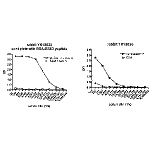
Figure 1 shows evaluation of rabbit antisera. A. Preimmune (open boxes) and
bleed 2
(filled boxes) rabbit antisera were tested for binding to BSA-DSE3 peptide. B.
Bleed 2
antiserum was evaluated for binding to BSA (triangles) and denatured PrP
(circles).
Figure 2 shows anti-peptide binding of seven anti-DSE3 monoclonal antibodies.
Each
antibody was evaluated for binding to a BSA-irrelevant peptide (triangles) and
BSA-
DSE3 peptide (circles). A positive control anti-BSA antibody bound to both BSA-
peptides.
Figure 3 shows anti-PrP binding of seven anti-DSE3 monoclonal antibodies, Each
antibody was evaluated for binding to denatured recombinant PrP (circles) and
His-
tagged captured PrP (triangles). A control anti-PrP antibody bound to both
denatured PrP
and His-tagged captured PrP.
Figure 4 shows anti-PrP antibody binding to tumor and normal cells. The DSE3
ab120
antibody or a control anti-PrP antibody was incubated at 10 ug/mL with various
cells.
Antibody binding was detected using an anti-rabbit IgG-AF488 or anti-mouse IgG-
AF488
6
SUBSTITUTE SHEET (RULE 26)
CA 02877505 2014-12-11
WO 2013/185215
PCT/CA2013/000569
secondary antibody, followed by fluorescence evaluation by flow cytometry (BD
FACS
Canto II).
Figure 5 shows titration of antibody binding to tumor and normal cells. The
DSE3 ab120
antibody or a control anti-PrP antibody (6H4) was incubated at varying
concentrations
with three tumor and two normal cells. Antibody detection was as described for
Figure 4.
Figure 6 reveals the conformational state of PrP resident on the surface of
various tumour
cell lines as determined by proteinase K titration. As shown, proteinase K
sensitivity is
high for the N-terminal region (upward triangles), low for the C-terminal
region
(downward triangles), and intermediate for the rigid loop region (squares)
within the
forms of PrP tested.
Figure 7 shows that the PrP presented by ovarian tumour cells is more
sensitive to
proteinase K digestion than is the PrP presented by normal, ovarian epithelial
cells.
Figure 8 shows the effect of paclitaxel treatment on antibody binding to
normal (dashed
lines) and ovarian tumour (solid lines) cells.
Figure 9 reveals the binding characteristics of AMF-I c-120 conjugated to
urease. A.
AMF-lc-120 (squares) and AMF-1c-120/urease conjugate (triangles) binding to
specific
peptide (filled) and non-specific peptide (open). B. AMF-lc-120 (squares) and
AMF-1c-
120/urease (triangles) binding to denatured PrP (filled) and captured His-PrP
(open);
Figure 10 shows binding of AMF-1c-120 and AMF-1c-120/urease conjugate to tumor
and
normal cells. AMF-lc-120 or AMF-1c-120/urease conjugate was incubated at
varying
concentrations with three tumor and five normal cells. Antibody binding was
detected
using an anti-rabbit IgG-AF488 secondary antibody, followed by fluorescence
evaluation
by flow cytometry.
Figure I 1 reveals the cytotoxicity of AMF-lc-120/urease in vitro. AMF- lc-
120, AMF-lc-
120/urease conjugate or urease were incubated with tumor cells for two hours.
Cells were
washed twice and then incubated with 20mM urea for 30 minutes. Cell viability
was
evaluated by addition of WST-1 followed by measuring absorbance after 16-20
hours.
7
SUBSTITUTE SHEET (RULE 26)
CA 02877505 2014-12-11
WO 2013/185215
PCT/CA2013/000569
Figure 12 shows the effect of lc-120/urease therapy on ES-2 tumor growth in
immunocompromised Rag2M mice. Mice were treated iv three times weekly with
vehicle
(squares), 1 c-120/urease at 183.3 jig/kg (diamonds), 1 c-120/urease at 368.7
jig/kg
(circles) or taxotere (triangles). Tumor growth was monitored by measuring
tumor
dimensions with calipers. Tumor volumes were calculated according to the
equation L x
W2 / 2.
Detailed Description of the Invention and Preferred Embodiments
As used herein, the term "PrP" refers to a mature human protein that comprises
the
expressed and processed product of the PRP gene, wherein the mature protein is
designated as residues 1-230 of UniProtKB/Swiss-Prot P04156. For present
purposes, the
term -PrP' further includes naturally occurring variants of this protein that,
in a misfolded
state, retain binding affinity for the present antibodies.
An "isolated antibody", as used herein, refers to an antibody that is
substantially free of
other antibodies having different antigenic specificities (e.g., an isolated
antibody that
specifically binds misfolded PrP is substantially free of antibodies that
specifically bind
antigens other than PrP proteins). An isolated antibody that specifically
binds a
misfolded human PrP protein may, however, have cross-reactivity to other
antigens, such
as misfolded PrP proteins from other species, but shows little or essentially
no affinity for
binding wild type human PrP. Moreover, an isolated antibody can be
substantially free of
other cellular material and/or chemicals. An isolated antibody also can be
substantially
free of other proteins of human origin.
The present invention relates to PrP antibodies that display an affinity and
preference for
binding to a form of PrP that presents an epitope comprising all or an
antibody-binding
part (comprising at least 5 contiguous residues) of the sequence MDEYSNQNN
(SEQ ID
No. 14) (sometimes referenced as DSE3). This region of the PrP protein is
referred to as
the "rigid loop", and represents residues 166-174 of the human prion protein.
In its
normal conformation, this epitope lies cryptically within the prion protein,
but becomes
accessible to the antibody when PrP misfolds, as a result for instance of
local
8
SUBSTITUTE SHEET (RULE 26)
CA 02877505 2014-12-11
WO 2013/185215
PCT/CA2013/000569
environmental shifts in conditions such as temperature or pH, or as a result
of aberrant
protein trafficking within the host cell or as a result of phenomena not yet
understood.
This misfolded form of PrP is found, for instance, on the surface of some PrP+
cancer
cells. Its presence on disease cells provides a therapeutic and diagnostic
target for the
present antibody, and means for achieving this are provided by the present
invention.
Thus, there is provided an antibody that comprises, as key features, an
affinity for binding
to the rigid loop of PrP, an affinity for binding to ovarian cancer cells that
are misfolded
PrP+, a preference for binding to ovarian cancer cells that are misfolded PrP+
relative to
normal ovarian epithelial cells, and complementarity determining regions
having the
sequences first recited above.
In embodiments, the antibody is an intact antibody comprising features common
to all
natural antibodies, e.g., a heavy chain and a light chain, each chain having a
constant
region and a variable region, each variable region comprising framework
regions (FRs)
and complementarity determining regions (CDRs). In the alternative, the
antibody is
provided as a fragment that is either monovalent or is bivalent, i.e., an
antibody fragment
comprising both "arms" of an intact antibody, joined through a linker that can
be
represented by the hinge region of the antibody or any equivalent. Such
bivalent
fragments include F(ab)2 fragments and any other bivalent fragment that
retains
preference for binding to misfolded PrP. In particular embodiments, the
bivalent
fragment is a F(ab')2 fragment, generated for instance by papain-based
digestion of the
parent antibody using standard procedures for digestion and subsequent
fragment
isolation. In the alternative, the fragment can be a so-called single chain Fv
(scFv),
consisting of the variable light and variable heavy antibody domains joined by
an amino
acid linker, or a bivalent form of a so-called diabody prepared using a 5
amino acid linker
such as SGGGG between the light and heavy chain variable domains and a C-
terminal
cysteine modification to GGC to give a final diabody product as VL-SGGG-VII-
GGC.
Still other bivalent fragments can be prepared by coupling the light and heavy
chain
variable domains through thioether linkages such as bis-maleimidomethyl ether
(BMME),
N.N'-p-phenylene dimaleimide (PDM and N,N'-bismaleimidohexane BM H), to
stabilize
the F(ab')2 fragments.
9
SUBSTITUTE SHEET (RULE 26)
CA 02877505 2014-12-11
WO 2013/185215
PCT/CA2013/000569
In the intact antibody or bivalent fragment, the CDRs comprise or consist of
the following
amino acid sequences:
For the heavy chain:
CDR1 TYAMG (SEQ ID No. 1)
CDR2 VITKSGNTYYASWAKG (SEQ ID No. 2)
CDR3 YGIGVSYYDI (SEQ ID No. 3)
For the light chain:
CDR1 QSSQSLYNKNWLS (SEQ Ill No, 4)
CDR2 KASTLES (SEQ ID No. 5)
CDR3 QGEFSCSSADCTA (SEQ ID No. 6)
Some variation is tolerable within these sequences, such as one or two
conservative
amino acid substitutions per CDR, and as many as 1, 2 or 3 CDRs having such
substitutions, but usually no more than about 5 substitutions within the CDRs
collectively. It will be appreciated that the conservative amino acid families
include (i) G,
A, V, L and 1; (ii) D and E; (iii) A, S and T; (iv) H, K and R; (v) N and Q;
and (vi) F, Y
and W. Thus, "conservative sequence modifications" can be made, and include
amino
acid modifications that do not significantly affect or alter the binding
characteristics of
the antibody containing the amino acid sequence. Such conservative
modifications
include amino acid substitutions, additions and deletions. Modifications
can be
introduced into an antibody of the invention at the genetic level by standard
techniques
known in the art, such as site-directed mutagenesis and PCR-mediated
mutagenesis.
In addition to the recited three CDRs present in each of the light and heavy
chain variable
regions, the heavy and light chains of the intact antibody comprise four
intervening
framework regions that present the CDRs in a conformation suitable for binding
to the
rigid loop of PrP, and constant regions that confer antibody effector
function. The CDRs
can be integrated into any suitable acceptor antibody, by grafting the present
CDRs into
the acceptor antibody, in accordance with practices and techniques well
established for
the production of chimeric, humanized and human antibodies.
It is well known in the art that the CDR3 domain alone can determine the
binding
specificity of an antibody for a cognate antigen and that multiple antibodies
can
SUBSTITUTE SHEET (RULE 26)
predictably be generated having the same binding specificity based on a common
CDR3
sequence. See, e.g., Klimka et al., British J. of Cancer 83(2):252-260 (2000);
Beiboer et
al., J. MoL Biol. 296:833-849 (2000); Rader etal., Proc. Natl. Acad. Sci.
U.S.A. 95:8910-
8915 (1998); Barbas etal., J. Am. Chem. Soc. 116:2161-2162 (1994); Barbas
etal., Proc.
Natl. Acad. Sci. U.S.A. 92:2529-2533 (1995); Ditzel et al., I Immunot 157:739-
749
(1996); Berezov et al., BIAjournal 8:Scientific Review 8 (2001); Igarashi et
al., J.
Biochem (Tokyo) 117:452-7 (1995); Bourgeois et al., J. Virol 72:807-10 (1998);
Levi et
al., Proc. Natl. Acad. Sci. U.S.A. 90:4374-8 (1993); Polymenis and Stoller, J.
Immunol.
152:5218-5329 (1994) and Xu and Davis, Immunity 13:37-45 (2000). See also, US
Patents Nos. 6,951,646; 6,914,128; 6,090,382; 6,818,216; 6,156,313; 6,827,925;
5,833,943; 5,762,905 and 5,760,185.
Accordingly, in one embodiment, the invention provides antibodies comprising
one or
more heavy and/or light chain CDR3 domains from the particular antibody
described
herein, wherein the antibody is capable of specifically binding to misfolded
human PrP.
Preferably, such antibodies (a) are capable of competing for binding with; (b)
retain the
functional characteristics; (c) bind to the same epitope; and/or (d) have a
similar binding
affinity as the particular antibodies described herein. In another embodiment,
the
antibodies of the invention may further include the CDR2 domain of the heavy
and/or
light chain variable region of the particular antibodies described herein, or
of another PrP
antibody, wherein the antibody is capable of specifically binding to misfolded
human PrP.
In another embodiment, the antibodies of the invention further may include the
CDR1 of
the heavy and/or light chain variable region of the particular antibodies
described herein,
or the CDR1 of the heavy and/or light chain variable region of another
misfolded human
PrP antibody, wherein the antibody is capable of specifically binding to
misfolded human
PrP.
To permit their use as cytotoxins per se, to inhibit directly the growth or
proliferation of
misfolded PrP+ disease cells presenting the MDEYSNQNN (SEQ ID No. 14) epitope,
the
antibodies can exert their anti-cancer activity through endogenous mechanisms
such as
complement-mediated cytotoxicity (CDC) and/or antibody-dependent cellular
cytotoxicity (ADCC). It will be appreciated that the antibodies can be
engineered or
selected to have altered effector function, to enhance effectiveness in
treating cancer.
11
CA 2877505 2019-10-04
CA 02877505 2014-12-11
WO 2013/185215
PCT/CA2013/000569
Cysteine residues, for instance, may be introduced to the Fe region to allow
interchain
disulfide bond formation. The resulting homodimeric antibody may have improved
internalization capacity, and more importantly may have increased complement
dependent cytotoxicity (CDC) and/or ADCC activities. Homodimeric antibodies
with
enhanced anti-tumour activity may also be prepared using heterobifunctional
cross-
linkers as described in Wolff et al, Cancer Research 53:2560-2565 (1993).
Alternatively,
an antibody can be engineered which has dual Fe regions and enhanced CDC and
ADCC
activity.
Particularly suitable acceptor antibodies are antibodies already known to have
PrP
binding affinity. Such donor antibodies are most desirably of human origin,
but they can
also derive from acceptor antibodies of non-human origin, including mouse,
rat, rabbit,
goat, sheep, primate and the like. It will be appreciated that human antibody
acceptor
sequences different from those exemplified herein can be identified and used
to
accommodate the presently desired CDRs. This is achieved by modeling the
structure of
a preferred antibody using for instance the Swiss-
Model
[http://swissmodel.expasy.org/repository] or similar software and selecting,
from among
the numerous human antibody sequences available in public databases, a human
acceptor
antibody sequence that, with CDR sequences altered as herein preferred,
approximates
the same structural conformation as the preferred antibodies. In embodiments,
the
acceptor antibodies, and the resulting present antibodies, are of the IgG1
isotype, but they
may also be IgG2 or IgG4. Moreover, the isotype of the antibody, as dictated
by the
constant region, can be manipulated to alter or eliminate the effector
function of the
resulting antibody. That is, the constant region of the present antibodies is
either wild
type human antibody constant region, or a variant thereof that incorporates
amino acid
modifications, i.e., amino acid additions, substitutions or deletions that
alter the effector
function of the constant region, such as to enhance serum half-life, reduce or
enhance
complement fixation, reduce or enhance antigen-dependent cellular cytotoxicity
and
improve antibody stability. The number of amino acid modifications in the
constant
region is usually not more than 20, such as 1-10 e.g., 1-5 modifications,
including
conservative amino acid substitutions.
In embodiments, the half-life of the antibody is improved by incorporating one
or more
amino acid modifications, usually in the form of amino acid substitutions, for
instance at
12
SUBSTITUTE SHEET (RULE 26)
CA 02877505 2014-12-11
WO 2013/185215
PCT/CA2013/000569
residue 252, e.g., to introduce Thr, at residue 254, e.g., to introduce Ser,
and/or at residue
256 e.g., to introduce Phe. Still other modifications can be made to improve
half-life,
such as by altering the CHI or CL region to introduce a salvage receptor
motif, such as
that found in the two loops of a CH2 domain of an Fe region of an IgG. Such
alterations
are described for instance in US 5869046 and US 6121022.
Altered Clq binding, or reduced complement dependent cytotoxicity, can be
introduced
by altering constant region amino acids at locations 329, 331 and 322, as
described in US
6194551. The ability of the antibody to fix complement can further be altered
by
introducing substitutions at positions 231 and 239 of the constant region, as
described in
W094/029351.
The framework regions of the light and heavy chains of the present antibodies
and
fragments also desirably have the sequence of a human antibody variable
region, but
incorporating the CDRs herein specified. In embodiments, the heavy chain
variable
region is human IgG4 in origin, which is generally considered to be inert for
effector
function. In specific embodiments, the heavy chain variable region is that of
human IgG,
such as the human IgG1 antibody variant having the sequence designated Genbank
gi
2414502. Alternatively, the heavy chain variable region is that of human IgG4
antibody
species designated Genbank gi 2414502.
The framework regions of the heavy and light chains of the present antibodies
may also
incorporate amino acid modifications, i.e., amino acid deletions, additions or
substitutions, which further improve upon the properties of the antibody or
fragment, in
accordance with techniques established for antibody humanization. Such
framework
modifications can be modeled on the framework regions of antibody sequences
provided
in public databases, and on framework regions of antibodies known to bind PrP,
such as
those antibodies referenced in the background section hereof. Preferred
framework
substitutions are those which yield antibodies having a greater preference for
binding
misfolded PrP associated with disease cells.
Framework modifications can also be made to reduce immunogenicity of the
antibody or
to reduce or remove T cell epitopes that reside therein, as described for
instance by Carr
et al in US2003/0153043.
13
SUBSTITUTE SHEET (RULE 26)
CA 02877505 2014-12-11
WO 2013/185215
PCT/CA2013/000569
Antibodies of the invention can also be altered in the variable region to
eliminate one or
more glycosylation sites, and/or to improve physical stability of the
antibody. For
example, in one embodiment, the physical stability of the antibody is improved
by
substituting the serine at position 228 of the variable region with a proline
residue (i.e.,
the antibody has a variable region comprising a S228P mutation). The S228P
alteration
significantly stabilizes the antibody structure against the formation of
intrachain disulfide
bonds. In another embodiment, the variable region is altered to eliminate one
or more
glycosylation sites resident in the variable region. More particularly, it is
desirable in the
sequence of the present antibodies to eliminate sites prone to glycosylation.
This is
achieved by altering the occurrence of one or more N-X-(S/T) sequences that
occur in the
parent variable region (where X is any amino acid residue), particularly by
substituting
the N residue and/or the S or T residue.
Antibodies of the invention can be engineered to include a variety of constant
regions. In
one embodiment, the antibody comprises a constant region the sequence of which
corresponds to the constant region of an antibody of human origin, such as a
human IgG1
constant region. In a particular embodiment, the constant region is inert for
effector
function (e.g., essentially devoid of effector function). In a specific
embodiment the
constant region is a human IgG4 constant region.
In accordance with embodiments of the present invention, the heavy and light
chain
variable regions comprise a heavy chain variable region of SEQ ID No.8, and/or
a light
chain variable region having SEQ ID No.7, as follows:
Light chain variable region (VL):
MDTRAPTQLLGLLLLWLPGATFAQVLTQTPSPVSAAVGGTVTINCQSSQSLYNKNWLSWYQKKPGQPPKLL
YKASTLESGVSSRFKGSGSGTQFTLTI SGVQCDDAATYYCQGE FS CSSADCTAFGGGTEVVV [SEQ ID
No. 7]
Heavy chain variable region (VH):
METGLRWLLLVAVLKGVQCQSVEESGGHLVTPGTPLTLTCTVSGIDLSTYANGWVRQAPGKGLEW I GVI TKS
GNTYYASWAKGRFAIS KTSTTVDLKITS PTTEDTATYFCGRYG IGVSY YDIWGPGTLVTVS SGQ [SEQ ID
No.8]
14
SUBSTITUTE SHEET (RULE 26)
CA 02877505 2014-12-11
WO 2013/185215
PCT/CA2013/000569
Thus, the antibody may be of any useful class, including IgA, IgD, IgE, IgG
and IgM, and
of any isotype including IgGl, IgG2, IgG3, and IgG4. Preferred antibodies are
IgG1 . In
more specific and preferred embodiments, the entire light and heavy chains of
the intact
antibody are set out below as SEQ ID Nos. 9 and 10, respectively:
Entire Light chain:
MDTRAPTQLLGLLLLWLPGATFAQVLTQTPSPVSAAVGGTVTINCQSSQSLYNKNWLSWYQKKPGQPPKLLI
YKASTLESGVSSRFKGSGSGTQFTLTISGVQCDDAATYYCQGEFSCSSADCTAFGGGTEVVVKGDPVAPTVL
IFPPAADQVATGTVTIVCVANKYFPDVTVTWEVDGTTQTTGIENSKTPQNSADCTYNLSSTLTLTSTQYNSH
KEYTCKVTQGTTSVVQSFNRGDC[SEQ1E) No. 9];
Entire Heavy chain:
METGLRWLLLVAVLKGVQCQSVEESGGHLVTPGTPLTLTCTVSGIDLSTYAMGWVRQAPGKGLEWIGVITKS
GNTYYASWAKGRFAISKTSTTVDLKITSPTTEDTATYFCGRYGIGVSYYDIWGPGTLVTVSSGQPKAPSVFP
LAPCCGDTPSSTVTLGCLVKGYLPEPVTVTWNSGTLTNGVRTFPSVRQSSGLYSLSSVVSVTSSSQPVTCNV
AHPATNTKVDKTVAPSTCSKPTCPPPELLGGPSVFIFPPKPKDTLMISRTPEVTCVVVDVSQDDPEVQFTWY
INNEQVRTARPPLREQQFNSTIRVVSTLPIAHQDWLRGKEFKCKVHNKALPAPIEKTISKARGQPLEPKVYT
MGPPREELSSRSVSLTCMINGFYPSDISVEWEKNGKAEDNYKTTPAVLDSDOSYFLYSKLSVPTSEWQRGDV
FTCSVMHEALHNHYTQKSISRSPGK [SEQIC)No.10];
In yet another embodiment, the Fe region is modified to increase the ability
of the
antibody to mediate antibody dependent cellular cytotoxicity (ADCC) and/or to
increase
the affinity of the antibody for an Fey receptor by modifying one or more
amino acids at
the following positions: 238, 239, 248, 249, 252, 254, 255, 256, 258, 265,
267, 268, 269,
270, 272, 276, 278, 280, 283, 285, 286, 289, 290, 292, 293, 294, 295, 296,
298, 301, 303,
305, 307, 309, 312, 315, 320, 322, 324, 326, 327, 329, 330, 331, 333, 334,
335, 337, 338,
340, 360, 373, 376, 378, 382, 388, 389, 398, 414, 416, 419, 430, 434, 435,
437, 438 or
439. This approach is described further in PCT Publication WO 00/42072.
Moreover,
the binding sites on human IgG1 for FcyR1, FcyRII, FcyRIII and FcRn have been
mapped
and variants with improved binding have been described (see Shields et al.
(2001)J. Biol.
Chem. 276:6591-6604). Specific mutations at positions 256, 290, 298, 333, 334
and 339
were shown to improve binding to FcyRIII. Additionally, the following
combination
mutants were shown to improve FeyRIII binding: T256A/S298A, 5298A/E333A,
S298A/K224A and S298A/E333A/K334A.
Antibodies of the present disclosure can contain one or more glycosylation
sites in either
the light or heavy chain variable region. Such glycosylation sites may result
in increased
immunogenicity of the antibody or an alteration of the pK of the antibody due
to altered
SUBSTITUTE SHEET (RULE 26)
CA 02877505 2014-12-11
WO 2013/185215
PCT/CA2013/000569
antigen binding (Marshall et al (1972) Annu Rev Biochem 41:673-702; Gala and
Morrison
(2004) J lmmunol 172:5489-94; Wallick et al (1988) J Exp Med 168:1099-109;
Spiro
(2002) Glycobiology 12:43R-56R; Parekh et al (1985) Nature 316:452-7; Mimura
et at
(2000) Mol Immunol 37:697-706). Glycosy lation has been known to occur at
motifs
containing an N-X-S/T sequence. In some instances, it is preferred to have an
antibody
that does not contain variable region glycosylation. This can be achieved
either by
selecting antibodies that do not contain the glycosylation motif in the
variable region or
by mutating residues within the glycosylation region.
For example, aglycoslated antibodies can be made (i.e., which lack
glycosylation).
Glycosylation can be altered to, for example, increase the affinity of the
antibody for
antigen. Such carbohydrate modifications can be accomplished by, for example,
altering
one or more sites of glycosylation within the antibody sequence. For example,
one or
more amino acid substitutions can be made that result in elimination of one or
more
variable region framework glycosylation sites to thereby eliminate
glycosylation at that
site. Such aglycosylation may increase the affinity of the antibody for
antigen. See, e.g.,
U.S. Patent Nos. 5,714,350 and 6,350,861.
Additionally or alternatively, the antibody can have an altered type of
glycosylation, such
as a hypofucosylated antibody having reduced amounts of fucosyl residues or an
antibody
having increased bisecting GleNac structures. Such altered glycosylation
patterns have
been demonstrated to increase the ADCC ability of antibodies. Such
carbohydrate
modifications can be accomplished by, for example, expressing the antibody in
a host cell
with altered glycosylation machinery. Cells with altered glycosylation
machinery have
been described in the art and can be used as host cells in which to express
recombinant
antibodies of the invention to thereby produce an antibody with altered
glycosylation.
For example, the cell lines Ms704, Ms705, and Ms709 lack the
fucosyltransferase gene,
FUT8 (a (1,6)-fucosyltransferase), such that antibodies expressed in the
Ms704, Ms705,
and Ms709 cell lines lack fucose on their carbohydrates. The Ms704, Ms705, and
Ms709
FUT8 -ir cell lines were created by the targeted disruption of the FUT8 gene
in CI10/DG44
cells using two replacement vectors (see U.S. Patent Publication No.
20040110704 and
Yamane-Ohnuki et al. (2004) Biotechnol Bioeng 87:614-22). As another example,
EP
1,176,195 describes a cell line with a functionally disrupted FUT8 gene, which
encodes a
fueosyl transferase, such that antibodies expressed in such a cell line
exhibit
16
SUBSTITUTE SHEET (RULE 26)
CA 02877505 2014-12-11
WO 2013/185215
PCT/CA2013/000569
hypofucosylation by reducing or eliminating the a-1,6 bond-related enzyme. EP
1,176,195 also describes cell lines which have a low enzyme activity for
adding fucose to
the N-acetylglucosamine that binds to the Fe region of the antibody or does
not have the
enzyme activity, for example the rat myeloma cell line YB2/0 (ATCC CRL 1662).
PCT
Publication WO 03/035835 describes a variant CHO cell line, Lec13 cells, with
reduced
ability to attach fucose to Asn(297)-linked carbohydrates, also resulting in
hypofucosylation of antibodies expressed in that host cell (see also Shields
et al. (2002)J.
Biol. Chem. 277:26733-26740). Antibodies with a modified glycosylation profile
can
also be produced in chicken eggs, as described in PCT Publication WO
06/089231.
Alternatively, antibodies with a modified glycosylation profile can be
produced in plant
cells, such as Lonna. Methods for production of antibodies in a plant system
are
disclosed in the U.S. Patent application corresponding to Alston & Bird LLP
attorney
docket No. 040989/314911, filed on August 11, 2006. PCT Publication WO
99/54342
describes cell lines engineered to express glycoprotein-modifying glycosyl
transferases
(e.g., J3(1,4)-N-acetylglucosaminyltransferase III (GnTIII)) such that
antibodies expressed
in the engineered cell lines exhibit increased bisecting G1cNac structures
which results in
increased ADCC activity of the antibodies (see also Umana et al. (1999) Nat.
Biotech.
17:176-180). Alternatively, the fucose residues of the antibody can be cleaved
off using a
fucosidase enzyme; e.g., the fucosidase a-L-fucosidase removes fucosyl
residues from
antibodies (Tarentino etal. (1975) Biochem. 14:5516-23).
In addition, the antibody can be pegylated, for example, to increase the
biological (e.g.,
serum) half-life of the antibody. To pegylate an antibody, the antibody, or
fragment
thereof, typically is reacted with polyethylene glycol (PEG), such as a
reactive ester or
aldehyde derivative of PEG, under conditions in which one or more PEG groups
become
attached to the antibody or antibody fragment. Preferably, the pegylation is
carried out
via an acylation reaction or an alkylation reaction with a reactive PEG
molecule (or an
analogous reactive water-soluble polymer). As used herein, the term
"polyethylene
glycol" is intended to encompass any of the forms of PEG that have been used
to
derivatize other proteins, such as mono (Cl-do) alkoxy- or aryloxy-
polyethylene glycol
or polyethylene glycol-maleimide. In certain embodiments, the antibody to be
pegylated
is an aglycosylated antibody. Methods for pegylating proteins are known in the
art and
can be applied to the antibodies of the invention. See, e.g,, EP 0 154 316 and
EP 0 401
384.
17
SUBSTITUTE SHEET (RULE 26)
CA 02877505 2014-12-11
WO 2013/185215
PCT/CA2013/000569
Thus, the present invention includes antibodies that comprise the CDR
sequences first
recited above, and otherwise can be chimeric, humanized, human or otherwise
engineered
antibodies.
The antibodies and binding fragments are useful both for diagnostic purposes,
including
in vivo imaging to identify endogenous sites of misfolded PrP, and for sample
testing to
detect misfolded PrP as a soluble protein or as a cell-borne surface protein.
The
antibodies and binding fragments are also useful for therapeutic purposes to
treat diseases
in which misfolded PrP is implicated.
For either purpose, the antibody or binding fragment can be conjugated to an
appropriate
agent, to form an immunoconjugate. Agents appropriate for treating disease
include
cytotoxic agents or toxins that include chemotherapeutics and
radiotherapeutics. For
diagnostic purposes, appropriate agents are detectable labels that include
radioisotopes or
fluorescent markers for whole body imaging, and radioisotopes, enzymes,
fluorescent
labels and the like for sample testing. In these diagnostic approaches, the
agent can serve
as a label either directly, as such, or indirectly as an agent that will bind
a desired label
such as a labeled secondary antibody that binds the agent.
For diagnostics, the detectable labels can be any of the various types used
currently in the
field of in vitro diagnostics, including particulate labels including
biotin/streptavidin,
metal sols such as colloidal gold, radioactive isotopes such as 1125 or Tc99
presented for
instance with a peptidic chelating agent of the N2S2, N3S or N4 type,
chromophores
including fluorescent markers such as FITC and PE, luminescent markers,
phosphorescent markers and the like, as well as enzyme labels that convert a
given
substrate to a detectable marker, and polynucleotide tags that are revealed
following
amplification such as by polymerase chain reaction. Suitable enzyme labels
include
horseradish peroxidase, alkaline phosphatase and the like. For instance, the
label can be
the enzyme alkaline phosphatase, detected by measuring the presence or
formation of
chemiluminescence following conversion of 1,2 dioxetane substrates such as
adamantyl
methoxy phosphoryloxy phenyl dioxetane (AMPPD), disodium 3-(4-
(methoxyspiro{1,2-
dioxetane-3,2'-(5'-chloro)tricyclo {3.3.1.1 3,7}decan}-4-y1) phenyl phosphate
(CSPD), as
well as CDP and CDP-star or other luminescent substrates well-known to those
in the
18
SUBSTITUTE SHEET (RULE 26)
art, for example the chelates of suitable lanthanides such as Terbium(III) and
Europium(III). The detection means is determined by the chosen label.
Appearance of
the label or its reaction products can be achieved using the naked eye, in the
case where
the label is particulate or chromatic and accumulates at appropriate levels,
or using
instruments such as a spectrophotometer, a luminometer, a fluorimeter, and the
like, all in
accordance with standard practice.
Likewise, imaging agents may be included in the composition or in additional
compositions. Suitable imaging agents include commercially available agents
used in
positron emission tomography (PET), computer assisted tomography (CAT), single
photon emission computerized tomography, x-ray, fluoroscopy, and magnetic
resonance
imaging (MRI).
Imaging agents useful with the antibody to screen for endogenous cancer sites
or for
misfolded PrP in plaque or other forms include metals, radioactive isotopes
and
radioopaque agents (e.g., gallium, technetium, indium, strontium, iodine,
barium, bromine
and phosphorus-containing compounds), radiolucent agents, contrast agents,
dyes (e.g.,
fluorescent dyes and chromophores) and enzymes that catalyze a colorimetric or
fluorometric reaction. In general, such agents may be attached or entrapped
using a
variety of techniques as described above, and may be present in any
orientation. See, e.g.,
U.S. Pat. Nos. 6,159,443 and 6,391,280.
Contrast agents according to the present invention are useful in the imaging
modalities,
such as X-ray contrast agents, light imaging probes, spin labels or
radioactive units.
Examples of suitable materials for use as contrast agents in MRI include the
gadolinium
chelates currently available, such as diethylene triamine pentaacetic acid
(DTPA) and
gadopentotate dimeglumine, as well as iron, magnesium, manganese, copper, and
chromium. Examples of materials useful for CAT and x-rays include iodine based
materials, such as ionic monomers typified by diatrizoate and iothalamate, non-
ionic
monomers such as iopamidol, isohexol, and ioversol, non-ionic dimers, such as
iotrol and
iodixanol, and ionic dimers, for example, ioxagalte.
Preferred agents for use with PET scan include N13 and fluorodeoxyglucose
(FDG).
19
CA 2877505 2019-10-04
CA 02877505 2014-12-11
WO 2013/185215
PCT/CA2013/000569
For therapy, a cytotoxin can be conjugated with the antibody or binding
fragment through
non-covalent interaction, but more desirably, by covalent linkage either
directly or, more
preferably, through a suitable linker. In a preferred embodiment, the
conjugate comprises
a cytotoxin and an antibody or any binding fragment thereof. Immunoconjugates
of the
antibody and cytotoxin are made using a variety of bifunctional protein
coupling agents
such as N-succinimidy1-3-(2-pyridyldithiol) propionate, iminothiolane,
bifunctional
derivatives of imidoesters such as dimethyl adipimidate HC1, active esters
such as
disuccinimidyl suberate, aldehydes such as glutaraldehyde, bis-azido compounds
such as
bis-(p-diazoniumbenzoy1)-ethylenediamine), diisocyanates such as toluene 2,6-
d iisocyanate, and bis-active fluorine compounds (such as 1 ,5-d
ifluoro-2,4-
dinitrobenzene). C'4-labeled 1 soth
iocyano benzy1-3-methy ldiethy lene
triaminepentaacetic acid (MX-DTPA) is a chelating agent suitable for
conjugation of
radionuclide to the antibody. One
particularly useful linker is succinimidy1-(4-
iodoacetyl)aminobenzoate (SIAB) which is a mid-length crosslinker for amine-to-
sulfhydryl conjugation via N-hydroxysuccinimide ester and iodoacetyl reactive
groups.
The cytotoxin component of the immunoconjugate can be any agent that is
cytotoxic to
the cells targeted by the antibody such as a chemotherapeutic agent, a toxin
such as an
enzymatically active toxin of bacterial, fungal, plant or animal origin, or
fragments
thereof, or a small molecule toxin, or a radioactive isotope such as 212Bi,
1311, 1211n, 111In,
90Y and 186Re, or any other agent that acts to inhibit the growth or
proliferation of a target
disease cell.
Chemotherapeutic agents useful in such immunoconjugates include maytansinoids
such
as DM-1 and DM-4, adriamycin, doxorubicin, epirubicin, 5-fluoroouracil,
cytosine
arabinoside ("Ara-C"), cyclophosphamide, thiotepa, busulfan, cytoxin, taxanes,
e.g.
paclitaxel, and docetaxel, taxotere, methotrexate, cisplatin, melphalan,
vinblastine,
bleomycin, etoposide, ifosamide, mitomycin C, mitoxantrone, vincristine,
vinorelbine,
carboplatin, teniposide, daunomycin, carminomycin, aminopterin, dactinomycin,
mitomycins, esperamicins, 5-FU, 6-thioguanine, 6-mercaptopurine, actinomycin
D,
VP-16, chlorambucil, melphalan, and other related nitrogen mustards. Also
useful are
hormonal agents that act to regulate or inhibit hormone action on tumors such
as
tamoxifen and onapristone. Toxins and fragments thereof which can be used
include
SUBSTITUTE SHEET (RULE 26)
diphtheria A chain, nonbonding active fragments of diphtheria toxin, cholera
toxin,
botulinus toxin, exotoxin A chain (from Pseudomonas aeruginosa), ricin A
chain, abrin A
chain, modeccin A chain, alpha-sarcin, Aleurites .fordii proteins, dianthin
proteins,
Phytolaca americana proteins (PAPI, PAPII, and PAP-S), Momordica charantia
inhibitor, curcin, crotin, sapaonaria, officinalis inhibitor, gelonin,
saporin, mitogellin,
restrictocin, phenomycin, enomycin, and the tricothcenes. Small molecule
toxins include,
for example, calicheamicins, palytoxin and CC1065. Also useful are the taxanes
including taxol and paclitaxel.
In one particular aspect, the present invention provides immunoconjugates that
incorporate urease, as a cytotoxic component, conjugated with at least one
antigen
binding site of the antibody herein characterized. The incorporation of urease
as the
cytotoxic component of an immunoconjugate has been described in the literature
(see US
7211250 and US 7264800 both to Helix BioPharma Corporation). While urease
itself is
not cytotoxic, its cytotoxicity arises from its ability to convert urea to pH
elevating
compounds such as ammonia. Thus, the urease-based immunoconjugate may raise
the
pH of interstitial fluid to which the cancer cells are exposed, by addition of
urease to the
interstitial fluid in the subject. Urease can convert the substrate urea to
ammonia and
carbamate. This enzymatic activity may increase the pH making the environment
more
basic. The environment around a cancer cell is typically acidic (Webb, S. D.,
et al. (2001)
Novartis Found Symp. 240:169-81). Thus, by raising the pH of the extracellular
environment in this manner, growth of the cancer cell is inhibited.
Accordingly, addition
of the immunoconjugate in certain embodiments of the invention causes the pH
of the
interstitial fluid, and particularly that surrounding misfolded PrP+ disease
cells, to be
raised by at least 0.1 pH unit, e.g., 0.1-0.5 pH units or greater.
As used herein, the term "urease" refers to an enzyme having the enzymatic
activity of a
urea amidohydrolase (E.C. 3.5.1.5). Urease also includes proteins comprising
the entire
urease, subunits, or fragments thereof, and/or urease with amino acid
substitutions,
deletions or additions that preserve the urea amidohydrolase activity of the
polypeptide. A
truncated urease sequence as used herein is a fragment of urease that is free
from a
portion of the intact urease sequence beginning at either the amino or carboxy
terminus of
urease. Methods for isolating native urease and for identifying active
fragments and
21
CA 2877505 2019-10-04
modified urease polypeptides are given below.
In embodiments of the invention, the urease is jack bean urease having SEQ ID
No.13, as
shown below:
MKLSPREVEKLGLHNAGYLAQKRLARGVRLNYTEAVALIASQIM
EYARDGEKTVAQLMCLGQHLLGRRQVL PAVPHLLNAVQVEATFP
DGTKLVTVHD P I S RE NGE LQEAL FG S L L PVP S LDKFAE T KEDNR
IPGE ILCEDE CLTLNI GRKAVILKVTSKGDRP I QVGSHYTIF IEV
NPYL T FDRRKAYGMRLNIAAGTAVRFE PGDCKS VTLVS I EGNKV
IRGGNAIADGPVNETNLEAAMHAVRSKGFGHEEEKDASEGFTKE
DPNCPENTFIHRKEYANKYGPTTGDKIRLGDTNLLAEIEKDYAL
YGDECVFGGGKVIRDGMGQSCGHPPAISLDTVITNAVI IDYTGI
IKAD I GI KDGL IAS I GKAGNPD IMNGVFSNM I I GANTEVIAGEG
LIVTAGAIDCHVHYI CPQLVYEAIS SG I TTLVGGGTGPAACTRA
TTCTPS P TQMRLMLQ S TDDLPLNEGFTGKGS S SKPDELHE I I KA
GANCLKLHEDWGSTPAAIDNCLTIAEHHDIQ INIHTDTLNEAGF
VEHS IAAFKGRT IHTYHS EGAGGGHAPD I I KVCGIKNVL PS STN
PTRPLTSNTIDEHLDMLMVCHHLDRE I PEDLAFAHSRIRKKTIA
AEDVLND I GAI S I IS SD SQAMGRVGEV I SRTWQ TADKMKAQ TGP
LKCD S S DNDNFR I RRY IAKYT INPA IANGF S QYVGSVEVGKLAD
LVMWKPSFFGTKPEMVIKGGMVAWADIGDPNAS I PT PE PVKMRP
MYGTLGKAGCALS TAFVSKAALDQRVNVLYGLNKRVEAVSNVRK
LTKLDMKLNDALPE I TVDPESYTVKADGKLLCVSEATTVPLSRN
YFLF (SEQ ID No.13 )
Alternatively, the urease may be of any origin, including, e.g., bacteria,
plants, fungi and
viruses. A number of studies have provided detailed information about the
genetics of
ureases from a variety of evolutionarily diverse bacteria, plants, fungi and
viruses
(Mobley, H. L. T. et al. (1995) Microbiol. Rev. 59: 451-480; Eur, J. Biochem.,
175, 151-
165 (1988); Labigne, A. (1990) International publication No. WO 90/04030;
Clayton, C.
L. et al. (1990) Nucleic Acid Res. 18, 362; and U.S. Pat. Nos. 6,248,330 and
5,298,399).
Of particular interest is urease that is found in plants (Sirko, A. and
Brodzik, R. (2000)
Acta Biochim Pol 47(4):1189-95). One exemplary plant urease is jack bean
urease.
The amino acid sequences of other useful urease sequences are available in
public
databases, e.g., Entrez (www.ncbi.nlm.nih.gov/Entrez/). Additionally, primers
that are
useful for amplifying ureases from a wide variety of organisms may be utilized
by
employing the CODEHOP (COnsensus-DEgenerate Hybrid Oligonucleotide Primer) as
described in Rose, et al. (1998) Nucl. Acids Res. 26:1628.
22
CA 2877505 2019-10-04
CA 02877505 2014-12-11
WO 2013/185215
PCT/CA2013/000569
Thus useful forms of urease include the naturally occurring forms as well as
functionally
active variants thereof. Two general types of amino acid sequence variants are
contemplated. Amino acid sequence variants are those having one or more
substitutions
in specific amino acids which do not destroy the urease activity. These
variants include
silent variants and conservatively modified variants which are substantially
homologous
and functionally equivalent to the native protein. A variant of a native
protein is
"substantially homologous" to the native protein when at least about 80%, more
preferably at least about 90%, even more preferably at least about 95%, yet
even more
preferably 98%, and most preferably at least about 99% of its amino acid
sequence is
identical to the amino acid sequence of the native protein. A variant may
differ by as few
as 1 or up to 10 or more amino acids.
A second type of variant includes size variants of urease which are isolated
active
fragments of urease. Size variants may be formed by, e.g., fragmenting urease,
by
chemical modification, by proteolytic enzyme digestion, or by combinations
thereof.
Additionally, genetic engineering techniques, as well as methods of
synthesizing
polypeptides directly from amino acid residues, can be employed to produce
size variants.
By -functionally equivalent" is intended that the sequence of the urease
variant defines a
chain that produces a protein having substantially the same biological
activity as the
native urease. Such functionally equivalent variants that comprise substantial
sequence
variations are also encompassed by the invention. Thus, a functionally
equivalent variant
of the native urease protein will have a sufficient biological activity to be
therapeutically
useful. Methods are available in the art for determining functional
equivalence. Biological
activity can be measured using assays specifically designed for measuring
activity of the
native urease protein, as exemplified herein.
Because of the degeneracy of the genetic code, a multitude of nucleic acid
sequences
encoding urease may be produced, some of which may bear minimal sequence
homology
to known urease nucleic acid sequences. Such "silent variations" are one
species of
"conservatively modified variations", discussed below. The invention embraces
any
possible codon variation of nucleic acid sequences encoding a polypeptide with
urease
activity. As well, urease polypeptides include one or more conservatively
modified
variations (or simply "conservative variations") of the sequences of known
urease
23
SUBSTITUTE SHEET (RULE 26)
CA 02877505 2014-12-11
WO 2013/185215
PCT/CA2013/000569
polypeptide sequences. Such conservative variations comprise substitutions,
additions or
deletions that alter, add or delete a single amino acid or a small percentage
of amino
acids. One of ordinary skill in the art will recognize that an individual
substitution,
deletion, or addition that substitutes, deletes, or adds a single amino acid
or a small
percentage of amino acids (typically less than 5%, more typically less than
4%, 2%, 1%,
or less) in a sequence typically constitutes conservative variations where
such changes
result in the deletion of an amino acid, addition of an amino acid, or
substitution of an
amino acid with a chemically similar amino acid.
The urease protein sequences, including conservatively substituted sequences,
can be
present as part of larger polypeptide sequences such as occur upon the
addition of one or
more domains for purification of the protein (e.g., poly his segments, FLAG
tag
segments, etc.), e.g., where the additional functional domains have little or
no effect on
the activity of the urease protein portion of the immunoconjugate, or where
the additional
domains can be removed by post synthesis processing steps, such as by
treatment with a
protease.
The active agents may be joined together in any order to form the
immunoconjugate.
Thus, where the urease may be joined to either the amino or carboxy termini of
the
targeting antibody. The antibody may also be joined to an internal region of
the urease, or
conversely, the urease may be joined to an internal location of the antibody,
as long as the
attachment does not interfere with the respective activities of the molecules.
The targeting antibody and the urease may be attached by any of a number of
means well
known to those of skill in the art. Typically, the active agent is conjugated,
either directly
or through a linker (spacer), to the antibody. However, where both the
antibody and the
urease are entirely genetically encoded, it may be preferable to recombinantly
express the
chimeric molecule as a fusion protein.
The procedure for attaching an agent to an antibody or other polypeptide
targeting
molecule will vary according to the chemical structure of the agent.
Polypeptides
typically contain a variety of functional groups; e.g., carboxylic acid (COOH)
or free
amine ( __ NH2) groups, which are available for reaction with a suitable
functional group
on a urease to bind the antibody thereto.
24
SUBSTITUTE SHEET (RULE 26)
CA 02877505 2014-12-11
WO 2013/185215
PCT/CA2013/000569
Alternatively, the antibody and/or urease may be derivatized to expose or
attach
additional reactive functional groups. The derivatization may involve
attachment of any
of a number of linker molecules. The linker is capable of forming covalent
bonds to both
the antibody and the urease. Suitable linkers include those first mentioned
above, and
particularly straight or branched-chain carbon linkers, heterocyclic carbon
linkers, or
peptide linkers. The linkers may be joined to the constituent amino acids
through their
side groups (e.g., through a disulfide linkage to cysteine). However, in a
preferred
embodiment, the linkers will be joined to the alpha carbon amino and carboxyl
groups of
the terminal amino acids.
A bifunctional linker having one functional group reactive with a group on a
particular
agent, and another group reactive with an antibody, may be used to form the
desired
iminunoconjugate. Alternatively, derivatization may involve chemical treatment
of the
targeting moiety, e.g., glycol cleavage of the sugar moiety of the
glycoprotein antibody
with periodate to generate free aldehyde groups. The free aldehyde groups on
the
antibody may be reacted with free amine or hydrazine groups on an agent to
bind the
urease thereto. (see U.S. Pat. No, 4,671,958). Procedures for generation of
free sulfhydryl
groups on polypeptide, such as antibodies or antibody fragments, are also
known (see
U.S. Pat. No, 4,659,839).
1mmunoconjugates of the antibody and urease can be made using a variety of
bifunctional
protein coupling agents such as N-succinimidy1-3-(2-pyridyldithiol)
propionate,
iminothiolane, bifunctional derivatives of imidoesters such as dimethyl
adipimidate HCl,
active esters such as disuccinimidyl suberate, aldehydes such as
glutaraldehyde, bis-azido
compounds such as bis-(p-diazoniumbenzoyI)-ethylenediamine), diisocyanates
such as
toluene 2,6-diisocyanate, and bis-active fluorine compounds (such as 1,5-
difluoro-2,4-
dinitrobenzene).
In some circumstances, it is desirable to liberate the urease from the
antibody when the
conjugate has reached its target site. Therefore, conjugates comprising
linkages which are
cleavable in the vicinity of the target site may be used. Cleaving of the
linkage may be
prompted by enzymatic activity or conditions to which the conjugate is
subjected either
inside the target cell or in the environment of the target site. It should be
appreciated that
SUBSTITUTE SHEET (RULE 26)
when the target site is a tumor, a linker which is cleavable under conditions
present at the
tumor site (e.g. when exposed to tumor-associated enzymes or acidic pH) may be
used.
Useful linkers that are pH sensitive are described, for instance, in WO
86/001409, WO
94/020487, WO 2009/158668 and WO 2010/053596.
A number of other useful cleavable linkers are known to those of skill in the
art (see U.S.
4,618,492; 4,542,225, and 4,625,014.) The mechanisms for release of an active
agent
from these linker groups include, for example, irradiation of a photolabile
bond and acid-
catalyzed hydrolysis. U.S. 4,671,958, for example, includes a description of
immunoconjugates comprising linkers which are cleaved at the target site in
vivo by the
proteolytic enzymes of the patient's complement system.
The immunoconjugate thus can be produced by chemically conjugating the
antibody and
the urease. In the alternative, the immunoconjugate can be made recombinantly,
provided
its components are all genetically encoded. Generally, this involves creating
a DNA
sequences that encodes the immunoconjugate, placing the DNA in an expression
cassette
under the control of a particular promoter, expressing the protein in a host,
isolating the
expressed protein and, if required, renaturing the protein.
DNA encoding the immunoconjugate, as a fusion protein, may be prepared by any
suitable method, including, for example, cloning and restriction of
appropriate sequences
or direct chemical synthesis by methods such as the phosphotriester method of
Narang et
al. (1979) Meth. Enzymol. 68: 90-99; the phosphodiester method of Brown et al.
(1979)
Meth. Enzymol. 68: 109-151; the diethylphosphoramidite method of Beaucage et
al.
(1981) Tetra. Lett., 22: 1859-1862; and the solid support method of U.S. Pat.
No.
4,458,066.
Chemical synthesis produces a single stranded oligonucleotide. This may be
converted
into double stranded DNA by hybridization with a complementary sequence, or by
polymerization with a DNA polymerase using the single strand as a template.
Alternatively, subsequences can be cloned and the appropriate subsequences
cleaved
using appropriate restriction enzymes. The fragments can then be ligated to
produce the
desired DNA sequence.
26
CA 2877505 2019-10-04
CA 02877505 2014-12-11
WO 2013/185215
PCT/CA2013/000569
While the two molecules are preferably essentially directly joined together,
the molecules
may be separated by a peptide spacer consisting of one or more amino acids.
Generally
the spacer will have no specific biological activity other than to join the
proteins or to
preserve some minimum distance or other spatial relationship between them.
However,
the constituent amino acids of the spacer may be selected to influence some
property of
the molecule, such as the folding, net charge, or hydrophobicity.
The nucleic acid sequences encoding the fusion protein, consisting of an
antibody light
chain and a separate antibody heavy chain, at least one such chain
incorporating a
terminal urease, may be expressed in a variety of host cells that secrete the
expression
product, including E. coli, other bacterial hosts, yeast, and various higher
eukaryotic cells
such as the COS, CI 10 and HeLa cells lines and myeloma cell lines. The
recombinant
protein gene will be operably linked to appropriate expression control
sequences for each
host. For E. coli this includes a promoter such as the T7, trp, or lambda
promoters, a
ribosome binding site and preferably a transcription termination signal. For
eukaryotic
cells, the control sequences will include a promoter and preferably an
enhancer derived
from immunoglobulin genes, SV40, cytomegalovirus, etc., and a polyadenylation
sequence, and may include splice donor and acceptor sequences.
.. The plasmids of the invention can be transferred into the chosen host cell
by well-known
methods such as calcium chloride transformation for E. coli and calcium
phosphate
treatment or electroporation for mammalian cells. Cells transformed by the
plasmids can
be selected by resistance to antibiotics conferred by genes contained on the
plasm ids,
such as the amp, gpt, neo and hyg genes.
Once expressed and secreted, the recombinant fusion proteins can be purified
according
to standard procedures of the art, including ammonium sulfate precipitation,
affinity
columns, column chromatography, gel electrophoresis and the like (see, R.
Scopes (1982)
Protein Purification, Springer-Verlag, N.Y.; Deutscher (1990) Methods in
Enzymology
Vol. 182: Guide to Protein Purification., Academic Press, Inc. N.Y.).
Substantially pure
compositions of at least about 90 to 95% homogeneity are preferred, and 98 to
99% or
more homogeneity are most preferred for pharmaceutical uses. Once purified,
partially or
to homogeneity as desired, the polypeptides may then be used therapeutically.
27
SUBSTITUTE SHEET (RULE 26)
According to one embodiment of the invention, the cancer cells are contacted
with an
imaging agent before or after, or both before and after being contacted with
the active
agent. For example, after urease has been targeted to the tumor cells, it may
have the
ability to modulate or regulate the tumor external environment, e.g., through
pH changes.
Imaging agents that favor a basic environment will then be more efficacious.
[0175] Both luminescent cyclen-based lanthamide chelates and those primarily
yielding
magnetic resonance signatures have been shown to be sensitive to changes in
pH.
Luminescent probes used for sensing pH changes typically detect changes in the
fluorescence lifetime of the lanthamide ion as a function of pH. Analogously,
magnetic
resonance contrast agents which modulate the water proton relaxivity via
changes in pH
are useful in the instant invention. In both cases, by changing the pH in a
given system,
one can envision agents with enhanced contrast.
Accordingly, a pH sensitive contrast agent is utilized at or near the cancer
cell. The
cancer cell or cells are also exposed to a urease composition containing
urease enzyme to
cause a change in pH at or near the cancer cell. In this way, a change in pH
causes the
nuclear magnetic resonance relaxation properties of water protons or other
nuclei in the
aqueous medium to be changed in a manner that is reflective of pH. Examples of
pH
sensitive contrast agents that may be utilized include those agents that
contain a
lanthamide metal, such as Ce, Pr, Nd, Sm, Eu, Gd, Db, Dy, Ho, Er, Tm, Yb, and
the like,
or another paramagnetic element, such as Fe, Mn, 170, or the like. Specific
contrast
agents that may be utilized include H (2)(17)0, GdDOTA-4AmP(5-) which is
described in
Magn Reson Med. 2003 February;49(2):249-57, and Fe(11I)meso-tetra(4-
sulfonatophenyl)porphine (Fe-TPPS4) as described in Helpem et al. (1987)
Magnetic
Resonance in Medicine 5:302-305 and U.S. Pat. No. 6,307,372. In addition, Gd
based
with polyion, as described in Mikawa et al. Acad. Radiol (2002) 9(suppl
1):S109¨S1111,
may be used in the invention.
As another alternative, a shift reagent may be provided in the aqueous medium
surrounding the cancer cell. The shift reagent is configured such that a
change in pH
affects the chemical shift properties of the water protons or other nuclei in
a manner that
is reflective of pH. The change in chemical shift properties may then be
measured using
nuclear magnetic resonance to determine whether the active agent is
biologically active.
28
CA 2877505 2019-10-04
CA 02877505 2014-12-11
WO 2013/185215
PCT/CA2013/000569
Exemplary shift reagents that may be used include those containing a
lanthamide metal,
such as Ce, Pr, Nd, Sm, Eu, Gd, Db, Dy, Ho, Er, Tm, or Yb, or another
paramagnetic
element. Examples of specific shift reagents that may be utilized include
Tm(DOTP) (5-),
the thulium (111) complex of
1,4,7,10-tetraazacy lododecane-N, N',N".N'"-
tetra(methylenephosphate). Dy(PPP) (2)(7)-dysprosium tripolyphosphate, and the
like.
In one embodiment of the invention, a dual-contrast-agent strategy using two
gadolinium
agents, such as the pH-insensitive GdDOTP(5-) and the pH-sensitive
GdDOTA-4AmP(5-), may be utilized to generate pH maps by MRI, as described in
Magn
Reson Med (2003) February;49(2):249-57.
Antibody Compositions
Therapeutic formulations of the antibody, binding fragment or the conjugate
are prepared
for storage by mixing the antibody or conjugate having the desired degree of
purity with
optional pharmaceutically acceptable carriers, excipients or stabilizers
(Remington's
Pharmaceutical Sciences, 16th edition, Osol, A. Ed. [1980]), in the form of
lyophilized
formulations or aqueous solutions. Acceptable carriers, excipients, or
stabilizers are
nontoxic to recipients at the dosages and concentrations employed, and include
buffers
such as phosphate, citrate, and other organic acids; antioxidants including
ascorbic acid
and methionine; preservatives (such as octadecyldimethy lbenzy I ammonium
chloride;
hexamethonium chloride; benzalkonium chloride, benzethonium chloride; phenol,
butyl,
or benzyl alcohol; alkyl parabens such as methyl or propyl paraben; catechol;
resorcinol;
cyclohexanol; 3-pentanol; and m-cresol); low molecular weight (less than about
10
residues) polypeptides; proteins such as serum, albumin, gelatin, or
immunoglobulins;
hydrophilic polymers such as polyvinylpyrrolidone; amino acids such as
glycine,
glutamine, asparagine, histidine, arginine or lysine; monosaccharides,
disaccharides, and
other carbohydrates including glucose, mannose, or dextrins; chelating agents
such as
EDTA; sugars such as sucrose, mannitol, trehalose or sorbitol; salt-forming
counter-ions
such as sodium; metal complexes (e.g., Zn-protein complexes); and/or non-ionic
surfactants such as TWEEN, PLURONICS or polyethylene glycol (PEG).
The active ingredients to be used for in vivo administration will be sterile.
This is readily
accomplished by filtration through sterile filtration membranes.
29
SUBSTITUTE SHEET (RULE 26)
CA 02877505 2014-12-11
WO 2013/185215
PCT/CA2013/000569
Diagnostically useful compositions comprising the antibody will incorporate a
carrier
suitable for diagnostic purposes, such as a solution of saline or buffered
saline including
phosphate buffered saline, together with any desired stabilizers or
preservatives. Of
course, the composition can be provided in a lyophilized form to prolong
storage stability.
Dosing and Administration
The antibody, binding fragment or immunoconjugate may be administered with a
physiologically tolerable, e.g., pharmaceutically-acceptable, diluent,
carrier, or excipient,
in unit dosage form, and as part of an overall treatment regimen adapted for
treatment or
diagnosis of a particular medical condition, or for imaging a subject for
diagnostic
purposes.
Any appropriate route of administration can be employed, for example,
parenteral,
intravenous, subcutaneous, intramuscular, intracranial, intraorbital,
ophthalmic,
intraventricular, intracapsular, intraspinal, intracisternal, intraperitoneal,
intranasal,
aerosol, or oral administration. Preferably, the antibody is administered
parenterally,
either by infusion or injection.
The antibody, and its binding fragments and conjugates, are useful in the
treatment and
detection of diseases and conditions that are associated with misfolded PrP.
It will thus
be appreciated that an effective amount of the antibody, fragment or
immunoconjugate is
an amount effective alone or as part of a treatment regimen that retards or
inhibits the
growth or proliferation of disease cells presenting with a misfolded form of
PrP in which
the MDEYSNQNN (SEQ ID No. 14) epitope is antibody-accessible.
The antibody is useful particularly in the treatment of a variety of cancers,
to inhibit the
growth or proliferation of cancer cells (i.e., to deplete cancer cells) and
tumours
comprising them, including hematopoietic cell cancers and solid tumours.
Conditions or
disorders to be treated include benign or malignant tumors (e.g., renal,
liver, kidney,
bladder, breast, gastric, ovarian, colorectal, prostate, pancreatic, lung,
vulva, and thyroid);
hepatic carcinomas; sarcomas; glioblastomas; and various head and neck tumors;
leukemias and lymphoid malignancies. Tumour cells that can usefully be treated
with the
present antibody, binding fragment or conjugate are identifiable as cells that
bind the
present antibody. In particular embodiments, the cancer cells are misfolded
PrP-
SUBSTITUTE SHEET (RULE 26)
CA 02877505 2014-12-11
WO 2013/185215
PCT/CA2013/000569
presenting cancer cells that include ovarian cancer cells and lymphoid cancer
cells.
The types of ovarian cancer that can be treated with the present antibodies,
fragments or
conjugates include those within the three major categories, according to the
kind of cells
from which they were formed, i.e., (1) epithelial tumors that arise from cells
that line or
cover the ovaries; (2) germ cell tumors that originate from cells that are
destined to form
eggs within thc ovaries; and (3) sex cord-stromal cell tumors that begin in
the connective
cells that hold the ovaries together and produce female hormones. Also
included are
tumors that are adjacent to ovarian tissues, such as extraovarian peritoneal
carcinoma
(intraperitoneal care inomatosis).
The common epithelial tumors begin in the surface epithelium of the ovaries
and account
for about 90% of all ovarian cancers. They are divided into a number of
subtypes
including serous, endometrioid, mucinous, and clear cell tumors¨that can be
further
classified as benign (noncancerous) or malignant (cancerous) tumors. Serous
tumors are
the most widespread forms of ovarian cancer. They account for 40% of common
epithelial tumors. About 50% of these tumors are malignant, 33% are benign,
and 17%
are of borderline malignancy. Serous tumors occur most often in women who are
between
40 and 60 years of age. Endometrioid tumors represent approximately 20% of
common
epithelial tumors. In about 20% of individuals, these cancers are associated
with
endometrial carcinoma (cancer of the womb lining). In 5% of cases, they also
are linked
with endometriosis, an abnormal occurrence of endometrium (womb lining tissue)
within
the pelvic cavity. About 80% of these tumors are malignant, and the remainder
usually is
of borderline malignancy. Endometrioid tumors occur primarily in women who are
between 50 and 70 years of age. Mucinous tumors make up about 1% of all common
epithelial tumors. Most (approximately 80%) of these tumors are benign, 15%
are of
borderline malignancy, and only 5% are malignant. Mucinous tumors appear most
often
in women between 30 to 50 years of age. Clear cell tumors account for about 6%
of
common epithelial tumors. Nearly all of these tumors are malignant.
Approximately one-
half of all clear cell tumors are associated with endometriosis. Most patients
with clear
cell tumors are between 40 and 80 years of age.
Also treatable with the present antibodies, fragments or conjugates are the
rare types of
ovarian tumours, such as Brenner tumors, undifferentiated tumors, and
transitional cell
31
SUBSTITUTE SHEET (RULE 26)
CA 02877505 2014-12-11
WO 2013/185215
PCT/CA2013/000569
tumors as well as germ cell tumours that arc formed from egg-making cells
within the
ovaries.
The antibody, and its binding fragments and conjugates, are useful also in the
treatment
and detection of the prion diseases, including particularly Creutsfeldt Jacob
Disease
(CJD). Particularly well suited are those antibodies that direct clearance of
aggregated
PrP upon binding thereto. Such antibodies include those recognized by
macrophages.
Dosage sizes and dosing regimens that are effective for this purpose are those
that reduce
the presence of PrP aggregates, as determined by any method useful therein,
such as
whole body imaging or in vitro screening using any agent that binds
selectively to
m isfolded PrP.
For use as an anti-cancer agent, the appropriate dosage will depend on the
particular type
of disease to be treated, as defined above, the severity and course of the
disease, whether
the agent is administered for preventative or therapeutic purposes, previous
therapy, the
patients clinical history and response to the agent, and the discretion of the
attending
physician. The agent is suitably administered to the patient at one time or
over a series of
treatments.
For example, depending on the type and severity of the disease, about 1 jig/kg
to 15
mg/kg (e.g., 0.1-20 mg/kg) of antibody or conjugate is a candidate dosage for
administration to the patient, whether, for example, by one or more separate
administrations, or by continuous infusion. A typical daily dosage is in the
range from
about 1 rig/kg to 100 mg/kg or more, depending on the factors mentioned above.
For
repeated administrations over several days or longer, depending on the
condition, the
treatment is sustained until a desired suppression of disease symptoms occurs.
However,
other dosage regimens may be useful. Unit doses can be in the range, for
instance of
about 5mg to 500mg, such as 50mg, 100mg, 150mg, 200mg, 250mg and 300mg. The
progress of anti-cancer therapy is monitored by conventional techniques and
assays.
In embodiments, the present antibodies can be administered by intravenous
infusion, such
as at an initial dose of 4mg/kg over 90 minutes, then 2 mg/kg over 30 minutes,
once
weekly for 52 weeks, with follow up as required.
32
SUBSTITUTE SHEET (RULE 26)
CA 02877505 2014-12-11
WO 2013/185215
PCT/CA2013/000569
When the urease-based immunoconjugate is used, additional guidance is
available for the
treatment of target cells including cancer cells that are misfolded PrP+. As
discussed
above, urease catalyzes the hydrolysis of urea, leading to the production of
carbamate and
ammonia. In an aqueous environment, the carbamate rapidly and spontaneously
decomposes to yield a second molecule of ammonia and one of carbon dioxide.
Urease
has a wide variety of functions. Its primary environmental role is to allow
organisms to
use external and internally generated urea as a nitrogen source. In plants
urease may
participate in the systemic nitrogen transport pathways and possibly act as a
toxic defense
protein.
The substrate for urease is urea, which is produced in the liver, carried in
the bloodstream
to the kidneys, and excreted in urine. Serum concentrations of urea in healthy
humans are
typically between one and 10 mM, but urea levels in urine may exceed 0.5 M
(Merck
Manual of Diagnosis and Therapy, Merck and Co., Inc., Rahway, N.J., 1999).
Urea is also
present in the secretions of the major and minor exocrine glands at
concentrations
approximately equivalent to serum, so a large proportion of circulating urea
is
translocated onto cell surfaces by secretory systems, or in tissue exudates
(Burne, R. A.,
and Chen, Y. M., Microbes and Infection, 2, 2000; 533-542). For example, adult
humans
secrete almost 1 liter of saliva per day containing 1-10 mM urea, and
approximately 20-
25% of all urea produced enters the intestinal tract rather than exiting the
body in urine
(Visek, W. J., Fed. Proc. 31 (1972) 1178-1193). There is no apparent active
efflux
mechanism for exocrine secretion of urea, so it is believed that the uncharged
urea
molecule simply follows water through the cells and tight junctions of the
epithelium. As
a consequence, the surfaces of cells in the human body are bathed in a fluid
which
contains urea (McLean R. J. C., et al. CRC, Crit. Rev. Microbiol. 16 (1988) 37-
79).
For the urease-based immunoconjugate, any effective administration regimen
regulating
the timing and sequence of doses may be used. Exemplary dosage levels for a
human
subject will depend on the mode of administration, extent (size and
distribution) of the
tumor, patient size, and responsiveness of the cancer to urease treatment.
Where a urease composition is injected directly into a tumor, an exemplary
dose is 0.1 to
1,000 international units urease activity per mm3 tumor. For example, and
assuming a
relatively uniform distribution of the urease in the tumor is achieved, a dose
of between
33
SUBSTITUTE SHEET (RULE 26)
CA 02877505 2014-12-11
WO 2013/185215
PCT/CA2013/000569
0.5 and 5 international units may be suitable. The placement of the injection
needle may
be guided by conventional image guidance techniques, e.g., fluoroscopy, so
that the
physician can view the position of the needle with respect to the target
tissue. Such
guidance tools can include ultrasound, fluoroscopy, CT or MRI.
The effectiveness or distribution of the administered urease conjugate dose
may be
monitored, during or after direct injection of urease into the tumor, by
monitoring the
tumor tissue by a tool capable of detecting changes in pH within the cancerous
tissue
region of the subject. Such tools may include a pH probe that can be inserted
directly into
the tumor, or a visualization tool, such as magnetic resonance imaging (MRI),
computerized tomography (CT), or fluoroscopy. MRI interrogation may be carried
out in
the absence of additional imaging agents, based simply on differences in
magnetic
properties of tissue as a function of pH. CT or fluoroscopic imaging may
require an
additional pH-sensitive imaging agent whose opacity is affected by the pH of
the tissue
medium. Such agents are well known to those of skill in the art.
Before any urease conjugate injection, the tumor tissue can be visualized by
its lower pH
relative to surrounding normal tissue. Thus, the normal tissue may have a
norrnal pH of
about 7.2, whereas the tumor tissue may be 0.1 to 0.4 or more pH units lower.
That is,
before any urease is injected, the extent of tumor tissue can be defined by
its lower pH.
Following urease conjugate administration, the pH of the tumor region having
urease will
begin to rise, and can be identified by comparing the resulting images with
the earlier pre-
dosing images.
By interrogating the tissue in this manner, the degree of change in pH and
extent of tissue
affected may be monitored. Based on this interrogation, the physician may
administer
additional composition to the site, and/or may administer composition at
additional areas
within the tumor site. This procedure may be repeated until a desired degree
of pH
changes, e.g.. 0.2 to 0.4 pH units, has been achieved over the entire region
of solid tumor.
Dosing by direct injection may be repeated by suitable intervals, e.g., every
week or twice
weekly, until a desired end point, preferably substantial or complete
regression of tumor
mass is observed. The treatment efficacy can be monitored, as above, by
visualizing
changes in the pH of the treated tissue during the course of treatment. Thus,
before each
34
SUBSTITUTE SHEET (RULE 26)
CA 02877505 2014-12-11
WO 2013/185215
PCT/CA2013/000569
additional injection, the pH of the tissue can be visualized to determine the
present
existing extent of tumor, after which changes in the pH of the tissue can be
used to
monitor the administration of the new dose of urease composition to the
tissue.
Where the urease conjugate is administered parenterally by a method other than
direct
injection, an exemplary dose of the urease is 100-100,000 international
units/kg urease
activity/kg subject body weight.
As noted above, imaging techniques that are sensitive to changes in tissue pH,
may be
used to monitor the effectiveness of the dose administered. Since such
targeting may take
several hours or more, the method may involve monitoring tumor pH, as above,
before
urease conjugate injection, and several hours, e.g., 12-24 hours following
dosing, to
confirm that the tumor site has been adequately dosed, as evidenced by rise in
pH of the
tumor region. Depending on the results of this interrogation, the method may
dictate
additional dosing until a desired rise in p1!, e.g., 0.2-0.4 pH units, is
observed. Once this
dose is established, the patient may be treated with a similar dose of the
urease
composition on a regular basis, e.g., one or twice weekly, until a change in
tumor size or
condition is achieved.
The frequency of dosing will depend on the pharmacokinetic parameters of the
agent and
the route of administration. Dosage and administration are adjusted to provide
sufficient
levels of the active agent or to maintain the desired effect. Accordingly, the
pharmaceutical compositions can be administered in a single dose, multiple
discrete
doses, continuous infusion, sustained release depots, or combinations thereof,
as required
to maintain a desired minimum level of the agent.
Pharmaceutical Combinations
The antibody, or a binding fragment or conjugate, can be administered to a
subject in
need thereof in combination with useful other agents. Administration "in
combination
with" one or more further therapeutic agents includes simultaneous
(concurrent) and
consecutive administration in any order. Other therapeutic regimens may be
combined
with the administration of the anti-cancer agents, e.g., antibodies or
conjugates, of the
instant invention. For example, the patient to be treated with such anti-
cancer agents may
also receive radiation therapy, such as external beam radiation.
Alternatively, or in
SUBSTITUTE SHEET (RULE 26)
CA 02877505 2014-12-11
WO 2013/185215
PCT/CA2013/000569
addition, a chemotherapeutic agent may be administered to the patient.
Preparation and
dosing schedules for such chemotherapeutic agents may be used according to
manufacturers' instructions or as determined empirically by the skilled
practitioner.
Preparation and dosing schedules for such chemotherapy are also described in
Chemotherapy Service Ed., M. C. Perry, Williams & Wilkins, Baltimore, Md.
(1992).
The chemotherapeutic agent may precede, or follow administration or the anti-
tumor
agent, e.g., antibody, or may be given simultaneously therewith. The antibody
may be
combined with any of the toxins described above with reference to the
conjugates, or any
other suitable drug particularly include irinotecan (CPT-11), cisplatin,
cyclophosphamide,
melphalan, dacarbazine, doxorubicin, daunorubicin, and topotecan, as well as
tyrosine
kinase inhibitors.
Particularly for use in treating ovarian cancer, the present antibody, binding
fragment or
immunoconjugate can be administered in combination with a taxane such as
paclitaxel
and/or carboplatin, or any other drug in use for ovarian cancer treatment.
It may be desirable to administer also antibodies or conjugates against other
tumor
associated antigens or their ligands, such as antibodies or agents that also
target the same
type of disease cell as targeted by the present antibody. Thus, the present
antibodies are
usefully administered in combination with agents already in use for the
treatment of
liquid tumours including particularly leukemias and lymphomas, as well as
solid tumours
including ovarian tissue tumours.
Thus, in embodiments, the present antibody, a binding fragment thereof or an
immunoconjugate based thereon can be used in combination with a
chemotherapeutic
agent that enhances binding of the antibody. It has been determined, for
instance, that
ovarian cancer cells incubated with paclitaxel display increased antibody
binding. Thus,
in one embodiment, the present invention comprises a treatment method in which
a
subject presenting with an ovarian cancer, including one having a misfolded
PrP
phenotype, is first treated with paclitaxel, and is then treated with the
antibody, fragment
or immunoconjugate of the present invention. Alternatively, the subject can
receive the
drugs simultaneously. Other anti-cancer agents that also cause an increase in
antibody
binding can be identified using the same in vitro methodology as exemplified
herein. It is
anticipated that taxanes other than paclitaxel, such as taxol, will be
similarly useful, for
36
SUBSTITUTE SHEET (RULE 26)
CA 02877505 2014-12-11
WO 2013/185215
PCT/CA2013/000569
instance.
In the present method, the urease-based immunoconjugate is administered for
instance to
a solid tumor, in an amount effective for the urease to raise the
extracellular pH of the
tumor fluid by at least 0.1 pH unit, e.g., 0.1 to 0.5 pH units or more. In
certain
embodiments, the extracellular pH of the fluid is raised to at least pH 7.0,
7.2, or higher.
The urease conjugate may be administered directly into the subject's tumor or
parenterally
other than by direct injection. Also as described above, the change in pH
produced by the
administration of urease conjugate may be monitored by determining changes in
pH in
tumor tissue and the extent of those changes, using imaging tools for
visualizing tumor
pH, or by direct pH measurements of the tumor.
Kits
In another embodiment of the invention, an article of manufacture containing
materials
useful for the diagnosis or treatment of the disorders described herein is
provided. The
article of manufacture comprises the present antibody, or binding fragment or
immunoconjugate thereof, in a container and suitably bearing a label. Suitable
containers
include, for example, bottles, vials, syringes, and test tubes. The containers
may be
formed from a variety of materials such as glass or plastic. The container
holds a
composition which is effective for detecting or treating the condition and may
have a
sterile access port (for example the container may be an intravenous solution
bag or vial
having a stopper pierceable by a hypodermic injection needle). The label on or
associated
with, the container indicates that the composition is used for treating a
cancer condition or
for treating a transmissible spongiform encephalopathy, such as CJD. The
article of
manufacture may further compromise a second container compromising a
pharmaceutically-acceptable buffer, such as phosphate-buffered saline,
Ringer's solution
and dextrose solution. It may further include other matters desirable from a
commercial
and use standpoint, including other buffers, diluents, filters, needles,
syringes, and
package inserts with instructions for use in accordance with the present
invention.
Control agents or standards useful in the method can also be included in the
kit, such as a
PrP preparation standard.
37
SUBSTITUTE SHEET (RULE 26)
CA 02877505 2014-12-11
WO 2013/185215
PCT/CA2013/000569
As noted antibodies as described herein may be used in a pharmaceutical
composition for
the treatment, prophylaxis or amelioration of a prion-misfolding associated
disease or
disorder in a subject. Such conditions particularly include the transmissible
spongiform
encephalopathies that afflict humans, such as CJD. A pharmaceutical
composition
comprising a therapeutically effective amount of an antibody according to some
embodiments of the invention and a pharmaceutically acceptable excipient may
be
administered to a subject to treat the prion-misfolding associated disease or
disorder. The
antibody may inhibit the formation of PrPSc aggregates, or block the further
conversion
of PrPC to PrPSc isoforms. The pharmaceutical composition may be useful, for
example,
in reducing a neurotoxic effect of PrPSc formation and/or aggregation. The
pharmaceutical composition may further comprise an additive or agent that
increases the
permeability of the blood-brain barrier (for administration into the blood).
In the
alternative, the composition can be administered directly into the
cerebrospinal fluid.
Progression of disease can be monitored by administering the present antibody
in
complex with an imaging agent thereby to reveal the location and/or extent of
PrP
aggregation, as discussed further below.
It will be appreciated that the present antibody can be used to treat all
subjects who could
benefit from the present method include mammals including humans as well as
livestock,
and pets provided of course that these subjects produce PrP in a misfolded
form that
retains immunoreactivity for the present PrP antibodies.
Detection and Diagnosis
Antibodies and fragments thereof that bind selectively to the target epitope
are used, in
accordance with an aspect of the invention, to screen cancer or other cells to
detect those
which present misfolded PrP. In a preferred embodiment, screening is applied
to a
sample of cancer cells taken from a subject that is a candidate for PrP
antibody therapy.
Subjects testing positive for cancer cells that present the misfolded form of
PrP can then
be scheduled for therapy with the present antibody or fragment, or an
imrnunoconjugate
thereof. Standard techniques, combined with the antibodies or other binding
agents
herein described can be used to screen cancer cells. Desirably, the antibodies
incorporate
a detectable label. The label may be detectable by itself. (e.g., radio-
isotope labels or
fluorescent labels) or, in the case of an enzymatic label, may catalyze
chemical alteration
of a substrate compound or composition which is detectable. Radionuclides that
can serve
38
SUBSTITUTE SHEET (RULE 26)
CA 02877505 2014-12-11
WO 2013/185215
PCT/CA2013/000569
as detectable labels include, for example, 1-131, 1-123, 1-125, Y-90, Re-188,
Re-186, At-
211, Cu-67, Bi-212, and Pd-109.
In situ detection of the binding to cancer cells bearing misfolded PrP can
also be
performed using the present antibody or fragment, by immunofluorescence or
immunoelectron microscopy. For this purpose, a histological specimen is
removed from
the patient, and a labeled form of the present antibody is applied to it,
preferably by
overlaying the antibody on a biological sample, in keeping with standard
immunohistochemistry techniques. This procedure also allows for distribution
of the PrP
antigen to be examined within biopsied tumour tissue, to reveal only those
sites at which
PrP is presented in misfolded form. It will be apparent for those skilled in
the art that a
wide variety of histological methods are readily available for in situ
detection.
More particularly, misfolded PrP antibodies or binding fragments of the
present invention
may be used to monitor the presence or absence of antibody reactivity in a
biological
sample, e.g., a tissue biopsy from brain, skin, liver, heart, kidney,
pancreas, bowel,
spleen, muscle, fat, skin, ovary and the like, from a cell, or from fluid such
as
cerebrospinal fluid, blood including plasma, urine, seminal fluid, and the
like, using
standard detection assays. Immunological assays may involve direct detection,
and are
particularly suited for screening large amounts of samples for the presence of
cancer cells
that present misfolded PrP. For example, antibodies may be used in any
standard
immunoassay format (e.g., EL1SA, Western blot, immunoprecipitation, flow
cytometry or
R1A assay) to measure complex formation. Any appropriate label which may be
directly
or indirectly visualized may be utilized in these detection assays including,
without
limitation, any radioactive, fluorescent, chromogenic (e.g., alkaline
phosphatase or
horseradish peroxidase), or chemiluminescent label, or hapten (for example,
digoxigenin
or biotin) which may be visualized using a labeled, hapten-specific antibody
or other
binding partner (e.g., avidin). Exemplary immunoassays are described, e.g., in
Ausubel et
al., supra, Harlow and Lane, Antibodies: A Laboratory Approach, Cold Spring
Harbor
Laboratory, New York (1988), and Moynagh and Schimmel, Nature 400:105, 1999.
For
example, using the antibodies described herein, misfolded PrP is readily
detected at the
cell surface using standard flow cytometry methods. Samples found to contain
labeled
complex compared to appropriate control samples are taken as indicating the
presence of
misfolded PrP, and are thus indicative of a cancer or other disease amenable
to treatment
39
SUBSTITUTE SHEET (RULE 26)
CA 02877505 2014-12-11
WO 2013/185215
PCT/CA2013/000569
with the present antibodies.
When the urease-based conjugate is used, the subject can be interrogated with
a
diagnostic tool capable of detecting changes in extracellular pH in a
subject's tissue, as
described above. The diagnostic tool is preferably a pH-sensitive diagnostic
agent, such
as an imaging, contrast or shift reagent, as described above, capable of
localizing in the
tumor that may be administered prior to, following or concurrently with the
active agent.
A tissue region is identified within the subject that shows an elevation in
extracellular pH
following the administration. Any tool capable of identifying the diagnostic
agent may be
used to detect the agent, such as MRI, PET scan, and the like as described
above.
In one embodiment, the method includes administering urease conjugate to the
subject
employed in an anti-tumor therapy, and the identification is used for
detecting the
localization of urease in a solid tumor. The identifying may be used for
monitoring the
change in size and shape of the tumor in response to urease conjugate
administration.
In one embodiment employing PET scan, the subject is administered 13N-labelled
ammonia. The patient is then administered urease conjugate in an amount
effective to
reach the tumor site. The urease hydrolyzes urea to produce non-labelled
ammonia. Over
time, the labelled ammonia is diluted or displaced, causing a gradual clearing
on the scan.
In another embodiment employing PET scan, the subject is administered 13N-
labelled
urea. The patient is then administered urease conjugate in an amount effective
to reach
the tumor site. The urease hydrolyzes the labelled urea to produce labelled
ammonia,
which could be detected on the scan.
The present antibody is produced suitably by recombinant DNA means, as
exemplified
herein. For production, there is provided a DNA molecule that encodes the
heavy chain
of the present antibody, and a DNA molecule that encodes the light chain
thereof. The
DNA further encodes any suitable signal peptide suitable for expression of a
secretable
chain precursor that enables proper externalization with folding and disulfide
formation to
elaborate the desired antibody as a secreted, dimerized and processed protein.
To this
end, the present invention provides, in one embodiment, a polynucleotide
comprising a
sequence that encodes the light chain variable region of a preferred antibody,
as set out in
SEQ ID No. 7. Also provided, in another embodiment, is a polynucleotide
comprising a
SUBSTITUTE SHEET (RULE 26)
CA 02877505 2014-12-11
WO 2013/185215
PCT/CA2013/000569
sequence that encodes the heavy chain variable region of a preferred antibody,
as set out
in SEQ ID No. 8.
In more specific embodiments, the present invention provides a polynucleotide
that
encodes the entire light chain (SEQ ID No. 9) and a polynucleotide that
encodes the entire
heavy chain (SEQ ID No. 10) of the presently preferred antibody, as recited
below:
Heavy chain
DNA Fragment with HindIII at tend and NotI at 3'end
ATGGAGACTGGGCTGCGCTGGCTTCTCCTGGTCGCTGTGCTCAAAGGTOTCCAGTGTCAGTCGGTGGAGGAG
TCCGGGGGTCACCTGGTCACGCCTGGGACACCCCTGACACTCACCTGCACAGTCTCTGGAATCGACCTCAGT
ACCTATGCAATGGGCTGGGTCCGCCAGGCTCCAGGGAAGGGGCTCGAGTGGATCGGAGTCATTACTAAAAGT
GGTAACACATACTACGCGAGCTGGGCGAAAGGCCGATTCGCCATCTCCAAAACCTCGACCACGGTGGATCTA
AAGATCACCAGTCCGACAACCGAGGACACGGCCACCTATTTCTGTGGCAGATATGGTATTGGTGTTTCTTAC
TATGACATCTGGGGCCCAGGCACTCTGGTCACCGTCTCCTCA
GGGCAACCTAAGGCTCCATCAGTCTTCCCACTGGCCCCCTGCTGCGGGGACACACCCAGCTCCACGGTGACC
CTGGGCTGCCTGGTCAAAGGGTACCTCCCGGAGCCAGTGACCGTGACCTGGAACTCGGGCACCCTCACCAAT
GGGGTACGCACCTTCCCGTCCGTCCGGCAGTCCTCAGGCCTCTACTCGCTGAGCAGCGTGGTGAGCGTGACC
TCAAGCAGCCAGCCCGTCACCTGCAACGTGGCCCACCCAGCCACCAACACCAAAGTGGACAAGACCGTTGCG
CCCTCGACATGCAGCAAGCCCACGTGCCCACCCCCTGAACTCCTGGGGGGACCGTCTGTCTTCATCTTCCCC
CCAAAACCCAAGGACACCCTCATGATCTCACGCACCCCCGAGGTCACATGCGTGGTGGTGGACGTGAGCCAG
GATGACCCCGAGGTGCAGTTCACATGGTACATAAACAACGAGCAGGTGCGCACCGCCCMCCGCCGCTACGG
GAGCAGCAGTTCAACAGCACGATCCGCGTGGTCAGCACCCTCCCCATCGCGCACCAGGACTGGCTGAGGGGC
AAGGAGTTCAAGTGCAAAGTCCACAACAAGGCACTCCCGGCCCCCATCGAGAAAACCATCTCCAAAGCCAGA
GGGCAGCCCCTGGAGCCGAAGGTCTACACCATGGGCCCTCCCCGGGAGGAGCTGAGCAGCAGGTCGGTCAGC
CTGACCTGCATGATCAACGGCTTCTACCCTTCCGACATCTCGGTGGAGTOGGAGAAGAACGGOAAGGCAGAG
GACAACTACAAGACCACGCCGGCCGTGCTGGACAGCGACGGCTCCTACTTCCTCTACAGCAAGCTCTCAGTG
CCCACGAGTGAGTGGCAGCGGGGCGACGTCTTCACCTGCTCCGTGATGCACGAGGCCTTOCACAACCACTAC
ACGCAGAAGTCCATCTCCCGCTCTCCGGGTAAATGA [SEC) ID No.11)
Light Chain:
DNA Fragment with HindIII at 5'end and NotI at 3'end
ATGGACACGAGGGCCCCCACTCAGCTGCTGGGGCTCCTGCTGCTCTGGCTCCCAGGTGCCACATTTGCCCAA
GTGCTGACCCAGACTCCATCCCCTGTGTCTGCAGCTGTGGGAGGCACAGTCACCATCAATTGCCAGTCCAGT
CAGAGTCTTTATAATAAGAACTGGTTATCCTGGTATCAGAAGAAACCAGGGCAGCCTCCTAAGCTCCTGATC
TACAAGGCATCCACTCTGGAATCTGGGGTCTCATCGCGGTTCAAGGGCAGTGGATCTGGGACACAGTTCACT
CTCACCATCAGCGGCGTGCAGTGTGACGATGCTGCCACTTACTACTGTCAAGGCGAATTTAGTTGTAGTAGT
GCTGATTGTACGGCTTTCGGCGGAGGGACCGAGGTGGTGGTCAAA
GGIGATCCAGTTGCACCTACTGTCCTCATCTTCCCACCAGCTGCTGATCAGGTGGCAACTGGAACAGTCACC
ATCGTGTGTGTGGCGAATAAATACTTTCCCGATGTCACCGTCACCTGGGAGGTGGATGGCACCACCCAAACA
ACTGGCATCGAGAACAGTAAAACACCGCAGAATTCTGCAGATTGTACCTACAACCTCAGCAGCACTCTGACA
CTGACCAGCACACAGTACAACAGCCACAAAGAGTACACCTGCAAGGTGACCCAGGGCACGACCTCAGTCGTC
CAGAGCTTCAATAGGGGTGACTGTTAG [SEQ ID No. 12]
41
SUBSTITUTE SHEET (RULE 26)
CA 02877505 2014-12-11
WO 2013/185215
PCT/CA2013/000569
It will be appreciated that polynucleotide equivalents also can be used, in
which
synonymous codons are replaced within the sequences provided, to produce the
present
antibodies.
In embodiments, there are also provided vectors that comprise polynucleotides
that
encode the heavy chain or the variable region thereof-and that encode the
light chain or
the variable region thereof To express the antibodies, the polynucleotides are
incorporated operably within expression vectors, i .e. operatively linked to
transcriptional
and translational control sequences. Expression vectors include plasmids,
retroviruses,
cosmids, and the like. The expression vector and expression control
sequences are
chosen to be compatible with the expression host cell used. The antibody light
chain gene
and the antibody heavy gene can be inserted into separate vectors. In a
preferred
embodiment, both genes are inserted into the same expression vector. The
antibody genes
are inserted into the expression vector by standard methods (e.g., ligation of
complementary restriction sites on the antibody gene fragment and vector, or
blunt end
ligation if no restriction sites are present).
A convenient vector is one that encodes a functionally complete human CH or CL
immunoglobulin sequence, with appropriate restriction sites engineered so that
any VH or
VL sequence can be easily inserted and expressed, as described above. In such
vectors,
splicing usually occurs between the splice donor site in the inserted J
region, and the
splice acceptor site preceding the human C region, and also at the splice
regions that
occur within the human CH exons. Polyadenylation and transcription termination
occur at
native chromosomal sites downstream of the coding regions. The recombinant
expression
vector can also encode a signal peptide that facilitates secretion of the
antibody chain
from a host cell. The antibody chain gene may be cloned into the vector such
that the
signal peptide is linked in-frame to the amino terminus of the antibody chain
gene. The
signal peptide can be an immunoglobulin signal peptide or a heterologous
signal peptide
(i.e., a signal peptide from a non-immunoglobulin protein).
Polynucleotides encoding the heavy chain and/or the light chain, and vectors
comprising
these can be used for transformation of a suitable mammalian host cell.
Methods for
introduction of heterologous polynucleotides into mammalian calls include
dextran-
mediated transfection, calcium phosphate precipitation, polybrene-mediated
transfection,
42
SUBSTITUTE SHEET (RULE 26)
CA 02877505 2014-12-11
WO 2013/185215
PCT/CA2013/000569
protoplast fusion, electroporation, encapsulation of the polynucleotide(s) in
liposomes,
biolistic injection and direct microinjection of the DNA into nuclei. In
addition,
polynucleotides may be introduced into mammalian cells by viral vectors.
Mammalian cell lines useful as hosts for expression of the antibody-encoding
polynucleotides include many immortalized cell lines available from the
American Type
Culture Collection (ATCC). These include, inter alia, Chine hamster ovary
(CHO) cells,
NSO, SP2 cells, HeLa cells, baby hamster kidney (BHK) cells, monkey kidney
cells
(COS), human hepatocellular carcinoma cells (e.g., HepG2), A549 cells, 3T3
cells, and a
number of other cell lines. In a specific embodiment, the polynucleotides are
expressed in
a HEK293 host. Mammalian host cells include human, mouse, rat, dog, monkey,
pig,
goat, bovine, horse, and hamster cells. Cell lines of particular preference
are selected
through determining which cell lines have high expression levels. Other cell
lines that
may be used are insect cell lines, such as S19 cells, amphibian cells,
bacterial cells, plant
cells and fungal cells. When recombinant expression vectors encoding the heavy
chain or
antigen-binding portion thereof are introduced into mammalian host cells, the
antibodies
are produced by culturing the host cells for a period of time sufficient to
allow for
expression of the antibody in the host cells or, more preferably, secretion of
the antibody
into the culture medium in which the host cells are grown. Antibodies can be
recovered
from the culture medium using standard protein purification methods.
The antibodies of the invention can be obtained as human monoclonal
antibodies. Such
human monoclonal antibodies can be generated using transgenic or
transchromosomic
mice carrying parts of the human immune system rather than the mouse system.
These
transgenic and transchromosomic mice include mice referred to herein as the
HuMAb
Mouse and KM Mouse , respectively, and are collectively referred to herein as
"human
Ig mice."
The HuMAb Mouse (Medarex , Inc.) contains human immunoglobulin gene miniloci
that encode unrearranged human heavy (a and y) and K light chain
immunoglobulin
sequences, together with targeted mutations that inactivate the endogenous a
and x chain
loci (see e.g., Lonberg et al. (1994) Nature 368(6474): 856-859). Accordingly,
the mice
exhibit reduced expression of mouse IgM or x, and in response to immunization,
the
introduced human heavy and light chain transgenes undergo class switching and
somatic
mutation to generate high affinity human IgGx monoclonal antibodies (Lonberg
et al.
43
SUBSTITUTE SHEET (RULE 26)
(1994), supra; reviewed in Lonberg (1994) Handbook of Experimental
Pharmacology
113:49-101; Lonberg, N. and Huszar, D. (1995) Intern. Rev. lmmunol. 13: 65-93,
and
Harding and Lonberg (1995) Ann. N.Y. Acad. Sci. 764:536-546). Preparation and
use of
the HuMAb Mouse , and the genomic modifications carried by such mice, is
further
described in Taylor et al. (1992) Nucleic Acids Research 20:6287-6295; Chen et
al.
(1993) International Immunology 5: 647-656; Tuaillon et al. (1993) Proc. Natl.
Acad. Sci.
USA 90:3720-3724; Choi et al. (1993) Nature Genetics 4:117-123; Chen et al.
(1993)
EMBO J. 12: 821-830; Tuaillon et al. (1994) J. Immunol. 152:2912-2920; Taylor
et al.
(1994) International Immunology 6: 579-591; and Fishwild et al. (1996) Nature
Biotechnology 14: 845-851. See further, U.S. Patent Nos. 5,545,806;
5,569,825;
5,625,126; 5,633,425; 5,789,650; 5,877,397; 5,661,016; 5,814,318; 5,874,299;
5,770,429;
and 5,545,807; PCT Publication Nos. WO 92/03918; WO 93/12227; WO 94/25585; WO
97/13852; WO 98/24884; WO 99/45962 and WO 01/14424.
In another embodiment, the human antibodies are raised using a mouse that
carries human
immunoglobulin sequences on transgenes and transchomosomes, such as a mouse
that
carries a human heavy chain transgene and a human light chain transchromosomc.
This
mouse is referred to herein as a "KM mouse ," and is described in detail in
PCT
Publication WO 02/43478. A modified form of this mouse, which further
comprises a
homozygous disruption of the endogenous Fc.x.R11B receptor gene, is also
described in
PCT Publication WO 02/43478 and referred to herein as a "KM/FCGR2D mouse ." In
addition, mice with either the HCo7 or HCo12 heavy chain transgenes or both
can be
used.
Additional transgenic animal embodiments include the Xenomouse (Abgenix, Inc.,
U.S.
Patent Nos. 5,939,598; 6,075,181; 6,114,598; 6,150,584 and 6,162,963). Further
embodiments include "TC mice' (Tomizuka et al. (2000) Proc. Natl. Acad. Sci.
USA
97:722-727) and cows carrying human heavy and light chain transchromosomes
(Kuroiwa et al. (2002) Nature Biotechnology 20:889-894; PCT Publication WO
02/092812).
44
CA 2877505 2019-10-04
Human monoclonal antibodies also can be prepared using SCID mice into which
human
immune cells have been reconstituted such that a human antibody response can
be
generated upon immunization. See, e.g., U.S. 5,476,996 and 5,698,767.
Antibodies of the invention also can be produced in a host cell transfectoma
using, for
example, a combination of recombinant DNA techniques and gene transfection
methods
as is well known in the art (e.g., Morrison, S. (1985) Science 229:1202). In
one
embodiment, DNA encoding partial or full-length light and heavy chains
obtained by
standard molecular biology techniques is inserted into one or more expression
vectors
such that the genes are operatively linked to transcriptional and
translational regulatory
sequences. In this context, the term ''operatively linked" is intended to mean
that an
antibody gene is ligated into a vector such that transcriptional and
translational control
sequences within the vector serve their intended function of regulating the
transcription
and translation of the antibody gene.
The term "regulatory sequence" is intended to include promoters, enhancers and
other
expression control elements (e.g., polyadenylation signals) that control the
transcription
or translation of the antibody chain genes. Such regulatory sequences are
described, e.g.,
in Goeddel (Gene Expression Technology. Methods in Enzymology 185, Academic
Press, San Diego, CA (1990)). Preferred regulatory sequences for mammalian
host cell
expression include viral elements that direct high levels of protein
expression in
mammalian cells, such as promoters and/or enhancers derived from
cytomegalovirus
(CMV), Simian Virus 40 (SV40), adenovirus, (e.g., the adenovirus major late
promoter
(AdMLP) and polyoma. Alternatively, nonviral regulatory sequences can be used,
such
as the ubiquitin promoter or T-globin promoter. Still further, regulatory
elements
composed of sequences from different sources, such as the SRa promoter system,
which
contains sequences from the SV40 early promoter and the long terminal repeat
of human
T cell leukemia virus type 1 (Takebe et al. (1988) Mol. Cell. Biol. 8:466-
472). The
expression vector and expression control sequences are chosen to be compatible
with the
expression host cell used.
The antibody light chain gene and the antibody heavy chain gene can be
inserted into the
same or separate expression vectors. In preferred embodiments, the variable
regions are
CA 2877505 2019-10-04
CA 02877505 2014-12-11
WO 2013/185215
PCT/CA2013/000569
used to create full-length antibody genes of any antibody isotype by inserting
them into
expression vectors already encoding heavy chain constant and light chain
constant
regions of the desired isotype such that the VI-I segment is operatively
linked to the CH
segment(s) within the vector and the VL segment is operatively linked to the
CL segment
within the vector. Additionally or alternatively, the recombinant expression
vector can
encode a signal peptide that facilitates secretion of the antibody chain from
a host cell.
The antibody chain gene can be cloned into the vector such that the signal
peptide is
linked in-frame to the amino terminus of the antibody chain gene. The signal
peptide can
be an immunoglobulin signal peptide or a heterologous signal peptide (i.e., a
signal
peptide from a non-immunoglobulin protein).
In addition to the antibody chain genes and regulatory sequences, the
recombinant
expression vectors of the invention can carry additional sequences, such as
sequences that
regulate replication of the vector in host cells (e.g., origins of
replication) and selectable
marker genes. The selectable marker gene facilitates selection of host cells
into which the
vector has been introduced (see, e.g., U.S. Pat. Nos. 4,399,216; 4,634,665 and
5,179,017).
For example, typically the selectable marker gene confers resistance to drugs,
such as
G418, hygromycin or methotrexate, on a host cell into which the vector has
been
introduced. Preferred selectable marker genes include the dihydrofolate
reductase
(DHFR) gene (for use in dhfr-host cells with methotrexate
selection/amplification) and
the neo gene (for G418 selection).
For expression of the light and heavy chains, the expression vector(s)
encoding the heavy
and light chains is transfected into a host cell by standard techniques. The
various forms
of the term "transfection" are intended to encompass a wide variety of
techniques
commonly used for the introduction of exogenous DNA into a prokaryotic or
eukaryotic
host cell, e.g., electroporation, calcium-phosphate precipitation, DEAE-
dextran
transfection and the like. Although it is theoretically possible to express
the antibodies of
the invention in either prokaryotic or eukaryotic host cells, expression of
antibodies in
eukaryotic cells, and most preferably mammalian host cells, is the most
preferred because
mammalian cells are more likely than prokaryotic cells to assemble and secrete
a properly
folded and immunologically active antibody.
46
SUBSTITUTE SHEET (RULE 26)
CA 02877505 2014-12-11
WO 2013/185215
PCT/CA2013/000569
Preferred mammalian host cells for expressing the recombinant antibodies of
the
invention include Chinese Hamster Ovary (CHO cells) (including dhfr- CHO
cells,
described in Urlaub and Chasin, (1980) Proc. Natl. Acad. Sci. USA 77:4216-
4220, used
with a DHFR selectable marker, e.g., as described in R. J. Kaufman and P. A.
Sharp
(1982) J. Mol. Biol. 159:601-621), NSO myeloma cells, COS cells, HEK293 cells
and
SP2 cells. In particular, for use with NSO myeloma cells, another preferred
expression
system is the GS gene expression system disclosed in WO 87/04462, WO 89/01036
and
EP 338,841. When recombinant expression vectors encoding antibody genes are
introduced into mammalian host cells, the antibodies are produced by culturing
the host
cells for a period of time sufficient to allow for expression of the antibody
in the host
cells or, more preferably, secretion of the antibody into the culture medium
in which the
host cells are grown. Antibodies can be recovered from the culture medium
using
standard protein purification methods.
It is likely that antibodies expressed by different cell lines or in
transgenic animals will
have different glycosylation from each other. However, all antibodies encoded
by the
polynucleotides provided herein, or comprising the amino acid sequences
provided herein
are part of this invention.
Embodiments are now described in the following examples.
Examples
The cell lines NCI-H929, HL-60, K562, Z138, OVCAR-3 and Du145 were obtained
from
the American Type Culture Collection. The cell lines C33A, SKOV-3, ES-2,
NCl/ADR-
Res and DoHH2 were provided by the BC Cancer Agency. Implanted tumors were
provided also by the BC Cancer Agency. Peripheral blood leukocytes were
prepared
from fresh blood provided by normal healthy donors. Other normal primary cells
were
obtained from ScienCell. Anti-PrP 6H4 antibody was obtained from Prionics.
NCl/ADR-Res is derived from OVCAR-8. It is an ovarian carcinoma. OVCAR-3 and
SKOV-3 are ovarian adenocarcinoma. ES-2 and LTL-382 are ovarian clear cell
carcinoma. LTL-409 is ovarian dysgerminoma.
47
SUBSTITUTE SHEET (RULE 26)
Generation of monoclonal antibodies
Peptides comprising the sequence MDEYSNQNN (SEQ ID No. 14) were synthesized
using standard methods and then coupled to carrier proteins. Prepared
immunogens
included both KLH-Cys-MDEYSNQNN and OVA-Cys-MDEYSNQNN.
New Zealand white rabbits were immunized subcutaneously with 0.4 mg peptide-
KLH
conjugates in complete Freund's adjuvant. After the initial immunization,
animals were
boosted several times every 2-3 weeks. The rabbit with the best titer in
immunoassay was
intravenously boosted with peptide antigen again, four days before the removal
of the
spleen. The hybridoma fusion was performed using conventional PEG cell fusion
methodology. Splenocytes were harvested from the immunized rabbit and fused
with
rabbit plasmacytoma cells 240E-W2 (US 5675063) using PEG4000 (Sigma Chemical,
St.
Louis, MO) and selected by HAT (hypoxanthinc, aminopterin, and thymidine). At
the end
of selection hybridoma supernatants were collected and evaluated in various
assays.
Selected hybridomas were subsequently subcloned by limited dilution to obtain
monoclonal hybridomas.
The antibody heavy and light chain genes for monoclonal ab120 were cloned from
the
hybridoma cells. Total RNA was extracted and reverse-transcribed to cDNA using
the
Qiagen TurboCaptureTm mRNA kits. DNA fragments for L chain and the variable
region
(VH) of H chain of rabbit IgG were amplified by PCR with rabbit H and L chain
primers.
The L chain fragment was cloned into pTT5 mammalian expression vector and the
VH
fragment fused in-frame to the constant region of H chain pTT5 Heavy chain
vector For
each hybridoma clone, three plasmid DNA clones for H and L chains were
sequenced and
expressed as recombinant RabMAb for characterization.
Plamids encoding the IgG heavy and light chains of ab120 were isolated from
transformed E. coli using EndoFree plasmid purification kit (Qiagen). Human
HEK-
293-6E cells were used for transient expression of ab120 antibody. The
antibody
plasmids were transfected into cells at logarithmic growth phase using
FreeStyleTM MAX
Reagent 293 fectin (Invitrogen, Cat: 51-0031) and cultured in FreeStyleTM 293
Expression Medium (Invitrogen, Cat: 12338-18) according to manufacturer's
instructions.
The transfected cells were grown at 37 C with 5% CO2 in an orbital shaker for
7 days.
The antibody secreted into the culture medium was collected by spinning at
7000 rpm for
48
CA 2877505 2019-10-04
15 minutes to remove cell debris. The cleared culture supernatant was purified
by
protein A chromatography (HiTrap-rm rProtein A FF, GE healthcare, CAT: 17-5080-
01)
under endotoxin free condition. Antibodies were eluted from the column in
citrate elution
buffer (SIGMA, CAT: C2404-100G) and adjusted to neutral pH with sodium
bicarbonate
buffer. The antibody preparation was concentrated and exchanged into PBS
buffer. The
concentration of IgG and endotoxin level in the final antibody preparation
were
determined by OD 280nm quantitation and Tachypleus Amebocyte Lysate gel clot
assay
(Zhanjiang A&C Biological Ltd), respectively.
Monoclonal antibody ab120 was purified by protein A. Purified antibody was
filter-
sterilized and stored at 4C in PBS buffer (pH 7.4). The protein concentration
was
determined by UV absorption 280 nm) assay and PBS buffer was used a blank
buffer.
The final concentration is the means from triplicate readings, and was given a
QC
requirement of > 2 mg/ml.
To measure protein purity, SDS-PAGE was performed with Bio-Rad mini
electrophoresis
system according to the manufacturer's instructions. The gel was then stained
with
Coomassie brilliant blue. The resolving gel was 12% acrylamide and the
stacking gel
was 4% acrylamide, with sample loading at 4 ug/lane. The assayed sample showed
2
bands (Heavy chain and Light chain) in reduced SDS-PAGE, and one band (whole
IgG
molecule) in non-reduced SDS-PAGE.
Endotoxin level was also assessed by the Gel Clot Tachypleus Ameboycte Lysate
(TAL)
kit using endotoxin standards and endotoxin-free water. Results indicated an
endotoxin
level of < lEU/m1 protein.
Thus in a preferred embodiment, the antibody is provided as a preparation that
exhibits
(a) < about 1 EU/ml protein, (b) a concentration of greater than about 2
mg/ml, (c) and
migration as a single protein band when measured by non-reducing SDS-PAGE at a
loading dose of 4ug/lane and detected at 280nm.
Anti-peptide ELISA
MaxisorpTM 96-well plates were coated overnight at 2-8 C with 100 ng/well of
BSA-peptide
in PBS. After blocking with PBST/casein, primary antibodies were added and
incubated
49
CA 2877505 2019-10-04
CA 02877505 2014-12-11
WO 2013/185215
PCT/CA2013/000569
for I hour at room temperature. Rabbit antibodies were detected using goat
anti-rabbit
IgG-11RP and TMB substrate. After stopping the reaction with 0.25M sulfuric
acid,
absorbance was measured at 450 nm.
Denatured PrP ELISA
Recombinant PrP (Alicon) was mixed with LDS sample buffer (Life Technologies)
and
sample reducing agent (Life Technologies) and heated at 80 C for 20 minutes.
After
cooling for 15 minutes, Maxisorp 96-well plates were coated with 100 ng/well
of
denatured PrP and incubated at 2-8 C overnight. After blocking with PBST/BSA,
primary
antibodies were added and incubated for 1 hour at room temperature. Remaining
steps
were as described for anti-peptide ELISAs.
His-PrP capture ELISA
Maxisorp 96-well plates were coated overnight at 2-8 C with 100 ng/well of
goat anti-
His-6 antibody (QED) in PBS. After blocking with PBST/BSA, His-PrP (Alicon)
was
added and incubated for 1 hour at room temperature. Addition of primary
antibody and
remaining steps were as described for anti-peptide ELISAs.
Cell preparation for FACS
Adherent tumor cell lines and primary cells were detached from flasks using
non-
enzymatic cell-dissociation buffer (Invitrogen). Peripheral blood mononuclear
cells were
prepared from fresh citrated blood on the day of collection using standard
Ficoll
centrifugation methods. Other primary cells were frozen in 10% DMSO and thawed
on
the day of testing. Implanted tumors were surgically removed from mice. Tumors
were
chopped with scissors and then treated with collagenase/hyaluronidase
(Worthington
Biochemical) while shaking at 37 C for 30 minutes. Individual tumor cells were
collected
by passing the mixture through a 40um screen.
FACS
Cells with Fe receptors were treated with 10% normal human scrum to block the
receptors. Cells were incubated with primary antibodies for 30 minutes at 2-8
C.
Following washing, cells were incubated with goat anti-rabbit AF488 for 30
minutes at 2-
8 C. After the final wash, cells were incubated in 1 g/mL propidium iodide.
Cells were
SUBSTITUTE SHEET (RULE 26)
analyzed using either a Becton Dickinson FACSCaliburTM or a Becton Dickinson
FACS
Canto Itrm and FCS Express Software (DeNovo Systems).
Affinity measurements for antibodies binding to peptide, denatured PrP or
tumor cells
Binding of antibodies to peptide or denatured PrP was performed by ELISA as
described
above. Binding of antibodies to tumor cells was performed by FACS as described
above.
Antibodies were titrated to provide binding curves. EC50 values were
calculated using
GraphPad software.
Proteinase K treatment of cells
Adherent tumor cell lines were detached from flasks using non-enzymatic cell-
dissociation buffer (lnvitrogen). Primary cells were frozen in 10% DMSO and
thawed on
the day of testing. Cells were treated with proteinase K at varying
concentrations for 30
minutes at 37 C. Cells were then washed and antibody binding determined by
FACS, as
described above.
Paclitaxel treatment of cells
Cells were plated in 6-well plates and allowed to adhere overnight. The
following day,
media was removed and replaced with fresh media containing various
concentrations of
paclitaxel. Cells were incubated overnight at 37C/5%CO2. The following day
cells were
detached using non-enzymatic cell dissociation buffer, washed, and antibody
binding was
evaluated by FACS, as described above.
Immunoconjugate Examples
Synthesis
The immunoconjugate was formed by conjugating Ab lc-120 with urease using STAB
(succinimidyl ¨ (4-iodoacetyl) aminobenzoate) as linker. STAB is a
mid-length
crosslinker for anime-to-sulfhydryl conjugation via N-hydroxysuccinimide (NHS)
ester
and iodoacetyl reactive groups. It yields a spacer arm of about 10.6 Angstroms
in length.
It is available commercially from Thermo Scientific, and its use in
conjugation is
described for instance by Hermanson, Bioconjugate Techniques, 1996, San Diego,
Academic Press pp 542, 553, 568.
51
CA 2877505 2019-10-04
CA 02877505 2014-12-11
WO 2013/185215
PCT/CA2013/000569
First, antibody lc-120 was activated with SIAB (molar ratio SIAB:IgG = 3.8:1)
at the pH
of the original buffer matrix, for 70 minutes. The reaction was then quenched
for ten
minutes at room temperature with addition of Tris-HCl buffer, to a final
concentration of
5mM. The resulting solution was chilled with ice/water, and chilled high
purity urease
(5ing/ml, ¨OC, GMP grade jack bean urease) was added while vortexing. Protein
molar
ratios were 1:2/IgG:HPU. Tris-HC1 (200mM, pH 8.45) was added at 1/10 volume to
adjust the pH to 8.0-8.3, over a period of 90 minutes. For stability,
hydrolyzed SIAB was
added to coup most of the surface hydrosulfite of urease. The molar ratio was
1:7
(urease:hydro-SIAB), room temperature, 30 minutes.
The reaction was then quenched by adding cysteine solution (100mM in 200mM
Tris-
HCI buffer, pH 8.45) to a final concentration of 5mM, room temperature, 10
minutes. The
resulting mixture was subjected to SEC separation with a GE healthcare
Superose 6
10/300 column, and the fractions were collected. Fractions F 1 0-13minutes
were pooled
and dialyzed (MWCO 12-14 kD) against 20mM arginine buffer containing 1%
sucrose
and 0.2mM EDTA, pH 7Ø Collected samples were then analyzed by SDS-PAGE, by
protein assay with BCA protocol, by urease-enzyme activity assay with the tube
protocol,
and by ELISA binding assay to reveal the immunoconjugate is active (Figures 9
and 10).
Results of these tests revealed the following:
Protein concentration of 0.5mg/m1 by BCA:
Urease enzyme activity of 1030 U/ml;
Urease specific activity of 2060 U/mg;
Average conjugation ratio of 2IgG/urease;
Total product: 3mg in 6.0m1 solution
Buffer compositions: 20nM arginine, 0.2mM EDTA, 1% sucrose, pH 7.0
Activity Assay of Urease and Urease Conjugate
The enzymatic activity of urease or urease conjugate was carried out in a
coupled enzyme
reaction with glutamate dehydrogenase (GLDH). The amount of NADH oxidized was
determined by measuring the change in absorbance at 340 nm (Kaltwasser, H. and
Schlegel, H. G., Anal. Biochem., 16, 132, 1966). The reagents used were: 0.10
M
Potassium phosphate buffer, pH 7.6; 1.80 M Urea prepared in phosphate buffer;
0.025 M
52
SUBSTITUTE SHEET (RULE 26)
CA 02877505 2014-12-11
WO 2013/185215
PCT/CA2013/000569
Adenosine-5'-diphosphate (ADP) (10.7 mg/ml) in buffer; 0.008 M NADH (5 mg/m1)
in
phosphate buffer; 0.025 M a-Ketoglutarate (3.7 mg/ml) in phosphate buffer;
Glutamate
dehydrogenase (GLDH) solution, free from ammonium ions; 50 U/m1 phosphate
buffer
prepared fresh prior to assay. Urease solution was prepared by dissolving in
phosphate
buffer to yield a concentration of 0.1-0.5 U/ml. This solution was prepared
fresh prior to
assay.
Assay was initiated by adding the following 2.0 mL of Phosphate buffer 2.40
ml, 0.10 ml
each of urea, ADP, NADH, GLDH and a-Ketoglutarate in a cuvette. The
spectrophotometer was adjusted to 340 nm and 25 C. The cuvette with the added
ingredients was placed in the spectrophotometer at 25 C. for 5 minutes to
attain
temperature equilibration and then establish blank rate, if any, at 340 nm.
To initiate the enzymatic reaction 0.1 ml of the urease solution was added to
the cuvette.
The changes in the absorbance at 340 nm were recorded for 15 min. Enzyme
activity was
correlated with a decrease in absorbance at 340 nm per min.
In vitro cytotoxicity assay
Reagents were incubated with tumor cells for two hours. Cells were washed
twice and
then incubated with 20mM urea for 30 minutes. Cell viability was evaluated by
addition
of WST-1 followed by measuring absorbance after 16-20 hours.
Preclinical efficacy study
ES-2 cells were grown in cell culture. On study day 0, 5 x 10⁶ cells were
implanted
subcutaneously. Once tumors reached on average 100 mm3, iv dosing was
initiated, with
3 doses weekly. Tumor growth was monitored by measuring tumor dimensions with
calipers. Tumor volumes were calculated according to the equation L x W2 / 2.
Mice were
terminated when tumors reached 800 mm3, or were severely ulcerated.
RESULTS
The ProMisim algorithm (described in WO 2010/040209) was used to identify DSEs
for
human PrP. DSE3 is called the rigid loop epitope, and it is located between [3-
sheet 2 and
a-helix 1.
53
SUBSTITUTE SHEET (RULE 26)
CA 02877505 2014-12-11
WO 2013/185215
PCT/CA2013/000569
Anti-DSE3 antibodies were developed using the specific sequence MDEYSNQNN (SEQ
ID No. 14), and two different immunogens, i.e., KLH-Cys-DSE3 and OVA-Cys-DSE3.
Rabbits were immunized as described in the Materials and Methods and the
antisera from
the rabbits were evaluated. Rabbits made excellent responses to the immunogen
peptides
(Figure la). In addition, antisera showed excellent binding to full-length
denatured PrP
(Figure 1 b). After performing fusions, monoclonal antibodies were generated.
Seven
recombinant rabbit monoclonal antibodies raised against DSE3 were then fully
evaluated.
Antibodies were tested for binding to the immunogen peptides, and all seven
antibodies
showed excellent titers (Figure 2). EC50 values for peptide binding were
determined by
ELISA. All antibodies showed very high affinity for peptides, with EC5os in
the 10-11M
range (Table 1).
All antibodies were then tested by ELISA for binding to denatured full-length
recombinant PrP (Figure 3). One antibody exhibited a titer for denatured PrP
that was
similar to the anti-peptide affinities, in the 10-11 M range (Table 1). Thus,
the preferred
present antibody exhibits preferably an EC50 by this test that is at least
better than
10-1 M.
Table 1
Antibody EC50 to peptide (M) EC50 to denatured protein (M)
DSE3 abl 5.65E-11 1.09E-07
DSE3 ab90 5.43E-11 2.22E-10
DSE3 ab94 5.79E-11 2.20E-10
DSE3 ab116 1.66E-10 5.52E-07
DSE3 ab119 5.25E-11 8.90E-08
DSE3 ab120 6.55E-11 8.80E-11
DSE3 ab166 5.24E-11 1.73E-10
54
SUBSTITUTE SHEET (RULE 26)
CA 02877505 2014-12-11
WO 2013/185215
PCT/CA2013/000569
The remaining antibodies showed lower affinity to denatured protein (le to le
M
range) than to peptide. All antibodies were also tested by EL1SA for binding
to captured
His-tagged PrP (Figure 3). None of the antibodies showed binding to captured
Flis-PrP.
All seven antibodies were tested for binding to a panel of eleven tumor cell
lines, six
implanted primary human tumors, and nine normal cells (Table 2).
Table 2
Average S/N
Antibody
(10 ug/mL) Tumor Cell Lines Primary tumors passaged Normal Cells
(n=11) in NOD-SCID mice (n=6) (n=9)
DSE3 abl 1.05 1.20 1.07
DSE3 ab90 1.48 1.06 1.76
DSE3 ab94 1.03 1.04 1.04
DSE3 abl 1 6 1.08 1.07 1.10
DSE3 abl 19 1.03 1.17 1.03
DSE3 ab120 2.28 1.48 1.39
DSE3 ab166 1.06 1.10 1.21
Only two antibodies showed binding to tumor cell lines (DSE3 ab90 and DSE3
ab120).
When tested for binding to normal cells. DSE3 ab90 showed more binding to
normal cells
than to tumor cells. Although DSE3 ab120 also showed a small amount of binding
to
normal cells, this was less than the amount of binding observed against both
tumor cell
lines and passaged primary tumors. The binding of DSE ab120 was particularly
strong
against ovarian tumor cells (Figure 4), as the antibody bound well to five of
six ovarian
tumors tested, but did not bind to normal ovarian epithelial cells from three
different
donors. PrP is expressed on all ovarian cells tested (Figure 4), although to
varying
degrees. In order to account for the differences in overall PrP levels, the
binding of DSE3
ab120 was normalized to the binding of the control PrP antibody, 6H4 (Table
3).
SUBSTITUTE SHEET (RULE 26)
CA 02877505 2014-12-11
WO 2013/185215 PCT/CA2013/000569
Table 3
Ave SIN with 6H4 Ave S/N with Normalized
Cell ID Cell type control PrP ab DSE3 ab120 DSE3 ab 120
(A) (B) binding
(B-1)/(A-1)"100
ES-2 52.45 4.43 10.44
OVCAR-3 10,89 2.17 11.15
Ovarian tumor
cell line
SKOV-3 42.02 3.62 6.33
NC liADR-Res 48.89 2.57 3.13
LTL-409 Ovarian tumors 3.51 2.79 71.56
implanted and
LTL-382 propagated in mice 1.88 1.09 9.84
HOEpiC Donor
8.31 1.16 2.22
1
HOEpiC Normal ovarian
14.17 1.27 2.02
Donor 2 epithelium
HOEpiC
15.10 1.32 2.25
Donor 3
For the six tumor cells, normalized DSE3 ab120 binding ranged from a low of
3.1 to a
high of 71.6 (average = 18.8). However, the normalized DSE3 ab120 binding
ranged only
from 2.0 to 2.3 for the normal ovarian cells.
In order to determine the affinity of DSE3 ab120 to tumor cells, antibody
titrations were
performed on three ovarian tumor cell lines (Figure 5). Antibody titrations
were also
performed on two types of normal cells, and confirmed the earlier findings
that DSE3
ab120 does not bind to these normal cells. Even though up to 40 ug/mL of
antibody was
tested, binding saturation was not reached on the tumor cells and affinities
could not be
determined. In the same experiments, the PrP control antibody 6H4 was also
titrated and
56
SUBSTITUTE SHEET (RULE 26)
CA 02877505 2014-12-11
WO 2013/185215
PCT/CA2013/000569
binding saturation was reached (Figure 5). For 6H4, the average calculated
EC50 is
1.7x10⁻⁸ M and there was no significant difference in the EC50 on tumor and
normal
cells. Since the binding of DSE3 ab120 to tumor cells is of lower affinity
than 6H4, the
EC50 for DSE3 ab120 must be lower than 1.7x10⁻⁸ M, and thus at least one log
lower
than the binding of DSE3 ab120 to denatured PrP (8.6x10⁻¹ M).
To further investigate the conformation of PrP expressed on the cell surface
of ovarian
tumor cells, three ovarian tumor cell lines were treated with a range of
concentrations of
proteinase K (PK) to determine PK sensitivity of the PrP protein expressed by
these cells.
The natively unfolded N-terminal domain is PK sensitive (Figure 6, 3F4
epitope). The
C-terminal structured domain (al) is proteinase K-resistant (Figure 6, 6H4
epitope). The
rigid loop (S2-a2) has intermediate PK sensitivity (Figure 6, DSE3 ab120
epitope),
indicating that the rigid loop loses its compact native structure by two
criteria: DSE3
ab120 accessibility, and PK sensitivity. The PK EC50 values for each tumor
line and
epitope were calculated (Table 4).
Table 4
EC50 (mg/mL)
(Average of 4 or 5 experiments)
Tumor Line 3F4 6H4 DSE3 ab120
ES-2 0.0121 1.1788 0.0385
SKOV-3 0.0111 1.3320 0.2157
NCI-ADR Res 0.0126 0.8170 0.4380
Average 0.0119 1.1093 0.2307
PK sensitivity was also evaluated for PrP expressed by normal ovarian cells
(Figure 7).
The PK EC50 values are significantly different between tumor and normal cells,
57
SUBSTITUTE SHEET (RULE 26)
indicating that PrP is partially denatured (misfolded) at the surface of
ovarian tumor cells
compared to normal cells.
The effect of a chemotherapeutic, paclitaxel, on PrP conformation was explored
by
incubating ovarian cells overnight with increasing concentrations of
paclitaxel, followed
by detection using DSE3 ab120. As shown in Figure 8, paclitaxel treatment
increases the
binding of DSE3 ab120 on the ovarian tumors tested, but not on the three
normal ovarian
cells obtained from three independent donors. The level of total PrP generally
remains
constant, as shown by binding of the pan-PrP antibody 6H4, and thus the
paclitaxel is
inducing structural changes in PrP at the cell surface of ovarian tumors, but
not normal
cells.
In order to facilitate efficacy in vivo, AMF-lc-120 was conjugated to urease.
After
conjugation, antibody binding to peptide, denatured PrP, and cells was
evaluated (Figures
9 and 10) and showed that antibody binding is maintained upon urease
conjugation.
Activity of the AMF-1c-120/urease in vitro was tested by incubating tumor cell
lines with
antibody alone, antibody/urease, or urease alone. Although some toxicity was
observed
with urease alone, considerably more toxicity was mediated by AMF-1c-
120/urease.
Thus, AMF-1c-120/urease is cytotoxic in vitro.
The efficacy of AMF-1c-120/urease in vivo was tested in the ES-2 xenograft
model
(Figure 12), and suggests an effect on tumor growth.
58
CA 2877505 2019-10-04