Note: Descriptions are shown in the official language in which they were submitted.
CA 02877827 2014-12-22
WO 2014/004509 PCT/US2013/047619
CELL PATHOLOGY TUBES AND ASSOCIATED CELL PROCESSING METHODS
Related Applications
[0001] This application claims priority to and the benefit of U.S.
Provisional
Application Serial Number 61/664,985, filed June 27, 2012, the contents of
which are hereby
incorporated by reference as if recited in full herein.
Field of the Invention
[0002] This invention relates to cell pathology tubes for cell collection
and/or
processing.
Background of the Invention
[0003] Compared to core or open biopsies, fine needle aspirates (FNA) are
a quick
and relatively safe method of biopsy to provide samples for evaluation of
clinically
suspicious mass lesions. FNA are often a first step in evaluating whether a
patient has a
malignancy. Routine collections during FNA include paired smears (Diff-Quick
and H&E)
and cell block (clot block or cell disk). Each collection is from a single
pass or aspiration.
The paired smears are routinely performed on most of the passes to rapidly
evaluate the
morphology of the suspicious lesion. The cell block is used to process the
specimen in a
manner to allow for ancillary testing. For example, the cell block can be used
to evaluate
immunohistochemistry, FISH, PCR and the like, as well as to assess morphology.
The
morphology of blocks may be diagnostically complimentary in that they often
provide more
data with regard to the architectural arrangement of cells compared to smears.
The
evaluation of cells from FNAs typically employs very limited cell material. It
is a known
problem that the quality of the cell block can deteriorate compared to paired
cell smears from
the same procedure. Unfortunately, this deterioration can impair additional,
sometimes
confirmatory, testing or analysis that can be carried out on the specimen.
This can result in
an inconclusive diagnosis typically requiring additional diagnostic procedures
at increased
risk and/or cost to the patient and may delay specific therapy.
[0004] In the past, the cell blocks have been prepared using samples that
are expelled
onto glass slides and placed in formalin within a short time frame. The cell
block is allowed
to "clot" together to make a relatively solid specimen that can be processed
to make formalin-
CA 02877827 2014-12-22
WO 2014/004509 PCT/US2013/047619
fixed paraffin embedded (FFPE) tissue samples. This cell block preparation
process can
result in undue cell loss resulting in decreasing (and sometimes no) cell
yields in the FFPE
tissue samples.
Summary of Embodiments of the Invention
[0005] Embodiments of the invention provide tubular devices with
integrated cell
beds (e.g., paraffin beds) that can directly receive cells from a FNA or other
sources at a
collection site without requiring the use of a glass slide.
[0006] Embodiments of the invention provide tubular devices that can be
used for the
collection, transfer and subsequent centrifuge processing of tissue samples to
form a cell
block with suspended cells.
[0007] Aspects of the invention are directed to methods of collecting and
processing a
biosample. The methods include: (a) providing a tubular container with an
internal cavity
having a cell bed residing above a closed bottom end of the container; (b)
inserting a
biosample comprising cells into the tubular container so that cells reside on
the cell bed; (c)
placing the tubular container with the cell bed and biosample in a centrifuge;
(d) centrifuging
the biosample in the tubular container so that cells from the biosample
deposit as a pellet
against the cell bed; (e) inserting a liquid matrix material in the tubular
container above the
cell bed; (f) forming a solid cell block of the liquid matrix material holding
distributed cells
therein above the cell bed; and (g) removing the solid cell block with the
cell bed from the
tubular container.
[0008] The cell bed can be a solid, shape-changeable, moldable material
that is able
to change in shape in response to forces above those applied during the
centrifuging step and
can retain that shape during the inserting, centrifuging, forming and removing
steps.
[0009] The cell bed can include solid paraffin and extends across the
tubular
container.
[0010] The cell bed can have a middle portion with a substantially
conical shape that
merges into an outer cylindrical upwardly extending outer portion that
conformably attaches
to an inner surface of a sidewall of the tube and the cell bed defines a
closed solid cell bed
surface.
[0011] Inserting the biosample can be carried out by depositing cells
from a fine
needle aspirate directly from a needle holding the aspirate onto the cell bed.
[0012] Placing the tubular container in the centrifuge can optionally be
carried out by
first placing the tubular container holding the biosample on the cell bed in a
coupler, adapter
2
CA 02877827 2014-12-22
WO 2014/004509 PCT/US2013/047619
or larger tube forming a centrifuge assembly, then placing the assembly in a
bucket of the
centrifuge and centrifuging the assembly.
[0013] The cell block can include cells from a fine needle aspirate
tissue sample. The
method can include, before the centrifuging step, inverting the tubular
container with the cell
bed, then attaching the tubular container to an open end of an elongate body
comprising a
liquid, then inverting the tubular container with the cell bed while attached
to the elongate
body with the liquid so that the cell bed is at a lower end of the tubular
container, then
centrifuging the biosample in the tubular container.
[0014] Other embodiments are directed to cell pathology containers. The
containers
can have a tubular body having an open interior space and open opposing first
and second
end portions. A base is removably attached to the second end portion of the
tubular body. A
solid cell bed resides in the tubular body proximate the base.
[0015] The cell bed can have a substantially planar bottom.
[0016] The cell bed can have a solid, shape-changeable, moldable material
that is able
to retain a defined conical or frustoconical shape.
[0017] The cell bed can include solid paraffin and extend across the
tubular container
to define a closed cell bed surface.
[0018] The cell bed can have a middle portion with a substantially
conical shape.
[0019] The cell bed can have a middle portion that merges into an outer
cylindrical
upwardly extending outer portion that conformably attaches to an inner surface
of a sidewall
of the tube and the cell bed can define a closed solid cell bed surface.
[0020] The solid cell bed can be pre-formed in the tubular body and/or
base and can
define a closed solid cell bed surface, and wherein the container is enclosed
in sterile
packaging.
[0021] The base can include an internal substantially planar surface that
holds the cell
bed.
[0022] The base can hold a spacer that rises above a lower portion of the
base and
extends into the tubular body, and the spacer can include an upper
substantially planar
surface that holds the cell bed.
[0023] The base can include an annular recess that surrounds the planar
surface and
engages a lower portion of the tubular body.
[0024] The tubular body first portion can releasably attach to either or
both of a cap
or elongate body having a length that is greater than that of the tubular
body.
3
CA 02877827 2014-12-22
WO 2014/004509 PCT/US2013/047619
[0025] The base and cap can have respective ledges of substantially
common
diameter that extend radially outward from a centerline of the container to
reside a distance
beyond a diameter of the tubular body.
[0026] The container can have a volume that is between about 10 mL to
about 100
mL. The tubular body has threads on upper and lower portions thereof, the
lower portion
configured to threadably attach to the base.
[0027] The tubular body can be sterile and configured to hold human or
animal cell
samples.
[0028] The first end portion of the tubular body can be attached to an
open end of the
elongate body. The elongate body can have a tapered segment that merges into a
lower
segment having a greater outer diameter. The elongate body can have a
removable end cap
on an end opposing the open end.
[0029] Embodiments of the invention provide cell disks that can be used
to make
pathology or diagnostic specimens, e.g., surgical pathology FFPE specimens
from any kind
of cellular source, e.g., suspension. This applies to body fluids (pleural,
pericardial, lung
washing, etc.). These fluids have diagnostic utility, but also might be the
only specimen in
certain cases.
[0030] Special stains for amyloid or microorganisms can also be performed
on any
collected specimen, e.g., any FFPE specimens. This can be adapted to fluids
for diagnosis.
[0031] Other aspects are directed to methods of collecting a biosample.
The methods
include: (a) providing a tubular container with an internal cavity having a
pre-formed solid
paraffin cell bed residing above a closed bottom end of the container; (b)
inserting a needle
with a fine needle aspirate sample comprising cells into the tubular container
so that cells
reside on the cell bed; and either (c)(i) placing a cap on the container
before or after inserting
the biosample or (c)(ii) attaching the tubular container with the cell bed on
an upper portion
of an elongate body with liquid and cells from other passes of FNA .
[0032] Yet other methods are directed to methods of processing a
biosample. The
methods include: (a) obtaining a tubular container with an internal cavity
having a solid cell
bed residing above a closed bottom end of the container and a biosample
comprising cells
that reside on the cell bed, wherein the tubular container with the solid cell
bed and
biosample is obtained from a collection site; (b) centrifuging the biosample
in the obtained
tubular container so that cells from the biosample deposit as a pellet against
the cell bed; (c)
inserting a liquid matrix material in the tubular container above the cell
bed; (d) forming a
4
CA 02877827 2014-12-22
WO 2014/004509 PCT/US2013/047619
solid cell block of the liquid matrix material holding distributed cells
therein above the cell
bed; and (e) removing the solid cell block with the cell bed from the tubular
container.
[0033] Still other aspects of the invention are directed to methods of
making cell beds
for cell pathology containers. The methods include forming solid paraffin into
a defined cell
bed shape so that the cell bed has a middle portion with a substantially
conical or
frustoconical shape that merges into an outer cylindrical upwardly extending
outer portion
that conformably attaches to an inner surface of a sidewall of the tube and
the cell bed defines
a closed solid cell bed surface.
[0034] The tubular container holding the biosample on the cell bed can
include an
elongate body attached to an upper end thereof. The elongate body can include
liquid with
supplemental cells from needles used to obtain additional fine needle
aspirates of target tissue
corresponding to the biosample on the cell bed. The centrifuging can be
carried out to form a
pellet of the biosample cells and the supplemental cells.
[0035] It is noted that aspects of the invention described with respect
to one
embodiment, may be incorporated in a different embodiment although not
specifically
described relative thereto. That is, all embodiments and/or features of any
embodiment can
be combined in any way and/or combination. Applicant reserves the right to
change any
originally filed claim or file any new claim accordingly, including the right
to be able to
amend any originally filed claim to depend from and/or incorporate any feature
of any other
claim although not originally claimed in that manner. These and other objects
and/or aspects
of the present invention are explained in detail in the specification set
forth below.
[0036] Other systems and/or methods according to embodiments of the
invention will
be or become apparent to one with skill in the art upon review of the
following drawings and
detailed description. It is intended that all such additional systems,
methods, and/or devices
be included within this description, be within the scope of the present
invention, and be
protected by the accompanying claims.
Brief Description of the Drawings
[0037] Other features of the present invention will be more readily
understood from
the following detailed description of exemplary embodiments thereof when read
in
conjunction with the accompanying drawings.
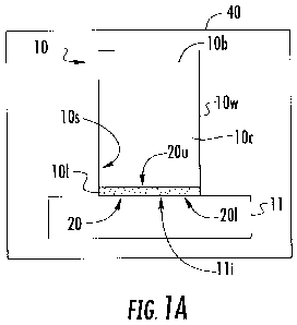
[0038] Figure lA is a schematic illustration of a biosample container
with an
integrated cell bed according to embodiments of the present invention.
CA 02877827 2014-12-22
WO 2014/004509 PCT/US2013/047619
[0039] Figure 1B is a section view of a portion of a biosample container
with an
example of a shaped cell bed according to embodiments of the present
invention.
[0040] Figures 2A-2C are schematic illustrations of an exemplary sequence
of cell
collection operations that may be used with biosample containers according to
embodiments
of the present invention.
[0041] Figures 3A-3F are schematic illustrations of an exemplary sequence
of cell
processing operations that can be carried out using the biosample containers
according to
embodiments of the present invention.
[0042] Figures 4A-4C are schematic illustrations of an exemplary sequence
of post-
processing, cell block removal operations that can be carried out according to
embodiments
of the present invention.
[0043] Figure 5 is a schematic illustration of a finished cell block
(e.g., cell disk) on
the cell bed in the containers shown in Figures 2A, 3A and 4A according to
embodiments of
the present invention.
[0044] Figure 6A is a partial exploded view of another example of a
container
according to embodiments of the present invention.
[0045] Figure 6B is a section view of another example of a container
according to
embodiments of the present invention.
[0046] Figure 6C is an exploded schematic illustration of an exemplary
container
with interchangeable tubular bodies according to embodiments of the present
invention.
[0047] Figure 6D is an exploded schematic illustration of another
exemplary
container with stackable container segments according to embodiments of the
present
invention.
[0048] Figure 7A is a side perspective view of another example of a
biosample
container according to embodiments of the present invention.
[0049] Figure 7B is a side view of the device shown in Figure 7A.
[0050] Figure 7C is a top view of the device shown in Figure 7A.
[0051] Figure 7D is a bottom view of the device shown in Figure 7A.
[0052] Figure 7E is a section view taken along lines 7E-7E in Figure 7A.
[0053] Figure 8A is a front view of a container assembly that holds a
collection
container with the integrated cell bed therein according to embodiments of the
present
invention.
[0054] Figure 8B is a front perspective view of the container assembly
shown in
Figure 8A.
6
CA 02877827 2014-12-22
WO 2014/004509 PCT/US2013/047619
[0055] Figure 9A is a front view of a biosample tube having an alternate
top or cap
configuration according to embodiments of the present invention.
[0056] Figure 9B is a sectional view of the device shown in Figure 9A.
[0057] Figure 9C is a front view of a biosample tube similar to that
shown in Figure
9A, having an alternate top configuration for processing according to
embodiments of the
present invention.
[0058] Figure 9D is a sectional view of the biosample tube shown in
Figure 9C.
[0059] Figure 10A is a front perspective view of the container assembly
(sans upper
lid) adjacent an exemplary centrifuge according to embodiments of the present
invention.
[0060] Figured 10B and 10C are top perspective views of the container in
position in
the centrifuge according to embodiments of the present invention.
[0061] Figure 11 is a schematic exploded view of a container according to
embodiments of the present invention.
[0062] Figure 12 is a schematic illustration of a container with an
example of an
alternate lid configuration according to embodiments of the present invention.
[0063] Figure 13A is a top perspective view of an exemplary mold for
creating
formed cell beds according to embodiments of the present invention.
[0064] Figure 13B is a schematic illustration of another forming
apparatus according
to embodiments of the present invention.
[0065] Figure 13C is a section view taken along lines 13C-13C in Figure
13A.
[0066] Figure 14 is a schematic partial section view of yet another cell
bed forming
device according to embodiments of the present invention.
[0067] Figures 15A-151 are schematic illustrations of a sequence of
operations that
may be used to carry out embodiments of the invention and also illustrating
another
embodiment of a container according to embodiments of the present invention.
[0068] Figure 16 is a flow chart of exemplary operations that can be used
to process
cells according to embodiments of the present invention.
[0069] Figures 17-27 are digital photographs of exemplary processing
steps using a
collection and processing container with an integrated cell bed according to
embodiments of
the present invention.
Detailed Description of Embodiments of the Invention
[0070] The present invention now is described more fully hereinafter with
reference
to the accompanying drawings, in which eMbodiments of the invention are shown.
This
7
CA 02877827 2014-12-22
WO 2014/004509 PCT/US2013/047619
invention may, however, be embodied in many different forms and should not be
construed
as limited to the embodiments set forth herein; rather, these embodiments are
provided so that
this disclosure will be thorough and complete, and will fully convey the scope
of the
invention to those skilled in the art.
[0071] Like numbers refer to like elements throughout. In the figures,
the thickness
of certain lines, layers, components, elements or features may be exaggerated
for clarity.
Broken lines illustrate optional features or operations unless specified
otherwise. One or
more features shown and discussed with respect to one embodiment may be
included in
another embodiment even if not explicitly described or shown with another
embodiment.
[0072] The terminology used herein is for the purpose of describing
particular
embodiments only and is not intended to be limiting of the invention. As used
herein, the
singular forms "a", "an" and "the" are intended to include the plural forms as
well, unless the
context clearly indicates otherwise. It will be further understood that the
terms "comprises"
and/or "comprising," when used in this specification, specify the presence of
stated features,
integers, steps, operations, elements, and/or components, but do not preclude
the presence or
addition of one or more other features, integers, steps, operations, elements,
components,
and/or groups thereof. As used herein, the term "and/or" includes any and all
combinations
of one or more of the associated listed items. As used herein, phrases such as
"between X
and Y" and "between about X and Y" should be interpreted to include X and Y.
As used
herein, phrases such as "between about X and Y" mean "between about X and
about Y." As
used herein, phrases such as "from about X to Y" mean "from about X to about
Y."
[0073] Unless otherwise defined, all terms (including technical and
scientific terms)
used herein have the same meaning as commonly understood by one of ordinary
skill in the
art to which this invention belongs. It will be further understood that terms,
such as those
defined in commonly used dictionaries, should be interpreted as having a
meaning that is
consistent with their meaning in the context of the specification and relevant
art and should
not be interpreted in an idealized or overly formal sense unless expressly so
defined herein.
Well-known functions or constructions may not be described in detail for
brevity and/or
clarity.
[0074] It will be understood that when an element is referred to as being
"on",
"attached" to, "connected" to, "coupled" with, "contacting", etc., another
element, it can be
directly on, attached to, connected to, coupled with or contacting the other
element or
intervening elements may also be present. In contrast, when an element is
referred to as
being, for example, "directly on", "directly attached" to, "directly
connected" to, "directly
8
CA 02877827 2014-12-22
WO 2014/004509 PCT/US2013/047619
coupled" with or "directly contacting" another element, there are no
intervening elements
present. It will also be appreciated by those of skill in the art that
references to a structure or
feature that is disposed "adjacent" another feature may have portions that
overlap or underlie
the adjacent feature.
[0075] Spatially relative terms, such as "under", "below", "lower",
"over", "upper"
and the like, may be used herein for ease of description to describe one
element or feature's
relationship to another element(s) or feature(s) as illustrated in the
figures. It will be
understood that the spatially relative terms are intended to encompass
different orientations of
the device in use or operation in addition to the orientation depicted in the
figures. For
example, if the device in the figures is inverted, elements described as
"under" or "beneath"
other elements or features would then be oriented "over" the other elements or
features.
Thus, the exemplary term "under" can encompass both an orientation of over and
under. The
device may be otherwise oriented (rotated 90 degrees or at other orientations)
and the
spatially relative descriptors used herein interpreted accordingly. Similarly,
the terms
"upwardly", "downwardly", "vertical", "horizontal" and the like are used
herein for the
purpose of explanation only unless specifically indicated otherwise.
[0076] It will be understood that, although the terms first, second, etc.
may be used
herein to describe various elements, components, regions, layers and/or
sections, these
elements, components, regions, layers and/or sections should not be limited by
these terms.
These terms are only used to distinguish one element, component, region, layer
or section
from another region, layer or section. Thus, a first element, component,
region, layer or
section discussed below could be termed a second element, component, region,
layer or
section without departing from the teachings of the present invention. The
sequence of
operations (or steps) is not limited to the order presented in the claims or
figures unless
specifically indicated otherwise.
[0077] The term "about" means that the recited number or value can vary
by +/- 20%.
[0078] The term "biosample" refers to human or animal tissue, blood or
other solid or
liquid samples that have cellular material. The cellular material can be
limited cellular
material, obtained from an FNA or other specimens including, for example,
washings,
lavages, and endoscopic procedures. Embodiments of the invention can be used
for
immunohistochemistry (IHC) or other studies including RNA and DNA analysis,
research or
studies including FISH, PCR and the like and/or to assess morphology.
Embodiments of the
invention may be used with or for stains, such as "special stains" like Gram
stains, Reticulin,
Mucin and may others as is well known to those of skill in the art.
9
CA 02877827 2014-12-22
WO 2014/004509 PCT/US2013/047619
[0079] Embodiments of the invention provide cell disks that can be used to
make
surgical pathology FFPE specimens from any kind of suspension. This applies to
body fluids
(pleural, pericardial, lung washing, etc.). These fluids have diagnostic
utility, but also might
be the only specimen in certain cases.
[0080] Special stains for amyloid or microorganisms can also be performed
on any
collected specimen, e.g., any FFPE specimens. This can be adapted to fluids
for diagnosis.
[0081] Embodiments of the invention may be suitable for veterinarian use,
medical
human use or research for human and/or with laboratory animals.
[0082] The term "sterile" means that the noted device or material meets or
exceeds
defined medical guidelines of cleanliness and is substantially (if not
totally) without
contaminants so as to be suitable for medical uses (e.g., diagnosis).
[0083] Turning now to the figures, Figure lA illustrates a cell collection
tube 10 with
a base 11, an internal cavity 10c and an internal cell bed 20 held proximate
the bottom 10/ of
the tube 10. The cell bed 10 can be an inert cell bed. The term "inert cell
bed" refers to a
solid material that can support processed cell material above the base 11,
typically in a block
form, while preserving the cells, typically without chemically interacting
with the cells. Post
collection and after processing, the cell bed 20 with patient cells C can be
removed,
substantially intact, e.g., scraped, pushed or otherwise disengaged from the
base 11 without
disrupting the collected cells thereon for cell evaluation.
[0084] As shown in Figure 1A, the cell bed 20 can be substantially planar
(e.g., the
top and bottom can be flat similar to a flat block). As shown in Figure 1B,
the cell bed 20
can have an upper portion 20u with a defined three-dimensional body shape 20s.
The body
shape 20s can include a substantially conical or frustoconical center portion
20c for
facilitating cell collection during centrifugation. The center portion 20c can
taper to have a
valley that holds the cells/pellet 100. The cell bed 20 can extend across an
entire interior
cavity space 10c at the bottom portion of the tube 10/ to define a closed
surface cell bed
above the base 11. In some embodiments, the cell bed 20 can include an
upwardly extending
outer sidewall 20w that rises a distance above the laterally extending portion
and conforms to
the inner surface 10i of the tube sidewall lOw at the bottom portion of the
tube 10/. The outer
perimeter sidewalls 20w can inhibit some samples from being attracted to
hydrophilic plastic
(polymer) walls of the cylinder 10b that can facilitate forming the cell plug
in the desired cell
disk or block form. In addition, or alternatively, the taller walls 20w can
stay with the
cylinder body 10b when separated from the base 11, with the lower portion of
the cell bed 20
CA 02877827 2014-12-22
WO 2014/004509 PCT/US2013/047619
intact and remaining attached to the walls 20w. In other embodiments, the cell
bed walls 20w
may detach from the lower cell bed 20 when the base 11 is removed.
[0085] In some embodiments, the tube body 10b, base 11 and cap 12 can
comprise a
molded polymeric material that is sterilized for use. The tube body 10b, base
11 and/or cap
12 may also comprise other suitable materials, including, for example, glass.
[0086] In some embodiments, the cell bed 20 has a lower surface 20s that
can be
substantially planar 20p. The base 11 can also have an internal surface lli
that is
substantially planar. However, the base surface lli can have other shapes and
may include
narrow channels or slots or can include "waves" or dimples and the like. The
surface lli can
be configured to allow for ease of removal of the cell bed 20 with cells in a
"cell block" form
100 (Figures 4A, 4B), post-processing. The cell bed lower surface 20s can
reside against the
base surface lli or may reside a distance above the base 11 attached to an
inner surface lOs
of a sidewall lOw of the tube 10.
[0087] The internal base surface lli can include a non-stick material
and/or coating
that reduces sliding friction and/or otherwise facilitates the removal of the
cell bed 20 from
the base 11 with the cell block 100 (Figure 4C) for conventional cell
evaluation after
processing. In some embodiments, the cell bed 20 with the cell block 100 can
be pushed off
the base or pushed up and out of the tube body once the base is removed.
Optionally, a thin
flexible liner 110 (Figure 4C) can reside on the surface lli to allow the cell
block 100 to be
lifted or more readily slid off the base 11 (Figure 4C). The liner 110, where
used, can be
adhesively attached to the internal surface of the base lli and a user can
lift to peel an edge
and dislodge the cell block 100.
[0088] The cell bed 20 can be a monolithic solid layer of an inert
material. In other
embodiments, the cell bed 20 can comprise a plurality of stacked layers of
different solid
materials or a mixture of materials. The cell bed 20 can comprise paraffin or
other suitable
material alone or in combination with one or more other materials. In some
embodiments,
the cell bed 20 is defined by a monolithic paraffin body. In some embodiments,
DNase
and/or RNase inhibitors may be added to the fixative or other liquid solutions
and/or paraffin
that may improve future molecular testing.
[0089] In some embodiments, the base 11 is detachable, e.g., releasably
attachable to
a bottom portion 10/ of the tube 10. This releasable attachment can be by any
suitable
attachment configuration including, for example, threaded attachment, bayonet
or frictional
fit, snap fit, hooks, VELCRO, adhesive attachment, frangible attachments, any
of which may
optionally also employ 0-rings, compatible sealant, wax or grease or washers
to promote a
11
CA 02877827 2014-12-22
WO 2014/004509 PCT/US2013/047619
sufficient fluid-tight seal. For frangible attachments, the tube body 10b
and/or base 11 can be
integrally attached and configured to preferentially tear or detach about a
defined zone when
exposed to sufficient compressive, torsion or tensile forces.
[0090] The tube 10 can be provided in a sterile package 40 for onsite
collection of a
specimen from a patient. The term "onsite" refers to a collection location of
a patient such as
a surgical or biopsy room, doctor's office or hospital or laboratory site. The
tube 10 can
include a lid or cap 12 (Figure 2C) that is provided in the package 40 or in a
separate
package. In other embodiments, non-sterile uses are contemplated.
[0091] The base 11 can be provided in the package 40 pre-assembled or pre-
formed
in the tube 10 and/or base 11 as shown. Alternatively, it may be provided
separately for
attachment at the use (collection) site (Figure 11). As such, the cell bed 20
may be provided
as a separate component in the package 40 or a separate package (held in a
rigid container so
as to protect the preformed shape). In other embodiments, the cell bed 20 is
pre-assembled
into the base 11 or bottom 10/ of the tube body 10b. In some particular
embodiments, the
cell bed 20 can be formed in the tube 10 at the collection site if a suitably
reliable press or
formation system can be included in a collection kit.
[0092] In some embodiments, the cell bed 20 can be pre-formed in the tube
10 with a
defined three-dimensional shape, in package 40, so that the tube 10 is ready
for use at a cell
collection site. The package 40 can hold a single tube or multiple tubes 10.
The cap 12
(Figure 2C) can be on the respective tube 10 to maintain the sterility until
use.
Figures 2A-2C illustrate exemplary steps that can be carried out at a
collection site to collect
cells for subsequent evaluation. As shown in Figure 2A, a sample with cells C
is introduced
into the internal cavity 10c of the tube 10 and rest on the cell bed 20. The
cells C may
comprise aspirated cells (unclotted) from a FNA using a needle 75 that is
directly inserted
into the tube 10, in some embodiments. Figure 2B illustrates that the cells C
may then clot
(onsite). Figure 2C illustrates that a supernatant, e.g., a solution of
fixative liquid 15 that
may comprise formalin or other suitable fixative material such as Zinc can be
introduced into
the tube 10. Other fixatives may include, but are not limited to, saline,
alcohols, acetone,
mercury based reagents, and even media (Lennox broth, RPMI, etc.). The vessel
10 can be
provided unfilled and a user can select the appropriate fixative or several or
a particular type
may be prepackaged in a kit which may be ordered for use. Any media used in
the tube body
10b should be sterilized.
12
CA 02877827 2014-12-22
WO 2014/004509 PCT/US2013/047619
[0093] A lid 12 can be attached to the tube 10 and transported or stored.
As shown,
there may be undesired floating cells F in the solution above the clotted
cells on the cell bed
20.
[0094] The lid 12 can be a rigid closed lid that is attached after the
fixative 15 is
introduced. However, in some embodiments the lid 12 can include a liquid entry
port to
allow the liquid to enter while the lid remains on. The lid 12 can include a
luer lock or luer
slip connection fitting that engages with a male syringe luer lock or slip
fitting to provide the
liquid entry hat allows the liquid to be introduced through the port.
[0095] Figures 3A-3F illustrate steps that can be carried out at a
cytology laboratory
or other suitable research or clinical laboratory. Figure 3A shows the tube 10
after
centrifugation in a centrifuge 200 (Figure 10). The centrifuge can be a
standard laboratory
centrifuge, typically a low speed centrifuge that permits the separation of
the fixative from
the cells and allows the cells to form a cell pellet P as is known to those of
skill in the art.
The centrifuge may be configured to process standard 50 mL or 100 mL conical
tubes and the
tube 10 can be placed therein alone or with an adapter. That is, the tube 10
may include a
sleeve, adapter, or coupler or may have an external integrated size and/or
design that allows it
to be placed directly into the "bucket" or standard receptacle of the
centrifuge.
[0096] The fixative liquid 15 is removed (e.g., the formalin is decanted)
as shown in
Figure 3B and a rinse solution 18 can be added to the tube 10 as shown in
Figure 3C. The
fixative liquid 15 can be removed and the rinse 18 added via any suitable
technique that
leaves the cells C and/or pellet P in the tube 10 including aspiration tubing,
pipette
withdrawal, decanting and the like. Typically, the supernatant should be
aspirated gently
with a vacuum rather than being decanted (which refers to tipped and poured)
to minimize or
reduce trauma to the cell pellet. As before, the rinse solution or other
liquids can be removed
or added with the lid 12 off as shown in Figure 3B or with the lid remaining
on the tube
using a liquid entry and/or retraction port. It is also contemplated that
different lids having
the same or different configurations may be used at different points in the
process.
[0097] In some embodiments, a clot blot formed during the collection/post-
collection
can be used as a cap for a rinse vessel.
[0098] Figure 3D illustrates that the tube 10 with the rinse 18 can then
be centrifuged.
Figure 3E illustrates that the rinse 18 can be decanted or otherwise removed
or withdrawn,
leaving cells C on the cell bed 20, typically in pellet form P. Figure 3F
illustrates that a
matrix material 28 can be added to form a cell block 100. The term "matrix
material" refers
to a specimen-processing gel (that may be aqueous) that encapsulates and
suspends histologic
13
CA 02877827 2014-12-22
WO 2014/004509 PCT/US2013/047619
and cytologic specimens in a solidified medium. The matrix material 28 can
include one or
more of agar, agarose gel or "histogel" solid at ambient temperature,
Methoce118, Matrix
Gel , OCT compounds, paraffin, denatured and non-denatured collagen,
fibronectin,
laminin,plasma and thrombin and other mixtures. Other matrixes for cell
immobilization can
also be used. For a discussion of cell blocks and ethanol formalin fixative
and other fixatives,
see, e.g., Nathan et al., Improved Preparation and Its Efficacy in Diagnosing
Cytology, Am J
Clin Pathol, 2000; 1114, 599-606, the contents of which are hereby
incorporated by reference
as if recited in full herein.
[0099] Figure 4A illustrates that the base 11 and body of the tube 10b
are detached
from each other, exposing the cell block 100 on the cell bed 20. The cell
block 100 with the
cell bed 20 are removed from the base 11 as shown in Figure 4B. The cell block
100 with
cell bed 20 can be detached, removed or separated in any suitable manner
including, for
example, scraping, sliding or lifting (e.g., using a liner 110, Figure 4C).
[00100] Figure 5 illustrates the resulting cell block 100 (also termed
cell disk) that
includes the cell bed 20 ready for routine processing. The cell block 100 can
be sliced or cut
for preparing slides for staining or other diagnostic protocols. There may be
an increased
number of cells in the cell block or slices thereof that may promote
diagnostic capability over
smears alone.
[00101] Figure 6A illustrates the tube 10 with the base 11 detached from
the tube body
10b. In this example, the base 11 includes a raised center pedestal lip that
forms the inner
surface lli that holds the cell bed 20. The perimeter of the pedestal lip can
include threads
that engage an inner surface of the tube body or may frictionally engage the
tube body using
seals such as an 0-ring(s) and the like (not shown). Typically, the base 11
with the cell bed
20 on the pedestal is attached to the tube body 10b and packaged for use at a
collection site.
[00102] Figure 6B illustrates that the tube 10 can include a spacer 13
that rises a
distance above the lower portion of the base 11. Figure 6B also illustrates
that the base 11
can attach to the tube body 10b at a location above the bottom of the base 11.
The spacer 13
can include a substantially planar or flat surface for holding the cell bed
20. The base 11 and
tube body 10b can threadably couple together. In the embodiment shown, the
base 11
includes external threads 34 and the tube body 10b includes internal threads
38. However, as
noted above, other coupling configurations may be used.
[00103] Figure 6C illustrates that the base 11 can interchangeably attach
to two
different tube bodies 10b2, 10b2, and/or the different tube bodies can have
different volumes.
Thus, for example, the smaller tube body 10b1 can be used at the collection
site and for
14
CA 02877827 2014-12-22
WO 2014/004509 PCT/US2013/047619
transport to the cytology lab. The larger tube body 10b2 can be used at the
cytology lab for
processing in the centrifuge, for example. The same or differently configured
caps or lids 12
may be used for each tube body 10b1, 10b2. In other embodiments, different
volume tube
bodies 10131, 10b2 can be provided in a package and selected for use at the
collection site
allowing for increased flexibility corresponding to the specimen type (e.g.,
urine, blood
plasma or serum versus FNA).
[00104] Figure 6D illustrates that the tube body 10b can include several
segments
10b1, 10b2 that attach together to provide a different volumetric capacity.
Thus, for example,
one segment 10b1 can be attached and used with the cap or lid 12 at the
collection site and for
transport. The lid/cap 12 can be removed and the second body 10b2 can be
attached to the
first body 10b1 at the cytology lab and the stacked segments can define the
tube body 10b
used for centrifugation. Where more than one tube body segment 10b1, 10b2 is
used, one or
both can be detached from each other and/or the base 11 to expose the cell bed
20 with the
cell block 100 for access/removal of the cell block 100 for subsequent
processing and
analysis.
[00105] Figure 7A is an exploded view of another example of a tube 10. In
this
example, the base 11 has an annular open space 33 with female threads 34 that
surrounds the
internal surface lli that holds the cell bed 20. The female threads 34 matably
engage
external male threads 38 on the bottom portion of the tube 10/.
[00106] Figures7B-7E illustrate that the tube 10 can include top and
bottom indicia
12m, llm so that a user knows which end is "up" before using or opening. In
some
embodiments, a visually transparent window may be provided in the tube or cap
or base or
the device may be transparent or translucent. The cell bed 20 can have a
substantially conical
shape with the lowest peak facing the base 11 along an axially projecting
centerline of the
tube body 10b. The outerwalls of the cell bed 20w can extend above the center
portion of the
cell bed 20 in a substantially straight vertical orientation so as to conform
to the sidewalls of
the tubular body 10b. The sidewalls 20w can cover more than a major portion
(e.g., greater
than 50%) of the enclosed fluid cavity 10c, leaving a minor portion of the
sidewalls lOw of
the tube body 10b below the cap 12 free of the cell bed material.
[00107] The tube 10 can have a defined capacity or volume. The tube 10 may
have a
volume or capacity between about 10 mL to about 200 mL, including about 20 mL,
about 30
mL, about 40 mL, about 50 mL, about 60 mL, about 70 mL, about 80 mL, about 90
mL, and
about 100 mL. The tubes 10 can be provided in different volumes/sizes for
different
CA 02877827 2014-12-22
WO 2014/004509 PCT/US2013/047619
applications. Where two segments 10b1, 10b2 are used, one can have a volume
between 10
mL to 25 mL and the other can have a volume between 25 mL and 100 mL, for
example.
[00108] The lid 12 can also be threadably attached to an upper portion of
the tube body
via threads 12t, 39. The base 11 and cap 12 can have a ledge 39, 139 with a
diameter that
defines a tight fit with a receptacle of a standard centrifuge or with a
standard tube, sleeve or
other adapter allowing the tube 10 to be placed in a centrifuge for
processing.
[00109] The cap and base ledges 39, 139 can be configured to have the same
outer
diameter size. The outer perimeter of the ledge can include a pattern of
circumferentially
spaced apart recesses or grooves 39g, 139g. The ledges 39, 139 can provide a
resilient fit to
provide for snug engagements using an overcoat, outerlayer or substrate of
resilient material
or just based on the groove configurations.
[00110] In some embodiments, the tube 10 is sized and configured as a 50
mL tube and
can snugly engage a centrifuge receptacle without the use of an adapter or
without a
customized sleeve or other adapter.
[00111] Figures 8A and 8B illustrate that the tube 10 can be placed inside
a larger
(standard) tube 50 for standard centrifuge processing. The ledges 39, 139 can
snugly engage
the sidewall of the tube 50. The tube 50 can include a cap 52 and the lower
portion 54 can
include a center with a conical internal shape 55. The tube 10 typically
resides over the
conical shape in the bottom portion of the standard tube 50. However, the tube
10 can be
placed above this location as well. The tube 10 can be slid in and out of the
tube 50 for
access.
[00112] Figures 9A and 9B illustrate another embodiment of a container 10'
where the
tube body 10b can releasably attach to an elongate body 150 which may be
attached to a
second cap 12b. As shown, the elongate body 150 is in fluid communication with
the tube
fluid cavity 10c. The second tube cap 12b can define a fluid port 153 that
allows fluid from
the elongate body 150 to be introduced or withdrawn. The tube body 10b with
the second
cap 12b can be configured to reside in a standard 50 mL tube 50 as shown. In
other
embodiments, the tube body 10b and second cap 12b can be configured to snugly
reside in a
centrifuge receptacle (e.g., bucket) without requiring the tube 50. The
elongate body 150 can
be integral with the tube cap 12b, e.g., a monolithic molded unitary
component. In other
embodiments, the elongate body can be configured to attach to the lid 12b in
other ways
including threaded, snap fit and the like, while providing a fluid-tight seal.
Figures 9C and
9D illustrate a similar configuration of a container 10' with an alternate top
configuration. In
some embodiments, the elongate body 150 may be attached at the collection site
as the
16
CA 02877827 2014-12-22
WO 2014/004509 PCT/US2013/047619
original cap. In other embodiments, a cap or lid 12 such as that shown in
Figure 7A can be
used at the collection site and/or for shipment and transport and the second
cap 12b or
elongate body 150 interchanged later for processing.
[00113] Figure 10A shows the tube 10 sitting over a spacer 30 snugly
attached to and
in a standard 50 mL tube 50 in preparation for centrifugation in a bucket 210
of the centrifuge
200. Figure 10B illustrates the tube 10 in the tube 50 in a standard swing
bucket 210 of the
centrifuge 200. Figure 10C illustrates centrifugation with the tube 10 on its
side (and
horizontal) rather than angled so that the specimen is collected in a middle
20c (Figure 1B)
of the cell bed 20.
[00114] Figure 11 illustrates the components of a tube 10 according to
some
embodiments. As noted above, the cell bed 20 can be pre-formed and provided
for assembly
onsite or may be pre-attached to the base 11 and/or tube body 10b. Typically,
the base 11
with the cell bed 20 is attached to the tube body 10b and packaged for use at
a collection site.
[00115] Figure 12 illustrates that the cap 12' can be configured to engage
a leur-lock
of a syringe 120 for introducing and/or removing different liquids.
[00116] Figures 13A and 13C illustrate examples of a mold that has mold
cavities
20c1-20c11 that can concurrently form a plurality of cell beds 20. Cell bed
material, in liquid,
solid, or semi-solid form, typically in flowable (fluid or gel) form can be
introduced into the
mold via one or more mold ports or through an open access region and molded
into the pre-
formed cell bed shape for use in the tube 10.
[00117] In some embodiments as shown in Figure 13B, a mechanical press 251
with
shaped mold members 253 can be configured to enter the tube body 10b with an
attached
base 11 held in a lower holding member 252 to allow concurrent cell bed
formation in
multiple tubes 10. The cell bed material 20m can be inserted in solid or
liquid form, typically
in a solid, but perhaps slightly heated form for aid in formation of the
desired cell bed shape.
[00118] In other embodiments, as shown in Figure 14, a respective cell bed
20 is
formed directly in an attached base 11 and tube body 10b using a shaped tamper
tool 260.
[00119] In some embodiments, the tubes 10 can be used to process cells for
human or
veterinary uses. In certain embodiments, the tubes 10 can be directed to
preparation of cells
for pathology review. While it is contemplated that the tubes 10 are
particularly suitable for
cells obtained by fine needle aspiration, it should be clear to one of skill
in the art that cellular
material captured by other means could also be collected and processed by the
tubes 10.
[00120] Cell material could also be collected by endoscopy, including but
not limited
to arthroscopy, bronchoscopy, colonoscopy, colposcopy, cystoscopy, ERCP
(endoscopic
17
CA 02877827 2014-12-22
WO 2014/004509 PCT/US2013/047619
retrograde cholangio-pancreatograthy), EGD (esophogealgastroduodensoscopy),
endoscopic
biopsy, gastroscopy, laparoscopy, laryngoscopy, proctoscopy and thoracoscopy.
Cells could
also be obtained from lavage procedures, including but not limited to
bronchoalveolar, breast
ductal, nasal, pleural, peritoneal, gastrointestinal, arthroscopic, and
urinary bladder lavages.
It is also contemplated that cells could be collected from catheters such as
those used in
infusion, cardiovascular, renal, bladder, urethral, hemodynamic monitoring,
neurological, and
other procedures which would be obvious to one of skill in the art. In some
embodiments,
cell samples can be from eye/cornea/globe aspirations,
endocervical/ectocervical/endometrial
curettages, cyst aspirations and urine. It is also contemplated that cell
samples can be for
xenografts from research and animal modeling as well as patient directed
therapy.
[00121] The cells can be from washings and spontaneously exfoliated
specimens
including bronchial washings, bronchoalveolar lavage, sputum pleural fluid,
pericardial fluid,
peritoneal fluid, peritoneal washing, ovarian cyst fluid, synovial fluid,
urine, brain cyst fluid,
cerebrospinal fluid. The cells can be for RNA/DNA research or analysis and may
include
live cells. With the use of appropriate media, the tubes 10 can act as a small
incubator to
keep cells alive (at least for a short period of time). DNase/RNase inhibitors
can be
introduced to the media to also preserve DNA/RNA. As is known, fixation alone
can help
with DNA/RNA preservation.
[00122] In particular embodiments, the cell samples are from endocervical
curettages
(ECC). In the past, conventional practice when the first slide from the
original paraffin block
is essentially noncontributory, is to take the fluid remaining in the specimen
jar and perform a
"ThinPrep" on it. These typically have many cells (squamous and glandular) but
there is no
architecture. Rather than place the minimal slime usually present in a
specimen jar in a
cassette, it is contemplated that those cells can be put in the tubular body
10b (or 150, Figure
9A, 15A) at the collection site. This should give better initial yield with
architecture present
and a source for immunos. This latter may not be inconsequential. For example,
when the
ECC contains a minute fragment of small cells with high N/C ratios, it is hard
to discern
whether they are not relevant (being potentially from the lower uterine
segment) or clump of
HGSIL cells. A single immuno--p16¨can be very useful in this scenario. The
term
"immuno" and plurals thereof refer to immunoperoxidase studies and include
antibodies
targeting specific epitopes to aid in tumor/disease differentiation. Also
known as (although
technically incorrect) immunohistochemistry: p16 is a protein/antigen with the
p16 antibody
in a cell having clinical significance.
18
CA 02877827 2014-12-22
WO 2014/004509 PCT/US2013/047619
[00123] Figures 15A-15I illustrate another embodiment of a container 10"
similar to
that shown in Figures 9A and 9B. In this embodiment, no external tube is
required for
centrifuge processing. As shown, the container 10" includes a tube body 10b
holding the cell
bed 20 and a base 11. The container 10" also includes an elongate body 150
that can attach
to the tube body 10b. The elongate body 150 can include a first end with a lid
155 and a
second opposing end 157 that is open. As shown in Figure 15A, the elongate
body 150 can
include a desired solution, such as saline or formalin 15. Figure 15B
illustrates that, as
before, a FNA sample can be inserted onto the cell bed 20. One or more needles
from other
passes obtained from the target tissue can be rinsed in the chamber of the
elongate body 150.
This is in contrast to ajar filled with saline used conventionally. As shown
in Figures 15C
and 15D, the tube body 10b and base 11 can be attached together after the
pellet P is dried
allowing the tube body 10b with cell bed 20 and pellet P to be inverted to
attach to the open
end 157 of the elongate body 150. The open end of the elongate body 157 can be
sized and
configured to be substantially the same as the size of the upper end of the
tube body. The ,
elongate body 157 can taper to a large size away from the open end as shown.
The tube body
10b can engage the open end of the elongate body 157 in a fluid tight manner
using
appropriate seals, threads, frictional fits and the like.
[00124] Figure 15E shows that the container 10" can be inverted so that the
cell bed
20 is in the lower portion of the container 10". The container 10" can be
centrifuged at an
appropriate revolution per minute and duration (e.g., about 2000 rpm for about
5 minutes) to
form a combined pellet Pc (Figure 15F) on the cell bed 20 (combined from cells
collected
from the rinse solution and the FNA direct deposit). Figure 15G illustrates
that the
supernatant can be aspirated as is conventional using a vacuum leaving only a
minimal
amount of fixative 15 (e.g., formalin or saline) in the tube body 10b above
the cell bed 20 so
as to not disrupt the cell pellet P. The aspiration can be carried out by
first removing lid 155
or by using a sealed port in the lid (not shown). As shown in Figure 15H, the
elongate body
150 can then be removed. A liquid matrix 28 can be added to the tube body 10b
and cells
resuspended. Figure 151 illustrates that the cell disk or block 100 with the
cell bed 20 can be
removed from the tube body 10b using, for example, a plunger 300. The cell
block 100 can
be processed as a histology specimen or other desired specimen.
[00125] Figure 16 is a flow chart of exemplary operations that can be used
to carry out
embodiments of the invention. A cell sample can be placed in a tube having a
cell bed (block
350). The sample is centrifuged while in the tube (block 360). A cell block is
removed from
the tube with the cell bed and solidified matrix holding the cells (block
370).
19
CA 02877827 2014-12-22
WO 2014/004509 PCT/US2013/047619
[00126] The cell sample can be placed with a needle with a fine needle
aspirate sample
directly in the tube (block 352). Liquid matrix material (e.g., specimen-
processing gel that
encapsulates and suspends histologic and cytologic specimens in a solidified
medium) can be
added to the tube after the centrifuging and the matrix material with the
cells distributed
therein can be solidified to form the cell block (block 365). The cell sample
can be collected
in the tube at a patient collection site and the tube can be transported to a
cytology lab for
processing (block 355).
[00127] The base of the tube can be separated from the tube body to expose
the cell
block and allow the cell block to be removed with the cell bed (block 368).
[00128] In some embodiments, generally summarized, rinses can be performed
in the
collection vessel 10 and the dedicated clot blot(s) can be generated as
discussed above. The
clot blot can be used as a cap for the rinse vessel. The entire apparatus can
be inverted (clot
block side down) and centrifuged. The supernatant can be aspirated leaving a
clot blot with
overlaying precipitated materials (button). A desired amount (not typically
calculated
precisely, but roughly, about 1: l/v:v to the button) of HistoGel can be
pipetted onto the
button and immediately spun again in a centrifuge. This time the collection
vessel is not
needed. This allows the HistoGel to permeate the cells and polymerize. Again,
this step is
carried out quickly to prevent polymerization before the centrifugation. This
leaves the cells
within a matrix of HistoGel. This action can prevent loss of cells in
downstream processing
procedures.
[00129] The present invention is explained in greater detail in the
following non-
limiting Examples.
EXAMPLES
[00130] Figures 17-27 are digital images of an exemplary tube 10 and
exemplary
processing that can be carried out using the tube (the centrifuge operations
were shown above
with respect to Figures 10A-10C). Figure 17 illustrates a FNA expelled
directly from a
needle into the cell bed 20 of the base 11 (which is attached to the tube body
10b). As shown
in Figure 18, the cell sample C can settle in the middle of the cell bed 20
and/or base 12 due
to the substantially conical or furstoconical shape of the cell bed 20. The
sample can be
allowed to dry and/or coagulate.
[00131] The supernatant (formalin) is aspirated and discarded being
careful not to
disrupt the pelleted sample P as shown in Figure 19. Figure 20 illustrates the
tube 10 with a
CA 02877827 2014-12-22
WO 2014/004509 PCT/US2013/047619
sample pellet P. Some supernatant may remain but does not interfere with
subsequent
processing.
[00132] After placing the tube 10 in the tube 50, and centrifuging (as
described above),
liquid histogel (melted agarose) can be pipetted up and down in the tube 10 as
shown in
Figure 21 using pipette 299 to resuspend the sample in the histogel. About 30
1AL of agarose
may be used for this resuspension although other amounts may also be
appropriate. Figure
22 illustrates that the suspension can be carried out relatively rapidly so
that the agarose does
not solidify. Figure 23 shows the tube with the suspended sample in a low
temperature
freezer (e.g., about -20C) to facilitate solidification, which can occur in
between about 2-5
minutes.
[00133] Figure 24 illustrates the cell block 100 (e.g., the cell bed 20
and the solid
histogel with cells) being removed from the tube 10b and/or base 11. Figure 25
shows that
the cell bed 20 may attach to the inner wall of the tube body 10b and may need
a plunger or
other push member 300 to push the cell bed with the cells, e.g., cell block
100 from the tube
body (typically out the top, pushing against the cell bed 20 rather than the
cells/histogel
(agarose) mixture 100.
[00134] Figure 26 illustrates the cell block 100 with cell bed 20 placed
on routine
tissue paper. As is known to those of skill in the art, it is standard
practice to wrap the sample
100 in tissue paper in preparation for tissue processing for small histology
specimens.
Figure 27 illustrates the tissue paper wrapped cell block 100 with cell bed 20
in a standard
histology cassette 310. The darker dots inside the wrapped cell block 100 are
cells C.
[00135] Although shown as a manual operation, it is contemplated that
machines may
be used to automatically carry out certain of the above steps.
[00136] Table 1 below is a list of 15 cases of data comparing an exemplary
collection
vessel with the standard methods. All cases were graded from 1 (poor) to 2
(adequate) to 3
(superior). The first row is the diagnosis. The 2nd row is the paired smear
quality (DQ and
H&E). The CBCS is the remnant material left on the traditional clotting slide.
The clot blot
is the standard method compared to the cell disk method (CD). Last level was
the last recut
level on the FFPE block. The method was overall graded for superiority. As
seen, the
standard method was never superior to the new CD method but equivocal in some
cases.
21
CA 02877827 2014-12-22
WO 2014/004509
PCT/US2013/047619
TABLE 1: DATA SUMMARY
Smear Clot Blot Cell Disk
Pathologic CBCS (1-
3) Last Best
Quality Quality Quality
Diagnosis Level Method
(1-3) (1-3) (1-3)
Oncocytoma 2 2 1 2 3 CD
RCC, clear cell 3 1 1 2 3 CD
Pleomorphic
3 2 1 2 3 CD
adenoma
Large cell
3 3 3 3 3 Tied
carcinoma
Adenocarcinoma 3 2 3 3 5 Tied
Scc 2 3 2 2 3 Tied
Urothelial
3 2 2 1 3 CD
carcinoma
Invasive ductal
3 1 3 3 3 CD
carcinoma
Hodgkin
3 3 3 3 9 CD
lymphoma
RCC, papillary 2 n/a 3 3 3 CD
Follicular
3 3 2 2 5 Tied
adenoma
Solid-
3 1 1 1 5 Tied
pseuodopapillary
Urothelial
2 1 1 2 3 CD
carcinoma
Oncocytoma 3 3 3 3 5 CD
Scc 3 2 1 2 5 CD
[00137] The
foregoing is illustrative of embodiments of the present invention and is
not to be construed as limiting thereof. Although a few exemplary embodiments
of this
invention have been described, those skilled in the art will readily
appreciate that many
modifications are possible in the exemplary embodiments without materially
departing from
the novel teachings and advantages of this invention. Accordingly, all such
modifications are
intended to be included within the scope of this invention as defined in the
claims. The
invention is defined by the following claims, with equivalents of the claims
to be included
therein.
22