Note: Descriptions are shown in the official language in which they were submitted.
CA 02878059 2014-12-29
WO 2014/022535 PCT/US2013/052971
POLYMERIC STRUCTURES CONTAINING STRAINED CYCLOALKYNE FUNCTIONALITY FOR
POST-FABRICATION AZIDEALKYNE CYCLOADDITION FUNCTIONALIZATION
CROSS-REFERENCE TO RELATED APPLICATION
[0001] This application claims priority to U.S. Patent Provisional
Application No.
61/677,691 filed on July 31, 2012, the contents of which are incorporated
herein by
reference.
FIELD OF THE INVENTION
[0002] The present invention generally resides in the art of
biocompatible polymeric
structures. More particularly, the present invention relates to biocompatible
polymeric
structures bearing strained cycloalkyne functionality that survives the
process of
fabricating the polymeric structures. The cycloalkyne functionality can be
beneficially
employed in a post-fabrication functionalization through
azidealkynecycloaddition.
BACKGROUND OF THE INVENTION
[0003] For more than two decades, tremendous advances in regenerative
medicine
have provided hope that materials will offer solutions to organ and tissue
shortages that
occur worldwide. However, overcoming the remaining challenges will require
many
additional innovations, especially in materials that possess specific
functionality that guide
the regenerative processes.
[0004] Nanofibrous scaffolds possessing mechanical properties, porous
microstructure, and dimensional similarity to collagen fibers have been used
to mimic the
natural extracellular matrix (ECM) and are highly relevant for tissue
engineering in a
number of different applications. Polymeric nanofibers have been fabricated
into a variety
of constructs and scaffolds using melt- or electrospinning processes that are
able to control
size, morphology and alignment by varying conditions including solvent,
concentration,
additives and electrode design.
[0005] For regenerative medicine applications, the polymeric precursors
used to
fabricate the nanofiber-based scaffolds should be both biocompatible and
biodegradable.
Many biodegradable and biocompatible polymers such as polyglycolic acid (PGA),
poly(lactic acid) (PLA), poly(lactide-co-glycotide) (PLGA) and poly(e-
caprolactone)
(PCL) have been widely investigated as fiber and nanofiber precursor
materials. Although
these degradable polymers meet several of the basic requirements for tissue
engineering
applications, bioactive molecules to guide cellular behavior and preserve cell
phenotype
-1-
CA 02878059 2014-12-29
WO 2014/022535 PCT/US2013/052971
are required for optimal performance. Specific functionalities that could
guide or direct
specific biological function need to be incorporated efficiently. There are
generally two
methods available for biomolecule functionalization: physical adsorption and
chemical
bonding. While physical adsorption risks the loss of biomolecules over time,
chemical
conjugation usually requires multi-step processing, and purification both of
which often
included harsh conditions.
[0006] Additionally, the derivitization of nanofibers often requires
multiple
procedures, including plasma treatment, wet chemical methods, surface graft
polymerization, and co-electrospinning of surface active agents with polymers.
Each of
these modifications is time and resource intensive to optimize and may lead to
immune
specific reactions and biocompatibility problems.
[0007] A new method that enables efficient, orthogonal and bio-system
friendly
functionalization is preferred. Copper-catalysed click chemistry has been used
for efficient
functionalization of polymers. However, the side effect of copper ions leads
to
biocompatibility problems. Recently, the discovery of strain-promoted azide
alkyne
cycloaddition has provided a robust chemical method for the efficient
conjugation of
biomolecules. This method has been widely used in bioimaging and
bioconjugation. The
present invention makes beneficial use of this click chemistry and provides
guidance for
the creation of fibrous scaffolds and their post fabrication
biofunctionalization through
such azide alkyne cycloaddition chemistry.
[0008] Peptides, growth factors and carbohydrates have each been
covalently tethered
to the surfaces of synthetic and naturally-derived polymers to stimulate
specific cell
functions. Concerns about the bioavailability, specificity and activity of the
tethered
species persist. Recent studies show that the desired biological response can
be obtained
with the appropriate tether. A number of synthetic degradable polymers,
including
poly(lactic acid) are presently utilized clinically; but, cellular systems do
not readily
interact directly with synthetic polymers through normal integrin mediated
assemblies.
Tyr-Ile-Gly-Ser-Arg (YIGSR), a bio-active peptide derived from laminin, was
shown to
promote cell attachment and laminin receptor binding. Graf, J.; Ogle, R. C.;
Robey, F. A.;
Sasaki, M.; Martin, G. R.; Yamada, Y.; Kleinman, H. K. Biochemistry 1987, 26,
6896-
6900. YIGSR-functionalized matrices have showed similar or superior cellular
effects to
Ile-Lys-Val-Ala-Val (IKVAV) peptide, a more commonly studied laminin-derived
peptide
and laminin-coated matrices. The art could benefit from incorporating YIGSR
into
nanofibers matrices to promote the directed differentiation of embryonic stem
cells into
-2-
CA 02878059 2014-12-29
WO 2014/022535 PCT/US2013/052971
neurons. However, the precise, regiospecific functionalization of degradable
polymers
with biological motifs capable of directing cellular function has been so
complicated with
regard to solvents, catalysts, residuals and processing methods, that they
have been
deemed translationally irrelevant. The recent evolution of click chemistry as
a method to
functionalize polymers and materials has enabled the derivatization of both
natural and
synthetic polymers in ways that were not previously possible.
SUMMARY OF THE INVENTION
[0009] A first embodiment of this invention provides a method of
creating
biocompatible polymeric structures comprising the steps of: providing a
biocompatible
polymer including a strained cycloalkyne end group; forming a polymeric
structure from
the biocompatible polymer such that the strained cycloalkyne end group remains
on the
biocompatible polymer; providing an azide tethered molecule; and, after said
step of
forming, reacting the azide tethered molecule with the cycloalkyne in an azide
alkyne
cyclo addition reaction to further functionalize the polymeric structure.
[0010] A second embodiment provides a method as in the first embodiment,
wherein
said step of providing a biocompatible polymer includes the step of
polymerizing
monomers through a ring-opening polymerization employing a ROP initiator
having a
strained cycloalkyne to create the biocompatible polymer including a strained
cycloalkyne
end group.
[0011] A third embodiment provides a method as in the first or second
embodiment,
wherein the ROP initiator includes a five to nine member strained cycloalkyne.
[0012] A fourth embodiment provides a method as in any of the first
through third
embodiments, wherein the ROP initiator further includes a reactive group
selected from an
hydroxyl group or amine group.
[0013] A fifth embodiment provides a method as in any of the first
through fourth
embodiments, wherein the reactive group is an hydroxyl group, and the monomers
polymerized in said step of polymerizing are cyclic esters.
[0014] A sixth embodiment provides a method as in any of the first
through fifth
embodiments, wherein the reactive group is an hydroxyl group, and the monomers
polymerized in said step of polymerizing are selected from lactones, lactides
and
glycolides.
-3-
CA 02878059 2014-12-29
WO 2014/022535 PCT/US2013/052971
[0015] A seventh embodiment provides a method as in any of the first
through sixth
embodiments, wherein the reactive group is an amine, and the monomers
polymerized in
said step of polymerization are N-carboxylic anhydrides.
[0016] An eighth embodiment provides a method as in any of the first
through
seventh embodiments, wherein the ROP initiator includes an 8-member
cycloalkyne.
[0017] A ninth embodiment provides a method as in any of the first
through eighth
embodiments, wherein the ROP initiator is selected according to the following
structure:
11
0H
II .
[0018] A tenth embodiment provides a method as in any of the first
through ninth
10 embodiments, wherein the ROP initiator is selected according to the
following structure:
11
1 le x___ oon........
W Z
wherein X is a urethane or carbonate, Y is methylene (CH2) group or ethoxy
(CH2CH20)
group, n is from 1 or more to 12 or less, and Z is an amine or hydroxyl or
hydroxyethyl.
[0019] An eleventh embodiment provides a method as in any of the first
through tenth
embodiments, wherein the ROP initiator is selected according to the following
structure:
=
1 0 cA i \ NH2
N
.
wherein n is from 1 to 5.
[0020] A twelfth embodiment provides a method as in any of the first
through
eleventh embodiments, wherein the ROP initiator is selected according to the
following
structure:
-4-
CA 02878059 2014-12-29
WO 2014/022535 PCT/US2013/052971
lik
1 10 cAscysoi-i
lik , n
wherein n is from 1 to 11.
[0021] A thirteenth embodiment provides a method as in any of the first
through
twelfth embodiments, wherein the ROP initiator is selected according to the
following
structure:
lik
110lik 01) scoH
n
wherein n is from 1 to 5.
[0022] A fourteenth embodiment provides a method as in any of the first
thirteenth
embodiments, wherein said step of forming a polymeric structure includes
processes
selected from the group consisting of electrospinning, melt-blowing, salt
leach scaffolding,
nanofibers by gas jet, ink jet printing and 3d printing.
[0023] A fifteenth embodiment provides a method as in any of the first
through
fourteenth embodiments, wherein the strained cycloalkyne survives said step of
forming.
[0024] A sixteenth embodiment provides a method as in any of the first
through
fifteenth embodiments, further including the step of storing the polymeric
structure after
said step of forming so as to preserve the strained cycloalkyne end group, and
performing
said step of reacting an azide tethered molecule after said step of storing
such that the
further functionalization of said step of reacting is carried out as
functionalization is
needed and such functionalization can be tailored to a desired functionality.
[0025] A seventeenth embodiment provides a method as in any of the first
through
sixteenth embodiments, wherein the azide-functionalized group is selected from
the group
consisting of azide-functionalized DNA, azide-functionalized peptides, azide-
functionalized proteins, azide-functionalized sugars, azide-functionalized
metal, azide-
functionalized nanoparticles and azide-functionalized antimicrobials.
[0026] An eighteenth embodiment provides a ring opening polymerization
initiator
according to the following structure:
-5-
CA 02878059 2014-12-29
WO 2014/022535 PCT/US2013/052971
li
1 I O x____ oon....._
W Z
wherein X is a urethane or carbonate, Y is methylene (CH2) group or ethoxy
(CH2CH20)
group, n is from 1 or more to 12 or less, and Z is an amine or hydroxyl or
hydroxyethyl.
[0027] A nineteenth embodiment provides a ring opening polymerization
initiator
according to the following structure:
0
wherein n is 5.
[0028]
BRIEF DESCRIPTION OF THE DRAWINGS
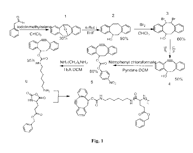
[0029] Fig. 1 is the reaction scheme for the synthesis of
dibenzocyclooctynol-
functionalized Poly(y-benzyl-L-glutamate) (DIBO-PBLG).
[0030] Fig. 2 provides SEC results of DIBO-PBLG with different feed
ratios. SEC
was run in DMF with LiBr (0.1mol/L) under 50 C.
[0031] Fig. 3 provides UV spectra of DIBO-PBLG before and after
electrospinning
indicating that the strained cyclooctyne survives the scaffold fabrication
process (top). A
second experiment measuring the residual DIBO left following a cycloaddition
reaction
indicates approximately 26% of the groups were able to react (bottom).
[0032] Fig. 4 provides SEM micrographs of gold nanoparticle (50 nm)
functionalized
fibers (A, B) and unmodified fibers as control (C, D). The scale bar for image
A, B, C, D
is 600 nm, 100 nm, 300 nm, and 200 nm respectively.
[0033] Fig. 5 provides 1H NMR and 13C NMR spectra of compound 4 of the
reaction
scheme of Fig. 1.
[0034] Fig. 6 provides 1H NMR and 13C NMR spectra of compound 5 of the
reaction
scheme of Fig. 1.
[0035] Fig. 7 provides ESI spectra of compound 6 of the reaction scheme
of Fig. 1.
-6-
CA 02878059 2014-12-29
WO 2014/022535 PCT/US2013/052971
[0036] Fig. 8 provides 1H NMR spectra of 4-dibenzocyclooctynol (DIBO)
and poly(e
-caprolactone) initiated using DIBO (bottom). The presence and end-
functionalization of
the DIBO is confirmed via the downfield shift of b from 4.7 to 5.6 ppm.
[0037] Fig. 9 provides a comparison of fluorescence emission of 9-
methyleneazidoanthracene (black) and the DIBO-PCL azide mixture (green).
[0038] Fig. 10 provides the UV-Visible adsorption spectra of DIBO-PCL
before and
after electrospinning showing that the strained cyclooctyne survives the
eelectrospinning
conditions.
[0039] Fig. 11 provides the 1H NMR spectra for the end-functionalized
polycaprolactone.
[0040] Fig. 12 provides graphs showing the influence of nanofiber
alignment and
YIGSR functionalization on differentiating mouse embryonic stem cells
embryonic (nanog
and oct3/4), neural progenitor (pax6 and nestin), and neural (neuron-specific
class III beta-
tubulin (TUJ1) and Microtubule-associated protein 2 (MAP2)) gene expression
over 3
days of culture. Sample groups include embryonic stem cells (ESC), random
unfunctionalized (RU), random YIGSR functionalized (RF), aligned
unfunctionalized
(AU) and aligned YIGSR functionalized (AF). The use of * indicates a p-
value<0.05
relative to ESC. The use of # indicates a p-value < 0.05 relative to 1 day
aligned YIGSR
functionalized sample. The use of & indicates a p-value < 0.05 relative to 3
day aligned
YIGSR functionalized sample.
[0041] Fig. 13 provides UV-vis spectra of polymer before electrospinning
and after
electrospinning, proving the survival of DIBO group in electrospinning.
[0042] Fig. 14 provides the reaction scheme for the synthesis of 0-(pent-
4-en- 1 -
yl)hydroxylamine hydrochloride.
[0043] Fig. 15 provides the reaction scheme for the synthesis for the di-
functionalized
polymer in one batch with copper-free click chemistry and oxime ligation.
DETAILED DESCRIPTION OF ILLUSTRATIVE EMBODIMENTS
[0044] The present invention provides methodologies for creating
polymeric
structures formed of polymers having strained cycloalkyne functionality that
survives the
creation process. After creation of the polymeric structure, the polymers are
functionalized
through strain-promoted azide alkyne cycloaddition copper free "click"
chemistry. The
polymeric structures bearing cycloalkyne functionality can be stored and post-
-7-
CA 02878059 2014-12-29
WO 2014/022535 PCT/US2013/052971
functionalized as needed though simple well defined azide alkyne cycloaddition
copper
free "click" chemistry.
[0045] The polymeric structures are formed at least in part of a
biocompatible
polymer including a strained cycloalkyne end group. As used herein, a strained
cycloalkyne is a five to nine member or greater cycloalkyne. In some
embodiments, the
strained cycloalkyne includes an 8-member cycloalkyne. In some embodiments,
the 8-
member cycloalkyne is a 4-dibenzocyclooctyne end group (DIBO end group).
[0046] In some embodiments, the cycloalkyne is an end group on a
biocompatible
and biodegradable polymer selected from polyglutamates, polylactones,
polylacatides,
polyglycolides and copolymers of any of the forgoing.
[0047] In some embodiments, the cycloalkyne is an end group on a polymer
selected
from poly-gamma-benzyl-L-glutamate, polycaprolactone, polylactic acid, and
polyglycolide and compolymers of any of the forgoing. In some embodiments,
cycloalkyne is an end group on a co-polymer selected from poly(lactide-co-
glycolide),
poly(lactide-co-caprolactone), poly(caprolactone-co-glycolide), and
poly(caprolactone-co-
3 -ketoc apro lactone).
[0048] In some embodiments, the biodegradable and biocompatible polymers
having
a cycloalkyne end group are formed by ring-opening polymerization (ROP). These
polymerizations employ what are termed herein "ROP initiator(s)" having a
strained
cycloalkyne as defined above. In some embodiments, the strained cycloalkyne
includes an
8-member cycloalkyne. In some embodiments, the 8-member cycloalkyne is a 4-
dibenzocyclooctyne (DIBO) group.
[0049] Notably, the ROP method of forming the polymer with cycloalkyne
end group
is particularly beneficial because cycloalkyne is not typically used as an
initiator, and, in
the case of biocompatible and biodegradable polymers, it provides a method of
functionalizing them, which the skilled artisan recognizes as being difficult
to do. The
strained cycloalkyne enables the artisan to derivatize the degradable polymer
using
conditions that do not require catalyst or additives and do not degrade the
polymer. Using
DIBO as an initiating system allows the artisan to control the stoichiometry
of functional
species in the polymer using molecular mass of the polymer as there is one
functional
group per chain.
[0050] In some embodiments, the ROP initiator includes a strained
cycloalkyne and
has a reactive group selected from an hydroxyl group and an amine group. With
hydroxyl
functionality, the ROP initiator is suitable for the ring opening
polymerization of cyclic
-8-
CA 02878059 2014-12-29
WO 2014/022535 PCT/US2013/052971
esters, such as lactones, lactides and glycolides. With amine functionality,
the ROP
initiator is suitable for the ring opening polymerization of monomers bearing
N-carboxylic
anhydrides, such as y-benzyl-L-glutamate N-carboxyanhydride.
[0051] In some embodiments, the cyclic esters are 5 to 9 member cyclic
lactones. In
some embodiments, the cyclic lactone is selected from e-caprolactone, 1,4,8-
trioxaspiro[4.6]-9-undecanone, y-butyrolactone, and 5-yalerolactone.
[0052] In some embodiments, the monomer is selected from glycolic acid
and
glycolide.
[0053] In some embodiments, the monomer is selected from lactide and
lactic acid.
[0054] In some embodiments, the ROP initiator is selected from 4-
dibenzocyclooctynol (DIBO):
1 le 0H
= .
[0055] In some embodiments, the ROP initiator is selected from DIBO
derivatives
thereof In some embodiments, the ROP initiator is an amine-functionalized DIBO
derivative or hydroxy-functionalized DIBO derivative. In some embodiments, the
ROP
initiator is chosen according to the following structure:
11
1 I e x__ oon........
W Z
wherein X is a urethane or carbonate linkage, Y is methylene (CH2) group or
ethoxy
(CH2CH20) group, n is from 1 or more to 12 or less, and Z is an amine or
hydroxyl or
hydroxyethyl.
[0056] In some embodiments, X is a urethane linkage, Y is methylene, n
is from 1 to
5, and Z is an amine, such that the ROP initiator has the following structure:
-9-
CA 02878059 2014-12-29
WO 2014/022535 PCT/US2013/052971
0
In a particular embodiment thereof, n is 5.
[0057] In some embodiments, X is a carbonate linkage, Y is methylene, n
is from 1 to
11, and Z is an hydroxyl, such that the ROP initiator has the following
structure:
11
cA
HrnCIF-1
*
In a particular embodiment thereof, n is from 1 to 6.
10 [0058] In some embodiments, X is a carbonate linkage, Y is CH2CH20, n
is from 1
to 5, and Z is an hydroxyethyl, such that the ROP initiator has the following
structure:
=
110 c):L) o_soF1
. n
In a particular embodiment thereof, n is 5.
[0059] In some embodiments, e-caprolatone monomers are polymerized
through ROP
by use of DIBO, above. In some embodiments, y-benzyl-L-glutamate N-
carboxyanhydride
is polymerized through ROP by use of:
0
0 c.L 1\NH2
.
-10-
CA 02878059 2014-12-29
WO 2014/022535 PCT/US2013/052971
wherin n is 5. In some embodiments, 1-lactide monomers are polymerized though
ROP by
use of DIBO. In yet other embodiments, DIBO is used as the initiator for the
ring-opening
copolymerization of e-caprolactone and 1,4,8-trioxaspiro[4.6]-9-
undecanone(TOSUO) to
yield the DIBO-(P(CL-co-OPD)).
[0060] The polymers bearing cycloalkyne end groups are formed into
polymeric
structures. These structures may be formed in any suitable manner taking into
account the
properties and limitations of any given polymer (e.g., solubility and heat
stability). In
some embodiments, the polymers bearing cycloalkyne end groups are formed into
polymeric structures by methods selected from electrospinning, melt-blowing,
salt leach
scaffolding, nanofibers by gas jet, ink jet printing and 3d printing.
[0061] In some embodiments, the polymers bearing cycloalkyne end groups
are
electrospun to form fibrous structures. Electrospinning is a well know process
in which the
polymer bearing cycloalkyne functionality is dissolved in an appropriate
solvent, the
resulting electrospinning solution is charged and drawn from a spinnerette to
a grounded
collector, and a fibrous mat is formed in the shape of the collector. In some
embodiments,
the electrospinning process can be used to create a porous fibrous matrix of
biodegradable
and biocompatible polymers bearing the strained cycloalkyne functionality.
[0062] Suitable solvents will be apparent to those of ordinary skill in
the art. In some
embodiments, the solvent is selected from aqueous solutions containing 10-50%
ethanol or
aqueous solutions containing 10-50% methanol.
[0063] By choosing suitable fabrication processes for forming polymeric
structures,
the cycloalkyne end group functionality survives the fabrication process. This
functionality is then available for post-fabrication functionalization through
azide alkyne
cycloaddition copper free "click" chemistry. The click chemistry is well
known, and, a
desired functionality can be imparted to the polymer by employing azide
tethered
molecules for reaction with the alkyne through cycloaddition. As used herein,
and azide
tethered molecule is a molecule that bears a reactive azide group.
[0064] In particular embodiments, the azide-tethered molecule is
selected from the
group consisting of azide-functionalized DNA, azide-functionalized peptides,
azide-
functionalized proteins, azide-functionalized sugars, azide-functionalized
metal, azide-
functionalized nanoparticles and azide-functionalized antimicrobials.
[0065] The polymeric structures of this invention can be stored after
they are
fabricated and the strained cycloalkyne end group preserved. Thus the
polymeric
structures can be functionalized as needed. Thus stock polymeric structures
can be tailored
-11-
CA 02878059 2014-12-29
WO 2014/022535 PCT/US2013/052971
to desired end uses with specifically chosen azide-tethered molecules. The
strained
cyclooctyne reacts only with azide groups. Therefore any conditions where the
strained
cyclooctyne is available and the azide functionalized molecule is soluble will
result in a
covalent cyclization. It occurs within a few minutes at room temperature and
the rate of
reaction increases with increasing temperature.
[0066] These and other aspects of this invention are experimentally
evidenced by
aspects of the following experiments.
EXPERIMENTAL
Example 1:
Post-Assembly Derivatization of Electrospun Nanofibers via Strain-Promoted
Azide
Alkyne Cycloaddition
[0067] In this work, 4-dibenzocyclooctynol (DIBO) functionalized with a
primary
amine group was used as an initiator for the ring opening polymerization of y-
benzyl-L-
glutamate N-carboxyanhydride (Bz-L-GluNCA) to yield a 4-dibenzocyclooctynol
functionalized polyey-benzyl-L-glutamate) (DIBO-PBLG). PBLG is a versatile,
degradable material that can adopt a-helix and 3-sheet conformations, which is
being
investigated for cell adhesion and proliferation when used with protein pre-
adsorption
techniques. The high binding affinity of calcium to PBLG is also promising for
bone
regeneration applications.
[0068] The DIBO-PBLG was synthesized as described in Fig. 1. The
strained DIBO
precursor was synthesized according to previously described methods. The DIBO
was
derivatized with p-nitrophenyl chloroformate and further reacted with excess
hexamethylene diamine to yield the primary amine-derivatized DIBO compound, 6
(Fig.
1). y-Benzyl-L-glutamate-N-carboxyanhydride (Bz-L-GluNCA) was synthesized and
purified by flash chromatography according to previously reported procedures.
[0069] The DIBO initiator was used immediately after purification. In a
series of
polymerizations the amine derivatized DIBO was used as an initiator for the
ring opening
polymerization of Bz-L-GluNCA in anhydrous DMF under nitrogen for 3 days to
yield
DIBO functionalized PBLG. The molecular weight of DIBO-PBLG increased linearly
with increasing feed ratio from 50:1 to 500:1. The corresponding molecular
weight
distribution increased from 1.14 to 1.29, which demonstrated that these
polymerizations
are well controlled and exhibit linear growth kinetics with increasing feed
ratio. The
-12-
CA 02878059 2014-12-29
WO 2014/022535 PCT/US2013/052971
various feed ratio conditions and the resulting molecular weights and
molecular weight
distributions of the resulting polymers were measured via DMF phase SEC and
are shown
in Fig. 2. The high molecular mass shoulders which appear at increasing feed
ratios are
consistent with what has been reported by others using amine based initiators
for the
polymerization of PBLG and is most likely due to water initiated polymer.
[0070] The DIBO-derivatized PBLG was then used as a functional precursor
for
electrospinning of nanofibers to generate a copper-free clickable scaffold.
DIBO-PBLG
(Mn=128K, PDI=1.29) and the unmodified PBLG (mol wt 150,000-350,000, Sigma)
were
both prepared in a 12 wt% 1,4-dioxane solution. Each polymer solution was held
in a glass
pipette and was electrospun from the orifice having an inner diameter of 300
um at the tip.
The electric potential was 6 kV over a 22 cm tip-to-collector distance for the
modified
polymer, and 12 kV over a 27 cm tip-to-collector distance for the unmodified
polymer. A
proper positive air pressure was applied on the surface of the solution to
maintain the
feeding rate. Fibers were collected on conductive glass slides for the
subsequent
fluorescence measurements, on silicon wafers for SEM observation, and on
copper grids
for TEM observation. Each type of collector was placed on top of a large
grounded
aluminum foil mat for the collection of electrospun fibers. Samples were
silver coated
with a sputter coater (by SPI Supplies, Pennsylvania, USA) before the SEM
observation.
From SEM micrographs, it was observed that fibers were obtained with diameters
near 1
um.
[0071] To confirm the survival of the strained DIBO group following the
electrospinning process, the fibers were dissolved in DMF and UV-Visible
spectra of the
solutions showed the presence of optical transitions near 306 nm which
correspond to the
alkyne group in DIBO (Fig. 3) before and after the fabrication process. Having
confirmed
the survival of DIBO group through electrospinning, the availability of DIBO
on the
surface of fibers for biofunctionalization was next investigated.
[0072] The extent of DIBO available on the surface of the nanofibers to
react was
quantified using UV-visible spectroscopy. A solution of 9-
methyleneazidoanthracene in
methanol, with is a non-solvent for PBLG was allowed to react with a mat of
electrospun
fibers. Concurrently, a solution of 9-methyleneazidoanthracene in N,N-dimethyl
formamide, with is a good solvent for PBLG was allowed to react with an
equivalent
solution of the DIBO-initiated PBLG. The absorbance of the nanofiber post-
functionalization and PBLG-DIBO with identical concentrations were measured
following
the reaction and from the reduction in absorbance of the alkyne transition in
DIBO, the
-13-
CA 02878059 2014-12-29
WO 2014/022535 PCT/US2013/052971
fraction of the DIBO group on the surface of fiber which is available for
derivatization is
26 5%.
[0073] A post-assembly experiment utilizing an azide containing
fluorescence probe
(Chremo 488 azide) was conducted. A glass coverslip containing fibers was
immersed in a
0.4% (mg/mL) solution of fluorescence probe for 5 min. Following the brief
incubation,
the fiber-loaded coverslip was removed, washed with methanol and dried under
nitrogen.
Using fluorescence microscopy, it was found that the azide functionalized
fluorescence
probe reacted with DIBO group on the surface of fibers. Underivatized PBLG
exhibited no
fluorescence following the methanol. These experiments indicate the
availability of the
DIBO groups on the surface for functionalization through strain-promoted azide-
alkyne
cycloaddition.
[0074] Further proof of the existence of DIBO group on the surface of
the fibers was
obtained from SEM micrographs of the fibers following immersion in a solution
of azide
functionalized gold nanoparticles. Fiber loaded silicon wafers were immersed
in a solution
of azide functionalized gold nanoparticles (50 nm, Nanocs, diluted 500 times)
for 5 h, after
which the wafer was washed with nanopure water (18 MQ/cm-1) and dried under
vacuum.
The SEM images clearly showed the presence of gold nanoparticles on the
surface of the
fibers, which confirmed that the DIBO group on the surface of fibers are
available for
functionalization (Fig. 4). A control experiment was performed using fibers
composed of
unmodified PBLG which showed conclusively that the nonspecific physical
adsorption of
gold nanoparticles to the surface of fibers is negligible (Fig. 4).
[0075] Additional experiments with transmission electron microscopy
(TEM)
confirmed the existence and availability of DIBO groups on the surface of the
fibers.
TEM grids loaded with nanofibers were immersed in an azide-functionalized gold
nanoparticle solution for lh at ambient temperature, after which the grid was
washed with
nanopure water (18 MQ/cm-l) and dried under vacuum. TEM images showed that
gold
nanoparticles are present on the surface of the nanofibers. Control
experiments with
unmodified nanofibers showed that no nonspecific physical adsorption of gold
nanoparticles was evident following immersion in the nanoparticle containing
solution.
[0076] Thus, the utility of an amine-derivatized DIBO unit as an initiator
for ring
opening polymerization is demonstrated for the first time. Additionally, the
DIBO group
survives an electrospinning procedure. The resulting nanofiber scaffold is
then available
for post-assembly functionalization with any number of azide-derivatized
molecules. The
availability of copper-free click chemistry based biofunctionalization sites
on the surface
-14-
CA 02878059 2014-12-29
WO 2014/022535 PCT/US2013/052971
of nanofibers offers versatile approach to create highly functional scaffolds
useful in a
variety of promising applications in regenerative medicine.
[0077] The synthetic details and characterization of the DIBO and amine-
derivatized
DIBO are included below.
General methods and materials
[0078] Chemicals and solvents were purchased from Sigma-Aldrich and
Acros and
were used without further purification. All reactions were performed in
anhydrous
conditions under an atmosphere of Argon. Flash chromatography was performed on
silica
gel (Sorbent Technologies Inc., 70-230 mesh). The fluorescence probe Chromeo
488 azide
was purchased from Active Motif, the azide functionalized goldnanoparticles
(50nm) was
purchased from Nanocs, 1H and 13C NMR spectra were acquired using a Varian
NMRS
500 and Varian NMRS 300. UV spectra were measured with SynergyTM MX from
BioTek, SEC results were obtained using HLC-8320GPC from TOSOH, SEM images
were acquired using JEOL-JSM-7401F with operating voltage as 4 kV, TEM images
were
obtained from a Philips TECNAI TEM with an accelerating voltage of 120 kV.
2,3 :6,7-Dibenzo-9-oxabicyclo [3.3.1] nona-2,6-diene (1)
[0079] A 250 mL flask was flame dried and charged with argon.
Phenylacetaldehyde
(18.52 g, 0.154 mol) and 100 mL of chloroform (anhydrous) were then added via
syringe.
The reaction flask was cooled in an ice bath. Trimethylsilyl iodide (25 mL,
37.5 g, 0.188
mol) was added to the solution and the reaction was allowed to stand at 5 c
for 7 days.
The reaction was monitored by TLC. After 7 days, sodium thiosulfate (1.0 M,
160 mL)
and chloroform (200 mL) were added, and the mixture was stirred until the
iodine color
was discharged. The organic phase was separated, dried (sodium sulfate), and
concentrated in vacuum. Chromatography on silica gel eluting with chloroform
yielded 6.1
g of the crystalline ether compound (35%).
3-Hydroxy-2',3',2",3"-tetramethoxly,-2 :5,6-dibenzocyclocta- 1,5,7-triene (2)
[0080] 2,3:6,7-Dibenzo-9-oxabicyclo[3.3.1]nona-2,6-diene 1 (2.00 g, 5.84
mmol) in
anhydrous THF (60 mL) was placed into a three-necked round bottom flask and
cooled in
an ice bath under argon. n-butyl lithium (4.92 mL, 2.5 M, 12.4 mmol) was added
slowly
via syringe. The reaction mixture was stirred at room temperature under argon
for 4 h. The
reaction was quenched by careful addition of water and extracted with 2XSOmL
CHC13.
-15-
CA 02878059 2014-12-29
WO 2014/022535 PCT/US2013/052971
The combined organic phases were washed with 30 mL of brine, dried over
Na2SO4,
concentrated under vacuum and purified by column chromatography on silica gel
CHC13
to yield 1.83 g of 3-Hydroxy-2',3',2",3"-tetramethoxly,-2 :5,6-dibenzocyclocta-
1,5,7-
triene (90%).
11,12-Dibromo-5,6,11,12-tetrahydro-dibenzo [a,e] cycloocten-5-ol (3)
[0081] Bromine (0.51 mL, 10 mmol) was added dropwise to a stirred
solution of 2
(2.22 g, 10 mmol) in CHC13 (50 mL). After stirring the mixture for 0.5 h, TLC
analysis
indicated completion of the reaction. The solvent was evaporated under reduced
pressure
and the residue was purified by flash chromatography over silica gel (2:1/1:2,
v/v,
hexanes/CH2C12) to yield 3 as a light-yellow oil (60%).
5,6-Dihydro-11,12-didehydro-dibenzo la,e]cycloocten-5-ol (4)
[0082] Lithium diisopropylamide in tetrahydrofuran (2.0 M; 8.0 mL, 16
mmol) was
added dropwise to a stirred solution of 3 (1.53 g, 4.0 mmol) in
tetrahydrofuran (40mL)
under an atmosphere of argon. The reaction mixture was stirred for 0.5 h,
after which it
was quenched by the dropwise addition of water (0.5 mL). The solvents were
removed
under reduced pressure, and the residue was purified by flash chromatography
on silica gel
(hexanes/ CH2C12 2:1/0:1, v/v) to yield 4 as a white amorphous solid (0.52 g,
60%). Fig. 5
provides 1H NMR and 13C NMR spectra of compound 4.
Carbonic acid, 5,6-dihydro-11,12-didehydro-dibenzola,e]cycloocten-5-y1 ester,
4-
nitrophenyl ester (5)
[0083] 4-Nitrophenyl chloroformate (0.4 g, 2 mmol) and pyridine (0.4 mL,
5 mmol)
were added to a solution of 4 (0.22 g, 1 mmol) in CH2C12 (30 mL). After being
stirred for
4 h at room temperature, the mixture was washed with brine (2>< 10 mL) and the
organic
layer was dried (MgSO4). The solvents were evaporated under reduced pressure,
and the
residue was purified by silica gel column chromatography (hexane/ethyl
acetate, 10:1, v/v)
to afford 5 (0.32 g, 82%). Fig. 6 provides 1H NMR and 13C NMR spectra of
compound 5.
6-Aminohexyl-carbamic acid 5,6-dihydro-11,12-didehydrodibenzola,e]cycloocten-5-
y1 ester (6)
-16-
CA 02878059 2014-12-29
WO 2014/022535 PCT/US2013/052971
[0084] To a 50m1 solution of Hexamethylenediamine(151mg, 1.3mmol) and
13.2u1
TEA in CH2C12 was added 5 (50mg, 0.26mmol). After stirring for 3h, the organic
phase
was washed with 10 x 20m1 water, and dried over Na2SO4. Solvent was removed
under
vacuum and was purified by flash chromatography on silica gel(CH2C12/CH3OH,
3:1
with 0.5% isopropylamine) to yield 6 (3 1 mg, 65%). Fig. 7 provides ESI
spectra of
compound 6.
Synthesis of DIBO functionalized Polyy-Benzyl-L-glutamate
[0085] 7-Benzyl-L-glutamate N-carboxyanhydride(Bz-L-GluNCA) and 6 were
dissolved in anhydrous DMF in flame dried schlenk flask. After three freeze,
pump, thaw
cycles the polymerization stood under nitrogen for 3 days. The polymer was
precipitated
in ethyl ether and dried under vacuum.
Electrospinning
[0086] DIBO-PBLG and the unmodified PBLG were both prepared in a 12 wt% 1,4-
dioxane solution. Each polymer solution was held in a glass pipette and was
electrospun
from the orifice with an inner diameter 300 um on the tip. The electric
potential was 6 kV
over a 22 cm tip-to-collector distance for the modified polymer, and 12 kV
over a 27 cm
tip-to-collector distance for the unmodified polymer. A proper positive air
pressure was
applied on the surface of the solution to maintain the feeding rate. Fibers
were collected on
conductive glass slides for the following fluorescence test, on silicon wafers
for SEM
observation, and on copper grids for TEM observation. Each kind of collector
was placed
on top of a large grounded aluminum foil for the collection of electrospinning
fibers.
Samples were silver coated with an SPI Sputter Coater before the SEM
observation.
UV Spectra
[0087] UV spectra of DIBO-PBLG before and after electrospinning were
measured in
DMF solution using a plate reader.
Example 2:
4-Dibenzocyclooctynol (DIBO) as an initiator for poly(c-caprolactone): copper
free
clickable polymer and nanofiber-based scaffolds
[0088] In this work, 4-dibenzocyclooctynol (DIBO) was used to initiate
the
polymerization of c¨caprolactone (c¨CL). The methodology results in an end-
-17-
CA 02878059 2014-12-29
WO 2014/022535 PCT/US2013/052971
functionalized PCL that is easily functionalized with azide-derivatized
compounds under
metal-free conditions. This is significant as post synthetic modification and
purification of
PCL have been limited due to its degradability.
[0089] The polymerizations were carried out under traditional Sn-based
catalytic
conditions using stannous octoate:
/
0 /r1
Stannot1:5 Octi-mte 0
A
............... -OH BOT
DIBO (28 mg, 0.13 mmol), c¨CL (2.50 g, 21.93 mmol) and freshly distilled
stannous
octoate (0.053 mmol). Following addition to a flame dried schlenk flask and
three cycles
of freeze-pump-thaw degassing, the reactions were heated at 80 C with
reaction times
that varied from 4 h to 20 h. The polymerization conditions yielded living
characteristics
with nearly linear increases in molecular mass with time.
[0090] Fig. 8 shows the 1H NMR spectra of the DIBO initiator (top) and
resulting
PCL polymer (bottom). The retention of the phenyl resonances (-7.3), the
methylene
(CH2) from the strained ring (2.9, 3.1) and the downfield shift of b (CHOH)
from 4.6 to
5.6 shows that DIBO successfully initiated the polymerization of PCL, and
survived intact
during the polymerization process. Size exclusion chromatography (SEC)
eluograms
indicated the molecular mass of DIBO¨PCL increased linearly with increased
polymerization time from 4 h to 20 h, while the polydispersity remained narrow
and mono
modal in molecular mass distribution.
[0091] The reactivity of the DIBO group at the end of the PCL chain
following the
polymerization was confirmed using a metal-free click reaction between DIBO
and 9-
methyleneazidoanthracene. Briefy, DIBO¨PCL (Mn = 4.5 kDa, 7.7 mg, 1.7 x10⁻³
mmol)
was added to a 9-methyleneazidoanthracene solution in DMSO (500 L, 1.4x10⁻⁴
mmol).
After 15 min, the solution was diluted 100 fold and the fluorescence emission
spectrum
was acquired.
[0092] Following the mixing of the azide solution and the DIBO-PCL
solution, a ¨7-
fold increase in the fluorescence intensity resulted (Fig. 9). A solution of
the azide
molecule with identical concentration was used as the control.
-18-
CA 02878059 2014-12-29
WO 2014/022535 PCT/US2013/052971
[0093] The DIBO-PCL (Mn=20k) was then used to generate nanofibers via an
electrospinning process. A DIBO-PCL solution 40% (wt/mL) in DCM/DMF (4/1) was
subjected to well-defined electrospinning conditions of 5.5 kv voltage, and a
10 cm plate
height. PCL nanofibers with diameters near 500 nm were successfully obtained
using
these conditions.
[0094] Fig. 10 shows the UV-visable absorption spectra of the DIBO-PCL
polymer
before and after electrospinning. The 7E-7E* optical transition at 306 nm from
the alkyne
group did not change position or relative intensity following the
electrospinning process,
demonstrating that DIBO groups survived.
[0095] The reactivity of the DIBO group on the surface of fibers was
substantiated
using a reaction with an azide-containing fluorescence probe (Chemo 488
azide). A glass
slide coated with fibers was immersed in a 0.4% (mg/mL) solution of this
fluorescence
dye for 5 min at ambient temperature. The glass slide was then washed with
water and
dried under nitrogen. Fluorescence images of DIBO-PCL nanofibers showed
covalent
derivatization with 9-methyleneazidoanthracene. The resulting nanofibers are
highly
fluorescent indicating that DIBO groups are present on the surface of the
nanofibers and
are able to react with the azide-containing dye. Very little fluorescence was
observed in a
control experiment using fibers from unmodified PCL under identical conditions
indicating nonspecific adsorption of the fluorescence probe is negligible, and
the
fluorescence is due to the copper-free click reaction between the dye and DIBO
group on
the surface of the fibers.
[0096] 4-dibenzocyclooctynol (DIBO) as an initiator for the ring-opening
polymerization of e-caprolactone yielded an DIBO end-functionalized PCL
polymer. The
DIBO group survives the polymerization conditions and offers efficient,
orthogonal and
biocompatible functionalization opportunities for both the polymer and polymer-
derivatized biomaterials. The combination of PCL and DIBO enables large-scale
production of a new type of easily functionalizable nanofiber-based scaffold
with versatile
regenerative medicine applications.
Methods and materials
[0097] Chemicals and solvents were purchased from either Sigma-Aldrich
or Acros
and were used without further purification. All reactions were performed in
anhydrous
conditions under an atmosphere of Argon.Flash chromatography was performed on
silica
-19-
CA 02878059 2014-12-29
WO 2014/022535 PCT/US2013/052971
gel (Sorbent Technologies Inc., 70-230 mesh). Stannous octoate (Aldrich) and e-
caprolactone (Acros Organics) were distilled before use.
[0098] Size exclusion chromatographic analyses (SEC) were performed
using a
Waters 150-C Plus instrument equipped with three HR-Styragel columns [100 A,
mixed
bed (50/500/103/104 A), mixed bed (103, 104, 106 A)], and three detectors
including a
differential refractometer (Waters 410), a differential viscometer (Viscotek
100), and a
laser light scattering detector (Wyatt Technology, DAWN EOS, 2, = 670 nm). THF
was
used as eluent with a flow rate of 1.0 mL/min at 30 C. The Molecular weight
and
polydispersity were calculated according to light scattering data.
[0099] 1H Nuclear Magnetic Resonance (NMR) spectra was acquired using a
Varian
Mercury 300 NMR and 500 NMR spectrometer. UV-Vis spectra were measured using a
SynergyTM MX plate reader from BioTek. SEM images were acquired using JEOLJSM-
7401F with operating voltage as 1 kV. Fluorescence images were acquired using
a CKX41
microscope (Olympus, Center Valley, PA).
[00100] The synthesis of 4-dibenzocyclooctynol (DIBO) has already been
experimentally shown in Example 1 and is not repeated here.
[00101] DIBO (28 mg, 0.13 mmol), c-CL (2.50 g, 21.93 mmol) and freshly
distilled
stannous octoate (0.053 mmol) were added to a flame dried schlenk flask. After
three
cycles of freeze-pump-thaw degassing, the reactions were heated at 80 C with
varied
reaction times. Following the designated polymerization time, the reaction was
quenched
in liquid nitrogen, dissolved in THF and precipitated in cold methanol.
Molecular Mass,
mass distribution, UV visible and NMR spectroscopy were collected as described
above.
The 1H NMR spectra for the end-functionalized PCL is shown in Fig. 11.
Example 3:
Directed Differentiation and Neurite Extension of mouse Embryonic Stem Cell on
Aligned Poly(lactide) Nanofibers Functionalized with YIGSR Peptide
[00102] Here we report a versatile and potentially transformative
approach to the
creation of nanofeatured poly-L-lactides (PLLAs) capable of being derivatized
post-
fabrication with any azide functionalized molecule. PLLA is a widely accepted
and
applied material in regenerative medicine applications, however the creation
of suitably
functional variants with which to direct cell behavior remains challenging.
Electrospinning was used to created nanofiber matrices with fiber diameters
near the size
of the nanotopography shown to promote stem cell neural differentiation and
neurite
-20-
CA 02878059 2014-12-29
WO 2014/022535 PCT/US2013/052971
extension, with functionalization of the scaffolds with the YIGSR peptide
undertaken
using metal-free alkyne-azide cycloaddition. Such an approach presents a mild
and
efficient method for the creation of functional scaffolds that avoids PLLA
degradation and
enables the fabrication of nanofiber mats in a translationally-relevant
manner. Herein, we
demonstrate the direct differentiation of mouse embryonic stem cells to neural
lineages on
aligned YIGSR nanofiber matricies in a manner that can readily be scaled and
translated to
other applications and the clinic.
Materials
[00103] Chemicals and solvents were purchased from Sigma-Aldrich were used
without further purification unless specifically noted. All reactions were
performed in
anhydrous conditions under an atmosphere of Argon. Flash chromatography was
performed on silica gel (Sorbent Technologies Inc., 70-230 mesh). 9-
fluorenylmethoxycarbonyl (Fmoc) protected amino acids, Fmoc-Amino acid loaded-
Wang
resin and -(1H-B enzotriazol-1 -y1)-1,1,3,3 - tetramethyluronium
hexafluorophosphate
(HBTU) were purchased from NovaBiochem. 4-Dibenzyocyclooctynol (DIBO) was
synthesized according to previous literature and dried over P205 under vacuum
for 72
hours before use (DIBO synthesis in accordance with Ngalle Eric Mbua, J. G.,
Margreet
A. Wolfert, Richard Steet, Geert-Jan Boons chembiochem 2011, 12, 1912 ¨ 1921;
Jung,
M. E.; Mossman, A. B.; Lyster, M. A. The Journal of Organic Chemistry 1978,
43, 3698-
3701). 1,8-diazabicyclo[5.4.0]undec-7-ene (DBU) was freshly distilled into
sealable
ampoules from CaH2 under an inert atmosphere. 1-Lactide (1-LA, Purac) was
first
dissolved in methylene chloride, passed through a silica plug and then stirred
over
anhydrous Mg504. The solution was then filtered and the methylene chloride
removed in
vacuo. The resulting solid was taken up in hot toluene before evaporation to
dryness in
vacuo on a rotary evaporator. The resulting crystalline solid was then
transferred to a
Schlenk flask and dissolved in anhydrous hot toluene and the solvent was then
removed in
vacuo on a vacuum manifold. The crystalline white solid was then taken up in
anhydrous
methylene chloride and transferred by cannula onto activated 3 A molecular
sieves and left
to stand for 24 h before being transferred for a second time onto fresh 3 A
sieves for a
further 24 h. The methylene chloride/1-LA solution was then transferred to a
Schlenk flask
using a filter cannula and the methylene chloride removed in vacuo. Finally,
the resulting
white solid was recrystallised from hot (70 C) toluene and stored in a glove
box at
-21-
CA 02878059 2014-12-29
WO 2014/022535 PCT/US2013/052971
ambient temperature. Deuterated chloroform (CDC13) was purchased from Apollo
Scientific Ltd and distilled from CaH2 before use.
General Considerations
[00104] Unless otherwise stated, all manipulations were carried out in a
nitrogen filled
glove box. 1H and 13C NMR spectra were recorded on either a Bruker DPX-400
spectrometer at 298 K. Chemical shifts are reported as 6 in parts per million
(ppm) and
referenced to the chemical shift of the residual solvent resonances (CHC13: 1H
6 = 7.26
ppm; 13C 6 = 77.16 ppm). Size exclusion chromatography (SEC) was conducted on
Varian
GPC 50 instrument fitted with a differential refractive index detector and a
mixed-D
column set comprising of a short guard column (Varian Polymer Laboratories
PLGel 5
M, 50 x 7.5 mm) and two further chromatographic columns (Varian Polymer
Laboratories PLGel 5 M, 300 x 7.5 mm). The mobile phase was CHC13 (HPLC
grade) at
a flow rate of 1.0 mL min-1. SEC samples were calibrated against Varian
Polymer
Laboratories Easi-Vials linear poly(styrene) standards (162 ¨ 2.4 x 10⁵ g mo1-
1) using
Cirrus v3.3 software.
Typical DIBO-terminated PLLA synthesis
[00105] In a glove box, 1-LA (2 g, 1.39 x 10⁻² mol, 200 eq.) and DIBO
(15.3 mg, 6.95
x 10-5 mol, 1 eq.) were added to a glass vial equipped with magnetic stirrer
bar and
dissolved in methylene chloride (13.9 mL). Under rapid stirring, DBU (21.12
mg, 1.39 x
10 mol, 2 eq.) was then added. After 30 min the reaction was quenched by the
addition of
Dowex 50w x8 (20-50 mesh) acidic resin followed by precipitation of the
polymer into
cold hexanes. The precipitate was isolated by filtration before being
redissolved in CHC13
and precipitated a further two times into cold hexanes. The resulting white
solid was then
subjected to high vacuum for 48 h to remove traces of solvent (1.5 g, 74%). 1H
NMR (400
MHz, CDC13, ppm): 6 = 7.52 (m, 8H, DIBO C6H4), 5.59 (s, 1H, CH(OH)), 5.16 (q,
1H,
OCH(CH3)C(0)), 4.35 (m, 1H, CH(CH3)0H), 3.00 (m, 2H, DIBO CH2), 1.58 (d, 3H,
OCH(CH3)C(0)). 13C NMR (100 MHz CDC13, ppm): 6 = 169.8, 69.2, 16.8. GPC
(CHC13,
poly(styrene) standards): Table 1.
Electrospinning
[00106] The DIBO-terminated PLLA (DIBO-PLLA) was dissolved in a 1:4 (v/v)
N,N-
dimethylformamide /dichloromethane solution to yield a clear, slightly viscous
solution of
-22-
CA 02878059 2014-12-29
WO 2014/022535 PCT/US2013/052971
25% (w/v) concentration. The solution was shaken and left overnight to ensure
homogeneity. The solution vial was sealed with Parafilm (Pechiney Plastic
Packaging,
Chicago, IL) to retain the concentration of the solution. The solution was
held in a tapered
tube made by heating and drawing a glass pipet to an outer diameter of 0.4 mm.
The air
pressure above the solutions was adjusted to control the flow. The solutions
inside the jets
were connected to high voltage supplies (0-60 kV, ES60, Gamma High Voltage
Research,
Ormond Beach, FL), and the sample collectors were grounded. A voltage of 12 kV
was
applied to the DIBO-PLLA solutions, and the tip-to-screen distance was 20 cm.
For
random fibers, aluminum foil was used as the grounded collector. Round glass
cover slides
(18 mm diameter, 18 CIR, Fisher Scientific, Pittsburgh, PA) were placed on the
aluminum
foil to collect the fibers. For aligned fibers, a special sheet of stainless
steel collector was
fabricated and used having elongated openings of 20 mm x 70 mm. The aligned
fibers
were collected on glass coverslips placed in the gaps of the stainless steel
collector.
Peptide Synthesis
[00107] The Br-GYIGSR was synthesized using standard FMOC conditions on a
CEM
Discovery microwave peptide synthesizer. The N-terminus was derivatized with 6-
Bromohexanoic acid as described previously. Moore, N. M.; Lin, N. J.; Gallant,
N. D.;
Becker, M. L. Biomaterials 2010, 31, 1604-1611. The crude Br-terminated
peptide was
purified by reverse phase HPLC. The Br end group was substituted with an azide
group in
a 1:2 solution of methanol: water containing 18-crown-6 (0.05 eq) stirred at
23 C
overnight. The azide-substituted peptide was purified by dialysis to eliminate
the NaN3
residue, and a pale yellow solid was obtained following lyophilization. This
substitution
was confirmed by ESI-Mass spectra (MW=789.3Da).
Nanofiber Functionalization
[00108] The N3-GYIGSR peptide was dissolved in 1:2 water/methanol (v/v)
to yield a
0.5 mg/mL solution. The glass slides covered with electrospun fibers were
carefully
dipped into the peptide solution three times and rinsed with a 1:2
water/methanol solution.
The functionalized fibers were dried overnight and sterilized with ethylene
oxide. The
sterilized nanofibers were degassed for 3 days.
Lowry Assay
-23-
CA 02878059 2014-12-29
WO 2014/022535 PCT/US2013/052971
YIGSR concentration on the fiber was determined using the Lowry assay as
previously
described. Miller, J. S.; Shen, C. J.; Legant, W. R.; Baranski, J. D.;
Blakely, B. L.; Chen,
C. S. Biomaterials 2010, 31, 3736-3743. Briefly, 1.0 mg peptide functionalized
polymer
was dissolved in 1.000 mL of DMSO. A standard curve was created by dissolving
1
ug/mL, 2 ug/mL, 5 ug/mL, 10 ug/mL or 20 ug/mL of YIGSR peptide in DMSO
containing
with 1.0 mg of polymer per mL. Total protein within each sample was measured
using a
Dc Protein assay (Biorad, Hercules, CA) according to manufacture protocol.
Scanning Electron Microscopy
[00109] The fiber dimensions and alignment were evaluated by SEM (JSM-
7401F,
JEOL, Peabody, MA). The acceleration voltage was set at lkV. The fibers were
not
sputter coated prior to imaging. The fiber diameters and angles were
calculated by
measuring over 100 fibers using Image J.
D3 mouse Embryonic Stem Cell Culture and Seeding
[00110] D3 mouse ESC [Doetschman, T.; Eistetter, H.; Katz, M.; Schmidt,
W.;
Kemler, R. J Embryol Exp Mophol 1985, 87, 27-45] were cultured on 0.1% gelatin-
coated
tissue culture flasks in ESC media (DMEM supplemented with 10% FBS, 10-4M p-
mercaptoethanol, 0.224 lag/mL L-glutamine, 1.33 lag/mL HEPES, and 1,000 units/
mL
human recombinant LIF). Nanofiber-loaded coverslips were placed in a 12 well
plate,
sterilized with ethylene oxide and then wet with 80% F-12/ 20% Neurobasal
media
(Invitrogen, Grand Island, NY) for 1 h. D3 cells (325,000) were seeded evenly
on each
sample in 400 [IL of fresh neural media (80% F-12/ 20% Neurobasal media with
N2 and
B27 supplements, 10 mM sodium pyruvate and 1 ILIM retinoic acid). The media
was
changed every other day for the duration of the experiment.
Immunofluorescence and Alkaline Phosphatase Quantification
[00111] Immunofluorescence was conducted as previously described. Smith
Callahan,
L. A.; Ma, Y.; Stafford, C. M.; Becker, M. L. Biomaterials Science 2013.
Briefly, cells
cultured on nanofiber-coated coverslips were fixed at designated time points
with 4%
paraformaldehyde/PBS, washed, and stored at 4 C in PBS. Nonspecific antibody
binding
was blocked by incubating in 10% goat serum, then the gradients were exposed
to Neuron-
specific class III beta-tubulin (TUJ1) (PRB-435p, Covance, 1:500), followed by
appropriate secondary antibody conjugated with Alexaflour 544 (Invitrogen).
DAPI was
-24-
CA 02878059 2014-12-29
WO 2014/022535 PCT/US2013/052971
used to stain the cell nuclei. Images were taken with an automated IX81
microscope
(Olympus). Cellular density at each position was determined by using the
automated
counting function of ImageJ to count nuclei in images at each position from at
least 5
separate samples. Statistical averages of neurite lengths from the nucleus
were made from
measurements of at least 250 cells from at least 3 separate samples per group
using ImageJ
(National Institute of Health, Bethesda, MD, USA). Fraction of cells
expressing TUJ1 was
determined using the ImageJ cell counter and dividing the total number of
cells expressing
TUJ1 staining by the number of nuclei in at least 10 fields of view per sample
and 1000
cells per sample group at each time point. Quantification of alkaline
phosphatase was
determined with a SensoLyte pNNP Alkaline Phosphatase assay kit (AnaSpec,
Fremont,
CA) according to the manufacturer's protocol. For normalization, total protein
was
measured in samples with a Dc Protein assay (Biorad, Hercules, CA) according
to the
manufacturer's protocol.
Semi-Quantitative PCR
[00112] Total
RNA was isolated from at least three replicates using an RNeasy Mini
Kit (Qiagen, Hilden, Germany) according to the manufacturer's protocol after
cells were
harvested from material samples with a cell scraper. RNA samples with an
optical density
ratio of absorbance at 260 nm (RNA) over that at 280 nm (protein) greater than
1.9 were
used to make cDNA. Based on the absorbance reading at 260 nm, 0.5 mg of RNA
from
each sample was used to make cDNA using a 2710 thermal cycler (Applied
Biosystems)
with TaqMan reverse transcription reagents. The thermocycler program was as
follows: 10
min incubation at 25 C, 30 min reverse transcription at 48 C, and 5 min
inactivation at
95 C. Two microliters of each reaction was subject to PCR using AmpliTaq Gold
DNA
polymerase (Applied Biosystems) for each of the following:
Nanog (5'-agggtctgctactgagatgctctg-3' and 5'-atcttctgatcctggcaag-3'); Oct 1/4
(5' -
ggggatccgatggcatactgtggacctcag-3' and 5' -gggaattcgcttcgggcacttcagaaac-3');
pax6 (5' -
aagggcggtgagcagatgt-3' and 5' -gcatgctggagctggttgg-3'); Nestin (5' -
gtgcctctggatgatg-3'
and 5' -ttgaccttcctccccctc-3'); TUJ1 (5' -
tcactgtgcctgaacttacc-3' and 5' -
ggaacatagccgtaaactgc-3 ' ); Microtubul e- as s oc iated protein
2 (MAP2) (5 ' -
gaggcagaagctccaaga-3' and 5'-ctggacccactccacaaact-3'); Oligodendrocyte
transcription
factor 1 (OLIG1) (5'-tgcgcgcgagaaggccgaag-3' and 5' -cccagccagccctcacttg-3');
Glial
fibrillary acidic protein (GFAP) (5'-gaggcagaagctccaaga-3' and 5'-
gctctagggactcgttcgtg-
-25-
CA 02878059 2014-12-29
WO 2014/022535 PCT/US2013/052971
3'); Glyceraldehyde 3-phosphate dehydrogenase (GAPDH) (5' -
accacagtccatgccatcac-3'
and 5' -tccaccaccctgttgctgta-3').
[00113] The cycling conditions were 94 C for 5 mm followed by 94 C for
30 s, 55 C
for 60 s, 72 C for 60 sec for Nanog and Oct 3/4, 94 C for 30 sec, 55 C for
30 sec, 72 C
for 60 sec for pax6 and TUJ1; 94 C for 30 sec, 55 C for 30 sec, 72 C for 45
sec for
Nestin; 94 C for 30 sec, 56 C for 30 sec, 72 C for 60 sec for GFAP and
OLIG01; and
94 C for 30 sec, 62 C for 30 sec, 72 C for 60 sec for GAPDH. All
amplifications were
run for 35 cycles and followed by a 10 mm extension at 72 C. The PCR product
was
loaded in a 1% agarose gel stained with ethidium bromide and run for 45 min at
100 V.
Fluorescent images of gels were obtained with a Biospectrum Imaging System
(UVP,
Upland, CA). The relative densities of the bands were analyzed with
VisionWorks LS
(UVP) and normalized to GAPDH density for the sample.
Statistics
[00114] All experiments were conducted at least 3 times (n > 3) with
multiple
substrates as noted in each section. All quantitative data are presented as
the average
standard deviation. One-way analysis of variance (ANOVA) with Tukey post hoc
analysis
was performed where applicable. Significance was set at a p-value of less than
0.05.
Results
[00115] Synthesis of end-functional poly(L-lactide)s, PLLAs, was
undertaken by the
ring-opening polymerization of L-lactide catalyzed by 1,8-
diazabicyclo[5.4.0]undec-7-ene
(DBU) using 4-dibenzyocyclooctynol (DIBO) as the initiator, as follows:
1\1
0
IL OH CN
n4.\..._o _____________ 0 0
Di.
W +
410 0 10 2n
[00116] This technique simplifies the removal of catalytic residues and
enables the
targeting of high molecular weight PLLAs with high end group fidelity. PLLAs
with
targeted degrees of polymerization of 170, 200 and 370 were obtained within 30
min and
-26-
CA 02878059 2014-12-29
WO 2014/022535 PCT/US2013/052971
yielded polymers of 41.1, 59.5 and 87.4 kDa with narrow molar mass
distributions (Dm
<1.13).
[00117] Nanofibrous matrices, which emulate the size scale of the native
ECM, were
shown to promote cellular attachment, proliferation and differentiation more
effectively
than traditional tissue engineering matrices. Electrospinning is one method
capable of
generating nanofibers on the size scale of ECM. Using DIBO-PLLA, both random
and
aligned fibers were fabricated; the average fiber diameter was 345 nm 51 nm
and 338
nm 63 nm, respectively. The aligned matrices possessed high degree of
alignment with a
narrow angular distribution of oriented fibers ¨ 89.5 6.5 . Functionalized
nanofibers
were found to have 4.94 ug 2.76 ug (average standard deviation) of YIGSR
peptide
per 1000 ug of functionalized fiber.
Table 1. Summary of size exclusion chromatography results for synthesized 4-
dibenzocyclooctynol (DIBO) terminated-L-lactide
Sample [Win DP Mn (Da) (Mw) Dm
1 175 170 41 100 43 500 1.06
2 350 200 59 500 67 500 1.13
3 450 370 87 400 90 200 1.03
[00118] Fiber diameter, alignment and functionalization can affect neural
progenitor
differentiation and neurite extension5,6,28-30. A significantly higher
fraction of ESC
cultured on aligned functionalized samples (0.75 0.07 (average standard
deviation))
compared to ESC cultured on both the random unfunctionalized (0.53 0.03; p-
value =
0.001) and functionalized samples (0.53 0.02; p-value = 0.001) expressed
TUJ1 after 1
day of differentiation, while ESC cultured on the aligned unfunctionalized
samples
expressed an intermediate fraction TUJ1 (0.63 0.03). After 3 days of
differentiation, a
significantly higher fraction of ESC cultured on aligned functionalized
samples (0.71
0.01) expressed TUJ1 compared to ESC cultured on the random unfunctionalized
(0.56
0.01, p-value=0.04). The random functionalized (0.61 0.01) and aligned
unfunctionalized (0.61 0.08) expressed intermediate fractional values.
Previous studies
on the effects of fiber alignment on stem cell differentiation to neural
lineages have
diverged with some indicating similarity to our results or others indicating
that alignment
does not affect the fraction of cells expressing TUJ1. Differences between
cell lines and
-27-
CA 02878059 2014-12-29
WO 2014/022535 PCT/US2013/052971
differentiation protocols, especially temporally, most likely contribute to
this discrepancy.
Average neurite length of ESC cultured on aligned functionalized samples was
significantly longer than that of ESC cultured on both the random
unfunctionalized and
unfunctionalized samples. The results are similar to previous studies that
have found fiber
alignment and the incorporation of YIGSR improved neurite extension compared
to
control. Subtle differences in cell density are unlikely to impact these
results since a
similar amount of DNA was isolated from all sample groups at each time point
studied
(data not shown). However, fiber density cannot be controlled precisely
between samples
and may contribute to differences between groups.
[00119] Gene expression for embryonic stem cell markers, nanog and Oct 3/4,
was
higher in the starting ESC population and decreased with increased exposure to
neural
differentiation media in ESC in all sample groups. In combination with
alkaline
phosphatase quantification which decreased relative to the total protein of
each sample
over time (data not shown) indicates that all sample groups support ESC
differentiation.
Although all sample groups supported ESC differentiation, ESC on the aligned
functionalized samples showed a significant decrease of Oct 3/4 mRNA
expression
compared to the ESC starting population after 1 day, while the ESC on the
other groups
required 3 days of culture show a significant decrease in Oct 3/4 mRNA
expression
relative to the ESC starting population. PAX6 mRNA expression, a neural
progenitor
marker, was up regulated in ESC on all sample groups compared to the ESC
starting
population after 1 day of differentiation (Fig. 12). Similarly, nestin mRNA
expression,
another neural progenitor marker, was found to be up regulated after 1 day of
neural
differentiation in ESC cultured on random unfunctionalized, aligned
unfunctionalized, and
aligned functionalized samples compared to the ESC starting population (Fig.
12). After 3
days of neural culture, mRNA expression of both pax6 and nestin decreased in
ESC on all
sample groups indicating differentiation of the ESC beyond neural progenitors.
After 3
days of differentiation, mRNA expression of TUJ1, an immature neuron marker,
was
significantly increased in ESC cultured on the aligned functionalized samples
compared to
ESC cultured on the other sample groups (Fig. 12). MAP2 mRNA expression, a
mature
neuron marker, was also up regulated in ESC cultured on aligned functionalized
samples
after 3 days of differentiation compared to ESC cultured on the
unfunctionalized random
and aligned samples (Fig. 12). GFAP, an astrocyte marker and Oligo 1, an
oligodendrocyte
marker, were not detectable by PCR in ESC cultured on any of the sample groups
indicating that those cell types were not yet present in the cultures (data
not shown). While
-28-
CA 02878059 2014-12-29
WO 2014/022535
PCT/US2013/052971
fiber alignment has been shown to effect gene expression in stem cells,
studies examining
the effects of fiber-aligned or YIGSR peptide-functionalized mats on the gene
expression
of ESC undergoing neural differentiation could not be located in the
literature.
Conclusions
[00120] Synthetic polymers functionalized with biological moieties have
widespread
applications in clinical regenerative medicine. Through the end-
functionalization of PLLA
with DIBO, a method has been developed for the facile, post-fabrication
functionalization
of tissue engineering matrices in a translationally-relevant manner. Increased
neurite
length and neural gene expression of ESC cultured on functionalized matrices
compared to
gene expression of ESC cultured on unfunctionalized matrices indicated the
functionalization is bioavailable to the cells and DIBO PLLA offers a
clinically relevant
material for the production of functional synthetic polymers. The post-
fabrication strategy
we demonstrate herein is a transformative approach to medical ideas and
innovations that
many have ignored clinically due to characterization challenges, difficulty of
processing
bioactive species and concerns about regulatory pathways for combination
products.
Example 4:
One Batch Difunctionalization of Poly (c-caprolactone) copolymer derivatized
nanofiber scaffold with Strain-Promoted Azide Alkyne Cycloaddition and Oxime
Chemistry
[00121] In this paper, two orthogonal chemical reactions, copper-free
click chemistry
and oxime reaction are combined to generate an efficient, biocompatible one
batch method
of difunctionalized nanofiber based scaffolds. DIBO was used as the initiator
for the ring-
opening copolymerization of e-caprolactone and 1,4,8-trioxaspiro[4.6]-9-
undecanone(TOSUO) to yield the DIBO-(P(CL-co-OPD)). Post-polymerization
deprotection proceeded under mild condition to recover the reactive ketone
group. Further
di-functionalization of the polymer was carried out using azide terminated PEG
and 0-
(pent-4-en-1-yl)hydroxylamine hydrochloride in one batch. The feasibility of
easy
functionalization of nanofiber based scaffold is also substantiated with
fluorescence
probes Chemo 488 azide (green) and Alexa Fluor 568 hydrazide (red).
-29-
CA 02878059 2014-12-29
WO 2014/022535 PCT/US2013/052971
General methods and materials
[00122] All chemicals and solvents were purchased from Sigma-Aldrich or
Acros and
were used without further purification unless otherwise noted. All reactions
were
performed in anhydrous conditions under an atmosphere of Argon. Flash
chromatography
was performed on silica gel (Sorbent Technologies Inc., 70-230 mesh). Stannous
octoate
(Aldrich) and e-caprolactone (Acros Organics) were distilled prior to use. 4-
dibenzocyclooctynol (DIBO) was synthesized according to methods described
previously.26'35 Chemo 488 azide was acquired from Active Motif and Alexa
Fluor 568
was acquired from Life Technologies.
Instrument Information
[00123] Size exclusion chromatographic analyses (SEC) were performed
using a
Waters 150-C Plus instrument equipped with three HR-Styragel columns [100 A,
mixed
bed (50/500/103/104 A), mixed bed (103, 104, 106 A)], and three detectors
including a
differential refractometer (Waters 410), a differential viscometer (Viscotek
100), and a
laser light scattering detector (Wyatt Technology, DAWN EOS, 2, = 670 nm). THF
was
used as eluent with a flow rate of 1.00 mL/min at 30 C. The molecular mass
and
molecular mass distribution were calculated from light scattering data.
[00124] 1H Nuclear Magnetic Resonance (NMR) spectra were acquired on
Varian
Mercury 300 NMR or 500 NMR spectrometers. UV-Vis spectra were measured using a
SynergyTM MX plate reader from BioTek.
[00125] Fluorescence images were acquired using an inverted IX81.
Scanning electron
microscopy (SEM) images were acquired using JEOL-JSM-7401F with operating
voltage
as 1 kV.
Synthesis of the monomer 1,4,8-trioxaspiro14.61-9-undecanone(TOSUO).
[00126] This monomer was synthesized according to known methods described
previously. Tian, D.; Dubois, P.; Grandfils, C.; Jerome, R. Macromolecules
1997, 30, 406.
Synthesis of 0-(pent-4-en-1-yl)hydroxylamine hydrochloride (Fig. 14)
[00127] 5-Bromo-1-pentene (3 g, 20 mmol, 1 eq.), N-hydroxyphthalimide (5
g, 30
mmol, 1.5 eq) and NaHCO3 (3.2 g, 30 mmol, 1.5 eq.) were suspended in 100 mL
THF.
The deep red reaction system was refluxed at 80 C for 24 h with magnetic
stirring.
Solvent was removed under reduced pressure, and the solid residue was
redissolved in 200
-30-
CA 02878059 2014-12-29
WO 2014/022535 PCT/US2013/052971
mL CHC13. This solution was washed with saturated sodium carbonate solution
until the
aqueous phase was colorless. The organic phase was collected and dried with
MgSO4 and
then evaporated under reduced pressure. The crude product was purified by
flash column
chromatography on silica gel to afford the product as a white solid (3.4 g,
yield 75%). The
product from previous step (2.3 g, 10 mmol, 1 eq.) and hydrazine monohydrate
(1 mL, 20
mmol, 1.5 eq.) were added into 100 mL of diethyl ether. The suspension was
stirred at
ambient temperature for 24 h, and then filtered to remove the solid byproduct.
The
collected organic phase was dried over MgSO4. After acidification with dry HC1
gas, the
product was afforded as a white solid. (1.3 g, yield 92%). The product was
confirmed by
1H NMR and 13C NMR spectra.
Synthesis of DIBO terminated copolymer
[00128] DIBO (28.0 mg, 0.13 mmol), -caprolactone (2.50 g, 21.9 mmol)
1,4,8-
trioxaspiro[4.6]-9-undecanone (TOSUO) (138.0 mg, 0.80 mmol) and freshly
distilled
stannous octoate (0.053 mmol) were added to a flame dried schlenk flask. After
three
cycles of freeze-pump-thaw degassing, the reactions were heated at 80 C for
24 h. The
product was confirmed by 1H NMR spectroscopy, UV-vis spectroscopy and SEC.
Recovery of reactive ketone group
[00129] Triphenylcarbenium tetrafluoroborate (235 mg, 71 mmol) was added to
the
polymer solution (470 mg/ 14 mL), after 2h under the protection of argon, the
product was
obtained by precipitating in cold methanol. The product was confirmed by 1H
NMR
spectra and UV-vis spectra.
One batch di-functionalization of the polymer
[00130] CH3O-PEG-N3 (Mn=lk, 5 mg, 0.005 mmol, 2eq), 0-(pent-4-en-1-
yl)hydroxylamine hydrochloride (12mg, 87mmol, 2eq), triethylamine (9.5 L,
68mmol,
1.5eq) and 4-methylbenzenesulfonic acid (2mg) were added to deprotected
polymer in
THF solution (50/3 mg/mL). After 5h, the solid was removed by centrifuge and
product
was recovered by precipitating in cold methanol. The product was confirmed by
1H NMR
spectra.
Electrospinning
-31-
CA 02878059 2014-12-29
WO 2014/022535 PCT/US2013/052971
[00131] Electrospinning was conducted under 10 kv, 10 cm tip-receiver
height with
40% (w/v) solution in 4:1 DCM/DMF. SEM images were acquired using JEOLJSM-
7401F with operating voltage as 1 kV.
UV-vis Spectroscopy.
[00132] UV-vis spectra of DIBO-P(CL-co-OPD)), deprotected copolymer and
nanofiber obtained from electrospinning were measured to ensure the survival
of DIBO
group from polymerization, deprotection reaction and electrospinning
respectively.
One batch di-functionalization of nanofiber scaffold
[00133] The glass slides with deposited nanofibers were immersed in the
solution of
Chemo 488 azide (0.4% mg/mL) and Alexa Fluor 568 (0.2% mg/mL, with 2 mg 4-
methylbenzenesulfonic as catalyst) for 5 min. After the glass slide was
removed, it was
washed with water and methanol. FITC and TRITC modes were used to image the
fluorescence of resulted fibers. Control experiments to remove the effect of
physical
adsorption of fluorescence probe were carried out with nanofibers generated
from
unmodified PCL.
Polymerization and Deprotection
[00134] 1H NMR spectra indicates DIBO survived the polymerization process
and
initiated the polymerization in accordance with previous results. According to
SEC results,
the molecular weight of the yielded polymer is about Mn=l9k with PDI around
1.23. The
content of the OPD monomer is calculated to be about 10% according to the
integration of
peak 6=2Ø Recovery of ketone group after polymerization was achieved with
triphenylcarbenium tetrafluoroborate. The OCH2CH20 peak in the 1H NMR
disappeared
and the CH2 adjacent to ketone group shifted to two peaks yielding evidence
for successful
deprotection.
[00135] The UV-vis absorption peak of the alkyne in DIBO at about 306 nm
is
characteristic for DIBO functional group, which has been used for reaction
kinetic study
of DIBO with azide. UV-vis spectra was used to verify the existence of DIBO
group. The
UV-vis spectrum demonstrated that the polymer obtained from polymerization had
the R-
R* optical transition at 306 nm from the alkyne characterstic of the DIBO
group. This
means again the DIBO group survived the polymerization, which is in accordance
with
previous result. After deprotection to recover the ketone group, the polymer
was measured
-32-
CA 02878059 2014-12-29
WO 2014/022535 PCT/US2013/052971
again by UV-vis spectra to see whether DIBO is still intact, and the 306 nm
peak indicates
that the DIBO is still in the polymer after the deprotection reaction.
One batch di-functionalization of the copolymer
[00136] A change of the chemical shift regime .3=2.5-2.7 also indicated the
one batch
di-functionalization happened successfully. So in this case, we synthesized a
biocompatible and biodegradable PCL based copolymer available for easy di-
functionalization in one batch.
Nanofiber based scaffold fabrication
[00137] SEM measurements were conducted after electrospinning to image
the
morphology of the obtained fibers. According to SEM image, we generated
nanofibers
with diameter around 600 nm. To check the viability of DIBO group, UV-vis
measurement is used. Fig. 13 indicates that there is no change to the 306 nm
peak in the
absorbance before and after electrospinning. So again we proved that the DIBO
group
survived the process of electrospinning.
Di-functionalization of the scaffold
[00138] A green fluorescence was detected under FITC mode proving that
the copper-
free click reaction happened at the surface of nanofibers, which is in
accordance with
previous research. While keeping the sample and settings in microscope
unchanged, just
switching from FITC to TRITC mode, red fluorescence was also observed. This
result
substantiates the oxime reaction is also successful on the surface of
nanofibers. With
consideration to the control experiment, little fluoresnece was observed which
could be
due to physical adsorption of the fluorescence probe. The physical adsorption
of
fluorescence probe is negligible and the fluorescence on the surface of fibers
is due to the
covalent conjugation of fluorescence molecules and the surface reactive sites.
The
fluorescence images demonstrates the nanofibers are di-functionalized using
two efficient
orthogonal reactions.
[00139] This manuscript describes the synthesis of a P(CL-co-OPD) copolymer
with
end-capped DIBO group. Successful post-polymerization modification recovers
the
reactive ketone group for oxime ligation while keeping the DIBO group intact.
One batch
di-functionalization works for both the polymer and polymer derivatized
nanofiber which
were characterized with 1H NMR and fluorescence. The methodology described in
this
-33-
CA 02878059 2014-12-29
WO 2014/022535 PCT/US2013/052971
paper offers potential applications for efficient, orthogonal and
biocompatible
functionalization of tissue engineering scaffolds with two types or even more
of bioactive
molecules. This research provides versatile approaches to highly
functionalized scaffold
for regenerative medicine applications.
-34-