Note: Descriptions are shown in the official language in which they were submitted.
CA 02878140 2014-12-30
WO 2014/006378
PCT/GB2013/051718
A device for augmenting blood flow, tissue perfusion and fluid distribution by
neuromuscular stimulation in horses and other non-human mammals
FIELD OF THE INVENTION
The invention relates to a method and device for neuromuscular stimulation of
one or more
muscles, or muscle groups, with one or more beneficial effects selected from
increasing
arterial, venous and capillary blood flow, increasing flux of interstitial
fluid and lymphatic
drainage, increasing tissue oxygenation, enhancing heat distribution,
enhancing the
distribution of pharmaceutical products, and enhancing performance and
recovery from
exertion in horses. Aspects of the invention relate to methods of operating
such a device,
and to methods and apparatus for clinical application of the device in
individuals affected by
pathological processes and those free from disease.
BACKGROUND TO THE INVENTION
A method and device for reduction or treatment of deep vein thrombosis (DVT)
are
described in international patent application W02006/054118. A method and
device for
reducing peripheral vascular resistance in the blood circulation of a patient
are described in
international patent application W02010/070332. W02006/054118 and
W02010/070332
are incorporated herein by reference.
W02006/054118 relates to a device for reduction or treatment of DVT in a
patient. The
device is designed to be placed directly against the skin of the patient.
Consequently, the
electrodes of the device would not be able to penetrate through the hair of a
non-human
mammal to make contact with the skin. Additionally, the electrodes are not
designed to
engage and grip the hair and skin of a non-human mammal. Therefore, the device
of
W02006/054118 is not suitable for the treatment of neuromuscular stimulation
of one or
more muscles, or muscle groups of a non-human mammal such as a horse.
W02010/070332 relates to a device for preventing diastolic flow reversal in a
patient. The
device is designed to be placed directly against the skin of the patient.
Consequently, the
electrodes of the device would not be able to penetrate through the hair of a
non-human
mammal to make contact with the skin. Additionally, the electrodes are not
designed to
engage and grip the hair and skin of a non-human mammal. Therefore, the device
of
W02010/070332 is not suitable for the treatment of neuromuscular stimulation
of one or
more muscles, or muscle groups of a non-human mammal such as a horse.
1
CA 02878140 2014-12-30
WO 2014/006378
PCT/GB2013/051718
US 6,615,080 relates to a neuromuscular electrical stimulation (NMES) device
for the
prevention of DVT by stimulating the musculature on the sole of the foot of a
patient. The
electrodes of the device, when in use, are designed to push through the
material of a sock
worn by a patient and consequently make contact with the skin of the patient.
The
electrodes are not designed to engage and grip hair of a non-human mammal.
Consequently, the device would not be suitable for the treatment of
neuromuscular
stimulation of one or more muscles, or muscle groups of a non-human mammal
such as a
horse.
W001/003768 relates to a transcutaneous electro neuro or muscular stimulation
unit. The
electrodes of the device are designed to be placed directly against the skin
of a patient.
W099/64105 relates to a portable adjustable stimulator for preventing DVT. The
electrodes
of the device are designed to be placed directly against the skin of a patient
and are held
against the skin of the patient by a cuff or sleeve. GB 2 404 858 relates to a
DVT and
circulation therapy device. The electrodes of the device when in use are
designed to be
placed directly against the skin of the patient. US 2010/0076533 relates to a
device for
transmitting an electrical stimulation to a bodily tissue of a patient. The
electrodes of the
device when in use are designed to be placed directly against the skin of the
patient. US
5,487,759 relates to an electrical nerve and muscle stimulation and associated
support
device. The electrodes of the device when in use are designed to be placed
directly against
the skin of the patient. The electrodes of the devices of W099/64105, GB 2 404
858, US
2010/0076533 and US 5,487,759 would not be able to penetrate through the hair
of a non-
human mammal to make contact with the skin. Additionally, the electrodes are
not designed
to engage and grip the hair and skin of a non-human mammal. Consequently, the
devices
would not be suitable for the treatment of neuromuscular stimulation of one or
more
muscles, or muscle groups of a non-human mammal such as a horse.
GB 2 454 481 relates to a dressing with an integral electrical stimulation
unit. The apparatus
is designed to be placed directly against the skin of a patient for the
treatment of tissue
damage such as a laceration or incision. GB 2 422 549 relates to flexible
electrodes
comprising a honey-comb mesh designed to be placed directly against the skin
for the
stimulation of wound healing. The electrodes of the devices of GB 2 454 481
and GB 2 422
549 would not be able to penetrate through the hair of a non-human mammal to
make
contact with the skin. Additionally, the electrodes are not designed to engage
and grip the
hair and skin of a non-human mammal. Consequently, the device would not be
suitable for
the treatment of neuromuscular stimulation of one or more muscles, or muscle
groups of a
non-human mammal such as a horse.
2
CA 02878140 2014-12-30
WO 2014/006378
PCT/GB2013/051718
The equine hoof, on contact with the ground during locomotion, dissipates the
energy of
impact by compression of the frog and the underlying digital cushion, and by
distortion of the
hoof especially at the heels. In
turn, the venous plexus at the heels is compressed
resulting in displacement of blood from the hoof and augmentation of perfusion
within the
limb. Consequently, vascular perfusion of the equine limb is enhanced by
exercise and
locomotion, but is reduced by restriction of locomotion and confinement.
Similarly, horses
that are confined to a stable frequently develop distal limb oedema, most
commonly affecting
the hind limbs, through relative stasis of interstitial fluid and lymphatic
drainage. Under
circumstances in which horses require prolonged periods of confinement due to
ailments
and maladies, due to the requirement for isolation, or for other reasons that
result in
restricted activity, these factors may have a deleterious impact on the
health, welfare or
performance of the horse. In particular, this will be true in clinical
situations in which tissue
perfusion and interstitial fluid drainage are already compromised, the
requirement for 'box
confinement' exacerbating this compromise with detrimental consequences for
the
individual.
Compromise of the flow of blood and interstitial fluid affect the rate of
healing of superficial
injuries and lesions in deeper structures by limiting tissue oxygenation and
the transfer of
cells, solutes and metabolites. This may influence the recovery from disease
affecting a
variety of structures including skin, muscles, tendons, ligaments and joints.
In addition, in
situations in which the flux of blood and interstitial fluid is compromised,
delivery of
veterinary pharmaceutical products and other medicinal agents from blood to
tissues may be
sub-optimal or inadequate. Furthermore, in horses exhibiting oedema,
discomfort may be
experienced, together with a reduced inclination for locomotion, resulting in
further
exacerbation of compromised fluid flux within tissues. These factors may have
deleterious
repercussions for the recovery of horses from injuries or diseases
irrespective of whether
confinement is a feature of their management.
It is believed that the method and device as described herein for augmenting
fluid flux in and
around tissues by neuromuscular stimulation will have wide ranging benefits to
horses
suffering clinical disease through promotion of tissue oxygenation, cell,
solute and metabolite
transfer, heat distribution and distribution of medicinal products. In
particular, the health and
welfare of horses exhibiting oedema through stasis of interstitial fluid,
impaired lymphatic
drainage or other mechanisms will be improved.
It is further believed that the method and device as described herein will
provide beneficial
effects in horses with compromised cardiovascular output due to a number of
different
3
CA 02878140 2014-12-30
WO 2014/006378
PCT/GB2013/051718
pathological or physiological states that result in sub-optimal tissue
perfusion. In particular,
the compromised cardiovascular output may result from disease or physiological
states
involving the heart and vasculature, or other organs and systems that have an
influence on
cardiac output or blood flow. In addition, it may result from postural affects
e.g. associated
with recumbence during general anaesthesia, or it may result from the
influence of medicinal
products e.g. agents used for general anaesthesia.
It is further believed that the method and device as described herein will
provide benefits to
performance horses that are free from clinical disease. Such horses include
racehorses,
and horses involved in competitions at professional or amateur levels
including but not
limited to dressage, cross-country, show jumping, endurance and all types of
harness and
carriage events. These benefits can be elicited by application of the method
and device to
horses in transit and confined to stables prior to competition, and to horses
following exertion
in training or competition. In the former scenario, the flux of fluid in and
around tissues will
be enhanced despite restriction of locomotion, thereby limiting the
physiological and
metabolic consequences of confinement that otherwise might exist. In the
latter scenario,
augmentation of tissue perfusion and fluid flux will aid recovery of muscles
from the
physiological and metabolic effects of exertion. In particular, it is believed
that the method
and device will limit the development of delayed onset muscle soreness (DOMS)
that
typically occurs in the days following exertion, this being of particular
significance for horses
in the late stages of training prior to any competition in which optimal
performance is
required. Although paucity of information exists for DOMS in all species and
conjecture
exists regarding its mechanism of onset, including in man, it is accepted to
occur in all
species used for athletic activities, such as racing, and can be alleviated by
neuromuscular
stimulation using the method and device.
In summary, it is believed that the method and device described herein will
provide benefits
to horses subjected to restriction of locomotion, horses that have sustained
diseases or
injuries that may or may not be complicated by infection, inflammation and
oedema, horses
with generalised oedema due to systemic disease, and in horses with
compromised general,
regional or local blood flow. In addition, irrespective of the existence of
concurrent disease
or pathological processes, it is believed that the method and device will aid
the distribution to
tissues of medicinal products administered by systemic route, it will counter
the adverse
effect on tissue perfusion associated with certain postures, it will counter
the adverse effect
on tissue perfusion associated with certain medicinal products, and it will
enhance
competitive performance and aid recovery from exertion in horses.
4
CA 02878140 2014-12-30
WO 2014/006378
PCT/GB2013/051718
It should be emphasised that the method and device, although described herein
for the
benefit of horses, could be used on non-human mammals of other species due to
similarities
in anatomy, physiology and pathology that exist between the non-human
mammalian
species such as a dog, cat, goat, sheep, horse and a cow.
SUMMARY OF THE INVENTION
According to the first aspect of the invention, there is provided a device for
improving blood
and lymphatic circulation in the limb or soft tissues in or around the axial
skeleton of a non-
human mammal, comprising positive and negative electrodes for administering an
electrical
stimulus to the limb or axial skeletal muscles of a non-human mammal; a power
supply
connected to the electrodes; and control means for activating the electrodes;
wherein the
device comprises a flexible substrate on which are mounted the electrodes, the
power
supply, and the control means; and wherein each electrode comprises a flexible
member
having engagement means which when in use engages and grips the hair and skin
of a non-
human mammal , such that electrical contact between the electrodes and skin of
the non-
human mammal is achieved.
The terms animal and non-human mammal are used interchangeably herein. The non-
human mammal is preferably an animal selected from dog, cat, horse, goat,
sheep, cow;
most preferably a horse. We believe however that the device and methods
described herein
may be used generally with other non-human mammals as well.
The electrodes of the device comprise an engagement means. The engagement
means can
be any means of engaging and gripping the hair and skin of the animal, such
that electrical
contact between the electrodes and skin of the animal is achieved. It is
important in certain
embodiments that the electrodes not only penetrate through the hair of the
animal and make
contact with the skin but they are also held in place against the skin. This
is achieved by the
engagement means gripping both the hair and skin of the animal. A device that
cannot grip
the hair and skin of the animal would not be held securely in place and would
be more easily
displaced or removed by the animal, and may have a greater risk of failing to
maintain
electrical contact with the skin.
The stimulus may be applied directly to the muscles, or indirectly via
stimulation of a suitable
nerve. For example, in one embodiment, a favoured approach is to stimulate
musculature of
the pelvic limb by stimulation of the common peroneal nerve, and in another
embodiment
musculature of the thoracic limb is stimulated by stimulation of the radial
nerve. Unless
otherwise specified, it will be appreciated that all reference herein to
stimulation of a limb or
a muscle is intended to encompass both direct stimulation and indirect
stimulation. In
5
CA 02878140 2014-12-30
WO 2014/006378
PCT/GB2013/051718
addition, it will be appreciated that the device may be used to stimulate any
nerve and cause
contraction of the respective muscle or muscles innervated by that nerve.
It will be appreciated that the reference to electrical stimulus or electrical
stimuli and stimulus
and stimuli are to be taken to be used interchangeably and should be
understood to mean
the electrical stimulation of a muscle or muscle group.
Preferably the device is used to apply an electrical stimulus to the limb of
an animal or to the
axial skeleton to elicit an isometric contraction of the muscles. An isometric
contraction of
the muscles should be understood to mean muscular contraction without muscle
shortening
and movement in any joint about which the muscle is attached.
The device allows contraction of limb muscles to take place even when the
animal, such as
a horse, is confined in a horse box for example, and so maintain effective
blood flow and
lymphatic flow by virtue of muscular action in the manner of a vascular pump.
The control means of the device preferably repeatedly activates the electrodes
for
repeatedly administering an electrical stimulus to the muscles. Conveniently
the stimulus is
administered repeatedly for the duration of a journey or other temporary
period of immobility,
or for the duration that augmentation of tissue flux is deemed desirable.
In a preferred embodiment, the device is applied to stimulate the common
peroneal nerve in
the pelvic limb to elicit isometric contraction of the muscles innervated by
that nerve.
In another preferred embodiment, the device is applied to stimulate the radial
nerve in the
thoracic limb to elicit isometric contraction of the muscles innervated by
that nerve.
In another embodiment, the device is applied to stimulate any peripheral nerve
of the pelvic
limb to elicit isometric contraction of any muscle innervated by that nerve.
For the sake of
clarity, any peripheral nerve can be interpreted to include all nerves and
their respective
branches or divisions that are derived from nerves or neurones of the
lumbosacral plexus or
spinal cord segments L4 to S4 including the femoral nerve, obturator nerve,
cranial gluteal
nerve, caudal gluteal nerve, sciatic nerve and pudendal nerve, together with
additional
nerves that innervate muscles of the trunk and viscera. It should be
recognised that inter-
species variation may exist in the precise pathway of innervation of muscles
in the pelvic
limb and the nomenclature used to describe the various nerves at any
particular location
within the limb may vary between the species.
6
CA 02878140 2014-12-30
WO 2014/006378
PCT/GB2013/051718
In another embodiment the device is applied to stimulate any peripheral nerve
of the thoracic
limb to elicit isometric contraction of any muscle innervated by that nerve.
For the sake of
clarity, any peripheral nerve can be interpreted to include all nerves and
their respective
branches or divisions that are derived from nerves of the brachial plexus or
spinal cord
segments C5 to T2 including the suprascapular nerve, subscapular nerve,
musculocutaneous nerve, axillary nerve, radial nerve, median nerve, and ulna
nerve,
together with additional nerves that innervate muscles of the trunk. It should
be recognised
that inter-species variation may exist in the precise pathway of innervation
of muscles in the
thoracic limb and the nomenclature used to describe the various nerves at any
particular
location within the limb may vary between the species.
In a further embodiment, the device is applied to stimulate any nerve that
innervates a
muscle or plurality of muscles attached to the axial skeleton such that
vascular flux is
augmented by virtue of the effect on vessels within or in the vicinity of the
stimulated muscle.
A typical electrical stimulus may be at a current of between 0 to 100 mA,
preferably 0 to 50
mA, more preferably 1 to 40 mA, and most preferably between 1 to 30 mA. Other
examples
of stimulus currents include between 15 and 30 mA.
The stimulus may be an AC waveform, although it is preferably a DC waveform,
more
preferably a pulsed DC waveform. The stimulus may have a frequency of 0.01 to
100 Hz,
preferably 0.1 to 80 Hz, more preferably 0.1 to 50 Hz; and more preferably
still 0.1 to 5 Hz.
The most preferred frequencies are 0.5-5 Hz, 1-5 Hz, preferably 1-3 Hz; for
example, 1, 2 or
3 Hz. In other embodiments, the frequency may be from 30 to 60 Hz, and more
preferably 40
to 50 Hz. Alternatively, a stimulus with a frequency from 0.1 to 1 Hz, or from
0.33 to 1 Hz
may be used. The precise desired frequency may depend on the purpose of the
method, the
desired physiological mode of action it is intended to cause, and the general
physical
condition, age, sex, and weight of the subject, among other factors.
It should also be understood that great care needs to be taken when providing
an electrical
stimulation to the limb or limbs of a horse to ensure that the stimulation is
being well
tolerated and not causing undue distress or discomfort.
Specific examples of preferred stimuli include 20 mA, at a frequency of 5 Hz,
30 mA at 3 Hz,
and 28 mA at 1 Hz. The most preferred stimulus is believed to be 25 mA at 1
Hz. Other
stimuli may of course be used.
7
CA 02878140 2014-12-30
WO 2014/006378
PCT/GB2013/051718
The stimulus may be applied for a duration between 0 and 1000 ms, between 100
and 900
ms, between 250 and 750 ms, between 350 and 650 ms, or between 450 and 550 ms.
In
certain embodiments, the stimulus may be applied for up to 5000 ms, up to 4000
ms, up to
3000 ms, or up to 2000 ms. Other durations may be used; again this may depend
on the
details of the patient or the mode of action intended. Other preferred
durations include from
50 to 600 ms. In certain embodiments, yet shorter durations may be used, for
example from
25 las to 800 s.
Characteristics of the stimulus may vary over time. For example, a single
stimulus may
increase in current over the duration of the stimulus. Preferably the increase
is gradual up to
a peak; the stimulus may then either be maintained at the peak; terminate at
the peak; or
decrease in a gradual manner. Alternatively, where repeated stimuli are
applied,
characteristics of the stimuli may vary between different stimuli. For
example, successive
stimuli may be applied at increasing levels of current. Again, these
successive stimuli may
increase up to a peak gradually, followed by maintenance at that peak, or
decrease from the
peak. A cycle of increasing stimuli may be repeated a number of times. In
another example,
the pulse frequency may vary over time such that a pattern or rhythm of pulses
is provided
which, itself may vary over time. In preferred embodiments, each stimulus is a
single pulse,
rather than multiple brief pulses.
Stimuli may be applied at a plurality of locations on the muscles. For
example, stimuli may
be applied along the main (long) axis of the limb. Such stimuli may be applied
simultaneously, or preferably sequentially such that a 'wave' of stimuli
proceeds along the
limb. Preferably, such a wave proceeds upward toward the body of the animal.
This wave
effect serves to generate a corresponding wave of muscle contraction which
wave may help
to promote blood flow away from the limb. However, in a preferred embodiment
of the
invention a stimulus is applied at a single point on the limb, for example to
stimulate the
common peroneal nerve, as described above. "A single point" may include
stimulation by
more than one electrode, for example, a pair of positive and negative
electrodes, with a
sufficiently small separation (for example, 10-50 mm, or up to 30 mm centre-to-
centre
separation) such that the stimulation is experienced at a point by the animal
rather than over
a larger area. Particularly good results have been obtained using a centre-to-
centre
separation of 70 mm; in preferred embodiments this is in combination with 25
mA, 1 Hz
stimulation, with pulse widths in the range of 40 us to 112 us.
8
CA 02878140 2014-12-30
WO 2014/006378
PCT/GB2013/051718
In certain embodiments one electrode substantially encloses the other
electrode; preferably
the positive electrode substantially encloses the negative electrode.
In certain embodiments one electrode has a greater surface area than the other
electrode;
preferably the positive electrode has a larger surface area than the negative
electrode.
By "substantially encloses" is meant that one electrode surrounds at least
66%, preferably at
least 75%, more preferably at least 85%, more preferably at least 90%, and
most preferably
100% of the perimeter of the other. It is not essential that the one electrode
be entirely
enclosed by the other.
In some embodiments the electrodes are in the form of concentric, or
substantially
concentric, circles. In others, the electrodes are generally elongate,
preferably generally
quadrilateral, such as rectangular, or C-shaped or U-shaped.
In a preferred embodiment each electrode is formed by a flexible member
possessing
adhesive properties in addition to the engagement means such that each
electrode is
inherently capable of attachment to skin or hair. Alternatively, the
attachment of each
electrode to skin or hair may be facilitated by employment of a separate
component or
substance to achieve adhesion.
In an alternative embodiment the engagement means comprises a flexible member
having a
generally helical shape; preferably the flexible member is a spring and more
preferably the
flexible member is a compression spring. In this embodiment, the engagement
means is
formed by the coils of the helical shape. The engagement means in this example
functions
by the coils of the compression spring being flexed open and so engaging the
skin through
the hair. When the compression spring is allowed to close, returning to its
resting state, the
coils grip the skin and hair held within each coil, holding the flexible
member to the animal
and making electrical contact between the spring and the skin.
In another embodiment each electrode is formed by a flexible member having
engagement
means for penetrating the hair of the animal, such that electrical contact
between the
electrodes and skin is achieved. For example, the engagement means may
comprise teeth.
Preferably the teeth have a rounded profile. The rounded profile of each tooth
is devoid of
sharp edges or projections that could gouge the underlying skin. Importantly
this ensures
the safety of the animal when the device is in use and provides greater
comfort when the
9
CA 02878140 2014-12-30
WO 2014/006378
PCT/GB2013/051718
device is fitted, providing a more tolerable experience for the animal. In one
embodiment the
flexible member comprises a spring with a flattened profile. By using springs
with a flattened
profile it is possible to maximise the total surface area of skin contact. It
should be
understood that the flattened profile is meant to mean that the spring does
not form a perfect
circle when viewed in cross section, instead the spring has at least one side
and preferably
two sides that are flattened to provide an increased surface area that can
make contact with
the skin. This has the benefit of increasing the surface area of the skin in
contact with the
flexible member, which has the effect of reducing the charge density and
reducing the risk of
accidental burning of the skin.
In one embodiment the engagement means, for example a spring, is bent
substantially 900.
Bending the springs through 90 has the effect of opening up the coils of the
spring which
may promote their ability to penetrate in between the hair of the animal.
In one embodiment the engagement means comprises grooves cut into the surface
of the
flexible member; preferably the grooves are cut at 90 to 45 to the surface
of the flexible
member. The engagement means in its resting state is closed and by bending or
flexing the
flexible member the grooves open and can be placed against the leg. When the
flexible
member is released and allowed to return to its resting state the grooves
close fastening
onto the skin and hairs of the animal. Furthermore, in addition to gripping or
holding the
device in place the engagement means additionally ensures that the electrodes
penetrate
through the hair of the animal and make contact with the skin. This contact
with the skin is
essential so as to generate an isometric contraction of the muscles when the
device is in
use. A device that cannot grip the hair and skin of the animal would not be
held securely in
place and would be more easily displaced or removed by the animal.
Consequently, a
device that is held against an animal solely by means of an adhesive applied
directly to the
electrodes and the surface of the flexible substrate may not penetrate through
the hair of the
animal and make sufficient contact to the skin so as to cause an isometric
contraction of the
muscles when the device is in use.
In another embodiment the dimension of the area occupied by each electrode is
5cm x 5cm.
In a further embodiment the dimension of the area occupied by each electrode
is 3cm x 2cm.
In one embodiment the dimension of the area occupied by each electrode is 2cm
x 1cm. It
should be recognised that electrodes of other dimensions may be utilised.
In one embodiment the positive and negative electrodes are separated by 10 to
50 mm;
preferably the positive and negative electrodes are separated by approximately
15 to 35mm;
CA 02878140 2014-12-30
WO 2014/006378
PCT/GB2013/051718
most preferably they are separated by 20 to 30mm. The separation provides a
greater level
of tolerance when the device is in place and the muscles are being stimulated.
Separation is
measured centre-to-centre of the electrodes. In a particularly preferred
embodiment, the
centre-to-centre separation is 70 mm.
Preferably the device is intended for use in conjunction with a conductive
gel. The gel has
the surprising effect of reducing the charge density, thereby improving the
tolerance of an
animal to the device. The conductive gel should be applied to the engagement
means of the
device. In certain embodiments, the device further comprises a conductive gel.
In another embodiment the device further comprises a conductive gel pad;
preferably the
conductive gel pad is a hydrogel pad. The use of the hydrogel on denuded skin
has the
surprising effect of reducing the charge density, thereby improving the
tolerance of an animal
to the device.
In a further embodiment the electrodes are directly printed into the flexible
substrate.
In one embodiment of the device the flexible member can be removed from the
device and
reconnected as required. The flexible member being removable is a preferred
feature of the
device as it allows the flexible member to be washed and cleaned in between
use.
Constructing the device so that the flexible member can be removed allows
different flexible
members to be used. This has the added benefit of allowing flexible members
differing in
size and shape to be used for different methods of use. The different uses
include adapting
the engagement means for different animals. In certain embodiments, therefore,
the device
comprises a plurality of interchangeable flexible members.
It is plausible that the ability to vary the electrode separation and/or
exchange electrodes will
be beneficial if a single device is used for a number of different clinical
indications or at a
number of different anatomical locations. For example, under certain
circumstances it may
be desirable to provide a high charge density over a restricted surface area
which may be
achieved with small electrodes, whereas with electrodes possessing a large
surface area the
converse effect is achieved. In addition, under some circumstances it may be
desirable to
provide a restricted charge field created by electrodes in close proximity,
whereas under
other circumstances it may be desirable to provide a larger charge field
created by
electrodes with a greater distance of separation.
11
CA 02878140 2014-12-30
WO 2014/006378
PCT/GB2013/051718
This may be achieved by, for example, providing a movable electrode on the
device which is
detachable and replaceable; or for example by having the ability to
effectively shorten the
flexible substrate such as by providing a loop in the substrate which may be
enlarged or
reduced in order to shorten or lengthen the substrate.
Where different sized flexible members are used, this should be understood to
mean any
variation in the length and width of the flexible member, including specific
aspects of the
flexible member. For example, where the flexible member is a wire
mesh/hydrogel
construct, the composition of the flexible member may vary, and where the
flexible member
is a spring the engagement means may also vary. In the embodiment where the
flexible
member is a spring the width of each coil may vary. The distance between each
coil may
also vary from one spring to another, though it should be noted that generally
the distance
between each coil for an individual spring will be generally the same.
Therefore the
engagement means can be adapted to suit the animal it is intended to be
attached to.
Preferably the spring selected has a specific gap size between each coil of
about 5 mm to
about 50 mm.
Preferably different flexible members can each comprise a different sized
engagement
means. By providing flexible members with different sized engagement means it
is possible
to engage the skin through hair of different lengths at the time of use. This
is preferable as it
may not always be possible or convenient to trim the coat of the animal (for
example, a
horse) prior to using the device and, by providing flexible members with
different
engagement means, flexibility and convenience is provided for the device's
use.
Preferably each electrode of the device is approximately 5cm x 5cm or more
preferably each
electrode is approximately 2cm x 1cm.
Preferably the positive and negative electrodes are separated by approximately
10 to 70cm,
more preferably the positive and negative electrodes are separated by
approximately 10 to
30 mm. Even more preferably the positive and negative electrodes are separated
by
approximately 20mm.
In a preferred embodiment of the device the flexible member is washable in
between use;
preferably the flexible member is a spring that can be washed. Ideally the
spring is a
compression spring.
12
CA 02878140 2014-12-30
WO 2014/006378
PCT/GB2013/051718
In one embodiment the flexible member is connected to the power supply by a
conductive
contact; preferably the conductive contact is copper tape.
In one embodiment of the device the nerve being stimulated is the common
peroneal nerve.
In another embodiment of the device the nerve being stimulated is the radial
nerve.
In a further embodiment of the device the nerve being stimulated is any nerve
of the thoracic
or pelvic limbs, or any nerve that innervates one or more muscles connected to
the axial
skeleton for which there is enhancement of the flow of blood or lymph, or for
which there is
enhancement of tissue fluid flux, by virtue of the effect of muscle
contraction on vessels
within or in the vicinity of the contracting muscle.
In another embodiment the control means can be used to select a duration of
activity when
the device will administer an electrical impulse.
The flexible substrate is preferably a substantially elongated strip.
Preferably the flexible substrate is clear.
In one embodiment the flexible substrate has a recessed slot corresponding to
the position
of each electrode.
In another embodiment the flexible substrate has recessed slots corresponding
to the
position of each electrode.
In another embodiment of the device the flexible substrate has electrodes
permanently
located along the strip.
In another embodiment of the device the appropriate positioning of the device
is facilitated
by a flexible template through which the appropriate locations for attachment
of each
electrode can be identified and marked.
In one embodiment the device further comprises an attachment means for
attaching the
device to the animal.
13
CA 02878140 2014-12-30
WO 2014/006378
PCT/GB2013/051718
In one embodiment the attachment means is an adhesive strap; preferably the
attachment
means is adhesive porous polyurethane foam.
In a further aspect of the invention there is provided a method of reducing or
preventing
oedema in the limb of a horse, the method comprising administering one or more
electrical
stimuli to a plurality of limb muscles sufficient to cause isometric
contraction of the muscles.
In a further aspect of the invention there is provided a method of reducing or
preventing
lymphangitis and cellulitis in the limb of a horse, the method comprising
administering one or
more electrical stimuli to a plurality of limb muscles sufficient to cause
isometric contraction
of the muscles.
In a further aspect of the invention there is provided a method of reducing or
preventing
oedema, lymphangitis or cellulitis, the method comprising administering one or
more
electrical stimuli to a plurality of muscles attached to the axial skeleton
sufficient to cause
isometric contraction of the muscles.
In a further aspect of the invention there is provided a method of enhancing
tissue
oxygenation and the transfer of cells, solutes and metabolites in the tissues
of a horse, the
method comprising administering one or more electrical stimuli to a plurality
of limb muscles,
or muscles attached to the axial skeleton, sufficient to cause isometric
contraction of the
muscles.
In a further aspect of the invention there is provided a method of enhancing
the distribution
of pharmaceutical products or, other administered agents of natural or
synthetic origin, to the
tissues of a horse, the method comprising administering one or more electrical
stimuli to a
plurality of limb muscles, or muscles attached to the axial skeleton,
sufficient to cause
isometric contraction of the muscles.
In a further aspect of the invention there is provided a method of enhancing
tissue perfusion
by recruitment of muscles to act as vascular pumps in a horse, the method
comprising
administering one or more electrical stimuli to a plurality of limb muscles,
or muscles
attached to the axial skeleton, sufficient to cause isometric contraction of
the muscles.
In a further aspect of the invention there is provided a method of improving
perfusion in
tissues sub-optimally or inadequately vascularised as a consequence of
disease, posture or
the administration of pharmaceutical products, the method comprising
administering one or
14
CA 02878140 2014-12-30
WO 2014/006378
PCT/GB2013/051718
more electrical stimuli to a plurality of limb muscles, or muscles attached to
the axial
skeleton, sufficient to cause isometric contraction of the muscles.
In a further aspect of the invention there is provided a method of improving
the rate or
completeness of recovery from a wound, a lesion, disease or injury, the method
comprising
administering one or more electrical stimuli to a plurality of limb muscles,
or muscles
attached to the axial skeleton, sufficient to cause isometric contraction of
the muscles.
In a further aspect of the invention there is provided a method of improving
the performance
of a horse, the method comprising administering one or more electrical stimuli
to a plurality
of limb muscles, or muscles attached to the axial skeleton, sufficient to
cause isometric
contraction of the muscles.
In a further aspect of the invention there is provided a method of improving
the recovery time
of a racehorse after competing, the method comprising administering one or
more electrical
stimuli to a plurality of limb muscles, or muscles attached to the axial
skeleton, sufficient to
cause isometric contraction of the muscles.
In a further aspect of the invention there is provided a method of improving
the recovery time
of a competition horse after competing, the method comprising administering
one or more
electrical stimuli to a plurality of limb muscles, or muscles attached to the
axial skeleton,
sufficient to cause isometric contraction of the muscles.
In a further aspect of the invention there is provided a method of assisting
in the training of a
horse, the method comprising administering one or more electrical stimuli to a
plurality of
limb muscles, or muscles attached to the axial skeleton, sufficient to cause
isometric
contraction of the muscles.
In a further aspect of the invention there is provided a method of training a
horse, the
method comprising administering one or more electrical stimuli to a plurality
of limb muscles,
or muscles attached to the axial skeleton, sufficient to cause isometric
contraction of the
muscles.
CA 02878140 2014-12-30
WO 2014/006378
PCT/GB2013/051718
In a further aspect of the invention there is provided a kit comprising a
device comprising at
least one positive electrode with engagement means; at least one negative
electrode with
engagement means; at least one power supply that can be connected to the
electrodes; at
least one flexible substrate on which the electrodes can be mounted; and at
least one control
means for activating the electrodes to administer an electrical stimulus to
the muscle
sufficient to cause the muscles to contract isometrically.
BRIEF DESCRIPTION OF THE DRAWINGS
Figure la shows an embodiment of a device for improving blood and lymphatic
circulation in
a limb of an animal.
Figure lb shows another embodiment of a device for improving blood and
lymphatic
circulation in a limb of an animal.
Figure 2 shows an alternative embodiment of a device for improving blood and
lymphatic
circulation in a limb of an animal.
Figure 3 shows the cradle and control module of the device.
Figure 4 shows a side profile of the device illustrated in Figures lb and 2
with the electrodes
14, 16 mounted in place.
Figure 5 shows a compression spring.
Figure 6 shows a compression spring with a flattened profile.
Figure 7 shows a removable electrode with a flattened profile
Figure 8 shows an illustration of the control processor
Figure 9 shows the device embodied by Figures la and lb held in place by
adhesive porous
polyurethane foam.
Figure 10 shows a diagram of how to identify the position of the common
peroneal nerve.
Figure 11 shows the transparent flexible plastic template for positioning and
applying the
device.
Figure 12 shows an embodiment of the invention in which one of the electrodes
is
detachable and may be located in one of a plurality of positions in order to
adjust the inter-
electrode distance. One detachable electrode is shown, but a number of
interchangeable
electrodes of different sizes and shapes may be envisaged.
Figure 13 shows another embodiment of the invention in which the effective
inter-electrode
distance may be varied by virtue of an adjustable loop in the flexible
substrate which may be
enlarged or reduced in size as appropriate.
DETAILED DESCRIPTION
16
CA 02878140 2014-12-30
WO 2014/006378
PCT/GB2013/051718
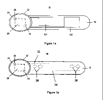
Referring first of all to Figure la, this shows an embodiment of a device for
improving blood
and lymphatic circulation in a limb of an animal. The device 10 includes a
flexible substrate
12, having two electrodes, 101 and 102, connected to a control means 24 which
includes a
power cell 26, a control processor 28, and an external LED 30. The control
means 24 is
mounted within a cradle 22 which is integral to the flexible substrate 12.
Referring to Figure lb, this shows another embodiment of a device for
improving blood and
lymphatic circulation in a limb of an animal. The device 10 includes a
flexible substrate 12,
having two pairs of connecting slots 36, 38 to which electrodes can be
mounted. Copper
tape 32, 34 embedded within the flexible substrate connects the electrodes to
the control
means 24 which includes a power cell 26, a control processor 28, and an
external LED 30.
The control means 24 is mounted within a cradle 22 which is integral to the
flexible substrate
12.
An alternate embodiment of the device illustrated in Figure lb is shown in
Figure 2, where
the connecting slots 36, 38 are replaced by a single connecting slot per
electrode 40, 42.
The cradle 22 and control module 24 are shown in more detail in Figure 3. In
some
embodiments, the control module 24 may be removable from the cradle 22, with a
pair of
detents 44 and corresponding recesses 46 allowing the cradle and control
module to
interlock. The control module and cradle carry corresponding electrical
contact surfaces 48,
50, 52, 54 which provide for electrical communication between the control
module 24 and
the first and second electrodes.
Figure 4 shows a side profile of the device illustrated in Figures lb and 2
with the electrodes
14, 16 mounted in place. In a preferred embodiment the electrodes 14, 16 are
formed by a
compression spring 56 (Figure 5) with a constant gap 58 size in between each
coil 60. To
engage the electrodes 14, 16 with an animal the compression spring 56 is
flexed opening
the gap 58 between each coil 60 and placed against the skin and hair of the
animal.
In another embodiment of the device the compression springs have a flattened
profile
(Figure 6). At least one surface 70, preferably two opposing surfaces 70, 72
are flattened.
The flattened surfaces 70, 72 ensure that the maximum surface area of the
electrode 14, 16
is in contact with the skin.
In a further embodiment of the device the electrode can be removed from the
device. The
electrodes in this embodiment of the device further comprise a connecting
means 74, 76 that
17
CA 02878140 2014-12-30
WO 2014/006378
PCT/GB2013/051718
engages the electrode to the device (Figure 7). The connecting means 74, 76 of
the
electrode 14, 16 are designed to engage the copper tape 32, 34. By compressing
the
compression spring 56 enables the connecting means 74, 76 to be inserted into
the
connecting slot(s) 36, 38 or 40, 42. When the compression spring 56 is
released it returns to
its resting state pressing the connecting means 74, 76 into the connecting
slot(s) 36, 38 or
40, 42. The pressure the compression spring 56 exerts engages the connecting
means 74,
76 with the copper tape 32, 34.
A schematic illustration of the control processor 28 is shown in Figure 8. The
processor 28
includes a timer module 44, a data store 46, a program store 48, and a logic
unit 50.
In use, the device is operated as follows. The flexible member 12 is attached
to an animal's
fore limb, hind limb or other appropriate region of anatomy, such that the
first electrode 14
and the second electrode 16 are located in the vicinity of the nerve(s) and
muscle(s) to be
stimulated. A button is pressed to activate the device.
The program store 48 is preloaded with an operating program arranged to
activate the
electrodes each minute using a 40 Hz pulsed DC of 20 mA for 0.1 second. Both
electrodes
are activated simultaneously. The timer module 44 serves to generate
appropriate timing
signals, while the logic unit 50 executes the program of the program store 48.
As the electrodes 14, 16 are activated; the animal's muscles are stimulated to
achieve
isometric contract. Simultaneously with each activation of the electrodes, the
LED 30 on the
outer surface of the control module 24 is also activated; this provides a
visual confirmation
that the device is operating.
The control module 24 may be provided to the user in a sealed form, to be
discarded when
the power cell 26 is depleted. A replacement control module may then be
fitted. In certain
embodiments of the invention, a range of different control modules may be
available, with a
range of different pre-programmed patterns for activating the electrodes. A
user may select a
different module based on a number of different conditions (for example,
animal's physical
health, length of journey, size of limb of the animal, and the like).
Alternatively, the control
module 24 may be partly user-programmable, to allow selection of one of a
number of preset
programmes from a single control module.
18
CA 02878140 2014-12-30
WO 2014/006378
PCT/GB2013/051718
The device 10 in Figures la and lb can be held in place by adhesive porous
polyurethane
foam such as Animal Polster 80. Figure 9 shows the device 10 held in place by
Animal
Polster 80 on the hind limb 82 of a horse.
The skilled person will understand that further variations on the invention
described herein
are possible. For example, rather than using Animal Polster, an adhesive
conductive gel
could be used. Alternatively, the device may be used on denuded skin by
placing the device
against a gel pad such as hydrogel and holding the device in place by use of
Animal Polster
or another type of adhesive material.
Other variations will be apparent to the skilled person.
It is envisioned that the device can be used on one or both of the forelimbs
and / or one or
both of the hind limbs and/or any other appropriate anatomic region, or any
combination
thereof. The device preferably is used to stimulate the radial nerve in the
forelimb, the
common peroneal nerve in the hind limb, or any nerve innervating one or more
limb muscles
or muscles attached to the axial skeleton the stimulation of which enhances
blood or
lymphatic circulation by virtue of the effects of contraction on vessels
within or around the
contracting muscle.
The radial nerve emerges between the medial and long heads of the triceps
muscle,
rounding the caudal surface of the humerus to gain the lateral aspect of the
forelimb where it
detaches branches to the extensor muscles of the carpus and digit: the
extensor carpi
radialis, the common digital extensor, the lateral digital extensor and the
ulnaris lateralis.
Correct placement of the device will elicit a neuromuscular effect in the
digital extensor
muscles.
The common peroneal nerve emerges between the biceps femoris muscle and the
lateral
head of the gastrocnemius muscle. It divides into superficial and deep
branches caudal to
the lateral collateral ligament of the stifle and these branches innervate the
digital extensor
muscle group on the cranio-lateral aspect of the tibia. Figure 10 shows a
diagram of how to
identify the position of the common peroneal nerve. The soft tissue boundaries
of the leg 84
are shown around the joint with the imaginary line 86 through the tibial
tuberosity and the
common peroneal nerve and its branches 88.
Useful landmarks in identifying a suitable position for attachment of the
device on the hind
limb are the tibial tuberosity and the fibular head: the common peroneal nerve
courses in a
19
CA 02878140 2014-12-30
WO 2014/006378
PCT/GB2013/051718
caudo-proximal to cranio-distal direction caudal to the fibula head. A bony
prominence on
the tibial tuberosity is easily palpated and an imaginary horizontal line from
this point defines
the level at which the device should be attached. Along this imaginary line
the fibula is
easily palpated. The peroneal nerve in the vicinity of its bifurcation lies
just caudal to the
fibula at this level.
The device has to be stably attached to the limb(s) of a horse, or other
appropriate
anatomical region, so that the device will be retained in the correct position
for the duration
of use. It should be observed that the lateral surface of the equine proximal
crus has a fairly
uniform convex curved conformation when the horse is standing squarely, but
when walking
the area cranial to the fibula varies considerably in its degree of curvature.
To retain the device in the correct position the device may be temporarily
fixed in position.
The inherent adhesive properties of the device alone may not be sufficient to
hold the device
in place.
The device may be held in place with adhesive tape, though it was found that
use of
adhesive tape is not well tolerated by horses due to the constrictive
sensation that the tape
creates. Alternatively, the device may be held in place with an elastic strap,
though it was
found that downward displacement of the elastic strap by the directional
conformation of hair
resulted in slippage of the device.
The use of adhesive gels produced a successful attachment of the device that
was well
tolerated. The device when attached by the use of an adhesive gel has the
potential to be
knocked and for the device to be partially or fully knocked free of the
limb(s).
The use of adhesive porous polyurethane foam such as Animal Polster proved to
be
surprisingly effective at retaining the device in position for prolonged
periods. The device
was left in position for 14 hours and was held firmly in position for the
duration of that time.
Animal Polster can be readily peeled away from the skin and hair of a horse
despite the
good adhesion. A slight residue may be left on any skin and hair to which it
has been
adhered. A medical grade adhesive remover which contains an emollient to
prevent the
skin from drying out can be purchased.
Use of the device on coarse cut hair reduced the effectiveness of the device
to stimulate the
radial or common peroneal nerves. Surprisingly it was found that the addition
of a small
CA 02878140 2014-12-30
WO 2014/006378
PCT/GB2013/051718
amount of conductive gel to the site of electrode contact had the effect of
enhancing
conduction and thereby stimulating the radial or peroneal nerves. This is
beneficial for
horses suffering with conditions that cause tenderness and increased
sensitivity in the legs
as it means that minimal handling is required to prepare the device for
placement. It is also
of benefit to competition horses which often have specific clip patterns and
the use of the
device should not affect the pattern or cosmetic appearance of the animal.
It is envisioned that a transparent flexible plastic template (Figure 11) is
provided with the
device. This template would be correctly aligned to the limb by referencing
the bony
landmarks as described. The conductive gel could be applied to the areas of
the skin that
would be in contact with the electrode. This application would be achieved by
applying the
conductive gel through holes corresponding to the position of each electrode.
Conductive coupling gel, such as ultrasound transmission gel, is a water based
macromolecular gel that improves electrode/skin contact. A typical product is
composed of
water (90%), carbomer 940 (5%), sodium hydroxide (2%), triethanolamine (2%),
antiseptic
(0.5%), and edible paint (0.5%).
It is envisaged therefore that a small amount of conductive gel would be
applied to bridge
any gap between the skin surface and the electrodes that might exist as a
consequence of
the presence hair or any other obstacle impairing electrode/skin contact. The
device would
then be positioned in place to stimulate the target nerve. Adhesive porous
polyurethane
foam such as Animal Polster or Polster Plast is then placed over the device to
adhere or
hold the device in place for the duration of use. The device can be easily
removed by
peeling the adhesive porous polyurethane foam away and removing any excess
adhesive
from the hair or skin with a medical grade adhesive remover which contains an
emollient to
prevent the skin from drying out.
The device would provide a suitable method for use on horses with conditions
where there is
an excessive build up of interstitial fluid in the forelimb(s) or hind
limb(s). Such methods of
use include but are not limited to reducing or preventing oedema,
lymphangitis, or cellulitis
by stimulation of the common peroneal nerve, the radial nerve, or other
appropriate nerves
of the fore or hind limbs. The device could be used in conjunction with other
methods of
treatment such as physiotherapy, sessions on a horse-walker and hydrotherapy.
The device would provide a suitable method for use on horses with conditions
where there is
inadequacy of tissue perfusion, tissue oxygenation and distribution to tissues
of
21
CA 02878140 2014-12-30
WO 2014/006378
PCT/GB2013/051718
pharmaceutical products or other agents. Such methods of use may be
appropriate under
circumstances including but are not limited to when disease or injury has been
sustained or
when tissue perfusion is compromised by the effects of posture or the
administration of
particular pharmaceutical products. The device could be used in conjunction
with other
methods of treatment.
The device would also provide a suitable method for limiting the vascular
effects of
confinement on a horse. It is envisioned that the device would be used during
transportation
of a horse to an event and prior to the event when the horse may be kept in
confined
conditions before competing.
It should be understood by reference to competitive events and competition
that all levels of
events are included from the professional to the amateur competing for fun.
The
augmentation of tissue perfusion by neuromuscular stimulation would prepare
the horse for
periods of potentially intense activity. The device therefore provides a
method of keeping
the horse in the best possible condition ahead of such periods of potentially
intense activity
during competition and provides a method of helping the horse achieve the best
possible
results during competition.
The device also provides a method for augmentation of blood and lymphatic
circulation by
neuromuscular stimulation of a horse when it is being stabled for any
prolonged periods of
time. The device could be used for example when it is not possible to allow
the horse to
freely roam around and thus naturally activate the musculovenous pumps of the
limb. The
method described therefore would be used as a prophylactic method for avoiding
the
excessive build up of interstitial fluid.
The device would also provide a suitable method for limiting delayed onset
muscle soreness
following exertion of any horse during competition, training for competition,
or recreation. It
is envisioned that the device would be used in this respect following physical
exertion.
The life of the battery could also vary depending on its intended use. When
the device is
intended for use in a method to reduce the excessive interstitial fluid in
cases of oedema,
lymphangitis and cellulitis a long battery life -32 hours for use in four,
eight hour overnight
sessions is envisioned.
For use on performance horse a device with a shorter battery life could be
more appropriate
and a battery life of 4 to 8 hours; preferably 6 hours, allowing a method of
use to keep a
22
CA 02878140 2014-12-30
WO 2014/006378
PCT/GB2013/051718
horse in the best possible condition ahead of a competitive event and during
the period
following physical exertion.
A proof of principle study was undertaken to evaluate the vascular effects of
electro-
stimulation of the common peroneal nerve in three horses.
Brief summary of the method
= Each horse was acclimatised to room temperature for at least 30 minutes
prior to collection of data.
= Hair was clipped at three sites: over the lateral femur, over the common
peroneal nerve, and over the lateral aspect of the tibia distal to common
peroneal nerve.
= Electrodes were attached over the common peroneal nerve, and laser
Doppler fluxmetry sensors were attached at the femoral and tibial sites.
= Following acclimatisation, sequentially each horse was stimulated,
allowed to
rest for 15 minutes, and then walked at a constant speed for 5 minutes.
Vascular flux was assessed by laser Doppler fluxmetry (Moor Instruments
DRT4) at the femoral and tibial sites:
= At rest before activation of the stimulator;
= During stimulation of the common peroneal nerve;
= Immediately following cessation of stimulation;
= Immediately following cessation of walking.
Results
The laser Doppler fluxmetry output parameters recorded in this study included
flux,
concentration, speed and temperature.
Concentration: the concentration of red blood cells in the sampled volume of
tissue.
[Increase in concentration implies an increase in the volume of sampled tissue
that is
occupied by blood vessels, i.e. there is blood vessel dilation through
engorgement of blood,
and vice versa].
Speed: the speed of movement of red blood cells in the sampled volume of
tissue.
Flux: the product of red blood cell concentration and speed in the sampled
volume of
tissue.
Temperature: the temperature recorded in the sampled volume of tissue.
23
CA 02878140 2014-12-30
WO 2014/006378 PCT/GB2013/051718
1. Results (flux)
MEAN FLUX DATA FROM THE LATERAL FEMUR
Baseline During
HORSE value stimulation Immediately
post stimulation Immediately post walking
1 72.3 171.4 145.4 125.0
2 210.8 229.3 206.7 159.0
3 55.6 165.5 148.5 124.3
AVERAGE 112.9 188.7 166.9 136.1
MEAN FLUX DATA FROM THE LATERAL TIBIA
Baseline During
HORSE value stimulation Immediately
post stimulation Immediately post walking
1 75.3 110.7 80.1 93.0
2 85.9 131.6 84.4 51.8
3 70.3 112.2 80.2 90.5
AVERAGE 77.2 118.2 81.6 78.4
2. Results (concentration)
CONCENTRATION DATA FROM THE LATERAL FEMUR
During Immediately post
Immediately post
HORSE stimulation stimulation
walking
1 213.1 231.9 220.8 295.0
2 212.8 259.4 196.2 305.6
3 205.4 226.8 215.3 295.5
AVERAGE 210.4 239.4 210.8 298.7
CONCENTRATION DATA FROM THE LATERAL TIBIA
Baseline During Immediately post
Immediately post
HORSE value stimulation stimulation walking
1 248.7 420.7 243.4 411.5
2 244.1 950.7 238.8 380.3
3 188.1 463.6 238.2 416.4
AVERAGE 227.0 611.7 240.1 402.7
3. Results (speed)
SPEED DATA FROM THE LATERAL FEMUR
Baseline During Immediately post
Immediately post
HORSE value stimulation stimulation walking
1 17.0 37.1 33.3 21.4
2 50.6 45.2 53.4 28.0
3 13.8 36.7 35.1 21.2
24
CA 02878140 2014-12-30
WO 2014/006378 PCT/GB2013/051718
I AVERAGE I 27.1 I 39.7 I 40.6 I 23.5 I
SPEED DATA FROM THE LATERAL TIBIA
Baseline During Immediately post Immediately post
HORSE value stimulation stimulation walking
1 18.0 14.0 16.8 12.6
2 19.1 7.7 19.5 7.6
3 19.2 12.8 17.3 12.1
AVERAGE 18.8 11.5 17.9 10.8
4. Results (temperature)
TEMPERATURE DATA FROM THE LATERAL FEMUR
Baseline During
HORSE value stimulation
Immediately post stimulation Immediately post walking
1 28.5 29.8 29.3 24.3
2 30.4 32.4 33.7 27.7
3 28.5 29.9 29.9 24.3
Averaged 29.1 30.7 31.0 25.4
TEMPERATURE DATA FROM THE LATERAL TIBIA
Baseline During
HORSE value stimulation
Immediately post stimulation Immediately post walking
1 28.4 29.5 29.36 24.8
2 30.5 30.3 29.1 27.0
3 28.2 29.2 29.3 24.9
Averaged 29.0 29.7 29.3 25.6
Conclusions
= Common peroneal nerve stimulation caused an increase in vascular
perfusion
in the equine pelvic limb and this increase was seen both proximally and
distally.
= Similar trends in flux, concentration, speed and tissue temperature were
seen
in all horses.
= Over the femur, electro-stimulation caused an increase above baseline
levels
in mean flux of 67.2%, averaged over three horses, which persisted for an
undetermined period after cessation of stimulation.
= Over the tibia, electro-stimulation caused an increase above baseline
levels in
mean flux of 53.1%, averaged over three horses, which immediately returned to
baseline levels after cessation of stimulation.
CA 02878140 2014-12-30
WO 2014/006378
PCT/GB2013/051718
= Over the femur, flux was augmented by a combination of increased
concentration and increased speed.
= Over the tibia, speed reduced below baseline levels during stimulation
but this
effect was more than offset by an increase in concentration, the overriding
effect
being an increase in flux. A similar fall in speed at tibial level was seen in
the data
collected immediately post walking. It is possible that the reduction is speed
directly
reflects the increase in concentration, i.e. the increase in blood vessel
diameter.
= Commensurate with increases in blood flow and the thermal energy
dissipated by blood, electro-stimulation caused elevation in tissue
temperature, this
being most noticeable at femoral level. Immediately
post walking, tissue
temperatures below baseline levels were recorded, probably as a consequence of
skin cooling via evaporative losses that occurred as a consequence of sweating
(walking is an active process which provoked sweating in all horses).
= Increases in concentration above baseline levels were seen over the femur
and tibia on cessation of walking. This may have been a consequence of
vasodilation induced by vasoactive chemicals released during homeostasis in
response to an increased demand for tissue oxygenation and metabolite
clearance
during active exercise. This increase in concentration above baseline levels
was not
seen on cessation of electro-stimulation at either site, perhaps reflecting
that
recruitment of muscle pumps by electro-stimulation is relatively passive and
without
the exertion and tissue demands that accompany active exercise.
In the horse, the thoracic limbs carry a disproportionate amount of the body
weight whereas
the pelvic limbs contribute disproportionately to forward thrust and
propulsion. Augmentation
of blood flow in the pelvic limb, therefore, would be of particular benefit to
racehorses and
performance horses engaged in a wide range of equestrian pursuits. However,
injuries such
as tendon and ligament strains most frequently affect the thoracic limb and it
would be
desirable to have a device that can promote healing and repair by augmenting
blood flow in
the thoracic limb of performance and recreational horses alike. During
explorative work
involving one Thoroughbred horse, successful stimulation of the radial nerve
was achieved,
there being concomitant visible pulsatile contraction in the innervated muscle
group.
26