Note: Descriptions are shown in the official language in which they were submitted.
CA 02879185 2015-01-14
DESCRIPTION
Title of Invention: METHOD FOR DETECTING CANCER
Technical Field
[0001]
The present invention relates to a method for detecting a cancer with CAPRIN-1
as a
tumor marker.
Background Art
[0002]
Cancer is the leading cause of death. This disease is currently treated
principally by
surgical therapy in combination with radiation therapy and/or chemotherapy.
Owing to
previous advances in medical technology, cancer is now a disease highly
curable if early
detected, depending on its type. Therefore, there is a demand for a method for
detecting a
cancer which places neither physical nor economic burdens on cancel pativitt
and can be
achieved by convenient tests.
[0003]
Recently, methods for assaying tumor products such as tumor markers have been
widely available. The tumor products refer to, for example, tumor-related
antigens, enzymes,
particular proteins, metabolites, oncogenes, oncogene products, and tumor
suppressor genes.
Carcinoembr)onic antigen CEA, glycoprotein CA19-9, prostate-specific antigen
PSA,
cialcitonin (peptide hormone produced in the thyroid gland), and the like are
exploited as tumor
markers in cancer diagnosis for some cancers. For many types of cancers,
however, tumor
markers useful in cancer diagnosis have not yet been found. In addition, a
large majority of
currently known tumor markers is present only in very small amounts (of the
order of pg/mL)
in body fluids and requires highly sensitive assay methods or special
techniques for detecting
these markers. Under such circumstances, it can be expected that doors will be
opened for
1
CA 02879185 2015-01-14
diagnostic use for various types of cancers if a novel cancer testing approach
capable of highly
sensitively detecting various types of cancers by convenient operation can be
provided.
[0004]
Meanwhile, in spite of recent development of novel surgical techniques or
discovery of
novel anticancer agents, the existing cancer treatment has an insufficiently
improved outcome.
This is because an effective cancer diagnosis technique has not been
established for many
cancers, except for some cancers. Inability to detect these cancers early is
partly responsible
for this situation.
[0005]
With recent advances in molecular biology or cancer immunology, antibodies
specifically reacting with cancer, molecular targeting drugs for cancer
antigens related to
malignant transformation or cancer exacerbation, and the like have been
identified, raising
expectations on specific cancer therapy targeting cancer antigens.
[0006]
Among others, a plurality of antibody drugs for cancer treatment targeting
antigenic
proteins on Lancer cells have been launched and used in the cancer treatment.
These
antibody drugs have received attention because of their certain efficacy as
cancer-specific
therapeutic agents. A large majority of antigenic proteins targeted by the
drugs, however, are
also expressed in normal cells. As a result of administering the antibodies,
cancer cells as
well as normal cells expressing the antigens are therefore damaged, resulting
in undesired
adverse reactions. In addition, the effects of cancer treatment differ very
largely among
Individuals due to various factors of the individual cancer patients. For
example, surgery,
chemotherapy, or radiation therapy largely varies in the treatment and
prognosis depending on
the stages of cancers. Different persons are known to have distinctive
sensitivities to the
same therapeutic drug for cancers. This indicates that a certain drug is
effective for some
patients but ineffective for others due to the diversity of individuals.
[0007]
Thus, as for some therapeutic drugs, their administration to cancer patients
is
determined by measuring in advance the expression of disease-related genes or
proteins in the
2
CA 02879185 2015-01-14
patients and evaluating whether a particular drug is effective for a patient
expressing a
particular gene or protein. Specifically, the presence of a cancer antigen in
a sample, for
example, serum or tissue, derived from a cancer patient is tested in clinical
practice by use of a
detection method for assaying a disease-related gene or protein of a certain
kind of cancer.
Then, the administration of a cancer antigen-specific therapeutic drug is
determined. For
example, cancer tissues from a large bowel cancer patient are evaluated by an
immunohistochemical staining EGFR detection method "EGFR pham) (Dako)" to
predict the
effectiveness of Erbitux(R) (cetuximab) for the large bowel cancer. Then, the
administration
of Erbitux is determined. Further, cancer tissues from a breast cancer patient
are evaluated
by an immunohistochemical staining Her2 detection method "HercepTest" to
predict the
effectiveness of Herceptin(R) (trastuzumab) for the breast cancer. Then, the
application of
Herceptin is determined.
[0008]
Incidentally, companion animals have been raised recently as family members
and
often have lifestyles similar to those of their owners. For this reason, from
the occurrence of
cancers in companion animals, it can reportedly be predicted that their owners
have the high
risk of developing cancers in the future.
[0009]
Dogs, typical companion animals, are known to age 7 times more quickly than
humans.
Reportedly, the number of dogs currently raised is approximately 6.7 million
in Japan and
approximately 17.64 million in the USA. Rabies shots as well as combined
vaccines such as
quintuple, septuple, or octuple combination shots are generally available,
leading to decreased
rates of highly lethal infections including canine parvovirus infection,
canine distemper
infection, canine parainfluenza virus infection (kennel cough), canine
adenovirus type 2
infection (kennel cough), canine infectious hepatitis, canine coronavirus
infection, and
leptospirosis. An average dog life-span has therefore been increased, and 7-
year-old or older
dogs account for 35.5% of the total number of pet dogs. The causes of death
such as cancer,
hypertension, and heart disease are ever increasing in dogs, as in humans. In
the USA,
approximately 4 million dogs are yearly diagnosed with cancers. Also in
Japan,
3
CA 02879185 2015-01-14
=
approximately 1.6 million dogs allegedly have some potential tumor. Checkup
examination,
however, is not very common in companion animals, unlike humans. This leads to
the late
detection of disease. In most cases, their owners notice pets' symptoms for
the first time after
tumors have already become large, and then visit animal hospitals. If such
large tumors are
malignant, even surgical therapy (e.g., surgical operation) or medication
using anticancer
agents or the like is very often too late to cure the tumors. Tumors confirmed
by
veterinarians to be malignant are generally treated with anticancer agents
without surgery.
Even in the case of performing surgery, it is required to secure surgical
margins or to take
stringent measures for surgery such as measures against the spread of blood or
cells during
surgery_ Desirably, treatment with anticancer agents is initiated immediately
after surgery,
and a follow-up is also performed at short intervals. Thus, the medication
using therapeutic
drugs for cancers is also essential for the cancer-affected companion animals.
A detection
method, if any, for assaying a disease-related gene or protein of a certain
kind of cancer
permits more effective treatment than ever and is advantageous both for owners
and for
veterinarians.
[0010]
Cytoplasmic- and proliferation-associated protein I (CAPRIN-1) is an
intracellular
protein known to be expressed upon activation or cell division of resting
normal cells and to
form cytoplasmic stress granules with intracellular RNAs to participate in the
regulation of
transport and translation of mRNAs. Meanwhile, it has been found that: CAPRIN-
1 is highly
expressed on the membrane surface of breast cancer cells; and an antibody
against CAPRIN-1
exerts strong antitumor effects on breast cancer cells (Patent Literature 1).
According to
another report, the expression of CAPRIN-1 in a patient-derived sample can be
measured
using an antibody binding to CAPRIN-1 expressed on cell surface to thereby
detect a cancer
and to evaluate the grade of the cancer (Patent Literature 2). Specifically,
the report states
that a plasma membrane protein CAPRIN-1 may serve as a target for cancer
treatment or the
like. As mentioned above, due to the diversity of cancer patients, it is
required to test the
presence of CAPRIN-1 in a cancer patient-derived sample for determining the
administration
of a CAPRIN-1-targeting therapeutic drug, for example, an antibody.
Nonetheless, there
4
CA 02879185 2015-01-14
exists no report on a method for detecting CAPRIN-1 for the application of
such a specific
therapeutic drug, or there exists no reagent for detecting a cancer using a
cancer patient-
derived sample.
Citation List
Patent Literature
[0011]
Patent Literature 1: W02010/016526
Patent Literature 2: W02010/016527
Summary of Invention
Technical Problem
[0012]
An object of the present invention is to provide a cancer detection approach
useful in
the diagnosis of a cancer. Another object of the present invention is to
provide a method for
detecting a cancer which involves determining the presence and the amount of
CAPRIN-1 in a
sample of a cancer patient in order to determine the administration of a
CAPR1N-1-targeting
drug to the cancer patient, and a drug and a kit for the diagnosis of a
cancer.
Solution to Problem
[0013]
As a result of conducting diligent studies, the present inventors have
obtained cDNAs
encoding proteins binding to antibodies present in cancer-bearing organism-
derived serum by
SEREX using a dog testis-derived cDNA library and the serum of cancer-bearing
dogs, and
prepared dog CAPRIN- Is having the amino acid sequences shown in SEQ ID NOs:
6, 8, 10,
12, and 14 on the basis of the cDNAs. The present inventors have also prepared
human
CAPRIN-is having the amino acid sequences shown in SEQ ID NOs: 2 and 4 on the
basis of
human homologous genes of the obtained genes. Consequently, the present
inventors have
found that: genes encoding these proteins are specifically expressed in dog
and human testis,
CA 02879185 2015-01-14
respectively, and in malignant cancer cells (see Example 1 mentioned later);
and monoclonal
antibodies prepared using, as antigens, recombinant polypeptides prepared on
the basis of the
amino acid sequences of these proteins can bind to CAPRIN-1 in various cancer
tissues and
damage cancer cells having CAPRIN-1 on their surface. As a result, the present
inventors
have gained the finding that CAPRIN-1 can be used as a target for cancer
treatment. The
present inventors have further found that CAPRIN-1 can be specifically
detected from cancer
patient-derived samples by use of the monoclonal antibodies mentioned above.
Specifically,
the present invention provides a method for detecting a cancer, comprising
measuring the
expression of CAPRIN-1 using a predetermined anti-CAPRIN-1 antibody for
application to a
sample separated from an organism. In addition, the present invention has
established a
method for detecting CAPRIN-1 in a cancer patient-derived sample and
evaluating the
expression level thereof by an immunological assay method using any of the
monoclonal
antibodies mentioned above, for example, by ELISA for cancer patient-derived
serum using a
predetermined anti-CAPRIN-1 monoclonal antibody or an immunohistochemical
staining
method for cancer tissues. The present inventors have also found that as a
result of
evaluating a cancer-derived sample by this mcthod, the applicability of a
CAPRIN-1-targeting
drug to a patient is indicated if the expression of CAPRIN-1 and a high
abundance thereof are
found in the patient. On the basis of these findings, the present invention
has been completed.
[0014]
The present invention provides a method for detecting a cancer which is
applied to a
sample separated from an organism, the method comprising detecting CAPRIN-1 in
the
sample and measuring the amount thereof. The present invention also provides a
diagnosis
method comprising measuring the expression level of CAPRIN-1 in a tissue
before
administration of a CAPRIN-1 -targeting drug to a patient to thereby predict
the effectiveness
thereof and reveal the applicability of a therapeutic drug against CAPRIN-1
(e.g., whether a
CAPRIN-1-targeting drug, for example, an antibody, can be applied to the
cancer patient).
The present invention further provides a drug or kit for the diagnosis of a
cancer, comprising
an antibody capable of antigen-antibody reaction with CAPRI-1, or an antigen-
binding
fragment thereof.
6
81785116
[0015]
Specifically, the present invention has the following aspects:
[0016]
(1) A method for detecting a cancer expressing CAPRIN-1, comprising measuring
the
expression level of CAPRIN-1 in a biological sample through antigen-antibody
reaction using
an antibody having immunological reactivity with a polypeptide the amino acid
sequence of
which consists of the sequence shown in SEQ ID NO: 66, or an antigen-binding
fragment
thereof, wherein the measured CAPRIN-1 expression level being higher than that
of a healthy
individual indicates the presence of the cancer.
[0017]
(2) The method for detecting a cancer according to (1), wherein the CAPRIN-1
to be
measured is (a) a polypeptide having the amino acid sequence shown in any even-
numbered
SEQ ID NO of SEQ ID NOs: 2 to 28 in the Sequence Listing, or (b) a polypeptide
having
85% or higher sequence identity to the polypeptide having the amino acid
sequence shown in
any even-numbered SEQ ID NO of SEQ ID NOs: 2 to 28 in the Sequence Listing.
[0018]
(3) The method for detecting a cancer according to (1) or (2), wherein the
biological
sample is derived from a human, a dog, or a cat.
[0019]
(4) The method for detecting a cancer according to any of (1) to (3), wherein
the
biological sample is derived from a dog, and the CAPRIN-1 to be measured has
the amino
acid sequence shown in SEQ ID NO: 6, 8, 10, 12, or 14.
[0020]
(5) The method for detecting a cancer according to any of (1) to (3), wherein
the
biological sample is derived from a human, and the CAPRIN-1 to be measured has
the amino
acid sequence shown in SEQ ID NO: 2 or 4.
[0021]
(6) The method for detecting a cancer according to any of (1) to (5), wherein
said
cancer is capable of being treated with said antibody when using it as a
cancer therapeutic
drug.
7
Date recu/Date Received 2020-04-14
81785116
[0022]
(7) The method for detecting a cancer according to any of (1) to (6), wherein
the
measurement of the expression level of CAPRIN-1 is carried out using an
immunological
assay method.
[0023]
(8) The method for detecting a cancer according to (7), wherein the
immunological
assay method is enzyme-linked immunosorbent assay (ELISA) and/or an
immunohistochemical staining method.
[0024]
(9) The method for detecting a cancer according to any of (1) to (8), wherein
the
sample is a body fluid, a tissue, or a cell.
[0025]
(10) The method for detecting a cancer according to any of (1) to (9), wherein
the
cancer is at least one cancer selected from the group consisting of breast
cancer, brain tumor,
esophagus cancer, stomach cancer, lung cancer, liver cancer, kidney cancer,
thyroid gland
cancer, spleen cancer, pancreas cancer, large bowel cancer, skin cancer, ovary
cancer, uterus
cancer, prostate cancer, bladder cancer, testis cancer, osteosarcoma, and
fibrosarcoma.
[0026]
(11) The method for detecting a cancer according to any of (1) to (10),
wherein the
antibody or the antigen-binding fragment thereof is a monoclonal antibody
having a heavy
chain variable region comprising the amino acid sequence shown in SEQ ID NO:
70 and a
light chain variable region comprising the amino acid sequence shown in SEQ ID
NO: 71, or
an antigen-binding fragment thereof.
[0027]
(12) An agent for diagnosis of a cancer, comprising an antibody having
immunological
reactivity with a polypeptide having the amino acid sequence shown in SEQ ID
NO: 66, or an
antigen-binding fragment thereof.
8
Date recu/Date Received 2020-04-14
81785116
[0028]
(13) The agent for diagnosis of a cancer according to (12), wherein the
antibody or the
antigen-binding fragment thereof is a monoclonal antibody having a heavy chain
variable
region comprising the amino acid sequence shown in SEQ ID NO: 70 and a light
chain
variable region comprising the amino acid sequence shown in SEQ ID NO: 71, or
an antigen-
binding fragment thereof.
[0029]
(14) A method for selecting an individual-specific therapeutic drug for a
cancer,
comprising: measuring the expression level of CAPRIN-1 in a biological sample
using an
antibody having immunological reactivity with a polypeptide consisting of the
amino acid
sequence shown in SEQ ID NO: 66, or an antigen-binding fragment thereof; and,
if the
expression level is statistically significantly higher than that of a healthy
individual, selecting
a CAPRIN-1-targeting drug as a therapeutic drug for a cancer suitable for
administration to
the individual from which the biological sample is derived.
[0030]
(15) The method for selecting an individual-specific therapeutic drug for a
cancer
according to (14), wherein the CAPRIN-1-targeting drug is an antibody having
immunological reactivity with CAPRIN-1, or an antigen-binding fragment
thereof.
[0030a]
(16) A method of identifying a CAPRIN-1-expressing cancer that is amenable to
treatment with an anti-CAPRIN-1 antibody or an antigen-binding fragment
thereof, the
method comprising carrying out the method defined in any one of (1) to (11)
for detecting a
cancer expressing CAPRIN-1, wherein the cancer being identified as a CAPRIN-1-
expressing
cancer indicates that the cancer is amenable to treatment with the antibody or
antigen-binding
fragment thereof.
[0031]
The present application claims the priority from Japanese Patent Application
No. 2012-160763.
9
Date Recue/Date Received 2021-01-22
81785116
Advantageous Effects of Invention
[0032]
The present invention provides a novel method for detecting a cancer,
comprising
measuring the expression of CAPRIN-1 in a sample separated from a cancer
patient. As
specifically shown in Examples mentioned later, antibodies prepared using, as
antigens,
recombinant polypeptides prepared on the basis of the amino acid sequence of
CAPRIN-1
(also referred to as Caprin-1 or CAPRIN-1 protein) specifically react with
CAPRIN-1 in the
body fluids (e.g., serum) or tissues of cancer patients. As also described
later in Examples,
CAPRIN-1 itself is specifically expressed at high levels in various cancer
tissues. The
presence and the amount of CAPRIN-1 in a sample separated from a cancer
patient can
9a
Date recu/Date Received 2020-04-14
CA 02879185 2015-01-14
a
therefore be measured to thereby detect a cancer. In addition, the presence or
absence of
sensitivity to a CAPRIN-1-targeting drug such as a CAPRIN-1-targeting
therapeutic drug, for
example, an antibody drug, can be determined in advance to thereby select a
patient to which
this drug is applicable. Specifically, the expression and the amount of CAPRIN-
1 can be
measured in advance by the application of the present invention to a cancer
patient to thereby
provide more efficient treatment using an antibody against CAPRIN-1.
Brief Description of Drawing
[0033]
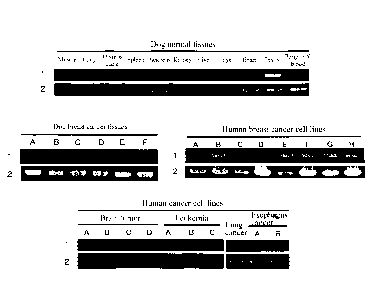
[Figure 1] Figure 1 is a diagram showing the expression patterns of a CAPRIN-1-
encoding
gene in normal tissues and tumor cell lines. Reference number 1 depicts the
expression
patterns of the gene encoding the CAPRIN-1 protein. Reference number 2 depicts
the
expression patterns of the GAPDH gene. The uppermost panel shows the results
for dog
normal tissues. The left middle panel shows the results for dog breast cancer
tissues. The
right middle panel shows the results for human breast cancer cell lines. The
lowermost panel
shows the results for various human cancer cell lines.
Description of Embodiments
[0034]
The method for detecting a cancer according to the present invention comprises
measuring the amount (expression level) of CAPRIN-1 (CAPRIN-1 protein) in a
sample
separated from an organism (biological sample). The measurement of the
expression level of
CAPRIN-1 in a sample of a cancer patient can be carried out by use of, for
example, an
immunological assay method which involves detecting CAPRIN-1 using an antibody
against
CAPRIN-1 (anti-CAPRIN-1 antibody). Various immunological assay methods
applicable to
the measurement of the expression level of CAPRIN-1 are well known in the art.
Examples
thereof include immunohistochemical analysis, Western blot analysis,
immunoprecipitation,
molecular binding assay, ELISA, and biochemical enzyme activity assay. The
results of
measuring the expression level of CAPR1N-1 by such an assay method can also
indicate, for
CA 02879185 2015-01-14
=
example, the presence of CAPRIN-1 in the sample, the ratio of cells expressing
CAPRIN-1,
the distribution of expression sites in tissues, and expression intensity on a
site basis. In this
context, the "expression level" used herein includes the intracellular
accumulation level and
abundance of the protein.
[0035]
The results of measuring the expression level of CAPRIN-1 in the sample can be
classified into scores shown in Examples. A higher score indicates that CAPRIN-
1 is
contained in a larger amount in the biological sample (e.g., cancer tissue or
cancer serum) of a
cancer patient. In the present invention, the term "measurement" or "assay"
encompasses all
of detection and qualitative, quantitative, and semiquantitative approaches.
[0036]
The amino acid sequence shown in SEQ ID NO: 6, 8, 10, 12, or 14 is the amino
acid
sequence of dog CAPRIN 1. The dog CAPRIN-1 having this amino acid sequence has
been
found by SEREX using a dog testis-derived cDNA library and serum derived from
cancer-
bearing dogs, and identified from the cDNA library as a polypeptide binding to
an antibody
specifically present ill the serum derived from cancer-bearing dogs (see
Example 1).
CAPRIN-1 itself having the amino acid sequence of SEQ ID NO: 6, 8, 10, 12, or
14 can also
be assayed as an antigen in dog tissues by the method mentioned above to
thereby diagnose
the presence or absence of sensitivity to a CAPRIN-1-targeting drug (see
Examples).
[0037]
In this context, the phrase "having an (the) amino acid sequence" used herein
means
that amino acid residues are arranged in the order presented in predetermined
amino acid
sequence information. Thus, for example. the "polypeptide having the amino
acid sequence
shown in SEQ ID NO: 2" means a polypeptide of 709 amino acid residues in size
in which the
amino acid residues are linked according to the amino acid sequence Met Pro
Ser Ala ...
(snip) ... Gin Gin Val Asn as shown in SEQ ID NO: 2. Also, for example. the
"polypeptide
having the amino acid sequence shown in SEQ ID NO: 2" is abbreviated to the
"polypeptide of
SEQ ID NO: 2". The same holds true for the phrase "having a (the) nucleotide
sequence".
11
CA 02879185 2015-01-14
The term "having" in the phrase "having an (the) amino acid sequence" and
"having a (the)
nucleotide sequence" may be replaced with the term "consisting or.
[0038]
The "polypeptide" used herein refers to a molecule that is formed through the
peptide
bonds of a plurality of amino acids, and encompasses not only a polypeptide
molecule
constituted by a large number of amino acids but a low-molecular-weight
molecule having a
small number of amino acids (oligopeptide or peptide) and a full-length
protein. The
polypeptide according to the present invention also encompasses the full-
length proteins of
CAPRIN-1 having the amino acid sequences shown in even-numbered SEQ ID NOs of
SEQ
ID NOs: 2 to 30.
[0039]
In the method of the present invention, additional mammalian CAPRIN-1 other
than
the dog CAPRIN-1 of SEQ ID NO: 6, g. 10, 12, or 14 can also be measured. In
the present
specification. such non-doe mammalian CAPRIN-1 is also referred to as a
"homolog" of dog
CAPR1N-1. The term "CAPRIN-1" encompasses CAPRIN-1 derived from not only dogs
but
other mammals. Examples of the additional mammalian CAPRIN-1 that may be
measured in
the method of the present invention include, but not limited to, human CAPRIN-
1 and cat
CAPRIN-1.
[0040]
As specifically described below in Examples, mRNA encoding human CAPRIN-1 is
significantly expressed at high levels in human testis and cancer cells, as
with the dog
CAPRIN-1 of SEQ ID NO: 6, 8, 10, 12, or 14. An anti-human CAPRIN-1 antibody,
however, is not detected in the bodies of healthy humans. An anti-cat CAPRIN-1
antibody is
not detected in the bodies of healthy cats. but is detected only in cancer-
bearing cats. Thus,
the applicability of a CAPRIN-1-targeting drug to a non-dog mammal can also be
determined
by the measurement of the expression level of CAPR1N-1 derived from the non-
dog mammal.
[0041]
The nucleotide sequence encoding human CAPRIN-1 and the amino acid sequence
thereof are shown in SEQ ID NO: 1 or 3 and SEQ ID NO: 2 or 4, respectively, in
the
12
CA 02879185 2015-01-14
=
Sequence Listing. The sequence identity of human CAPRIN-1 to dog CAPRIN-1 is
94% for
the nucleotide sequence and 98% for the amino acid sequence. Since the amino
acid
sequence identity of CAPRIN-1 is as very high as 98% even between dogs and
humans, which
are genetically distantly related mammals from each otherõ many non-human and
non-dog
mammalian CAPRIN-1 proteins have high (approximately 85% or higher) sequence
identity to
the human or dog CAPRIN-1. The CAPRIN-1 whose expression level is to be
measured in
the method of the present invention may be, but not particularly limited to, a
protein having
preferably 85% or higher, more preferably 95% or higher sequence identity to
the amino acid
sequence of the dog or human CAPRIN-1 shown in SEQ ID NO: 2, 4, 6, 8, 10, 12,
or 14.
[0042]
An antigenic substance, such as a protein, which has a complicated structure
with a
large molecular weight, usually contains a plurality of epitopes differing in
structure on its
molecule. Thus, plural types of antibodies that respectively recognize and
bind to a plurality
of epitopes on such an antigenic substance are produced in vivo. In other
words, antibodies
produced in vivo against the antigenic substance (e.g., protein) are
polyclonal antibodies,
which arc mixtures of plural types of antibodies. The antibodies found by the
present
inventors to be specifically present in serum derived from cancer-affected
organisms and to
specifically bind to recombinant CAPRIN-1 through antigen-antibody reaction
are also
polyelonal antibodies. The term "poly-clonal antibody" used in the present
invention refers to
an antibody that is found in serum derived from an organism containing an
antigenic substance
in the body, and has been induced in the organism against the antigenic
substance.
[0043]
Specific examples of a preferred polypeptide for use as an antigen for
obtaining an anti-
CAPRIN-1 antibody include polypeptides of even-numbered SEQ ID NOs of SEQ ID
NOs: 2
to 30 and fragments thereof. Particularly, an anti-CAPRIN-1 antibody that is
obtained using,
as an antigen, the polypeptide of SEQ ID NO: 2, 4, 6, 8, 10, 12, 14, 16, 18,
20, 22, 24, 26, 28,
or 30 or a fragment thereof comprising the amino acid sequence shown in SEQ ID
NO: 66
containing a preferred epitope and specifically binds to (i.e., has
immunological reactivity
13
CA 02879185 2015-01-14
55232-54
with) a polypeptide having the amino acid sequence shown in SEQ ID NO: 66 can
be
preferably used in the method of the present invention.
[0044] The nucleotide sequences of polynucleotides encoding
polypeptides consisting
of the amino acid sequences shown in even-numbered SEQ ID NOs of SEQ ID NOs: 2
to 30
(i.e., SEQ ID NOs: 2, 4, 6, ... 28, and 30) are shown in odd-numbered SEQ ID
NOs of
SEQ ID NOs: 1 to 29 (i.e., SEQ ID NOs: 1, 3, 5, ... 27, and 29), respectively.
[0045] It is widely known to those skilled in the art that even a
protein derived from a
protein antigen by the substitution, deletion, addition, or insertion of a
small number of amino
acid residues in the amino acid sequence of the protein may generally have
almost the same
antigenicity as that of the original protein. Thus, a polypeptide having a
sequence derived
from the amino acid sequence of CAPRIN-1 by the substitution, deletion,
addition, and/or
insertion of a small number of (preferably 1 or several) amino acid residues
can also be used
in the production of an anti-CAPRIN-1 antibody, as with the polypeptides
consisting of the
amino acid sequences shown in even-numbered SEQ ID NOs of SEQ ID NOs: 2 to 30,
as
.. long as the polypeptide has 80% or higher or 85% or higher, preferably 90%
or higher, more
preferably 95% or higher, further preferably 98% or higher sequence identity
to the original
sequence and specifically binds to a polyclonal antibody against CAPRIN-1
through antigen-
antibody reaction (hereinafter, this polypeptide is also referred to as a
"specifically-reactive
modified polypeptide" for the sake of convenience). Preferably, the
specifically-reactive
modified polypeptide has an amino acid sequence derived from the amino acid
sequence of
CAPRIN-1 by the substitution, deletion, addition, and/or insertion of 1 or
several amino acid
residues. The term "several" used herein refers to an integer of 2 to 10,
preferably an integer
of 2 to 6, more preferably an integer of 2 to 4. The "sequence identity" used
herein for the
amino acid sequence refers to the percentage of a value determined by dividing
the number of
matched amino acid residues by the total number of amino acid residues in the
best-matching
alignments of the amino acid residues in two amino acid sequences to be
compared. For the
alignments, if necessary, one or both of these two sequences to be compared
can be gapped.
14
CA 02879185 2015-01-14
Such sequence alignments can be carried out using a well known program, for
example,
BLAST. FASTA, or CLUSTAL W (Karlin and Altschul, Proc. Natl. Acad. Sci.
U.S.A., 87:
2264-2268. 1993; and Altschul et al.. Nucleic Acids Res., 25: 3389-3402,
1997).
[0046]
Twenty types of amino acids constituting a natural protein can be divided
according to
similar properties into the following groups: neutral amino acids having a low
polar side chain
(Gly, Ile, Val, Leu, Ala, Met, and Pro); neutral amino acids having a
hydrophilic side chain
(Asn, Gin, Thr, Ser, Tyr, and Cys); acidic amino acids (Asp and Glu); basic
amino acids (Arg,
Lys, and His): and aromatic amino acids (Phe. Tyr, Trp, and His). It is known
that
substitution within each of these groups, i.e., conservative substitution,
does not change the
properties of the poly-peptide in most cases. Thus, in the case of
substituting the amino acid
residues of CAPRIN-1, a member in each of these groups can be substituted by
another
member in the same group so that the binding activity against the appropriate
antibody is
likely to be maintained. In the present invention, however, the modified form
may have non-
conservative substitution as long as the modified form is provided with
immunity-inducing
activity equivalent to or substantially equivalent to that of the unmodified
form.
[0047]
The polypeptide used in the present invention can be synthesized according to
chemical
synthesis methods, for example, Fmoc (fluorenylmethyloxycalbonyl) and tBoc (t-
butyloxycarbonyl) methods (Seikagaku Jikken Koza (Biochemical Experimentation
Course in
English) 1, the Japanese Biochemical Society ed., Protein Chemistry IV,
Chemical
Modification and Peptide Synthesis, Tokyo Kagaku Donn Co., Ltd. (Japan),
1981). Also, the
polypeptide can be synthesized by routine methods using various commercially
available
peptide synthesizers. Alternatively, the polypeptide can be easily prepared
using genetic
engineering approaches known in the art (Sambrook et al., Molecular Cloning,
the 2nd edition,
Current Protocols in Molecular Biology (1989), Cold Spring Harbor Laboratory
Press;
Ausubel et al., Short Protocols in Molecular Biology, the 3rd edition, A
compendium of
Methods from Current Protocols in Molecular Biology (1995), John Wiley & Sons:
etc.). For
example, RNA is extracted from a tissue expressing a gene encoding the human
CAPRIN-1 of
CA 02879185 2015-01-14
SEQ ID NO: 2 or a homolog thereof. From this RNA, cDNA of the gene is prepared
by RT-
PCR. The full-length cDNA or a desired partial fragment thereof is
incorporated into
expression vectors, which can then be transferred to host cells to obtain the
polypeptide of
interest. The nucleotide sequences of cDNAs encoding the dog CAPRIN-1 proteins
of SEQ
ID NOs: 6, 8. 10, 12, and 14 are shown in SEQ ID NOs: 5, 7, 9, 11, and 13,
respectively.
The nucleotide sequences of cDNAs encoding the human CAPRIN-1 proteins of SEQ
ID
NOs: 2 and 4 as human homologs thereof are shown in SEQ ID NOs: 1 and 3,
respectively.
Primers for use in RT-PCR can therefore be easily designed with reference to
these nucleotide
sequences. As mentioned later, a gene encoding non-human mammalian CAPRIN-1
can be
amplified with primers designed with reference to the nucleotide sequence of
any odd-
numbered SEQ ID NO of SEQ ID NOs: 5 to 29. Thus, cDNA encoding, for example.
cat
CAPRIN-1, can also be easily prepared by the same approach as above. The RNA
extraction,
RT-PCR, the incorporation of cDNA into vectors, and the transfer of the
vectors to host cells
can be carried out, for example, by well known methods as described below.
Also, the
vectors or host cells used are well known, and various products are
commercially available.
[0048]
The host cells may be any cell capable of expressing the above polypeptide.
Examples of prokaryotic cells include E. co/i. Examples of eukaryotic cells
include: cultured
mammalian cells such as monkey kidney cells COSI, Chinese hamster ovary cells
CHO, a
human embryonic kidney cell line HEK293, and mouse embryonic skin cell line
NIH3T3; and
budding yeast, fission yeast cells, silkworm cells, and Xenopus egg cells.
[0049]
In the case of using prokaryotic cells as the host cells, the expression
vectors used have
an origin that permits replication in the prokaryotic cells, a promoter, a
ribosomal binding site,
a multicloning site, a terminator, a drug resistance gene, an auxotrophic
complementary gene,
etc. Examples of expression vectors for E. coli can include pUC series,
pBluescript II, pET
expression systems, and pGEX expression systems. DNA encoding the above
polypeptide
can be incorporated into such expression vectors, with which prokaryotic host
cells are then
transformed, followed by the culture of the obtained transformants so that the
polypeptide
16
CA 02879185 2015-01-14
encoded by the DNA is expressed in the prokaryotic host cells. In this
respect, the
polypeptide may be expressed as a fusion protein with an additional protein.
In this context,
the DNA encoding the above polypeptide can be obtained, for example, by the
preparation of
cDNA by RT-PCR as mentioned above. Alternatively, the DNA may be synthesized
by
routine methods using commercially available nucleic acid synthesizers as
mentioned later.
The nucleotide sequences of cDNAs of genes encoding the CAPRIN-1 proteins of
SEQ ID
NOs: 2 and 4 are shown in SEQ ID NOs: 1 and 3, respectively, in the Sequence
Listing.
[0050]
In the case of using eukaryotic cells as the host cells, expression vectors
for eukaryotic
cells having a promoter, a splicing region, a poly(A) addition site, etc. are
used as the
expression vectors. Examples of such expression vectors can include pKA1,
pCDM8,
pSVK3. pMSG, pSVL, pBK-CMV, pBK-RSV, EBV vector, pRS, pcDNA3, and pYES2
vectors. In the same way as above, the DNA encoding the above polypeptide used
in the
present invention can be incorporated into such expression vectors, with which
eukaryotic host
cells are then transformed, followed by the culture of the obtained
transformants so that the
polypeptide encoded by the DNA is expicssed in the cukaiyvtic host cells. In
the case of
using expression vectors such as III-ND/VS-His, pFLAG-CMV-2, pEGFP-N1, or
pEGFP-C1,
the polypeptide may be expressed as various fusion proteins tagged with His
tag (e.g., (His)6 to
lHiS)10), FLAG tag, myc tag, HA tag, GFP, or the like.
[0051]
The expression vectors can be transferred to the host cells using well known
methods
such as electroporation, a calcium phosphate method, a liposome method, a DEAE
dextran
method, microinjection, viral infection, lipofection, and binding with cell-
penetrating peptides.
[0052]
The polypeptide of interest can be isolated and purified from the host cells
by a
combination of separation operations known in the art. Examples thereof
include treatment
with a denaturant (e.g., urea) or a surfactant, ultrasonication, enzymatic
digestion, salting-out,
solvent fractionation and precipitation, dialysis, centrifugation,
ultrafiltration, gel filtration,
17
CA 02879185 2015-01-14
SDS-PAGE, isoelectric focusing electrophoresis, ion-exchange chromatography.
hydrophobic
chromatography, affinity chromatography, and reverse-phase chromatography.
[0053]
The polypeptides obtained by these methods also include their forms of fusion
proteins
with other arbitrary proteins. Examples thereof can include fusion proteins
with glutathione-
S-transferase (GST) or His tag. Such polypeptides in the fon-n of fusion
proteins are also
encompassed by the specifically reactive added poly-peptide mentioned above.
The
polypeptides expressed in transformed cells may undergo various intracellular
modifications
after translation. Such posttranslationally modified polypeptides may be used
as long as
these polypeptides have binding activity against the polyclonal antibody
against CAPRIN-1 .
Examples of such posttranslational modifications can include N-terminal
methionine
elimination, N-terminal acety-lation, glycosylation, intracellular protease-
mediated limited
degradation. myristoylation, isoprenylation, and phosphorylation.
[0054]
The CAPRIN-1 as mentioned above or a fragment thereof can be used as an
antigen to
prepare an aati-CAPRIN-1 antibody. The anti-CAPRIN-I antibody used in the
present
invention may be a polyclonal antibody or may be a monoclonal antibody. A
monoclonal
antibody is more preferred.
[0055]
In the method of the present invention, an anti-CAPR1N-1 antibody specifically
binding to (having immunological reactivity with) a polypeptide comprising the
amino acid
sequence shown in SEQ ID NO: 66, among the anti-CAPRIN-1 antibodies obtained
as
mentioned above, can be preferably used in analysis such as the measurement of
the
expression level of CAPRIN-1. Such an anti-CAPRIN-1 antibody can bind to human
or dog
CAPRIN-1 (e.g., a polypeptide having the amino acid sequence shown in any even-
numbered
SEQ ID NO of SEQ ID NOs: 2 to 30) or a homolog thereof (e.g., a polypeptide
having 85% or
higher sequence identity to the polypeptide having the amino acid sequence
shown in any
even-numbered SEQ ID NO of SEQ ID NOs: 2 to 30) as a target.
[0056]
18
CA 02879185 2015-01-14
The anti-CAPRIN-1 antibody having immunological reactivity with a polypeptide
comprising the amino acid sequence shown in SEQ ID NO: 66 can be obtained as a
polyclonal
antibody by: immunizing an animal with the above CAPRIN-1 or a fragment
thereof
comprising the amino acid sequence shown in SEQ ID NO: 66; and screening the
produced
polyclonal antibodies for the immunological reactivity with the polypcptide of
SEQ ID NO: 66.
Alternatively, the anti-CAPR1N-1 antibody having immunological reactivity with
a
polypeptide comprising the amino acid sequence SEQ ID NO: 66 can also be
obtained as a
monoclonal antibody by: immunizing an animal with the above CAPRIN-1 or the
fragment
thereof; preparing monoclonal antibody-producing hybridomas using its
immunocytes such as
spleen cells: and further screening for an antibody having immunological
reactivity with the
polypeptide of SEQ ID NO: 66.
[0057]
The animal to be immunized can be any non-human animal having spleen cells or
the
like that permit preparation of hybridoma cells. Examples thereof include
mice, rats,
hamsters, rabbits, and chickens. A mouse can be used more preferably.
[0058]
The immunization method involves immunizing the animal with, for example,
CAPRIN-1 or a fragment thereof conjugated with a carrier protein such as
keyhole limpet
hemocyanin (1c1_,H), casein, or serum albumin as an immunogen together with an
adjuvant to
thereby induce an antibody against CAPRIN-1. More specifically, the above
CAPRIN-1 or
fragment thereof is subcutaneously or intraperitoneally administered several
times together
with an adjuvant to, for example, a 4- to 10-week-old mouse. After
confirmation of an
elevated antibody titer in blood, only CAPRIN-1 or a fragment thereof is
intravenously or
intraperitoneally administered to the mouse for a boost. At day 3 to 10,
blood, ascites, or
spleen cells can be collected. In this case, serum obtained from the collected
blood, or the
ascites contains polyclonal antibodies including anti-CAPRIN-1 antibodies. The
obtained
polyclonal antibodies can be screened by routine methods such as affinity
chromatography for
the binding to the poly-peptide of SEQ ID NO: 66 to select an antibody having
immunological
reactivity with the polypeptide of SEQ ID NO: 66.
19
CA 02879185 2015-01-14
[0059]
Examples of the adjuvant can include complete Freund's adjuvants, incomplete
Freund's adjuvants, mixtures of aluminum hydroxide gel and pertussis vaccine,
MPL + TDM
adjuvant (Sigma-Aldrich Corp.), Titer Max Gold (Vaxel Inc.), and GERBU
adjuvant (GERBU
otechnik GmbH).
[0060]
The antibody titer in blood can be measured by: collecting blood from the eye-
ground
venous plexus or tail vein of the immunized animal; and examining the presence
or absence of
the CAPRIN-1-reactive antibody in the obtained blood by an immunological assay
method.
[0061]
The spleen cells can be collected 3 to 10 days after boosting from the
immunized
animal which had been found to have an elevated antibody titer in blood and
then boosted, and
Fused with myeloma cells to prepare hybridoma cells capable of growing
autonomously
These hybridomas can be screened for hybridoma cells producing antibodies
having the
specificity of interest to prepare monoclonal antibodies in large amounts.
[0062]
For the cell fusion, for example, 5P2/0, P3-X63Ag8-U1 (P3-111), P3-X63-Ag8653
(653), P3-X63-Ag8 (X63), or P3iNS1/1-Ag4-1 (NS1) can be used as the myeloma
cells.
These cell lines are available from, for example, ATCC (American Type Culture
Collection),
ECACC (European Collection of Cell Cultures), or Riken BioResource Center.
[0063]
The cell fusion of the spleen cells with the myeloma cells can be carried out
by:
washing the cells of both lines; then mixing the myeloma cells and the spleen
cells at a ratio of
t:1 to 10; and adding thereto polyethylene glycol or polyvinyl alcohol having
an average
molecular weight of 1000 to 6000 as a fusion promoter or using a commercially
available cell
fusion apparatus based on electrical stimulation (e.g., electroporation).
[0064]
After the completion of the treatment for cell fusion, the fused cells are
washed by
suspension in a medium and cloned by a limiting dilution method or by a colony
formation
CA 02879185 2015-01-14
method in a methylcellulose medium. In this context, examples of the limiting
dilution
method can include a method which imolves, for example. diluting the cells to
103 to 107
cells/mL and then inoculating the dilution at 102 to 106 cells/well to a 96-
well microplate for
cell culture, followed by the culture of the cells.
[0065]
The culture medium for the hybridoma cell cloning is preferably supplemented
with a
HAT supplement in order to selectively obtain only the fused cells of
interest. More
specifically, the hybridoma cells of interest can be obtained and cloned
according to the
methods described in Antibodies: A Laboratory Manual (Cold Spring Harbor
Laboratory,
1988) or Selected Methods in Cellular Immunology (W.H. Freeman and Company,
1980).
[0066]
The screen for the hybridoma cells producing the antibody having immunological
reactivity with the polypeptide of SEQ ID NO: 66 can be performed for as
follows: for
example. CAPRIN-1 or a fragment thereof is immobilized onto a carrier, to
which the culture
supernatant (containing anti-CAPRIN-1 antibodies produced by the hybridoma
cells) of each
Itybliduina cell line i tlicii added. Aftet reaction under conditions of 4 to
37 C for a time
long enough to form an antibody/antigen complex, a secondary antibody labeled
with, for
example. an enzyme, a dye, or a radioisotope is contacted with the formed
antibody/antigen
complex and reacted under conditions of 4 to 37 C for a time long enough to
form an
antibody/antigen/secondary antibody complex. The presence or absence of the
formed
antibody/antigen/secondary antibody complex is further detected using a signal
from the
enzyme, dye, or radioisotope label on the secondary antibody as an indicator.
An anti-
CAPRIN-1 antibody confirmed to form the complex can be selected as the
antibody of interest
so that hybridoma cells producing this antibody of interest are screened for.
[0067]
The hybridoma cells thus selected are used to condition a serum-free medium
and
monoclonal antibodies can be prepared from the resulting culture supernatant.
For the large-
scale preparation of monoclonal antibodies, for example, 0.5 mL of pristane
(2,6,10,14-
letramethylpentadecane) is intraperitoneally administered to a 6- to 8-week-
old nude mouse or
21
CA 02879185 2015-01-14
SC1D mouse. After rearing for 2 weeks, the hybridoma cells are
intraperitoneally
administered thereto at a dose of 5 x 10⁶ to 2 x 10 cells/mouse. Monoclonal
antibodies can
be prepared from ascites generated by rearing for 10 to 21 days.
[0068]
The thus-obtained anti-CAPRIN-1 antibody having immunological reactivity with
the
polypeptide having the amino acid sequence shown in SEQ ID NO: 66, or an
antigen-binding
fragment thereof can be used in the present invention. The antigen-binding
fragment of the
antibody means any antibody fragment that retains the ability to bind to the
antigen.
Examples thereof include Fv, scFv, Fab. Fab', and F(ab)2. The anti-CAPRIN-1
antibody or
the antigen-binding fragment may be conjugated with a metal such as manganese
or iron_
[0069]
In the method of the present invention, CAPRIN-1 that may be contained in the
sample
obtained from an organism (biological sample) is assayed. As mentioned above,
cancer cells
have been found to have a significantly high expression level (accumulation
level) of
CAPRIN-1 as an antigen. CAPRIN-1 itself can be assayed in cancer cells or
cancer tissues to
[hereby determine die applicability of a CAPRIN-l-taigcting drug to thc
patient having a high
expression level of CAPRIN-1. This is as specifically described below in
Examples.
[0070]
The polypeptide in the biological sample can be easily assayed, as mentioned
above, by
a well known immunological assay method based on antigen-antibody reaction
using the anti-
CAPRIN-1 antibody or the antigen-binding fragment thereof. As mentioned above,
not only
the dog CAPRIN-1 of SEQ ID NO: 6 but its homologs in other mammals, for
example, non-
dog mammalian CAPRIN-1 (e.g., human CAPRIN-1 of SEQ ID NO: 2 or 4 or cat
CAPRIN-1),
can be assayed using an antibody capable of antigen-antibody reaction with,
for example, the
dog CAPRIN-1 of SEQ ID NO: 6, or an antigen-binding fragment thereof, due to
cross-
reactivity of the antibodies.
[0071]
The organism from which the biological sample is derived or to which the
method of
the present invention is applied is a mammal and is preferably a human, a dog,
or a cat.
22
CA 02879185 2015-01-14
[0072]
Examples of the biological sample that is subjected to the method of the
present
invention typically include, but not limited to, body fluids, tissues, and
cells. The "body
fluid" used herein refers to a biological sample in a liquid state. Examples
thereof include
blood (including serum, plasma, and interstitial fluid). lymph, ascites,
pleural effusion, spinal
fluid, sputum, lacrimal fluid, nasal discharge, saliva, urine, vaginal fluid,
and semen. The
body fluid may additionally include, for example, peritoneal washings with
saline. The body
fluid used as the biological sample in the present invention is preferably
serum, plasma, ascites,
or pleural effusion.
[0073]
For example, the expression level of CAPRIN-1 in the biological sample is
measured
using the anti-CAPRIN-1 antibody. If the expression level is higher
(preferably, statistically
significantly higher) than that of a healthy individual, this biological
sample is indicated to
contain cancer cells or cancer tissues. In the present invention, the "healthy
individual" refers
to a cancer-unaffected, normal individual of the same organism species as the
test subject.
[0074]
According to one embodiment, the anti-C AP RIN-1 antibody may be
immunohistochemically tested for its reactivity with CAPRIN-1 in a tissue
sample by an
immunological assay method well known to those skilled in the art using a
paraformaldehyde-
or acetone-fixed frozen section or parafon-naldehyde-fixed paraffin-embedded
section of a
tissue such as a tissue obtained from a patient during surgical operation or
from an animal
carrying a xenograft tissue inoculated with a cell line expressing CAPRIN-1
either
spontaneously or after transfection.
[0075]
The expression level (accumulation level or abundance) of CAPRIN-1 in the
sample
thus immunohistochemically stained can be quantitatively determined by
numerical scoring
based on staining patterns. Two or more scores are preferably set. In the most
preferred
aspect, the staining patterns arc classified into 4 scores. For example,
CAPRIN-1 expressed
on the surface of cancer cells in a tissue sample is stained by a usual
immunohistochemical
23
CA 02879185 2015-01-14
staining method, and the amount thereof is given any of 4 scores reflecting
its staining pattern.
In such a case, each score is set as follows.
[0076]
- Score 0 (without CAPRI-N-1 overexpression): Positive staining of the cell
membrane
is not observed or is observed in less than 10% of the cancer cells.
[0077]
- Score 1 (without CAPRIN-1 overexpression): Faint, almost unperceivable
staining of
the cell membrane is observed in 10% or more of the cancer cells, and these
cancer cells are
partially stained only at their cell membranes.
[0078]
- Score 2 (with CAPRIN-1 overexpression): Weak to moderate complete
positive
staining of the cell membrane is observed in 10% or more of the cancer cells,
or strong
complete positive staining of the cell membrane is observed in 10 4 or more
and 309/0 or less
of the cancer cells.
[0079]
- Score 3 (with CAPRIN-1 ovcrcxpression): Strong complete positive staining
of the
cell membrane is observed in 30% or more of the cancer cells.
[0080]
This score system is specified by American Society of Clinical Oncology (USA)
and
approved by The Japanese Society of Pathology (Japan). A similar scoring
system is also
exploited in "HercepTest" for quantitatively determining the abundance of a
cancer antigen
1-1er2 in samples of patients. The quantification of Her2 is specified by the
ASCO1CAP Her2
testing guidelines. In Japan, a guideline for Her2 testing including this
scoring system is also
specified by the pathological committee for trastuzumab.
[0081]
The ratio of stained cancer cells after immunohistochemical staining as
indicated in
each score can be determined by: counting at least 500 cells in the field of
view using a light
microscope with sensitivity increased to 4 times, 10 times, or 20 times;
measuring cells that
24
CA 02879185 2015-01-14
exhibit stain images of their cell membranes as described in each score; and
making a trial
calculation according to the following expression.
[0082]
The number of positive cells / The total number of cells (approximately 500
cells) x
100
In these scoring criteria, biological samples with scores 2 and 3 can be
determined to
contain CAPRIN-1-expressing cancer tissues.
[0083]
For the immunohistochemical staining, the antigen-antibody reaction of the
anti-
CAPRIN-1 antibody can he visualized by various methods. For example, the anti-
CAPRIN-
1 antibody is reacted with a secondary antibody labeled with an enzyme such as
horseradish
peroxidase or alkaline phosphatase, and the reaction (e.g., color reaction,
chemiluminescence,
or chemical fluorescence) of the enzyme can be induced to thereby visualize
the binding of the
anti-CAPRIN-1 antibody to CAPRIN-1. A fluorescent label, a radioisotope label,
a biotin
label, or the like can be used in the labeling of the secondary antibody.
[0084]
CAPRIN-1 has been found to be a plasma membrane protein that is expressed on
the
surface of cancer cells. Since organisms contain many proteolytic enzymes, the
extracellular
region of CAPRIN-1 expressed on cancer cells in the body of a cancer patient
is separated
from the cancer cells upon degradation. The extracellular region thus
separated is therefore
present in larger amounts in the outside of the cells, compared with the
intracellular region of
CAPRIN-1. Accordingly, CAPRIN-1 present not only in cancer tissues but in body
fluids or
cell populations derived from cancer-affected individuals (e.g., cancer
tissues fixed on slide
glass or the serum of cancer patients) can be detected by the detection of
CAPRIN-1 using an
anti-CAPR1N-1 antibody or an antigen-binding fragment thereof capable of
binding more
strongly to the extracellular region of CAPRIN-1 present on the surface of
cancer cells. Thus,
in the present invention, an anti-CAPRIN-1 antibody binding to a portion
expressed on the
surface of cancer cells (CAPRIN-1 extracellular region) in the CAPRIN-1
protein is preferably
used. Examples of the partial peptide of CAPRIN-1 recognized by such an
antibody include
CA 02879185 2015-01-14
partial peptides consisting of a sequence in extracellular regions in the
amino acid sequences
shown in even-numbered SEQ ID NOs of SEQ ID NOs: 2 to 30 in the Sequence
Listing
except for SEQ ID NOs: 6 and 18. Such sequence in these extracellular regions
corresponds
to a sequence of 7 or more consecutive amino acids in the region of amino acid
residues (aa)
50 to 98 or amino acid residues (aa) 233 to 344 based on SEQ ID NO: 2 as a
reference.
Specifically, such a preferred anti-CAPRN-1 antibody binds to a partial
peptide of CAPRIN-1
comprising, for example, a sequence in the amino acid sequences of SEQ ID NOs:
43, 61, and
52 located in the extracellular region of CAPRIN-1 expressed on cancer cells.
Also, an anti-
CAPRIN-1 antibody particularly preferably used binds to a peptide comprising
an amino acid
sequence having SO% or higher, preferably 85% or higher, more preferably 90%
or higher.
further preferably 95% or higher sequence identity to any of these amino acid
sequences.
The anti-CAPR1N-1 antibody used in the method of the present invention, which
specifically
binds to (has immunological reactivity with) the polypeptide comprising the
amino acid
sequence shown in SEQ ID NO: 66, can bind to the extracellular region of
CAPRIN-1. Thus,
CAPRIN-1 can be detected with high sensitivity by use of the method of the
present invention.
The anti-CAPRIN-1 antibudy 1,ct...ifically binding to (having immunological
rcactivity with)
the polypeptide comprising the amino acid sequence shown in SEQ ID NO: 66 is
further
preferably an antibody (preferably a monoclonal antibody) having a heavy chain
variable
region comprising the amino acid sequence shown in SEQ ID NO: 70 and a light
chain
variable region comprising the amino acid sequence shown in SEQ ID NO: 71, or
an antigen-
binding fragment thereof.
[0085]
The cancer to be detected by the method of the present invention is a cancer
overexpressing CAPRIN-1 and examples thereof include, but not limited to,
breast cancer,
brain tumor, esophagus cancer, stomach cancer, lung cancer, liver cancer,
kidney cancer,
thyroid gland cancer, spleen cancer, pancreas cancer, large bowel cancer, skin
cancer, ovary
cancer, uterus cancer (uterine cervix cancer and uterine body cancer),
prostate cancer, bladder
cancer, testis cancer, and osteosarcoma. Other examples thereof can include,
but not limited
to, squamous cell cancer of the head and neck, melanoma, various types of
adenocarcinomas,
26
CA 02879185 2015-01-14
hepatocellular cancer, basal cell cancer, acanthoma-like gingival tumor, tumor
mass in the oral
cavity. perianal gland cancer, tumor mass of the anal sac, anal sac apocrine
adenocarcinoma,
Sertoli cell carcinoma, vaginal vestibule cancer, sebaceous cancer, sebaceous
epithelioma,
sebaceous adenoma, sweat gland cancer, adenocarcinoma in the nasal cavity,
adenocarcinoma
of the nose, bronchial adenocarcinoma, ducal cancer, mammary gland cancer.
mammary
complex carcinoma, malignant mixed tumor of the mammary gland. intraductal
papillary
adenocarcinoma, fibrosarcoma, hemangiopericytoma, chondrosarcoma, soft tissue
sarcoma,
histiocytic sarcoma, myxosarcoma, primitive sarcoma, lung cancer, mastocytoma,
cutaneous
leiomyoma, intraperitoneal leiomyoma, leiomyoma, chronic lymphocytic leukemia,
lymphoma,
gastrointestinal lymphoma, lymphoma of the digestive organ, small/medium cell
lymphoma,
adrenal medullary tumor, granulosa cell tumor, and pheochromocytoma.
[0086]
In the method of the present invention, if the measured CAPRIN I expression
level is
higher (preferably, statistically significantly higher) than that of a healthy
individual, the
presence of a cancer that can be specifically bound by the anti-CAPRIN-1
antibody used in the
ineasuicuiciii (i.e., a eanect that can be targeted by the antibody as a
therapeutic drug for the
cancer) in the organism (individual) from which the biological sample is
derived, is indicated.
Based on this, the expression level of CAPRIN-1 in a cancer patient-derived
biological sample
can be measured by the method of the present invention and compared with that
of a healthy
individual to determine whether a CAPRIN-1-targeting drug is applicable to the
cancer in the
patient (e.g., whether the cancer in the patient can be targeted by the
antibody as a therapeutic
drug for the cancer).
[0087]
Thus, the present invention enables an identification of a cancer patient that
can be
expected to get therapeutic effects by the administration of a CAPRIN-1-
targeting drug
including a CAPRIN-1-targeting antibody, and thus the provision of more
effective cancer
treatment.
[0088]
27
CA 02879185 2015-01-14
According to one embodiment, the present invention relates to a method for
selecting
an individual-specific therapeutic drug for a cancer, comprising: measuring
the expression
level of CAPRIN-1 in a biological sample using an antibody having
immunological reactivity
with a polypeptide having the amino acid sequence shown in SEQ ID NO: 66; and,
if the
expression level is higher (preferably, statistically significantly higher)
than that of a healthy
individual, selecting a CAPRIN-1-targeting drug, preferably an antibody having
immunological reactivity with CAPRIN-1 or an antigen-binding fragment thereof,
as a
therapeutic drug for a cancer suitable for administration to the individual
from which the
biological sample is derived. This selection of the individual-specific
therapeutic drug for a
cancer realizes so-called tailor-made medicine, which offers cancer therapy
optimized for an
individual patient.
[0089]
The term "statistically significantly" used herein means that statistically
treated
quantitative difference between the two is a significant difference.
Specifically, examples
thereof include the case where a significance level is smaller than 5%, 1%, or
0.1%. The
method of verification is nut particularly limited as long as thc method is
known in thy art and
is capable of determining the presence or absence of significance. For
example, a Student's t
test or multiple comparison test method can be used.
[0090]
The present invention also provides a drug or kit for the diagnosis of a
cancer,
comprising, as a reagent, an anti-CAPRIN-1 antibody (particularly, an anti-
CAPRIN-1
antibody having immunological reactivity with a polypeptide having the amino
acid sequence
shown in SEQ ID NO: 66) or an antigen-binding fragment thereof for use in the
measurement
of the expression of CAPRIN-1 according to the present invention. In this
case, the drug or
kit for the diagnosis of a cancer may further comprise, for example, various
additives useful in
the stabilization or the like of the antibody or the antigen-binding fragment.
The anti-
CAPRIN-1 antibody or the antigen-binding fragment may be conjugated with a
metal such as
manganese or iron. Such a metal-conjugated antibody or antigen-binding
fragment, when
administered to the body. accumulates to a site containing a larger amount of
the antigenic
28
CA 02879185 2015-01-14
protein. Accordingly, the presence of cancer cells producing the antigenic
protein can be
detected by the MRI measurement or the like of the metal.
Examples
[0091]
Hereinafter, the present invention will be described more specifically with
reference to
Examples. However, the scope of the present invention is not intended to be
limited by these
Examples.
[0092]
Example 1: Analysis of CAPRIN-1 expression in each tissue
The expression of the CAPRIN-1 gene in dog and human normal tissues and
various
cancer tissues and cancer cell lines was examined by RT-PCR according to
Example 1(4) of
W02010/016526. As a result, its strong expression vkas observed in the testis
among the
normal tissues of the healthy dog. Also, the expression was observed in dog
breast cancer
(Figure 1) and adenocarcinoma tissues. Further, the expression of the gene was
also
confirmed in human tissues. As a result, the expression was confirmed only in
the testis
among the normal tissues, as with the dog CAPRIN-1 gene, but was detected in
many types of
cancer cell lines including 8 human breast cancer cell lines (ZR75-1, MCF7,
T47D, SK-BR-3,
VIDA-MB-157, BT-20. MDA-MB-231Y. and MRK-nu-1) as well as a brain tumor cell
linc, a
leukemia-derived cell line, a lung cancer cell line, and an esophagus cancer
cell line (Figure 1).
These results demonstrated that CAPRIN-1 is not expressed in normal tissues
except for the
testis, but is expressed in many cancer cells including breast cancer cells.
[0093]
Example 2: Preparation of antibody against CAPR1N-1
(1) Preparation of mouse anti-human CAPR1N-1 monoclonal antibody
100 i.tg of human CAPRIN-1 having the amino acid sequence of SEQ ID NO: 2 as
prepared in Example 3 of W02010/016526 was mixed with an equal amount of
MPL+TDM
adjuvant (Sigma-Aldrich Corp.). This mixture was used as an antigen solution
per mouse.
This antigen solution was intraperitoneally administered to each 6-week-old
Balb/c mouse
29
CA 02879185 2015-01-14
(Japan SLC, Inc.). Then, 3 boosters were performed every 1 week. Three days
after the
final immunization, the spleen of the mouse was excised and ground between two
sterilized
glass slides. Procedures of washing with PBS(-) (Nissui Pharmaceutical Co.,
Ltd.) and
removing the supernatant by centrifugation at 1500 rpm for 10 minutes were
repeated three
times to obtain spleen cells. The obtained spleen cells were mixed with mouse
myeloma
cells SP2/0 (purchased from ATCC) at a ratio of 10:1. 200 uL of an RPMI1640
medium
containing 10% FBS was heated to 37 C and mixed with 800 1.tL of PEG1500
(Boehringer
fngelheim GmbH), and the PEG solution thus prepared was added to the cell
mixture, which
was then left standing for 5 minutes for cell fusion. After removal of the
supernatant by
centrifugation at 1700 rpm for 5 minutes, the cells were suspended in 150 ml
of an RPMI1640
medium containing 15% FBS supplemented with 2% equivalent of a HAT solution
(Life
fechnoloOes. Inc./Gibco) (HAT selective medium). This suspension was
inoculated to
fifteen 96-well plates (Nune) at 100 H.LAvell. The spleen cells and the
myeloma cells were
fused by culture under conditions of at 37 C for 7 days in 5% CO2 to obtain
hybridomas.
[0094]
The prcparcd hybridomas were screened for thc binding affinity of antibodies
produced
by the hybridomas against CAPRIN-1 as an indicator. The 1 ug/m1 CAPRIN-1
protein
solution was added to a 96-well plate at 100 4/well and left standing at 4 C
for 18 hours.
Each well was washed three times with PBS-T. Then, a 0.5% bovine serum albumin
(BSA)
solution (Sigma-Aldrich Corp.) was added thereto at 400 4/well and left
standing at room
temperature for 3 hours. The solution in each well was discarded, and each
well was washed
three times with 400 tL of PBS-T. Then, the culture supernatant of each
hybridoma obtained
above was added thereto at 100 4/well and left standing at room temperature
for 2 hours.
Each well was washed three times with PBS-T. Then, HRP-labeled anti-mouse IgG
(II+L)
antibodies (Life Technologies. Inc.) diluted 5000-fold with PBS were added
thereto at 100
L/well and left standing at room temperature for 1 hour. Each well was washed
three times
with PBS-T. Then, a TMB substrate solution (Thermo Fisher Scientific Inc.) was
added
thereto at 100 4/well and left standing for 15 to 30 minutes to cause color
reaction. After
the color development, the reaction was terminated by the addition of 1 N
sulfuric acid at 100
CA 02879185 2015-01-14
[IL/well. The absorbance was measured at 450 nm and 595 nm using an absorption
spectrometer. As a result, several hybridomas producing antibodies having high
absorbance
were selected as candidate lines of the hybridoma of interest.
[0095]
The selected hybridomas were added to a 96-well plate at a density of 0.5
cells/well
and cultured in the plate. One week later, hybridomas forming single colonies
in the wells
were observed. The cells in these wells were further cultured, and the cloned
hybridomas
were screened for the binding affinity of antibodies produced by the
hybridomas against
CAPRI-1 as an indicator. The 1 lig/m1 CAPRIN-1 protein solution was added to a
96-well
plate at 100 [it,/well and left standing at 4 C for 18 hours. Each well was
washed three times
with PBS-T. Then, a 0.5% BSA solution was added thereto at 400 [IL/well and
left standing
at room temperature for 3 hours. The solution in each well was discarded, and
each well was
washed three times with 400 uL of PBS-T. Then, the culture supernatant of each
hybridoma
obtained above was added thereto at 100 laLlwell and left standing at room
temperature for 2
hours. Each well was washed three times with PBS-T. Then, FIRP-labeled anti-
mouse IgG
(II I L) antibodies (Life Technologies, Inc.) diluted 5000 fold with PBS were
added thereto at
100 1..11_,/vvell and left standing at room temperature for 1 hour. Each well
was washed three
times with PBS-T. Then. a TMB substrate solution (Thermo Fisher Scientific
Inc.) was
added thereto at 100 p.L/well and left standing for 15 to 30 minutes to cause
color reaction.
After the color development, the reaction was terminated by the addition of 1
N sulfuric acid
at 100 L/well. The absorbance was measured at 450 nm and 595 nm using an
absorption
spectrometer. As a result, a plurality of hybridoma lines producing monoclonal
antibodies
reactive with CAPRIN-1 were obtained. The culture supernatants of these
hybridomas were
purified using a protein G carrier and 150 types of monoclonal antibodies
binding to CAPRIN-
1 were obtained.
[0096]
Next, these monoclonal antibodies were screened for the reactivity with the
surface of
breast cancer cells expressing CAPRIN-1. Specifically, 106 cells of a human
breast cancer
cell line MDA-MB-231V were centrifuged in a 1.5-ml microcentrifuge tube. 100
1iL of the
31
CA 02879185 2015-01-14
supernatant of each hybridoma obtained above was added thereto and left
standing for 1 hour
on ice. After washing with PBS, FITC-labeled goat anti-mouse IgG antibodies
(Life
Technologies, Inc.) diluted 500-fold with PBS containing 0.1% fetal bovine
serum were added
thereto and left standing for 1 hour on ice. After washing with PBS, the
fluorescence
intensity was measured using FACSCalibur (Becton, Dickinson and Company). On
the other
hand, the same operation as above was performed with the addition of a medium
instead of the
antibodies as a control. As a result, 10 monoclonal antibodies (41 to 410)
having stronger
fluorescence intensity than that of the control, i.e., reactive with the
surface of breast cancer
cells, were selected. The respective sequences of the heavy chain and light
chain variable
regions of these monoclonal antibodies are shown in SEQ ID NOs: 44 to 60. The
monoclonal antibody 41 comprises the heavy chain variable region of SEQ ID NO:
44 and the
light chain variable region of SEQ ID NO: 45; the monoclonal antibody 42
comprises the
heavy chain variable region of SEQ ID NO: 44 and the light chain variable
region of SEQ ID
NO: 46; the monoclonal antibody #3 comprises the heavy chain variable region
of SEQ ID
NO: 44 and the light chain variable region of SEQ ID NO: 47; the monoclonal
antibody 44
comprises the heavy chain variable region of SEQ ID NO: 44 and the light chain
variable
region of SEQ ID NO: 48; the monoclonal antibody 45 comprises the heavy chain
variable
region of SEQ ID NO: 49 and the light chain variable region of SEQ ID NO: 50;
the
monoclonal antibody #6 comprises the heavy chain variable region of SEQ ID NO:
51 and the
light chain variable region of SEQ ID NO: 52; the monoclonal antibody #7
comprises the
heavy chain variable region of SEQ ID NO: 53 and the light chain variable
region of SEQ ID
NO: 54; the monoclonal antibody #8 comprises the heavy chain variable region
of SEQ ID
NO: 55 and the light chain variable region of SEQ ID NO: 56; the monoclonal
antibody #9
comprises the heavy chain variable region of SEQ ID NO: 57 and the light chain
variable
region of SEQ ID NO: 58; and the monoclonal antibody #10 comprises the heavy
chain
variable region of SEQ ID NO: 59 and the light chain variable region of SEQ ID
NO: 60.
[0097]
(2) Identification of peptide in CAPRIN-1 bound by mouse anti-CAPRIN-1
antibody reactive
with cancer cell surface
CA 02879185 2015-01-14
The cancer cell surface-reactive mouse anti-CAPRIN-1 monoclonal antibodies 41
to
410 obtained above were used to identify partial sequences in CAPRIN-1
recognized thereby.
[0098]
First, DTT (Fluka) was added at a final concentration of 10 mM to 1001,t1_, of
a 1 [igiuL
protein solution of recombinant CAPRIN-1 dissolved in PBS, and reacted at 95 C
for 5
minutes to reduce disulfide bonds in the CAPRIN-1 proteins. Next, 20 mM (final
concentration) iodoacetamide (Wako Pure Chemical Industries, Ltd.) was added
thereto,
followed by the alkylation reaction of thiol groups at 37 C for 30 minutes
under shading
conditions. 50 jag each of the mouse anti-CAPRIN-1 monoclonal antibodies #1 to
410 was
added to 40 vg of the obtained reduced alkylated CAPR1N-1 proteins. The total
amount of
each mixture was adjusted to 1 mL with a 20 mM phosphate buffer (p1-1 7.0).
The resulting
mixture was reacted overnight at 4 C while mixed by stirring.
[0099]
Next, trypsin (Promega K.K.) was added at a final concentration of 0.2 ug to
each
reaction mixture and reacted at 37 C for 1 hour, 2 hours, 4 hours. or 12
hours. Then, the
reaction mixture was mixed with protein A-glass beads (GE 1-lealtheare Bio
Sciences Ltd.)
blocked with PBS containing 1% BSA (Sigma-Aldrich Corp.) and washed with PBS
in
advance, 1 mM calcium carbonate, and NP-40 buffer (20 mM phosphate buffer (pH
7.4), 5
(11M EDTA, 150 mM NaC1, 1% NP-40) and reacted for 30 minutes.
[0100]
Each reaction solution was washed with a 25 mM ammonium carbonate buffer (pH
8.0),
followed by the elution of antigen-antibody complexes using 100 uf, of 0.1%
formic acid.
The eluate was analyzed by LC-MS using Q-TOF Premier (Waters-MicroMass). This
analysis followed the protocol attached to the instrument.
[0101]
As a result, the polypeptide of SEQ ID NO: 61 was identified as a partial
CAPRIN-1
sequence recognized by all of the mouse anti-human CAPRIN -1 monoclonal
antibodies 41 to
4.10. In the polypeptide of SEQ ID NO: 61, the peptide of SEQ ID NO: 62 was
further
identified as a partial sequence recognized by the monoclonal antibodies 41 to
44, 45 to 47,
33
CA 02879185 2015-01-14
and 49. As a partial sequence peptide, the peptide of SEQ ID NO: 63 was
further found to be
recognized by the monoclonal antibody 410.
[0102]
(3) Preparation of chicken anti-human CAPRIN-1 monoclonal antibody
300 pg of human CAPR1N-1 having the amino acid sequence of SEQ ID NO: 2 as
prepared in Example 3 of W02010/016526 was mixed with an equal amount of a
complete
Freund's adjuvant. This mixture was used as an antigen solution per chicken.
The antigen
solution was intraperitoneally administered to 7-week-old chickens. Then, 7
boosters were
performed every 4 weeks to complete immunization. Four days after the final
shot, the
spleen of each chicken was excised and ground between two sterilized glass
slides.
Procedures of washing with PBS(-) (Nissui Pharmaceutical Co., Ltd.) and
removing the
supernatant by centrifugation at 1500 rpm for 10 minutes were repeated three
times to obtain
spleen cells. The obtained spleen cells were mixed with light chain-deficient
chicken
myeloma cells established from chickens by transformation using avian
reticulocndotheliosis
virus, at a ratio of 5:1. 200 [IL of an IMDM medium containing 10% FBS was
heated to
37 C and mixed with 800 1.- of PEG1500 (Boehringer Inselheim GmbH), and the
PF.G
solution thus prepared was added to the cell mixture, which was then left
standing for 5
minutes for cell fusion. After removal of the supernatant by centrifugation at
1700 rpm for 5
minutes, the cells were suspended in 300 ml of an 1MDM medium containing 10%
PBS
supplemented with 2% equivalent of a HAT solution (Gibco) (HAT selective
medium). This
suspension was inoculated to thirty 96-well plates (Nunc) at 100 pL/well. The
spleen cells
and the chicken myeloma cells were fused by culture at 37 C for 7 days in 5 70
CO2 to obtain
hybridomas.
[0103]
The prepared hybridomas were screened for the binding affinity of antibodies
produced
by the hybridomas against CAPRIN-1 proteins as an indicator. the 1 i.tg/m1
CAPRIN-1
protein solution was added to a 96-well plate at 100 jiL/well and left
standing at 4 C for 18
hours. Each well was washed three times with PBS-T. Then, a 0.5% bovine serum
albumin
(BSA) solution (Sigma-Aldrich Corp.) was added thereto at 400 L/well and left
standing at
34
CA 02879185 2015-01-14
room temperature for 3 hours. The solution in each well was discarded, and
each well was
washed three times with 400 tilL of PBS-T. Then, the culture supernatant of
each hybridoma
obtained above was added thereto at 100 4/well and left standing at room
temperature for 2
hours. Each well was washed three times with PBS-T. Then, HRP-labeled anti-
chicken
IgY antibodies (Sigma-Aldrich Corp.) diluted 5000-fold with PBS were added
thereto at 100
pt/well and left standing at room temperature for 1 hour. Each well was washed
three times
with PBS-T. Then, a TIVIB substrate solution (Thermo Fisher Scientific Inc.)
was added
thereto at 100 iaL/well and left standing for 15 to 30 minutes to cause color
reaction. After
the color development, the reaction was terminated by the addition of 1 N
sulfuric acid at 100
uL/well. The absorbance was measured at 450 nm and 595 nm using an absorption
spectrometer. As a result, several hybridomas producing antibodies having high
absorbance
were selected as candidate lines of the hybridoma of interest.
[0104]
The selected hybridomas were added to a 96-well plate at a density of 0.5
cells/well
and cultured in the plate. One week later, hybridomas forming single colonies
in the wells
were observed. The cells in these wells were further cultured, and the cloned
hybridomas
were screened for the binding affinity of antibodies produced by the
hybridomas against
CAPRIN-1 proteins as an indicator. The 1 tag/ml CAPRIN-1 protein solution was
added to a
96-well plate at 100 uL/well and left standing at 4'C for 18 hours. Each well
was washed
three times with PBS-T. Then, a 0.5% BSA solution was added thereto at 400
pt/well and
left standing at room temperature for 3 hours. The solution in each well was
discarded, and
each well was washed three times with 400 iaL of PBS-T. Then, the culture
supernatant of
each hybridoma obtained above was added thereto at 100 iaL/well and left
standing at room
temperature for 2 hours. Each well was washed three times with PBS-T. Then,
HRP-
labeled anti-chicken IgY antibodies (Sigma-Aldrich Corp.) diluted 5000-fold
with PBS were
added thereto at 100 pt/well and left standing at room temperature for 1 hour.
Each well
was washed three times with PBS-T. Then, a TMB substrate solution (Thermo
Fisher
Scientific Inc.) was added thereto at 100 iL/well and left standing for 15 to
30 minutes to
cause color reaction. After the color development, the reaction was terminated
by the
CA 02879185 2015-01-14
addition of 1 N sulfuric acid at 100 4/well. The absorbance was measured at
450 nm and
595 urn using an absorption spectrometer. As a result, a plurality of
hybridoma lines
producing monoclonal antibodies reactive with CAPRIN-1 proteins were obtained
as
candidate lines of the hybridoma of interest.
[0105]
Next, these monoclonal antibodies were screened for the reactivity with the
surface of
breast cancer cells expressing CAPRIN-1. Specifically, 5 x 10 cells of a human
breast
cancer cell line MDA-MB-231V were centrifuged in a 1.5-ml microcentrifuge
tube. 100 [IL
of the culture supernatant of each hybridoma obtained above was added thereto
and left
standing for 1 hour on ice After washing with PBS, FITC-labeled goat anti-
chicken IgG
(H+L) antibodies (SouthernBiotech) diluted 30-fold with PBS containing 0.1%
FBS were
added thereto and left standing for 1 hour on ice. After washing with PBS, the
fluorescence
intensity was measured using FACSCalibur (Becton, Dickinson and Company). On
the other
hand, the same operation as above was performed using a medium for hybridoma
culture to
prepare a control sample. As a result, 1 monoclonal antibody (chicken anti-
human CAPRIN-
1 monoclonal antibody #11) having stronger fluorescence intensity than that of
the control, i.e.,
reactive with the surface of breast cancer cells expressing CAPRIN-1, was
selected.
[0106]
(4) Preparation of mouse-chicken chimeric recombinant antibody
The gene amplification fragment of the heavy chain variable region (SEQ ID NO:
64)
of the chicken anti-human CAPRIN-1 monoclonal antibody #11 obtained in the
preceding
paragraph (3) was treated at both ends with restriction enzymes, then
puritied, and inserted
according to a routine method into a pcDNA4/myc-His vector (Life Technologies,
Inc.) into
which a chicken antibody-derived leader sequence and a mouse IgCil H chain
constant region
have been inserted. Also, the gene amplification fragment of the light chain
variable region
(SEQ ID NO: 65) of the chicken anti-human CAPRIN-1 monoclonal antibody #11 was
treated
at both ends with restriction enzymes, then purified, and inserted according
to a routine
method into a pcDNA3.1imye-His vector (Life Technologies, Inc.) into which a
chicken
36
CA 02879185 2015-01-14
antibody-derived leader sequence and a mouse IgG1 L chain constant region have
been
inserted.
[0107]
Next, the recombinant vector having the gene insert of the heavy chain
variable region
of the chicken anti-human CAPRIN-1 monoclonal antibody 411 and the recombinant
vector
having the gene insert of the light chain variable region of the chicken anti-
human CAPRIN-1
monoclonal antibody 411 were introduced into CHO-K 1 cells (obtained from
Riken Cell
Bank). Specifically. 2 x 10⁵ CIIO-K1 cells were cultured in 1 ml of Ham's F12
medium
(Life Technologies, Inc.) containing 10% FBS per well of a 12-well culture
plate, and washed
with PRS(-). Then, 1 ml of fresh Ham's F12 medium containing 10% FBS per well
was
added thereto. The vectors (250 ng each) dissolved in 30 pit of OptiMEM (Life
Technologies. Inc.) was mixed with 30 tL of Polyfeet transfection reagent
(Qiagen N.V.), and
this mixture was added to each well. The CHO-K1 cells cotransfected with the
recombinant
vectors were cultured in a Ham's F12 medium containing 10% FBS supplemented
with 200
ng/m1 Zeocin (Life Technologies, Inc.) and 200 jig/m1 Geneticin (Roche
Diagnostics K.K.)
and then inoculated to a 96-well plate at a density of 0.5 cells/well to
prepare a cell line stably
producing a mouse-chicken chimeric anti-human CAPRIN-1 monoclonal antibody 412
having
the variable regions of the chicken anti-human CAPRIN-1 monoclonal antibody
411 and the
constant regions of mouse IgGl. The prepared cell line was cultured for 5 days
in a 150-cm2
flask at a density of 5 x 10⁵ cells/ml using 30 ml of a scrum-free OptiCHO
medium (Life
Technologies, Inc.) to obtain a culture supernatant containing 412.
[0108]
(5) Identification of CAPRIN-1 epitope recognized by mouse-chicken chimeric
anti-human
CAPRIN-1 monoclonal antibody 412
The cancer cell surface-reactive mouse-chicken chimeric anti-human CAPRIN-1
monoclonal antibody 412 obtained in the paragraph (4) was used to identify a
CAPRIN-1
epitope region recognized thereby. 100 lug of recombinant CAPRIN-1 proteins
was
dissolved in a protein inhibitor-free dissolving buffer and reacted with the
mouse-chicken
chimeric anti-human CAPRIN-1 monoclonal antibody 412. To this solution, a
digestive
37
CA 02879185 2015-01-14
=
enzyme trypsin or chymotrypsin was added, followed by digestion reaction at a
suitable
temperature. After the reaction, a protein G Sepharose carrier was added
thereto, then
reacted, and precipitated by centrifugation operation. After removal of the
supernatant, the
carrier was washed with a dissolving buffer and PBS and dissolved in 0.1%
formic acid, and
the supernatant was recovered. The recovered supernatant sample was applied to
a reverse-
phase column (HLB Extraction Cartridge (Waters-OASIS)) to obtain an antibody-
free sample
solution. The obtained sample was subjected to reverse-phase liquid
chromatography
(Chromatography Nanosytem (KYA Technologies Corp.)) to recover a solution
containing
only peptides. The solution was introduced to a tandem-type mass spectrometer
Quadrupole-
TOF Mass Spectrometer (Waters-MicroMass) and analyzed by MS/MS to detect the
peptides
contained in the sample. As a result, a peptide consisting of the amino acid
sequence of SEQ
ID NO: 66 was identified as a partial CAPR1N-1 sequence recognized by the
mouse-chicken
chimeric anti human CAPRIN-1 monoclonal antibody i412. The chicken anti-CAPRIN-
1
monoclonal antibody #11 has the same heavy chain and light chain variable
regions as those of
the mouse-chicken chimeric anti-human CAPR1N-1 monoclonal antibody #12 and as
such,
recognizes this peptide consisting of the amino acid sequence of SEQ ID NO: 66
as a partial
CAPRIN-1 sequence.
[0109]
(6) Preparation of human-chicken chimeric anti-human CAPRIN- 1 antibody
The gene amplification fragment of the heavy chain variable region (SEQ 1D NO:
64)
of the chicken anti-human CAPRIN-1 monoclonal antibody #11 obtained in the
preceding
paragraph (3) was treated at both ends with restriction enzymes, then
purified, and inserted
according to a routine method into a pcDNA4/mye-His vector (Life Technologies,
Inc.) into
which a chicken antibody-derived leader sequence comprising SEQ ID NO: 67 and
a human
IgG1 H chain constant region comprising SEQ ID NO: 68 have been inserted.
Also, the gene
amplification fragment of the light chain variable region (SEQ ID NO: 65) of
the chicken anti-
human CAPRIN-1 monoclonal antibody 411 was treated at both ends with
restriction enzymes,
then purified, and inserted according to a routine method into a pcDNA3.1/myc-
His vector
(Life Technologies, Inc.) into which a chicken antibody-derived leader
sequence comprising
38
CA 02879185 2015-01-14
SEQ ID NO: 68 and a human IgG1 L chain constant region comprising SEQ ID NO:
69 have
been inserted.
[0110]
Next, the recombinant vector having the gene insert of the heavy chain
variable region
of the chicken monoclonal antibody 411 and the recombinant vector having the
gene insert of
the light chain variable region of the chicken monoclonal antibody 411 were
introduced into
CHO-Kl cells (obtained from Riken Cell Bank). Specifically, 2 x 10⁵ CHO-K1
cells were
cultured in 1 ml of Ham's F12 medium (Life Technologies, Inc.) containing 10%
FBS per well
of a 12-well culture plate, and washed with PBS(-). Then, 1 ml of fresh Ham's
F12 medium
containing 10% FBS per well was added thereto. The vectors (250 ng each)
dissolved in 30
ut of OptiMEM (Life Technologies, Inc.) was mixed with 30 juL of Polyfect
transfection
reagent (Qiagen N.V.), and this mixture was added to each well. The CHO-Kl
cells
cotransfected with the recombinant vectnrc were cultured in a 1-Tam's F12
medium containing
10% FBS supplemented with 200 1.1.g/m1 Zeocin (Life Technologies, Inc.) and
200 vg/m1
Geneticin (Roche Diagnostics K.K.) and then inoculated to a 96-well plate at a
density of 0.5
cells/well to prepare a cell line stably producing a human-chicken chimeric
anti-human
CAPRIN-1 antibody 413 having the variable regions of the chicken anti-human
CAPRIN-1
monoclonal antibody 411 and the constant regions of human IgGl. The prepared
cell line
was cultured for 5 days in a 150-cm2 flask at a density of 5 x 10⁵ cells/m1
using 30 ml of a
serum-free OptiCHO medium (Life Technologies, Inc.) to obtain a culture
supernatant
containing the antibody 413.
[0111]
(7) Preparation of mouse anti-human CAPRIN-1 monoclonal antibody 414
In the same way as in the paragraph (1), a fusion protein of the amino acid
sequence of
SEQ ID NO: 66 identified in the paragraph (5) and a carrier protein KLH
(keyhole limpet
hemocyanin) was mixed as an immunogen with an equal amount of an adjuvant
TiterMax
Gold (CytRx Corp.), and this mixture was subcutaneously administered at a dose
of 20
rig/shot to each mouse at 7-day intervals. After administration with four
shots in total, spleen
cells were obtained from the mouse 3 days after the final immunization and
fused with mouse
39
CA 02879185 2015-01-14
myeloma cells in the same way as in the paragraph (1) to prepare hybridomas.
Then,
antibodies were screened using, as an indicator, the reactivity of each
antibody contained in
the culture supernatants of the prepared hybridomas with a 1 1.1g./m1 CAPRIN-1
protein
solutions prepared in Example 3 of W02010/016526 or a fusion protein of the
amino acid
sequence of SEQ ID NO: 66 used as an immunogen and a carrier protein KLH. The
1 lag/m1
CAPRIN-1 protein solution prepared in Example 3 of W02010/016526 and the
fusion protein
(30 jig/m1) of the amino acid sequence of SEQ ID NO: 66 and a carrier protein
KLII were
separately added at 100 pL/well to 96-well plates and left standing at 4 C for
18 hours. Each
well was washed with PBS-T. Then, a Block Ace (DS Pharma Biomedical Co., Ltd.)
solution was added thereto at 400 (IL/well and left standing at room
temperature for 3 hours.
The solution in each well was removed, and each well was washed with PBS-T.
Then, the
culture supernatant of each hybridoma obtained above was added thereto at 100
pt/well and
left standing at room temperature for 2 hours_ Each well was wached with PRS-T
Then,
FRP-labeled anti-mouse IgG (H+L) antibodies (Life Technologies, Inc.) diluted
5000-fold
with PBS were added thereto at 100 pt/well and left standing at room
temperature for 1 hour.
Each well was washed with PBS-T. Then, a TMB substrate solution (Thermo Fisher
Scientific Inc.) was added thereto at 100 aL/well and left standing for 5 to
30 minutes to cause
color reaction. After the color development, the reaction was terminated by
the addition of 1
N sulfuric acid at 100 pt/well. The absorbance was measured at 450 nm and 595
nm using
an absorption spectrometer. As a result, hybridomas producing antibodies
having high
absorbance were selected.
[0112]
The selected hybridomas were added to a 96-well plate at a density of 0.3
cells/well
and cultured in the plate. One week later, hybridomas forming single colonies
in the wells
were observed. The cells in these wells were further cultured, and the cloned
hybridomas
were screened in the same way as above with the binding affinity of antibodies
produced by
the hybridomas to the amino acid sequence of SEQ ID NO: 66 as a partial CAPRIN-
1
sequence as an indicator to obtain hybridomas producing antibodies against the
amino acid of
SEQ ID NO: 66.
CA 02879185 2015-01-14
[0113]
Monoclonal antibodies produced by the obtained hybridomas were screened for
the
reactivity with the surface of breast cancer cells expressing CAPRI'-1.
Specifically. 106
cells of a human breast cancer cell line MDA-MB-231 were centrifuged in a 1.5-
ml
microcentrifuge tube. 100 pi- of the culture supernatant of each hybridoma
obtained above
was added thereto and left standing for 1 hour on ice. After washing with PBS,
FITC-labeled
goat anti-mouse IgG antibodies (Life Technologies, Inc.) diluted 500-fold with
PBS
containing 0.1% FBS were added thereto and left standing for 1 hour on ice.
After washing
with PBS, the fluorescence intensity was measured using FACSCalibur (Becton,
Dickinson
and Company). On the other hand, the same operation as above was performed
using instead
of the antibodies a sample of the serum of each untreated 6-week-old Balb/c
mouse diluted
500-fold with a medium for hybridoma culture, or using secondary antibodies
alone for
reaction as a negative control. As a result, a mouse anti-human CAPRIN-1
monoclonal
antibody #14 having stronger fluorescence intensity than that of the negative
control, i.e.,
reactive with the surface of breast cancer cells, was obtained. The monoclonal
antibody #14
comprises thc heavy chain variable region of SEQ ID NO: 70 and the light chain
variable
region of SEQ ID NO: 71.
[0114]
The obtained mouse anti-human CAPRIN-1 monoclonal antibody #14 was examined
for its specific reaction with the amino acid sequence of SEQ ID NO: 66 that
is a partial
CAPRIN-1 sequence used as an immunogen. 30 pig/m1 of a polypeptide consisting
of the
amino acid sequence of SEQ ID NO: 66 in a 0.1 M aqueous sodium carbonate
solution and 30
ug/m1 of a poly-peptide consisting of a partial CAPRIN-1 sequence free from
the amino acid
sequence of SEQ ID NO: 66 in a 0.1 M aqueous sodium carbonate solution were
separately
added to 96-well plates Immobilizer Amino for ELISA (Nune) at a concentration
of 100 ug/m1
and reacted all night and all day at 4 C to bind the peptides to the wells. A
0.1 M aqueous
sodium carbonate solution containing 10 mM ethanolamine was added to the
resulting
peptide-bound well and left standing at room temperature for 1 hour. The
solution in each
well was removed, and each well was then washed with PBS-T. Then, a Block Ace
solution
41
CA 02879185 2015-01-14
was added thereto at 400 pt/well and left standing at room temperature for 3
hours. The
solution in each well was removed, and each well was washed with PBS-T. Then,
the culture
supernatant containing the mouse monoclonal antibody #14 was added thereto at
50 pL/well
and reacted at room temperature for 1 hour. Then, each well was washed with
PBS-T, and
HRP-labeled anti-mouse IgG (H-'-L) antibodies (Life Technologies, Inc.)
diluted 5000-fold
with a Block Ace solution were added thereto at 50 laL/well and left standing
at room
temperature for 1 hour. Each well was fully washed with PBS-T. Then, a TMB
substrate
solution (Thermo Fisher Scientific Inc.) was added thereto at 100 pL/well and
left standing for
to 30 minutes to cause color reaction. After the color development, the
reaction was
terminated by the addition of 1 N sulfuric acid at 100 !IL/we11. The
absorbance was
measured at 450 nm and 595 nm using an absorption spectrometer. As a result,
the mouse
monoclonal antibody 1414 did not react with the partial CAPRIN-1 sequence free
from the
amino acid sequence of SEQ ID NO: 66, but specifically reacted only with the
amino acid
sequence of SEQ ID NO: 66. Thus, the polypeptide of SEQ ID NO: 66 was
confirmed to
contain an epitope region recognized by the mouse anti-human CAPRIN-1 antibody
#14.
[0115]
Example 3: Analysis of CAPRIN-1 protein expression on cancer cell
Next, 8 human breast cancer cell lines (ZR75-1, MCF7, T47D, SK-BR-3, MDA-MB-
157, BT-20, MDA-M13-231V, and MRK-nu-1) confirmed to have a high rate of
CAPRIN-1
gene expression were examined for their expression of CAPRIN-1 proteins on the
cell surface.
5 x 10⁵ cells of each human breast cancer cell line were centrifuged in a 1.5-
ml
microcentrifuge tube. 2 lag (5 lal) of the mouse-chicken anti-human CAPRI-N-1
monoclonal
antibody (#12) prepared in Example 2(4) was added thereto, further mixed by
the addition of
95 tl of PBS containing 0.1% fetal bovine serum, and left standing for 1 hour
on ice. After
washing with PBS, the cells were mixed by the addition of 2 jil of Alexa 488-
labeled goat
anti-mouse IgG antibodies (Life Technologies, Inc.) and 98 !al PBS containing
0.1% fetal
bovine serum (FBS) and left standing for 30 hours on ice. After washing with
PBS, the
fluorescence intensity was measured using FACSCalibur (Becton, Dickinson and
Company).
On the other hand, the same operation as above was performed using mouse IgG1
instead of
42
CA 02879185 2015-01-14
the mouse-chicken anti-human CAPRIN-1 monoclonal antibody (#12), as a control.
As a
result, the cancer cell lines supplemented with the mouse-chicken anti-human
CAPRIN-1
monoclonal antibody (#12) all exhibited fluorescence intensity at least 35%
stronger than that
of the control. This demonstrated that CAPRIN-1 proteins are expressed on the
cell
membrane surface of the human cancer cell lines. The above rate of enhancement
in
fluorescence intensity was indicated by the rate of increase in mean
fluorescence intensity
(MFI) in each cell line and calculated according to the following expression.
[0116]
Rate of increase in mean fluorescence intensity (Rate of enhancement in
fluorescence
intensity) (%) = ((MFI of cells reacted with the anti-CAPRTN-I antibody) -
(Control MFI)) /
(Control MF1) x 100
[0117]
Also, the fluorescence intensity was measured for 3 kidney cancer cell lines
(Caki-1,
Caki-2, and A498), a bladder cancer cell line (T24), an ovary cancer cell line
(SKOV3), a lung
cancer cell line (QG56), a prostate cancer cell line (PC3), a uterine cervix
cancer cell line
(FIcla), a fibrosarcoma cell line (IIT1080), 2 brain tumor cell lines (T98G
and U87N1G), a
gastric cancer cell line (MNK28), a large intestinal cancer cell line (Lovo),
and pancreatic
cancer cell lines (Capan-2. M1APaCa-2, Panc-1, and BxPC-3) using the same
approach as
above. As a result, all the cancer cell lines had fluorescence intensity at
least 35% stronger
than that of the control.
[0118]
As with the results obtained above, the CAPRIN-1 expression on cancer cell
surface
was also confirmed using the human-chicken chimeric anti-human CAPRIN-1
monoclonal
antibody (#13) obtained in Example 2(6) or the mouse anti-human CAPRIN-1
monoclonal
antibody (#14) obtained in Example 2(7).
[0119]
Example 4: Selection of optimum antibody for CAPR1N-1 detection
(1) Selection of antibody using human breast cancer tissue
43
81785116
31 breast cancer tissue samples of a paraffin-embedded human breast cancer
tissue
array (Medical & Biological Laboratories Co., Ltd.) were used in
immunohistochemical
staining. The human breast cancer tissue array was treated at 60 C for 3 hours
and then
placed in a staining bottle filled with xylene, and procedures of replacing
xylene with a fresh
one every 5 minutes were performed three times. Subsequently, the same
operation as in
xylene was performed using ethanol and PBS-T. The human breast cancer tissue
array was
placed in a staining bottle filled with a 10 inM citrate buffer solution (pH
6.0) containing
0.05% TweeT20, treated at I25'C for 5 minutes, and then left standing at room
temperature
for 40 minutes or longer. Excess water around a section was wiped off with a
Kirnwipe.
The section on a glass slide was encircled with a Dako pen (Dako), and an
appropriate amount
of Peroxidase Block (Dako) was added dropwise thereto. The glass slide was
left standing at
room temperature for 5 minutes and then placed in a staining bottle filled
with PBS-T, and
procedures of replacing PBS-T with a fresh one every 5 minutes were performed
three times.
A PBS-T solution containing 10% FBS was applied thereto as a blocking
solution, and the
glass slide was left standing at room temperature for 1 hour in a moist
chamber. Next, 10
1.4/m1 of the mouse anti-human CAPRIN-1 monoclonal antibody #8 or #14 prepared
in
Example 2 in a PBS-T solution containing 5% FBS was applied thereto. The glass
slide was
left standing overnight at 4 C in a moist chamber. After washing with PBS-T
for 10 minutes
three times, an appropriate amount of Peroxidase Labelled Polymer Conjugated
(Dako) was
added dropwise thereto, and the glass slide was left standing at room
temperature for 30
minutes in a moist chamber. After washing with PBS-T for 10 minutes three
times, a DAB
staining solution (Dako) was applied thereto, and the glass slide was left
standing at room
temperature for approximately 10 minutes. Then, the staining solution was
discarded, and
the glass slide was washed with PBS-T for 10 minutes three times. After
rinsing with
distilled water, the glass slide was placed in 70%, 80%, 90%, 955/o, and 100%
ethanol
solutions in this order for 1 minute per solution, and finally left standing
overnight in xylene.
The glass slide was taken out and the section was embedded in Glyeergel
Mounting Medium
(Dako), followed by observation. The expression level of CAPRJN-1 in the
tissues Was
evaluated according to the criteria given below. A slide that exhibited
positive results was
44
CA 2879185 2019-10-04
CA 02879185 2015-01-14
selected, and its CAPRIN-1 stain image was observed. First, the CAPRIN-1 stain
image of
cancer cells in the tissues, the intensity of positive staining, and the ratio
of positive cells were
observed by use of a x 4 objective lens of a light microscope. Next, the
objective lens was
changed to a x 10 or x 20 lens, and examination was made on whether the
positive results
were localized to the cell membrane or the cytoplasm. The detection results
were evaluated
in this way and classified into scores 0 to 3. The details of the scores are
as follows.
[0120]
- Score 0 (without CAPRIN-1 overexpression): Positive staining of the cell
membrane
is not observed or is observed in less than 10% of the cancer cells.
[0121]
- Score 1 (without CAPRINA overexpression): Faint, almost unperceivable
staining of
the cell membrane is observed in 10% or more of the cancer cells, and these
cancer cells are
partially stained only at their cell membranes.
[0122]
- Score 2 (with CAPRIN-1 overexpression): Weak to moderate complete
positive
staining of the cell membrane is observed in 10% or more of the cancer cells,
or strong
complete positive staining of the cell membrane is observed in 10% or more and
30% or less
of the cancer cells.
[0123]
- Score 3 (with CAPRIN-1 overexpression): Strong complete positive staining
of the
cell membrane is observed in 30% or more of the cancer cells.
[0124]
A cancer tissue was determined to be CAPRINA -positive, if its assay results
were
given score 2 or 3.
[0125]
As a result, the expression of CAPRIN-1 in the breast cancer tissues was
successfully
confirmed using any of the antibodies. The results of immunohistochemical
staining using
the antibody 1=8 exhibited score 2 for 14 samples and score 3 for 1 sample,
and therefore the
number of CAPRIN-1-positive samples was 15 samples. The result of
immunohistochemical
CA 02879185 2015-01-14
staining using the antibody 414 exhibited score 2 for 18 samples and score 3
for 8 samples,
and therefore the number of CAPRIN-1-positive samples was 26 samples. Thus,
the
antibody 414 was selected for the detection of CAPRIN-1 using human cancer
tissues.
(0126]
1,2) Detection of CAPRIN-1 on various human normal tissues by
immunohistochemical
staining method using antibody #14
A human normal tissue array (US Biomax, Inc.) (including brain, thyroid gland,
lung,
spleen, kidney, esophagus, stomach, large bowel, pancreas, muscle, skin,
salivary gland, ovary,
uterus, mammary gland, placenta, bone marrow, testis, and prostate tissues)
was used in
immunohistochemical staining_ Excess water around a section was wiped off with
a
Kimwipe. The section on a glass slide was encircled with a Dako pen (Dako),
and an
appropriate amount of Peroxidase Block (Dako) was added dropwise thereto. The
glass slide
was left standing at room temperature for 5 minutes and then placed in a
staining bottle filled
with PBS-T, and procedures of replacing PBS-T with a fresh one every 5 minutes
were
performed three times. A PBS-T solution containing 10% FBS was applied thereto
as a
blocking solution, and the glass slide was left standing at 100111 tempciatuie
fin I how in a
moist chamber. Next, 10 jig/m1 of the mouse anti-human CAPRIN-1 monoclonal
antibody
414 prepared in Example 2 in a PBS-T solution containing 5% FBS was applied
thereto. The
glass slide was left standing overnight at 4 C in a moist chamber. After
washing with PBS-I
for 10 minutes three times, an appropriate amount of Peroxidase Labelled
Polymer Conjugated
(Dako) was added dropwise thereto, and the glass slide was left standing at
room temperature
for 30 minutes in a moist chamber. After washing with PBS-T for 10 minutes
three times, a
DAB staining solution (Dako) was applied thereto, and the glass slide was left
standing at
room temperature for approximately 10 minutes. Then, the staining solution was
discarded,
and the glass slide was washed with PBS-T for 10 minutes three times. After
rinsing with
distilled water, the glass slide was placed in 70%, 80%, 90%, 95%, and 100%
ethanol
solutions in this order for 1 minute per solution, and finally left standing
overnight in xylene.
The glass slide was taken out and the section was embedded in Glycergel
Mounting Medium
(Dako), followed by observation.
46
CA 02879185 2015-01-14
[0127]
The expression level of CAPRIN-1 in the tissues was evaluated according to the
criteria
given below. A slide that exhibited positive results was selected, and its
CAPRIN-1 stain
image was observed. First. the CAPRIN-1 stain image of cancer cells in the
tissues, the
intensity of positive staining, and the ratio of positive cells were observed
by use of a x 4
objective lens of a light microscope. Next, the objective lens was changed to
ax 10 or x 20
lens, and examination was made on whether the positive results were localized
to the cell
membrane or the cytoplasm. The detection results were evaluated in this way
and classified
into scores 0 to 3. The details of the scores are as follows.
[012R]
- Score 0 (without CAPRIN-1 overexpression): Positive staining of the cell
membrane
is not observed or is observed in less than 10% of the cancer cells.
[0129]
- Score 1 (without CAPRIN-1 overexpression): Faint, almost unperceivable
staining of
the cell membrane is observed in 10% or more of the cancer cells, and these
cancer cells are
pal tidily sunned only ut tlich tx11 ineintnanc.
[0130]
- Score 2 (with CAPRIN-1 overexpression): Weak to moderate complete
positive
staining of the cell membrane is observed in 10% or more of the cancer cells,
or strong
complete positive staining of the cell membrane is observed in 10% or more and
30% or less
of the cancer cells.
[0131]
- Score 3 (with CARRIN-1 overexpression): Strong complete positive staining of
the
cell membrane is observed in 30% or more of the cancer cells. A cancer tissue
with score 2
or 3 was determined to be CAPRIN-1-positive.
[0132]
The uterus and prostate tissues were given score 1, whereas the other tissues
were all
given score 0. Thus, the expression of CAPRIN-1 was not observed in the human
normal
tissues.
47
81785116
[0133]
(3) Detection of CAPRN-1 protein on various human cancer tissues by
immunohistochemical
staining method using mouse anti-human CAPRIN-1 antibody #14
Various cancer tissues of a paraffin-embedded human cancer tissue array (US
Biomax,
Inc.) were used in immunohistochemical staining. The human cancer tissue array
was treated
at 60 C for 3 hours and then placed in a staining bottle filled with xylem,
and procedures of
replacing xylene with a fresh one every 5 minutes were performed three times.
Subsequently,
the same operation as in xylene was performed using ethanol and PBS-T. The
human cancer
tissue array was placed in a staining bottle filled with a 10 nliVI citrate
buffer solution (pH 6.0)
containing 0.05% Twee-11[20, treated at 125 C for 5 minutes, and then left
standing at room
temperature for 40 minutes or longer. Excess water around a section was wiped
off with a
Kimwipe. The section on a glass slide was encircled with a Dako pen (Dako),
and an
appropriate amount of Peroxidase Block (Dako) was added dropwise thereto. The
glass slide
was left standing at room temperature for 5 minutes and then placed in a
staining bottle filled
with PBS-T, and procedures of replacing PBS-T with a fresh one every 5 minutes
were
performed three times. A PBS-T solution containing 10% FBS was applied thereto
as a
blocking solution, and the glass slide was left standing at room temperature
for 1 hour in a
moist chamber. Next, 10 jug/m1 of the mouse anti-human CAPR1N-1 monoclonal
antibody
414 prepared in Example 2 in a PBS-T solution containing 5% FBS was applied
thereto. The
glass slide was left standing overnight at 4 C in a moist chamber. After
washing with PBS-T
for 10 minutes three times, an appropriate amount of Peroxidase Labelled
Polymer Conjugated
(Dako) was added dropwise thereto, and the glass slide was left standing at
room temperature
for 30 minutes in a moist chamber. After washing with PBS-T for 10 minutes
three times, a
DAD staining solution (Dako) was applied thereto, and the glass slide was left
standing at
room temperature for approximately 10 minutes. Then, the staining solution was
discarded,
and the glass slide was washed with PBS-T for 10 minutes three times. The
glass slide was
rinsed with distilled water and placed in 70%, 80%, 90%, 95%, and 100% ethanol
solutions in
this order for 1 minute per solution, and finally left standing overnight in
xylene. The glass
48
CA 2879185 2019-10-04
CA 02879185 2015-01-14
slide was taken out and the section was embedded in Glycergel Mounting Medium
(Dako),
Followed by observation.
[0134]
The expression level of CAPRIN-1 in the tissues was evaluated according to the
criteria
given below. A slide that exhibited positive results was selected, and its
CAPRIN-1 protein
stain image was observed. First, the CAPR1N-1 stain image of cancer cells in
the tissues, the
intensity of positive staining, and the ratio of positive cells were observed
by use of a x 4
objective lens of a light microscope. Next, the objective lens was changed to
a x 10 or x 20
lens, and examination was made on whether the positive results were localized
to the cell
membrane or the cytoplasm. The detection results were evaluated in this way
and classified
into scores 0 to 3. The details of the scores are as follows.
[0135]
- Scorc 0 (without CAPR1N-1 overexpression): Positive staining of the cell
membrane
is not observed or is observed in less than 10% of the cancer cells.
[0136]
- score I (without CAPRIN-1 overexpression): Faint, almost unpeiccivablc
stainilig of
the cell membrane is observed in 10% or more of the cancer cells, and these
cancer cells are
partially stained only at their cell membranes.
[0137]
- Score 2 (with CAPRIN-1 overexpression): Weak to moderate complete positive
staining of the cell membrane is observed in 10% or more of the cancer cells,
or strong
complete positive staining of the cell membrane is observed in 10% or more and
30% or less
of the cancer cells.
[0138]
- Score 3 (with CAPRIN-1 overexpression): Strong complete positive staining of
the
cell membrane is observed in 30% or more of the cancer cells.
[0139]
A cancer tissue was determined to be CAPRIN-1-positive, if its assay results
were
given score 2 or 3.
49
81785116
[0140]
As a result, CAPRIN-1 was shown to be positive in 16 out of 22 brain tumor
tissue
samples (64%), 19 out of 32 lung cancer tissue samples (59%), 18 out of 21
uterus cancer
tissue samples (86%), 10 out of 16 esophagus cancer tissue samples (63%), 27
out of 30
kidney cancer tissue samples (90%), 14 out of 17 liver cancer tissue samples
(82%), 11 out of
15 thyroid gland cancer tissue samples (73%), 10 out of 14 stomach cancer
tissue samples
(71%), 17 out of 19 pancreas cancer tissue samples (89%), 13 out of 13
prostate cancer tissue
samples (100%), 12 out of 14 bladder cancer tissue samples (86%), 11 out of 14
large bowel
cancer tissue samples (79%), 24 out of 30 skin cancer tissue samples (80%),
and 16 out of 21
breast cancer tissue samples (76%).
[014I]
(4) Detection of CAPRIN-1 protein on dog breast cancer tissue by
immunohistochernieal
staining method using mouse anti-human CAPRIN-I antibody #14
100 frozen breast cancer tissue samples of dogs pathologically diagnosed as
malignant
breast cancer were used in immunohistochemical staining. Each frozen dog
breast cancer
tissue was sliced into 10 to 20 gm sections using Cryostat (Leica Biosystems),
mounted on a
glass slide, and dried in air, together with the glass slide, for 30 minutes
using a hair dryer to
prepare a glass slide with a tissue slice mounted thereon. Next, the glass
slide was placed in
TM
a staining bottle filled with PBS-T (saline containing 0.05% Tween 20 ), and
procedures of
replacing PBS-T with a fresh one every 5 minutes were performed three times.
Excess water
around a section was wiped off with a Kimwipe. The section on the glass slide
was encircled
with a Dako pen (Dako). Then, a PBS-T solution containing 10% fetal bovine
serum was
applied thereto as a blocking solution, and the glass slide was left standing
at room
temperature for 1 hour in a moist chamber. Next, 10 ughnl of the mouse anti-
human
CAPRiN-1 monoclonal antibody #8 or #14 prepared in Example 2 in a blocking
solution, and
this solution was applied thereto. The glass slide was left standing overnight
at 4 C in a
moist chamber. After washing with PBS-T for 10 minutes three times, MOM biotin-
labeled
anti-IgG antibodies (Vectastain) diluted 250-fold with a blocking solution
were applied thereto,
and the glass slide was left standing at room temperature for 1 hour in a
moist chamber.
CA 2879185 2019-10-04
CA 02879185 2015-01-14
Nfter washing with PBS-T for 10 minutes three times, avidin-biotin ABC reagent
(Vectastain)
was applied thereto. and the glass slide was left standing at room temperature
for 5 minutes in
a moist chamber. After washing with PBS-T for 10 minutes three times, a DAB
staining
solution (10 mg of DAB -I- 10 pit of 30% H202/ 50 ml of 0.05 1\4 Tris-HC1 (pH
7.6)) was
applied thereto, and the glass slide was left standing at room temperature for
30 minutes in a
moist chamber. The glass slide was rinsed with distilled water. A hematoxylin
reagent
tDako) was applied thereto, and the glass slide was left standing at room
temperature for 1
minute and then rinsed with distilled water. The glass slide was placed in
70%, 80%, 90%,
95%, and 100% ethanol solutions in this order for 1 minute per solution, and
then left standing
overnight in xylene. The glass slide was taken out and the section was
embedded in
Glycergel Mounting Medium (Dako), followed by observation. The expression
level of
CAPRIN-1 in the tissues was evaluated according to the criteria given below. A
slide that
exhibited positive results was selected, and its CAPRIN-1 stain image was
observed. First,
the CAPRIN-1 stain image of cancer cells in the tissues, the intensity of
positive staining, and
the ratio of positive cells were observed by use of a x 4 objective lens of a
light microscope.
Next, the objective lens was changed to a x 10 or x 20 lens, and examination
was made on
whether the positive results were localized to the cell membrane or the
cytoplasm. The
detection results were evaluated in this way and classified into scores 0 to
3. The details of
the scores are as follows.
[0142]
- Score 0 (without CAPRIN-1 overexpression): Positive staining of the cell
membrane
is not observed or is observed in less than 10% of the cancer cells.
[0143]
- Score 1 (without CAPRIN-1 overexpression): Faint, almost unperceivable
staining of
the cell membrane is observed in 10% or more of the cancer cells, and these
cancer cells are
partially stained only at their cell membranes.
[0144]
- Score 2 (with CAPRIN-1 overexpression): Weak to moderate complete
positive
staining of the cell membrane is observed in 10% or more of the cancer cells,
or strong
51
81785116
complete positive staining of the cell membrane is observed in 10% or more and
30% or less
of the cancer cells.
[0145]
- Score 3 (with CAPRLN-1 overexpression): Strong complete positive staining of
the
cell membrane is observed in 30% or more of the cancer cells.
[0146]
A cancer-bearing dog tissue was determined to be CAPRIN-1-positive and to be
expected to get effective therapeutic effects by the administration of a
CAPRIN-1-targeting
drug, if its assay results were given score 2 or 3.
[0147]
As a result, the expression of CAPRIN-1 in the dog breast cancer tissues was
successfully shown. using any of the antibodies. Specifically, the results
of
immunohistochemical staining using the antibody #8 exhibited score 2 for 69
samples and
score 3 for 11 samples, and thus the number of CAPRIN-1-positive samples was
80 samples
(80%). The result of immunohistochemical staining using the antibody #14
exhibited score 2
for 46 samples and score 3 for 36 samples, and thus the number of CAPRIN-1-
positive
samples was 82 samples (82%).
[0148]
(5) Detection of CAPRIN-1 on cat breast cancer tissue by immunohistochemical
staining
method using mouse anti-human CAPRIN-1 antibody #14
30 frozen breast cancer tissue samples of cats pathologically diagnosed as
malignant
breast cancer were used in immunohistochemical staining. Each frozen cat
cancer tissue was
sliced into 10 to 20 um sections using Cryostat (Leica Biosystems), mounted on
a glass slide,
and dried in air, together with the glass slide, for 30 minutes using a hair
dryer to prepare a
glass slide with a tissue slice mounted thereon. Next, the glass slide was
placed in a staining
bottle filled with PBS-T (saline containing 0.05% Tween 201M), and procedures
of replacing
PBS-T with a fresh one every 5 minutes were performed three times. Excess
water around a
section was wiped off with a Kimwipe. The section on the glass slide was
encircled with a
Dako pen (Dako). Then, a PBS-T solution containing 10% fetal bovine serum was
applied
52
CA 2879185 2019-10-04
CA 02879185 2015-01-14
thereto as a blocking solution, and the glass slide was left standing at room
temperature for 1
hour in a moist chamber. Next. 10 pg/m1 of the mouse anti-human CAPRIN-1
monoclonal
antibody #8 or #14 prepared in Example 2 in a blocking solution was applied
thereto. The
glass slide was left standing overnight at 4 C in a moist chamber. After
washing with PBS-T
For 10 minutes three times, MOM biotin-labeled anti-IgG antibodies
(Vectastain) diluted 250-
fold with a blocking solution were applied thereto, and the glass slide was
left standing at
room temperature for 1 hour in a moist chamber. After washing with PBS-T for
10 minutes
three times, avidin-biotin ABC reagent (Vectastain) was applied thereto, and
the glass slide
was left standing at room temperature for 5 minutes in a moist chamber. After
washing with
PBS-T for 10 minutes three times, a DAB staining solution (10 mg of DAB + 10
pi, of 30%
14202/ 50 ml of 0.05 M Tris-HC1 (pH 7.6)) was applied thereto, and the glass
slide was left
standing at room temperature for 30 minutes in a moist chamber. The glass
slide was rinsed
with distilled water. A hematoxylin reagent (Dako) was applied thereto, and
the glass slide
was left standing at room temperature for 1 minute and then rinsed with
distilled water. The
glass slide was placed in 70%, 80%, 90%, 95%, and 100% ethanol solutions in
this order for 1
minute per solution, and then left standing overnight in xylcnc. The glass
slide was taken out
and the section was embedded in Glycergel Mounting Medium (Dako), followed by
observation. The expression level of CAPRIN-1 in the tissues was evaluated
according to the
criteria given below. A slide that exhibited positive results was selected,
and its CAPRIN-1
stain image was observed. First, the CAPR1N-1 stain image of cancer cells in
the tissues, the
intensity of positive staining, and the ratio of positive cells were observed
by use of a x 4
objective lens of a light microscope. Next, the objective lens was changed to
a x 10 or x 20
lens, and examination was made on whether the positive results were localized
to the cell
membrane or the cytoplasm. The detection results were evaluated in this way
and classified
into scores 0 to 3. The details of the scores are as follows.
[0149]
- Score 0 (without CAPRIN-1 overexpression): Positive staining of the cell
membrane
is not observed or is observed in less than 10% of the cancer cells.
[0150]
53
CA 02879185 2015-01-14
- Score I (without CAPR1N-1 overexpression): Faint, almost unperceivable
staining of
the cell membrane is observed in 10% or more of the cancer cells, and these
cancer cells are
partially stained only at their cell membranes.
[0151]
- Score 2 (with CAPRIN-1 overexpression): Weak to moderate complete
positive
staining of the cell membrane is observed in 10% or more of the cancer cells,
or strong
complete positive staining of the cell membrane is observed in 10% or more and
30% or less
of the cancer cells.
[0152]
- Score 3 (with CAPRIN-1 overexpression): Strong complete positive staining of
the
cell membrane is observed in 30% or more of the cancer cells.
[0153]
A cancer bearing cat tissue was determined to be CAPRIN-1-positive and to
expected
to get effective therapeutic effects by the administration of a CAPRN-1-
targeting drug, if its
assay results were given score 2 or 3.
[0154]
As a result, the expression of CAPRIN-1 in the cat breast cancer tissues was
successfully shown using any of the antibodies.
Specifically, the results of
immunohistochemical staining using the antibody #8 exhibited score 2 for 20
samples and
score 3 for 4 samples, and thus the number of CAPRIN-1-positive samples was 24
samples
(80%). The result of immunohistochemical staining using the antibody #14
exhibited score 2
for 18 samples and score 3 for 9 samples, and thus the number of CAPRIN-1 -
positive samples
was 27 samples (90%).
[0155]
Example 5: Correlation of CAPRIN-1 expression evaluated using cancer sample
with
antitumor effect of antibody against CAPR1N-1 - I
(1) Detection of CAPRIN-1 by immunohistochemical staining method using cancer
tissue derived from cancer-bearing mouse in which mouse cancer cells were
transplanted
54
CA 02879185 2015-01-14
Two mouse-derived cancer cell lines (B16F10 and EMT-6) were subcutaneously
transplanted (each for 5 mice) into the dorsal regions of 26 Balb/c mice
(Japan SLC, Inc.) and
grown until the size of tumor became approximately 7 mm in diameter. Three
subjects were
selected from each of these two mouse groups respectively having the 2 types
of transplanted
cancer cells. A tumor mass was excised from each mouse, cut open in PBS, and
perfusion-
fixed overnight in a 0.1 M phosphate buffer solution (pH 7.4) containing 4%
paraformaldehyde (PFA). The perfusate was discarded. The tissue surface of
each organ
was rinsed with PBS. A PBS solution containing 10% sucrose was added to a 50-
ml
centrifuge tube, each cancer tissue was then placed therein and shaken at 4 C
for 2 hours using
a rotor. The solution was replaced with a PBS solution containing 20% sucrose,
and the
sample was left standing at 4 C until the cancer tissue was precipitated.
Then, the solution
was replaced with a PBS solution containing 30% sucrose, and the sample was
left standing at
4 C until the cancer tissue was precipitated. The cancer tissue was taken out,
and desired
portions were cut off with a surgical knife. Next, OCT compound (Tissue Tek)
was poured
onto the tissue surface and spread over the surface. Then, the tissue was
placed on Cryomold.
The Cryomold was placed on dry ice to quickly fteeze the tissue, theft sliced
into 10 to 20
sections using Cryostat (Leica Biosystems), mounted on a glass slide, and
dried in air. together
with the glass slide, for 30 minutes using a hair dryer to prepare a glass
slide with a tissue slice
mounted thereon. On the next day, the glass slide was washed with PBS(-) three
times.
PBS(-) containing 5% goat serum was applied thereto as a blocking solution,
and the glass
slide was left standing at room temperature for 1 hour in a moist chamber.
Next, 10 [tg/m1 of
the mouse anti-human CAPRIN-1 monoclonal antibody #8 or #14 prepared in
Example 2 in a
PBS(-) solution was applied thereto. The glass slide was left standing
overnight at 4 C in a
moist chamber. After washing with PBS(-) for 5 minutes five times, an
appropriate amount
of Peroxidase Labelled Polymer Conjugated (Dako) was added dropwise thereto,
and the glass
slide was left standing at room temperature for 30 minutes in a moist chamber.
After
washing. with PBS-T for 5 minutes six times, a DAB staining solution (Dako)
was applied
thereto, and the glass slide was left standing at room temperature for
approximately 10
minutes. Then, the staining solution was discarded, and the glass slide was
washed with
CA 02879185 2015-01-14
PBS(-) for 5 minutes three times. Then, the section on the glass slide was
embedded in
Glycergel Mounting Medium (Dako), followed by observation. As a result of
scoring as
described in Example 4, the results of immunohistochemical staining using the
antibody #8
exhibited score 1 both for the melanoma-derived cells B1 6F10 and for the
breast cancer-
derived cells EMT-6. Thus, CAPR1N-1 expression was not detected. On the other
hand,
the results of immunohistochemical staining using the antibody #14 exhibited
score 1 for the
cancer cells B16F10. but exhibited score 3 for the cancer cells EMT-6.
[0156]
(2) Antitumor effect of antibody against CAPRIN-1
The human-chicken chimeric anti-human CAPRIN-1 monoclonal antibody 413 was
studied for its antitumor effect using the cancer-bearing mice prepared in the
preceding
paragraph (1). Of the cancer-bearing mice in which each cancer cell line
(B16F10 or EMT-
6) was transplanted, 5 cancer-bcaring mice in each group underwent the
intraperitoneal
administration of the antibody #13 at a dose of 200 jag (200 [iL) per mouse.
Then, the
antibody was intraperitoneally administered at the same dose as above to each
cancer-bearing
mouse a total of 3 times for 2 days. The size of tumor was imastacd cycly day,
and the
antitumor effect of the antibody #13 was observed (study group). On the other
hand, PBS(-)
was administered instead of the antibody to the remaining 5 cancer-bearing
mice, which were
in turn used as a control group.
[0157]
As a result of observing the antitumor effect, the tumor volumes of the cancer
cell
316F10-transplanted mice in the study group receiving the antibody #13 were
increased to
approximatel). 150%, 200%, 370%, and 630% at days 4, 6, 8, and 11,
respectively, with the
tumor volume at the start of antibody administration defined as 100%. On the
other hand, the
tumor volumes of the cancer cell EMT-6-transplanted mice in the study group
were reduced to
51% at day 4, approximately 31% at day 6, and 9% at day 8 with the tumor
volume at the start
of antibody administration defined as 100%, and their tumors were almost
completely
regressed by days 10 to 14. The tumor volumes of both tumor-transplanted mice
in the
56
CA 02879185 2015-01-14
control group receiving PBS(-) were increased to approximately 230%, 290%,
470%, and
800% at days 4, 6. 8. and 11, respectively.
[0158]
From the results mentioned above. the results of measuring the expression of
CAPRIN-
1 using the antibody #8 were not shown to correlate with cancer therapeutic
effects based on
the antitumor activity of the antibody, whereas the results of measuring the
expression of
CAPRIN-1 using the antibody #14 were shown to correlate with cancer
therapeutic effects
based on the antitumor activity of the antibody. Specifically, the results of
measuring the
expression level of CAPRI'-1 using the antibody 414 exhibited score 3 for the
EMT-6
transplant-derived cancer tissues, which indicates CAPRIN-1 overexpression,
and
pharmacological effects based on the antitumor activity of the administered
antibody were
shown. On the other hand, the results of measuring the expression level of
CAPRIN-1 using
the antibody *11 exhibited score 1 for the transplanted B16F10-derived cancer
tissues, which
indicates that the expression of CAPRIN-1 was not observed. In addition, the
antibody #13
having antitumor activity did not produce pharmacological effects when
administered to the
cum.:et-beating inicx in which the cancel cells D161710 were transplanted.
[0159]
These results indicated that a cancer or an individual determined to have a
high
expression level of CAPRIN-1 in a cancer tissue by detection of CAPRIN-1 in
the cancer
tissue using the antibody #14 of the present invention specifically binding to
CAPRIN-1, can
get high therapeutic effects by administering the anti-CAPRIN-1 antibody
according to the
present invention, based on the antitumor effect of the antibody.
[0160]
Example 6: Correlation of CAPRIN-1 expression evaluated using cancer sample
with
antitumor effect of antibody against CAPRIN-I - II
[1) Detection of CAPRIN-1 by immunohistochemical staining method using cancer
tissue
derived from cancer-bearing mouse in which mouse cancer cells were
transplanted
Two mouse-derived cancer cell lines (B16 and CT26) were subcutaneously
transplanted (each for 5 mice) into the dorsal regions of 26 Balb/c mice
(Japan SLC. Inc.) and
57
CA 02879185 2015-01-14
grown until the size of tumor became approximately 7 mm in diameter. Three
subjects were
selected from each of these two mouse groups respectively having the 2 types
of transplanted
cancer cells. A tumor mass was excised from each mouse, cut open in PBS, and
perfusion-
fixed overnight in a 0.1 M phosphate buffer solution (pH 7.4) containing 4%
paraformaldehyde (PFA). The perfusate was discarded. The tissue surface of
each organ
was rinsed with PBS. A PBS solution containing 10% sucrose was added to a 50-
ml
centrifuge tube, each cancer tissue was then placed therein and shaken at 4 C
for 2 hours using
a rotor. The solution was replaced with a PBS solution containing 20% sucrose,
and the
sample was left standing at 4 C until the cancer tissue was precipitated.
Then, the solution
was replaced with a PBS solution containing 30% sucrose, and the sample was
left standing at
4 C until the cancer tissue was precipitated. The cancer tissue was taken out,
and desired
portions were cut off with a surgical knife. Next, OCT compound (Tissue Tek)
was poured
onto the tissuc surface and spread over the surface. Then, the tissue was
placed on Cryornold.
The Cryomold was placed on dry ice to quickly freeze the tissue, then sliced
into 10 to 20 [im
sections using Cryostat (Leica Biosystems), mounted on a glass slide, and
dried in air, together
with the glass slide, for 30 minutes using a hair dryer to prepare a glass
slide with a tissue slice
mounted thereon. On the next day, the glass slide was washed with PBS(-) three
times.
PBS(-) containing 5% goat serum was applied thereto as a blocking solution,
and the glass
slide was left standing at room temperature for 1 hour in a moist chamber.
Next. 10 pgi'ml of
the mouse anti-human CAPRIN-1 monoclonal antibody #8 or #14 prepared in
Example 2 in a
PBS(-) solution was applied thereto. The glass slide was left standing
overnight at 4 C in a
moist chamber. After washing with PBS(-) for 5 minutes five times, an
appropriate amount
of Peroxidase Labelled Polymer Conjugated (Dako) was added dropwise thereto,
and the glass
slide was left standing at room temperature for 30 minutes in a moist chamber.
After
washing with PBS-T for 5 minutes six times, a DAB staining solution (Dako) was
applied
thereto, and the glass slide was left standing at room temperature for
approximately 10
minutes. Then, the staining solution was discarded, and the glass slide was
washed with
PBS(-) for 5 minutes three times. Then, the section on the glass slide was
embedded in
Glycergel Mounting Medium (Dako), followed by observation. As a result of
scoring as
58
CA 02879185 2015-01-14
described in Example 4, the results of immunohistochemical staining using the
antibody #8
exhibited score 0 for the melanoma cells B16 and score 1 for the large bowel
cancer cells
CT26. Thus. CAPRIN-1 expression was not detected. On the other hand, the
results of
immunohistochemical staining using the antibody #14 exhibited score 0 for the
cancer cells
B16, but exhibited score 2 and thus to be positive for the cancer cells CT26.
[0161]
(2) Antitumor effect of antibody against CAPRIN-1
The human-chicken chimeric anti-human CAPR1N-1 monoclonal antibody #13 was
studied for its antitumor effect using the cancer-bearing mice prepared in the
preceding
paragraph (1) Of the cancer-hearing mice in which each cancer cell line (B16
or CT26) was
transplanted, 5 cancer-bearing mice in each group underwent the
intraperitoneal administration
of the antibody #13 at a dose of 200 lag (200 I.) per mouse. Then, the
antibody was
intraperitoneally administered at the same dose as above to each cancer-
bearing mouse a total
of 3 times for 2 days. The size of tumor was measured every day, and the
antitumor effect of
the antibody 413 was observed (study group). On the other hand, PBS(-) was
administered
instead of the antibody to the temaining 5 cancer-bearing mice, which were in
turn used as a
control group.
[0162]
As a result of observing the antitumor effect, the tumor volumes of the cancer
cell 516-
transplanted mice in the study group receiving the antibody #13 were increased
to
approximately 170%, 220%, 390%, and 680% at days 4, 6, 8, and 11,
respectively, with the
tumor volume at the start of antibody administration defined as 100%. On the
other hand, the
tumor volumes of the cancer cell CT26-transplanted mice in the study group
were reduced to
65% at day 4. approximately 41% at day 6, and 17% at day 8 with the tumor
volume at the
start of antibody administration defined as 100%, and their tumors were almost
completely
regressed by days 10 to 14. The tumor volumes of both tumor-transplanted mice
in the
control group receiving PBS(-) were increased to approximately 230%, 290%,
470%, and
800% at days 4, 6, 8, and II, respectively.
[0163]
59
81785116
From the results mentioned above, the results of measuring the expression of
CAPRIN-
1 using the antibody #8 were not shown to correlate with cancer therapeutic
effects based on
the antitumor activity of the antibody, whereas the results of measuring the
expression of
CAPRIN-1 using the antibody #14 were shown to correlate with cancer
therapeutic effects
based on the antitumor activity of the antibody. Specifically, the results of
measuring the
expression level of CAPR1N-1 using the antibody #14 exhibited score 2 for the
CT26
transplant-derived cancer tissues, which indicates CAPRIN-1 overexpression,
and
pharmacological effects based on the antitumor activity of the administered
antibody were
shown. On the other hand, the results of measuring the expression level of
CAPRI:N-1 using
the antibody #14 exhibited score 0 for the transplanted B16-derived cancer
tissues, which
indicates that the expression of CAPRIN-1 was not observed. In addition, the
antibody 413
having antitumor activity did not produce pharmacological effects when
administered to the
cancer-bearing mice in which the cancer cells B16 were transplanted.
[0164]
These results indicated that a cancer or an individual determined to have a
high
expression level of CAPRIN-1 in a cancer tissue by detection of CAPRIN-1 in
the cancer
tissue using the antibody 1#14 of the present invention specifically binding
tu CAPRIN-1, eau
get high therapeutic effects by administering the anti-CAPR1N-1 antibody
according to the
present invention, based on the antitumor effect of the antibody.
Industrial Applicability
[0165]
The present invention can be utilized for the diagnosis of a cancer and for
the
determination of administration of a CAPRIN-1-targeting drug such as a CAPRIN-
1-specific
therapeutic drug.
[0166]
CA 2879185 2019-10-04
CA 02879185 2015-01-14
Free Text of Sequence Listing
[0167]
SEQ ID NOs: 31 to 36, and 38 to 42: Primer
61