Note: Descriptions are shown in the official language in which they were submitted.
CA 02879987 2015-01-22
WO 2014/025787 PCT/US2013/053799
-1-
MATRIX AND SYSTEM FOR PRESERVING BIOLOGICAL SPECIMENS FOR
QUALITATIVE AND QUANTITATIVE ANALYSIS
CROSS-REFERENCE TO RELATED APPLICATIONS
[0001] This application claims priority to U.S. Provisional
Application No. 61/680,193
filed August 6, 2012, the entire contents of which are incorporated by
reference herewith.
FIELD OF THE INVENTION
[0002] This invention relates generally to biological specimen
preserving matrix and
system, devices, and methods for use therewith. More specifically, the
invention relates to a
matrix and system for collection, storage and recovery of nucleic acids such
as viral DNA and
RNA specimens for subsequent quantitative and qualitative laboratory analysis
such as viral
load, genotyping, and antiviral drug resistance testing.
BACKGROUND OF THE INVENTION
[0003] Biological specimens are often collected, transported and
stored for analysis of
the levels and concentrations of various analytes contained therewithin.
Conventionally, liquid
suspensions of biological specimens are stored in sealed airtight tubes under
refrigeration.
Liquid sample collection, handling, transportation and storage has many
problems associated
with it, for example: the cost of refrigeration (typically by dry ice) in
remote collection centers;
the risk of container breakage or leakage which causes loss of sample and the
danger of
infection; sample instability during shipment and storage; refusal of
transport carriers to accept
liquid biohazard shipments; and collection of adequate sample volume to ensure
quantities
compatible with laboratory methods of subsequent qualitative and quantitative
analyses. The
costs of addressing the above problems are substantial.
[0004] Dried blood spot (DBS) and dried plasma spot (DPS) sampling on
filter paper
are alternative methods to the liquid sampling procedures, and have been used
worldwide with
some success. Since the 1980s, manufacturers such as Schleicher and Schuell
Corp., Bio-Rad,
Boehringer Mannheim Corp., and Whatman, Inc., have been producing filter
papers for DBS
and DPS sampling. In using these commercially available biological sampling
filter paper
systems, a blood or plasma spot is placed in one or more designated areas of
the filter paper,
CA 02879987 2015-01-22
WO 2014/025787 PCT/US2013/053799
-2-
allowed to dry, and then mailed along with a test request form to the
laboratory. Commonly
used filter papers are known to those of ordinary skill in the art, such as
Whatman 3 MM,
GF/CM30, GF/QA30, S&S 903, GB002, GB003, or GB004. Several categories of
blotting
materials for blood specimen collection are available, e.g., S&S 903 cellulose
(wood or cotton
derived) filter paper and Whatman glass fiber filter paper. However, certain
disadvantages
have been associated with these commercially available filter papers.
Specifically, certain of
these commercially available and commonly used materials lack characteristics
which provide
precision values and accuracy that are preferred for carrying out certain
qualitative and
quantitative biological assays.
[0005] Genetic material can be extracted and isolated from prior art DBSs
in sufficient
quantities for use in genetic analysis. For instance, DBS has been used for
the detection of
prenatal human immunodeficiency virus (HIV) infection by the polymerase chain
reaction
(PCR) (Cassol, et al., J. Clin Microbiol. 30 (12): 3039-42, 1992). DPS and DBS
have also
been used with limited success for HIV RNA detection and quantification
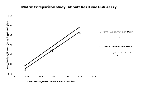
(Cassol, et al., J.
Clin. Microbiol. 35: 2795-2801, 1997; Fiscus, et al., J. Clin. Microbiol. 36:
258-60, 1998;
O'Shea, et al., AIDS 13: 630-1, 1999; Biggar, et al., J. Infec. Dis. 180 1838-
43, 1999;
Brambilla, et al., J. Clin. Microbiol. 41(5): 1888-93, 2003); HIV DNA
detection and
quantification (Panteleefe, et al., J. Clin. Microbiol. 37: 350-3, 1999;
Nyambi, et al., J. Clin.
Microbiol. 32: 2858-60, 1994); and HIV antibody detection (Evengard, et al.,
AIDS 3: 591-5,
1989; Gwirin, et al., JAMA 265: 1704-08, 1991). HCV RNA detection and
genotyping are
also reported using DBS (Solmone et al., J. Clin. Microbio. 40 (9): 3512-14,
2002). Although
these studies provide a good correlation with titers using DPS or DBS is
obtained as compared
with conventional liquid plasma samples, a loss of viral titers may occur
after room temperature
storage (Cassol, et al., J. Clin. Microbiol. 35: 2795-2801, 1997; Fiscus, et
al., J. Clin.
Microbiol. 36: 258-60, 1998). DBS and DPS samples are clearly less expensive
and less
hazardous to transport than liquid samples.
[0006] However, the procedure of analyte microextraction from DBS and
DPS on filter
paper suffers from a number of disadvantages. For example, microextraction of
sufficient
DNA or RNA from filter paper involves reconstitution in a liquid medium under
certain
vigorous procedures, e.g., vortex and centrifugation that damages the genetic
analytes of
interest. Furthermore, the fibers and other components of the filters become
dislodged into the
CA 02879987 2015-01-22
WO 2014/025787 PCT/US2013/053799
-3-
reconstitution solution, and require further centrifugation separation and/or
can impede the
ability to isolate the genetic material, such as by blocking genetic material
from adhering to a
separation column. Such prior microextraction procedures require a high
standard of technical
assistance, and even then do not consistently provide results with a desired
level of sensitivity,
reproducibility, quantification and specificity.
[0007] Furthermore, the sample volume used for DBS and DPS on filter
paper is
limited, typically to 50-200u1 spots, and considerable difficulty in analyte
detection and
accurate quantification and reproducibility can be encountered, particularly
when the
concentration of the desired analyte material is low in the sample. Also in
the prior art, there is
a lack of deliberate inhibition of enzymes and chemicals which degrade the
analytes, such as
genetic material contained therewithin. Even in the presence of a
bacteriostatic agent there are
conditions that permit enzymatic, nonenzymatic and autolytic breakdown of the
genetic
material. Furthermore, microextraction of genetic material from DBS or DPS on
filter papers
is considerably more difficult if absorption of high molecular weight DNA or
RNA is required.
Although the introduction of new material and transportation methods
continuously improve
the ways samples are handled, the quantity and quality of the sample available
for subsequent
analysis are still of great concern to researchers and clinicians alike.
[0008] U.S. Patent No. 7,638,099 provides an advantageous alternative
system for
biological sample collection, storage and transportation. The reference
suggests the use of
cellulose acetate fibers and hydrophilic polymer fibers as being advantageous
for an absorbent
matrix material. However, further improvements are desired for certain
situations, such as to
achieve more accurate and reproducible quantification of viral load in a
sample.
[0009] Thus, there is a need for an improved device for collection,
storage and
transportation of liquid suspension of biological specimens containing
analytes of interest in a
dry state, especially in large field studies and for application in settings
where collection,
centrifugation, storage and shipment can be difficult, as is often the case in
developing
countries. In addition, there is a need for improved recovery of viral
specimens for subsequent
analysis that provides precision values and accuracy of detection,
reproducibility and
quantification of the analytes of interest contained therewithin.
CA 02879987 2015-01-22
WO 2014/025787 PCT/US2013/053799
-4-
SUMMARY OF THE INVENTION
[0010] This invention fulfills in part the need to provide a safe,
convenient and simple
device and method for preserving, storage and transportation of biological
specimens
containing analytes of interest. The invention also fulfills in part the need
to recover biological
specimens containing analytes of interest for subsequent analysis that
provides more desirable
sensitivity and specificity of detection. More particularly, the invention
provides an improved
matrix storage material comprising hydrophobic polyolefm polymers for use as a
device,
system, and method for accurate and reproducible quantification of viral load
in a patient. The
invention provides a novel device and method for preserving, storing, and
transporting a liquid
suspension of biological specimens in a dry state and further reconstituting
the analytes of
interest contained in the biological specimens for use in research and site
validated clinical
testing.
[0011] In certain embodiments, the absorbent polyolefm matrix
comprises hydrophobic
polymers, including polyethylene. In certain embodiments, the absorbent
polyolefm fiber
matrix comprises a hydrophobic polyethylene surface coating. In certain
embodiments, the
matrix comprises a plurality of polyolefin fiber strands, wherein each
individual fiber strand
within the absorbent polyolefin fiber matrix is composed of a core and an
outer sheath. In
certain embodiments, the core of each fiber comprises polypropylene, and the
outer coating
sheath of each fiber comprises polyethylene. In certain embodiments, each
individual fiber
strand within the polyolefm fiber matrix is composed of a core of each strand
comprising about
50% polypropylene and a hydrophobic outer sheath surrounding the core of each
strand
comprising about 50% polyethylene.
[0012] Based on the data presented, the invention provides that a
hydrophobic
polyolefm fiber matrix is superior compared to previous dried collection
devices for absorption,
preservation, stabilization, and subsequent recovery of nucleic acid for
quantification and
qualification. Without wishing to be bound by theory, it is believed that
these surprising results
are due to the properties of the embedded hydrophobic interstices, or pockets
within the
polyolefm matrix. These pockets provide a reservoir for the analyte to reside
while excluding
water from the analyte, e.g., nucleic acid, providing a stable environment
during storage. The
improved hydrophobic polyolefin matrix further allows polar solvents to
evaporate more
consistently and efficiently. Therefore, the improved polyolefm matrix retains
analytes and
CA 02879987 2015-01-22
WO 2014/025787 PCT/US2013/053799
-5-
suspended particles inside the matrix better than for example a cellulose
matrix. Contrary to
the teachings in the prior art that hydrophilic polymer surfaces in the matrix
are more desirable,
it has been discovered that hydrophobic polyolefin surfaces in the matrix are
surprisingly
advantageous. Thus, a substantially intact viral nucleic acid, for example,
can be eluted from
the reconstituted matrix with great efficiency permitting a surprisingly
accurate degree of
quantification and qualification of the viral load in the biological sample.
[0013]
The polyolefm fiber matrix of the invention absorbs greater than 0.05 ml of a
liquid suspension of biological specimens absorbed and dried thereon. In
certain embodiments,
the polyolefm fiber matrix absorbs at least 0.1, ml or 0.5, ml of the liquid
suspension. In yet
other embodiments, the polyolefm fiber matrix absorbs at least 1 ml, 1.5 ml,
2.0 ml, 2.5 ml, 3.0
ml, or more, of the liquid suspension of biological specimens.
[0014]
The invention provides that the absorbent polyolefm fiber matrix, while
harboring numerous hydrophobic pockets, is able to be compressed by applying
force against
the matrix by at least 10% of the volume of the matrix to release a portion of
the re-suspended
biological specimen stored therewithin. In other embodiments, the matrix is
able to be
compressed by at least 20%, 50%, 75%, 80%, 85%, 90%, or 95% or more of the
volume of
the matrix to release a portion of the liquid suspension of biological
specimen stored in the
matrix. In other words, the matrix is at least 10% porous or defines at least
10% available
space, including numerous hydrophobic pockets within the polyolefin fiber
matrix, for the
storage of a biological specimen therein.
[0015] In
certain embodiments, the polyolefin fiber matrix is three-dimensional in a
variety of different shapes, including but not limited to, a cylinder, disk,
cube, sphere, pyramid,
cone, concave, indented, invaginated or other shapes and surface textures
suitable for
absorption and fitting inside a container. In certain embodiments, the matrix
is in the shape of a
cylinder about 18 mm to 24 mm, or 21 mm, in length, and 5 mm to 15 mm, or 9
mm, in
diameter, with a density of about 0.01 g/cc to 0.1 g/cc, or about 0.077 g/cc.
In certain
embodiments, a majority of the polyolefm fiber sizes are in the range of about
1-100 microns,
10-50 microns, or 20-25 microns and contain numerous hydrophobic pockets.
[0016]
The invention provides a device and methods that allow for biological testing
of
air-dried bodily fluid samples without the need for refrigerated or frozen
shipping and storage.
CA 02879987 2015-01-22
WO 2014/025787 PCT/US2013/053799
-6-
The inventive device and methods provide the capability to significantly
reduce the costs of
shipping infectious materials worldwide, especially those associated with
large clinical trials.
Moreover, the inventive device and methods for preserving biological specimens
are applicable
to and include a wide range of esoteric and standard clinical testing,
including qualitative and
quantitative nucleic acid analysis.
[0017] In certain embodiments, the invention provides a device, and
method of use
thereof, for preserving and recovering a biological specimen containing
analytes of interest.
More particularly, the device comprises a first enclosed container defming an
interior space
having side walls, a bottom and an openable and sealable lid or cap. In
certain embodiments,
the first enclosed container is a tube having a sealable cap with an absorbent
three-dimensional
polyolefm fiber matrix mounted therein. In certain embodiments, an interior of
the tube or cap
has an internal surface extension with the absorbent three-dimensional matrix
removably
mounted thereon.
[0018] In certain embodiments, the invention further comprises a
second enclosed
compression container with a syringe barrel shape or any other suitable shape
for receiving
therein the matrix for reconstitution, compression, and release of the
analytes of interest, e.g.,
intact viral RNA or DNA. In certain embodiments, only one container is
required for storage,
transportation, reconstitution, and release of the analytes of interest from
the matrix.
[0019] In certain embodiments, the device may optionally comprise a
desiccant inside
the enclosed container in vaporous communication with the matrix to maintain a
dried state of
the matrix and integrity of the biological specimen and analytes of interest
it contains on the
matrix. Exemplary suitable desiccant includes, but is not limited to,
montmorillonite clay,
lithium chloride, activated alumina, alkali alumino-silicate, DQ11 Briquettes,
silica gel,
molecular sieve, calcium sulfate, or calcium oxide. In certain embodiments,
the desiccant
indicates its moisture content by colorimetric means. In other embodiments,
since unlike the
hydrophilic cellulose acetate matrix where the solvents are not released as
efficiently, the
hydrophobic polyolefin fiber matrix of the invention allows the solvents to
evaporate more
consistently and efficiently, and a desiccant is not necessary.
[0020] According to the invention, the analytes of interest include,
but are not limited
to, nucleic acids, proteins, carbohydrates, lipids, whole cells, cellular
fragments, whole virus or
CA 02879987 2015-01-22
WO 2014/025787 PCT/US2013/053799
-7-
viral fragments. In certain embodiments, the analytes of interest are nucleic
acids including
either or both DNA and RNA molecules. The invention particularly provides
improved
systems and methods for the detection and quantitation of RNA, e.g., whole
virus for
determining viral load and genotyping in a biological specimen or subject.
[0021] In certain embodiments, the nucleic acid of interest is HCV or other
single
stranded RNA viruses. In certain embodiments, the nucleic acid of interest is
HIV or other
retroviruses. In certain embodiments, the nucleic acid of interest is HBV or
other double
stranded DNA viruses. In certain embodiments, the nucleic acid of interest is
Influenza or
other double stranded RNA viruses. In certain embodiments, the nucleic acid of
interest is
Parvovirus B19 or other single stranded DNA viruses. In certain embodiments,
the nucleic
acid of interest is contained within the HCV genome or the genome of other
single stranded
RNA viruses. In certain embodiments, the nucleic acid of interest is contained
within the HIV
genome or the genome of other retrovirus. In certain embodiments, the nucleic
acid of interest
is HBV genome or the genome of other double stranded DNA viruses. In certain
embodiments, the nucleic acid of interest is Influenza genome or the genome of
other double
stranded RNA viruses. In certain embodiments, the nucleic acid of interest is
Parvovirus B19
or the genome of other single stranded DNA virus.
[0022] According to the invention, the biological specimens include,
but are not limited
to, whole blood, plasma, urine, saliva, sputum, semen, vaginal lavage, bone
marrow,
cerebrospinal fluid, other physiological or pathological body liquids, or any
of the combinations
thereof. In certain embodiments, the biological specimen is human body fluid,
such as whole
blood containing the analytes of interest, such as nucleic acids, including
either or both DNA
and RNA molecules. In certain embodiments, the analytes of interest are
nucleic acids and the
biological specimens comprise at least 5 ng to 1 p g either or both DNA or RNA
molecules. In
yet other embodiments, the biological specimen is contained in liquid
suspension. According to
the invention, the liquid suspension includes, but is not limited to, cell
suspension, liquid
extracts, tissue homogenates, media from DNA or RNA synthesis, saline, or any
combinations
thereof.
[0023] The invention further provides a system and method for
preserving and
recovering a biological specimen containing analytes of interest, such as RNA,
from the matrix
in the device provided by the invention. In certain embodiments, the method
comprises the
CA 02879987 2015-01-22
WO 2014/025787 PCT/US2013/053799
-8-
following steps of providing a device comprising an absorbent matrix comprised
of
hydrophobic polyolefm fibers, wherein in certain embodiments each strand of
fiber is comprised
of a core and an outer sheath surface, wherein said core of each strand
comprises
polypropylene, and said outer sheath surface of each strand comprises
polyethylene. In the
method the matrix can be provided with a dried biological specimen contained
thereon obtained
from a volume of at least 0.05 ml of an evaporated liquid suspension
comprising a liquid and
the biological specimen containing analytes of interest. The method further
comprises
reconstituting the biological specimen on the matrix with a controlled volume
of a
reconstitution medium; and removing the biological specimen from the matrix,
such as by
compressing the matrix.
[0024] In certain embodiments, the reconstitution solution is water
medium. In other
embodiments, the reconstitution buffer comprises 1X phosphate buffered saline
(PBS) or
nuclease-free water optionally comprising sodium azide or other antimicrobial
agent. In yet
other embodiments, the reconstitution buffer is a "lysis" buffer. The
reconstitution buffer may
also include any number or combinations of available biological preservatives
or blood
anticoagulants including, but not limited to, ethylenediaminetetraacetic acid
(EDTA), sodium
citrate, and heparin.
[0025] In one embodiment, the method can comprise removing the matrix
from the
container prior to compressing the matrix in a second container, e.g., a
syringe barrel. In yet
another embodiment, the compression of the matrix is achieved by applying
force against the
hydrophobic polyolefin matrix within the same container to release the
analytes of interest.
According to the invention, the hydrophobic polyolefin matrix in the
compression device is
capable of compressing by at least 10%, 20%, 25%, 50%, 55%, 60%, 65%, 70%,
75%, 80%,
85%, 90% or more of the volume of the matrix to release a portion of the
biological specimen
suspended in the matrix.
[0026] The invention further provides a kit for preserving a liquid
suspension of a
biological specimen containing analytes of interest and for follow-up recovery
and analysis. In
certain embodiments, the kit includes the compression device provided by the
present invention
and instructions for preserving the biological specimens containing analytes
of interest. The kit
can further comprise a stabilizing solution to inhibit degradation of the
analytes. The kit can
further comprise a reconstitution medium, a compression device and further
instructions for
CA 02879987 2015-01-22
WO 2014/025787 PCT/US2013/053799
-9-
recovery the analytes of interest contained in the biological specimen. In
certain embodiments,
the compression device comprises a tube with a syringe barrel shape that
contains a plunger
with a cup attached that the matrix is permanently adhered to allowing
compression of the
matrix to be achieved by applying force to the plunger, and wherein at least
10% to 90%, or
greater, of the volume of the matrix is compressed to release a portion of the
bound biological
specimen.
[0027] The invention further provides subsequent analysis using the
recovered
biological specimen containing analytes of interest. In certain embodiments,
the analytes of
interest are RNA molecules that are detected or analyzed using analytical and
diagnostic
methods known in the art. In certain embodiments, the analytes of interest are
intact virus,
such as HCV or HIV, and the biological specimen recovered from the device is
used for
evaluation and analytical measurements with reproducibility, accuracy, and
precision.
BRIEF DESCRIPTION OF THE DRAWINGS
[0028] Fig. lA is a perspective view of an assembled device according
to one
embodiment of the invention. Fig. 1B is a perspective view of a disassembled
device according
to one embodiment of the invention ready for sample addition.
[0029] Fig. 2 illustrates addition of sample to the polyolefm matrix
of a device
according to one embodiment of the invention.
[0030] Fig. 3 illustrates addition of sample to the polyolefm matrix
of a device
according to one embodiment of the invention.
[0031] Fig. 4 is a perspective view of preparing to transfer the
polyolefin matrix of a
device according to one embodiment of the invention into an empty syringe
barrel.
[0032] Fig. 5 is a perspective view of completed delivery of the
polyolefm matrix into
the syringe barrel.
[0033] Fig. 6 illustrates rehydration of the polyolefm matrix by a pipette
tip gently
placed on the top of the matrix and slowly dispensing reconstitution buffer.
CA 02879987 2015-01-22
WO 2014/025787 PCT/US2013/053799
-10-
[0034] Fig. 7A illustrates insertion of the plunger into the syringe
barrel. Fig. 7B
illustrates application of pressure to the syringe plunger. Fig. 7C
illustrates compression of the
polyolefm matrix plug. Fig. 7D illustrates completion of sample recovery.
[0035] Fig. 8 provides a linear regression analysis for matrix
comparison studies using
the Abbott REALTIME HBV assay.
[0036] Fig. 9 provides a sample correlation and HCV viral load using
fresh plasma and
plasma samples processed through the ViveST devices of the invention.
[0037] Fig. 10 provides a sample correlation and HIV-1 viral load
using fresh plasma
and plasma samples processed through the ViveST devices of the invention and
recovered with
mLysis.
[0038] Fig. 11 provides a sample correlation and HIV-1 viral load
using fresh plasma
and plasma samples processed through the ViveST devices of the invention and
recovered with
water.
[0039] Fig. 12 provides a HCV analytical measurement range
determination using the
Abbott REALTIME HCV assay for samples processed through the ViveST devices of
the
invention.
[0040] Fig. 13 provides comparisons of frozen plasma and plasma
samples processed
through the ViveST devices of the invention using the Roche COBAS
AmpliPrep/COBAS
TaqMan HCV assay.
[0041] Fig. 14 provides comparisons of frozen plasma and plasma samples
processed
through the VivesST devices of the invention using the Roche COBAS
AmpliPrep/COBAS
TaqMan HIV assay.
[0042] Fig. 15 provides a HIV-1 analytical measurement range
determination using the
Abbott REALTIME HIV-1 assay for samples processed through the ViveST devices
of the
invention.
CA 02879987 2015-01-22
WO 2014/025787 PCT/US2013/053799
-11-
[0043] Fig. 16 provides a linear regression analysis of HCV 7-day
stability studies at
ambient conditions using the Abbott REALTIME HCV assay for samples processed
through
the ViveST devices of the invention.
[0044] Fig. 17 provides comparisons of target and actual HCV titers in
the 7-day
stability studies at ambient conditions using the Abbott REALTIME HCV assay,
viewed by
concentration level.
[0045] Fig. 18 provides comparisons of HCV titers analyzed on initial
test point (Day
1) in the nominal frozen plasma not processed through the ViveST devices of
the invention,
and the HCV titers of the plasma samples stored on the ViveST device for 7
days and
processed through the ViveST devices thereafter.
[0046] Fig. 19 provides a linear regression analysis of HCV 21-day
stability studies at
ambient storage condition using the Abbott REALTIME HCV assay for samples
processed
through the ViveST devices of the invention.
[0047] Fig. 20 provides comparisons of target and actual HCV titers in
the 21-day
stability studies at ambient storage condition using the Abbott REALTIME HCV
assay, view
by concentration level.
[0048] Fig. 21 provides a linear regression analysis of HCV 21-day
stability studies at
4 C storage condition using the Abbott REALTIME HCV assay for samples
processed through
the ViveST devices of the invention.
[0049] Fig. 22 provides comparisons of target and actual HCV titers in the
21-day
stability studies at 4 C storage condition using the Abbott REALTIME HCV
assay, view by
concentration level.
[0050] Fig. 23 provides a linear regression analysis of HCV 21-day
stability studies at
40 C/75% RH storage condition using Abbott REALTIME HCV assay for samples
processed
through the ViveST devices of the invention.
[0051] Fig. 24 provides comparisons of target and actual HCV titers in
the 21-day
stability studies at 40 C/75% RH storage condition using the Abbott REALTIME
HCV assay,
view by concentration level.
CA 02879987 2015-01-22
WO 2014/025787 PCT/US2013/053799
-12-
[0052] Fig. 25 provides comparisons of target and actual HCV titers in
the 21-day
stability studies, view by storage condition after 21 days storage.
[0053] Fig. 26 provides a linear regression analysis of HIV-1
stability studies at ambient
conditions using the Abbott REALTIME HIV-1 assay for samples processed through
the
ViveST devices of the invention.
[0054] Fig. 27 provides comparisons of target and actual HIV-1 titers
in the HIV-1
stability studies at ambient conditions using the Abbott REALTIME HIV-1 assay,
view by
concentration level.
[0055] Fig. 28 provides a linear regression analysis of HCV 62-day
stability studies at
ambient storage conditions using the Abbott REALTIME HCV assay for samples
processed
through the ViveST devices of the invention.
[0056] Fig. 29 provides comparisons of target and actual HCV titers in
the 62-day
stability studies at ambient storage condition using the Abbott REALTIME HCV
assay, view
by concentration level.
[0057] Fig. 30 provides a linear regression analysis of HCV 62-day
stability studies at
4 C storage condition using the Abbott REALTIME HCV assay for samples
processed through
the ViveST devices of the invention.
[0058] Fig. 31 provides comparisons of target and actual HCV titers in
the 62-day
stability studies at 4 C storage condition using the Abbott REALTIME HCV
assay, view by
concentration level.
[0059] Fig. 32 provides a linear regression analysis of HCV 62-day
stability studies at
40 C/75% RH storage conditions using the Abbott REALTIME HCV assay for samples
processed through the ViveST devices of the invention.
[0060] Fig. 33 provides comparisons of target and actual HCV titers in
the 62-day
stability studies at 40 C/75% RH storage conditions using the Abbott REALTIME
HCV assay,
view by concentration level.
CA 02879987 2015-01-22
WO 2014/025787 PCT/US2013/053799
-13-
[0061] Fig. 34 provides comparisons of target and actual HCV titers in
the 62-day
stability studies, view by storage condition after 62 days storage.
[0062] Fig. 35 provides a linear regression analysis of HIV-1 62-day
stability studies at
ambient storage conditions using the Abbott REALTIME HIV-1 assay for samples
processed
through the ViveST devices of the invention.
[0063] Fig. 36 provides comparisons of target and actual HIV-1 titers
in the 62-day
stability studies at ambient storage conditions using the Abbott REALTIME HIV-
1 assay, view
by concentration level.
[0064] Fig. 37 provides a linear regression analysis of the HIV-1 62-
day stability
studies at 4 C storage condition using the Abbott REALTIME HIV-1 assay for
samples
processed through the ViveST devices of the invention.
[0065] Fig. 38 provides comparisons of target and actual HIV-1 titers
in the 62-day
stability studies at 4 C storage conditions using the Abbott REALTIME HIV-1
assay, view by
concentration level.
[0066] Fig. 39 provides a linear regression analysis of HIV-1 62-day
stability studies a
40 C/75% RH storage conditions using the Abbott REALTIME HIV-1 assay for
samples
processed through the ViveST devices of the invention.
[0067] Fig. 40 provides comparisons of target and actual HIV- ltiters
in the 62-day
stability studies at 40 C/75% RH storage conditions using the Abbott REALTIME
HIV-1
assay, view by concentration level.
[0068] Fig. 41 provides comparisons of target and actual HIV-1 titers
in the 62-day
stability studies, view by storage condition after 62 days storage.
[0069] Fig. 42 provides a linear regression analysis for target and
achieved frozen
plasma samples.
[0070] Fig. 43 provides a probit analysis for HIV-1 limited of detection
(LOD)
evaluation.
CA 02879987 2015-01-22
WO 2014/025787 PCT/US2013/053799
-14-
[0071] Fig. 44 provides a regression analysis for samples processed
through the
ViveST device of the invention and the frozen plasma using the Roche COBAS
TaqMan HCV
Test (v2.0).
[0072] Fig. 45 provides a linear regression analysis of HCV 7-day
stability studies at
ambient storage conditions using the Roche COBAS TaqMan HCV Test (v 2.0) for
samples
processed through the ViveST devices of the invention.
[0073] Fig. 46 provides comparisons of target and actual HCV titers in
the 7-day
stability studies at ambient storage conditions using the Roche COBAS TaqMan
HCV Test (v
2.0).
[0074] Fig. 47 provides a linear regression analysis for target and
achieved frozen
plasma samples.
[0075] Fig. 48 provides a probit analysis for HCV LOD/LOQ Studies.
[0076] Fig. 49 illustrates 1.0 mL loaded on 3 matrixes. Picture taken
at time of loading
demonstrating that all matrixes completely absorbed all material.
[0077] Fig. 50 illustrates 1.0 mL, 1.5 mL, and 2.0 mL loaded. Picture taken
at time of
loading. With 1.5 mL it was observed that specimen was not completely absorbed
and liquid
pooled in the inner rim of the inverted cap. With 2.0 mL it was observed that
specimen was
not completely absorbed and liquid flowed over the inner rim of the inverted
cap and pooled in
the outer rim of the inverted cap.
[0078] Fig. 51 illustrates 1.0 mL, 1.5 mL, and 2.0 mL loaded. Picture taken
30
minutes after loading. With 1.5 mL it was observed that all specimens were
completely
absorbed by the matrix. With 2.0 mL it was observed that specimen was not yet
completely
absorbed and liquid still pooled in the outer rim of the inverted cap.
[0079] Fig. 52 illustrates 1.0 mL, 1.5 mL, and 2.0 mL loaded. Picture
taken after
drying overnight. All matrixes were dried. With 1.5 mL matrix it was observed
that all
specimens were completely absorbed by the matrix and dried. With 2.0 mL it was
observed
that the matrix did not absorb all specimens and dried specimen was visible in
the outer rim of
the inverted cap.
CA 02879987 2015-01-22
WO 2014/025787 PCT/US2013/053799
-15-
[0080] Fig. 53 provides a box plot of results of HIV-1 concentration
study using the
Abbott REALTIME HIV-1 Assay.
[0081] Fig. 54 provides a scatter plot of Samples (n=180) processed
through the
ViveST device of the invention using the Abbott REALTIME HCV Assay.
[0082] Fig. 55 provides a regression analysis for samples processed through
the
ViveST devices of the invention and the frozen plasma sample using the Abbott
REALTIME
HBV Assay.
[0083] Fig. 56 provides a linear regression analysis of HBV 60-day
stability studies at
ambient storage conditions using the Abbott REALTIME HBV assay for samples
processed
through the ViveST device of the invention.
[0084] Fig. 57 provides comparisons of target and actual titers in the
60-day stability
studies at ambient storage conditions using the Abbott REALTIME HBV Assay.
[0085] Fig. 58 provides a linear regression analysis for target and
achieved frozen
plasma samples.
[0086] Fig. 59 provides a probit analysis for HBV LOD/LOQ studies.
DETAILED DESCRIPTION OF THE INVENTION
[0087] The invention may be understood more readily by reference to
the following
detailed description of the preferred embodiments of the invention and the
Examples included
herein. However, before the present devices, materials, and methods are
disclosed and
described, it is to be understood that this invention is not limited to
specific embodiments of the
devices, materials and methods, as such may, of course, vary, and the numerous
modifications
and variations therein will be apparent to those skilled in the art. It is
also to be understood
that the terminology used herein is for the purpose of describing specific
embodiments only and
is not intended to be limiting.
[0088] The invention provides a device and method for collection, storage
and
transportation of a liquid suspension of a biological specimen containing an
analyte of interest.
More particularly, the present invention provides a device and method for
collection, storage
CA 02879987 2015-01-22
WO 2014/025787 PCT/US2013/053799
-16-
and transportation of a liquid suspension containing a biological specimen in
a dry state that is
convenient and simple to use. As used herein, the terms "a" or "an" mean one
or more than
one depending upon the context in which they are used. For example, "an
analyte" in a sample
refers to a particular type of analyte of interest (e.g., such as intact HCV
or HIV RNA), of
which there may be numerous copies within the sample. Where a sample is
referred to as
containing an analyte, it is understood that the sample may contain many other
types of
analytes of interest also.
[0089] According to the invention, the time period for which
biological specimen may
be preserved may be as short as the time necessary to transfer a sample of
biological specimen
from a collection source to the place where subsequent analysis is to be
performed. Therefore,
the invention provides that such preservation can occur for a period of
several minutes, hours,
days, months, or greater. The temperature conditions under which a biological
specimen may
be stored in the device provided by the invention are not limited. Typically,
samples are
shipped and/or stored at ambient or room temperature, for example, from about
15 C to about
40 C, preferably about 15 C to 25 C. In another embodiment the samples may be
stored in a
cool environment. For example, in short-term storage, the samples can be
refrigerated at about
2 C to about 10 C. In yet another example, the samples may be refrigerated at
about 4 C to
about 8 C. In another example, in long-term storage, the samples can be frozen
at about -80 C
to about -10 C. In yet another example, the samples can be frozen from about -
60 C to about
-20 C. In addition, the device may preferably but not necessarily be stored in
dry or desiccated
conditions or under an inert atmosphere.
[0090] In certain embodiments, the invention provides a device
comprising a first
enclosed container defining an interior space having side walls, a bottom and
an openable and
sealable lid or cap with an absorbent three-dimensional hydrophobic polyolefin
fiber matrix
disposed inside the first enclosed container. The invention can further
comprise a second
container with a syringe barrel-shape or any other suitable shape, and a
plunger contained
therewith, wherein the matrix can be placed therein for compression and
release of the analyte
of interest. In certain embodiments, the matrix can be loaded with a
biological specimen and
dried, and placed into a single container which serves both a protective, dry
transportation
vessel and is configured for compression of the reconstituted matrix for
release of the analyte
of interest.
CA 02879987 2015-01-22
WO 2014/025787 PCT/US2013/053799
-17-
[0091] The shape of the first or second container is not limited, but
can be cylindrical,
rectangular, or tubular for example. Materials for construction of the
containers are not
limited, but can be plastic, metal foil, laminate comprising metal foil,
metallized film, glass,
silicon oxide coated films, aluminum oxide coated films, liquid crystal
polymer layers, and
layers of nano-composites, metal or metal alloys, acrylic, and amorphous
carbon for example.
In certain embodiments, the invention provides a first enclosed container
having a threaded
screw cap. In other embodiments, the lid or cap can remain attached to the
first enclosed
container such as a flip-top fashion. In yet other embodiments, the lid or cap
may also be cork-
like or any other openable configuration. The lid or cap can also provide an
air-tight seal when
the first enclosed container is closed.
[0092] The device also comprises a hydrophobic polyolefin fiber matrix
for retaining
the biological specimen, drying the analyte of interest therein,
reconstitution and release of the
analyte. In certain embodiments, the hydrophobic matrix is made from polyolefm
fibers that
can be quality-controlled during manufacturing. As used herein, the term
"polyolefin fiber
matrix" refers to a fiber matrix made of at least one type polyolefm polymer
produced from a
simple olefin (also called an alkene with the general formula CõH2õ) as a
monomer. The term
"hydrophobic" polyolefm surface is used to describe a polyolefm surface that
generally repels
water or resists wetting, for example, as would result from minimal or
substantially absent
hydrogen bonding or other chemical bonding interactions between the polyolefm
surface and
water molecules. A hydrophobic polyolefin surface generally lacks the
molecular entities or
substituents to interact with the polar solvents, in particular water, or with
other polar groups.
In one aspect, the hydrophobicity of a polyolefin surface can be quantified by
the contact angle,
0c, which is the angle between the polyolefm surface and the tangent to the
water surface at the
contact point, that is, where the water/air (or water/vapor) interface meets
the polyolefm
surface. For example, the polyolefin surface can be considered "hydrophobic"
if the water
contact angle is greater than about 850. In another aspect, the polyolefin
surface can be
considered "hydrophobic" if the water contact angle is greater than about 90';
alternatively,
greater than about 950; alternatively, greater than about 1000; alternatively,
greater than about
105'; alternatively, greater than about 110'; alternatively, greater than
about 115'; or
alternatively, greater than about 120 .
CA 02879987 2015-01-22
WO 2014/025787 PCT/US2013/053799
-18-
[0093] In certain embodiments, the hydrophobic polyolefm fiber matrix
comprises
fibers that have a hydrophobic first polyolefin, such as polyethylene surface.
In certain
embodiments the surface can be a coating or sheath substantially disposed on a
core of a
second polyolefin, such as polypropylene. The relative amounts of each polymer
can range
from 10%-90% polyethylene and 10%-90% polypropylene, and in some embodiments
about
50% polyethylene and about 50% polypropylene by weight. The hydrophobic
polymer fibers
are bound together and shaped as is known in the art and commercially
available, such as from
Filtrona Porous Technologies, with pore sizes ranging from 2 microns to 100
microns.
[0094] In certain embodiments, the hydrophobic polyolefm matrix of the
invention is an
absorbent material to which the liquid suspension of biological specimen
containing analytes of
interest will be retained and which does not inhibit evaporation of the
solvent (e.g., water or
other fluids) for storage or subsequent reconstitution and analysis of the
analytes of interest
applied thereto. The matrix of the invention comprises hydrophobic polyolefm
surfaces of a
porous nature to provide entrainment of the liquid suspension in the matrix.
As used herein,
the term "entrain" and derivatives thereof means that the liquid suspension of
a polar solvent
and analytes can be temporarily entrapped within the interstices, or pores, of
the matrix without
substantial reliance on chemical and/or physical interactions such that a
polar solvent like water
can evaporate and leave the suspended analytes remaining in the matrix.
[0095] A matrix suitable for this purpose includes, but is not limited
to, a matrix that
comprises or is composed of hydrophobic polyolefm homopolymers and copolymers.
Particularly suited are polymers of ethylene alone, combined or copolymerized
with an alpha-
olefm polymer. Examples of the alpha-olefm polymer include, but are not
limited to,
propylene, 1-butene, 2-butene, 3-methyl- 1-butene, isobutylene, 1-pentene, 2-
pentene, 3-
methyl-1 -pentene, 4-methyl-l-pentene, 1 -hexene, 2-hexene, 3-hexene, 3-ethyl-
l-hexene, 1-
heptene, 2-heptene, 3-heptene, the four normal octenes, the four normal
nonenes, or the five
normal decenes. In another aspect, the alpha-olefin polymer may be selected
from 1-butene, 1-
pentene, 1-hexene, 1-octene, 1-decene, or styrene. In certain embodiments, the
hydrophilic
olefm polymers form the core, and hydrophobic polymers such as made with
polyethylene form
the outer sheath surface of each strand of the polyolefin fiber matrix of the
invention.
[0096] Any ratio of polymers can be employed to prepare the suitable
polyolefm
polymer matrix for use herein. For example, ethylene can be used from about 5
to about 95
CA 02879987 2015-01-22
WO 2014/025787 PCT/US2013/053799
-19-
mole percent for the outer sheath surface of each strand, and any of the
suitable monomers can
constitute the balance of the mole percent of the alpha olefms for the core of
each strand.
Thus, ethylene can be used from about 5, 10, 15, 20, 25, 30, 35, 40, 45, 50,
55, 60, 65, 70, 75,
80, 85, 90, or 95 mole percent to prepare suitable material, with any of the
suitable monomers
can constitute the balance of the mole percent of the alpha olefins.
Alternatively, ethylene can
be used from about 5 to about 95 mole percent, about 15 to about 85 mole
percent, or about
25 to about 75, about 35 to about 65, or about 45 to about 55 mole percent,
with any of the
suitable monomers making up the balance of the mole percent of the alpha
olefms. In certain
embodiments, polyethylene is used for the outer sheath surface, and
polypropylene is used for
the core, of each strand within the polyolefm fiber matrix of the invention.
Polyolefin polymers
can be low density or high density, highly branched or substantially
unbranched, and the like, as
long as the polymer can withstand the methods used to prepare and use the
disclosed devices
and methods. In certain embodiments, the density of the resulting polyolefm
fiber matrix of the
invention is about 0.077 grams/cc.
[0097] Thus, the polyolefm fiber matrix of the invention has an ability to
absorb a liquid
suspension readily and quickly, as well as to release the biological specimen
containing analytes
of interest consistently, efficiently, and precisely. In certain embodiments,
the polyolefm fiber
matrix can absorb at least 0.05 ml, 0.1 ml, 0.2 ml, 0.3 ml, 0.4 ml, 0.5 ml,
0.6 ml, 0.7 ml, 0.8 ml,
or 0.9 ml, 1.0 ml, 1.5 ml, 2.0 ml, 2.5 ml, 3.0 ml, or greater, sample of a
liquid suspension of a
biological specimen containing an analyte of interest. The term "absorb" and
"adsorb" are used
interchangeably, and means that the liquid suspension is incorporated into or
onto the
polyolefm fiber matrix in such a way as to be readily removed from the matrix
leaving the
analytes of interest behind.
[0098] The volume of the polyolefm matrix may or may not expand upon
absorption of
the liquid suspension, and may or may not contract upon drying. However, a
liquid saturated
matrix can be compressed to release entrained fluid containing analyte, due to
its porosity, by
at least about 10%, 20%, 30%, 40%, 50%, 60%, 70%, 75%, 80%, 90%, or more of
its
saturated volume. Volumetric compression is one convenient technique for
release of the
reconstituted biological specimen, however, any other means, such as
centrifugation or vacuum
pressure, can alternatively be employed to release the biological specimen
from the matrix.
CA 02879987 2015-01-22
WO 2014/025787 PCT/US2013/053799
-20-
[0099]
Therefore, as used herein, the term "compress," "co mpres s able,"
"compression," and other derivatives of the word "compress" means that the
volume of the
saturated matrix is reduced as compared to the original volume of the
saturated matrix while a
force or a pressure is applied to the matrix. As used herein, the term "a
portion of the
biological specimen" means at least some of the biological specimen contained
in the liquid
suspension is released from the matrix. In certain embodiments, the matrix is
compressed until
the maximum volume of the reconstituted biological specimen is released from
the matrix.
[00100] In
certain embodiments, the polyolefin fiber matrix is three-dimensional in a
shape such as cylinder, cube, sphere, pyramid or cone. In certain embodiments,
the matrix is in
the shape of a cylinder about 21 mm in length and 9 mm in diameter, with a
weight of about
0.103 grams. However, the matrix can be widened, lengthened, or shortened to
achieve any
needed volume capacity.
Polyolefin fiber sizes can vary, but are generally about 1-100n or
20-25 microns.
[00101] In
certain embodiments, for reconstitution and recovery of the analytes the
matrix is mounted or placed within a container or syringe barrel into which is
received a
plunger, wherein the matrix is compressed by applying force to the plunger
against the matrix
to release reconstituted biological suspension through a port for example. In
yet other
embodiments, the matrix can be removable from the enclosed container and the
plunger. As
used herein, the term "removable" means that the matrix can be detached or
separated from the
container and the plunger.
[00102] As
used herein, the term "liquid suspension" refers to any liquid medium and
mixture containing biological specimens. This includes, for example, water,
saline; cell
suspensions of humans, animals and plants; extracts or suspensions of
bacteria, fungi, plasmids,
viruses; extracts or suspensions of parasites including helminthes, protozoas,
spirochetes; liquid
extracts or homogenates of human or animal body tissues, e.g., bone, liver,
kidney, brain;
media from DNA or RNA synthesis; mixtures of chemically or biochemically
synthesized DNA
or RNA, and any other sources in which any biological specimen is or can be in
a liquid
medium.
[00103] As
used herein, the term "biological specimen" refers to samples, either in
liquid
or solid form, having dissolved, suspended, mixed or otherwise contained
therein, any analytes
CA 02879987 2015-01-22
WO 2014/025787 PCT/US2013/053799
-21-
of interest, for example, genetic material. As used herein, the term "genetic
material" refers to
nucleic acids that include either or both deoxyribonucleic acid (DNA) or
ribonucleic acid
(RNA). The term "biological specimen" also refers to whole blood, plasma,
serum, lymph,
synovial fluid, bone marrow, cerebrospinal cord fluid, semen, saliva, urine,
feces, sputum,
.. vaginal lavage, skin scrapings, hair root cells, or the like of humans or
animals, physiological
and pathological body liquids, such as secretions, excretions, exudates and
transudates; any
cells or cell components of humans, animals, plants, bacteria, fungi,
plasmids, viruses, parasites,
or the like that contain analytes of interest, and any combination thereof.
[00104] As used herein, the term "analytes of interest" refers to any
micro- or macro-
.. molecules in the biological specimen that are interested to be detected or
analyzed. These
include, for example, nucleic acids, polynucleotides, oligonucleotides,
proteins, polypeptides,
oligopeptides, enzymes, amino acids, receptors, carbohydrates, lipids, cells,
any intra- or extra-
cellular molecules and fragments, virus, viral molecules and fragments, or the
like. In certain
embodiments, the analytes of interest are nucleic acids including either or
both DNA or RNA.
.. As used herein, the term "nucleic acids" or "polynucleotide" refers to RNA
or DNA that is
linear or branched, single or double stranded, a hybrid, or a fragment
thereof. The term also
encompasses RNA/DNA hybrids. The term also encompasses coding regions as well
as
upstream or downstream noncoding regions. In addition, polynucleotides
containing less
common bases, such as inosine, 5-methylcytosine, 6-methyladenine,
hypoxanthine, and other
.. are also encompassed. Other modifications, such as modification to the
phosphodiester
backbone, or the 2'-hydroxy in the ribose sugar group of the RNA are also
included. The
nucleic acids/polynucleotides may be produced by any means, including genomic
preparations,
cDNA preparations, in vitro synthesis, RT-PCR, and in vitro or in vivo
transcription. In
certain embodiment, the nucleic acids are either or both viral DNA or RNA, for
example, DNA
.. or RNA from human immunodeficiency virus (HIV), hepatitis B virus (HBV),
hepatitis C virus
(HCV), or any other human or animal viral pathogen
[00105] In certain embodiments, the compression device provided by the
present
invention may optionally include a desiccant, either a natural or synthetic
desiccant, inside the
container to maintain the dried state of the matrix and integrity of the
analytes of interest on the
.. matrix within the enclosed container. In certain embodiments, the desiccant
is in vaporous
communication with the matrix in the compression device having a dye indicator
reactive with
CA 02879987 2015-01-22
WO 2014/025787 PCT/US2013/053799
-22-
moisture whereby the desiccant changes to a bright color when exposed to
humidity or
moisture. In certain embodiments, the desiccant is in vaporous communication
with the matrix
so that an air permeable barrier is formed in-between the desiccant and the
matrix inside the
container. The desiccant used in the device is commonly known in the art,
including but is not
limited to montmorillonite clay, lithium chloride, activated alumina, alkali
alumino-silicate,
DQ11 Briquettes, silica gel, molecular sieve, calcium sulfate, and calcium
oxide. The desiccant
can be provided with a colorimetric indicator of water content. A desiccant
may not be needed
inside the device with the hydrophobic polyolefin fiber matrix of the
invention.
[00106] The polyolefm fiber matrix of the invention may optionally
include a
composition absorbed to the matrix wherein the composition protects against
degradation of
the analytes of interest contained in the biological specimens. As used
herein, the term
"protects against degradation of the analytes of interest" means that a matrix
in the device of
the invention maintains the stored analytes of interest contained in the
biological specimens in a
substantially nondegraded form, providing that the analytes of interest are
suitable for many
different types of subsequent analytical procedures. Protection against
degradation may
include protection against substantial damaging of analytes of interest caused
by chemical or
biological agents including action of bacteria, free radicals, nucleases,
ultraviolet radiation,
oxidizing agent, alkylating agents, or acidic agents (e.g., pollutants in the
atmosphere). In
certain embodiments, the composition absorbed on the matrix of the invention
may include one
or more of a weak base, a chelating agent, a protein denaturing agent such as
a detergent or
surfactant, a nuclease inhibitor, and a free radical trap. In the case where
the stored analyte of
interest is RNA, particularly unstable RNA, the composition may include RNase
inhibitors and
inactivators, genetic probes, complementary DNA or RNA (or functionally
equivalent
compounds), proteins and organic moieties that stabilize RNA or prevent its
degradation.
[00107] Another composition which protects against degradation which may be
optionally used is an oxygen scavenger element. As used herein, the term
"oxygen scavenging
element" refers to is a substance that consumes, depletes or reduces the
amount of oxygen
from a given environment without negatively affecting the samples of
interests. Suitable
oxygen scavenging elements are well-known to those skilled in the art. Non-
limiting examples
of oxygen scavenging elements include but are not limited to compositions
comprising metal
particulates reactive with oxygen such as transition metals selected from the
first, second or
CA 02879987 2015-01-22
WO 2014/025787 PCT/US2013/053799
-23-
third transition series of the periodic table of the elements, and include
manganese II or III, iron
II or III, cobalt II or III, nickel II or III, copper I or II, rhodium II, III
or IV, and ruthenium.
The transition metal is preferably iron, nickel or copper. An example of an
iron oxygen
scavenging element is D500 from Multisorb. Other commercially available oxygen
scavengers
may also be purchased from companies such as Mitsubishi, Dow, or the like.
Other examples
of oxygen scavenging element may be enzymes which consumes, depletes or
reduces the
amount of oxygen from the given environment without negatively affecting the
samples of
interests.
[00108] In other embodiments, the compression device may optionally
comprise a
modified atmosphere such as nitrogen or argon through a well-known gas purging
process
prior to sealing, shipping, or storing. The term "modified atmosphere" refers
to any replacing
or altering normal atmospheric gas compositions with at least one inert gas or
gas which does
not degrade the sample of interests.
[00109] As used herein, a "weak base" suitable for the composition of
the invention may
be a Lewis base which has a pH of about 6 to 10, preferably about pH 8 to 9.5.
The weak base
suitable for the composition of the invention may, in conjunction with other
components of the
composition, provide a composition pH of 6 to 10, preferably, about pH 8.0 to
9.5. Suitable
weak bases according to the invention include organic and inorganic bases.
Suitable inorganic
weak bases include, for example, an alkali metal carbonate, bicarbonate,
phosphate or borate
(e.g., sodium, lithium, or potassium carbonate). Suitable organic weak bases
include, for
example, tris-hydroxymethyl amino methane (Tris), ethanolamine,
triethanolamine and glycine
and alkaline salts of organic acids (e.g., trisodium citrate). A preferred
organic weak base is a
weak monovalent organic base, for example, Tris. The weak base may be either a
free base or
a salt, for example, a carbonate salt. It is believed that the weak base may
provide a variety of
functions, such as protecting the analytes of interest from degradation,
providing a buffer
system, ensuring proper action of the chelating agent in binding metal ions,
and preventing the
action of acid nucleases which may not be completely dependent on divalent
metal ions for
functioning.
[00110] As used herein, a "chelating agent" is any compound capable of
complexing
multivalent ions including Group II and Group III multivalent metal ions and
transition metal
ions (e.g., Cu, Fe, Zn, Mn, etc). In certain embodiments, the chelating agent
is ethylene
CA 02879987 2015-01-22
WO 2014/025787 PCT/US2013/053799
-24-
diamine tetraacetic acid (EDTA), citrate or oxalate. It is believed that one
function of the
chelating agent is to bind multivalent ions which if present with the stored
biological specimen
may cause damage to the analytes of interest, especially to nucleic acids.
Ions which may be
chelated by the chelating agent include multivalent active metal ions, for
example, magnesium
and calcium, and transition metal ions, for example, iron. Both calcium and
magnesium are
known to promote nucleic acid degradation by acting as co-factors for enzymes
which may
destroy nucleic acids (e.g., most known nucleases). In addition, transition
metal ions, such as
iron, may readily undergo oxidation and reduction and damage nucleic acids by
the production
of free radicals or by direct oxidation.
[00111] The composition can further include a protein denaturing agent if
the analytes of
interest are nucleic acids. As used herein, a "protein denaturing agent"
functions to denature
non-nucleic acids compounds, for example, nucleases. If the protein denaturing
agent is a
detergent or a surfactant, the surfactant may also act as a wetting agent to
facilitate the uptake
of a sample by the dry solid matrix. The terms "surfactant" and "detergent"
are synonymous
and may be used interchangeably throughout the specification. Any agent that
denatures
proteins without substantially affecting the nucleic acids of interest may be
suitable for the
invention. In certain embodiments, protein denaturing agents include
detergents. As used
herein "detergents" include ionic detergents, preferably anionic detergents.
An anionic
detergent suitable for the invention may have a hydrocarbon moiety, such as an
aliphatic or
aromatic moiety, and one or more anionic groups. Particularly, suitable
anionic detergents
include sodium dodecyl sulphate (SDS) and sodium lauryl sarcosinate (SLS). The
ionic
detergent causes inactivation of a microorganism which has protein or lipid in
its outer
membranes or capsids, for example, fungi, bacteria or viruses. This includes
microorganisms
which may be pathogenic to humans or which may cause degradation of nucleic
acids. It is
believed that inactivation of a microorganism by a detergent is a result of
destruction of the
secondary structure of the organisms external proteins, internal proteins,
protein containing
membranes, or any other protein necessary for viability. However, the
detergent may not
inactivate some forms of organisms, for example, highly resistant bacterial
spores and
extremely stable enteric virions.
[00112] The composition may optionally include a free radical trap. As used
herein, a
"free radical trap" is a compound which is sufficiently reactive to be
preferred, over a DNA
CA 02879987 2015-01-22
WO 2014/025787 PCT/US2013/053799
-25-
molecule or a component thereof, as a reactant with a free radical, and which
is sufficiently
stable not to generate damaging free radicals itself. Examples of a suitable
free radical trap
include: uric acid or a urate salt, mannitol, benzoate (Na, K, Li or tris
salt), 1-3 dimethyl uric
acid, guanidine, guanine, thymine, adenine, cytosine, in N-acetyl-histidine,
histidine,
deferoxamine, dimethyl sulfoxide, 5'5 dimethyl pyrroline-N-oxide, thiocyanate
salt and
thiourea. Suitable free radical traps include mannitol, thiocyanate salts,
uric acid or a urate salt.
It is believed that the longer the period of time for which the nucleic acid
is to be stored the
more likely that a free radical trap may be advantageously included in the
composition absorbed
to the solid matrix. Even if the nucleic acid is only to be stored for a
matter of minutes, a free
radical trap may still be incorporated into the composition. It is believed
that one function of
the free radical trap may be to trap nucleic acid damaging free radicals. For
example, when the
free radical trap used is uric acid or urate salt it may be converted to
allantoin which may also
act as a free radical trap that accepts free radicals that would otherwise
damage nucleotide
bases, for example, guanine. In certain embodiments, the free radical trap
reacts with free
radicals regardless of source (including free radicals present in the air).
Free radicals may be
generated through oxidation or reduction of iron in biological specimen, such
as blood.
Typically, free radicals are believed to be generated by spontaneous oxidation
of the groups
which are present, for example, in denatured serum protein of blood. Free
radicals may also be
generated by radiation such as UV light, x-rays and high-energy particles. In
addition, free
radical traps which are also a weak acid, e.g. uric acid, may also function as
a component of the
buffering system provided by the weak base discussed above. Also, the free
radical trap may
enhance removal of a stored sample of nucleic acids if in situ processing is
not desired.
[00113] Referring to Figs. 1A & 1B, an exemplary compression device of
the invention
for preserving liquid suspension of biological specimen containing analytes of
interest is shown.
The container 20 is cylindrical and has side walls 22, a bottom 24 and an
openable lid 26, which
sealingly engages the container 20 opening. The lid 26 has an extension 28
that holds a
removable matrix 30 inside the container 20. The polyolefin fiber matrix 30 is
a cylinder
capable of absorbing 1 ml of a liquid suspension of a biological specimen and
compress by at
least 50% of the volume of the saturated matrix to release a portion of the
biological specimen.
A desiccant 40 may be optionally placed inside the container 20, separated
with the matrix 30
by an optional air permeable barrier 42, for in vaporous communication with
the matrix 30 to
control humidity or moisture therein.
CA 02879987 2015-01-22
WO 2014/025787 PCT/US2013/053799
-26-
[00114] The invention further provides a method for preserving and
recovering a
biological specimen comprising: (a) providing a dried biological specimen in a
device
comprising a container defming an interior space having side walls, a bottom
and an openable
and sealable lid with an absorbent three-dimensional polyolefm matrix mounted
inside the
container, wherein the polyolefm matrix comprises a plurality of interstices
with a hydrophobic
polyolefm surface and has contained therein the dried biological specimen
obtained from an
evaporated volume of at least 0.1 ml of a liquid suspension comprising a
solvent and the
biological specimen absorbed and dried on the matrix; (b) reconstituting the
biological
specimen on the polyolefm matrix with a controlled volume of a reconstitution
media; and (c)
removing the biological specimen and reconstitution media from the polyolefin
matrix by
compressing the matrix. Any suitable and/or commonly available drying methods,
such as
vacuum dry, low heat dry, low pressure dry, and fan dry, can be used in the
inventive method.
[00115] In certain embodiments, the polyolefin matrix comprises a
plurality of fibers
having a substantially hydrophobic surface. In certain embodiments, the fibers
within the
polyolefm matrix have a polyethylene surface. In other embodiments, the fibers
within the
polyolefm matrix comprise polypropylene coated with polyethylene. In certain
embodiments,
the polypropylene and polyethylene are present in approximately equal amounts
in each fiber
strand.
[00116] Referring to Fig. 1B, the lid 26 of the container 20 has a lid
extension 28
holding a polyolefin fiber matrix 30 which may be permanently mounted in a cup
that is
attached to a plunger contained within the second enclosed container. A liquid
suspension of
any biological specimen containing analytes of interest is added on the top of
the polyolefm
fiber matrix 30 and is allowed to fully absorb into the matrix 30 (Fig. 3).
The lid 26 with the
matrix 30 having bound biological specimen thereon is allowed to air-dry, and
then
reassembled with the container 20 for preservation at ambient temperature.
[00117] The method of the invention further optionally includes an
intermediate step of
applying a stabilizing composition to the polyolefm fiber matrix to protect
the analytes of
interest against degradation. Depending upon the analytes of interest, the
stabilizing
composition, as discussed above, may include but is not limit to one or more
of a weak base, a
chelating agent, a protein denaturing agent such as a detergent or surfactant,
a nuclease
inhibitor, and a free radical trap. Particularly for protection of unstable
RNA, the stabilizing
CA 02879987 2015-01-22
WO 2014/025787 PCT/US2013/053799
-27-
composition may include RNase inhibitors and inactivators, genetic probes,
complementary
DNA or RNA (or functionally equivalent compounds), proteins and organic
moieties that
stabilize RNA or prevent its degradation.
[00118] The invention further provides a method for recovering from the
polyolefm fiber
matrix in the compression device the biological specimen containing analytes
of interest. In
certain embodiments, the method includes the following steps: a) applying
reconstitution
medium to the matrix to rehydrate the bound biological specimen containing
analytes of
interest, and b) compressing the matrix to release a portion of the biological
specimen.
According to the present invention, the reconstitution medium is molecular-
grade water. In
other embodiments, the reconstitution medium includes the components of 1X
phosphate
buffered saline (PBS) or nuclease-free water optionally with the addition of
sodium azide or
other antimicrobial agent. The reconstitution medium may also include any
number or
combinations of available biological preservatives or blood anticoagulants
including but not
limited to ethylenediaminetetraacetic acid (EDTA), sodium citrate, and
heparin. PBS or
nuclease-free water serves as the sterile and neutral medium for the
rehydration, resuspension,
and recovery of the analyte(s) of interest from the matrix. When included,
antimicrobial agents
such as sodium azide prevent microbial growth and subsequent contamination
with RNases.
When included, biological preservatives such as EDTA, sodium citrate, and
heparin serve as
anticoagulants and or chelating agents.
[00119] In the embodiments shown in Figs. 4-7, the biological sample is
prepared for
analysis. Fig. 4 is a perspective view of preparing to transfer the polyolefm
fiber matrix 30 of
the device to an empty syringe barrel 52. Fig. 5 is a perspective view of
completed delivery of
the polyolefin fiber matrix 30 into the syringe barrel 52.
[00120] Fig. 6 illustrates rehydration of the polyolefm fiber matrix 30
by a pipette tip 53
gently placed on the top of the matrix 30 and slowly dispensing reconstitution
buffer. Fig. 7A
illustrates insertion of the plunger 54 into the syringe barrel 54. Fig. 7B
illustrates application
of pressure to the syringe plunger 42. Fig. 7C illustrates compression of the
matrix 30. Fig. 7D
illustrates completion of sample recovery from the matrix 30.
[00121] In certain embodiments, the analytes of interest are nucleic
acids including either
or both DNA or RNA molecules. In certain embodiments, the liquid suspension of
biological
CA 02879987 2015-01-22
WO 2014/025787 PCT/US2013/053799
-28-
specimen contains at least about 5 attograms or 1 p g isolated DNA or RNA
molecules. As
used herein, the term "isolated," "isolation," and other derivatives of the
word "isolate" means
that the DNA or RNA molecules are substantially free from some of the other
cellular material
with which it is naturally associated, or culture medium when produced by
recombinant
techniques, or chemical precursors or other chemicals when chemically
synthesized.
[00122] The invention further provides that the analytes of interest
contained in the
biological specimen recovered from the polyolefin fiber matrix of the device
into the
reconstitution medium, such as molecular-grade water, are subject to
subsequent analysis. As
used herein, the term "subsequent analysis" includes any analysis which may be
performed on
recovered biological specimens stored in reconstitution medium. Alternatively,
the analytes of
interest contained in the biological specimen may be isolated, purified or
extracted prior to
analysis using methods known in the art. The analytes of interest may be
subjected to
chemical, biochemical or biological analysis. In one of the preferred
embodiments, the analytes
of interest are nucleic acids including either or both DNA or RNA molecules
that can be
detected or analyzed with or without prior extraction, purification or
isolation. DNA or RNA
extraction, purification or isolation, if necessary, is performed based on
methods known in the
art. Examples of subsequent analysis include polymerase chain reaction (PCR),
ligase chain
reaction (LCR), reverse transcriptase initiated PCR, DNA or RNA hybridization
techniques
including restriction fragment length polymorphism (RFLP), viral DNA or RNA
detection and
quantification, viral load tests, DNA or RNA genotyping, etc. "Subsequent
analysis" also
includes other techniques using genetic probes, genomic sequencing, enzymatic
assays, affmity
labeling, methods of detection using labels or antibodies and other similar
methods.
[00123] The invention also provides a kit for preserving a liquid
suspension of biological
specimen containing analytes of interest. The kit of the invention provides a
compression
device disclosed herein including one or more containers, one or more
polyolefm fiber
matrixes, and optionally desiccant, and instructions for the use thereof to
preserve biological
specimens. The kit may optionally include a stabilizing solution. Kits of the
invention can
further include a reconstitution medium, a compression device and further
protocols for
rehydration and recovery of the biological specimen. The container of the kit
may be any
container suitable for use during application of a liquid suspension of
biological specimen
containing analytes of interest to the matrix or during application and one or
more phases of
CA 02879987 2015-01-22
WO 2014/025787 PCT/US2013/053799
-29-
subsequent processing of a sample of the biological specimen.
Therefore, in certain
embodiments, a liquid suspension of biological specimen may be applied,
stored, transported
and further processed all in the same kit. Alternatively, a liquid suspension
may be applied to
the matrix where the matrix is removed from the kit container for processing
in a different
container.
[00124]
The kit may also include one or more of any of the polyolefin fiber matrix
disclosed herein. This includes one or more polyolefin fiber matrix with or
without
compositions for protection of analytes of interest contained in the
biological specimen. One
aspect of the kit of the invention is that the reconstituted biological
specimen containing
analytes of interest is released by compressing the matrix. This procedure
avoids vortexing and
centrifuging the sample, providing decreased chance of sample damage, human
labor costs and
matrix contamination of the sample. A compression device of the kit of the
present invention
may be any device that is used to provide a force or pressure on the matrix to
compress it. In
certain embodiments, the compression device comprising a plunger permanently
attached to the
polyolefm fiber matrix, wherein the matrix is compressed by applying force to
the plunger
against the matrix in the same kit container(s) where biological specimens are
prepared and
stored in. Alternatively, the compression device comprises a syringe separate
from the
polyolefm fiber matrix, wherein the matrix is removed from the container and
placed in the
syringe barrel and the force or pressure is applied to the plunger of the
syringe to compress the
matrix to release the reconstituted biological specimen.
[00125]
Throughout this application, various publications are referenced. The
disclosures of all of these publications and those references cited within
those publications in
their entireties are hereby incorporated by reference into this application in
order to more fully
describe the state of the art to which this invention pertains.
[00126] It
should also be understood that the foregoing relates to certain embodiments
of the present invention and that numerous changes may be made therein without
departing
from the scope of the invention. The invention is further illustrated by the
following examples,
which are not to be construed in any way as imposing limitations upon the
scope thereof. On
the contrary, it is to be clearly understood that resort may be had to various
other
embodiments, modifications, and equivalents thereof, which, after reading the
description
CA 02879987 2015-01-22
WO 2014/025787 PCT/US2013/053799
-30-
herein, may suggest themselves to those skilled in the art without departing
from the spirit of
the present invention and/or the scope of the appended claims.
EXAMPLES
EXAMPLE 1
One (1.0) ml Sample Preparation and Device Recovery Kit
Kit Components:
[00127] This example provides a kit for the preparation,
transportation, and recovery of
thirty-six (36) dry biological specimens from bodily fluids or tissue.
Materials and reagents for
the preparation and recovery of thirty-six (36) one (1.0) ml samples for dried
ambient
transportation include the following:
Component Quaithty
Device Kit containers (tubes) 36 each
Reconstitution Buffer 3 X 13 ml
Disposable 3 ml Syringes 36 each
ml Conical Centrifuge Tubes 36 each
Storage and Handling:
[00128] Upon receipt, all kit components are stored dry at room
temperature (15-25 C).
Only use device container tubes when the indicating desiccant is blue in
color. The device kit
15 container tubes should not be if the indicating desiccant appears white
or pink in color.
Materials, such as 1000 jul pipette, 1000 jul sterile DNase-free, RNase-free
pipette tips with
aerosol barrier, rack for holding 15 ml conical tubes, safety glasses,
laboratory coat, powder-
free disposable gloves and biohazard waste container, are also required but
are not provided by
the kit Safety Precautions: Disposable powder-free gloves are used to handle
all materials as
though capable of transmitting infectious agents. Utilize good laboratory
practices and
universal precautions relating to the prevention of transmission of blood
borne pathogens
(Centers for Disease Control. Update: Universal precautions for prevention of
transmission of
human immunodeficiency virus, hepatitis B virus and other blood borne
pathogens in healthcare
settings. MMWR, 1988; 37: 377-82, 387-8; National Committee for Clinical
Laboratory
Standards. Protection of laboratory workers from infectious disease
transmitted by blood, body
fluids, and tissue; approved guideline. NCCLS Document M29-A Villanova (PA):
NCCLS;
CA 02879987 2015-01-22
WO 2014/025787 PCT/US2013/053799
-31-
1997 Dec. 90p; Federal Occupational Safety and Health Administration.
Bloodborne
Pathogens Standard, 29 CFR 1910, 1030). Any spills suspected of potentially
containing
infectious agents were immediately cleaned up with 0.5% w/v sodium
hypochlorite (10% v/v
bleach). Dispose of all specimens and materials coming into contact with
specimens as though
they contain infectious agents. In the event that materials known or suspected
of containing
infectious agents are ingested or come in contact with open lacerations,
lesions, or mucous
membranes (eyes, nasal passages, etc.), consult a physician immediately.
EXAMPLE 2
Sample Preparation Using the Device Kit
[00129] The sample preparation steps were performed within a biological
safety cabinet
using sterile technique and universal precautions relating to the handling of
potentially
infectious materials. Before beginning the sample preparation process, the
protocol of using
the device kit that is illustrated in Figs. lA & 1B should be familiarized.
[00130] Before loading a sample liquid suspension of biological
specimen containing
analytes of interest, the cap from the device container was unscrewed,
inverted and placed on a
clean working surface with the absorbent matrix facing upwards (Figs. lA &
1B). About up to
1 nll of sample fluid was slowly added to the top of the matrix plug and
allowed it to fully
absorb into the matrix. The device kit matrix loaded with the sample fluid was
allowed to air-
dry. In general, air-drying within a biological safety cabinet takes
approximately 4.5 to 5
hours. Once the sample is completely dry, the cap holding the dried specimen-
containing
matrix was carefully reattached back to the device kit container tube. The
specimen is now
ready for shipment or storage at ambient temperature.
EXAMPLE 3
Sample Recovery Using the Device Kit
[00131] The sample recovery steps were also performed within a biological
safety
cabinet using sterile technique and universal precautions relating to the
handling of potentially
infectious materials. Basically, a sterile 3 or 5 ml disposable LUER-LOK
syringe (provided by
the kit) was inserted into a 15 ml collection tube (also provided by the kit).
The plunger was
removed from the syringe barrel. The absorbent matrix containing the dried
specimen was
transferred into the syringe barrel by pressing the matrix against the sterile
inside of the syringe
barrel's mouth with just enough pressure to break it free from the attached
cap and allow it to
CA 02879987 2015-01-22
WO 2014/025787 PCT/US2013/053799
-32-
fall freely to the bottom of the syringe (Figs. 4 & 5). The syringe barrel
with detached matrix
plug was placed into a 15 ml conical collection tube, which is further placed
into a rack. About
1 ml of Reconstitution Buffer (supplied by the kit) was applied slowly and
directly to the top of
the matrix plug to gently re-hydrate the dried specimen absorbed inside the
matrix (Fig. 6). It
is necessary to inspect the absorption rate and adjust the application speed
as needed while
adding the reconstitution buffer, and try not to allow buffer to collect at
the bottom of the
syringe without first being absorbed into the matrix because failing to fully
absorb the
reconstitution buffer may result in lower recovery yields. The re-hydrating
specimen was
allowed to incubate for at least 10 minutes at room temperature prior to
adding an additional
175 pl of Reconstitution Buffer to the top of the matrix plug.
[00132] The syringe plunger was re-inserted into the syringe barrel and
depressed with
firm even pressure until the plunger has completely compressed the matrix plug
and a
maximum volume of approximately 1 ml is collected inside the 15 ml collection
tube (Fig. 7A,
7B, 7C, & 7D). The syringe barrel, the plunger and the compressed matrix plug
were then
removed from the 15 ml collection tube and discarded into an appropriate waste
receptacle.
The 15 ml collection tube containing the newly recovered specimen was sealed
with the
provided screw cap. The reconstituted sample is ready for storage, testing, or
further
subsequent analysis.
EXAMPLE 4
ViveST Device Matrix Comparison Study
1. Purpose
[00133] The purpose of this study was to compare performance of the
ViveST devices
with the cellulose matrix to the ViveST devices of the invention with the
synthetic hydrophobic
polyolefm fiber matrix using the Abbott REALTIME HBV assay. HBV infectious
samples (3
levels, 5 replicates each) were loaded and stored for 7 days at ambient
conditions on both
matrixes. Specimens recovered from the matrixes were run simultaneously with
frozen samples.
2. Methodology
[00134] All testing on the Abbott REALTIME HBV assay was performed
according to
the FDA approved protocol (0.5mL) with no modifications. 1 mL HBV infectious
plasma was
CA 02879987 2015-01-22
WO 2014/025787 PCT/US2013/053799
-33-
loaded onto each matrix, stored for 7 days at ambient temperature and
recovered in 1 mL
molecular grade water.
[00135] HBV viral load results of frozen samples (3 levels, 5
replicates each) were
compared to specimen recovered from the ViveST devices with cellulose matrix
and the
ViveST devices of the invention having the hydrophobic polyolefm fiber matrix.
3. Equipment and Reagents
[00136] The following commercially available products/equipments were
utilized in the
course of this study: ViveST sample storage and transportation devices of the
invention
(Catalogue No. VST-1E, ViveBio LLC, Alpharetta, GA), and the ViveST devices
with the
cellulose matrix (ViveBio, LLC, Alpharetta, GA); BD 3 mL syringe- LUER-LOK
Tip: Ref
3096567 (Becton Dickenson; Franklin Lakes, NJ); General lab consumables and
equipment
(centrifuge tubes, sterile aerosol resistant pipette tips, pipettes, vortex,
centrifuge, etc.;
HYCLONE HYPURE molecular biology grade water (Catalogue No.: SH30538.02,
HyClone
Laboratory Inc., Logan, UT); Human plasma (Tennessee blood services; Memphis,
TN);
Abbott sample preparation system (4 x 24 Preps), List number: 06K12-024
(Abbott Molecular
Inc; Des Plaines, IL); Abbott REALTIME HBV AMP kit (Catalogue number: 02N40-
90,
Abbott Molecular Inc; Des Plaines, IL); Abbott REALTIME HBV Control kit
(Catalogue
number: 02N40-80, Abbott Molecular Inc; Des Plaines, IL); Abbott REALTIME HBV
Calibration kit (Catalogue number: 02N40-70, Abbott Molecular Inc; Des
Plaines, IL); Abbott
m2000sp System including m2000rt (Abbott Molecular Inc; Des Plaines,
Illinois), and
associated materials.
4. Experimental Design
[00137] A high titer HBV infectious plasma sample was diluted in normal
human plasma
to yield 3 concentrations (-5 LOG, ¨4 LOG and ¨3 LOG). 5 replicates of each
concentration
(n= 15 total) were loaded onto each of the ViveST devices with the cellulose
matrix and the
ViveST devices of the invention with the polyolefin fiber matrix. Identical
aliquots (3 levels, 5
replicates) were stored frozen (-80 C). All matrixes were dried overnight in a
laminar flow
hood, capped the following day and stored at ambient conditions for 7 days.
Specimens were
recovered from both sets of matrixes and analyzed concurrently with the frozen
specimens in a
single assay run as outlined in the Abbott REALTIME HBV assay package insert
and in
CA 02879987 2015-01-22
WO 2014/025787 PCT/US2013/053799
-34-
accordance with the bioMONTR Research Method (RM-008.00, Quantitation of HBV
RNA
using the Abbott REALTIME HBV assay).
5.
Results - A summary of the Abbott REALTIME HBV viral load results is provided
in Table 1 below.
Table 1. Summary of ViveST Matrix Comparison Study Data
Achieved HBV Concentration (LOG
Target HBV Target HBV IU/mL)
Level Concentration Concentration Replicate 2nd
1st
(LOG IU/mL) (IU/mL)
Frozen Generation Generation
Matrix
Matrix
a 4.94 4.93 4.69
b 4.90 4.93 4.69
c 5.02 4.91 4.55
1 5.00 100000 d 5.00 4.89 4.73
,
e 4.99 4.90
4.54
Average 4.97 4.91 4.64
Std Dev 0.05 0.02 0.09
95%C1 0.04 0.02 0.08
a 3.96 3.86 3.76
b 3.93 3.92 3.73
c 3.93 3.86 3.59
2 4.00 10000 d 3.88 3.86 3.80
,
e 3.84 3.87
3.64
Average 3.91 3.87 3.70
Std Dev 0.05 0.03 0.09
95%CI 0.04 0.02 0.08
a 2.81 2.90 2.78
b 2.94 2.93 2.74
c 2.85 2.90 2.80
3 3.00 1 000 d 2.97 2.87 2.67
,
e 2.94 2.88
2.61
Average 2.90 2.90 2.72
Std Dev 0.07 0.02 0.08
95%CI 0.06 0.02 0.07
[00138] Results of this example demonstrated an average reduction
across all
concentrations of: a) 0.03 LOG IU/mL between the frozen plasma and the plasma
samples
stored on the ViveST devices of the invention with the polyolefm fiber matrix;
and b) 0.24
LOG IU/mL between the frozen plasma and the plasma samples stored on the
ViveST devices
with the cellulose matrix. The Standard Deviations (LOG IU/mL) across all
concentrations
CA 02879987 2015-01-22
WO 2014/025787 PCT/US2013/053799
-35-
were: a) <0.07 for the frozen plasma; b) <0.03 for the plasma samples stored
on the ViveST
devices of the invention with the polyolefm fiber matrix; and c) <0.09 for the
plasma samples
stored on the ViveST devices with the cellulose matrix.
[00139] As shown in Figure 8, the linear regression analysis yielded:
a) R2 = 0.999998
for the frozen plasma as compared to the plasma samples stored on the ViveST
devices of the
invention with the polyolefm fiber matrix; and b) R2 = 0.9991 for the frozen
plasma as
compared to the plasma samples stored on the ViveST devices with the cellulose
matrix.
6. Final Conclusion
[00140] Therefore, this example provides that HBV infectious plasma
samples stored on
the ViveST devices of the invention with the polyolefm fiber matrix were
recovered and yielded
results similar to the frozen plasma. There was minimal loss (0.03 LOG IU/mL)
when
compared to frozen plasmas and very high reproducibility across all
concentrations (Std Dev <
0.03). In contrast, HBV infectious plasma samples stored on the ViveST devices
with the
cellulose matrix exhibited greater loss as compared to the frozen plasma (0.24
LOG IU/mL)
and a higher variability across all concentrations (Std Dev <0.09).
[00141] Therefore, the ViveST devices of the invention with the
polyolefm fiber matrix
provide better and superior sample recovery and minimized sample loss, as well
as providing
reproducibility across all concentration, as compared to the devices with the
cellulose matrix,
suggesting that the polyolefin fiber matrix retains analytes and suspended
particles inside the
matrix better than the cellulose matrix, and allows the solvents to evaporate
more consistently
and efficiently.
EXAMPLE 5
Extracted RNA versus Intact Virus
1. Experimental Design
[00142] The purpose of this experiment was to evaluate polyolefm matrix
ViveST
devices' binding and releasing properties of nucleic acid as compared to
intact virus. 1 mL
aliquots of HCV infectious plasma samples (N= 20), stored at -80 C, were used
for the study
and were designated as follows:
CA 02879987 2015-01-22
WO 2014/025787
PCT/US2013/053799
-36-
A = RNA was extracted using the EasyMAG system and loaded on the ViveST
devices with the cellulose fiber matrix, and then recovered with water and
analyzed with the
Abbott REALTIME HCV assay;
B = RNA was extracted using the EasyMAG system and loaded on the ViveST
devices of the invention with the polyolefm fiber matrix, and then recovered
with water and
analyzed with the Abbott REALTIME HCV assay;
C = Sample loaded on the ViveST devices with the cellulose fiber matrix, and
recovered with water and analyzed with the Abbott REALTIME HCV assay; and
D = Sample loaded on the ViveST devices of the invention with the polyolefin
fiber
matrix, and recovered with water and analyzed with the REALTIME Abbott HCV
assay.
2.1 Procedure - Nucleic acid isolation using EasyMAG followed by
processed through
either the ViveST devices with the cellulose fiber matrix or the polyolefin
fiber matrix.
a). Removed 1 mL plasma aliquots designated as "1A, 2A.. .20A" and
"1B,
2B...20B" from -80 C, thawed at room temperature;
b). Vortexed each sample to ensure adequate mixing;
c). Following the EasyMAG standard nucleic acid extraction protocol,
processed the 40 samples + 2 negative controls, after extraction, all samples
were diluted 1
mL volume using EasyMAG elution buffer;
d). Obtained 42 ViveST devices and labeled the cap of each with the sample
designations (i.e., lA - 20A, 1B - 20B, neg control - old matrix, neg control -
new matrix);
e). Loaded 1 ViveST device for each sample;
0. Dried the loaded matrix in the ViveST devices in a laminar flow
hood for at
least 12 hours but not more than 36 hours;
g). Recovered samples from the dried ViveST devices using water;
and
h). Froze the recovered samples at -80 C prior to proceeding to the Abbott
REALTIME HCV assay.
2.2 Procedure - Sample processed through the ViveST devices with
cellulose fiber
matrix or the polyolefin fiber matrix
CA 02879987 2015-01-22
WO 2014/025787
PCT/US2013/053799
-37-
a) Removed 1 mL plasma aliquots designated as "1C, 2C. ..20C" and "1D,
2D...20D" from -80 C, thawed at room temperature;
b) Vortexed each sample to ensure adequate mixing;
c) Obtained 42 ViveST devices and labeled the cap of each with the sample
designations (i.e., 1C - 20C, 1D - 20D, neg control - cellulose matrix, neg
control ¨
polyolefm fiber matrix);
d) Loaded 1 ViveST device for each sample;
e) Dried the loaded matrixes of the ViveST devices in a laminar flow hood
for
at least 12 hours but not more than 36 hours;
0 Recovered samples from the dried ViveST devices using water; and
g) Froze the recovered samples at -80 C prior to proceeding to the
Abbott's
REALTIME HCV assay.
2.3. Procedure - Abbott REALTIME HCV assay: Processed the samples following
the
Abbott REALTIME HCV package insert.
3. Results: The results are provided in the following Table 2.
CA 02879987 2015-01-22
WO 2014/025787 PCT/US2013/053799
-38-
Table 2
.......... ............
= Autzootc:i
imictuatipiii
=== =p?:,t::::=-..,I F..N,...i:-..l,i0i .t'.Ø;.:::::,:: D 5'
Ft:.,,,,:i. s.-..r.-::::0i
0:: IiiØ:_>i ,.,1=.: :::,,;:-..)::'!:i. ..... :.: = ..;':=......,.i
:::..,=:=jiii.... *4.1,-,.;,= qi:=,:, i 1:60 W:.::=,:..; C,1 V:....60
j.':Id ='.f.,.-::-..Z'al:: ::nV.:tr.:A N C,,Y,',Nifi la :i;gitailn.:46neta
;:i===,:fIS.: ::"...N:(2`.re::.:
i:.58::: ::..i:=.==,.' NV: .4:.,:;60:i 1Af4A.i:. 8:,,ii':..
V;:i:.:, Ai :t,.:iii:i :it6,.tIV!,,i,:: R::d.i:i :irtrit'illW.:e:VM1,6:i
KV '''''1''.!Ki iiF4:85 i =:if=-= : i ;===:' 44avi: 0.*;
=1:;,. i l, .v .,,o.g.ii ki,,:....,-...?..i.o...L.i!:=;0 NiNC
P.,-..u:'.2 L.i.';'. :.?::.::::w..,.:=.,. ;--.:,.:sui,.
::..:.ff:: .....1.1CV F:,:stt.s LC:,..::':::." .: Ditx.:1,:=-.:
:.:.:.... :.:1-1::r1L; .,,,..,,,,,,,,.. il,-;:',I.: ,,...:.7,v..",'-
.),.1.:,,,,,,,,,,,,.. ,,i:2::....,p ,,,,,... 4;-3:!7:;:i
Neg TL TNu TND TND
i9.904:9.. i.i
1:i: =: 3.22 2.85- Not Catculaled 3.72 3.69
.::=:= i:
3.71 3.84 3.39 3.66 3.63
:=:=:=:= i=i =
3.56 3.69 3.52 145 172 7.26
:.:.:.:. i1=
4,.02..__......__4;32..._......._.6:94.._......_.4:22.,_......_3713__....
..õ_7,14:05_,_
*" " " " 422 4.48 5.88 4.30 4.15
=
14. :.i 342 4.14 17.39 4.17 = = = = ..;=
3.91 -6.65
.... .......
16.25 2.73 2.36
itliii 5.24 5.31 1.32 523 4.96 -5.44
.... ..........
424 426 =.; - -
6.81 4.58 4.17 -9.83
En-cf 4.CK.1 Net 086:Waled 3.64 3.87
022
= = :14ii .: 4.52 4.64 2.59 4.58
4.45 -2.69
:=::::= :::
W ,i 4.54 4A7 -1.57 4.46 4.45 = = = =:=
= =
-0.22
3.85 4.39 12.30 4,38 4A9 . 245
:10ii ..: 4.24 4.53 e.4t7t J..?=3 4.12
iii20ii . 4 44 4.5u 1.33 4.4;3 4.1:3
:::::::=:=::
........:1x............:...............45,,,,,,,,,,,,,................4:74.....
.......................14:3D.,,,,,,,,,,,................4.
Z.46 ii:i 4:4-----....------.1.i
:6.22''''''''''''
4.20 4.58 8.30 4.61 4.54 -1.54
.. iii24ii 4.29 4.48 3.81 4.29 4.11 -4.38
25. 3.03 3.4,4-4 13.43 3.37 3.12=
Avefage4.23 .:....t4.2 a 18
: ...
** Sample Error on first m2000 run. The extracted RNA was rerun on the m2000.
Data not included in
calculations.
4. Conclusions
[00143] On average, for Extracted RNA, the polyolefm fiber matrix
yielded higher
recovery than the cellulose matrix (0.29 log IU/mL higher). Near the
clinically significant cut-
off to support that polyolefm matrix performs better for extracted RNA than
the cellulose
matrix.
[00144] On average, the polyolefin fiber matrix yielded higher recovery for
extracted
RNA as compared to the fresh plasma (0.25 log IU/mL higher). Near the
clinically significant
cut-off to support that polyolefin matrix performs better for extracted RNA
than the fresh
plasma.
[00145] Based on these data, the polyolefm fiber matrix is superior as
compared to the
cellulose matrix for absorption, preservation, stabilization, and subsequent
recovery of nucleic
acid. These surprising results are perhaps due to the properties of the
embedded hydrophobic
pockets within the polyolefm matrix. These pockets may provide a reservoir and
'safe haven'
CA 02879987 2015-01-22
WO 2014/025787 PCT/US2013/053799
-39-
for the nucleic acid to reside while excluding the water from the nucleic acid
providing a stable
environment for the nucleic acid during storage.
EXAMPLE 6
HCV Evaluation for Samples Processed Through the ViveST Devices of the
Invention
1. Experimental Design
[00146] lml aliquots of HCV infectious plasma samples (N= 19), stored
at -80 C, was
used for each part of the analysis and was designated as follows:
A = sample analyzed with the Abbott REALTIME HCV assay
B = sample processed through the ViveST devices of the invention with the
polyolefm fiber matrix (elute with water) and analyzed with the Abbott
REALTIME HCV
assay
C = sample analyzed with the Abbott HCV GT assay
D = sample processed through the ViveST devices of the invention with the
polyolefm fiber matrix (elute with water) and analyzed with the Abbott HCV GT
assay.
[00147] Additional aliquots of each plasma sample were maintained at -80 C
for
additional testing.
2.1 Procedure ¨ Sample processed through the ViveST devices of the
invention
a) Removed plasma aliquots designated as "1B, 2B...19B" and "1D,
2D...19D"
from -80 C, thawed at room temperature;
b) Vortexed each sample to ensure adequate mixing;
c) Obtained 38 ViveST devices of the invention with the polyolefm fiber
matrix
and labeled the cap of each with the sample designations (i.e., 1B -19B, 1D -
19D), obtained
2 additional ViveST devices and labeled each as negative controls;
d) Loaded 1 ViveST device of the invention with the polyolefm fiber matrix
for
each sample, loaded 1 mL normal (HCV negative) human plasma on the ViveST
devices
labeled as negative controls;
CA 02879987 2015-01-22
WO 2014/025787
PCT/US2013/053799
-40-
e) Dried the loaded matrix of the ViveST device in a laminar flow
hood for at
least 12 hours but not more than 36 hours;
0 Recovered samples from the dried ViveST devices using water;
and
g) Froze recovered samples at -80 C.
2.2 Procedure -Abbott m2000 REALTIME HCV assay
a) Removed plasma aliquots designated as "1A, 2A...19A" and the recovered
aliquots from the ViveST devices of the invention with the polyolefm fiber
matrix as "1B,
2B...19B and Neg Control" from -80 C, thawed at room temperature; and
b) Processed the samples following the Abbott REALTIME HCV package
insert.
2.3. Procedure -Abbott m2000 HCV GT assay
a) Removed plasma aliquots designated as "1C, 2C...19C" and the
recovered
aliquots from the ViveST device of the invention with the polyolefin fiber
matrix as "1D,
2D...19D and Neg Control" from -80 C, thawed at room temperature; and
b) Processed the samples following the Abbott HCV GT package insert.
3. Results - A summary of the results is provided in Tables 3 and 4
below.
CA 02879987 2015-01-22
WO 2014/025787 PCT/US2013/053799
-41-
Table 3: Summary of the Abbot REALTIME HCV and HCV GT Assay Results
ikau444.:, :ANii4k,
Aliquot .17V .:z:
Aliquot V' Aliquot 1)
, nalyze plasma W i t h Process plastft Analyze01***014es Process plasma
sainft
Abbott RI ',AI.TIMI '.. samples through W i t h through ViveST
(elute
::
!lic V Asay: Viveti'l (elute with AbbokYCWARAoggi with water)
and analyz0:
water) and analyeg with Abbott I ICV
cio
\kith Abbott ::Assay
14:fflkiiltit()0 I ICV Rest,i4taa l'estifis:6::enotSii* Results aenotS00
............ ( I l J/1111.) ( It J/1111.) Interpretation
Interpretation
t 3.92 3.66 2 2
Z i5.46 5.32 1, la 1, la
i* 4.58 4.24 1, 1a 1, la
* i3.6 3.34 1, la 1, la
$ 4.62 4.14 1, la 1, la
6
4.7 3.92 1, la 1, la
1: 5.39 5.02 1, la 1, la
*
4.85 4.59 1, la 1, la
g :
4.98
.. 4.69 1, la 1, la
i:IR:.2.96
, 2.8 2 2
4
I. .03 5.74 3 3 1
5.44 5.15 1, la 1, la
1 : 4.48 4.02 3 3
11 :5.07 4.77 3 3
i:$ i3.46 3.4 1, la 1, la
'.*:5 32
= 4.94 1, la 1, la
itt :.5.47 5.26 lib lib
* 112..91 2.64 1, la 1, la
it% :4.89 4.48 1 1
*g --- Not Detected --- Not Detected
061tita
Table 4: Results of Comparative Analysis Using the Abbott's REALTIME HCV
Testing
(Fresh Plasma versus Samples Processed through the ViveST Device of the
Invention)
Fresh Plasma, Mean Viral Load LOG IU/mL (n=19) 4.64
ViveST, Mean Viral Load LOG IU/mL (n=19) 4.32
Mean Difference, LOG IU/mL (Fresh vs. ViveST) -0.32
Std. Dev. LOG IU/mL 0.15
Correlation Cofficient (R) 0.98
4. Conclusion
[00148] HCV genotyping results demonstrated 100% concordance between
the plasma
samples recovered from the ViveST devices of the invention as compared to the
frozen plasma
with HCV genotypes 1, la, lb, 2, and 3 being tested (Table 3). HCV viral load
results showed
CA 02879987 2015-01-22
WO 2014/025787 PCT/US2013/053799
-42-
an average reduction of 0.32 log for the plasma removed from the ViveST
devices of the
invention as compared to the frozen plasma (Table 4 and Figure 9).
[00149] Based on dried blood/plasma spot data previously published, one
expects
approximately 0.5 ¨ 0.7 log reduction between quantitation from fresh plasma
versus a dried
collection device (Amellal et al., 2007, HIV Med. 8:396-400, discussing the
median loss was
significant and equivalent to 0.64 log copies/mL; Hamers et al., 2009,
Antiviral Therapy
14:619-29, discussing the median difference between DPS and plasma were 0.077
to 0.64 log
copies/mL). Here, this study demonstrated that the average viral RNA recovered
from the
plasma samples processed through the ViveST devices of the invention with the
polyolefm
matrix was surprising higher and more reproducible than previously expected
values based on
published literature.
EXAMPLE 7
HIV-1 and ViroSeq HIV-1 Genotyping Evaluation for Samples Processed through
the
ViveST Devices of the Invention
1. Experimental Design
[00150] lml aliquots of HIV-1 positive plasma samples (N=20), stored at
-80 C, was
used for each part of the analysis and was designated as follows:
A = sample analyzed with the Abbott REALTIME HIV-1 assay;
B = sample processed through the ViveST devices of the invention with the
polyolefm fiber matrix (elute with water) and analyzed with the Abbott
REALTIME HIV-1
assay;
C = sample processed through the ViveST devices of the invention with the
polyolefm fiber matrix (elute with mLysis buffer) and analyzed with the Abbott
REALTIME HIV-1 assay;
D = sample analyzed with the ViroSeq HIV-1 Pro & RT Genotypic assay;
E = sample processed through the ViveST devices of the invention with the
polyolefm fiber matrix (elute with water) and analyzed with the ViroSeq HIV-1
Pro & RT
Genotypic assay.
[00151] Additional aliquots of each plasma sample were maintained at -
80 C for
additional testing.
CA 02879987 2015-01-22
WO 2014/025787
PCT/US2013/053799
-43-
2.1 Procedure -Sampled processed through the ViveST devices of the
invention
a) Removed plasma aliquots designated as "1B, 2B...20B", "1C, 2C...20C" and
"1E, 2E...20E" from -80 C, thawed at room temperature;
b) Vortexed each sample to ensure adequate mixing;
c) Obtained 60 ViveST devices of the invention with the polyolefm fiber
matrix
and labeled the cap of each with the sample designations (i.e., lb -20b, lc -
20c, le -20e),
obtained 2 additional ViveST devices and labeled each as Negative Control;
d) Loaded 1 ViveST device for each sample, loaded 1 mL normal (HIV-
1
negative) human plasma on the ViveST devices labeled as Negative Controls;
e) Dried the loaded matrix in the ViveST devices in a laminar flow hood for
at
least 12 hours but not more than 36 hours;
0 Recovered samples "lb -20b", "le-20e" and "Neg Ctrl-water" from
the
dried ViveST devices using water, recovered samples "lc-20c" and "NegCtrl -
lysis" with
lysis buffer; and
g) Froze recovered samples at -80 C.
2.2. Procedure -Abbott m2000 REALTIME HIV-1 assay
a) Removed plasma aliquots designated as "1A, 2A.. .20A", and the
recovered
aliquots from the ViveST devices as "1B, 2B...20B", "1C...2C...20C", "Neg Ctrl
-water"
and "Neg Ctrl -lysis" from -80 C, thawed at room temperature; and
b) Processed the samples following the Abbott REALTIME HIV-1 assay
package insert.
2.3 Procedure -ViroSeq HIV-1 Pro & RT Genotypic Assay
a) Removed plasma aliquots designated as "1D, 2D...20D" and the recovered
aliquots from the ViveST devices as "1E, 2E...20E" from -80 C, thawed at room
temperature; and
b) Processed the samples following the ViroSeq HIV-1 Pro & RT Genotypic
assay package insert.
CA 02879 987 2015-01-22
WO 2014/025787 PCT/US2013/053799
-44-
3. Results - A summary of the results are provided in Tables 5 and 6
below.
Table 5: Summary of Abbott REALTIME HIV-1 and ViroSeq HIV-1 Genotyping Results
:.:m61.........ii.o......1.....1.
...... .......... ............ ............
.......... .......... .......... .......... ............
.......... ............
i0141 N1R 5100.*!Ofr . 41tiiilitk: 416quet:P: AlquoW
Alitiuot.E:
ID
....Analyze plasma with Process plasma samples Process plasma samples
,Ai1ely00asma samples with ViroSe.1.*M.Obcess p asrna samples tnrougn Vivkif:
*boll RealTime HIV-1 through ViveST (e u.e wi.l= througn ViveST
(elute =:=:=:=:':='==pro & RT Genolyoic Assay ''' 010.0 ana:yie with V
roSeq HIV-1 Pro kR.r
Assay wa1er) ago ana yze with with lysis)
are analyze Genotypic Assay
Abbott Rearime H1V-'. with Abbott RealTime
Assay HIV-1 Assay
.1181-1 ResuiSK: :.H1V-1 Res Its::
:1-11V-' Resu:s .diiii.PWiiW Oiiigla.4601046.iiiPiif .).
..
..
=1:0G (c/m1:)== = :.:LOG (c/mL) :: = = 'LOG (c/TL)
.:.:.:.:.:.:.:.:.:.:.:.:....:.:.:.:.:.:.:.:.:.:.:.:.i.r 5.45 4.68
4.61 = r
NR11: M41L, E44D, D67N, L74V, V1181, NRTI: M41L,
E44D, D67N, L74V, V1181,
. M184V, L210W, T215Y, K219N
M184V, L210W, 1215Y, K219N
..
...
1012tX1 1::: NNRTI: V1081, Y181C,
Y181L NNRTI: V1081, Y181C, Y181L
PI: L101, V32I, M46I, F53L,154V, Q58E, PI: L10I, V32I,
M46I, F53L,154V, Q58E,
... A71V, V82A, L9OM A71V, V82A,
L9OM
...
'
3.46 2.84 3.11 NRTI: M41L, E44D, D67N, K7OR,
M184V, NRTI: M41L, E44D, D67N, K7OR, M184V,
b1210 :Z L210W, 1215Y, K219E
L210W, T215Y, K219E
........
PI: L101, I54V, V82A PI: L101, I54V,
V82A
*.
b1211 .:':':':':':.0:::.::::::: 6.07 5.6 '
=
. 5.75 WTI: M411_, T215Y iNRTI :
M411_, 1215Y
,...,' .t:.:.:.:.:.:.:.:.:.......:.: 5.09 4.61 5.05
NRTI: M41L, E44D, L47V, L210W, T215Y NRTI: M41L, E44D, L47V, L210W, T215Y
:b.1218.: * NNRTI: Y188L NNRTI:
Y188L
PI: M46I, A71T, L9OM PI: M46I, A71T,
L9DM
õ
.................................... 3.54 3.15 3.07 NRTI:
M41L, E44D, D67N, T69D, V1181, NRTI: M41L, E44D, D67N, T69D, V1181,
: b1214 * Ol:-1:00N1- ' -
... -- .-. - Oi:' 'Aoki- ' ------ .--
b1215 :::'....41.=:: 3.16 2.64 2.97 NRTI:
M184V No arnp:ficatc^
2.12 1.57 2.03 Not analyzed due to low viral
load Not analyzed due to low viral load
-,..--_
61216 '8' ========== 4.57 4.14 4.37 Deletion @ T69
(no report) Deletion (4 T69 (no report)
',I--
b1217 9 2.87 2.54 3.41 No
amplification No amplification
61217 10 '. 2.22 1.29 2.28 Not analyzed due
to low viral load Not analyzed due to low viral load
(diluted)
__________ ,
b1218 ' .:1C:::::::::: 4.72 4.15 .
=
. 4.3 NNRTI: K103N NNRTI: K103N,
K2:18T
4.17 3.67 3.79 NRTI: T69D NRTI: 169D
b1219 12 NNRTI: K103N NNRTI:
K103N
............
14 =
b1220 .;:;:laM: 4.44 3.84 =
. 4.05 NRTI: V75I =
=
. No mutatons
intifioci
, = .
3.24 2.67 2.88 NRTI: D67N, M184V, 1.21 -i,,'
NRTI: D67N, M184V, I',.%=1-ic:fr215%,
NNRTI: K101Q, K103R, V179D, Y181C, NNRTI: K101Q,
K103R, V179D, Y181C,
G190A G190A
PI: L10F, M461V,154L, I84V, L9OM PI: L10F, M461V,
154L, 184V, L9OM
..
b1221 =':':':1C':':':':': 2.32 1.5 1.92 Not analyzed due
to low viral load Not analyzed due to low viral load
(diluted) ....
..t---
b1222 16 '':' 4.18 3.65 3.78 PI: A71V PI:
A71V
4---
61223 17 23 2.04 3.06 No
amplification No amplification
¨
: b1224 18 ....: 3.83 3.26 3.54 No Mutations
Identified No Mutations Identified
......4.-- '
4.19 3.41 3.77 NRTI: D67N, L74V, V118I, T215F,
K219Q NRTI: D67N, L74V, V118I, T215F, K219Q
..
= :=:=:=::::
b1225 :19::. NNRTI: L100I, K103N
NNRTI: L100I, K103N
PI: L101, G48V, I54V, A71V, V82A, L9OM PI: L101, G48V,
I54V, A71V, VB2A, L9OM
=A3.12(4?:........::::::::AIV:::::::::: 2.47 1.84 :
= 1.83 .
'
.
. No amplification .
. No
amplification
*All results are provided for RESEARCH USE ONLY and should not be used for
diagnostic purposes.
Table 6: Results of Comparative Analysis Using the Abbott's REAL,TIME HIV-1
Testing
(Fresh Plasma versus Samples Processed through the ViveST Devices)
Fresh Plasma, Mean LOG c/mI, (n=20) 3.74
Recovery with Recovery with
mI,ysis Water
ViveST, Mean LOG c/mI, (n=20) 3.48 3.15
-0.26 -0.59
Mean Difference, LOG c/mI, (Fresh vs
ViveST)
Std Dev, LOG c/mI, 0.31 0.15
Correlation Cofficient (R) 0.96 0.99
CA 02879987 2015-01-22
WO 2014/025787 PCT/US2013/053799
-45-
4. Conclusions
[00152] HIV-1 viral load results showed an average reduction of 0.26
log for the plasma
samples recovered from the ViveST devices of the invention with the polyolefm
fiber matrix
using mLysis buffer and 0.59 log reduction using water (Figure 10, Figure 11
and Table 6).
[00153] HIV-1 drug resistance mutations were identified in 10/17 pairs
(59%) and
demonstrated 100% concordance between the plasma samples recovered from the
ViveST
devices as compared to the frozen plasma. A mixture T215Y/C was identified in
1/17 in the
sample recovered from the ViveST device while corresponding plasma reported a
mutation
T215Y. A mutation at V75I was identified for 1/17 in the plasma sample while
the paired
sample recovered from the ViveST device was wild type. 1/17 pair demonstrated
deletion at
T69 preventing generation of ViroSeq report. 1/17 plasma sample had M184V but
results
were not generated for corresponding processed sample through the ViveST
device due to low
viral load. No genotypic results were generated for 3/17 paired samples due to
low viral loads
(Table 5).
[00154] Based on dried blood/plasma spot data previously published, one
expects
approximately 0.5 ¨ 0.7 log reduction between quantitation from fresh plasma
versus a dried
collection device (Amellal et al., 2007, HIV Med. 8:396-400, discussing the
median loss was
significant and equivalent to 0.64 log copies/mL; Hamers et al., 2009,
Antiviral Therapy
14:619-29, discussing the median difference between DPS and plasma were 0.077
to 0.64 log
copies/mL). Additionally, based on published data, one expects not to obtain
sufficient integrity
(quality and quantity) of amplicons for valid genotypic analysis results
especially from low
viremia HIV samples (Lofgren et al., 2009, AIDS 23: 2459-66, providing that
performance
was best for samples from patients with plasma RNA levels about 5,000
copies/mL for
detecting virologic failure; Hamers et al., 2009, Antiviral Therapy 14:619-29,
providing that
overall amplification success rates high for high VL (>3.0 to 4.0 log
copies/mL), but reduced
for lower VL (< 3.0 log, reduced sensitivity due to small volume used in spot,
compared to 140
mL for plasma).
[00155] Here, this study demonstrated the average viral RNA recovered
from the
ViveST device of the invention with the polyolefin matrix was surprisingly
higher (recovered
0.26 log) and more reproducible than previously expected values based on
published literature.
CA 02879987 2015-01-22
WO 2014/025787 PCT/US2013/053799
-46-
Additionally, results were obtained in 14/17 pairs analyzed (82%) across a
broad viral RNA
range, is a greater genotyping success rate than expected.
EXAMPLE 8
HCV Validation (Linearity and Precision) for Samples Processed Through the
ViveST Devices
of the Invention
1. Experimental Design
[00156] The purpose of this study was to validate the analytical
measurement range and
the precision for the samples processed through the ViveST devices of the
invention with the
polyolefm matrix using the Abbott REALTIME HCV assay. This example describes
the
verification of the analytical measurement range (linear range) and precision.
2. Precision (Inter and Intra-Assay Precision)
[00157] Three samples of varying viral load values (low, mid, and high
viral load) stored
in ViveST devices of the invention were tested in triplicate on three separate
assays performed
by two different operators on different days (N = 27 samples). Table 7
describes the
nomenclature of the experimental design for the precision validation assays.
3. Analytical Measurement Range
[00158] For testing analytical measurement range, a high titer sample
(4E6 IU/mL) was
serially diluted in normal human plasma to yield dilutions of 1:2, 1:20,
1:200, 1:2,000, 1:20,000
and 1:200,000, and processed through the ViveST devices of the invention with
the polyolefm
fiber matrix. Each dilution is tested in triplicate in a single run on the
m2000 platform (N=21).
Table 8 describes the nomenclature of the experimental design for the
analytical measurement
range validation assays. Additional aliquots of each plasma sample were
maintained at -80 C
for additional testing.
4. Procedures
a) Samples for the precision assays were HCV positive samples of
known
concentrations. Samples for the analytical measurement range assay were
prepared as
follows: high titer samples were diluted to make 6 serial dilutions resulting
in seven samples
with a concentration range of 1.3 ¨ 6.6 10g10 IU/mL. The samples were prepared
in
triplicate as indicated in Table 8;
CA 02879987 2015-01-22
WO 2014/025787 PCT/US2013/053799
-47-
b) Vortexed each sample to ensure adequate mixing;
c) Obtained 51 ViveST devices with the polyolefm fiber matrix and labeled
the
cap of each with the sample designations (Table 7);
d) Loaded 1 ViveST device for each sample (1.0 mL each), loaded 1 mL
normal (HCV negative) human plasma on the ViveST devices labeled as Negative
Controls;
e) Dried the loaded matrixes in the ViveST devices in a laminar flow hood
for
at least 12 hours but not more than 36 hours;
0 Recovered samples from the dried ViveST devices using water;
and
g) Processed the samples following the Abbott REALTIME HCV assay
package insert.
[00159] Results for the HCV precision assay are provided in Tables 7
and 9. Results for
the HCV analytical measurement range determination are provided in Table 8 and
Figure 12.
Table 7: HCV Precision Data using the Abbott REALTIME HCV Assay
VlveST Abtlait HCV Reproduc iti lity Data Surrnewy
HCV tire resuits
Assay Sample ID Level
;Icv251Ujrcti.).
S:LAVIA 216
=
R.i.NV 18 Lo., 2.16
RLAVIC 2.90
R:141.4.V1A 3.437
cr3 AV I 8 FAai 3.71
3.74
RHAVIA t6. t6
RHAµ.1f3 .................................. 5 17 ..
iC
6.18
215
F<LA=i28. "2.21
RLAV:IC 216
RMA,t2es, 3.86
Day 2 k$,IAv". &ce:1 3.58
FiWV2C 3.492
RH,i,V2A 544
RHik`.129 6.06
:RHAV2e 4.92
RLAV9, 2.2.1
RLAVS 2õ20
2.11
nay ElvtAV3A 3A
KlAv'M 0.1.94 3.84
---------------------------- ------------------------------ -- -------------
------
High
O.V38 5.10
----------------------------- 121-SAVSG 5.1.1 =
CA 02879987 2015-01-22
WO 2014/025787
PCT/US2013/053799
-48-
Table 8: Data for HCV Analytical Measurement Range Determination
using the Abbott REALTIME HCV Assay
Target
H CV titre
Concentration õ
Sample ID Dilution Replicate ii_oglE:
HCV titre ItErni.)
(logi.0 ILI/111W
L.AV7A 1 A 0.37
1_/-\\/7E 1 E 1.3. o.se
LAV7C 1 C 0.27
LAV.6A 2 A 1.73
i_A',./6E 2 E 2.3 1.78
LAV60 2 0 1.51
LAV5A 3 A 2.73
L,k V5B 3 E 3.3 2.73
LA,V50 3 0 2.73
LAV4A 4 A 3.74
LAV4B 4 B 4.3 3.6.4
LAV4 0 '
0 3.74
LA,V3A 5 A 4.77
I_AV3E 5 E 5.3 4.71
LAV3C 5 C 4.79
LAN 2A 6 A 5,84
1_1-\\/2E 8 E 6.3 5.82
LAV2C 6 C 5.87
L.AV1 A 7 A 6.32
i_A',./1E 7 IS 6.6 6.23
LAV1 0 7 0 6.14
Table 9: HCV Inter-assay and Intra-assay Precision Determination
using the Abbott REALTIME HCV Assay
ViveST Abbott HCV intra-assay and Inter-assay Precision
Intra-assay precision Inter-
assay precision
Concentration: Low Medium High
Low Medium High
Timepoint (Day) 1 2 3 1 2 3 1 2 3
Replicates (n) 3 3 3 3 3 3 3 3 3 9
9 9
Mean pogio WirriL) 2.21 2.18 2.22 3.71 3.6.3 3.63 5.15 5.03 5..10
2.29 3.56 5A0
Standard Deviation 0.08 0.03 0.10 0.04 0.05 0.02 0.01 0.05 0.02 0.07
0.05 0.06
Coefricent of variation MCV) 0.04 0.01 0.05 0.02 0.02 0.01 0.00 0.02 0.01
0.06 0.05 0.06
CA 02879987 2015-01-22
WO 2014/025787 PCT/US2013/053799
-49-
5. Conclusions
[00160]
This study determined that the analytical measurement range of HCV positive
samples processed through the ViveST devices of the invention is 20IU/mL ¨
4,000,000
IU/mL or 1.3 ¨ 6.6 10g10 IU/mL. Linear regression analysis R2 value is 0.9979
for the
analytical measurement range samples. The standard deviation for all precision
assays was <
0.2 10g10 IU/mL indicating robust reproducibility. The coefficient of
variation (%CV) at a
95% confidence level for inter-assay precision was <0.06% for all time points
for all sample
concentrations. The coefficient of variation (%CV) at a 95% confidence level
for intra-assay
precision was <0.05% for all time points for all sample concentrations.
[00161]
Previously published dried blood spot and dried plasma spot data indicated at
least 0.5 log. Standard Deviation for precision leading to highly variable
recovery and reduced
reproducibility for viral load analysis (Andreotti et al., 2010, Clin. Virol.
47: 4-7, discussing
that 10% cases (n=13) DBS RNA not detectable while measureable in plasma
(between 2.1 and
3.04 log). One DBS gave 2.74 log while corresponding plasma level was < 1.67
log. In all
other cases, undetectable RNA in plasma was not detectable in DBS (n=18)).
Here, this study
demonstrated surprising reproducibility across a broad viral load range
indicating very robust
storage and recovery of nucleic acid using the ViveST devices of the invention
with the
polyolefm matrix.
EXAMPLE 9
HCV Evaluation for Samples Processed through the ViveST Devices of the
Invention using
the Roche TaqMan HCV Assay
1. Experimental Design
[00162]
The purpose of this study was to evaluate HCV in samples processed through
the ViveST devices of the invention with the polyolefm matrix using the Roche
COBAS
AmpliPrep/COBAS TaqMan HCV assay.
For analysis using the Roche COBAS
AmpliPrep/COBAS TaqMan HCV assay, 1.2 mL aliquots of HCV positive plasma
samples
(N=20), were used for the analysis. Additional aliquots of each plasma sample
were
maintained at -80 C for additional testing.
CA 02879987 2015-01-22
WO 2014/025787
PCT/US2013/053799
-50-
2. Procedure
a) High titer samples were diluted in HCV negative normal human plasma to
generate samples as described in Table 10;
b) Vortexed each sample to ensure adequate mixing;
c) 1.2 mL of each
sample was stored frozen at -80 C (pending analysis) or
processed through the ViveST devices of the invention as indicated in Table
10;
d) Obtained 20 ViveST devices and labeled the cap of each with the sample
designations (Table 10);
e) Load 1 ViveST device for each sample (1.2 mL each);
0 Dried the
loaded matrixes in the ViveST devices in a laminar flow hood for
at least 12 hours but not more than 36 hours;
g) Recovered samples from the ViveST devices using 1.2 mL water; and
h) Froze the recovered samples at -80 C;
i) Recovered samples, and stored frozen plasma aliquots were analyzed using
the Roche COBAS AmpliPrep/COBAS TaqMan HCV assay.
3. Results - Results are provided in Table 10 below.
Table 10: Samples/Results for the Roche COBAS AmpliPrep/
COBAS TAQMAN HCV Assay
r õprocess re.,..,...:.:.::::
::::,..........:.1
,,,,,,,,,7111.,,,,,,,,,,,,,,,:::::.... .... I
sw . . .. , .: :: ...... ep
AnAy2f. piaseta w iflt: ....,:f.eace lp,asina:::
" Vtve31" z:c4te.
Roche COBAS. . sample proce.E:sed
watell and ansty.ze ,,, , ,, ... ,..
:.:ii14.6 ii!,1Zeefiere'1<,) rt7,17.!?.CcCvB.A.5µ,. "?.?...g:' '''''''',
' -. s'''''''.:..
:: :::...:.:.... = ...:...::: % Tar4DA:c*. HCVa...scC
....
HCV Results HC V Resuits
fog,. iliimL
log,, lUleiL log" !Wm!.
la 6.4.S a 37
6.6
lb 6 . 39 6.39 0.00
2a 6.11 6.06
6 a
a 5.13 5.00
3a 4 97 4.33
5.3
3b 4.93 4.85
43 4.12 3.89 -0.23
4b 4 05 3.75
5a 3.12 :
. 2.87
'3 3
5b 3.13 =
. 2.85
,
e.
2 2 pa 1.05 -1.03
.a
a 2.16 1.48 -0.68
7a <1.12 <1.12 ND
7o -.1 18 <1.18 ND
La 5.03 2.27 -0 76
2 75
Lb 2 91 2.62
Ma 4.55 :
. 3.89
4.43
Mb 4.51 =
. 3.95
He 6.D1 5.92
.3.07
Hb 5.97 5.90
Mean Dfference Iog,,, ,,i:ri,L). -0.32
ND = Difference not determined as result values were <15 IU/mL.
CA 02879987 2015-01-22
WO 2014/025787 PCT/US2013/053799
-51-
4. Conclusions
[00163] Average loss of HCV RNA observed when plasma was processed
through the
ViveST devices of the invention in the Roche COBAS AmpliPrep/COBAS TaqMan HCV
assay is 0.32 10g10 IU/mL (Table 10). The linearity when the plasma samples
were compared
with the plasma samples processed through the ViveST devices of the invention
over the
concentration range of 2.3-6.6 10g10 IU/mL had a linear regression analysis
value (R2) of
0.9874 (Figure 13).
[00164] Based on dried blood/plasma spot data previously published, one
expects
approximately 0.5 ¨ 0.7 LOG reduction between quantitation from fresh plasma
versus a dried
collection device (Amellal et al., 2007, HIV Med. 8:396-400, discussing the
median loss was
significant and equivalent to 0.64 log copies/mL; Hamers et al., 2009,
Antiviral Therapy
14:619-29, discussing the median difference between DPS and plasma were 0.077
to 0.64 log
copies/mL). Here, this study demonstrated that the average viral RNA recovered
using the
ViveST devices of the invention with the polyolefm matrix was surprising
higher and more
reproducible than previously expected values based on published literature.
EXAMPLE 10
HIV Evaluation for Samples Processed through the ViveST Devices of the
Invention
using the Roche HIV1 RNA TagMan Assay
1. Experimental Design
[00165] The purpose of this study was to evaluate HIV in the samples
processed
through the ViveST devices of the invention using the Roche HIV1 RNA TaqMan
assay. For
analysis using the Roche assay, 1.2 mL aliquots of HIV-1 positive plasma
samples (N=20),
stored at -80 C, were used and aliquots were designated as follows:
p = plasma sample analyzed with Roche HIV-1 RNA TaqMan assay
v = plasma sample processed through the ViveST devices with polyolefm fiber
matrix (elute with water) and analyzed with the Roche HIV-1 RNA TaqMan assay.
[00166] Additional aliquots of each plasma sample were maintained at -
80 C for
additional testing.
CA 02879987 2015-01-22
WO 2014/025787
PCT/US2013/053799
-52-
2. Procedures
a) Removed plasma aliquots to be processed through the ViveST devices from
-80 C, thawed at room temperature;
b) Vortexed each sample to ensure adequate mixing;
c) Obtained 20 ViveST devices and labeled the cap of each with the sample
designations (Table 11);
d) Loaded 1 ViveST device for each sample (1.2 mL each);
e) Dried the loaded matrixes in the ViveST devices in a laminar flow hood
for
at least 12 hours but not more than 36 hours;
0 Recovered samples from the ViveST devices using water (1.2mL each);
g) Froze recovered samples at -80 C; and
h) Recovered samples and stored frozen plasma aliquots were analyzed using
the Roche HIV-1 RNA TaqMan assay.
3. Results - Results are provided in Table 11 below and Figure 14.
Table 11: Samples/Results for the Roche HIV-1 RNA TaqMan Assay
Dtffrencè
giniple ID Roche HIV Roche HIV ii,:::i
NiveST -
. Plasma Results ViveGT Rastas pfasõa}
=i:.i:.......................................... ...... LOG (karmL)
....:. :.:.:.:.. LOG (cfml).:.:.:.:.ii:.:.:.:.:.:.:.:..
.:.:.:.:.:.:.:.:.:
b1209-21 4.41 4.13 -0.28
51210-2 3.51 3.09 -0.42
b1208-1 5.5 5.14
b1212-23 2.58 2.34 -0.24
b1213-4 5.33 5.17 -0.16
b1214-5 3.63 3.48 -0,15
b1215-7 2.38 <1.68 ND
b1215-24 3.43 3.18 -0.25
b1216-6 4.51 4,45 -0.06
b1217-10 2.46 1.94 -0.52
b1218-11 4.87 4.59 -0.28
b1219-12 4.32 4.29 -0,03
b1220-13 4.45 4.42 -0.03
b1221-15 2.11 2.12 0.01
b1222-16 4.45 4.53 0.08
b1223-17 3.46 3.01 -0.45
b1224-18 4.24 4.15 -0.09
b1225-19 4.26 3.75 -051
b1226-22 5.02 4.9 -0.12
b1227-20 2.46 2.24 -0.22
AVERAGE 3.87 173 4121
ND = Difference not determined as results values were <1.68 LOG c/mL.
CA 02879987 2015-01-22
WO 2014/025787 PCT/US2013/053799
-53-
4. Conclusions
[00167] Average loss of HIV RNA observed when the plasma sample was
processed
through the ViveST devices in the Roche HIV-1 RNA TaqMan assay is 0.21 log
c/mL (Table
11). The linearity when the plasma samples were compared with the plasma
samples processed
through the ViveST devices over the concentration range of ¨2.1 ¨ 5.5 log c/mL
has a linear
regression analysis value (R2) of 0.9717 (Figure 14).
[00168] Based on dried blood/plasma spot data previously published, one
expects
approximately 0.5 ¨ 0.7 log reduction between quantitation from fresh plasma
versus a dried
collection device (Amellal et al., 2007, HIV Med. 8:396-400, discussing the
median loss was
significant and equivalent to 0.64 log copies/mL; Hamers et al., 2009,
Antiviral Therapy
14:619-29, discussing the median difference between DPS and plasma were 0.077
to 0.64 log
copies/mL). Here, this study demonstrated that the average viral RNA recovered
using the
ViveST devices of the invention with the polyolefm matrix was surprising
higher and more
reproducible than previously expected values based on published literature.
EXAMPLE 11
HIV-1 Assay Validation (Linearity and Precision) for Samples Processed through
the
ViveST Devices of the Invention
1. Experimental Design
[00169] The purpose of this study was to validate the analytical
measurement range and
the precision for the samples processed through the ViveST devices of the
invention using the
Abbott REALTIME HIV-1 assay. This example describes the verification of the
analytical
measurement range (linear range) and precision.
2. Precision (Inter and Intra-Assay Precision)
[00170] Three samples of varying viral load values (low, mid, and high
viral load) stored
in the ViveST devices were tested in triplicate (at a minimum) on three
separate assays on
different days.
CA 02879987 2015-01-22
WO 2014/025787
PCT/US2013/053799
-54-
3. Analytical Measurement Range
[00171] For testing analytical measurement range, a high titer sample (-
8 log
copies/mL) was serially diluted in normal human plasma to yield dilutions of
1:10, 1:100,
1:1,000, 1:10,000, 1:100,000, 1:1,000,000, and 1:10,000,000 and processed
through the
ViveST devices of the invention with the polyolefin fiber matrix. Each
dilution was tested in
triplicate on a single run on the m2000 platform (N=21). Serial dilutions were
frozen, thawed,
and analyzed on the m2000 platform (N=21) for comparison. Additional aliquots
of each
plasma sample were maintained at -80 C for additional testing.
4. Procedure
a) Samples for the
precision assays were diluted from a HIV-1 positive sample
with concentration of ¨8 log copies/mL. Serial dilutions were made with
negative human
plasma to yield samples with concentrations of ¨5 log copies/mL, ¨4 log
copies/mL, and ¨3
log copies/mL. Samples for the analytical measurement range assay were
prepared as
follows: a high titer sample was serially diluted 7 times resulting in seven
samples with a
concentration range of 1 ¨ 7 log copies/mL. The samples were prepared in
triplicate;
b) Vortexed each sample to ensure adequate mixing;
c) Obtained the appropriate number of ViveST devices and labeled the cap of
each with the sample designations;
d) Loaded 1 ViveST device for each sample (1.15 mL each), loaded 1.15 mL
normal (HIV-1 negative) human plasma on the ViveST devices labeled as Negative
Controls. NOTE: The Abbott REALTIME HIV-1 0.6 mL application requires 1.1 mi,
sample; therefore, 1.15 mL sample was loaded/recovered on the ViveST devices
to ensure
adequate recovery;
e) Dried the loaded matrixes in the ViveST devices in a laminar flow hood
for
at least 12 hours but not more than 36 hours, designated the load/recovery
dates on the
assay worksheet, once dried, capped the ViveST devices and stored at ambient
laboratory
conditions;
0
Recovered samples from the dried ViveST devices using water (1.15 mL
each); and
CA 02879987 2015-01-22
WO 2014/025787
PCT/US2013/053799
-55-
g)
Processed the samples following the Abbott REALTIME HIV-1 package
insert (0.6mL application).
Results for the HIV-1 precision assay are provided in Table 12. Results for
the
HIV-1 analytical measurement range determination are provided in Table 13, and
Figure
15.
CA 02879987 2015-01-22
WO 2014/025787
PCT/US2013/053799
-56-
Table 12: HIV-1 Inter-Assay and Intra-Assay Precision Determination using the
Abbott
REALTIME HIV-1 Assay
'õ52 ip
-4- 0
g
rg
we,
Q
6 0 o
o
--
0
6 6
z
ts,
6 6
DE 6 No c):J
VI 0 6
'n
kti
ke, .13 8 3
- - d
fflrp
tr
tt)
6 6
6 6 6
6 6
ut
" 6 6
8 8
c .r1)
O
8
t t 171
3 8.
CA 02879987 2015-01-22
WO 2014/025787 PCT/US2013/053799
-57-
Table 13: HIV-1 Analytical Measurement Range Determination
using the Abbott REALTIME HIV-1 Assay
Target HIV- Samples
Frozen Difference
1titre processed
Samples
(10g10 through
Actual HIV- Actual HIV-
c/mL)
1 titre 1 titre
<1.6 ND Not Calculated
1 <1.6 ND Not Calculated
ND <1.6 Not Calculated
<1.6 1.88 Not Calculated
2 1.70 <1.6 Not Calculated
<1.6 ND Not Calculated
2.32 2.81 -0.49
3 2.48 2.70 -0.22
2.10 2.75 -0.65
3.13 3.71 -0.58
4 3.32 3.70 -0.38
3.07 3.67 -0.60
4.13 4.80 -0.67
5 4.24 4.80 -0.56
4.00 4.76 -0.76
5.06 5.84 -0.78
6 5.24 5.80 -0.56
5.08 5.76 -0.68
6.10 6.74 -0.64
7 6.04 6.75 -0.71
6.11 6.79 -0.68
Average difference: -0.60
ND = Target Not Detected
Not Calculated = Difference could not be calculated due to Target not detected
or Result of <1.6 log
c/mL.
5. Conclusions
[00172] All analysis was performed using the Abbott REALTIME HIV-1
assay (0.6
application). This study determined that the analytical measurement range of
HIV-1 positive
samples processed through the ViveST devices of the invention is 2 log
copies/mL - 7 log
copies/mL or 100 copies/mL - 10,000,000 copies/mL. Linear regression analysis
R2 value is
0.9944 for the analytical measurement range samples. A mean loss of 0.60 was
observed for
the HIV-1 samples processed through and recovered from the ViveST devices when
compared
to the frozen HIV-1 samples analyzed using the Abbott REALTIME HIV-1 m2000
system.
CA 02879987 2015-01-22
WO 2014/025787 PCT/US2013/053799
-58-
[00173] For the precision analysis: The standard deviation for all
assays was < 0.2 log
copies/mL indicating robust reproducibility. The coefficient of variation
(%CV) at a 95%
confidence level for inter-assay precision was <0.07% for all time points for
all sample
concentrations. The coefficient of variation (%CV) at a 95% confidence level
for intra-assay
precision was <0.14% for all time points for all sample concentrations.
EXAMPLE 12
7-Day Stability Studies for HCV Using the Abbott HCV Assay
1. Experimental Design
[00174] The purpose of this study was to assess the ViveST devices of
the invention
with the polyolefin fiber matrix for storage of HCV infectious samples at
ambient conditions
over a seven day period. HCV infectious samples at four concentrations were
added to the
ViveST devices of the invention on Day 0 and dried overnight. The ViveST
devices were then
be sealed by capping and stored at ambient conditions. Samples were recovered
and analyzed
with the Abbott REALTIME HCV assay on Day 1 (4 replicates each level), Day 3,
and Day 7
(5 replicates each level). As a control, one frozen plasma sample of each
level was analyzed on
Day 1. Negative controls were included with each time point. The assay design
is shown in
Table 14.
Table 14: Assay Design for HCV 7-Day Stability Studies at Ambient Conditions
Time point HCV titre !eve/ Repl;cates
4
2 4
Day 1
4 4
1
5
2 5
Day 3
4 5
3
2 5
Day 7
4 5
3
CA 02879987 2015-01-22
WO 2014/025787 PCT/US2013/053799
-59-
2. Procedures
a) Prepared HCV positive samples Levels 1-4 by linear dilution (Table 15),
used HCV negative plasma for negative controls;
b) Vortexed each sample to ensure adequate mixing;
c) Obtained 60 ViveST devices and labeled the cap of each with the sample
designations, obtained additional 3 ViveST devices for negative controls as
described in
Table 14;
d) Loaded 1 ViveST device for each sample (1.0 mL each) and 1 mL
negative
human plasma onto each negative control;
e) Dried the loaded matrixed in the ViveST devices in a laminar flow hood
for
at least 12 hours but not more than 36 hours;
0 Recovered samples from the ViveST devices using water; and
g) Processed the samples following the Abbott REALTIME HCV assay
package insert.
[00175] Results for the HCV 7-day stability studies at ambient conditions
are provided
in Tables 15-16 below and Figures 16-18.
CA 02879987 2015-01-22
WO 2014/025787 PCT/US2013/053799
-60-
Table 15: Raw Data for HCV 7-Day Stability Studies at Ambient Conditions using
the
Abbott REALTIME HCV Assay
Asszy pneers Re:suits
Tat.ctE,t Tame,:
HCV th,..y, KC 'µ.., tits-t4.
Th-fie E.:14::int c-cm.::-.-Enlyzt.ic.,n uo.o.,.:.,..rFtraii>.:m
Rep:fit...tate., f'slomegic'kiture
L
1,,E.,,,F,1: Eog Biem 41.3,,Fni_
ouere.1 4.1:.'cg 1:Uir.'0
F:rcczezz. pl.,35ma 1 -:-.1-.4. gi525
1-1-E errs', Ecror
1 .500,3 2.70 4 1 -I -C, 2.50. 4007
3.57 2-8E:3
Frszen pl'azrr5.3 1 : -2-A. 2.,57 47 12
2 254:3E 4 i -2-C 3874
3.23 1710
-1,.--2-E 3.22 1552
Day 1 Frcz,ars pi-a szra '3 1-3-A 3.37' 2088
1- 2.-S g74
3-3-c aa2 1545
4-3-D. 3.57 11.S5
:1-a-E a 00 gg4
F..-c..7.-er: plasma 1 -:.1--
4
2.53 424
825 -2 S0 4
2.5g 3g0
071 f.Og
0 = r....a..-. C. 0 1 ;$.;,-E,:3 roat ,7.1.7.<1s1):
2(.102
2.4-S 2,44 2740
2,..34.,3
3-1-D 3.43 3,324
a .20. 15.43
3-2-B 3.1 Fa' 1434
-.. 25.43C; 3.40 5 3-2-0 3.15 1444
3-2-3 a 33 1338
3.20. 152
724
3 125.:.1 13, tO 5 3-?-0 2.85 7'1g
3-3-E 2.87 7 "ZA
-...
4.5.S
3-4-6 2 52 424
4 'LW 5
3.55 2-53
3-4-E 2.51-: 4
23.ES
7-'4-E 20.82
7-1-0 3.43 2721
S.42 2E-,..27
7-/-E 3.4D 2571,3
7-2-4 21.15 142>
7- -,-Ez 11.g=a gs.5-1)
2 2.4-:.:K.1 3,40 57-2-0. '3_13 1345
7-2-0 "3.15 1414
'152S
S
7-4-A 2.45 285
7-4-D 2.55 353
4 823 2..830 5. 7-4-0 2.5,2 3,23
7-4-0 2.47 2g4
7-4-E 2.575 273
0 1 74,:E&
CA 02879987 2015-01-22
WO 2014/025787 PCT/US2013/053799
-61-
Table 16: Result Summary for the HCV 7-day Stability Studies at Ambient
Conditions
Using the Abbott REALTIME HCV Assay
Mean Mean Inas
Mean 4 ,F4.,, ,,,,, Mean Ices Mean
iffssilog
Taeget RCN' Mean HCV HCV tiSe
DfftetenceNt '''':_,T
,'e'''' N ViveST 11Yri_t v'veST
TitRe point Use ft.ori titte yesinte ,"3190011.f 0 % CSV
Vive.T vs Vwe'' 'f"" il..E::111
'188i'n;tiaTi .'"Ive Sr
itiltni:t tioci 1W:AL) Deviation
Sozen Magna 'n't'al titnepontt
tiff:en:Ant f'''') f'''zen
Sinepotett plasm
3.70 2.08
Fcezett
t:V.4, Ft=t riimnina: Not amlicaNe
Pia Silil3 2..1g 1:32
2.80 3:08
3,75 2t.-57 5.e3 c4. ,-,0 -
0.41
3.40 3.24 5.o3 a..1; 3.,3 -
0.43
Day 1 4 ist time point
3.1.0 3.02 5.0 5:55 al
220 2.8,-1 8.03 5.3,9 Lt,7 -
5.40
3.70 2.,..al 0.52 0L2
D 3.45 317 5.5-3 0.04 8+5 98 :. --, -0.07
ay '3
3.15 2. 5.53 0.04 85 ;3:5 -5.3
2.80 28 t105 0.35 84 97 -3.2
3.70 3.41 0.05 5.8'7 86 05 -4.7 -517
3.40 3.32 ' 3i18 0I3 ::K1 -3.8 -a 12
Dsy 7
3.13 28:8 0.36 O4 .87 4,3 -4.4 -U3
285 2.53 ' 5.58 0.43E 82 04 -5.0 4116
Minimum 2.80, ,...
5.32 552 82 94 .-a. 0 -017
maxsii UM 3.70 3.88 8.08 5.10 01 58 -2.2 -0.07
' 1143
3. Conclusions
[00176] A maximum loss of 0.57 log IU/mL was recorded (Table 16 and
Figure 18)
between the frozen plasma as compared to the plasma samples stored on the
ViveST devices of
the invention over a seven day period at ambient temperature. A linear fit (R2
> 0.99) was
retained over the course of the 7-day study as indicated by linear regression
analysis of each
level across all time points (Figure 16): R2 value of 0.9942 for Day 1
samples; R2 value of
0.9987 for Day 3 samples; and R2 value of 0.9924 for Day 7 samples. The
remarkable result
here is the fact that regardless of the level of viral RNA loaded onto the
polyolefin matrix in the
ViveST devices of the invention, a very reproducible, predicable, and
quantifiable loss was
demonstrated over time with R2 values at each viral RNA level >0.99.
EXAMPLE 13
21-Day Stability Studies for HCV with the Storage Condition Comparison
1. Experimental Design
[00177] The purpose of this study was to assess the ViveST devices of
the invention for
storage of HCV infectious samples at various storage conditions over a 21-day
period.
[00178] HCV infectious samples at four concentrations were added to
the ViveST
devices of the invention on Day 0 and dried overnight. The ViveST devices were
then sealed
CA 02879987 2015-01-22
WO 2014/025787 PCT/US2013/053799
-62-
by capping and moved stored at ambient conditions (lab bench), 4 C
(refrigerator), and
40 C/75% RH (microclirnate chamber). Samples (5 replicates each level) were
recovered from
the ViveST devices and analyzed with the Abbott REALTIME HCV assay on Day 1,
Day 3,
Day 7, Day 10, Day 14, and Day 21. As a control, frozen plasma samples (5
replicates each
level) were analyzed on Day 1. Negative controls were included with each time
point. The
assay design is shown in Table 17.
Table 17: Assay Design for HCV 21-Day Stability Studies
with Storage Condition Comparisons
Storage Days In Level 1 Level 2 Level 3 Level 4 Neg
Control
. .
Condition Storage
1 rozeil 0 5 5 5 5 1
1 5 5 5 5 1
3 5 5 5 5 1
7 5 5 5 5 1
Ambient 10 5 5 5 5 1
14 _ 5 5 5 5 1
21 5 5 5 5 1
EXTRA* 5 5 5 5 1
1 5 5 5 5 1
3 5 5 5 5 1
7 5 5 5 5 1
4 C 10 5 5 5 5 1
14 5 5 5 5 1
21 5 5 5 5 1
EXTRA* 5 5 5 5 1
1 5 5 5 5 , 1
3 ___________________________ 5 5 5 5 1
MiciuClimate 7 5 5 5 5 1
Chamber A
(40C/75% 1" 5 5 5 5 1
RH) 14 5 5 5 5 1
21 5 5 5 5 1
,
EXTRA* 5 5 5 ....................
5 1
* = hi the event of a run failure, one extra set of samples was made for each
storage condition. If not used, these
samples were stored for future analysis.
2. Procedure
a) Prepared HCV positive samples Levels 1-4 by diluting a high titer HCV
infectious plasma sample into HCV negative (normal) human plasma (Table 17),
use HCV
negative plasma for negative controls;
CA 02879987 2015-01-22
WO 2014/025787
PCT/US2013/053799
-63-
b) Vortexed each sample to ensure adequate mixing;
c) Obtained 441 ViveST devices of the invention and labeled the cap of each
with the sample designations, 21 of these ViveST devices were used for
negative controls
as described in Table 17, additional aliquots (5 for each level + 1 negative
control) were
stored at -80 C and tested concurrently with the Day 1 samples;
d) Loaded 1 ViveST device for each sample (1.0 mL each) and 1 mL negative
human plasma onto each negative control;
e) Dried the loaded matrixes in the ViveST devices in a laminar flow hood
for
at least 12 hours but not more than 36 hours, designated the load/recovery
dates/times on
the assay worksheet;
0 Recovered samples from the ViveST devices using water; and
g) Processed the samples following the Abbott REALTIME HCV assay
package insert.
3. Results - Results are provided in Table 18 - Table 20 and Figure 19 -
Figure 25.
Table 18: Results Summary for the HCV 21-Day Stability Study at Ambient
Storage
Condition using the Abbott REALTIME HCV Assay
Target Mean Results - Ambient
Storage (Days)
HCV titre
(log IU/mL) Frozen 1 3 7 10 14 21
4.67 4.64 4.42 4.39 4.21 4.27 4.18 4.16
4.38 4.38 4.09 4.09 3.95 3.91 3.87 3.93
4.07 4.07 3.75 3.63 3.66 3.61 3.60 3.60
3.77 3.77 3.39 3.41 3.33 3.38 3.35 3.30
Table 19: Results Summary for the HCV 21-Day Stability Study at 4 C Storage
Condition
using the Abbott REALTIME HCV Assay
Target Mean Results - 4oC Storage (Days)
HCV titre
(log
IU/mL) Frozen 1 3 7 10 14 21
4.67 4.64 4.45 4.38 4.28 4.36 4.32 4.37
4.38 4.38 4.11 4.09 4.01 4.01 4.01 4.06
4.07 4.07 3.79 3.76 3.68 3.71 3.70 3.71
3.77 3.77 3.51 3.45 3.37 3.45 3.38 3.38
CA 02879987 2015-01-22
WO 2014/025787 PCT/US2013/053799
-64-
Table 20: Results Summary for the HCV 21-Day Stability Study at 40 C/75% RH
Storage
Condition using the Abbott REALTIME HCV Assay
Target Mean Results - MIcroClimate Chamber Storage, 40oC/75% RH
(Days)
HCV titre
(log
IU/mL) Frozen 1 3 7 10 14 21
4.67 4.64 4.20 4.10 3.91 3.89 3.84 3.76
4.38 4.38 3.93 3.78 3.62 3.58 3.52 3.45
4.07 4.07 3.54 3.52 3.30 3.25 3.24 3.13
3.77 3.77 3.35 3.26 2.97 2.98 3.01 2.89
4. Conclusions
[00179] For samples stored at ambient temperature, a maximum loss of
0.51 LOG
IU/mL (range -0.23 to -0.51 LOG IU/mL) was recorded between the frozen plasma
and the
plasma samples stored on the ViveST devices of the invention over a 21-day
period. The
Standard Deviation across all levels/all test points ranged from 0.01 to 0.17.
For samples
stored at 4 C, a maximum loss of 0.40 LOG IU/mL (range -0.20 to -0.40 LOG
IU/mL) was
recorded between the frozen plasma and the plasma samples stored on the ViveST
devices of
the invention over a 21-day period. The Standard Deviation across all
levels/all test points
ranged from 0.02 to 0.09. For samples stored in the microclimate chamber at 40
C/75% RH, a
maximum loss of 0.93 LOG IU/mL (range -0.42 to -0.93 LOG IU/mL) was recorded
between
the frozen plasma and the plasma samples stored on the ViveST devices of the
invention over a
21-day period. The Standard Deviation across all levels/all test points ranged
from 0.01 to
0.11.
[00180] A linear fit (R2 > 0.98) was retained over the course of the 21-
day study as
indicated by linear regression analysis across all test points and all storage
conditions (Figures
19, 21, and 23). A summary of linear regression results for Day 21 analysis
is: R2 value of
0.9970 for ambient storage; R2 value of 0.9997 for 4 C storage; and R2 value
of 0.9959 for
40 C/75% RH storage.
CA 02879987 2015-01-22
WO 2014/025787 PCT/US2013/053799
-65-
EXAMPLE 14
28-Day Stability Studies for HIV at Ambient Conditions using the Abbott
REALTIME Using
HIV-1 Assay
1. Experimental Design
[00181] The purpose of this study was to assess the ViveST devices of the
invention for
storage of HIV-1 infectious samples at ambient conditions over at least a
twenty-eight day
period.
[00182] HIV-1 infectious samples at four concentrations were added to
the ViveST
devices of the invention on Day 0 and dried overnight. The ViveST devices were
then sealed
by capping and stored at ambient conditions. Samples were recovered and
analyzed with the
Abbott REALTIME HIV-1 assay on Days 1, 3, 7, 10, 14, 21, and 28. Five
replicates of four
concentration levels of ¨3, ¨4, ¨5, and ¨6 log copies/mL were analyzed at each
test point.
Negative controls were included with each time point. Additional aliquots of
each plasma
sample will be maintained at -80 C for additional testing.
2. Procedures
a) Prepared HIV-1 positive samples Levels 1-4 by linear dilution (Table
21),
used HIV-1 negative plasma for negative controls;
b) Vortexed each sample to ensure adequate mixing;
c) Obtained 140 ViveST devices of the invention and labeled the cap of each
with the sample designations, obtained an additional 7 ViveST devices for
negative
controls;
d) Load 1 ViveST for each sample (1.15 mL each) and 1.15 mL negative
human plasma onto each negative control; the Abbott REALTIME HIV-1 0.6 mL
application requires 1.1 mL sample, therefore, 1.15 mL sample was
loaded/recovered on
the ViveST devices to ensure adequate recovery;
e) Dried the loaded matrixes in the ViveST devices in a laminar flow hood
for
at least 12 hours but not more than 36 hours, designated the load/recovery
dates on the
assay worksheet; once dried, capped the ViveST devices and stored at ambient
laboratory
conditions;
CA 02879987 2015-01-22
WO 2014/025787
PCT/US2013/053799
-66-
0
Recovered samples from the ViveST devices using water (1.15 mL each);
and
g)
Processed the samples following the Abbott REALTIME HIV-1 assay
package insert (0.6 mL application).
3. Results - Results are provided in Table 21, Table 22, Figure 26, and
Figure 27.
Table 21: Mean HIV-1 Titers for HIV-1 Stability Studies at Ambient Conditions
using the
Abbott REALTIME HIV-1 Assay
Target HIV-1 Test points (Days Stored on the ViveST Devices)
titre (log
c/mL) Nominal 3 7 10 14 21 28
3 2.74 1.75 1.77 1.81 1.72 1.86 1.87
4 3.73 2.71 2.56 2.66 2.66 2.71 2.71
5 4.80 3.64 3.58 3.60 3.53 3.67 3.70
6 5.80 4.72 4.61 4.62 4.62 4.66 4.63
Note: Results of storage on the ViveST device for 1 Day were not presented.
There was a fatal run error on
the Abbott m2000sp resulting in the loss of all samples and no results were
obtained.
CA 02879987 2015-01-22
WO 2014/025787
PCT/US2013/053799
-67-
Table 22: Result Summary for the HIV-1 Stability Studies at Ambient Conditions
using the
Abbott REALTIME HIV-1 Assay
Mean loss
Target HIV Mean HIV HIV titre
(log c/mL)
Test point titre (log titre results Standard % CV
ViveST from
c/mL) (log c/mL) Deviation
frozen plasma
3.00 2.74 0.16 0.20
4.00 3.73 0.04 0.05 Not
Frozen Plasma
5.00 4.80 0.03 0.03 applicable
6.00 5.80 0.05 0.06
3.00 1.75 0.05 0.07* -0.99
4.00 2.71 0.06* 0.08 -1.02
Day 3
5.00 3.64 0.06 0.07 -1.15
6.00 4.72 0.03 0.03 -1.08
3.00 1.77 0.28 0.36* -0.97
4.00 2.56 0.08 0.10 -1.17
Day 7
5.00 3.58* 0.03 0.04 -1.22*
6.00 4.61 0.09 0.11 -1.19
3.00 1.81 0.13 0.16 -0.94*
4.00 2.66 0.11 0.14 -1.07
Day 10
5.00 3.60 0.06 0.08 -1.20
6.00 4.62 0.05 0.06 -1.18
3.00 1.72 0.10 0.14 -1.03*
4.00 2.66 0.13 0.16 -1.07
Day 14
5.00 3.53 0.15 0.19 -1.27
6.00 4.62 0.05 0.07 -1.18
3.00 1.86 0.17 0.21 -0.88
4.00 2.71 0.04 0.04 -1.02
Day 21
5.00 3.67 0.05 0.06 -1.13
6.00 4.66 0.04 0.04 -1.14
3.00 1.87 0.21 0.26 -0.87
4.00 2.71 0.06 0.07 -1.02
Day 28
5.00 3.70 0.08 0.10 -1.10
6.00 4.63 0.06 0.07 -1.17
Minimum 3.00 1.72 0.03 0.03 -1.27
Maximum 6.00 5.80 0.28 0.36 -0.87
Mean Loss (log c/mL) ViveST Compared to Frozen Plasma -1.09
* = Data previously reported incorrectly. Data has been corrected here-in
CA 02879987 2015-01-22
WO 2014/025787 PCT/US2013/053799
-68-
4. Conclusions
[00183] All analysis was performed using the Abbott REALTIME HIV-1
assay (0.6 ml.
application). For the analytical measure range testing, samples stored on the
ViveST devices
for 1 day resulted in a mean loss of 0.60 log c/mL when compared to frozen
samples (Example
11, HIV-1 Validation (Linearity and Precision)).
[00184] A mean loss of 1.09 log c/mL (Range = -0.87 log c/mL ¨ 1.27 log
copies/mL)
was recorded (Table 22) between the frozen plasma and the plasma samples
stored on the
ViveST devices of the invention over a 28-day period at ambient temperature. A
linear fit (R2
> 0.9963) was retained over the course of the 28 day study as indicated by
linear regression
analysis of each level across all time points (Figure 26): R2 value of 0.9989
for Day 3 samples;
R2 value of 0.9963 for Day 7 samples; R2 value of 0.9984 for Day 10 samples;
R2 value of
0.9979 for Day 14 samples; R2 value of 0.9988 for Day 21 samples; and R2 value
of 0.999 for
Day 28 samples.
EXAMPLE 15
62-Day Stability Studies for HCV with the Storage Condition Comparisons
1. Experimental Design
[00185] This study served to supplement the HCV 21-day stability
studies in Example
13 with additional data collected after 62 days of storage. During the
original study, one extra
set of samples were loaded onto the ViveST devices of the invention for each
storage
condition. These samples were not utilized during the original 21 day study;
therefore, they
remained stored at the relevant storage condition and were analyzed after 62
days. The
purpose of this study was to assess the ViveST devices of the invention for
storage of HCV
infectious samples at various storage conditions over a 60+ day period.
[00186] HCV infectious samples at four concentrations were added to the
ViveST
devices of the invention on Day 0 and dried overnight. The ViveST devices were
then sealed
by capping and moved stored at ambient conditions (lab bench), 4 C
(refrigerator), and
40 C/75% RH (microclimate chamber). Samples (5 replicates each level) were
recovered from
the ViveST devices and analyzed with the Abbott REALTIME HCV Assay on Day 1,
Day 3,
Day 7, Day 10, Day 14, Day 21, and Day 62. As a control, frozen plasma samples
(5
CA 02879987 2015-01-22
WO 2014/025787 PCT/US2013/053799
-69-
replicates each level) were analyzed on Day 1. Negative controls were included
with each time
point. The assay design is shown in Table 23.
Table 23: Assay Design for HCV 62-Day Stability Studies with Storage Condition
Comparisons
Storage DaYs in Level 1 Level 2 Level 3 Level 4 Neg
Control
Condition Storage
I :rozen 0 5 5 5 5 1
I _ 5 5 5 5 1
3 5 5 5 5 I
7 5 5 5 5 1
Ambient it) 5 5 5 5 1
14 5 5 5 5
21 5 5 5 5 1
62 5 5 5 5 1
1 5 5 5 5 1
3 5 5 5 5 1
7 5 5 5 5 1
4 C 10 5 5 5 5 1
14 5 5 5 5 1
__________________ 21 _ 5 5 5 5 1
62 5 I 5 5 5 1
5 5 5 5 I
3 5 5 5 5 1
MicroClimate 7 5 5 5 - 5
Chamber in c c 5 1
(40C/75% iu
RII) 14 5 5 5 5 1
21 5 5 5 5 1
62 5 5 5 5 I
2. Procedure
a) Prepared HCV positive samples Levels 1-4 by diluting a high titer HCV
infectious plasma sample into HCV negative (normal) human plasma (Table 23),
used HCV
negative plasma for negative controls;
b) Vortexed each sample to ensure adequate mixing;
c) Obtained 441 ViveST devices of the invention and labeled the cap of each
with the sample designations, 21 of these ViveST devices were used for
negative controls
as described in Table 23, additional aliquots (5 for each level + 1 negative
control) were
stored at -80 C and tested concurrently with the Day 1 samples;
CA 02879987 2015-01-22
WO 2014/025787 PCT/US2013/053799
-70-
d) Loaded 1 ViveST device for each sample (1.0 mL each) and 1 mL negative
human plasma onto each negative control;
e) Dried the loaded matrixes in the ViveST devices in a laminar flow hood
for
at least 12 hours but not more than 36 hours, designated the load/recovery
dates/times on
the assay worksheet;
0 Recovered samples from the ViveST devices using water; and
g) Processed the samples following the Abbott REALTIME HCV package
insert.
3. Results - Results are provided in Table 24 - Table 26 and Figure 28 -
Figure 34.
Table 24: Results Summary for the HCV 62-Day Stability Studies at Ambient
Storage
Condition using the Abbott REALTIME HCV Assay
Target HCV titre Mean Results - Ambient Storage
(log IU/mL) Frozen 1 3 7 10 14 21 62
4.67 4.64 4.42 4.39 4.21 4.27 4.18 4.16
4.06
4.38 4.38 4.09 4.09 3.95 3.91 3.87 3.93
3.80
4.07 4.07 3.75 3.63 3.66 3.61 3.60 3.60
3.58
3.77 3.77 3.39 3.41 3.33 3.38 3.35 3.30
3.24
Table 25: Results Summary for the HCV 62-Day Stability Studies at 4 C Storage
Condition
using the Abbott REALTIME HCV Assay
Target HCV Mean Results - 4oC Storage
titre (log
IU/mL) Frozen 1 3 7 10 14 21 62
4.67 4.64 4.45 4.38 4.28 4.36 4.32 4.37 4.31
4.38 4.38 4.11 4.09 4.01 4.01 4.01 4.06 4.02
4.07 4.07 3.79 3.76 3.68 3.71 3.70 3.71 3.64
3.77 3.77 3.51 3.45 3.37 3.45 3.38 3.38 3.36
Table 26: Results Summary for the HCV 62-Day Stability Studies at 40 C/75% RH
Storage Condition using the Abbott REALTIME HCV Assay
Target HCV Mean Results - MIcroClimate Chamber Storage
(40oC/75% RH)
titre (log
IU/mL) Frozen 1 3 7 10 14 21 62
4.67 4.64 4.20 4.10 3.91 3.89 3.84 3.76
3.32
4.38 4.38 3.93 3.78 3.62 3.58 3.52 3.45
3.01
4.07 4.07 3.54 3.52 3.30 3.25 3.24 3.13
2.72
3.77 3.77 3.35 3.26 2.97 2.98 3.01 2.89
2.45
CA 02879987 2015-01-22
WO 2014/025787 PCT/US2013/053799
-71-
4. Conclusions
[00187] For samples stored at ambient temperature, a maximum loss of
0.58 LOG
IU/mL (range -0.23 to -0.58 LOG IU/mL) was recorded between the frozen plasma
and the
plasma samples stored on the ViveST devices of the invention over a 62-day
period. The
Standard Deviation across all levels/all test points ranged from 0.01 to 0.17.
For samples
stored at 4 C, a maximum loss of 0.43 LOG IU/mL (range -0.20 to -0.43 LOG
IU/mL) was
recorded between the frozen plasma and the plasma samples stored on the ViveST
devices of
the invention over a 62-day period. The Standard Deviation across all
levels/all test points
ranged from 0.02 to 0.09. For samples stored in the microclimate chamber at 40
C/75% RH, a
maximum loss of 1.37 LOG IU/mL (range -0.42 to -1.37 LOG IU/mL) was recorded
between
the frozen plasma and the plasma samples stored on the ViveST devices of the
invention over a
62-day period. The Standard Deviation across all levels/all test points ranged
from 0.01 to
0.11. For samples stored in the microclimate chamber, an average loss of 0.88
LOG IU/mL
was recorded between the samples stored on the ViveST devices of the invention
for 1 day and
samples stored for 62 days.
[00188] A linear fit (R2 > 0.98) was retained over the course of the 62-
day study as
indicated by linear regression analysis across all test points and all storage
conditions (Figures
28, 30, and 32). A summary of linear regression results for Day 62 analysis
is: R2 value of
0.9918 for ambient storage; R2 value of 0.9977 for 4 C storage; and R2 value
of 0.9979 for
40 C/75% RH storage.
EXAMPLE 16
62-Day Stability Studies for HIV with Storage Condition Comparisons
1. Experimental Design
[00189] The purpose of this study was to assess the ViveST devices of
the invention for
storage of HIV-1 infectious samples at various storage conditions over a 62-
day period.
[00190] HIV-1 infectious samples at four concentrations were added to
the ViveST
devices of the invention on Day 0 and dried overnight. The ViveST devices were
then be
sealed by capping and moved stored at ambient conditions (lab bench), 4 C
(refrigerator), and
40 C/75% RH (microclimate chamber). Samples (5 replicates each level) were
recovered from
the ViveST devices and analyzed with the Abbott REALTIME HIV-1 assay on Day 1,
Day 3,
CA 02879987 2015-01-22
WO 2014/025787 PCT/US2013/053799
-72-
Day 7, Day 10, Day 14, Day 21, and Day 62. As a control, frozen plasma samples
(5
replicates each level) were analyzed on Day 1. Negative controls were included
with each time
point. The assay design is shown in Table 27.
Table 27: Assay Design for HIV-1 62-Day Stability Studies with Storage
Condition
Comparisons
Storage Days inNeg
Level 1 =Level 2 Level 3 Level 4
Condition Storage Control
Frozen 0 5 5 5 5 1
1 5 5 5 5 1
3 5 5 5 5 I
, -
7 5 5 5 5 I
Ambient 10 5 5 5 5 I
14 5 5 5 5 I
21 5 5 5 5 I
62 5 5 5 5 1
1 5 5 5 5 1
3 5 5 5 5 1
7 5 5 5 5 1
4 C 10 5 5 5 5 1
14 5 5 5 5 1
21 5 5 5 5 1
62 5 5 5 5 1
1 5 _ 5................................5
, 5 1
3 5 5 5 5 I
MieroClimate 7 5
5 5 ' 5 I
Chamber 10(4(re / 5 5 5 5 I
75%
1111) 21 5 5 5 5 I
62 5 5 5 5 , 1
2. Procedures
a) Prepared HIV-1 positive samples Levels 1-4 by diluting a high
titer HIV-1
infectious plasma sample into HIV-1 negative (normal) human plasma (Table 27),
used
HIV-1 negative plasma for negative controls;
b) Vortexed each sample to ensure adequate mixing;
c) Obtained 441 ViveST devices of the invention and labeled the
cap of each
with the sample designations, 21 of these ViveST devices were used for
negative controls
as described in Table 27, additional aliquots (5 for each level + 1 negative
control) were
stored at -80 C and tested concurrently with the Day 1 samples;
CA 02879987 2015-01-22
WO 2014/025787
PCT/US2013/053799
-73-
d)
Loaded 1 ViveST device for each sample (1.1 mL each) and 1.1 mL
negative human plasma onto each negative control, the Abbott REALTIME HIV-1
0.6 mL
application requires 1.1 mL sample, therefore, 1.15 mL sample was loaded on
each ViveST
device to ensure adequate recovery volume;
e) Dried the
loaded matrixes in the ViveST devices in a laminar flow hood for
at least 12 hours but not more than 36 hours, designated the load/recovery
dates/times on
the assay worksheet;
0
Recovered samples from the ViveST devices using water (1.15 mL each);
and
g) Processed the
samples following the Abbott REALTIME HIV-1 assay
package insert (0.6 mL application).
3. Results - Results are provided in Table 28 - Table 30 and Figure 35 -
Figure 41.
Table 28: Results Summary for the HIV-1 62-Day Stability Studies at Ambient
Storage
Condition using the Abbott REALTIME HIV-1 Assay
Target Mean Results - Ambient Storage
HIV-1 titre
(log c/mL) Frozen 1 3 7 10 14 21 62
5.00 4.89 4.21 4.15 4.09 4.12 4.10 4.07 3.98
4.70 4.59 3.94 3.90 3.87 3.82 3.79 3.83 3.77
4.40 4.32 3.63 3.67 3.57 3.63 3.57 3.61 3.51
4.10 4.01 3.28 3.35 3.30 3.27 3.27 3.27 3.23
Table 29: Results Summary for the HIV-1 62-Day Stability Studies at 4 C
Storage
Condition using the Abbott REALTIME HIV-1 Assay
Target Mean Results - 4 C Storage
HIV-1 titre
(log c/mL) Frozen 1 3 7 10 14 21 62
5.00 4.89 4.17 4.09 4.10 4.11 4.05 4.06 4.08
4.70 4.59 3.86 3.85 3.86 3.81 3.85 3.79 3.79
4.40 4.32 3.63 3.59 3.54 3.58 3.58 3.52 3.57
4.10 4.01 3.40 3.30 3.29 3.28 3.28 3.22 3.26
Table 30: Results Summary for the HIV-1 62-Day Stability Studies at 40 C/75%
RH
Storage Condition using the Abbott REALTIME HIV-1 Assay
Target H1V- Mean Results - MIcroClimate Chamber Storage (40 C/75% RH)
1 titre (log
c/mL) Frozen 1 3 7 10 14 21 62
5.00 4.89 4.04 3.96 3.89 3.86 3.83 3.74 3.20
4.70 4.59 3.79 3.70 3.64 3.62 3.55 3.47 3.12
4.40 4.32 3.49 3.43 3.37 3.31 3.28 3.25 2.86
4.10 4.01 3.22 3.17 3.07 2.99 2.97 2.99 2.57
CA 02879987 2015-01-22
WO 2014/025787 PCT/US2013/053799
-74-
4. Conclusions
[00191] For samples stored at ambient temperature, a maximum loss of
0.91 LOG c/mL
(range -0.65 to -0.91 LOG c/mL) was recorded between the frozen plasma and the
plasma
samples stored on the ViveST devices of the invention over a 62-day period.
The Standard
Deviation across all levels/all test points ranged from 0.02 to 0.13. For
samples stored at 4 C,
a maximum loss of 0.84 LOG c/mL (range -0.60 to -0.84 LOG c/mL) was recorded
between
the frozen plasma and the plasma samples stored on the ViveST devices over a
62-day period.
The Standard Deviation across all levels/all test points ranged from 0.02 to
0.07. For samples
stored in the microclimate chamber at 40 C/75% RH, a maximum loss of 1.69 LOG
c/mL
(range -0.79 to -1.69 LOG c/mL) was recorded between the frozen plasma the
plasma samples
stored on the ViveST devices of the invention over a 62-day period. The
Standard Deviation
across all levels/all test points ranged from 0.02 to 0.12. For samples stored
in the
microclimate chamber, an average loss of 0.70 LOG c/mL was recorded between
the samples
stored on the ViveST devices of the invention for 1 day and samples stored for
62 days.
[00192] A linear fit (R2 > 0.95) was retained over the course of the 62-day
study as
indicated by linear regression analysis across all test points and all storage
conditions (Figures
35, 37, and 39). A summary of linear regression results for Day 62 analysis
is: R2 value of
0.9953 for ambient storage; R2 value of 0.9961 for 4 C storage; and R2 value
of 0.9555 for
40 C/75% RH storage.
EXAMPLE 17
HIV-1 Limited of Detection (LOD) Evaluation using the Abbott HCV Assay
1. Experimental Design
[00193] The purpose of this experiment was to evaluate the limit of
detection (LOD) for
the ViveST devices of the invention to store HIV-1 infectious plasma samples
at ambient
conditions over a seven day period.
[00194] HIV-1 infectious plasma samples at six linear concentrations
(n=14 samples
each level) were added to the ViveST devices of the invention (Day 0), placed
in a laminar flow
hood and dried overnight. The ViveST devices were then be sealed by capping
and stored at
ambient conditions for seven days. Samples were recovered from the ViveST
devices on Day 7
and analyzed with the Abbott REALTIME HIV-1 assay. An additional aliquot of
each
CA 02879987 2015-01-22
WO 2014/025787 PCT/US2013/053799
-75-
concentration were stored frozen and analyzed concurrently with the recovered
samples from
the ViveST devices. The assay design is shown in Table 31. Additional aliquots
of each
plasma sample were maintained at -80 C for additional testing. All lot numbers
were recorded
on the assay worksheet.
Table 31: Assay Design for HIV-1 LOD Studies
HIV-1 Evaluation: Limit of Detection (LOD) ¨ Abbott HCV Assay
Target Concentration Target Concentration
Level Replicates
(c/mL) (LOG c/mL)
5 774 2.89 1 frozen
14 ViveST
6 387 2.59 1 frozen
14 ViveST
7 193 2.29 1 frozen
14 ViveST
8 97 1.99 1 frozen
14 ViveST
9 48 1.68 1 frozen
14 ViveST
24 1.38 1 frozen
14 ViveST
2.1 Procedures ¨ Sample processed through the ViveST devices of the
invention
a) Samples for the LOD assay were diluted from a HIV-1 positive sample with
concentration of ¨8 LOG c/mL, serial dilutions were made with negative human
plasma to
yield samples with concentrations are described in Table 31, approximately 20
mL of each
10 concentration was required;
b) Vortexed each sample concentration to ensure adequate mixing;
c) Obtained 84 ViveST devices of the invention and labeled the cap of each;
d) Loaded 14 ViveST devices for each sample concentration (1.15 mL each);
the Abbott REALTIME HIV-1 0.6 mL application requires 1.1 mL sample,
therefore, 1.15
mL sample was loaded on each ViveST device to ensure adequate recovery volume;
e) Pipetted 1.1 mL of each sample concentration into a sterile screw cap
tube
and stored at -80 C;
f) Dried the loaded matrixes in the ViveST devices in a laminar flow hood
for
at least 12 hours but not more than 36 hours, designated the load/recovery
dates/times on
the assay worksheet;
CA 02879987 2015-01-22
WO 2014/025787
PCT/US2013/053799
-76-
g) Capped and sealed the loaded ViveST devices and stored at ambient
laboratory conditions for 7 days; and
h) Recovered samples from the ViveST devices using water (1.15 mL each).
2.2
Procedure ¨ Abbott m2000 REALTIME HCV Assay: Processed the frozen samples
and the recovered samples from the ViveST devices following the Abbott
REALTIME
HIV-1 assay package insert (0.6 mL application).
3.
Results - Results for the HIV-1 infectious frozen plasma samples not processed
through the ViveST devices are presented in Table 32 and Figure 42. Results of
the HIV-1
infectious samples processed and recovered after storage on the ViveST devices
for 7 days
are presented in Table 33, Table 34 and Figure 43.
Table 32: HIV-1 LOD Studies for Frozen Plasma Samples Not Processed Through
the
ViveST Devices
Target titre Target titre Achieved FlIV-1 titre
Achieved FlIV-1
Level Sample ID
(c/mL) (LOG c/mL) (LOG c/mL) titre (c/mL)
10 24 1.38 frozen 10 TND TND
9 48 1.68 frozen 9 1.36 23
8 97 1.99 frozen 8 1.92 83
7 193 2.29 frozen 7 1.94 86
6 387 2.59 frozen 6 2.18 150
5 774 2.89 frozen 5 2.70 502
Table 33: Summary of the HIV-1 LOD Data (LOG c/mL) using the Abbott REALTIME
HIV-1 Assay
Percent Calculated Mean
Frozen Sample Viral Number Number
Detected Viral Load (LOG
Load (LOG c/mL) tested Detected
1%) c/mL)
Level
5 2.70 14 14 100% 1.82
6 2.18 14 10 71% 1.55
7 1.94 14 8 57% 1.36
8 1.92 14 7 50% 1.35
9 1.36 14 4 29% 1.20
10 TND 14 0 0% N/A
CA 02879987 2015-01-22
WO 2014/025787 PCT/US2013/053799
-77-
Table 34: Summary of the HIV-1 LOD Data (c/mL) using the Abbott REALTIME HIV-1
Assay
Percent
Frozen Sample Viral Number Number Calculated Mean
Load (c/mL) tested Detected DetectedViral Load
(c/mL)
Level (%)
502 14 14 100% 74
6 150 14 10 71% 42
7 86 14 8 57% 24
8 83 14 7 50% 27
9 23 14 4 29% 16
TND 14 0 0% N/A
4. Conclusions
[00195] A high titer HIV-1 positive sample was diluted in normal human
plasma to yield
5 dilutions of 6 concentrations. The diluted samples yielded slightly lower
values than expected;
however, linear regression analysis yielded an R2 value of 0.92067, indicating
the diluted
samples were acceptable for use in this study (Table 32 & Figure 42). A
maximum loss of 0.88
LOG c/mL of HIV-1 RNA was observed for the plasma samples stored on the ViveST
devices
for 7 days when compared to the frozen plasma (Table 33 & Table 34). Based on
the Probit
10 analysis of the data, when the plasma sample with a HIV-1 concentration
of 353 c/mL (2.55
LOG c/mL), was loaded on the ViveST device and stored (up to 7 days), that
sample was
detected with 95% probability (Figure 43). For all analysis, results reported
as <40 c/mL
(<1.60 LOG c/mL) by the Abbott data analysis software were manually calculated
using a
stored HIV-1 calibration curve.
EXAMPLE 18
Evaluation of Samples Processed through the ViveST Devices for Use in the
Vitro Seq HIV-1
Genotyping System (v2.0)
1. Purpose
[00196] The purpose of this study was to evaluate the samples processed
through the
ViveST devices of the invention for use in the ViroSeq HIV-1 Genotyping System
(v2.0). This
example describes the results of accuracy as compared to the frozen plasma not
processed
through the ViveST devices.
CA 02879987 2015-01-22
WO 2014/025787 PCT/US2013/053799
-78-
2. Methodology
[00197] The ViroSeq HIV-1 Genotyping System (v2.0) is a qualitative RNA-
based cycle
sequencing assay that detects HIV-1 genomic mutations. The assay detects
mutations in the
entire protease region and two-thirds of the reverse transcriptase region of
the HIV-1 pol gene.
The assay is based on five major processes: reverse transcription (RT);
polymerase chain
reaction (PCR); cycle sequencing; automated sequence detection; and software
analysis.
[00198] The protease and reverse transcriptase regions were amplified
to generate a 1.8
kb amplicon. The amplicon were used as a sequencing template for seven primers
that
generate an approximately 1.3 kb consensus sequence. The ViroSeq HIV-1
Genotyping
System (v2.8) software was used to compare the consensus sequence with the
known HXB-2
reference sequence to determine mutations present in the sample.
3. Experimental Design
[00199] The performance of the samples processed through the ViveST
devices of the
invention in the ViroSeq HIV-1 Genotyping System (v2.0) was evaluated for
accuracy as
compared to the frozen plasma. Comparative genotypic analysis was performed on
duplicate
aliquots of ten (10) paired HIV-1 plasma samples (frozen vs. samples processed
through the
ViveST devices) with viral loads ranging from 3.58 to 5.17 LOG c/ml. To assess
reproducibility, of the ten paired samples, replicates (neat, 1:2, and 1:4
dilutions) of two
samples and replicates (neat and 1:4 dilution) of one sample were analyzed.
Frozen plasma
samples were extracted via Et0H (manual extraction per the FDA approved
package insert).
The plasma samples processed through the ViveST devices of the invention were
extracted per
bioMONTR's research method (RM-005.00, Sequencing of HIV-1 Pro/RT Region Using
ViroSeq HIV-1 Genotyping System and the ABI Prism 3100/3130 Genetic Analyzer).
This
method utilizes an automated RNA extraction, paramagnetic silica particles
using NucliSENS
easyMag platform (bioMerieux, Inc.). All HIV-1 sequencing reactions were
processed on an
ABI PRISM 3100 Genetic Analyzer capillary platform (Applied Biosystems) and
data was
analyzed using ViroSeq software (v2.8). HIV-1 sequence homology was analyzed
via
bioMONTR's proprietary bioConT sequence analysis tool.
CA 02879987 2015-01-22
WO 2014/025787 PCT/US2013/053799
-79-
4. Results
[00200] Drug resistance mutations were 100% concordant (10/10 pairs) in
ViroSeq
HIV-1 generated reports between the plasma samples processed through the
ViveST devices of
the invention and the frozen plasma. HIV-1 drug resistance mutations were
identified in 4/10
pairs with WT virus detected in 6/10 paired specimens. For all of the paired
samples, there was
>99% concordance at the nucleotide level when comparing the plasma samples
processed
through the ViveST devices with the frozen plasma for the protease and reverse
transcriptase
regions (Table 35). For the replicate samples (neat, 1:2 and 1:4 dilutions),
the plasma samples
processed through the ViveST devices of the invention produced the identical
drug resistance
profile pattern regardless of the dilution analyzed.
Table 35. Results of ViroSeq Analysis
co-de-be-dee
Sample Information Viral Load NanoDrop Values Drug
Resistance Mutations Drug R Concordance at theesistance
Nucleotide Level
Mutations
Dilution purified PCR
Sample Level Replicate Assay Factor c/mL LOG c/mL iztodort
ingion NRTI NNRTI PI
1 ETON ViroSeq 19 No Mutations Identified
1 1 148,140 5.17
100% 99.92%
4 ViveST_easyMAG 14.1 No Mutations Identified
1 ETON ViroSeq 32.1 No Mutations Identified
1 2 1:2 74,080 4.87
100% 100.00%
2 ViveST_easyMAG 12.7 No Mutations Identified
1 ETOH ViroSeq 24.3 No Mutations Identified
3 1:4 37,040 457
100% 99.92%
2 ViveST_easyMAG 10 No Mutations Identified
1 ETON ViroSeq 18.3 No Mutations Identified
1 1 134,424 5.13
100% 99.08%
2 IfiveST_easyMAG 1.7 No Mutations Identified
1 ETON ViroSeq 48.8 No Mutations Identified
2 2 1:2 67,212 483
100% 99.62%
2 MveST_easyMAG 9.1 No Mutations Identified
1 ETOH ViroSeq 18.5 No Mutations Identified
3 1:4 33,606 4.53
10096 99.54%
2 MveST_easyMAG 7.3 No Mutations Identified
M41L, E440, 067N, FRI, L101, V32I, M46I,
F53L,
1 ETOH ViroSeq 11.6 L194,, V1181, M184V, V1081,
Y1811 I54V, Q58E, Ally,
1 15,176 4.18
100% 99.45%
L210W, T215V, K219N V82A, L9OM
1
M41L, E44D, D67N, UM!, L101, 832I, M46I,
F53L,
2 ViveST_easyMAG 10.9 L'24,!, 81181, M184V,
91081, V1811 I54V, Q58E, 57W,
L210W, T215Y, K219N V825, L9OM
3
M41L, E44D, D67N, LA:, L101, V32I, M46I,
F53L,
1 ETON ViroSeq 15.7 1.24V, V1181, M184V,
V1081, Y1811 I54V, Q58E, Ally,
L210W, T215V, K219N V82A, L9OM
3 1:4 3,794 3.58
100% 0.23%
M41L, E44D, D67N, I,211, L101, 832I, M46I,
F53L,
2 \fiveST_easyMAG 7.8 LT4V, 81181, M184V,
81081, Y1811 I54V, 058E, 5715,
L210W, T215Y, K219N V82A, L9OM
1 ETOH ViroSeq 4.1 M41L, T215Y
4 2 1:2 28,400 4.45
100% 99.00%
2 ViveST_easyMAG 4.1 M41L, T215Y
1 ETON ViroSeq 7.1
M41L, T69N, K7OR, M184V, K103N, V1081, L10F, V111, K43T,I84V, I54V,
L210W, T215F, K219E Y181C AllV, V825,
L9OM
5 1 1 24,336 439
100% 99.54%
2 ViveST_easyMAG 14.8 M41L, T69N, K7OR, M184V,
K103N, V1081, L10F, V111, K43T, I54V,
571V, V825, I84V,
L210W, T215F, K219E Y181C
L9OM
5. Conclusions
[00201] The use of the samples processed through the ViveST devices of
the invention
in the ViroSeq HIV-1 Genotyping System (v2.0) demonstrated 100% concordance
for drug
resistance mutations and greater than 99% at the nucleotide level as compared
to the frozen
plasma. Additionally, replicates of plasma samples processed through the
ViveST devices of
CA 02879987 2015-01-22
WO 2014/025787 PCT/US2013/053799
-80-
the invention generated identical drug resistance mutation patterns. The
results demonstrate
the ViveST devices' utility for transporting plasma obtained from HIV-1
positive individuals
for HIV-1 resistance testing.
EXAMPLE 19
The High Pure System Validation using the Roche TagMan HCV Assay
1. Purpose
[00202] The purpose of this study was to validate the ViveST devices of
the invention
for use with The High Pure System using the Roche COBAS TaqMan HCV (v 2.0)
assay.
This study describes the results of: precision studies; linearity (analytical
measurement range);
stability (7 days); accuracy as compared to the frozen plasma; and limit of
detection
(LOD)/limit of quantitation (LOQ).
2. Methodology
[00203] The Roche COBAS TaqMan HCV (v2.0) for use with The High Pure
System is
a quantitative RT-PCR based assay that uses RT-PCR to generate amplified
product from the
RNA genome of HCV in clinical specimens. The process is based on two major
steps: a)
extraction of viral RNA from plasma samples, and b) amplification with
concurrent detection of
viral RNA.
3. Experimental Design
[00204] All testing on the Roche COBAS TaqMan HCV (v2.0) for use with
The High
Pure System was performed according to FDA approved protocol (0.5 mL) with no
modifications. The Roche HCV assay requires 0.5 mL sample, therefore, 0.8 mL
sample was
loaded on/recovered from each ViveST device to ensure adequate sample volume.
All loaded
ViveST devices were stored at ambient temperature (RT). The performance of the
samples
processed through the ViveST devices of the invention in this assay was
evaluated for
precision, accuracy, analytical measurement range, stability, and limit of
detection (LOD)/limit
of quantitation (LOQ).
CA 02879987 2015-01-22
WO 2014/025787 PCT/US2013/053799
-81-
3.1 Precision (Inter and Intra-Assay Precision)
[00205] To assess inter- and intra-assay precision, HCV infectious
plasma samples with
varying viral load values (low, mid, and high viral load) were stored in
triplicate on the ViveST
devices of the invention, recovered, and tested with the Roche HCV assay on
different days (n
= 27). A summary of the results is provided in Table 36.
Table 36: Summary of Intra-Assay and Inter-assay Precision (Mean Values)
ViveST_Roche HCV Intra-assay and Inter-assay Precision
Intra-Assay Precision Inter-Assay
Precision
Concentratio
Low Medium High
Run # 1 2 3 1 2 3 1 2 3 Low Medium High
Days Stored 1 3 7 1 3 7 1 3 7
Replicates 3 3 3 3 3 3 3 3 3 9 9 9
3.
Mean 65 3.59 3.48 4.17 4.22 4.06 4.48 4.49 4.38 3.57 4.15 4.45
0.
Std Dev 15 0.04 0.07 0.13 0.04 0.07 0.01 0.03
0.09 0.11 0.11 0.07
0.
95% CI 17 0.04 0.08 0.15 0.04 0.08 0.01 0.03
0.10 0.07 0.07 0.04
Conclusion:
[00206] The inter-assay and intra-assay standard deviations (SDs)
achieved at mean
concentrations of -3.55, -4.15 and -4.45 LOG IU/mL are <0.15 log IU/mL
indicating robust
reproducibility. The 95% confidence interval (95% CI) for inter-assay
precision was +/- 0.07
for all time points for all sample concentrations. The 95% confidence interval
(95%CI) for
intra-assay precision was +/- 0.17 for all time points for all sample
concentrations.
3.2 Analytical Measurement Range and
Accuracy
[00207] For testing analytical measurement range, a high titer HCV
infectious plasma
sample (-6 log copies/mL) was serially diluted in normal human plasma (7
levels). Each level
was loaded onto the ViveST devices of the invention in triplicate, stored for
7 days, recovered,
and tested on a single run (n = 21). For accuracy as compared to the frozen
plasma, identical
serial dilutions were frozen (in triplicate), thawed, and analyzed (N=21).
Results are provided
in Table 37 and Figure 44.
CA 02879987 2015-01-22
WO 2014/025787 PCT/US2013/053799
-82-
Table 37: HCV Linearity Using the Roche COBAS TaqMan HCV Test (v2.0)
Actual Results Difference
ViveST Results ViveST Results Standard
Sample ID Frozen Samples Frozen Vs
(LOG IU/mL) Average (LOG Deviation (LOG
(LOG IU/mL) ViveST
IU/mL) IU/mL)
3.46
Level 7 3.92 3.55 3.55 0.10 -0.37
3.65
3.76
Level 6 4.28 3.79 3.78 0.02 -0.50
3.78
4.06
Level 5 4.59 4.14 4.11 0.05 -0.48
4.14
4.34
Level 4 5.01 4.37 4.40 0.07 -0.61
4.48
4.82
Level 3 5.36 4.83 4.76 0.11 -0.60
4.64
5.09
Level 2 5.70 5.14 5.16 0.08 -0.54
5.25
5.43
Level 1 6.08 5.44 5.46 0.04 -0.62
5.51
Minimum Loss -0.37
Maximum Loss -0.62
-0.53
Average Loss (7 day storage)
Conclusion
[00208] This study
confirmed good sample correlation across a range of -3 to -6 LOG
(samples processed through the ViveST devices as compared to the frozen
plasma) with linear
regression analysis yielding an R2 value of 0.9954. An average reduction of
0.53 LOG IU/mL
HCV RNA was observed for samples stored on the ViveST devices for 7 days at
ambient
condition (RT) prior to recovery and analysis when compared to the frozen
plasma.
CA 02879987 2015-01-22
WO 2014/025787 PCT/US2013/053799
-83-
3.3 Stability
[00209] To assess stability, HCV infectious plasma samples with varying
viral load
values (low, mid, and high viral load) were analyzed on the Roche HCV assay
after being
stored at ambient condition (RT) on the ViveST devices for 1, 3, and 7 days
(n= 27). Results
are provided in Table 38, Figure 45 and Figure 46.
Table 38: Results Summary for the HCV 7-Day Stability Studies at Ambient
Storage Conditions using the Roche COBAS TaqMan HCV Test (v2.0)
Target HCV titre Mean Results - Ambient Storage
(LOG IU/mL) Frozen 1 3 7
4.70 4.94 4.48 4.49 4.38
4.30 4.63 4.17 4.22 4.06
3.78 3.96 3.65 3.59 3.48
Conclusion
[00210] For samples stored at ambient temperature (RT), a maximum
reduction of 0.57
LOG IU/mL (range 0.31 to 0.57 LOG IU/mL) was recorded between the frozen
plasma and
the plasma samples stored on the ViveST devices of the invention over a 7-day
period (Table
38). The Standard Deviation across all levels/all test points ranged from 0.01
to 0.15. A linear
fit (R2> 0.97) was retained over the course of the 7-day study as indicated by
linear regression
analysis across all time points (Figure 45).
3.4 Limit of Detection (LOD)/Limit of Quantitation (LOQ)
[00211] For determination of the LOD/LOQ, HCV infectious plasma was
diluted in
HCV negative human plasma to yield dilutions of approximately 40 to 440 IU/mL.
To confirm
the HCV RNA concentration, the diluted samples were analyzed and linear
regression analysis
was performed. 20 replicates of each concentration were then loaded onto the
ViveST devices
of the invention and stored for 7 days at ambient condition (RT). After
recovery, samples were
tested using a single lot of extraction and amplification reagents. The Probit
analysis was
performed to determine the 95% hit rate.
CA 02879987 2015-01-22
WO 2014/025787 PCT/US2013/053799
-84-
Table 39. HCV LOD/LOQ Studies for Frozen Plasma Samples Not Processed
through ViveST using the Roche COBAS TaqMan HCV Assay
Achieved
Target Achieved
Target HCV
titre Sample HCV
Level titre Titre
(LOG ID titer
(IU/mL) (LOG
IU/mL) (IU/mL)
IU/mL)
6 12.5 1.10 frozen 6 37 1.55
5 25 1.40 frozen 5 72 1.85
4 50 1.70 frozen 4 142 2.14
3 75 1.88 frozen 3 178* 2.25*
2 100 2.00 frozen 2 215 2.32
1 200 2.30 frozen 1 436 2.64
* = Error during analysis. Value estimated using values of Level 2 and Level
4.
Table 40. Summary of HCV LOD/LOQ Data in the Roche COBAS TaqMan HCV Assay
Reported
Frozen Sample Percent Mean
Viral
Number Number Percent Number
Viral Load Quantitated
Load
tested Detected Detected (%) Quantitated
(LOG IU/mL) (%)
(LOG
Level
IU/mL)
3 2.25 20 20 100% 20 100%
1.89
4 2.14 20 20 100% 15 75%
1.58
1.85 20 20 100% 0 0% N/A
6 1.55 20 15 70% 1 5%
1.57
Frozen Sample Percent
Reported
Number Number Percent Number Mean Viral
Viral Load Quantitated
tested Detected Detected (%) Quantitated Load
(IU/mL) (%)
Level
(IU/mL)
3 178 20 20 100% 20 100% 84
4 142 20 20 100% 15 75% 41
5 72 20 20 100% 0 0% N/A
6 37 20 15 70% 1 5% 37
5
Conclusion
[00212] For
the LOD/LOQ study, the diluted plasma samples yielded slightly higher
HCV viral load values than expected; however, linear regression analysis
yielded an R2 value of
0.9946, indicating the diluted samples were acceptable for use with the
LOD/LOQ study (Table
39 and Figure 47). Based on the Probit analysis of the data from the plasma
samples processed
through the ViveST devices, when the plasma sample with a HCV RNA
concentration of 161
IU/ml (2.21 LOG IU/mL), was loaded on the ViveST devices of the invention and
stored for 7
CA 02879987 2015-01-22
WO 2014/025787 PCT/US2013/053799
-85-
days at ambient condition (RT), that sample was quantitated with 95%
probability (Figure 48).
Probit values were recalculated to determine the limit of detection (LOD).
This required
calculating the viral load values for the samples that were detected but not
quantitated by the
AmpliLink software (i.e., results <25 IU/mL).
5. Final Conclusions
[00213] The use of the ViveST devices of the invention with the Roche
COBAS
TaqMan HCV Test (v2.0) for use with The High Pure System demonstrated
acceptable
precision, reproducibility, accuracy, and stability. The results indicate that
after 7 days storage
at an ambient condition (RT) ¨0.55 LOG IU/mL reduction in HCV concentration is
observed
for the samples stored and processed through the ViveST devices of the
invention as compared
to the frozen plasma. The concentration of HCV RNA quantitated with 95%
probability after
7 days was 161 IU/mL. The results demonstrate the ViveST devices' utility for
storing HCV
infectious samples for viral load testing.
EXAMPLE 20
Concentration Study for the ViveST Devices of the Invention
1. Purpose
[00214] The purpose of this study was to determine if more than 1.0 mL
of specimen
(i.e., up to 2.0 mL) can be successfully loaded onto the ViveST devices of the
invention. All
specimens, regardless of load volume, were recovered in 1.0 mL to ascertain if
sensitivity is
improved by concentrating biological specimens. This study describes the
results of specimen
load volume experiments, as well as results of analyzing 'concentrated
specimens' compared to
'non-concentrated specimens'.
2. Methodology
[00215] HIV-1 infectious plasma was loaded on/recovered from the ViveST
device of
the invention. Recovered specimens were analyzed as outlined in the Abbott
REALTIME
HIV-1 Assay package insert and in accordance with the bioMONTR Research Method
(RM-
002.00 Quantitation of HIV-1 RNA Using the Abbott REALTIME HIV-1 Assay).
CA 02879987 2015-01-22
WO 2014/025787 PCT/US2013/053799
-86-
3. Experimental Design
3.1 Specimen Loading Volume
[00216] To assess the maximum volume of plasma that can be successfully
loaded onto
the ViveST devices of the invention with the polyolefm matrix, HIV-1
infectious plasma (1.0
mL, 1.5 mL and 2.0 mL) was pipetted onto the top of each polyolefin matrix of
the individually
labeled ViveST device. As described below, pictures were taken to document the
results.
[00217] Conclusion: 1.0 mL of plasma was loaded onto the polyolefm
matrix of the
ViveST devices of the invention and was completely absorbed at the time of
loading.
Additonal volume, up to 1.5 mL, was loaded but was not completely absorbed
until
approximately 30 minutes after loading. Any volume above 1.5 mL was not appear
to be
absorbed by the matrix. This excess volume appeard to dry on the interior
surface of the cap
and could not be recovered for analysis.
3.2 Abbott REALTIME HIV-1
[00218] To assess concentration of HIV-1 infectious plasma using the
ViveST devices
of the invention, an HIV-1 infectious plasma sample at a concentration of
¨2.08 LOG c/mL
(-120 c/mL) was analyzed. 10 replicates at lmL and 10 replicates at 1.5 mL
each were
pipetted onto the top of each polyolefin matrix of each individually labeled
ViveST device.
[00219] The loaded matrixes were dried overnight in a laminar flow hood
at ambient
temperature. Devices were capped and stored 4 days at ambient temperature
prior to recovery.
All specimens were recovered using 1 mL molecular grade water and analyzed
according to the
Abbott REALTIME HIV-1 package insert (0.5 mL application). The results are
provided in
Table 41 and Figure 53.
CA 02879987 2015-01-22
WO 2014/025787 PCT/US2013/053799
-87-
Table 41: Individual Results of HIV-1 Concentration Study using the Abbott
REALTIME
HIV-1 Assay
Results Results Mean Mean LOG
Specimen ID c/mL LOG c/mL c/mL c/mL Std Dev
lmL HIV 41 1.61
lmL HIV 12 1.07
lmL HIV 51 1.71
lmL HIV 116 2.07
lmL HIV 54 1.73
48 1.60 0.28
lmL HIV 69 1.84
lmL HIV 34 1.53
lmL HIV 30 1.47
lmL HIV 49 1.69
lmL HIV 20 1.31
1.5 mL HR/ 147 2.17
1.5 mL HR/ 146 2.17
1.5 mL HR/ 112 2.05
1.5 mL HR/ 132 2.12
1.5 mL HR/ 120 2.08
142 2.14 0.12
1.5 mL HR/ 142 2.15
1.5 mL HR/ 85 1.93
1.5 mL HR/ 154 2.19
1.5 mL HR/ 250 2.4
1.5 mL HR/ 136 2.13
[00220] Conclusion: An average value of 2.14 LOG c/mL was obtained when
1.5
of a low titer HIV-1 infectious plasma sample was loaded on the ViveST devices
of the
invention and recovered using 1.0 mL of molecular grade water compared to an
average value
of 1.6 LOG c/mL when 1 ml. was loaded and recovered using 1.0 mL molecular
grade water.
These results indicate that the ViveST devices of the invention may be used to
concentrate
virus in plasma specimens.
4. Final Conclusions
[00221] Up to 1.5 mL of plasma can be successfully loaded on the
polyolefm matrix of
the ViveST devices of the invention. Volumes in excess of 1.0 mL were not
immediatley
absorbed but can be fully absorbed into the matrix after -30 minutes. Viral
targets were
concentrated using the ViveST devices of the invention by recovering a volume
less than that
loaded. However, there was not a direct proportional relationship between the
results obtained
CA 02879987 2015-01-22
WO 2014/025787 PCT/US2013/053799
-88-
with 1 mL input compared to 1.5 mL input indicating that target concentration
with the
ViveST devices of the invention could have a more meaningful application for
qualitative
assays (postive/negative tests).
EXAMPLE 21
Low Titer HCV Study
1. Purpose
[00222] The purpose of this study was to evaluate performance of the
ViveST devices of
the invention for storage of low titer HCV infectious plasma. This study
describes the results
of the low titer HCV study.
2. Methodology
[00223] All testing on the Abbott REALTIME HCV assay was performed
according to
the FDA approved protocol (0.9 mL) with no modifications. 1 mL HCV infectious
plasma was
loaded onto the ViveST devices, dried, stored for 3, 4, 5, or 7 days at an
ambient temperature
and recovered in 1 mL molecular grade water. HCV viral load results of frozen
samples were
compared to the samples stored and processed through the ViveST devices.
3. Experimental Design
[00224] A panel of low titer HCV infectious plasma samples (HCV Type
lb) was
purchased from Qnostics. Material was shipped on dry ice and stored at -80 C
pending
analysis. Qnostics provided the test results: 1.76 LOG IU/mL when tested
against the WHO
2nd International Standard; 2.14 LOG IU/mL when tested against the WHO 4th
International
Standard; and assigned value of 100 IU/mL.
[00225] To confirm the viral load of the purchased material, 45 samples
were thawed
and analyzed without being processed through the ViveST devices (i.e., frozen
samples). To
evaluate the performance of samples stored and processed through the ViveST
devices of the
invention, 180 samples were thawed, loaded onto the ViveST devices (1 mL
each), dried, and
stored at ambient conditions. 45 samples were recovered from the ViveST
devices using 1 mL
molecular grade water after storage for 3 days, 4 days, 5 days, and 7 days.
Frozen samples and
all recovered samples were analyzed in the Abbott REALTIME HCV assay in
accordance with
CA 02879987 2015-01-22
WO 2014/025787 PCT/US2013/053799
-89-
the package insert and the bioMONTR Research Method (RM-003.00 Quantitation of
HCV
RNA Using the Abbott REALTIME HCV Assay).
4. Results
[00226] A summary of the Abbott REALTIME HCV viral load results for the
frozen
samples and samples stored and processed through the ViveST devices of the
invention is
provided in Table 42 (LOG IU/mL) and Table 43 (IU/mL) below.
[00227] The average concentration of the Qnostics panel samples based
on testing 45
frozen samples was 1.80 LOG IU/mL (69 IU/mL) with a range of 1.56-2.20 LOG
IU/mL (37-
158 IU/mL). The average viral load is below the Qnostics' assigned value of
100 IU/mL.
100% of the samples stored and processed through the ViveST devices of the
invention were
detected with an average viral load of: 1.35 LOG IU/mL (23 IU/mL) when stored
for 3 days at
ambient temperature (n = 45); 1.29 LOG IU/mL (21 IU/mL) when stored for 4 days
at ambient
temperature (n = 45); 1.27 LOG IU/mL (20 IU/mL) when stored for 5 days at
ambient
temperature (n = 45); and 1.26 LOG IU/mL (19 IU/mL) when stored for 7 days at
ambient
temperature (n= 45).
[00228] The Standard Deviations across all assays were: 0.17 LOG IU/mL
for frozen
samples (n = 45); 0.12 LOG IU/mL when stored for 3 days at ambient temperature
(n = 45);
0.17 LOG IU/mL when stored for 4 days at ambient temperature (n = 45); 0.19
LOG IU/mL
when stored for 5 days at ambient temperature (n = 45); and 0.13 LOG IU/mL
when stored for
7 days at ambient temperature (n = 45).
[00229] The average reduction in viral load for the samples stored and
processed
through the ViveST devices of the invention, when compared to the frozen
plasma, was: 0.45
LOG IU/mL when stored for 3 days at ambient temperature (n = 45); 0.51 LOG
IU/mL when
stored for 4 days at ambient temperature (n = 45); 0.53 LOG IU/mL when stored
for 5 days at
ambient temperature (n = 45); and 0.54 LOG IU/mL when stored for 7 days at
ambient
temperature (n= 45).
[00230] As shown in Figure 54, a scatter plot indicates no noticeable
trends other than
all the samples stored and processed through the ViveST devices yield lower
results than the
frozen plasma. While the average for the frozen plasma (n = 45) was 1.80 LOG
IU/mL, all the
CA 02 87 9987 2015-01-22
WO 2014/025787 PCT/US2013/053799
-90-
samples stored and processed through the ViveST devices (n=180) yielded
results between
0.71 LOG IU/mL - 1.77 LOG IU/mL. Results reported as <1.08 LOG IU/mL (<12
IU/mL) by
the Abbott REALTIME HCV data analysis software were manually calculated using
a stored
HCV calibration curve.
Table 42. Summary of Low Copy HCV Study (LOG IU/mL)
Abkiptt RealTime FICV 1_Ctq ItnralL)
Replicate of pasties
1-1CV p.11WI $P.T.Ti?' FROZEN 13aT 3 Day 4 Day 5 Day 7
1 1.82 1.44 1.2R 1.53 1.21
2 1.81 1.13 1..5.93 1.39
3 1.75 I. 1.1.5 1A7 1.C19
4 2.11 1.55 1.'17 I. 1.51
5 1.7R 1.4C 1. 1.79 1..53
6 1.851 1.4C 1.29 1.7f3 1.34
7 2.00 1.39 0.91 1.11 1.26
8 2.12 1.33 5.33 1.32
9 1.75 1.4'3 1)7 I. 1.42
1.72 1.32 1.1.1 1.34 1.31
11 1.79 1.35 1.5'1 1.42
12 131 1.27 1. 1.5'5 1.23
1.3 1.64 1.18 1 .5.5 1.5'6 1.31
14 1.83 1.72 1..37 1.15 1.0b
15 1.81 1.41 1.21 1.15 1.D5
lfi 2.17 1.42 1.75 1.25
17 1.651 1..55 1.19 1.5'6 1.39
13 1.83 1.14 1.41 1.43 1.33
19 1.11 1.:11 1.44 1.0f, 1..)9
75 2.25 1.38 1.27. 1.12 1.33
71 1.75 1.77 1.44 1.71 1.23
77 2.00 1. 1..1,41 0.33
7.3 1.64 1. 1..45.71 1.13
24 1.89 1.51 1.15 1.25 1.23
75 1.63 1.7.8 1.43 1A2 1.17
7fi 1.75 1.37 1..4 1.14 1.D3
77 2.19 1.43 1.1.5 1,46 1.3
73 1.63 1.37 1.10 5.91 1.7
29 1.85 1.5 i 1.10 1.1.3 1.13
35 1.82 1.43 1.49 107 1.43
31 1.851 1.39 1.39 1.37
32 1.73 1. 1.. 1.52 1.37
3.3 1.73 1.54 1.1R 1.9 1.D7
34 1.55 1.11C 1.39 1.9 1.21-1
35 1.59 1.36 1.46 1.71 1.15
36 1.73 1.77 1.27 47 1.11
37 1.69 1.44 1.2.5 1.21 1.21
5'3 1.53 1.4 1..1 1.55 1.35
39 1.51 1.71 1.20 'I I. ) 1.4
4C1 1.69 C.98 1.151 1.9 1.15
41 1.65 1.7.9 1.416 1.35 1 .C16
42 1.77 1.21 1..1.81 1.33
4A 2.03 1.39 1.1.5 1.45 1.24
44 1.71 1.55 1.47 1.V1 1.55
as 1.61 1.17 1.73 147
'
'
'
Average ( n = 451 tsa 1.35 1.251 1.27 1.26
5tri Dev 5,17 C.12 n.1.7 5.151
95% CI {}Ø5 C.t V. i K0.5 {1.U.S Wle.:
CA 02879987 2015-01-22
WO 2014/025787 PCT/US2013/053799
-91-
Table 43. Summary of Low Copy HCV Study (IU/mL)
Aaott Rea ITirnefICV (16,1/m14
Replicate of gnostics
H CV pa nal Sarnp les
FROZEN Day 3 Day 4 Day 5 Day 7
1 fiki- 27 '19 34 'I ii
2 78- 15
3 57 22 14 30 1.2
4 129 .3.5 1.5 11 20
25 1.2 20 .34
fi 77 25 20 18. 22
7 1(10 2.1 8 15 1.8
8 133 21 20 a 21
21
53 21 13 22 70
11 .52 2.3 20 20 2fi
17 fi5 19 .30 22 19
13 44 15 11 23 2a
14 11, 1/ 21 14 11
fi4 2fi. lfi, 14 11
lfi 149 2fi- 33 '18 18
17 49 3fi 1.5. 23 24
18 fi7 14 2E 30 22
19 .51 2(1 2.1 11 1.9
158 24 17 13 21
21 .51 19 27 1fi 17
22 1.,10 34 23 2fi 8
23 44 35 22 S 15
24 fi4 20 14 18. 11
48- 1.3 27 27 15
7,9 50 23 22 14 11
27 15 .30 14 79 20
28 48. 23 1.3 8
iii
2J 11 24 1.3 14 14
fifi. 27 :31 12 27
31 78 24 10 25 21
.32 '50 20 22 33 23
33 5.3 34 15 20 12
34 31 20 24 20 18.
3.9 39 23 29 'Hi 14
3fi 53 19 19 .30 13
37 49 27 1.8. '16 1.fi
38 38 29 _2 35 22
.3'..1 41 lb. lfi 24 25
z1C'e 49 10 1.5 20 14-
41 40 V) 45 2.. 11
47 .98- lfi 73 22 71
43 10fi 24 14 28. 17
44. bl 3.11 2-1 12 22
45 49 15 23
Average ( n 7 '151 fig 23 21_ 20 19
std oev 32 fi 9 8 5
95% CI ej 2 3 2 2
CA 02879987 2015-01-22
WO 2014/025787 PCT/US2013/053799
-92-
5. Final Conclusions
[00231] Frozen plasma samples (n = 180) with an average viral load of
1.80 LOG
IU/mL (69 IU/mL) were stored on the ViveST devices of the invention for up to
7 days. Upon
recovery, 100% of these samples were detected using the Abbott's REALTIME HCV
assay.
While there was some reduction in viral load, the recovery was very
reproducible regardless of
storage time. These data support the use of a correction factor of 0.5 LOG
IU/mL to
normalize/align the viral load with values that would be obtained from frozen
plasma for the
samples stored and processed through the ViveST devices of the invention.
EXAMPLE 22
Validation of the Samples Processed through the ViveST Devices
for Use with the Abbott REALTIME HBV Assay
1. Purpose
[00232] The study purpose was to validate the samples processed through
the ViveST
devices of the invention for use in the Abbott REALTIME HBV assay. This study
describes
the results of: precision and accuracy studies; linearity (analytical
measurement range); stability
(7 days); accuracy as compared to the frozen plasma; and limit of detection
(LOD)/limit of
quantitation (LOQ).
2. Methodology
[00233] The Abbott REALTIME HBV assay is an in vitro polymerase chain
reaction
(PCR) based assay for the quantitation of Hepatitis B Virus (HBV) DNA in human
plasma
(EDTA) from chronically HBV-infected individuals. The process is based on two
major steps:
a) extraction of viral DNA from plasma samples; and b) amplification with
concurrent detection
of viral DNA.
3. Experimental Design
[00234] All testing on the Abbott REALTIME HBV assay was performed
according to
the FDA approved protocol (0.5 mL) with no modifications, and in accordance
with the
bioMONTR Research Method (RM 008.00, Quantitation of HBV DNA Using the Abbott
RealtTime HBV Assay). The Abbott HBV assay (0.5 mL protocol) requires 0.7 ¨
1.2 mL
sample, therefore, 1.0 mL sample was loaded on/recovered from the ViveST
devices of the
CA 02879987 2015-01-22
WO 2014/025787 PCT/US2013/053799
-93-
invention to ensure adequate sample volume. All loaded ViveST devices were
stored at an
ambient temperature (RT). The performance of samples stored and processed
through the
ViveST devices of the invention in this assay was evaluated for
precision/accuracy, analytical
measurement range, stability, and limit of detection (LOD)/limit of
quantitation (LOQ).
4.1 Precision (Inter and Intra-Assay Precision)
[00235] To assess inter- and intra-assay precision, HBV infectious
plasma samples with
varying viral load values (low, mid, and high viral load) were stored in
triplicate on the ViveST
devices, recovered, and tested with the Abbott REALTIME HBV assay on different
days (n =
27). A summary of the results is provided in Table 44 below.
Table 44: Summary of Intra-Assay and Inter-assay Precision (Mean Values)
Abbott REALTIME HBV_Intra-assay and Inter-assay Precision
Intra-Assay Precision Inter-Assay
Precision
Concentration Low Medium High
Run # 1 2 3 1 2 3 1 2 3 Low Medium High
Days Stored 1 4 7 1 4 7 1 4 7
Replicates 3 3 3 3 3 3 3 3 3 9 9 9
Mean (log
IU/mL) 3.61 3.57 3.66 4.74 4.70 4.83 5.81 5.82 5.84 3.61 4.76 5.82
Standard
Deviation 0.04 0.05 0.06 0.02 0.13 0.10 0.02 0.04 0.03 0.06 0.10 0.03
95%
Confidence
Interval 0.04 0.05 0.07 0.02 0.14 0.11 0.03 0.04 0.03 0.04 0.06 0.02
[00236] Conclusion: The inter-assay and intra-assay standard deviations
(SDs) achieved
at mean concentrations of -3.6, -4.7 and -5.8 LOG IU/mL are <0.13 log IU/mL
indicating
robust reproducibility. The coefficient of variation (%CV) at a 95% confidence
interval for
inter-assay precision was <0.06% for all time points for all sample
concentrations. The
coefficient of variation (%CV) at a 95% confidence level for intra-assay
precision was <0.14%
for all time points for all sample concentrations.
4.2 Analytical Measurement Range and Accuracy
[00237] For testing analytical measurement range, a high titer HBV
infectious plasma
sample (-7 log IU/mL) was serially diluted in normal human plasma (7 levels).
Each level was
loaded onto the ViveST devices in triplicate, stored for 7 days, recovered,
and tested on a
single run (n = 21). For accuracy compared to frozen plasma, identical serial
dilutions were
CA 02879987 2015-01-22
WO 2014/025787 PCT/US2013/053799
-94-
frozen (in triplicate), thawed, and analyzed (N=21). Results are provided in
Table 45 and
Figure 55.
Table 45: HBV Linearity Using the Abbott REALTIME HBV Assay
ViveST Device Processed Sample versus Frozen Plasma_Abbott REALTIME HBV (LOG
IU/mL)
Actual Average
Difference
Sample Results Results
ID Frozen Frozen ViveST Results ViveST
Frozen Vs
Samples Samples Results Standard ViveST
Average Deviation
1.14 1.07
Level 7 0.75 0.92 1.03 1.07 0.05 0.15
0.87 1.12
2.15 2.15
Level 6 1.97 2.09 2.02 2.09 0.07 0.00
2.15 2.09
2.81 2.83
Level 5 2.90 2.86 2.74 2.82 0.07 -0.04
2.86 2.88
3.79 3.82
Level 4 3.77 3.80 3.74 3.76 0.05 -0.04
3.85 3.73
4.93 4.83
Level 3 4.82 4.87 4.81 4.84 0.04 -0.02
4.85 4.89
6.00 5.90
Level 2 5.85 5.93 5.88 5.89 0.01 -0.04
5.93 5.88
7.00 6.96
Level 1 6.90 6.97 7.00 6.97 0.03 -0.01
7.02 6.94
Minimum Loss 0.15
Maximum Loss -0.04
Average Loss (7 day storage) 0.00
[00238] Conclusion: This study confirmed exceptional sample correlation
across a range
of -1 to -7 LOG (samples stored and processed through the ViveST devices of
the invention
as compared to the frozen plasma) with linear regression analysis yielding an
R2 value of
0.99706. On average, sample recovered from the ViveST devices of the invention
after being
CA 02879987 2015-01-22
WO 2014/025787 PCT/US2013/053799
-95-
stored at an ambient condition (RT) for 7 days yielded equivalent viral load
values as compared
to frozen plasma.
4.3 Stability
[00239] To assess stability, HBV infectious plasma samples with varying
viral load
values (low, mid, and high viral load) were analyzed on the Abbott REALTIME
HBV assay
after being stored at an ambient condition (RT) on the ViveST devices of the
invention for 1, 4,
7, 14, 30, and 60 days. For accuracy as compared to the frozen plasma,
identical serial
dilutions were frozen (in triplicate), thawed, and analyzed (N=21). Results
are provided in
Table 46, Figure 56 and Figure 57.
Table 46. Results Summary for the HBV 60-Day Stability Studies at Ambient
Storage
Conditions using the Abbott REALTIME HBV Assay
Frozen Mean Results (LOG IU/mL): Ambient Storage
(Days)
Target HBV titre
Level (LOG
(LOG IU/mL)
IU/mL) 1 4 7 14 30 60
1 5.97 5.81 5.81 5.82 5.84 5.91 5.83 5.83
2 4.97 4.71 4.74 4.70 4.78 4.83 4.71 4.77
3 3.97 3.71 3.61 3.57 3.66 3.74 3.71 3.70
[00240] Conclusion: For samples stored on the ViveST devices of the
invention over a
60-day period at an ambient conditions(RT) there was no reduction of HBV DNA
when
compared to the frozen plasma (Table 46 and Figure 57). The Standard Deviation
across all
levels/all test points ranged from 0.02 to 0.13. A linear fit (R2 > 0.99) was
retained over the
course of the 60-day study as indicated by linear regression analysis across
all time points
(Figure 56).
4.4 Limit of Detection (LOD)/Limit of Quantitation (LOQ)
[00241] For determination of the LOD/LOQ, HBV infectious plasma was
diluted in
HBV negative human plasma to yield dilutions of approximately 1.5 to 50 IU/mL.
To confirm
the HBV DNA concentration, the diluted samples were analyzed and linear
regression analysis
was performed. 15 replicates of each concentration were then loaded onto the
ViveST devices
of the invention and stored for 7 days at an ambient condition (RT). After
recovery, samples
were tested using a single lot of extraction and amplification reagents.
Probit analysis was
performed to determine the 95% hit rate.
CA 02879987 2015-01-22
WO 2014/025787 PCT/US2013/053799
-96-
Table 47. HBV LOD/LOQ Studies for the Frozen Plasma Samples Not Processed
through the
ViveST Devices using the Abbott REALTIME HBV Assay
Frozen Plasma: Determination HBV LOD results
Achieved Average
Target Target Achieved Average
HBV titre HBV titre
Level titre titre (log Sample ID HBV
titre HBV titer
(log (log
(IU/mL) IU/mL) (IU/mL) (IU/mL)
IU/mL) IU/mL)
1.01 10
6 1.5 0.18 frozen L6 0.67 6
0.32 2
0.76 6
3 0.48 frozen L5 0.79 7
0.82 7
4 6 0.78 frozen L4 1.1 13 0.99 11
0.88 8
1.47 30
3 12 1.08 frozen L3 1.17 19
0.86 7
1.77
2 25 1.40 frozen L2 1.68 58 49
1.59 39
1 50 1.70 frozen Li 1.85 1.85 70 70
Table 48. Summary of LOD/LOQ Data in the Abbott REALTIME HBV Assay
Frozen Sample
Number Number Percent Mean Viral Load
Level Viral Load
tested Detected Detected (%) (LOG
IU/mL)
(LOG IU/mL)
6 0.67 15 14 93% 0.62*
5 0.79 15 15 100% 0.71*
4 0.99 15 15 100% 1.06*
3 1.17 15 15 100% 1.42
2 1.68 15 15 100% 1.72
1 1.85 15 15 100% 1.97
Frozen Sample
Number Number Percent Mean Viral Load
Level Viral Load
tested Detected Detected (%) (IU/mL)
(IU/mL)
6 6 15 14 93% 4*
5 7 15 15 100% 6*
4 11 15 15 100% 12*
3 19 15 15 100% 27
2 49 15 15 100% 54
1 70 15 15 100% 95
*Results reported as <1.00 LOG IU/mL (<10 IU/mL) by the Abbott REALTIME HBV
data analysis
software were manually calculated using a stored HBV calibration curve.
[00242] Conclusion: For the LOD/LOQ study, the diluted plasma samples
yielded
5 slightly higher HBV viral load values than expected; however, linear
regression analysis yielded
an R2 value of 0.9575, indicating the diluted samples were acceptable for use
with the
LOD/LOQ study (Table 47 and Figure 58). As summarized in Table 48, 14 of 15
samples or
93% with an estimated viral load of 6 IU/mL were detected and yielded a
manually calculated
CA 02879987 2015-01-22
WO 2014/025787 PCT/US2013/053799
-97-
mean viral load of 4 IU/mL. All samples (15 of 15) with an estimated viral
load of 7 IU/mL
were detected and yielded a manually calculated mean viral load of 6 IU/mL.
[00243] Probit analysis was performed on all the samples stored and
processed through
the ViveST devices of the invention and analyzed and quantitated by the Abbott
REALTIME
HBV data analysis software. Based on this analysis, when the plasma sample
with a HBV
DNA concentration of 13 IU/ml (1.10 LOG IU/mL) was loaded on the ViveST
devices of the
invention and stored for 7 days at an ambient condition (RT), that sample was
quantitated with
95% probability (Figure 59). The Probit analysis was performed using only
sample values
quantitated by the Abbott Software (i.e., >10 IU/mL).
5. Final Conclusions
[00244] The use of the samples stored and processed through the ViveST
devices of the
invention with the Abbott REALTIME HBV assay demonstrated acceptable
precision,
reproducibility, accuracy, and stability. The results confirm that HBV
infectious plasma stored
on the ViveST devices of the invention yields results comparable to those
obtained from the
frozen plasma not processed through the ViveST devices of the invention.
[00245] Other embodiments and uses are apparent to one skilled in the
art in light of the
present disclosures. Those skilled in the art will appreciate that numerous
changes and
modifications can be made to the embodiments of the invention and that such
changes and
modifications can be made without departing from the spirit of the invention.
It is, therefore,
intended that the appended claims cover all such equivalent variations as fall
within the true
spirit and scope of the invention.