Note: Descriptions are shown in the official language in which they were submitted.
CA 02880691 2017-01-05
METHODS AND COMPOSITIONS FOR IN VIVO INDUCTION OF PANCREATIC BETA CELL
FORM ATION
DESCRIPTION
PRIORITY CLAIM
[001] This Application is a non-provisional application claiming priority
to U.S.
provisional application 61/678,077 filed July 31, 2012.
REFERENCE TO SEQUENCE LISTING
[002] A
sequence listing is being submitted
electronically with this application.
BACKGROUND
[003] Most medical drug treatments have utilized a reductionist approach:
one molecule
for one cellular pathophysiological condition. Although the reductionist
approach has proven
successful for monogenic diseases, it has failed for complex diseases.
Physicians have
recognized that a combination of approaches is required to treat complex
disorders such as
type 1 or type 2 diabetes. One treatment for diabetes is the administration of
insulin
injections, which dates back to 1922. However, insulin injections do not stop
the
development of diabetic complications (e.g., retinopathy, neuropathy,
nephropathy,
cardiovascular disease, and stroke) in many type 1 and type 2 diabetic
patients. The
treatment cost of these diabetic complications is enormous and contributes in
a major way to
the increased cost health care in diabetic patients.
[004] Although advances have been made in biomedical research, scientists
and
clinicians are still looking for effective treatments for diabetes. In certain
forms of diabetes
beta cells are damaged, deficient, or depleted. Potential treatments for
diabetes include drug-
based therapies and cell-based therapies, both of which have their
limitations. Drug-based
therapies usually treat symptoms only and patients arc chronically dependent
on them. Cell-
i
CA 02880691 2015-01-30
WO 2014/022455 PCT/US2013/052820
based therapies are hampered by the scarcity of cells and their source, immune
rejection, and
high manufacturing and distribution costs.
[005] Cell-based therapy is one approach to the treatment of diabetes and
other
conditions in which a reduction in pancreatic beta cell number or beta cell
function is
causative or contributory (D'Amour et al., Nature Biotech 24:1392-1401, 2006;
Kroon et al.,
Nature Biotech 26:443-452, 2008). Multicomponent cocktails is one method for
reproducing
embryonic precursors of beta cells, for example a cocktail of transcriptional
factors has been
used in stem cell research (Eminli et al., Nature Genetics 41:968-976, 2009)
or a viral vector
cocktail has been used more recently in the mouse (Zhou et al., Nature 455:
627-632, 2008).
In general these cells are not fully developed in their response to glucose,
and although the
cells contain and express insulin, they fail to secrete insulin in the
presence of glucose or in
response to changes in glucose concentration.
[006] Thus, there remains a need for methods of treating diabetes, such as
producing
beta cells that express and secret insulin in vivo in a subject.
SUMMARY
[007] The methods described herein induce pancreatic beta cell formation in
vitro or in
vivo. In certain aspects the methods induce pancreatic beta cell formation in
adult subjects
without dedifferentiating cells to recapitulate the embryonic pathway. In
further aspects the
methods induce pancreatic beta cell formation in cells that are at various
stages of
differentiation. In other aspects the methods can be used to in vitro to
induce beta cell
formation. Certain embodiments of the approach described herein specifically
target the
post-embryonic induction of pancreatic beta cell formation without reproducing
the
embryonic formation process of the pancreas - the embryonic formation process
leads to the
generation of multiple pancreatic endocrine cell types. The ability to
generate new beta cells
in vivo in adult subjects can provide a novel therapeutic approach for the
treatment of patients
with type 1 and 2 diabetes mellitus, as well as other types of diabetes. The
ability to increase
the number of pancreatic beta cells in adult subjects can be therapeutic,
prophylactic, and/or
curative in regards to diabetes.
2
CA 02880691 2017-01-05
[008] Certain embodiments are directed to compositions and methods that
modulate and
integrate three levels of beta cell physiology: (i) glucose metabolism, (ii)
membrane receptor
function, and (iii) transcriptional factors. In certain aspects, the methods
described herein
target post-embryonic induction processes of pancreatic beta cell formation.
Since the
embryonic process leads to multiple endocrine cell types, the post-embryonic
methods
described herein are designed to induce primarily or only the formation of
beta cells. In
certain aspects beta cells are formed in vivo in organs or tissues, such as
the pancreas, or in
vitro without causing formation of detectable levels of other endocrine cell
types (e.g., alpha
cells that secrete glucagon or delta cells that secrete somatostatin). The
inventors are not
aware of any reports in which pancreatic beta cell formation is induced in
vivo in an adult
subject without inducing other pancreatic endocrine cells types. This ability
to generate beta
cells in vivo in adult subjects provides a novel therapeutic approach for the
treatment of
patients with type I and 2 diabetes mellitus, as well as other types of
diabetes.
[009] Certain embodiments employ a gene transfer approach to modulate
intracellular
targets for pancreatic beta cell formation. Other embodiments use therapeutic
agents that
mimic the cellular process modulated by the gene transfer methodology. Still
other
embodiments use a combination of gene transfer and therapeutic agents.
[010] In certain aspects, glucokinase (GK) (GenBank Accession No. NP
034422.2
(GI:31982798) or NP 000153.1 (GI:4503951).
), functional segments or variants thereof, or an
activator of GK activity is provided to increase the glucose metabolic rate.
Use of other GK
nucleic acids transcribed from the GK gene (see GenBank accession NG_008847.1
) is also contemplated. In certain aspects a variant of GK
that maintains GK enzymatic activity can also be used. In a further aspect, an
inhibitor of
protein tyrosine phosphatase 1B (PTB1B) (e.g., an inhibitory RNA, anti-sense
DNA, small
molecule inhibitor, etc.) is provided to increase tyrosine kinase receptor or
tyrosine kinase
associated receptor activity. In still a further aspect, Pdx-1 (GenBank
Accession No.
NP 000200 (GI:4557673).
), a functional segment or variant thereof, or an activator of Pdx-1 activity
is
provided to target genes involved in beta cell formation. In certain aspects a
variant of Pdx-1
that maintains Pdx-1 transcription activating abilities can also be used. Use
of other Pdx-1
3
CA 02880691 2015-01-30
WO 2014/022455 PCT/US2013/052820
nucleic acids transcribed from the Pdx-1 gene (see GenBank accession NG
008183) is also
contemplated. In certain aspects, a nucleic acid encoding a protein of
interest is
administered. In a further aspect, each protein or inhibitor is comprised in
an individual and
separate expression cassette or expression vector. In other aspects, two or
more proteins are
encoded in a single expression cassette or expression vector.
[011] In certain aspects the beta cell inducing agent(s) are administered
directly to the
pancreas. In certain aspects, the beta cell inducing composition(s) are
administered via the
pancreatic duct. In a further aspect, beta cell inducing agents are
administered orally or
intravascularly.
[012] In a further aspect the beta cell inducing agent(s) are administered
to a cell in
vitro. In certain aspect the cell treated in vitro are cells that are
heterologous or autologous to
the subject being treated. In one aspect autologous cells are isolated from a
patient,
administered the inducing agent(s), and the in vitro treated cells are then
implanted in the
patient. In other aspects a heterologous cell is obtained, administered the
inducing agent(s),
and the in vitro treated cells are then implanted in the patient.
[013] In certain aspects, an organ, tissue, or cell target is one that can
be induced to
sense glucose level and secrete insulin. In certain aspects, a target cell or
tissue exhibits the
ability to induce or be engineered for expression of Glut 2 and/or
Glucokinase; expression of
proinsulin; and expression of protein convertases to cleave the proinsulin.
Cells are present
in the human body that have at least two characteristics of a beta cell. A gut
K cell is one
example of such a cell. Gut K cells express Glut 2, glucokinase, and protein
convertase,
therefore inducement of insulin expression is needed. In another example,
liver cells also
express Glut 2 and glucokinase.
[014] Certain embodiments are directed to methods of inducing beta cell
formation from
post-embryonic pancreatic cells in vivo. In certain aspects, the method
includes providing to
a pancreas in vivo, a combination of (i) a first agent that increases
glucokinase (GK) levels or
activity, (ii) a second agent that increases tyrosine receptor kinase
activity, and (iii) a third
agent that increases Pdx-1 mediated transcription.
4
CA 02880691 2015-01-30
WO 2014/022455 PCT/US2013/052820
[015] In certain aspects the first agent is a nucleic acid encoding
glucokinase. The
nucleic acid encoding glucokinase can be incorporated in a viral vector. In
certain aspects,
the viral vector is a lentivirus vector or other nucleic acid delivery vector
or particle. The
nucleic acid can comprise a posttranscriptional regulatory element 3' of the
coding sequence,
e.g., a posttranscriptional regulatory element of woodchuck hepatitis virus
(WPRE). In
certain embodiments a polypeptide comprising a protein transduction domain can
be
administered to a cell or subject. In certain aspect glucokinase is provided
as a recombinant
protein fusion comprising protein transduction domains. Protein transduction
domains (PTDs
or cell permeable proteins (CPP) or membrane translocating sequences (MTS))
are small
peptides that are able to ferry much larger molecules into cells independent
of classical
endocytosis. Many known PTDs bind to the same surface molecules (Heparan
Sulphate
Proteoglycans, HSPG) before internalization, and that internalization is
dependent on these
molecules. In further aspects, the first agent can be a small molecule
activator of
glucokinase. An activator of glucokinase can include, but is not limited to
R1440,
R00281675, R04389620 (Piragliatin), LY2121260, PSN-GK1, or GKA-50.
[016] In certain aspects, the second agent is an inhibitor of protein
tyrosine phosphatase
1B. The protein tyrosine phosphatase 1B inhibitor can be an shRNA inhibitor of
protein
tyrosine kinase phosphatase 1B. In certain aspects, the protein tyrosine
phosphatase 1B
inhibitor can be, but is not limited to Wyeth Research Inc., 32D; antisense
ISIS-PTP1BRX;
Abbott Laboratories, Inc., IsoxazoleTM; Abbott Laboratories, Inc., antisense
oligonucleotides
designed to downregulate expression of PTP1B; Merck Frosst Center for
Therapeutic
Research, selective inhibitors of PTP1B compound 1 and 3; Incyte Corporation,
Inc., (S)-
isothiazolidinone ((S)-IZD) heterocyclic phosphotyrosine; or Affymax, Inc.,
triaryl
sulfonamide based PTP1B inhibitors.
[017] In still further aspects, the third agent is a beta cell selective
transcriptional
activator. In certain aspects the transcriptional activator is a nucleic acid
encoding Pdx-1. In
certain aspect a transcriptional activator is administered to a cell or
subject as a recombinant
protein fusion with a protein transduction domain. In certain aspects other
transcriptional
activators used alone or in combination with one or more of NeuroD, Is11,
Nkx6.1, and/or
Pax4 can be used. In a further embodiment, the compound troglitazone can be
provided in
place of or in conjunction with Pdx-1 transcriptional activation.
CA 02880691 2015-01-30
WO 2014/022455 PCT/US2013/052820
[018] In certain aspects, the first, second, and third agents are provided
in a single
composition. In another aspect, the first, second, and third agents are
provided separately.
The agents can be administered almost simultaneously or within a 1, 2, 3, 4,
5, 6, 7, 8, 9, or
minute(s) or hour(s) administration window. In certain embodiments the first,
second, and
third agent are provided sequentially. In other embodiments the first, second,
and third agent
are provided simultaneously. In certain embodiments, the first and second
agents, first and
third agents, or the second and third agents are the same agent.
[019] In certain aspects, the first, second, and third agent are provided
by injection or
infusion into the pancreas, or other target organ or tissue. In a further
aspect, injection or
infusion into the pancreas is through the pancreatic duct.
[020] Other embodiments include methods of treating diabetes comprising:
providing a
therapeutic composition to a pancreas or other organ or tissue in vivo
comprising the agents
described above. In certain aspects the therapeutic composition comprises (i)
glucokinase
expression cassette configured to express a functional glucokinase protein,
(ii) a tyrosine
phosphatase 1B inhibitor, and (iii) a Pdx-1 expression cassette configured to
express a
functional Pdx-1 protein, wherein pancreatic beta cells are induced. In a
further embodiment,
the compounds troglitazone can be provided in conjunction with Pdx-1.
[021] Certain embodiments include methods of treating diabetes comprising:
obtaining a
target cell heterologous to a patient or isolating a autologous target cell
from a patient and
providing a therapeutic composition to the cell in vitro comprising the agents
described
above. In certain aspects the therapeutic composition comprises (i)
glucokinase expression
cassette configured to express a functional glucokinase protein, (ii) a
tyrosine phosphatase 1B
inhibitor, and (iii) a Pdx-1 expression cassette configured to express a
functional Pdx-1
protein, wherein pancreatic beta cells are induced. In a further embodiment,
the compounds
troglitazone can be provided in conjunction with Pdx-1. The methods further
comprise
implanting the treated target cell in a patient.
[022] In certain aspects, one, two, or more nucleic acids (i.e., genes) can
be used. In
certain aspects, three nucleic acids are used. In a further aspect, one, two,
or three nucleic
acids can be combined with one or more chemical agent. In still further
aspects, chemical
6
CA 02880691 2015-01-30
WO 2014/022455 PCT/US2013/052820
agents that positively or negatively modulate the target pathways can be used
without nucleic
acids.
[023] In certain embodiments, chemical agent combinations can include, but
are not
limited to chemical agent activators of GK in combination with chemical agent
inhibitors of
PTB1B and/or chemical agent activators of Pdx-1; or chemical agent activators
of Pdx-1 with
chemical agent inhibitors of PTB1B.
[024] In certain embodiments, nucleic acids can be used in combination with
chemical
agents. In certain aspects, one or more of a GK gene, PTB1B inhibitory nucleic
acid, and/or
a Pdx-1 activating nucleic acid can be used in combination with one or more
chemical agent
PTB1B inhibitor, chemical agent GK activator, and/or chemical agent Pdx-1
activator. As
used herein, the gene can refer to a nucleic acid encoding a therapeutic
nucleic acid such as
GK gene encoding the GK enzyme, the PTB1B gene encoding an inhibitory nucleic
acid, or a
Pdx-1 encoding an activator of the Pdx-1 pathway.
[025] Various combinations of agents include, but are not limited to
chemical agent GK
activator(s) + chemical agent PTP1B inhibitor(s); chemical agent GK
activator(s) + chemical
agent PTP1B inhibitor(s) + chemical agent Pdx-1 activator(s); chemical agent
Pdx-1
activator(s) + chemical agent PTP1B inhibitor(s); GK gene + PTP1B gene; GK
gene +
chemical agent PTP1B inhibitor(s); GK gene + PTP1B gene + chemical agent PTP1B
inhibitor(s); GK gene + chemical agent GK activator(s) + PTP1B gene; GK gene +
chemical
agent GK activator(s) + PTP1B gene + chemical agent PTP1B inhibitor(s); GK
gene +
chemical agent GK activator(s) + chemical agent PTP1B inhibitor(s); chemical
agent GK
activator(s) + PTP1B gene; chemical agent GK activator(s) + chemical agent
PTP1B
inhibitor(s); chemical agent GK activator(s) + PTP1B gene + chemical agent
PTP1B
inhibitor(s); GK gene + PTP1B gene + Pdx-1 gene; GK gene + chemical agent GK
activator(s) + PTP1B gene + Pdx-1 gene; GK gene + chemical agent GK activator
(s) +
PTP1B gene + chemical agent PTP1B inhibitor(s) + Pdx-1 gene; GK gene +
chemical agent
GK activator(s) + PTP1B gene + chemical agent PTP1B inhibitor(s) + Pdx-1 gene
+ chemical
agent Pdx-1 activator; GK gene + chemical agent GK activator(s) + PTP1B gene +
chemical
agent PTP1B inhibitor(s) + chemical agent Pdx-1 activator; chemical agent GK
activator(s) +
PTP1B gene + Pdx-1 gene; chemical agent GK activator(s) + PTP1B gene +
chemical agent
7
CA 02880691 2015-01-30
WO 2014/022455 PCT/US2013/052820
PTP1B inhibitor(s) + Pdx-1 gene; chemical agent GK activator(s) + PTP1B gene +
chemical
agent PTP1B inhibitor(s) + Pdx-1 gene + chemical agent Pdx-1 activator(s);
chemical agent
GK activator (s) + chemical agent PTP1B inhibitor(s) + chemical agent Pdx-1
activator(s);
chemical agent GK activator (s) + chemical agent PTP1B inhibitor(s) + chemical
agent Pdx-1
activator(s) + PTP1B gene; GK gene + chemical agent GK activator(s) + chemical
agent
PTP1B inhibitor(s) + chemical agent Pdx-1 activator (s); GK gene + chemical
agent GK
activator(s) + chemical agent PTP1B inhibitor(s) + PTP1B gene; GK gene +
chemical agent
GK activator(s) + chemical agent PTP1B inhibitor(s) + Pdx-1 gene; Pdx-1 gene +
PTP1B
gene; Pdx-1 gene + chemical agent PTP1B inhibitor(s); chemical agent Pdx-1
activator(s) +
PTP1B gene; chemical agent GK activator(s) + Pdx-1 gene + chemical agent PTP1B
inhibitor(s); chemical agent Pdx-1 activator(s) + Pdx-1 gene + PTP1B gene;
chemical agent
Pdx-1 activator(s) + Pdx-1 gene + chemical agent PTP1B inhibitor(s); PTP1B
gene +
chemical agent GK activator(s) + chemical agent Pdx-1 activator(s); chemical
agent PTP1B
inhibitor(s) + Pdx-1 gene + GK gene; chemical agent PTP1B inhibitor(s) + Pdx-1
gene +
PTP1B gene; chemical agent PTP1B inhibitor(s) + chemical agent Pdx-1
activator(s) + GK
gene; chemical agent PTP1B inhibitor(s) + chemical agent Pdx-1 activator(s) +
PTP1B gene;
chemical agent PTP1B inhibitor(s) + chemical agent GK activator(s) + PTP1B
gene; GK
gene + chemical agent GK activator(s) + Pdx-1 gene + chemical agent PTP1B
inhibitor(s);
chemical agent GK activator(s) + Pdx-1 gene + chemical agent PTP1B
inhibitor(s) + PTP1B
gene; GK gene + Pdx-1 gene + chemical agent Pdx-1 activator(s) + PTP1B gene;
GK gene +
Pdx-1 gene + chemical agent Pdx-1 activator(s) + chemical agent PTP1B
inhibitor(s); GK
gene + chemical agent Pdx-1 activator(s) + PTP1B gene + chemical agent PTP1B
inhibitor(s); GK gene + chemical agent GK activator(s) + Pdx-1 gene + chemical
agent Pdx-1
activator(s) + PTP1B gene; GK gene + chemical agent GK activator(s) + Pdx-1
gene +
chemical agent Pdx-1 activator(s) + chemical agent PTP1B inhibitor(s); GK gene
+ Pdx-1
gene + chemical agent Pdx-1 activator(s) + PTP1B gene + chemical agent PTP1B
inhibitor(s); Pdx-1 gene + chemical agent Pdx-1 activator(s) + PTP1B gene +
chemical agent
PTP1B inhibitor(s); or Pdx-1 gene + PTP1B gene + chemical agent PTP1B
inhibitor(s) + GK
gene.
[026] In certain embodiments, a single agent can (i) positively modulate
glucokinase
activity, and positively modulate tyrosine kinase receptor activity and/or
tyrosine kinase
8
CA 02880691 2015-01-30
WO 2014/022455 PCT/US2013/052820
associated receptor activity; (ii) positively modulate glucokinase activity,
and positively
modulate beta cell specific transcription; or (iii) positively modulate
tyrosine kinase receptor
activity and/or tyrosine kinase associated receptor activity (e.g., inhibit
PTB1B), and
positively modulate beta cell specific transcription.
[027] In certain aspect chemical agent GK activator(s) can act at two
levels increasing
the glucose metabolism rate and increasing the Pdx-1 mediated gene expression.
Chemical
agent PTB1B inhibitor(s) in combination with chemical agent GK activator(s)
can target each
of the three pathways described herein.
[028] In certain aspects, in disease states such as type II diabetes
chemical agent PTP1B
inhibitor(s) can act on both the tyrosine kinase receptor level and GK levels
in the presence
of insulin. In certain aspects a GK activator(s) can increase glucose
metabolism and Pdx-1
mediated transcriptional activation. For example, Rosiglitazone increases the
expression of
GK and Pdx-1 mediated effects. Chemical agent PTP1B inhibitor(s) in
combination with
insulin secretion competence can increase glucose metabolism and increase
tyrosine kinase
receptor activity. Furthermore, the family of PPAR-gamma activator(s) like
Rosiglitazone
increases GK expression and Pdx-1 expression. Thus, a single agent can be
administered to
modulate multiple target pathways.
[029] As used herein "target cell" and "target cells" refer to precursor
cells, isolated
cells, stem cells, cells of the pancreas or other organs or tissues that can
be induced to form
beta cells or beta cell-like cells. The cells can be beta cells or non-beta
cells prior to
inducement. A precursor cell is a cell that is not fully differentiated.
[030] As used herein, expression refers to mRNA levels (nucleic acid
expression) and/or
protein levels (protein expression). Oligonucleotides suitable to detect mRNA,
e.g., using
RT-PCR, can be designed using techniques routine in the art. Alternatively or
in addition,
protein expression can be assessed using any art-recognized technique (e.g.,
any antibody
based detection technique).
[031] As used herein, the term "treatment," when used in the context of a
therapeutic
strategy to treat a disease or disorder means any manner in which one or more
of the
symptoms of a disease or disorder are ameliorated or otherwise beneficially
altered. As used
9
CA 02880691 2015-01-30
WO 2014/022455 PCT/US2013/052820
herein, amelioration of the symptoms of a particular disease or disorder
refers to any
lessening, whether permanent or temporary, lasting or transient that can be
attributed to or
associated with treatment by the compositions and methods of the present
invention.
[032] The terms "effective amount" and "effective to treat," as used
herein, refer to an
amount or a concentration of one or more compounds or a pharmaceutical
composition
described herein utilized for a period of time (including in vivo acute or
chronic
administration, and periodic or continuous administration) that is effective
within the context
of its administration for causing an intended effect or physiological outcome.
[033] Effective amounts of one or more compounds, or a pharmaceutical
composition
for use in the present invention include amounts that promote beta cell
formation or maturity,
e.g., an increase in glucose-dependent insulin secreting cells or an increase
in glucose-
dependent secretion from a cell.
[034] The term "subject" is used throughout the specification to describe
an animal,
human or non-human, to whom treatment according to the methods of the present
invention
is provided. In certain aspects, the subject is human.
[035] The term "providing" is used according to its ordinary meaning "to
supply or
furnish for use." In some embodiments, a protein is provided directly by
administering the
protein, while in other embodiments, the protein is provided by administering
a nucleic acid
that encodes the protein. In other embodiments, an inhibitor such as an shRNA
can be
provided to reduce protein levels in a cell. In certain aspects the invention
contemplates
compositions comprising various combinations of therapeutic nucleic acids,
peptides, and/or
small molecules.
[036] Other embodiments of the invention are discussed throughout this
application.
Any embodiment discussed with respect to one aspect of the invention applies
to other
aspects of the invention as well and vice versa. Each embodiment described
herein is
understood to be embodiments of the invention that are applicable to all
aspects of the
invention. It is contemplated that any embodiment discussed herein can be
implemented with
respect to any method or composition of the invention, and vice versa.
Furthermore,
compositions and kits of the invention can be used to achieve methods of the
invention.
CA 02880691 2015-01-30
WO 2014/022455 PCT/US2013/052820
[037] The use of the word "a" or "an" when used in conjunction with the
term
"comprising" in the claims and/or the specification may mean "one," but it is
also consistent
with the meaning of "one or more," "at least one," and "one or more than one."
[038] Throughout this application, the term "about" is used to indicate
that a value
includes the standard deviation of error for the device or method being
employed to
determine the value.
[039] The use of the term "or" in the claims is used to mean "and/or"
unless explicitly
indicated to refer to alternatives only or the alternatives are mutually
exclusive, although the
disclosure supports a definition that refers to only alternatives and
"and/or."
[040] As used in this specification and claim(s), the words "comprising"
(and any form
of comprising, such as "comprise" and "comprises"), "having" (and any form of
having, such
as "have" and "has"), "including" (and any form of including, such as
"includes" and
"include") or "containing" (and any form of containing, such as "contains" and
"contain") are
inclusive or open-ended and do not exclude additional elements or method
steps.
[041] Other objects, features and advantages of the present invention will
become
apparent from the following detailed description. It should be understood,
however, that the
detailed description and the specific examples, while indicating specific
embodiments of the
invention, are given by way of illustration only, since various changes and
modifications
within the spirit and scope of the invention will become apparent to those
skilled in the art
from this detailed description.
DESCRIPTION OF THE DRAWINGS
[042] The following drawings form part of the present specification and are
included to
further demonstrate certain aspects of the present invention. The invention
may be better
understood by reference to one or more of these drawings in combination with
the detailed
description of the specification presented herein.
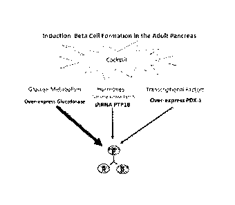
[043] FIG. 1. An example of an approach using an induction cocktail
comprising three
molecules to induce pancreatic beta cells formation in vivo in the adult
pancreas.
11
CA 02880691 2015-01-30
WO 2014/022455 PCT/US2013/052820
[044] FIG. 2A-2C. Illustrates the design and validation of Lentiviral
constructs, (A)
glucokinase, (B) Pdx-1, and (C) shRNA PTP1B.
[045] FIG. 3. In vivo over-expression of PDX-1 and GK, and suppression of
PTP1B
expression by the CNIP cocktail (Lenti-GCK + Lenti-Pdx-1 and Lenti-shRNA
PTP1B).
[046] FIG. 4. Quantitation of single beta cell staining in the adult
pancreatic tissue in
mice injected with a beta cell formation cocktail compared with a control
group injected with
placebo cocktail.
[047] FIG. 5. An example of a beta cell formation cocktail comprising three
molecules
(GK, PTP1B inhibitor, and Pdx - 1) induced proliferation in the adult mouse
pancreas
compared with the control adult mouse group injected with placebo.
[048] FIG. 6. Pancreatic beta cell mass was significantly increased in
adult mice
injected with a beta cell formation cocktail (GK, PTP1B inhibitor, and Pdx -
1) compared
with the control adult mice group injected with the placebo.
Immunofluorescence images of
insulin staining were captured using confocal microscopy. The beta cell and
total pancreatic
areas were quantified with Image J (NIH, Bethesda MD). Total beta cell mass
was calculated
as the total beta cell area expressed as a percentage of the total area of the
pancreas.
[049] FIG. 7. Illustrates the number of beta cell clusters in the pancreas
(cluster density)
in the adult mouse group injected with the beta cell formation cocktail
compared with the
control adult mouse group injected with the placebo. Cluster density was
determined as the
number of beta cell clusters divided by the total area of the pancreas.
[050] FIG. 8. Illustrates the fasting plasma insulin concentration in the
adult mouse
group injected with a beta cell formation cocktail (GK, PTP1B inhibitor, and
Pdx - 1)
compared with the adult mouse control group injected with placebo.
[051] FIG. 9 BrdU marker of proliferation in islets and exocrine tissue 4
weeks post-
injection of the cocktail (Lenti-GCK + Lenti-Pdx-1 + Lenti-shRNA PTP1B) or by
two
molecules or each molecule individually. The figure is representative of an
area of the
pancreas examined in ten sections per animal (n=3 to 4 for each group),
separated by 200 pm.
The results are expressed as the fold-increase in number of BrdU-labeled cells
compared with
12
MI
CA 02880691 2015-01-30
WO 2014/022455 PCT/US2013/052820
controls. Confocal laser microscopy was used for analysis. Lenti = Lentivirus;
GCK =
glucokinase; PTP1B = protein-tyrosine phosphatase 1B. Data are presented as
mean SE.
*=p<0.001 CNIP cocktail vs control; =p<0.05 CNIP cocktail vs [GK + PTP1B]; #
=p<0.01
[GK + PTP1B] vs control.
[052] FIG. 10 Beta cell mass 4-weeks post-injection with the cocktail CNIP
(Lenti-
GCK + Lenti-Pdx-1 + Lenti-shRNA PTP1B) or by two molecules or each molecule
individually compared to cocktail control and each other (n=3 to 4 for each
group). Total
pancreatic and insulin positive staining areas of each section were measured
using Image J
(NIH, Bethesda, USA). Beta cell mass was calculated as the ratio of total
insulin positive
area to total pancreatic area of all sections, multiplied by the pancreatic
tissue wet weight.
The figure is representative of an area of the pancreas examined in ten
sections per animal,
separated by 200 lam. Confocal laser microscopy was used for analysis. Lenti =
Lentivirus;
GCK = glucokinase; PTP1B = protein-tyrosine phosphatase 1B. *=p < 0.001 = CNIP
cocktail
vs control; p<0.001 CNIP cocktail vs [GK + Pdx-1]; p<0.001 CNIP cocktail vs
[PTP1B +
Pdx-1]; U =p<0.05 CNIP cocktail vs [GK + PTP1B]. # =p < 0.05 [GK + PTPIB] vs
[PTP1B + Pdx-1); p<0.05 [GK + PTP1B] vs control. Data are presented as mean
SE.
DESCRIPTION
[053] Certain methods described herein represent a concept that contradicts
the
scientific doctrine of one molecule to one cellular control process. In
certain aspects, the
methods include the integration of three levels of cellular physiology:
metabolism, membrane
receptor function, and gene transcription. The integration of multiple levels
of cellular
physiology produces a synergistic effect on beta cell formation. Synergy
requires that
multiple molecules work together to produce an effect that is greater than the
sum of their
individual effects. Using the synergistic approach described herein, the
inventors have
successfully induced pancreatic beta cell formation in the adult pancreas. The
ability to
generate beta cells in vivo in adult animals and humans provides a novel
therapeutic approach
for the treatment of subjects with type 1 and type 2 diabetes mellitus.
I. Methods of Treating Diabetes
[054] The inventors have demonstrated that, utilizing "Cellular Networking
Integration
& Processing" (CNIP), pancreatic beta cell formation can be increased in vivo
in adult
13
CA 02880691 2015-01-30
WO 2014/022455 PCT/US2013/052820
subjects. According to the CNIP approach, the inventors intervene at three
major levels in
cell processing: (1) first, at the level of intracellular carbohydrate
metabolism, (2) second, at
the level of the membrane receptor function, (3) third, at the level of gene
expression. By
targeting all three levels, one can generate a synergistic interaction that
induces beta cell
formation. The inventors refer to one example of this method as "Syner-III,"
with Syner
being the prefix from the Greek name synergos and III is Roman number three.
[055] The CNIP approach is designed to mimic the formation of beta cells in
adult
subjects and not to reprogram the cell at the stage of embryonic development.
The inventors
note that the cocktail of transcriptional factors used in stem cell research
or in the viral vector
cocktail used more recently in the mouse model (Zhou et al., Nature 455: 627-
32, 2008) are
used to generate beta cells by reproducing the embryonic stage of development.
In contrast,
the CNIP approach is designed to act in the adult state and utilizes a
mechanism that
integrates the three levels of cellular regulation to induce beta cell
formation.
[056] The methods described herein induce pancreatic beta cell formation in
vivo in
adult subjects without dedifferentiating cells to recapitulate the embryonic
pathway. The
CNIP approach specifically targets the post-embryonic induction of pancreatic
beta cell
formation without reproducing the embryonic formation process of the pancreas -
the
embryonic formation process leads to the generation of multiple pancreatic
endocrine cell
types. The ability to generate new beta cells in vivo in adult subjects can
provide a novel
therapeutic approach for the treatment of patients with type 1 and 2 diabetes
mellitus, as well
as other types of diabetes. The ability to increase the number of pancreatic
beta cells in adult
subjects can be therapeutic, prophylactic, and/or curative in regards to
diabetes.
[057] In certain embodiments, compositions and methods described herein can
be
applied to tissues other than the pancreas. In certain aspects, compositions
described herein
can be delivered into the gut endocrine K cells and be an able to form insulin-
like beta cell
that would secrete insulin in response to an elevation of blood glucose. In a
further aspect,
the compositions described herein can be delivered to the liver to induce
formation of beta
cells that respond to glucose. In still other aspects, the compositions
described herein can be
applied at the last step of stem cell differentiation and/or dedifferentiation
to form beta cells.
In certain aspects, the compositions described herein can be delivered to
various cells and/or
14
CA 02880691 2015-01-30
WO 2014/022455 PCT/US2013/052820
tissues in the body to form beta cells and therefore, are not limited to the
specific examples
described herein.
[058] In certain embodiments target cells are treated in vitro. Target
cells are those cells
that have the capability or can be induced to have the capability of forming
beta cells.
Methods for providing or obtaining such target cells are known in the art and
include either
providing tissue containing target cells and isolating the target cells by
methods known in the
art, e.g. with the help of cell surface specific antibodies and using a FACS
(cell sorter) or
cultivation of the cells under specific conditions allowing the growth of
target cells. In
certain aspects there are suitable target cell lines (Lieber et al., Int J
Cancer 15(5):741-47,
1975).
[059] Any cell being capable of differentiating into pancreatic beta cells
can be used as
a target cell of the method of the invention. This includes precursor cells
derived from
human or animal (e.g., mammal) tissue. In certain embodiments the target cell
is an
autologous target cell, i.e., it contains the same genetic information as
cells of the subject
being treated. In certain aspects the target cell has not been genetically
modified prior to the
treatment being administered. In certain aspects a target cell is selected
from the group
consisting of a pancreatic precursor cell, a small intestine precursor cell, a
liver precursor
cell, a precursor cell derived from the pancreatic duct population, precursor
of
neuroendocrine cell, and a pancreatic stem cell. This includes all somatic
differentiated cells
from a human or animal tissue. In certain aspects a target cell is selected
from the group
consisting of somatic differentiated cell from the liver, endocrine gut cell,
pancreatic duct
cell, exocrine and endocrine pancreatic cell, and neuroendocrine cell
[060] Once target cells have been obtained or provided, the cells can be
grown and
manipulated in an in vitro cell culture system, which includes standard cell
culture systems
like tissue culture dishes and 6-well, 24-well or 96-well plates. Culture
conditions will
depend on the target cell and the person skilled in the art will know how to
cultivate the cells.
A. Glucose Metabolism
[061] Glucose metabolism is the first aspect in the CNIP approach to
inducing beta cell
formation in the adult pancreas or other organs or tissues. Glucose is the
major energy source
CA 02880691 2015-01-30
WO 2014/022455 PCT/US2013/052820
utilized by the mammalian cell, and metabolism of glucose provides the energy
for cellular
function and proliferation (Bohnsack and Hirschi, Annu Rev Nutr. 24: 433-453,
2004).
Inhibition of glycolysis stops cell cycle progression, documenting the
necessity of glucose
metabolism to induce proliferation (Newcomb et al., Eukaryot. Cell. 2:143-149,
2003).
Factors that induce pancreatic beta cell formation in vivo include an increase
in glucose
metabolism (Bernard et al., FASEB J. 13:1195-1205, 1999; Alonso et al.,
Diabetes 56:1792-
1801, 2007). Glucose infusion in adult rats for a period of only 24 h
increased beta cell
number by ¨50% (Bernard et al., FASEB J. 13:1195-1205, 1999). Furthermore,
glucose
promotes beta cell survival by suppressing a constitutive apoptotic program in
vitro (Hoorens
et al., J. Clin. Invest. 98:1568-1574, 1996). Glucose metabolism primes the
pancreas for
induction of pancreatic beta cell formation.
[062] In certain aspects, the rate of glucose metabolism is increased by
providing a
nucleic acid encoding glucokinase, or increasing the activity of glucokinase
or other enzymes
or regulators. In certain embodiments the functions ascribed to the nucleic
acid described
herein can be provide by administering various chemical compounds or small
molecules that
increase glucose metabolism. Glucokinase activating compounds include, but are
not limited
to Roche Inc., compound R1440; Hoffinan-La Roche Inc., compound R00281675;
Hoffman-
La Roche Inc., compound R04389620 (Piragliatin); Eli Lilly Inc., compound
LY2121260;
OSI Pharmaceuticals, Inc., compound PSN-GKl; Astra-Zeneca, Inc., compound GKA-
50;
Pfizer Inc., glucokinase activators described in International Patent
publication
WO/2007122482); Merck-Banyu Inc., glucokinase activators described in
International
Patent publication WO/2003080585; Takeda Inc., glucokinase activators
described in
International Patent publication WO/200710434); Johnson & Johnson Inc.,
glucokinase
activator described in International Patent publication WO/2007075847); and
the like.
B. Receptor Tyrosine Kinases and Tyrosine-Kinase-Associated
receptors.
[063] Membrane receptor tyrosine kinase(s) and/or tyrosine-kinase-
associated receptors
are a second component of the CNIP approach to induce the formation of
pancreatic beta
cells in an adult subject. The second aspect in the generation of pancreatic
beta cells
following a physiological stimulus is for the cell to receive the message
through its
membrane receptors. The membrane receptors responsible for the stimulation of
pancreatic
beta cell mass are from the tyrosine kinase family of receptors and tyrosine-
kinase-associated
16
CA 02880691 2015-01-30
WO 2014/022455 PCT/US2013/052820
family of receptors. During pregnancy the pancreatic beta cell mass increases
in response to
the development of insulin resistance and increased fetal/placenta energy
demand and this
effect is mediated by increased prolactin, estrogen, and placental lactogen
secretion (Heit et
al., Annu. Rev Cell Dev. Biol. 22:311-338, 2006). The failure of the beta cell
to compensate
by augmenting its secretion of insulin leads to gestational diabetes. Islet
enlargement and
beta cell hyperplasia have been observed in autopsied pregnant humans (Van
Assche et al.,
Br. J. Obstet Gynaecol 85: 818-820, 1978). The hormonal stimuli (prolactin,
estrogen, and
placental lactogen) during pregnancy to increase the pancreatic beta cell mass
act through
tyrosine kinase associated receptors (Nielsen et al., Diabetes 50(Suppl. 1):
S25-S29, 2001).
Other hormones that increase beta cell mass also act through the tyrosine
kinase family of
receptors and include hepatocyte growth factor, platelet-derived growth
factor, growth
hormone, insulin, IGF-1 and EGF (Nielsen et al., Diabetes 50(Suppl. 1): S25-
S29, 2001). Of
note, the effect of these hormones on beta cell mass also relies on glucose
metabolism. In the
absence of glucose, the ability of hormones acting through the tyrosine kinase
family to
increase pancreatic beta cells mass is lost (Cousin et al., Biochem. J.
344:649-658, 1999).
Consequently, the CNIP approach combines the effect of glucose metabolism, and
membrane
tyrosine kinase(s) and/or tyrosine-kinase(s) associated receptors to induce
beta cell formation
in the adult pancreas.
[064] In certain embodiments the function(s) ascribed to the nucleic acids
described
herein can be provided by administering various chemical compounds or small
molecules
that increase tyrosine kinase receptor and/or tyrosine-kinase(s) associated
receptor activity
for beta cell formation in an adult pancreas. In certain aspects, inhibitory
nucleic acids such
as anti-sense DNA or inhibitory RNA molecules can be used. Such compounds
include, but
are not limited to PTP1B inhibitor compounds such as Wyeth Research Inc., 32D;
antisense
ISIS-PTP1BRX; Abbott Laboratories, Inc., Isoxazole; Abbott Laboratories, Inc.,
antisense
oligonucleotides designed to downregulate expression of PTP1B; Merck Frosst
Center for
Therapeutic Research, selective inhibitors of PTP1B compound 1 and 3; Incyte
Corporation,
Inc., (S)-isothiazolidinone ((S)-IZD) heterocyclic phosphotyrosine; Affymax,
Inc., triaryl
sulfonamide based PTP1B inhibitors; and the like.
[065] Other shRNA targeting tyrosine phosphatase family proteins can be
included
alone or in combination with shRNA PTP1B. PTP1B acts on the majority of the
tyrosine
17
CA 02880691 2015-01-30
WO 2014/022455 PCT/US2013/052820
kinase family of receptors that have been implicated in pancreatic beta cell
function in the
adult pancreas. However, T-cell protein tyrosine phosphatase (TCPTP) and SHP-
2, two other
members of intracellular protein phosphatase, have been shown to target
receptor tyrosine
kinases implicated insulin signaling (Tonks, Nat Rev. Mol Cell Biol 7:833-46,
2006). SHP-2
(SH2-domian containing phosphatase-2) is a ubiquitously expressed
intracellular protein
tyrosine phosphatase that contains two amino-terminal Src homology 2 (SH2)
domains.
SH2-2 binds to both the tyrosine-phosphorylated insulin receptor and IRS-1.
TCPTP exists
in two forms: an endoplasmic reticulum-targeted 48-kDa from (TC48) and a
nuclear 45-kDa
form (TC45). TC-PTP has been demonstrated to negatively regulate insulin
signaling and the
prolactin receptor (Aoki and Matsuda, J.Biol.Chem. 275:39718-26, 2000; Tonks,
Nat Rev.
Mol Cell Biol 7:833-46, 2006). Therefore, nucleic acids expressing shRNA-SHP-2
and
shRNA-TC-PTP can be used in the methods and compositions described herein.
C. Beta Cell Specific Transcription
[066] The third aspect in the CNIP approach is directed at the level of
gene expression
and involves a transcriptional activator or transcription factor, which is
utilized as an attractor
to converge and focus the glucose metabolism effect and metabolic/molecular
effects
generated by glucokinase, and the tyrosine kinase receptor(s) and tyrosine
kinase associated
receptor(s) to form beta cells in the adult pancreas. The term "transcription"
refers to the
process of copying a DNA sequence of a gene into an RNA product, generally
conducted by
a DNA-directed RNA polymerase using the DNA as a template. Every system has a
modulator attractor, like in physics. In a chaotic system, the direction of
the network
endpoint will follow the force of the attractor. From a simplistic view, the
impact target of a
projectile will depend on the initial force of propulsion combined with air
resistance and the
effect of gravity on the projectile. The initial forces, air resistance and
gravity, will act in
synergy as an attractor to determine the final destination of the projectile.
The living
organism is a nonlinear dynamic system that exists in a "chaotic" state. At
the transcriptional
level, the expression of a set of genes remains unchanged and those genes are
call
"housekeeping" genes. They carry out the routine functions of the cell,
whereas other classes
of genes are expressed in response to environmental stimuli. In the adaption
of pancreatic
beta cells to a physiological stress, upregulation of gene expression is
essential for the
induction of pancreatic beta cell formation (Bouwens and Rooman, Physiol Rev
85:1255-
18
CA 02880691 2015-01-30
WO 2014/022455 PCT/US2013/052820
1270, 2005). A key mediator of this adaptive response to a physiological
stress at the gene
expression level involves the activation of modular attractor(s) in the form
of transcription
factors (Albert and Barabasi, Rev Mod Physics 74:47-97, 2002; Albert and
Othmer, J Theor
Riot 223,1-18, 2003). Therefore, the CNIP approach includes transcription
factor(s) (as a
modular attractor) that have been implicated in the formation of beta cells in
the adult
pancreas in response to physiological stress.
[067] Pdx-1 overexpression alone could be used with a synergistic
convergence force of
other TFs to channel the CNIP gene expression pattern to induce pancreatic
beta cell
formation. Therefore, other TFs implicated in adult pancreatic beta cell
formation and that
can increase the effect of Pdx-1 on beta cell formation in vivo can be used in
the methods
described herein. In certain aspects, TFs implicated in beta cell formation
can be used. TFs
implicated in other endocrine cell formation can be excluded. The TFs
implicated in
pancreatic beta cell formation in the post-development period added in
combination with or
in place of Pdx-1 include: NeuroD, Is11, Nkx6.1, and Pax4. Anti-diabetic
compounds such as
anti-diabetic thiazolidinediones (e.g., troglitazone) can also be used in
conjunction with TFs
to increase beta cell formation. For example, troglitazone increases Pdx-1
expression in
mouse islets through the functional peroxisome proliferators-activated
receptor gamma
(PPARy) response element in the Pdx-1 promoter (Gupta et al., J Biol Chem.
283(47):32462-
70, 2008). Also contemplated is the induction of Pdx-1 via positive modulation
of the
PPARy response element in the promoter of the Pdx-1 gene.
D. Therapeutic Compositions
[068] In certain aspects, 1, 2, 3, or more of the therapeutic moieties
described herein can
be combined in one or more composition or administered in combination. In one
aspect, one
or more therapeutic moiety is provided as a cocktail of 1, 2, 3, or more
nucleic delivery
vector(s) and/or therapeutic agent(s). Such a cocktail can be administered
orally, locally, or
systemically as described herein.
[069] In a further aspect, 2, 3, or more of the therapeutic moieties can be
joined to create
a bi-valent, tri-valent, or tetra-valent composition. Such a composition can
be administered
orally, locally or systemically as described herein. In certain aspects, such
compositions are
administered orally. In other aspects the compositions are injected or infused
locally.
19
CA 02880691 2017-01-05
[070] In still a further aspect, 1, 2, 3, or more therapeutic moieties can
be joined in one
molecule by chemical adaptor systems. Such a composition can be administered
orally,
locally or systemically as described herein.
Nucleic Acid Compositions
[071] The term "nucleic acid vector" is used to refer to a carrier nucleic
acid molecule
into which a nucleic acid sequence can be inserted for introduction into a
cell where it can be
replicated, transcribed, and/or translated (i.e., expressed). A nucleic acid
sequence can be
"exogenous," which means that it is foreign to the cell into which the vector
is being
introduced or that the sequence is "endogenous" to the cell but in a position
within the host
cell in which the sequence is ordinarily not found. In certain aspects an
exogenous vector can
encode an endogenous nucleic acid. Nucleic acid vectors include plasmids,
cosmids, viral
genomes, and other expression vectors (bacteriophage, animal viruses, and
plant viruses),
artificial chromosomes (e.g., YACs), and the like. Given the current
disclosure, one of skill
in the art would be well equipped to construct a vector through standard
recombinant
techniques (see, for example, Maniatis et al., Molecular Cloning: A laboratory
Manual. Cold
Spring Harbor Laboratory, New York., 1989; Ausubel et al., Current Protocols
in Molecular
Biology, New York City, NY, John Wiley & Sons, Inc., 1994
).
[072] The term "expression vector" refers to any type of genetic construct
comprising a
nucleic acid coding for an RNA capable of being transcribed. In some cases,
RNA molecules
are then translated into a protein, polypeptide, or peptide. In other cases,
these sequences are
not translated, for example, in the production of inhibitory RNA, antisense
molecules, or
ribozymes. Expression vectors can contain a variety of "control sequences,"
which refer to
nucleic acid sequences necessary for the transcription and possibly
translation of an operably
linked coding sequence in a particular host cell. In addition to control
sequences that govern
transcription and translation, vectors and expression vectors may contain
nucleic acid
sequences that serve other functions as well and are described herein.
[073] Certain aspects involve the use of nucleic acids encoding beta cell
inducing
components. Examples of nucleic acids include GK as provided in SEQ ID NO:1
and SEQ
ID NO:4; PTB1B shRNA as provided in SEQ ID NO:2; and Pdx-1 as provided in SEQ
ID
CA 02880691 2015-01-30
WO 2014/022455 PCT/US2013/052820
NO:3; or the equivalent as would be recognized by one skilled in the art. In
certain aspects
the nucleic acid comprise a nucleotide sequence that is 80, 85, 90, 95, 98, or
100% identical
to SEQ ID NO:1, 2, 3, and/or 4. In certain embodiments, nucleic acids of the
invention
encode proteins that are 80, 85, 90, 95, 98, or 100% identical to the proteins
of SEQ ID NO:5
(GK) or SEQ ID NO:6 (Pdx-1) and maintain the appropriate activity.
[074] The sequences may be modified, given the ability of several different
codons to
encode a single amino acid, while still encoding for the same protein or
polypeptide.
Optimization of codon selection can also be undertaken in light of the
particular organism
used for expression.
A. Promoters and Enhancers
[075] A "promoter" is a control sequence that is a region of a nucleic acid
sequence at
which initiation and rate of transcription are controlled. It may contain
genetic elements at
which regulatory proteins and molecules may bind, such as RNA polymerase and
other
transcription factors, to initiate the specific transcription a nucleic acid
sequence. The
phrases "operatively positioned," "operatively linked," "under control," and
"under
transcriptional control" mean that a promoter is in a correct functional
location and/or
orientation in relation to a nucleic acid sequence to control transcriptional
initiation and/or
expression of that sequence.
[076] A promoter generally comprises a sequence that functions to position
the start site
for RNA synthesis. Additional promoter elements regulate the frequency of
transcriptional
initiation. Typically, these are located in the region 30-110 by upstream of
the start site,
although a number of promoters have been shown to contain functional elements
downstream
of the start site as well. To bring a coding sequence "under the control of" a
promoter, one
positions the 5' end of the transcription initiation site of the
transcriptional reading frame "
downstream'' of (i.e., 3' of) the chosen promoter. The "upstream" promoter
stimulates
transcription of the DNA and promotes expression of the RNA. The spacing
between
promoter elements frequently is flexible, so that promoter function is
preserved when
elements are inverted or moved relative to one another. Depending on the
promoter, it
appears that individual elements can function either cooperatively or
independently to
activate transcription. A promoter may or may not be used in conjunction with
an
21
CA 02880691 2017-01-05
"enhancer," which refers to a cis-acting regulatory sequence involved in the
transcriptional
activation of a nucleic acid sequence.
[077] A promoter may be one naturally associated with a nucleic acid
sequence, as may
be obtained by isolating the 5' non-coding sequences located upstream of the
coding segment
and/or exon. Such a promoter can be referred to as "endogenous" or
"homologous."
Similarly, an enhancer may be one naturally associated with a nucleic acid
sequence, located
either downstream or upstream of that sequence. Alternatively, certain
advantages will be
gained by positioning the nucleic acid under the control of a recombinant,
exogenous, or
heterologous promoter, which refers to a promoter that is not normally
associated with a
nucleic acid sequence in its natural environment. A recombinant or
heterologous enhancer
refers also to an enhancer not normally associated with a nucleic acid
sequence in its natural
environment. Such promoters or enhancers may include promoters or enhancers of
other
genes, and promoters or enhancers isolated from another virus, or prokaryotic
or eukaryotic
cell, and promoters or enhancers not "naturally occurring," i.e., containing
different elements
of different transcriptional regulatory regions, and/or mutations that alter
expression.
[078] Naturally, it will be important to employ a promoter and/or enhancer
that
effectively directs the expression of the DNA segment in the organelle, cell,
tissue, organ, or
organism chosen for expression. Those of skill in the art of molecular biology
generally
know the use of promoters, enhancers, and cell type combinations for protein
expression,
(see, for example Sambrook et al., Molecular Cloning: A Laboratory Manual,
vol. 1. 2nd
edition. Cold Spring Harbor Laboratory Press, 1989 ). The
promoters employed may be constitutive, tissue-specific, inducible, and/or
useful under the
appropriate conditions to direct high level expression of the introduced DNA
segment, such
as is advantageous in the large-scale production of recombinant proteins
and/or peptides.
The promoter may be heterologous or endogenous.
[079] Additionally any promoter/enhancer combination (as per, for example,
the
Eukaryotic Promoter Data Base EPDB, world-wide-web at epd.isb-sib.ch/) could
also be
used to drive expression. Use of a T3, T7, or SP6 cytoplasmic expression
system is another
possible embodiment. Eukaryotic cells can support cytoplasmic transcription
from certain
22
CA 02880691 2015-01-30
WO 2014/022455 PCT/US2013/052820
bacterial promoters if the appropriate bacterial polymerase is provided,
either as part of the
delivery complex or as an additional genetic expression construct.
[080] In certain aspects, a nucleic acid of the invention can comprise a
non-inducible or
inducible promoter that will be expressed specifically in the pancreatic
tissues. Such non-
inducible promoters include tissue-specific pancreas promoters from the
insulin gene,
glucagon gene, amylase gene, etc. Such inducible promoters include pancreas
specific
promoters under the control of the glucose response element or pancreas
specific promoter
under the control of a response element that is inducible by chemical,
peptide, ligand, or
metabolites.
B. Initiation Signals
[081] A specific initiation signal also may be required for efficient
translation of coding
sequences. These signals include the ATG initiation codon or adjacent
sequences.
Exogenous translational control signals, including the ATG initiation codon,
may need to be
provided. One of ordinary skill in the art would readily be capable of
determining this and
providing the necessary signals. It is well known that the initiation codon
must be "in-frame"
with the reading frame of the desired coding sequence to ensure translation of
the entire
insert. The exogenous translational control signals and initiation codons can
be either natural
or synthetic. The efficiency of expression may be enhanced by the inclusion of
appropriate
transcription enhancer elements.
C. Multiple Cloning Sites
[082] Vectors can include a multiple cloning site (MCS), which is a nucleic
acid region
that contains multiple restriction enzyme sites, any of which can be used in
conjunction with
standard recombinant technology to digest the vector. "Restriction enzyme
digestion" refers
to catalytic cleavage of a nucleic acid molecule with an enzyme that function
only at specific
locations in a nucleic acid molecule. Many of these restriction enzymes are
commercially
available. Use of such enzymes is widely understood by those of skill in the
art. Frequently,
a vector is linearized or fragmented using a restriction enzyme that cuts
within the MCS to
enable exogenous sequences to be ligated to the vector. "Ligation" refers to
the process of
forming phosphodiester bonds between two nucleic acid fragments, which may or
may not be
23
CA 02880691 2017-01-05
contiguous with each other. Techniques involving restriction enzymes and
ligation reactions
are well known to those of skill in the art of recombinant technology.
D. Termination Signals
[083] The vectors or constructs of the present invention will generally
comprise at least
one termination signal. A "termination signal" or "terminator" is comprised of
the DNA
sequences involved in specific termination of an RNA transcript by an RNA
polymerase.
Thus, in certain embodiments a termination signal that ends the production of
an RNA
transcript is contemplated. A terminator may be necessary in vivo to achieve
desirable
message levels.
[084] Terminators contemplated for use in the invention include any known
terminator
of transcription described herein or known to one of ordinary skill in the
art, including but not
limited to, the termination sequences such as bovine growth hormone terminator
or viral
termination sequences, such as the 5V40 terminator. In certain embodiments,
the termination
signal may be a lack of transcribable or translatable sequence, such as due to
a sequence
truncation.
E. Post-Transcriptional Regulatory Elements (PRE)
[085] Post-transcriptional regulation is the control of gene expression at
the RNA level,
i.e., between the transcription and the translation of the gene. In certain
aspects, the
Woodchuck Hepatitis Virus Post-transcriptional Regulatory Element (WPRE) is
used.
WPRE increases the levels of nuclear transcripts and facilitates RNA export.
WPRE may
facilitate other steps in RNA processing, directing RNAs that would normally
be degraded
within the nucleus to be efficiently expressed. The WPRE can also function to
facilitate the
generation of RNA-protein complexes that would protect newly synthesized
transcripts from
degradation in the nucleus. (Zufferey et al., Journal of Virology, 73: 2886-
2892, 1999 and US
Patent 6284469 ).
F. Polyadenylation Signals
[086] In expression, particularly eukaryotic expression, one will typically
include a
polyadenylation signal to effect proper polyadenylation of the transcript. The
nature of the
polyadenylation signal is not believed to be crucial to the successful
practice of the invention,
24
CA 02880691 2015-01-30
WO 2014/022455 PCT/US2013/052820
and any such sequence may be employed. Preferred embodiments include the SV40
polyadenylation signal or the bovine growth hormone polyadenylation signal,
convenient and
known to function well in various target cells. Polyadenylation may increase
the stability of
the transcript or may facilitate cytoplasmic transport.
G. Origins of Replication
[087] In order to propagate a vector in a host cell, it may contain one or
more origins of
replication sites (often termed "on"), which is a specific nucleic acid
sequence at which
replication is initiated. Alternatively an autonomously replicating sequence
(ARS) can be
employed if the host cell is yeast.
H. Selectable and Screenable Markers
[088] In certain embodiments of the invention, cells containing a nucleic
acid construct
of the present invention may be identified in vitro or in vivo by including a
marker in the
expression vector. Such markers would confer an identifiable change to the
cell permitting
easy identification of cells containing the expression vector. Generally, a
selectable marker is
one that confers a property that allows for selection. A positive selectable
marker is one in
which the presence of the marker allows for its selection, while a negative
selectable marker
is one in which its presence prevents its selection. An example of a positive
selectable
marker is a drug resistance marker.
[089] Usually the inclusion of a drug selection marker aids in the cloning
and
identification of transformants, for example, genes that confer resistance to
neomycin,
puromycin, hygromycin, DHFR, GPT, zeocin and histidinol are useful selectable
markers. In
addition to markers conferring a phenotype that allows for the discrimination
of
transformants based on the implementation of conditions, other types of
markers including
screenable markers such as GFP, whose basis is colorimetric analysis, are also
contemplated.
Alternatively, screenable enzymes such as herpes simplex virus thymidine
kinase (tk) or
chloramphenicol acetyltransferase (CAT) may be utilized. One of skill in the
art would also
know how to employ immunologic markers, possibly in conjunction with
fluorescence
assisted cell sorting (FACS) and/or immunohistochemistry. The marker used is
not believed
to be important, so long as it is capable of being expressed simultaneously
with the nucleic
CA 02880691 2015-01-30
WO 2014/022455 PCT/US2013/052820
acid encoding a gene product. Further examples of selectable and screenable
markers are
well known to one of skill in the art.
III. Polypeptide compositions
[090] Modifications and/or changes may be made in the amino acid
composition of
polypeptides, and thus the present invention contemplates variation in
sequences of the
polypeptides, and nucleic acids coding therefor, where they are nonetheless
able retain
substantial activity with respect to the therapeutic, preventative, and
curative aspects of the
present invention.
[091] The biological functional equivalent may comprise a polynucleotide
that has been
engineered to contain distinct sequences while at the same time retaining the
capacity to
encode the "wild-type" or standard peptide. This can be accomplished through
the
degeneracy of the genetic code, i.e., the presence of multiple codons, which
encode for the
same amino acids. In one example, one of skill in the art may wish to
introduce a restriction
enzyme recognition sequence into a polynucleotide while not disturbing the
ability of that
polynucleotide to encode a protein.
[092] In another example, a polynucleotide may encode a biological
functional
equivalent with more significant changes. Certain amino acids may be
substituted for other
amino acids in a protein structure without appreciable loss of interactive
binding capacity
with structures such as, for example, antigen-binding regions of antibodies,
binding sites on
substrate molecules, receptors, and such like. So-called "conservative"
changes do not
disrupt the biological activity of the protein, as the structural change is
not one that impinges
on the protein's ability to carry out its designed function. It is thus
contemplated by the
inventors that various changes may be made in the sequence of genes and
proteins disclosed
herein, while still fulfilling the goals of the present invention.
[093] In terms of functional equivalents, it is well understood by the
skilled artisan that,
inherent in the definition of a "biologically functional equivalent" protein
and/or
polynucleotide, is the concept that there is a limit to the number of changes
that may be made
within a defined portion of the molecule while retaining a molecule with an
acceptable level
of equivalent biological activity. Biologically functional equivalents are
thus defined herein
26
CA 02880691 2017-01-05
as those proteins (and polynucleotides) in selected amino acids (or
nucleotides) may be
substituted. In certain aspects, a polypeptide is 80, 85, 90, 92, 94, 96, 98,
or 100% identical
to the wildtype form of the polypeptide. In certain aspects, polypeptide(s)
80, 85, 90, 92, 94,
96, 98, or 100% identical to SEQ ID NO: 5 or 6 are used or nucleic acids
encoding the same.
[094] In general, the shorter the length of the molecule, the fewer changes
that can be
made within the molecule while retaining function. Longer domains may have an
intermediate number of changes. The full-length protein will have the most
tolerance for a
larger number of changes. However, it must be appreciated that certain
molecules or
domains that are highly dependent upon their structure may tolerate little or
no modification.
Function of a polypeptide can be determined by using various assays know to
detect the
activity of the polypeptide of interest.
[095] Amino acid substitutions are generally based on the relative
similarity of the
amino acid side-chain substituents, for example, their hydrophobicity,
hydrophilicity, charge,
size, and/or the like. An analysis of the size, shape and/or type of the amino
acid side-chain
substituents reveals that arginine, lysine, and/or histidine are all
positively charged residues;
that alanine, glycine, and/or serine are all a similar size; and/or that
phenylalanine,
tryptophan, and/or tyrosine all have a generally similar shape. Therefore,
based upon these
considerations, arginine, lysine, and/or histidine; alanine, glycine, and/or
serine; and/or
phenylalanine, tryptophan, and/or tyrosine are defined herein as biologically
functional
equivalents.
[096] To effect more quantitative changes, the hydropathic index of amino
acids may be
considered. Each amino acid has been assigned a hydropathic index on the basis
of their
hydrophobicity and/or charge characteristics, these are: isoleucine (+4.5);
valine (+4.2);
leucine (+3.8); phenylalanine (+2.8); cysteine/cystine (+2.5); methionine
(+1.9); alanine
(+1.8); glycine (-0.4); threonine (-0.7); serine (-0.8); tryptophan (-0.9);
tyrosine (-1.3);
proline (-1.6); histidine (-3.2); glutamate (-3.5); glutamine (-3.5);
aspartate (-3.5);
asparagine (-3.5); lysine (-3.9); and/or arginine (-4.5).
[097] The importance of the hydropathic amino acid index in conferring
interactive
biological function on a protein is generally understood in the art (Kyte &
Doolittle, 1982
). It is known that certain amino acids may be substituted for
27
CA 02880691 2015-01-30
WO 2014/022455 PCT/US2013/052820
other amino acids having a similar hydropathic index and/or score and/or still
retain a similar
biological activity. In making changes based upon the hydropathic index, the
substitution of
amino acids whose hydropathic indices are within 2 is preferred, those that
are within 1 are
particularly preferred, and/or those within 0.5 are even more particularly
preferred.
[098] It also is understood in the art that the substitution of like amino
acids can be
made effectively on the basis of hydrophilicity. As detailed in U.S. Patent
4,554,101, the
following hydrophilicity values have been assigned to amino acid residues:
arginine (+3.0);
lysine (+3.0); aspartate (+3.0 1); glutamate (+3.0 1); serine (+0.3);
asparagine (+0.2);
glutamine (+0.2); glycine (0); threonine (-0.4); proline (-0.5 1); alanine (-
0.5); histidine
(-0.5); cysteine (-1.0); methionine (-1.3); valine (-1.5); leucine (-1.8);
isoleucine (-1.8);
tyrosine (-2.3); phenylalanine (-2.5); tryptophan (-3.4). In making changes
based upon
similar hydrophilicity values, the substitution of amino acids whose
hydrophilicity values are
within 2 is preferred, those that are within 1 are particularly preferred,
and/or those within
0.5 are even more particularly preferred.
[099] In certain embodiments recombinant polypeptides as described herein
comprise
protein transduction domains. Protein transduction domains (PTDs) have the
ability to
translocate across biological membranes. The PTDs are relatively short (one-
to 35-amino
acid) sequences that confer this apparent translocation activity to proteins
and other
macromolecular cargo to which they are conjugated, complexed or fused. The HIV-
derived
TAT peptide (YGRKKRRQRRR (SEQ ID NO:7)), for example, has been used widely for
intracellular delivery of various agents ranging from small molecules to
proteins, peptides,
range of pharmaceutical nanocarriers and imaging agents. Alternatively,
receptor-mediated
endocytic mechanisms can also be used for intracellular drug delivery. For
example, the
transferrin receptor-mediated internalization pathway is an efficient cellular
uptake pathway
that has been exploited for site-specific delivery of drugs and proteins. This
is achieved
either chemically by conjugation of transferrin with therapeutic drugs or
proteins or
genetically by infusion of therapeutic peptides or proteins into the structure
of transferrin.
Naturally existing proteins (such as the iron-binding protein transferrin) are
very useful in
this area of drug targeting since these proteins are biodegradable, nontoxic,
and non-
immunogenic. Protein transduction domains include, but are not limited to,
PTDs derived
from proteins such as human immunodeficiency virus 1 (HIV-1) TAT (Ruben et
al., J. Virol.
28
CA 02880691 2015-01-30
WO 2014/022455 PCT/US2013/052820
63:1-8, 1989), e.g., GRKKRRQRRR (TAT 48-57, SEQ ID NO:8); the herpes virus
tegument
protein VP22 (Elliott and O'Hare Cell 88:223-33, 1997); the homeotic protein
of Drosophila
melanogaster Antennapedia (Antp) protein (Penetratin PTD; Derossi et al. J.
Biol. Chem.
271:18188-93, 1996); the protegrin 1 (PG-1) anti-microbial peptide SynB (e.g.,
SynBl,
SynB3, and Syn B4; Kokryakov et al. FEBS Lett. 327:231-36, 1993); and basic
fibroblast
growth factor (Jans FASEB J. 8:841-47, 1994). PTDs also include synthetic
PTDs, such as,
but not limited to, polyarginine peptides (Futaki et al., J. Mol. Recognit.
16:260-64, 2003;
Suzuki et al., J. Biol. Chem. 276:5836-40, 2001); transportan (Pooga et al.,
FASEB J. 12:67-
77, 1988; Pooga et al., FASEB J. 15:1451-53, 2001); MAP (Oehlke et al.,
Biochim. Biophys.
Acta. 1414:127-39, 1998); KALA (Wyman et al., Biochemistry 36:3008-17, 1997);
and other
cationic peptides, such as, for example, various 13-cationic peptides
(Akkarawongsa et al.,
Antimicrob. Agents and Chemother. 52(6):2120-29, 2008).
IV. Delivery Vectors
[0100] In certain aspects, components are provided to a pancreas or other
organ or tissue
by using nucleic acids that encode or express such components. Viral and non-
viral delivery
vectors can be used in the methods described herein (e.g., Syner-III). The
term "nucleic
acids", "nucleic acid molecules", "nucleic acid sequences", "nucleotide
sequences" and
"nucleotide molecules" are used interchangeably herein and, unless otherwise
specified, refer
to a polymer of deoxyribonucleic acids, including cDNA, DNA, PNA, or polymers
of
ribonucleic acids (RNA). Nucleic acid may be obtained from a cellular extract,
genomic or
extragenomic DNA, viral nucleic acids, or artificially/chemically synthesized
molecules. The
term can include double stranded or single stranded deoxyribonucleic or
ribonucleic acids.
A. Viral delivery
[0101] The ability of certain viruses to infect cells or enter cells via
receptor-mediated
endocytosis, and to express virally encoded genes have made them attractive
candidates for
the transfer of foreign nucleic acids into cells (e.g., mammalian cells).
Viruses may thus be
utilized that encode and express agents to increase the activity of glucose
metabolism,
increase tyrosine kinase receptor activity, and increase transcription of
genes associated with
beta cells. Non-limiting examples of virus vectors that may be used to deliver
nucleic acids
are described below.
29
CA 02880691 2017-01-05
[0102] Adenoviral Vectors. A particular method for delivery of the nucleic
acid involves
the use of an adenovirus expression vector. Although adenovirus vectors are
known to have
a low capacity for integration into genomic DNA, this feature is
counterbalanced by the high
efficiency of gene transfer afforded by these vectors. "Adenovirus expression
vector" is
meant to include those constructs containing adenovirus sequences sufficient
to (a) support
packaging of the construct and (b) to ultimately express a tissue or cell-
specific construct that
has been cloned therein. Knowledge of the genetic organization or adenovirus,
a 36 kb,
linear, double-stranded DNA virus, allows substitution of large pieces of
adenoviral DNA
with foreign sequences up to 7 kb (Grunhaus and Horwitz, Senfin. Virol. 3, 237-
252, 1992).
[0103] AAV Vectors. The nucleic acid may be introduced into the cell using
adenovirus-
assisted transfection. Increased transfection efficiencies have been reported
in cell systems
using adenovirus-coupled systems. Adeno-associated virus (AAV) has a low
frequency of
integration and it can infect non-dividing cells, thus making it useful for
delivery of genes
into mammalian cells in tissue culture or in vivo. AAV has a broad host range
for infectivity.
Details concerning the generation and use of rAAV vectors are described in
U.S. Patents
5,139,941 and 4,797,368.
[0104] Retroviral Vectors. Retroviruses have the ability to integrate their
genes into the
host genome, transferring a large amount of foreign genetic material,
infecting a broad
spectrum of species and cell types and of being packaged in special cell-
lines. In order to
construct a retroviral vector, a nucleic acid (e.g., one encoding a protein of
interest) is
inserted into the viral genome in the place of certain viral sequences to
produce a virus that is
replication-defective. In order to produce virions, a packaging cell line
containing the gag,
pol, and env genes but without the LTR and packaging components is
constructed. When a
recombinant plasmid containing a cDNA, together with the retroviral LTR and
packaging
sequences is introduced into a special cell line (e.g., by calcium phosphate
precipitation for
example), the packaging sequence allows the RNA transcript of the recombinant
plasmid to
be packaged into viral particles, which are then secreted into the culture
media. The media
containing the recombinant retroviruses is then collected, optionally
concentrated, and used
for gene transfer. Retroviral vectors are able to infect a broad variety of
cell types.
CA 02880691 2017-01-05
CA 02880691 2015-01-30
WO 2014/022455 PCT/US2013/052820
[0105] Lentiviruses are complex retroviruses, which, in addition to the
common retroviral
genes gag, poi, and env, contain other genes with regulatory or structural
function. Lentiviral
vectors are well known in the art (see, for example, U.S. Patents 6,013,516
and 5,994,136
). Some examples of lentivirus include the
Human Immunodeficiency Viruses: HIV-1, HIV-2 and the Simian Immunodeficiency
Virus:
Sly. Lentiviral vectors have been generated by multiply attenuating the HIV
virulence genes,
for example, the genes env, vif, vpr, vpu and nef are deleted making the
vector biologically
safe.
[0106] Recombinant lentiviral vectors arc capable of infecting non-dividing
cells and can
be used for both in vivo and ex vivo gene transfer and expression of nucleic
acid sequences.
For example, recombinant lentivirus capable of infecting a non-dividing cell
wherein a
suitable host cell is transfected with two or more vectors carrying the
packaging functions,
namely gag, pol and env, as well as rev and tat is described in U.S. Patent
5,994,136.
[0107] One may target the recombinant virus by linkage of an envelope
protein with an
antibody or a particular ligand for targeting to a receptor of a particular
cell-type. By
inserting a sequence (including a regulatory region) of interest into the
viral vector, along
with another gene that encodes the ligand for a receptor on a specific target
cell, for example,
the vector is now target-specific. Such viral vectors can be targeted to cells
of the pancreas.
[0108] Other Viral Vectors. Other viral vectors may be employed in the
methods of the
present invention. Vectors derived from viruses such as vaccinia virus
(Ridgeway, In:
Vectors: A survey of molecular cloning vectors and their uses, Rodriguez and
Denhardt
(Eds.), Stoneham: Butterworth, 467-492, 1988; Baichwal and Sugden, In: Gene
Tran.sfer,
Kucherlapati (Ed.), NY, Plenum Press, 117-148, 1986; Coupar et al., Gene 68:1-
10, 1988),
sindbis virus, cytomegalovirus, and herpes simplex virus may be employed. They
offer
several attractive features for various mammalian cells (Friedmann, Science,
244:1275-1281,
1989; Ridgeway, In: Vectors: A survey of molecular cloning vectors and their
uses,
Rodriguez and Denhardt (Eds.), Stoneham: Butterworth, 467-492, 1988; Baichwal
and
Sugden, In: Gene Transfer, Kucherlapati (Ed.), NY, Plenum Press, 117-148,
1986; Coupar et
al., Gene 68:1-10, 1988; Horwich et al., .1 Virol. 64:642-650, 1990).
31
CA 02880691 2015-01-30
WO 2014/022455 PCT/US2013/052820
[0109] Modified Viruses. A nucleic acid to be delivered may be housed
within an
infective virus that has been engineered to express or is coupled to a
specific binding ligand.
The virus particle will thus bind specifically to the cognate receptors of the
target cell and
deliver the contents to the cell. A novel approach designed to allow specific
targeting of
virus vectors was developed based on the chemical or genetic modification of a
virus by the
chemical addition or recombinant engineering of moieties to or in surface
proteins of the
virus. One such modification using lactose moieties can permit the specific
infection of
hepatocytes via sialoglycoprotein receptors.
[0110] Another approach to targeting of recombinant viruses was designed in
which
biotinylated antibodies against a viral surface protein and against a specific
cell receptor were
used. The antibodies can be coupled via biotin by using streptavidin (Roux et
al., Proc. Natl.
Acad. Sci. USA, 86:9079-83, 1989). Using antibodies against major
histocompatibility
complex class I and class II antigens, they demonstrated the infection of a
variety of human
cells that bore those surface antigens with an ecotropic virus in vitro (Roux
et al., Proc. Natl.
Acad. Sci. USA, 86:9079-83, 1989).
[0111] In certain aspects, an adaptor system can be used, i.e., a molecule
that binds both
the delivery vector and a target-pancreatic cell receptor to facilitate
transduction in the
pancreas. In a further aspect, use of a native viral vector receptor can be
fused to the
pancreas targeting ligand. In still another aspect, a bispecific antibody, two
antibodies
coupled together, can be coupled to the delivery vector resulting the delivery
vector having
specificity for the target pancreatic cells. In certain aspects, a targeting
moiety can be bound
to the delivery vector by chemical means. In further aspects, an antibody that
binds to a
genetically incorporated Ig-binding domain of the delivery vector can be used
to enhance
delivery. In further aspects, small targeting motifs can be inserted into the
capsid, envelope,
viral attachment, other viral surface protein to target the pancreatic cells.
[0112] In certain aspects, a viral construct can encode for two
heterologous protein
components (e.g., GK and Pdx-1) and express an shRNA PTP1B. In a further
aspect, each
component can be comprised in individual and separate viruses.
[0113] The compositions described herein can be delivered via the
pancreatic duct
through endoscopy. Briefly, the patient is sedated or anaesthetized, and a
flexible endoscope
32
CA 02880691 2015-01-30
WO 2014/022455 PCT/US2013/052820
is inserted through the mouth, down the esophagus, into the stomach, through
the pylorus into
the duodenum where the ampulla of Vater (the opening of the common bile duct
and
pancreatic duct) exists. The sphincter of Oddi is a muscular valve that
controls the opening
of the ampulla. The region can be directly visualized with the endoscopic
camera while
performing the procedure. A plastic catheter or cannula is inserted through
the ampulla, and
the beta cell inducing composition (e.g., Syner-III) is introduced into the
pancreatic bile duct
to target the pancreas. The beta cell forming composition can also be
delivered intravenously
by coupling a viral vector with a pancreas targeting moiety, e.g, modifying
the envelop
structure of viral vector to target only the pancreatic tissues.
B. Lipid-Mediated Transfection
[0114] In a further embodiment, a nucleic acid may be entrapped in a lipid
particle such
as, for example, a liposome. Liposomes are vesicular structures characterized
by a
phospholipid bilayer membrane and an inner aqueous medium. Multilamellar
liposomes
have multiple lipid layers separated by aqueous medium. They form
spontaneously when
phospholipids are suspended in an excess of aqueous solution. The lipid
components
undergo self-rearrangement before the formation of closed structures and
entrap water and
dissolved solutes between the lipid bilayers (Ghosh and Bachhawat, In: Liver
Diseases,
Targeted Diagnosis and Therapy Using Specific Receptors and Ligands, Wu et al.
(Eds.),
Marcel Dekker, NY, 87-104, 1991). Also contemplated is a nucleic acid
complexed with
Lipofectamine (Gibco BRL) or Superfect (Qiagen).
[0115] Lipid-mediated nucleic acid delivery and expression of foreign DNA
in vitro has
been very successful (Nicolau and Sene, Biochim. Biophys. Acta, 721:185-190,
1982; Fraley
et al., Proc. Natl. Acad. Sci. USA, 76:3348-3352, 1979; Nicolau et al.,
Methods Enzymol.
149:157-176, 1987). The feasibility of lipid-mediated delivery and expression
of foreign
DNA in cultured chick embryo, HeLa and hepatoma cells has also been
demonstrated (Wong
et al., Gene 10:87-94 1980).
[0116] In certain embodiments of the invention, a lipid particle may be
complexed with a
hemagglutinating virus (HVJ). This has been shown to facilitate fusion with
the cell
membrane and promote cell entry of lipid-encapsulated DNA (Kaneda et al.,
Science
243:375-378 1989). In other embodiments, a lipid particle may be complexed or
employed
33
CA 02880691 2015-01-30
WO 2014/022455 PCT/US2013/052820
in conjunction with nuclear non-histone chromosomal proteins (HMG-1) (Kato et
al., J. Biol.
Chem. 266:3361-3364, 1991). In yet further embodiments, a lipid particle may
be complexed
or employed in conjunction with both HVJ and HMG-1. In other embodiments, a
delivery
vehicle may comprise a ligand and a lipid particle.
[0117] In certain aspects, components described herein, including shRNA of
PTP1B and
nucleic acids encoding Pdx-1 and glucokinase can be delivered by liposomes.
Liposomes
containing the nucleic acids could be delivered intravenously and liberated
when it reaches
the pancreatic tissues. In certain aspects, nucleic acids of the invention are
incorporated into
liposomes that contain chemically coupled ligands that are presented on the
liposome surface.
The ligands specifically target pancreatic cells. Using this strategy, a
variety of ligands or
receptors, such as antibodies, growth factors, cytokines, hormones, and
toxins, can be
anchored on liposome surface so that the beta cell inducing components can be
targeted to
and introduced into pancreatic cells.
C. Receptor Mediated Transfection
[0118] Still further, a nucleic acid may be delivered to a target cell via
receptor-mediated
delivery vehicles. These take advantage of the selective uptake of
macromolecules by
receptor-mediated endocytosis that will be occurring in a target cell. In view
of the cell type-
specific distribution of various receptors, this delivery method adds another
degree of
specificity to the present invention.
[0119] Certain receptor-mediated gene targeting vehicles comprise a cell
receptor-
specific ligand and a nucleic acid-binding agent. Others comprise a cell
receptor-specific
ligand to which the nucleic acid to be delivered has been operatively
attached. Several
ligands have been used for receptor-mediated gene transfer (Wu and Wu, J.
Biol. Chem.
262:4429-4432, 1987; EP patent 0273085), which establishes the operability of
the
technique. In certain aspects of the present invention, a ligand will be
chosen to correspond
to a receptor specifically expressed on the target cell population.
[0120] In other embodiments, a nucleic acid delivery vehicle component of a
cell-specific
nucleic acid targeting vehicle may comprise a specific binding ligand in
combination with a
lipid particle. The nucleic acid(s) to be delivered are housed within the
lipid particle and the
34
CA 02880691 2017-01-05
CA 02880691 2015-01-30
WO 2014/022455
PCT/US2013/052820
specific binding ligand is functionally incorporated into the lipid layer. The
lipid particle will
thus specifically bind to the receptor(s) of a target cell and deliver the
contents to a cell. Such
systems have been shown to be functional using systems in which, for example,
epidermal
growth factor (EGF) is used in the receptor-mediated delivery of a nucleic
acid to cells that
exhibit upregulation of the EGF receptor.
[0121] In still
further embodiments, the nucleic acid delivery vehicle component of a
targeted delivery vehicle may be a lipid particle itself, which will
preferably comprise one or
more lipids or glycoprotcins that direct cell-specific binding. For example,
lactosyl-
ceramidc, a galactose-terminal asialgangliosidc, have been incorporated into
lipid particles
and observed to increase the uptake of the insulin gene by hepatocytes
(Nicolau et al.,
Methods Enzymol. 149:157-176, 1987). It is
contemplated that the tissue-specific
transforming constructs of the present invention can be specifically delivered
into a target cell
in a similar manner or as described herein.
D. Microproj ecti le Bombardment
[0122]
Microprojectile bombardment techniques can be used to introduce a nucleic acid
into at least one, organelle, cell, tissue or organism (U.S. Patents
5,550,318; 5,538,880;
5,610,042; and PCT Application WO 94/09699
). This method depends on the ability to accelerate DNA-coated
microprojectiles to
a high velocity allowing them to pierce cell membranes and enter cells without
killing them
(Klein et al., Nature 327, 70-73, 1987). There are a wide variety of
microprojectile
bombardment techniques known in the art, many of which are applicable to the
invention.
[0123] In this
microprojectile bombardment, one or more particles may be coated with at
least one nucleic acid and delivered into cells by a propelling force. Several
devices for
accelerating small particles have been developed. One such device relies on a
high voltage
discharge to generate an electrical current, which in turn provides the motive
force. The
microprojectilcs used have consisted of biologically inert substances such as
tungsten or gold
particles or beads. Exemplary particles include those comprised of tungsten,
platinum, and
gold. It is contemplated that in some instances DNA precipitation onto metal
particles would
not be necessary for DNA delivery to a recipient cell using microprojectile
bombardment.
However, it is contemplated that particles may contain DNA rather than be
coated with DNA.
CA 02880691 2015-01-30
WO 2014/022455 PCT/US2013/052820
DNA-coated particles may increase the level of DNA delivery via particle
bombardment but
are not, in and of themselves, necessary.
[0124] Naked plasmid DNA can be transferred into a cell by air pistol (Gene
Gun).
Syner-III plasmid could be injected via the pancreatic duct using the gene gun
approach with
endoscopy to reach the pancreatic tissues. The plasmid DNA bombardments
through a gene
gun enter the cell by physical pressure that opens membrane pores and/or by
facilitating
diffusion of the naked plasmid DNA though the cell membrane.
E. Nanoparticles
[0125] Nucleic acids of the invention can be incorporated into three-
dimensional, multi-
component structures of nanoparticles that target pancreatic or other tissues.
The
nanoparticles used include, but are not limited to liposomes, polymers,
proteins, micelles,
dendimers, quantum dots, nanoshells, nanocyrstals, gold nanoparticles,
paramagnetic
nanoparticles, and carbon nanotubes.
F. Hydrodynamic Gene Delivery
[0126] Naked plasmid DNA can be delivered via the pancreatic duct employing
hydrodynamic gene delivery. A balloon catheter can be placed in the pancreatic
duct. The
balloon catheter placed in the pancreatic duct is inflated for occlusion-
assisted infusion.
G. Electroporation
[0127] In certain aspects, a pair of electrodes can be placed on or in
pancreatic or other
tissues, and nucleic acids of the invention are deposited on the electrodes so
that the genetic
material is transferred into the tissues.
H. Ultrasound-Facilitated Gene Transfer
[0128] Nucleic acids of the invention can be incorporated into microbubbles
that are
injected intravenously. When the microbubbles reach the pancreas or other
tissue they are
targeted with ultrasound. As the micorbubbles expand and burst, they release
the nucleic
acids in the pancreas. The local shock waves cause nucleic acids to permeate
the nearby cell
membranes.
36
CA 02880691 2015-01-30
WO 2014/022455 PCT/US2013/052820
I. Gene Transfer by Needle
[0129] In certain aspects, nucleic acids of the invention are injected
directly into the
pancreas or other tissue using a needle.
V. Pharmaceutical Compositions
[0130] In light of the current specification, the determination of an
appropriate treatment
regimen (e.g., dosage, frequency of administration, systemic vs. local, etc.)
is within the skill
of the art. For administration, the components described herein will be
formulated in a unit
dosage form (solution, suspension, emulsion, etc.) in association with a
pharmaceutically
acceptable carrier. Such vehicles are usually nontoxic and non-therapeutic.
Examples of
such vehicles are water, saline, Ringer's solution, dextrose solution, and
Hank's solution.
Non-aqueous vehicles such as fixed oils and ethyl oleate may also be used. A
preferred
vehicle is 5% (w/w) human albumin in saline. The vehicle may contain minor
amounts of
additives, such as substances that enhance isotonicity and chemical stability,
e.g., buffers and
preservatives.
[0131] The therapeutic compositions described herein, as well as their
biological
equivalents, can be administered independently or in combination by any
suitable route.
Examples of parenteral administration include intravenous, intraarterial,
intramuscular,
intraperitoneal, and the like. The routes of administration described herein
are merely an
example and in no way limiting.
[0132] The dose of the therapeutic compositions administered to an animal,
particularly
in a human, in accordance with embodiments of the invention, should be
sufficient to result
in a desired response in the subject over a reasonable time frame. It is known
that the dosage
of therapeutic compositions depends upon a variety of factors, including the
strength of the
particular therapeutic composition employed, the age, species, condition or
disease state, and
the body weight of the animal.
[0133] Moreover, dose and dosage regimen, will depend mainly on the type of
biological
damage to the host, the type of subject, the history of the subject, and the
type of therapeutic
composition being administered. The size of the dose will be determined by the
route, timing
and frequency of administration as well as the existence, nature and extent of
any adverse
37
CA 02880691 2015-01-30
WO 2014/022455 PCT/US2013/052820
side effects that might accompany the administration of a particular
therapeutic composition
and the desired physiological effect. It is also known that various conditions
or disease
states, in particular, chronic conditions or disease states, may require
prolonged treatment
involving multiple administrations.
[0134] Therefore, the amount of the therapeutic composition must be
effective to achieve
an enhanced therapeutic index. If multiple doses are employed, the frequency
of
administration will depend, for example, on the type of subject. One skilled
in the art can
ascertain upon routine experimentation the appropriate route and frequency of
administration
in a given subject that are most effective in any particular case. Suitable
doses and dosage
regimens can be determined by conventionally known range-finding techniques.
Generally,
treatment is initiated with smaller dosages, which are less than the optimal
dose of the
compound. Thereafter, the dosage is increased by small increments until the
optimal effect
under the circumstances is obtained.
[0135] The therapeutic compositions for use in embodiments of the invention
generally
include carriers. These carriers may be any of those conventionally used and
are limited only
by the route of administration and chemical and physical considerations, such
as solubility
and reactivity with the therapeutic agent. In addition, the therapeutic
composition may be
formulated as polymeric compositions, inclusion complexes, such as
cyclodextrin inclusion
complexes, liposomes, microspheres, microcapsules, and the like, without
limitation.
[0136] The pharmaceutically acceptable excipients described herein, for
example,
vehicles, adjuvants, carriers, or diluents, are well known and readily
available. It is preferred
that the pharmaceutically acceptable carrier be one which is chemically inert
with respect to
the therapeutic composition and one that has no detrimental side effects or
toxicity under the
conditions of use.
[0137] The choice of excipient will be determined, in part, by the
particular therapeutic
composition, as well as by the particular method used to administer the
composition.
Accordingly, there are a wide variety of suitable formulations of the
pharmaceutical
composition used in the embodiments of the invention. For example, the non-
limiting
formulations can be injectable formulations such as, but not limited to, those
for intravenous,
subcutaneous, intramuscular, intraperitoneal injection, and the like, and oral
formulations
38
CA 02880691 2015-01-30
WO 2014/022455 PCT/US2013/052820
such as, but not limited to, liquid solutions, including suspensions and
emulsions, capsules,
sachets, tablets, lozenges, and the like. Non-limiting formulations suitable
for parenteral
administration include aqueous and non-aqueous isotonic sterile injection
solutions, including
non-active ingredients such as antioxidants, buffers, bacteriostats,
solubilizers, thickening
agents, stabilizers, preservatives, surfactants, and the like. The solutions
can include oils,
fatty acids, including detergents and the like, as well as other well known
and common
ingredients in such compositions, without limitation.
VI. Examples
[0138] The following examples as well as the figures are included to
demonstrate certain
embodiments of the invention. It should be appreciated by those of skill in
the art that the
techniques disclosed in the examples or figures represent techniques
discovered by the
inventors to function well in the practice of the described methods, and thus
can be
considered to constitute modes for its practice. However, those of skill in
the art should, in
light of the present disclosure, appreciate that many changes can be made in
the specific
embodiments which are disclosed and still obtain a like or similar result
without departing
from the spirit and scope of the invention.
EXAMPLE
BETA CELL FORMATION IN ADULT PANCREAS
A. Results
[0139] For illustration purposes, the therapeutic moieties include
Lentivirus-CMV-
Glucokinase (GK), Lentivirus-H1-shRNA PTP1B; and Lentivirus-CMV-PDX-1. The
control
composition includes Lentivirus-CMV-GFP and Lentivirus-H1-shRNA Scramble
injected at
the same concentration as the therapeutic composition. Four-weeks post-
injection in vivo
over-expression of PDX-1 and GK, and suppression of PTP1B expression was
detected in the
mouse pancreas. Glucokinase over-expression was detected in the islets and in
exocrine
tissues. Expression of glucokinase in the exocrine tissues confirms the over-
expression of the
glucokinase by the therapeutic composition since pancreatic tissues only
express glucokinase
in endocrine cells. Pdx-1 over expression is detected by using a c-Myc tag
incorporated into
the cDNA of Pdx-1 to differentiate between exogenous and endogenous
expression. shRNA
39
CA 02880691 2015-01-30
WO 2014/022455 PCT/US2013/052820
PTP1B co-expressing GFP was also detected. Protein expression was confirmed by
western
blot.
[0140] First Aspect of CNIP Approach: One goal of the CNIP approach is to
increase
glucose metabolism in the pancreas to induce pancreatic beta cell formation.
For glucose to
enter the glycolytic pathway, it first must enter the intracellular space
through a membrane
glucose transport system. The glucose transporters, Glut 1 and Glut 3, are
present in exocrine
and endocrine human pancreatic tissues, and Glut 2 is present in the
pancreatic beta cell
(Coppieters et al., Diabetes Metab Res Rev 27: 746-754, 2011). However,
glucose transport
is not the rate-limiting step for glucose entry into the glycolytic pathway
(Wasserman et al.,
J.Exp.Biol. 214:254-262, 2011). The first rate-limiting step for glucose
metabolism in the
glycolytic pathway is at the level of glucose phosphorylation by hexokinase.
The
phosphorylation of glucose by hexokinase produces the metabolite glucose 6-
phosphate (G-
6-P). The pancreatic beta cells contain a hexokinase type IV, named
glucokinase (GK).
Glucokinase is not allosterically inhibited by the accumulation of
intracellular G-6-P, in
contrast to other hexokinases. Therefore, for glucokinase, the amount and
activity of the
glucokinase enzyme regulates the rate of glucose flux through glycolysis. For
other members
of the hexokinase family, product inhibition by G-6-P is the key regulator of
enzyme activity
and, therefore, glucose entry into the glycolytic pathway. If glucose is not
phosphorylated by
hexokinase, it cannot undergo further metabolism and cannot generate any
signal to the
transcriptional machinery to induce gene expression (Doiron et al., J Biol
Chem. 269: 10213-
10216, 1994 and J Riot Chem. 271:5321-5324, 1996). Therefore, in certain
aspects
glucokinase can be included in a CNIP cocktail to induce pancreatic beta cell
formation.
[0141] The inventors designed a lentiviral vector construct expressing the
glucokinase
gene under control of the cytomegalovirus (CMV) promoter (FIG. 2A). The
lentiviral vector
construct included a posttranscriptional regulatory element of woodchuck
hepatitis virus
(WPRE) at the 3' untranslated region of coding sequence, which increased the
level of
expression of the transgene. WPRE functions within the nucleus to stimulate
gene
expression posttranscriptionally by increasing the levels of nuclear
transcripts and greatly
increasing the RNA half-life (Zufferrey et al., J. Virol. 73:2886-289, 1999).
The mouse
glucokinase (GCK) gene was subcloned to pEntCMV-WPRE vector and the insert was
verified by DNA sequencing. The pENT-GCK was treated with LR Clonase II enzyme
CA 02880691 2015-01-30
WO 2014/022455 PCT/US2013/052820
(Invitrogen) and ligated to a pLenti vector. The recombination product was
transformed into
E. coli cells. After overnight incubation, the positive clones were selected,
and plasmid DNA
was purified. The pEnt-GCK and pLenti-GCK were transfected into 293 cells. 48
hours
after transfection, the cells were lysed in SDS-PAGE buffer and subjected to 4-
20% SDS-
PAGE gel electrophoresis and analyzed by Western blotting. The Western blot
was carried
out using the anti-GCK antibody at a 1:1000 dilution, followed by a HRP
conjugated
secondary antibody. The Western blot membrane was developed using ECL
reagents.
[0142] The pLenti-GCK is used for the production of pure, high titer
lentiviral vector.
The Lenti-GCK will be injected directly into the pancreas of adult mice as
described above.
Adult mice C57BL/6 (8 weeks old; from Charles River, Wilmington, MA) will be
used for
the in vivo experiment targeting the pancreas with lentiviral vector
construct.
[0143] Increased glucokinase expression with a plasmid vector has been
shown to
increase the glucose-6-phosphate (G-6-P) pool (Doiron et al., J Riot Chem.
269: 10213-
10216, 1994). Glucose 6-phosphate is a key intermediate that sits at the
junction of several
metabolic pathways (glycolysis, gluconeogenesis, pentose phosphate pathway,
glycogenesis
and glycogenolysis). Doiron et al (1996) demonstrated that the glucose signal
to the
transcriptional machinery is mediated by xylulose 5-phosphate, which is a
metabolite
produced by the pentose phosphate pathway. As demonstrated by Doiron, xylulose
5-
phosphate is the major metabolite responsible for mediating transcriptional
machinery
induction by glucose metabolism. The key enzyme that regulates metabolic flux
through the
pentose phosphate pathway is glucose-6-phosphate dehydrogenase (G6PD). G-6-P
enters the
pentose phosphate pathway through the action of G6PD enzyme. Therefore, in the
development of our CNIP approach, the combination of Lenti-CGK and Lenti-G6PD
to
channel glucose 6-phosphate into the pentose phosphate pathway to enhance
xylulose-5-
phosphate formation that mediates the transcriptional effects of glucose
metabolism can be
used. Another important enzyme in the formation of xylulose 5-phosphate is
transketolase
(TK), which is located further down the pentose phosphate pathway. Therefore,
an
alternative approach will be to combine Lenti-TK with Lenti-CGK to increase
the effect of
glucose metabolism regulated gene expression. The synergistic action of all
molecules
(Lenti-CGK, Lenti-G6PD and Lenti-TK) can be used to activate the
transcriptional
machinery under the control of glucose metabolism and increase beta cell
formation.
41
CA 02880691 2015-01-30
WO 2014/022455 PCT/US2013/052820
[0144] Second Aspect of CNIP Approach: The second aspect of the CNIP
approach
includes increasing membrane receptor tyrosine kinase activity and tyrosine
kinase associated
receptor activity. Glucose metabolism is a major mechanism involved in the
formation of
beta cells in the adult pancreas. Another system implicated in beta cell
formation is ligand
binding to tyrosine kinase receptor(s). (Vasavada et at., Int J Biochem Cell
Biol. 38:931-950,
2006). The increase in pancreatic beta cell mass in response to physiological
stress (i.e.
pregnancy) is mediated by growth hormone, prolactin, and placental lactogen
working
through the prolactin receptor. The prolactin receptor does not have intrinsic
tyrosine kinase
activity but it interacts with members of the Janus kinase family of tyrosine
kinases.
Prolactin receptor is part of the family of tyrosine kinase associated
receptor(s). The Janus
kinase family of tyrosine kinases is responsible for the increase in beta cell
mass in response
to hormonal changes that occur in pregnancy (Vasavada et at., Int J Biochem
Cell Biol.
38:931-950, 2006). The action of insulin, insulin-like growth factors,
hepatocyte growth
factor, and epidermal growth factor also increase beta cell mass by activating
a membrane
bound tyrosine kinase receptor (Vasavada et at., Int J Biochem Cell Biol.
38:931-950, 2006).
Therefore, lactogens (including growth hormone, prolactin and placental
lactogen), insulin,
insulin-like factors, hepatocyte growth factor and epidermal growth factor
receptor require
the activation of tyrosine kinase(s) to increase pancreatic beta cells mass.
[0145] Protein-tyrosine phosphatase 1B (PTP1B) has been shown to inhibit
the ability of
insulin, insulin-like growth factors receptor, prolactin and hepatocyte growth
factor to
activate tyrosine kinase(s) (Aoki and Matsuda, J. Biol. Chem. 275:39718-39726,
2000;
Kakazu et al., Invest Opthalmol Vis Sci. 49:2927-2935, 2008; Tonks, Nat Rev.
Mol Cell Riot
7:833-46, 2006). Consequently, one molecule that can be included in a CNIP
cocktail is an
inhibitor or inhibitory RNA (e.g., shRNA) targeting PTP1B. The suppression of
PTP1B
protein production by shRNA will increase receptor tyrosine kinase activity
involved in beta
cell formation in the adult pancreas in response to binding of a physiological
concentration of
the corresponding hormone. The shRNA PTP1B has been introduced into a
lentivirus
construct using the same method described above for lenti-CGK under the
control of the H1
polymerase promoter (Doiron et al., Diabetologia 55:719-728, 2012) (FIG. 2C).
The
polymerase III promoter H1 is active ubiquitously in all cells, because of the
housekeeping
42
CA 02880691 2015-01-30
WO 2014/022455 PCT/US2013/052820
function of polymerase III. The lenti-shRNA-PTP1B was provided to the pancreas
as
described above.
[0146] Lentivirus shRNA PTP1B. A small hairpin RNAi was constructed into
pEQU6
vector. The target sequences are 20 nt in length (GCCAGGACATTCGACATGAA SEQ ID
NO:2). The shRNAi sequences were verified by DNA sequencing.
[0147] Co-transfection experiments were performed using the target gene
expression
plasmid pEnt-PTP1B and one pEQ-PTP1B-shRNA vector. The chart below describes
the
component of each reaction. 48 hours after transfection, cells were lysed in
SDS-PAGE
buffer and subjected to 4-20% SDS-PAGE gel electrophoresis and Western blot
analyses.
The Western blot was carried out using the anti-PTP1B antibody at a 1:1000
dilution. The
membrane was evaluated using ECL reagents. For each 6-well:
293 cells PTP1B-shRNA
mouse PTP1B cDNA 1.0
pEQ- PTP1B shRNA 2.0
pEQ-scramble-
3.0
shRNA
Total DNA 3.0 3.0
[0148] Third Aspect of CNIP Approach: A third aspect that can be included
in the
methods described herein includes increasing gene expression through
transcriptional
factor(s) to integrate the effect of glucose metabolism (Aspect 1) and
stimulation of tyrosine
kinase receptor family activity (Aspect 2) to induce beta cell formation in
the pancreas.
[0149] Different models have been proposed to explain the facilitated
diffusion of
transcriptional factors to bind to their target DNA sequence into the nucleus.
One model
proposes that the transcriptional factor (TF) binds the DNA by facilitated
diffusion. First, the
TF interacts with the DNA randomly at a non-specific site. After initial TF
interaction with
the DNA molecules, by facilitated diffusion, the TF moves from its initial non-
specific site to
its target sequence by 'sliding' along the DNA. As the TF rolls along the DNA
and finds its
corresponding binding site, it induces activation or inhibition of the
transcriptional
machinery. Irrespective of the model proposed to explain how the TF reaches
its DNA
binding site, all proposed models and experiments include facilitated
diffusion with random
movement of the TF to find its site of activation or inhibition in the
transcriptional
43
CA 02880691 2015-01-30
WO 2014/022455 PCT/US2013/052820
machinery. One action of glucose metabolism signaling to the transcriptional
machinery is to
increase the probability of the TF binding the DNA molecule (Doiron et al., J
Riot Chem.
271:5321-5324, 1996).
Indeed, glucose metabolism signaling to the transcriptional
machinery induces TF expression and TF translocation from the cytoplasm to the
nucleus
turns on glucose-induced genes (Doiron et al., J Riot Chem. 271:5321-5324,
1996).
Consequently, the biophysical mechanism by which TFs increase glucose-induced
gene
expression depends on the TF quantity level present in the nucleus. Indeed,
when the
quantity of TFs in the nucleus increases, the chances of interacting with DNA
molecules to
randomly find its specific binding site increased (Mitanchez et al., Endo Rev
18:520-540,
1997). Increasing expression of genes implicated in beta cell formation in the
adult pancreas
can be accomplished or enhanced by overexpressing key TFs that activate the
beta cell
formation pathway.
[0150] One
of the major transcriptional factors implicated in beta cell formation after
birth is Pdx-1 (Doiron and DeFronzo, Int J Endocrinol Metab. 9:356-357, 2011).
Pdx-1 has
distinct effects before and after birth in the pancreas (Doiron and DeFronzo,
Int J Endocrinol
Metab. 9:356-357, 2011). At the embryonic stage, Pdx-1 is essential for
pancreatic
development and it is expressed in endocrine and exocrine tissues. However,
after birth, Pdx-
1 is expressed only in pancreatic beta cells and somatostatin cells.
Therefore, in the adult
pancreas, Pdx-1 does not induce embryonic pancreatic development but it does
induce
pancreatic beta cell formation by its action to induce insulin gene expression
in response to
glucose metabolism signaling to the transcriptional machinery (Doiron and
DeFronzo, Int J
Endocrinol Metab. 9:356-357, 2011; Mitanchez et al., Endo Rev 18:520-540,
1997). Pdx-1
has been demonstrated in post-development to induce beta cell formation in
adult animals
(Bouwens and Rooman, Physiol Rev 85:1255-1270, 2005). Pdx-1 can be used in the
CNIP
approach for its post-embryonic action to enhance beta cell formation.
[0151] The
CNIP approach is designed to induce the post-embryonic development of beta
cell formation in the adult pancreas without activation of the embryonic
pathway of
pancreatic development. Others have used the embryonic pathway or stem cells
to recreate
the embryologic development of the pancreas. A disadvantage of inducing the
embryonic
pathway to augment beta cell formation is that it induces all of the endocrine
cell types,
including glucagon-producing alpha cells that have been shown to play an
important
44
CA 02880691 2017-01-05
pathogenic role in the glucose intolerance of both type 1 diabetes and type 2
diabetes
mellitus. The CNIP approach bypasses the embryonic pathway and stem cell
pathway to
induce pancreatic beta cell formation in the adult pancreas.
[0152] Pdx-1 expression and translocation from the cytoplasm to the nucleus
are induced
by glucose metabolism signaling to the transcriptional machinery (Mitanchez et
al., Endo Rev
18:520-540, 1997). As described, glucose metabolism is essential for beta cell
formation. To
create a cell type, the genetic expression profile of the cell has to be
changed. Gene induction
by glucose metabolism is an example of an epigenetic phenomena in which the
gene
expression profile is controlled and regulated by the glucose level in the
blood (Doiron et al.,
J Biol Chem. 271:5321-5324, 1996; Mitanchez et al., Endo Rev 18:520-540,
1997). The Pdx-
1 gene expression and translocation from the cytoplasm to the nucleus are
induced by glucose
metabolism. However, the effect of glucose metabolism signaling is to be
directed to the
transcriptional machinery involved with the formation of pancreatic beta
cells. The
overexpression of Pdx-1 will enhance the formation of beta cells by increasing
the amount of
Pdx-1 that can bind randomly to DNA molecules and find its specific DNA
binding site.
Thus, overexpression of Pdx-1 plays a central role in converging all of the
signaling
mechanisms in the CNIP cocktail to stimulate beta cell formation.
Overexpression of Pdx-1
in the pancreas results in the convergence of the molecules in the CNIP
cocktail to produce
beta cells before activating other effects of glucose metabolism signaling on
the
transcriptional machinery that are not related to beta cell formation (Doiron
et al., J Blot
Chem. 271:5321-5324, 1996; Mitanchcz et al., Endo Rev 18:520-540, 1997). The
Pdx-1
cDNA has been introduced into a lentivirus construct using the same method
described above
for lenti-CGK under the control of the CMV promoter. The in vivo methods of
injection
were used to target the adult mouse pancreas with lenti-CMV-Pdx-1.
[0153] Lentivirus CMV- Pdx-1 construct (FIG. 2B): The mouse PDX-1 genes
were
subcloned to pEntCMV-WPRE vector and inserts were verified by DNA sequencing.
The
pENT-PDX-1 were treated with LR Clonase"II enzyme (Invitrogen) and ligated to
a pLenti
vector. The recombination products were transformed into E. coli cells. After
incubation
overnight, the positive clones were selected, and plasmid DNA was purified.
* Trade-mark
CA 02880691 2015-01-30
WO 2014/022455 PCT/US2013/052820
[0154] The pEnt-PDX1 and pLenti-PDX1 were transfected into 293 cells. 48
hours after
transfection, the cells were lysed in SDS-PAGE buffer and subjected to 4-20%
SDS-PAGE
gel electrophoresis and Western blot analyses. The Western blot was carried
out using the
anti-Myc (for PDX1 construct) antibody at a 1:1000 dilution, followed by a HRP
conjugated
secondary antibody. Antibody binding was detected using ECL reagents.
[0155] The number of single or two insulin-positive cells in the exocrine
tissues were
used as an indication of beta cell formation by comparing the therapeutic
group to the control
group (FIG. 4). An increase of single or two insulin-positive cells were
detected in the
exocrine tissues for the therapeutic group.
[0156] The increase in single or two insulin positive cells in the
therapeutic group
compared with the control was correlated with the increase in beta cell
proliferation,
quantitated by the marker BrdU (FIG. 5). The BrdU marker demonstrated
proliferation in
islets and exocrine tissues.
[0157] Co-localization of the proliferation marker BrdU with insulin
positive cell
demonstrates the formation of new pancreatic beta cells in the group injected
with the
therapeutic composition. Only pancreatic beta (insulin) cell proliferation was
observed. No
alpha (glucagon) or delta (somatostatin) cell proliferation was detected. By
histologic
quantification, the therapeutic composition induced pancreatic beta cell
formation. Indeed, as
explained above, the therapeutic composition was designed to induce the post-
embryonic
formation of pancreatic beta cells without causing the formation of glucagon
or somatostatin
cells.
[0158] Beta cell mass was quantified in the therapeutic and control groups
(FIG. 6).
Pancreatic beta cell mass was significantly increased in adult mice injected
with the
therapeutic composition compared with the control adult mice group injected
with the control
placebo.
[0159] Beta cell cluster density in therapeutic group and control group was
quantified.
The therapeutic composition caused a significant increase in beta cell cluster
density
compared to the control (FIG. 7).
46
CA 02880691 2015-01-30
WO 2014/022455 PCT/US2013/052820
[0160] The increase in of pancreatic beta cell proliferation, beta cell
mass, and beta cell
cluster density was correlated with an increase in insulin production after
overnight fasting in
adult mice injected with the therapeutic composition compared to the control
placebo group
(FIG. 8).
B. Methods
[0161] In vivo method for targeting gene delivery to adult pancreas: To
study beta cell
formation in the adult animal, the adult pancreas was targeted in vivo using a
viral vector.
The methodology has been validated (Doiron et al., Diabetologia 55:719-728,
2012) and
employed in the CNIP approach to generate pancreatic beta cells in vivo.
[0162] An advantage of lentiviral vectors is that they do not activate
dendritic cells to a
significant extent. Furthermore, lentiviral vectors can (i) infect and
integrate into both
dividing and nondiving cells, (ii) provide high transduction efficiency and
sustain gene
expression in vivo, (iii) do not induce a significant host immune response,
and (iv) can be
successfully readministered. Importantly, the method of viral vector injection
in vivo into the
adult mouse pancreas permits one to evaluate new treatments and/or potential
cures for a
chronic disease that develops in adulthood and avoids the development of
compensatory
mechanisms that occur when a gene is deleted during embryonic development.
This
approach obviates some of the paradoxical findings that have been reported
with knock out
models, i.e. normal/near-normal muscle insulin sensitivity in mice in whom the
insulin
receptor is knocked out (See Kitamura et al., Annu. Rev. Physiol., 65:313-32,
2003) and the
homozygous null mutant for GLUT4 (GLUT4 -I-)(See Minokoshi et al., Journal of
Biol.
Chem., 278(36): 33609-12, 2003), which did not manifest a diabetic phenotype.
Therefore,
in certain embodiments a lentiviral vector is used to target the pancreas
directly.
[0163] In brief, a lentiviral construct can be introduced into the mouse
pancreas via the
intraductal route, as follows: a 32-gauge catheter (Braintree Scientific, Inc,
Braintree, MA) is
inserted into the cystic duct through a small opening in the gallbladder. The
catheter is then
advanced into the common bile duct and secured in place with a slipknot of 0/0
suture around
the bile duct and catheter to prevent vector reflux into the liver. With a
micro clamp placed
around the sphincter of Oddi to avoid leakage of the vector into the duodenum,
100 pi
lentiviral vector expressing green fluorescent protein (GFP) at 108 TU/ml is
slowly injected
47
CA 02880691 2015-01-30
WO 2014/022455 PCT/US2013/052820
into the pancreatic duct through the catheter. Two weeks post-infection, the
entire pancreas
is removed for histological examination. After 48 hours, injection of
lentivirus coding for
GFP under the control of cytomegalovirus (CMV) promoter specifically targeted
the
pancreatic tissues (Doiron et al., 2012). Quantitative morphometric analysis
of pancreatic
transduction by the lentivirus vector, based on GFP expression, showed that
60% of the tissue
expressed GFP. Expression was detected in the pancreas even after four weeks
(FIG. 3). The
lentivirus vector expressed green fluorescent protein was not found in any
other tissues in the
body including heart, lung, liver, brain, leg muscle, and kidney by histology
and PCR (data
not shown). Pancreatic tissue was stained with H&E to look for evidence of
inflammation
(pancreatitis) at day 2 and day 14 post-injection. No evidence of inflammation
was observed.
Following the lentiviral vector injection containing shRNA Grb10 or shRNA
scramble,
activity, daily food intake over the 14 days post-injection; (shRNA scramble
mice, 5.4 0.3
grams/day [n=5] versus shRNA Grb10 mice, 5.9 0.5 grams/day [n=6]), and
weight gain
were similar in the shRNA Grb10 and shRNA scramble groups. No diarrhea was
observed in
either the control or experimental groups after the lentivirus injection, and
pancreatic (lipase)
and hepatic (AST, ALT) enzymes were not elevated (Doiron et al., Diabetologia
55:719-728,
2012). These results demonstrate that lentivirus injection technique does not
cause adverse
gastrointestinal, pancreatic, or hepatic effects.
[0164] The inventors also constructed and produced a lentivirus expressing
glucokinase
(GK), Pdx-1 transcriptional factor, and shRNA targeting PTP1B. Male C57BL/6
mice
(Charles River, Wilmington, MA, USA) 8 weeks of age were used and maintained
on an ad
libitum diet of water and normal chow for all experiments. At day 1 post-
injection with
lentiviral vector, the mice were injected i.p. daily with BrdU (Sigma-Aldrich,
St Louis, MO,
USA) in PBS at a dose of 50 [tg/g body weight for 12 days to quantitate beta
cell
proliferation. At 4 weeks post-lentiviral injection, the entire pancreas was
removed for
histological examination.
[0165] Direct administration to the adult pancreas in vivo can be used to
over-produce a
protein(s) or to suppress production of a protein(s). In summary, the
inventors have
developed a methodological protocol to target the adult pancreas in vivo. The
technique will
be employed in validation studies of the CNIP approach to promote beta cell
formation. The
lentivirus injection method described above provides proof of concept that
adult pancreas can
48
CA 02880691 2015-01-30
WO 2014/022455 PCT/US2013/052820
be targeted directly. The data obtained using the lentivirus approach to
target/generate
pancreatic beta cells can be modified using small molecules and non-viral
therapeutics.
[0166] Animal Studies: Males C57BL/6 mice (Charles River, Wilmington, MA,
USA) 8
weeks of age were used and maintained on a diet of water and normal chow ad
libitum for all
experiments. At 1 day post-injection with lentiviral vector, the mice were
injected i.p. daily
with BrdU (Sigma-Aldrich, St Louis, MO, USA) in PBS at a dose of 50 [tg/g body
weight for
12 days. At 4 weeks post-lentiviral injection, the entire pancreas was removed
for
histological examination (see below).
[0167] Immunolluorescent and immunohistochemical analysis: Adult mouse
pancreatic
tissues were fixed by immersion in phosphate buffer 4 % paraformaldehyde - 1%
glutaraldehyde overnight at 4 C and subsequently embedded with Tissues-Tek OCT
compound for cryostat sectioning. The following primary antibodies were used:
anti-
somatostatin (G-10), anti-Ki-67 (M-19), anti-glucagon (K79bB10) and anti-
insulin A (C-12),
antibodies and control rabbit IgG (Santa Cruz Inc., Santa Cruz, CA, USA). For
proliferation
studies, pancreatic tissues were stained with either Ki67 (M-19) antibody
(Santa Cruz Inc.,
Santa Cruz, CA, USA) or with rat monoclonal BrdU antibody (Abcam Inc.,
Cambridge, MA,
USA). Antigen retrieval was performed for Ki67 and BrdU antibodies by boiling
sections for
min in 10 mM citrate buffer followed by cooling for 30 min to room
temperature. Nuclei
were counterstained with DAPI (Vector Laboratories, Inc., Burlingame, CA,
USA). The
fluorescent secondary antibodies used included donkey anti-goat-fluorescein,
goat anti-
mouse-fluorescein, goat anti-rabbit Texas red, and donkey anti-goat Texas red
(Santa Cruz
Inc., Santa Cruz, CA, USA). The beta cell area represents the surface area of
cells staining
positively for insulin immunostaning divided by the total pancreatic surface
scanned with
Olympus FV-1000 laser scanning confocal microscope. The insulin positive and
total
pancreatic areas were quantified with Image J (National Institutes of Health,
Bethesda, MD,
USA). Beta cell mass was calculated as beta-cell area multiplied by pancreatic
wet weight. At
least three mice were analyzed per condition. Pancreatic tissue was stained
with H & E to
look for evidence of inflammation (pancreatitis) at day 2 and day 14 post-
injection of the
Lentivirus.
49
CA 02880691 2017-01-05
[0168] Western Blot: For western blots, equal amounts of total protein were
separated on
a 10 and 15% SDS/PAGE and transferred onto nitrocellulose membranes. Membranes
were
then blocked with 5% nonfat milk in 0.1% TBS Tween-20* and probed with
specific
antibodies against Pdx-1 (Cell Signaling Technology, Danvers, MA, USA), PTP1B
(Abcam
Inc., Cambridge, MA, USA), glucokinase (Santa Cruz Inc, Santa Cruz, CA, USA),
and
GAPDH (G9545, Sigma Aldrich, St Louis, MO, USA). Membranes were then incubated
with HRP-conjugated secondary antibody (NA934) and developed with a
chemiluminescent
reagent (Amersham Bioscience, GE Healthcare, Pittsburgh, PA, USA).
[0169] Statistical Analysis: Results arc presented as mean SEM.
Statistical comparisons
were preformed with Student's unpaired t test or one-way ANOVA, where
appropriate.
Results were considered to be statistically significant when p < 0.05.
* Trade-mark