Note: Descriptions are shown in the official language in which they were submitted.
CA 02880791 2015-02-02
WO 2013/020074
PCT/US2012/049573
1
A BROADLY NEUTRALIZING HUMAN ANTIBODY THAT RECOGNIZES THE
RECEPTOR-BINDING POCKET OF INFLUENZA HEMAGGLUTININ
CROSS-REFERENCE TO RELATED APPLICATIONS
This application claims the benefit of U.S. Provisonal Patent Application No.
61/514,662, filed August 3, 2011, the contents of which are incorporated
herein by reference in
their entirety.
STATEMENT OF RIGHTS TO INVENTIONS MADE UNDER
FEDERALLY SPONSORED RESEARCH
This work was supported by grant nos. U19 AI067854 and P01 GM62580 from the
National Institutes of Health. The Government has certain rights in this
invention.
BACKGROUND OF THE INVENTION
The well-known seasonal drift of influenza virus antigenicity accounts for the
absence of
long-term immune protection in previously infected individuals. The
hemagglutinin (HA), a
trimeric surface glycoprotein that binds the viral receptor and promotes
fusion and penetration
from low-pH endosomes, is the principal surface antigen on influenza virions.
HA presents
conserved as well as variable epitopes, but neutralizing antibodies against
the latter dominate the
response to immunization and infection.
Accordingly, there is a need for developing broadly neutralizing therapeutics
that can
effectively treat or prevent drifted strains of influenza.
SUMMARY OF THE INVENTION
As described below, the present invention is based upon the discovery of novel
antibodies that broadly neutralize influenza antigenic variants. The invention
features
compositions and kits containing the novel antibodies, as well as methods for
using these novel
therapeutic molecules to treat or prevent influenza viral infection.
In aspects, the invention provides isolated anti-influenza antibody or
antibody fragment
that specifically binds to an epitope of an influenza hemagglutinin (HA).
Binding of the antibody or
antibody fragment to the influenza HA epitope reduces or inhibits influenza HA
binding to sialic
acid.
CA 02880791 2015-02-02
WO 2013/020074
PCT/US2012/049573
2
In embodiments, the epitope of influenza HA comprises a sialic-acid binding
domain.
In embodiments, the HA is H1 HA, H2 HA, H3 HA, or H5 HA (an HA from a human
adapted H5 strain).
In related embodiments, the influenza HA epitope comprises amino acids
corresponding to
CDR H1 residue 158; CDR H2 residues 158-160; CDR H3 residues 135-136, 190-195,
and 226;
CDR L1 residues 222, 225, and 227; and CDR L3 residues 187 and 189 from
A/Solomon
Islands/3/2006.
In related embodiments, the influenza HA epitope comprises the amino acids set
forth in
any one of SEQ ID NOs:17-44.
In related embodiments, the influenza HA epitope comprises the CH65-CH67
binding
residues in any one of SEQ ID NOs:17-44 (e.g., the CH65-CH67 binding residues
identified in
Figure 15).
In embodiments, the anti-influenza antibody or antibody fragment comprises a
variable heavy (VII) chain, and wherein the VH chain comprises an amino acid
sequence set
forth in SEQ ID NO: 9, SEQ ID NO: 10, SEQ ID NO: 11, or SEQ ID NO: 12.
In embodiments, the anti-influenza antibody or antibody fragment comprises one
or
more heavy chain CDR regions present in a variable heavy (VH) chain amino acid
sequence of
SEQ ID NO: 9, SEQ ID NO: 10, SEQ ID NO: 11, or SEQ ID NO: 12. In related
embodiments,
the one or more heavy chain CDR regions comprises a CDR3 region present in the
variable
heavy (VH) chain amino acid sequence of SEQ ID NO: 9, SEQ ID NO: 10, SEQ ID
NO: 11, or
SEQ ID NO: 12.
In embodiments, the anti-influenza antibody or antibody fragment comprises a
variable light (VL) chain, and wherein the VL chain comprises an amino acid
sequence set forth
in SEQ ID NO: 13, SEQ ID NO: 14, SEQ ID NO: 15, or SEQ ID NO: 16.
In embodiments, the anti-influenza antibody or antibody fragment comprises one
or
more light chain CDR regions present in a variable light (VL) chain amino acid
sequence of
SEQ ID NO: 13, SEQ ID NO: 14, SEQ ID NO: 15, or SEQ ID NO: 16. In related
embodiments, the one or more light chain CDR regions comprises a CDR3 region
present in
the variable heavy (VL) chain amino acid sequence of SEQ ID NO: 13, SEQ ID NO:
14, SEQ
ID NO: 15, or SEQ ID NO: 16.
CA 02880791 2015-02-02
WO 2013/020074
PCT/US2012/049573
3
In embodiments, the anti-influenza antibody or antibody fragment comprises i)
a
variable heavy (VH) chain amino comprising an amino acid sequence set forth in
SEQ ID NO:
10, and ii) a variable light (VL) chain comprising an amino acid sequence set
forth in SEQ ID
NO: 14.
In embodiments, the anti-influenza antibody or antibody fragment comprises a
variable heavy (VH) chain, wherein the CDR3 region of the VH chain comprises
Arg104,
Ser105, Va1106, Asp107, Tyr109, Tyr110, Tyr112, or a combination thereof.
In embodiments, the anti-influenza antibody is a monoclonal antibody or
antibody
fragment thereof.
In embodiments, the anti-influenza antibody is a humanized antibody.
In embodiments, the antibody fragment is an Fab fragment, an Fab' fragment, an
Fd
fragment, a Fd' fragment, an Fv fragment, a dAb fragment, an F(ab')2 fragment,
a single
chain fragment, a diabody, or a linear antibody.
In embodiments, the anti-influenza antibody or antibody fragment further
comprises an
agent conjugated to the anti-influenza antibody or antibody fragment thereof.
In related
embodiments, the agent conjugated to the antibody or antibody fragment thereof
is a therapeutic
agent or detectable label.
The therapeutic agent can be any therapeutic agent suitable for use with the
novel
antibodies. Such agents are well known in the art and include small molecules,
nanoparticles,
polypeptides, radioisotopes, inhibitory nucleic acids, and the like. In
embodiments, the
therapeutic agent is an antiviral agent or a toxin. In embodiments, the
therapeutic agent is an
siRNA, shRNA, or antisense nucleic acid molecule that reduces influenza virus
production.
The detectable label can be any detectable label suitable for use with the
novel
antibodies. Such labels are well known in the art and include labels that are
detected by
spectroscopic, photochemical, biochemical, immunochemical, physical, or
chemical means.
In embodiments, the detectable label is an enzyme, a fluorescent molecule, a
particle label, an
electron-dense reagent, a radiolabel, a microbubble, biotin, digoxigenin, or a
hapten or a
protein that has been made detectable.
In aspects, the invention provides pharmaceutical compositions containing at
least one
of the anti-influenza antibody or antibody fragments described herein. In
embodiments, the
CA 02880791 2015-02-02
WO 2013/020074
PCT/US2012/049573
4
pharmaceutical compositions contain a pharmaceutically acceptable carrier,
diluent, or
excipient.
In aspects, the invention provides isolated polynucleotides encoding an anti-
influenza
antibody or antibody fragments described herein. In related aspects, the
invention provides
expression vectors comprising such an isolated polynucleotide. In further
related aspects, the
invention provides host cells comprising such an expression vector.
In aspects, the invention provides methods for treating or preventing an
influenza virus
infection in a subject in need thereof. The methods involve administering to
the subject an effective
amount of an anti-influenza antibody or antibody fragment described herein, or
a pharmaceutical
composition containing the antibody or antibody fragment. The methods treat or
prevent
influenza virus infection in the subject, including reducing or alleviating
symptoms associated with
infection.
In aspects, the invention provides methods for neutralizing an influenza virus
in a subject in
need thereof. The methods involve administering to the subject an effective
amount of an anti-
influenza antibody or antibody fragment described herein, or a pharmaceutical
composition
containing the antibody or antibody fragment. The methods neutralize the
influenza virus in the
subject, thereby treating or prevent influenza virus infection in the subject,
including reducing or
alleviating symptoms associated with infection.
In aspects, the invention provides methods for establishment of influenza
virus infection in
a subject in need thereof. The methods involve administering to the subject an
effective amount of
an anti-influenza antibody or antibody fragment described herein, or a
pharmaceutical
composition containing the antibody or antibody fragment. The methods inhibit
establishment of
influenza virus infection in the subject, thereby preventing symptoms
associated with infection.
In aspects, the invention provides methods for inhibiting dissemination of
influenza virus
infection in a subject in need thereof. The methods involve administering to
the subject an effective
amount of an anti-influenza antibody or antibody fragment described herein, or
a pharmaceutical
composition containing the antibody or antibody fragment. The methods inhibit
dissemination of
influenza virus infection in the subject, thereby reducing or alleviating
symptoms associated with
infection.
CA 02880791 2015-02-02
WO 2013/020074
PCT/US2012/049573
In aspects, the invention provides methods for inhibiting influenza virus
entry into a cell in
a subject. The methods involve administering to the subject an effective
amount of an anti-
influenza antibody or antibody fragment described herein, or a pharmaceutical
composition
containing the antibody or antibody fragment. The methods inhibit influenza
virus entry into a cell
5 in the subject, thereby preventing symptoms associated with infection or
reducing or alleviating
symptoms associated with infection.
In aspects, the invention provides methods for inhibiting influenza virus
entry into a cell.
The methods involve contacting a cell having or at risk of developing
influenza virus infection with
an anti-influenza antibody or antibody fragment described herein, or a
pharmaceutical
composition containing the antibody or antibody fragment. The methods inhibit
influenza virus
entry into the cell.
In any of the above aspects, the subject has or is at risk of developing an
influenza
infection. In related embodiments, the subject is a mammal (e.g., human). In
related embodiments,
the subject is susceptible to viral infection (e.g., a pregnant female, a
young subject or an infant
subject, an elderly subject).
In any of the above aspects and embodiments, the anti-influenza antibody or
antibody
fragment, or the pharmaceutical composition is administered by intramuscular
injection,
intravenous injection, subcutaneous injection, or inhalation.
In aspects, the invention provides kits for treating or preventing influenza
virus
infection; kits for neutralizing influenza virus; kits for inhibiting
establishment of influenza virus
infection; kits for inhibiting dissemination of influenza virus infection; and
kits for inhibiting
influenza virus entry into a cell.
In embodiments, the kits contain an anti-influenza antibody or antibody
fragment
described herein.
In embodiments, the kits also contain a therapeutic agent. In related
embodiments, the
therapeutic agent inhibits influenza infection.
In embodiments, the kits also contain directions for using the kits in any of
the methods
described herein.
CA 02880791 2015-02-02
WO 2013/020074
PCT/US2012/049573
6
In any of the above embodiments, the influenza can be H1N1, H2N2, H3N2, or a
human adapted H5 influenza strain (i.e., an H5 influenza that has acquired
human-receptor
specificity; see Figure 14D for exemplary strains).
Additional objects and advantages of the invention will be set forth in part
in the
description which follows, and in part will be obvious from the description,
or may be learned
by practice of the invention. The objects and advantages of the invention will
be realized and
attained by means of the elements and combinations disclosed herein, including
those pointed
out in the appended claims. It is to be understood that both the foregoing
general description
and the following detailed description are exemplary and explanatory only and
are not restrictive
of the invention as claimed. The accompanying drawings, which are incorporated
herein and
constitute a part of this specification, illustrate several embodiments of the
invention and,
together with the description, serve to explain the principles of the
invention.
Definitions
To facilitate an understanding of the present invention, a number of terms and
phrases
are defined below.
As used herein, the singular forms "a", "an", and "the" include plural forms
unless the
context clearly dictates otherwise. Thus, for example, reference to "an
influenza antibody"
includes reference to more than one influenza antibody.
Unless specifically stated or obvious from context, as used herein, the term
"or" is
understood to be inclusive.
As used herein, the terms "comprises," "comprising," "containing," "having"
and the
like can have the meaning ascribed to them in U.S. Patent law and can mean
"includes,"
"including," and the like; "consisting essentially of" or "consists
essentially" likewise has the
meaning ascribed in U.S. Patent law and the term is open-ended, allowing for
the presence of
more than that which is recited so long as basic or novel characteristics of
that which is
recited is not changed by the presence of more than that which is recited, but
excludes prior
art embodiments.
The term "antibody" means an immunoglobulin molecule that recognizes and
specifically binds to a target, such as a protein, polypeptide, peptide,
carbohydrate,
CA 02880791 2015-02-02
WO 2013/020074
PCT/US2012/049573
7
polynucleotide, lipid, or combinations of the foregoing through at least one
antigen recognition
site within the variable region of the immunoglobulin molecule. As used
herein, the term
"antibody" encompasses intact polyclonal antibodies, intact monoclonal
antibodies, antibody
fragments (such as Fab, Fab', F(ab')2, and Fv fragments), single chain Fv
(scFv) mutants,
multispecific antibodies such as bispecific antibodies generated from at least
two intact
antibodies, chimeric antibodies, humanized antibodies, human antibodies,
fusion proteins
comprising an antigen determination portion of an antibody, and any other
modified
immunoglobulin molecule comprising an antigen recognition site so long as the
antibodies
exhibit the desired biological activity. An antibody can be of any the five
major classes of
immunoglobulins: IgA, IgD, IgE, IgG, and IgM, or subclasses (isotypes) thereof
(e.g. IgGl,
IgG2, IgG3, IgG4, IgAl and IgA2), based on the identity of their heavy-chain
constant domains
referred to as alpha, delta, epsilon, gamma, and mu, respectively. The
different classes of
immunoglobulins have different and well known subunit structures and three-
dimensional
configurations. Antibodies can be naked or conjugated to other molecules such
as toxins,
radioisotopes, and the like.
The basic four-chain antibody unit is a heterotetrameric glycoprotein composed
of two
identical light (L) chains and two identical heavy (H) chains. An IgM antibody
consists of 5 of
the basic heterotetramer unit along with an additional polypeptide called J
chain, and therefore
contains 10 antigen binding sites, while secreted IgA antibodies can
polymerize to form
polyvalent assemblages comprising 2-5 of the basic 4-chain units along with J
chain. In the case
of IgGs, the 4-chain unit is generally about 150,000 daltons. Each L chain is
linked to an H
chain by one covalent disulfide bond, while the two H chains are linked to
each other by one or
more disulfide bonds depending on the H chain isotype. Each H and L chain also
has regularly
spaced intrachain disulfide bridges. Each H chain has at the N-terminus, a
variable domain (VH)
followed by three constant domains (CH) for each of the a and y chains and
four CH domains for
v. and E isotypes. Each L chain has at the N-terminus, a variable domain (VL)
followed by a
constant domain (CL) at its other end. The VL is aligned with the VH and the
CL is aligned with
the first constant domain of the heavy chain (CH1). Particular amino acid
residues are believed
to form an interface between the light chain and heavy chain variable domains.
The pairing of a
VH and VL together forms a single antigen-binding site. For the structure and
properties of the
different classes of antibodies, see, e.g., Basic and Clinical Immunology, 8th
edition, Daniel P.
Stites, Abba I. Terr and Tristram G. Parslow (eds.), Appleton & Lange,
Norwalk, Conn., 1994,
page 71, and Chapter 6.
CA 02880791 2015-02-02
WO 2013/020074
PCT/US2012/049573
8
An "isolated antibody" is one that has been separated and/or recovered from a
component of its natural environment. Contaminant components of its natural
environment are
materials that would interfere with diagnostic or therapeutic uses for the
antibody, and may
include enzymes, hormones, and other proteinaceous or nonproteinaceous
solutes. In
embodiments, the antibody is purified: (1) to 80%, 85%, 90%, 95%, 99% or more
by weight of
antibody as determined by the Lowry method; (2) to a degree sufficient to
obtain at least 15
residues of N-terminal or internal amino acid sequence by use of a spinning
cup sequenator; or
(3) to homogeneity by SDS-PAGE under reducing or non-reducing conditions using
Coomassie
blue, silver stain, and the like. Isolated antibody includes the antibody in
situ within
recombinant cells since at least one component of the antibody' s natural
environment will not be
present. In embodiments, an isolated antibody will be prepared by at least one
purification step.
The term "antibody fragment" refers to a portion of an intact antibody and
refers to the
antigenic determining variable regions of an intact antibody. Examples of
antibody fragments
include, but are not limited to Fab, Fab', F(ab')2, and Fv fragments, linear
antibodies, single
chain antibodies, and multispecific antibodies formed from antibody fragments.
Papain digestion of antibodies produces two identical antigen-binding
fragments, called
"Fab" fragments, and a residual "Fe" fragment, a designation reflecting the
ability to crystallize
readily. The Fab fragment consists of an entire L chain along with the
variable region domain
of the H chain (VH), and the first constant domain of one heavy chain (CH1).
Each Fab fragment
is monovalent with respect to antigen binding, i.e., it has a single antigen-
binding site. Pepsin
treatment of an antibody yields a single large F(ab')2 fragment that roughly
corresponds to two
disulfide linked Fab fragments having divalent antigen-binding activity and is
still capable of
cross-linking antigen. Fab' fragments differ from Fab fragments by having
additional few
residues at the carboxy terminus of the CH1 domain including one or more
cysteines from the
antibody hinge region. Fab' -SH is the designation herein for Fab' in which
the cysteine
residue(s) of the constant domains bear a free thiol group. F(ab')2 antibody
fragments originally
were produced as pairs of Fab' fragments that have hinge cysteines between
them. Other
chemical couplings of antibody fragments are also known.
The "Fc" fragment comprises the carboxy-terminal portions of both H chains
held
together by disulfides. The effector functions of antibodies are determined by
sequences in the
Fc region, which region is also the part recognized by Fc receptors (FcR)
found on certain types
of cells.
CA 02880791 2015-02-02
WO 2013/020074
PCT/US2012/049573
9
An "Fv antibody" refers to the minimal antibody fragment that contains a
complete
antigen-recognition and -binding site either as two-chains, in which one heavy
and one light
chain variable domain form a non-covalent dimer, or as a single-chain (scFv or
sFv), in which
one heavy and one light chain variable domain are covalently linked by a
flexible peptide linker
so that the two chains associate in a similar dimeric structure. In this
configuration the
complementary determining regions (CDRs) of each variable domain interact to
define the
antigen-binding specificity of the Fv dimer. Alternatively a single variable
domain (or half of
an Fv) can be used to recognize and bind antigen, although generally with
lower affinity.
The term "diabodies" refers to small antibody fragments prepared by
constructing sFy
fragments (see preceding paragraph) with short linkers (about 5-10 residues)
between the VH
and VL domains such that inter-chain but not intra-chain pairing of the V
domains is achieved,
resulting in a bivalent fragment, i.e., fragment having two antigen-binding
sites. Bispecific
diabodies are heterodimers of two "crossover" sFy fragments in which the VH
and VL domains
of the two antibodies are present on different polypeptide chains. Diabodies
are described more
fully in, for example, EP 404,097; WO 93/11161; and Hollinger et al., Proc.
Natl. Acad. Sci.
USA, 90:6444-6448 (1993).
A "monoclonal antibody" refers to homogenous antibody population involved in
the
highly specific recognition and binding of a single antigenic determinant, or
epitope. This is in
contrast to polyclonal antibodies that typically include different antibodies
directed against
different antigenic determinants. The term "monoclonal antibody" encompasses
both intact and
full-length monoclonal antibodies as well as antibody fragments (such as Fab,
Fab', F(ab')2,
Fv), single chain (scFv) mutants, fusion proteins comprising an antibody
portion, and any other
modified immunoglobulin molecule comprising an antigen recognition site.
Furthermore,
"monoclonal antibody" refers to such antibodies made in any number of manners
including but
not limited to by hybridoma, phage selection, recombinant expression, and
transgenic animals.
The term "humanized antibody" refers to forms of non-human (e.g., murine)
antibodies
that are specific immunoglobulin chains, chimeric immunoglobulins, or
fragments thereof that
contain minimal non-human (e.g., murine) sequences. Typically, humanized
antibodies are
human immunoglobulins in which residues from the complementary determining
region (CDR)
are replaced by residues from the CDR of a non-human species (e.g. mouse, rat,
rabbit, hamster,
and the like) that have the desired specificity, affinity, and capability
(Jones et al., 1986, Nature,
321:522-525; Riechmann et al., 1988, Nature, 332:323-327; Verhoeyen et al.,
1988, Science,
CA 02880791 2015-02-02
WO 2013/020074
PCT/US2012/049573
239:1534-1536). In some instances, the Fv framework region (FR) residues of a
human
immunoglobulin are replaced with the corresponding residues in an antibody
from a non-human
species that has the desired specificity, affinity, and capability. The
humanized antibody can be
further modified by the substitution of additional residue either in the Fv
framework region
5 and/or within the replaced non-human residues to refine and optimize
antibody specificity,
affinity, and/or capability. In general, the humanized antibody will comprise
substantially all of
at least one, and typically two or three, variable domains containing all or
substantially all of the
CDR regions that correspond to the non-human immunoglobulin whereas all or
substantially all
of the FR regions are those of a human immunoglobulin consensus sequence. The
humanized
10 antibody can also comprise at least a portion of an immunoglobulin
constant region or domain
(Fc), typically that of a human immunoglobulin. Examples of methods used to
generate
humanized antibodies are described in U.S. Pat. 5,225,539.
The term "human antibody" means an antibody produced by a human or an antibody
having an amino acid sequence corresponding to an antibody produced by a human
made using
any technique known in the art. This definition of a human antibody includes
intact or full-
length antibodies, fragments thereof, and/or antibodies comprising at least
one human heavy
and/or light chain polypeptide such as, for example, an antibody comprising
murine light chain
and human heavy chain polypeptides.
"Hybrid antibodies" are immunoglobulin molecules in which pairs of heavy and
light
chains from antibodies with different antigenic determinant regions are
assembled together so
that two different epitopes or two different antigens can be recognized and
bound by the
resulting tetramer.
The term "chimeric antibodies" refers to antibodies wherein the amino acid
sequence of
the immunoglobulin molecule is derived from two or more species. Typically,
the variable
region of both light and heavy chains corresponds to the variable region of
antibodies derived
from one species of mammals (e.g., mouse, rat, rabbit, etc) with the desired
specificity, affinity,
and capability while the constant regions are homologous to the sequences in
antibodies derived
from another (usually human) to avoid eliciting an immune response in that
species.
A "variable region" of an antibody refers to the variable region of the
antibody light
chain or the variable region of the antibody heavy chain, either alone or in
combination. The
variable regions of the heavy and light chain each consist of four framework
regions (FR)
CA 02880791 2015-02-02
WO 2013/020074
PCT/US2012/049573
11
connected by three complementarity determining regions (CDRs) also known as
hypervariable
regions. The CDRs in each chain are held together in close proximity by the
FRs and, with the
CDRs from the other chain, contribute to the formation of the antigen-binding
site of antibodies.
There are at least two techniques for determining CDRs: (1) an approach based
on cross-species
sequence variability (see Kabat et al., Sequences of Proteins of Immunological
Interest (5th ed.,
1991, National Institutes of Health, Bethesda Md.)); and (2) an approach based
on
crystallographic studies of antigen-antibody complexes (see Al-lazikani et al.
J. Molec. Biol.
273:927-948 (1997)). In addition, combinations of these two approaches are
sometimes used in
the art to determine CDRs.
"Administering" is defined herein as a means of providing an agent or a
composition
containing the agent to a subject in a manner that results in the agent being
inside the subject's
body. Such an administration can be by any route including, without
limitation, oral,
transdermal (e.g., vagina, rectum, oral mucosa), by injection (e.g.,
subcutaneous, intravenous,
parenterally, intraperitoneally, intrathecal), or by inhalation (e.g., oral or
nasal). Pharmaceutical
preparations are, of course, given by forms suitable for each administration
route.
By "agent" is meant any small molecule chemical compound, antibody, nucleic
acid
molecule, or polypeptide, or fragments thereof.
By "analog" is meant a molecule that is not identical, but has analogous
functional or
structural features. For example, an amide, ester, carbamate, carbonate,
ureide, or phosphate
analog of an influenza antibody is a molecule that either: 1) does not destroy
the biological
activity of the influenza antibody and confers upon that influenza antibody
advantageous
properties in vivo, such as uptake, duration of action, or onset of action; or
2) is itself
biologically inactive but is converted in vivo to a biologically active
compound. Analogs
include prodrug forms of an influenza antibody. Such a prodrug is any compound
that when
administered to a biological system generates the influenza antibody as a
result of
spontaneous chemical reaction(s), enzyme catalyzed chemical reaction(s),
and/or metabolic
chemical reaction(s).
By "control" is meant a standard or reference condition.
The term "derivative" means a pharmaceutically active compound with equivalent
or
near equivalent physiological functionality to a given agent (e.g., an
influenza antibody). As
used herein, the term "derivative" includes any pharmaceutically acceptable
salt, ether, ester,
CA 02880791 2015-02-02
WO 2013/020074
PCT/US2012/049573
12
-
prodrug, solvate, stereoisomer including enantiomer, diastereomer or
stereoisomerically
enriched or racemic mixture, and any other compound which upon administration
to the
recipient, is capable of providing (directly or indirectly) such a compound or
an antivirally
active metabolite or residue thereof.
By "disease" is meant any condition or disorder that damages or interferes
with the
normal function of a cell, tissue, or organ.
The term "epitope" or "antigenic determinant" are used interchangeably herein
and
refer to that portion of an antigen capable of being recognized and
specifically bound by a
particular antibody. When the antigen is a polypeptide, epitopes can be formed
both from
contiguous amino acids and noncontiguous amino acids juxtaposed by tertiary
folding of a
protein. Epitopes formed from contiguous amino acids are typically retained
upon protein
denaturing, whereas epitopes formed by tertiary folding are typically lost
upon protein
denaturing. An epitope typically includes at least 3, and more usually, at
least 5 or 8-10
amino acids in a unique spatial conformation.
Competition between antibodies is determined by an assay in which the
immunoglobulin under test inhibits specific binding of a reference antibody to
a common
antigen. Numerous types of competitive binding assays are known, for example:
solid phase
direct or indirect radioimmunoassay (RIA), solid phase direct or indirect
enzyme
immunoassay (EIA), sandwich competition assay (see Stahli et al., Methods in
Enzymology
9:242-253 (1983)); solid phase direct biotin-avidin EIA (see Kirkland et al.,
J. Immunol.
137:3614-3619 (1986)); solid phase direct labeled assay, solid phase direct
labeled sandwich
assay (see Harlow and Lane, "Antibodies, A Laboratory Manual," Cold Spring
Harbor Press
(1988)); solid phase direct label RIA using 1-125 label (see Morel et al.,
Molec. Immunol.
25(1):7-15 (1988)); solid phase direct biotin-avidin EIA (Cheung et al.,
Virology 176:546-
552 (1990)); and direct labeled RIA (Moldenhauer et al., Scand. J. Immunol.
32:77-82
(1990)). Typically, such an assay involves the use of purified antigen bound
to a solid
surface or cells bearing either of these, an unlabeled test immunoglobulin and
a labeled
reference immunoglobulin. Competitive inhibition is measured by determining
the amount of
label bound to the solid surface or cells in the presence of the test
immunoglobulin. Usually
the test immunoglobulin is present in excess. Antibodies identified by
competition assay
(competing antibodies) include antibodies binding to the same epitope as the
reference
antibody and antibodies binding to an adjacent epitope sufficiently proximal
to the epitope
CA 02880791 2015-02-02
WO 2013/020074
PCT/US2012/049573
13
bound by the reference antibody for steric hindrance to occur. When a
competing antibody is
present in excess, it will inhibit specific binding of a reference antibody to
a common antigen
by at least 25%, 50, 75%, or more.
By "enhances" or "increases" is meant a positive alteration of at least about
10%,
25%, 50%, 75%, or 100% relative to a reference.
By "fragment" is meant a portion of a polypeptide or nucleic acid molecule.
This
portion contains at least 10%, 20%, 30%, 40%, 50%, 60%, 70%, 80%, or 90% of
the entire
length of the reference nucleic acid molecule or polypeptide. A fragment may
contain 10, 20,
30, 40, 50, 60, 70, 80, 90, or 100, 200, 300, 400, 500, 600, 700, 800, 900, or
1000 nucleotides
or amino acids.
"Hybridization" means hydrogen bonding, which may be Watson-Crick, Hoogsteen,
or reversed Hoogsteen hydrogen bonding between complementary nucleobases. For
example,
adenine and thymine are complementary nucleobases that pair through the
formation of
hydrogen bonds.
By "isolated polynucleotide" is meant a nucleic acid (e.g., a DNA) that is
free of the
genes which, in the naturally-occurring genome of the organism from which the
nucleic acid
molecule of the invention is derived, flank the gene. The term therefore
includes, for
example, a recombinant DNA that is incorporated into a vector; into an
autonomously
replicating plasmid or virus; or into the genomic DNA of a prokaryote or
eukaryote; or that
exists as a separate molecule (for example, a cDNA or a genomic or cDNA
fragment
produced by PCR or restriction endonuclease digestion) independent of other
sequences. In
addition, the term includes an RNA molecule that is transcribed from a DNA
molecule, as
well as a recombinant DNA that is part of a hybrid gene encoding additional
polypeptide
sequence.
By an "isolated polypeptide" is meant a polypeptide of the invention that has
been
separated from components that naturally accompany it. Typically, the
polypeptide is
isolated when it is at least 60%, by weight, free from the proteins and
naturally-occurring
organic molecules with which it is naturally associated. In embodiments, the
preparation is at
least 75%, at least 90%, or at least 99%, by weight, a polypeptide of the
invention. An
isolated polypeptide of the invention may be obtained, for example, by
extraction from a
natural source, by expression of a recombinant nucleic acid encoding such a
polypeptide; or
CA 02880791 2015-02-02
WO 2013/020074
PCT/US2012/049573
14
by chemically synthesizing the protein. Purity can be measured by any
appropriate method,
for example, column chromatography, polyacrylamide gel electrophoresis, HPLC
analysis,
and the like.
The terms "identical" or "percent identity" in the context of two or more
nucleic acids
or polypeptides, refer to two or more sequences or subsequences that are the
same or have a
specified percentage of nucleotides or amino acid residues that are the same,
when compared
and aligned (introducing gaps, if necessary) for maximum correspondence, not
considering
any conservative amino acid substitutions as part of the sequence identity.
The percent
identity may be measured using sequence comparison software or algorithms or
by visual
inspection. Various algorithms and software are known in the art that may be
used to obtain
alignments of amino acid or nucleotide sequences. One such non-limiting
example of a
sequence alignment algorithm is the algorithm described in Karlin et al.,
Proc. Natl. Acad.
Sci., 87:2264-2268 (1990), as modified in Karlin et al., Proc. Natl. Acad.
Sci., 90:5873-5877
(1993), and incorporated into the NBLAST and XBLAST programs (Altschul et al.,
Nucleic
Acids Res., 25:3389-3402 (1991)). In certain embodiments, Gapped BLAST may be
used as
described in Altschul et al., Nucleic Acids Res. 25:3389-3402 (1997). BLAST-2,
WU-
BLAST-2 (Altschul et al., Methods in Enzymology, 266:460-480 (1996)), ALIGN,
ALIGN-2
(Genentech, South San Francisco, California) or Megalign (DNASTAR) are
additional
publicly available software programs that can be used to align sequences. In
certain
embodiments, the percent identity between two nucleotide sequences is
determined using the
GAP program in GCG software (e.g., using a NWSgapdna.CMP matrix and a gap
weight of
40, 50, 60, 70, or 90 and a length weight of 1, 2, 3, 4, 5, or 6). In certain
embodiments, the
GAP program in the GCG software package, which incorporates the algorithm of
Needleman
and Wunsch (J. Mol. Biol. (48):444-453 (1970)) may be used to determine the
percent
identity between two amino acid sequences (e.g., using either a Blossum 62
matrix or a
PAM250 matrix, and a gap weight of 16, 14, 12, 10, 8, 6, or 4 and a length
weight of 1, 2, 3,
4, 5). In certain embodiments, the percent identity between nucleotide or
amino acid
sequences is determined using the algorithm of Myers and Miller (CABIOS, 4:11-
17 (1989)).
For example, the percent identity may be determined using the ALIGN program
(version 2.0)
and using a PAM120 with residue table, a gap length penalty of 12 and a gap
penalty of 4.
Appropriate parameters for maximal alignment by particular alignment software
can be
determined by one skilled in the art. In certain embodiments, the default
parameters of the
alignment software are used. In certain embodiments, the percentage identity
"X" of a first
CA 02880791 2015-02-02
WO 2013/020074
PCT/US2012/049573
amino acid sequence to a second sequence amino acid is calculated as 100 x
(Y/Z), where Y
is the number of amino acid residues scored as identical matches in the
alignment of the first
and second sequences (as aligned by visual inspection or a particular sequence
alignment
program) and Z is the total number of residues in the second sequence. If the
length of a first
5 sequence is longer than the second sequence, the percent identity of the
first sequence to the
second sequence will be longer than the percent identity of the second
sequence to the first
sequence.
As a non-limiting example, whether any particular polynucleotide has a certain
percentage sequence identity (e.g., is at least 80% identical, at least 85%
identical, at least
10 90% identical, and in some embodiments, at least 95%, 96%, 97%, 98%, or
99% identical) to
a reference sequence can, in certain embodiments, be determined using the
Bestfit program
(Wisconsin Sequence Analysis Package, Version 8 for Unix, Genetics Computer
Group,
University Research Park, 575 Science Drive, Madison, WI 53711). Bestfit uses
the local
homology algorithm of Smith and Waterman, Advances in Applied Mathematics 2:
482 489
15 (1981), to find the best segment of homology between two sequences. When
using Bestfit or
any other sequence alignment program to determine whether a particular
sequence is, for
instance, 95% identical to a reference sequence according to the present
invention, the
parameters are set such that the percentage of identity is calculated over the
full length of the
reference nucleotide sequence and that gaps in homology of up to 5% of the
total number of
nucleotides in the reference sequence are allowed.
In some embodiments, two nucleic acids or polypeptides of the invention are
substantially identical, meaning they have at least 70%, at least 75%, at
least 80%, at least
85%, at least 90%, and in some embodiments at least 95%, 96%, 97%, 98%, 99%
nucleotide
or amino acid residue identity, when compared and aligned for maximum
correspondence, as
measured using a sequence comparison algorithm or by visual inspection.
Identity can exist
over a region of the sequences that is at least about 5, at least about 10,
about 20, about 40-60
residues in length or any integral value therebetween, or over a longer region
than 60-80
residues, at least about 90-100 residues, or the sequences are substantially
identical over the
full length of the sequences being compared.
A "conservative amino acid substitution" is one in which one amino acid
residue is
replaced with another amino acid residue having a similar side chain. Families
of amino acid
residues having similar side chains have been defined in the art, including
basic side chains
CA 02880791 2015-02-02
WO 2013/020074
PCT/US2012/049573
16
(e.g., lysine, arginine, histidine), acidic side chains (e.g., aspartic acid,
glutamic acid),
uncharged polar side chains (e.g., glycine, asparagine, glutamine, serine,
threonine, tyrosine,
cysteine), nonpolar side chains (e.g., alanine, valine, leucine, isoleucine,
proline,
phenylalanine, methionine, tryptophan), beta-branched side chains (e.g.,
threonine, valine,
isoleucine) and aromatic side chains (e.g., tyrosine, phenylalanine,
tryptophan, histidine). For
example, substitution of a phenylalanine for a tyrosine is a conservative
substitution.
Preferably, conservative substitutions in the sequences of the polypeptides
and antibodies of
the invention do not abrogate the binding of the polypeptide or antibody
containing the amino
acid sequence, to the antigen(s). Methods of identifying nucleotide and amino
acid
conservative substitutions which do not eliminate antigen binding are well-
known in the art
(see, e.g., Brummell et al., Biochem. 32: 1180-1 187 (1993); Kobayashi et al.
Protein Eng.
12(10):879-884 (1999); and Burks et al. Proc. Natl. Acad. Sci. USA 94:.412-417
(1997)).
"Pharmaceutically acceptable" refers to approved or approvable by a regulatory
agency of the Federal or a state government or listed in the U.S. Pharmacopeia
or other
generally recognized pharmacopeia for use in animals, including humans.
"Pharmaceutically acceptable excipient, carrier or diluent" refers to an
excipient,
carrier or diluent that can be administered to a subject, together with an
agent, and which
does not destroy the pharmacological activity thereof and is nontoxic when
administered in
doses sufficient to deliver a therapeutic amount of the agent.
A "pharmaceutically acceptable salt" of an influenza antibody recited herein
is an acid
or base salt that is generally considered in the art to be suitable for use in
contact with the
tissues of human beings or animals without excessive toxicity, irritation,
allergic response, or
other problem or complication. Such salts include mineral and organic acid
salts of basic
residues such as amines, as well as alkali or organic salts of acidic residues
such as
carboxylic acids. Specific pharmaceutical salts include, but are not limited
to, salts of acids
such as hydrochloric, phosphoric, hydrobromic, malic, glycolic, fumaric,
sulfuric, sulfamic,
sulfanilic, formic, toluenesulfonic, methanesulfonic, benzene sulfonic, ethane
disulfonic, 2-
hydroxyethylsulfonic, nitric, benzoic, 2-acetoxybenzoic, citric, tartaric,
lactic, stearic,
salicylic, glutamic, ascorbic, pamoic, succinic, fumaric, maleic, propionic,
hydroxymaleic,
hydroiodic, phenylacetic, alkanoic such as acetic, HOOC-(CH2)õ-COOH where n is
0-4, and
the like. Similarly, pharmaceutically acceptable cations include, but are not
limited to
sodium, potassium, calcium, aluminum, lithium and ammonium. Those of ordinary
skill in
CA 02880791 2015-02-02
WO 2013/020074
PCT/US2012/049573
17
the art will recognize further pharmaceutically acceptable salts for the
antibodies provided
herein, including those listed by Remington 's Pharmaceutical Sciences, 17th
ed., Mack
Publishing Company, Easton, PA, p. 1418 (1985). In general, a pharmaceutically
acceptable
acid or base salt can be synthesized from a parent compound that contains a
basic or acidic
moiety by any conventional chemical method. Briefly, such salts can be
prepared by reacting
the free acid or base forms of these compounds with a stoichiometric amount of
the
appropriate base or acid in an appropriate solvent.
The term "patient" or "subject" refers to an animal which is the object of
treatment,
observation, or experiment. By way of example only, a subject includes, but is
not limited to,
a mammal, including, but not limited to, a human or a non-human mammal, such
as a non-
human primate, bovine, equine, canine, ovine, or feline.
As used herein, the terms "prevent," "preventing," "prevention," "prophylactic
treatment," and the like, refer to reducing the probability of developing a
disease or condition
in a subject, who does not have, but is at risk of or susceptible to
developing a disease or
condition.
By "reduces" is meant a negative alteration of at least about 10%, 25%, 50%,
75%, or
100% relative to a reference.
By "reference" is meant a standard or control condition.
By "specifically binds" is meant a compound or antibody that recognizes and
binds a
polypeptide of the invention, but which does not substantially recognize and
bind other
molecules in a sample, for example, a biological sample, which naturally
includes a
polypeptide of the invention.
That an antibody "specifically binds" to an epitope or protein means that the
antibody
reacts or associates more frequently, more rapidly, with greater duration,
with greater affinity,
or with some combination of the above to an epitope or protein than with
alternative
substances, including unrelated proteins. In certain embodiments,
"specifically binds"
means, for instance, that an antibody binds to a protein with a KD of about
0.1 mM or less,
but more usually less than about 1 i_EM. In certain embodiments, "specifically
binds" means
that an antibody binds to a protein at times with a KD of at least about 0.1
i_EM or less, and at
other times at least about 0.01 i_EM or less. Because of the sequence identity
between
homologous proteins in different species, specific binding can include an
antibody that
recognizes a particular protein in more than one species. It is understood
that an antibody or
CA 02880791 2015-02-02
WO 2013/020074
PCT/US2012/049573
18
binding moiety that specifically binds to a first target may or may not
specifically bind to a
second target. As such, "specific binding" does not necessarily require
(although it can
include) exclusive binding, i.e. binding to a single target. Generally, but
not necessarily,
reference to binding means specific binding.
As used herein, "substantially pure" refers to material which is at least 50%
pure (i.e.,
free from contaminants), more preferably at least 90% pure, more preferably at
least 95%
pure, more preferably at least 98% pure, more preferably at least 99% pure.
As used herein, the terms "treat," treating," "treatment," and the like refer
to reducing
or ameliorating a disorder and/or symptoms associated therewith. By
"ameliorate" is meant
decrease, suppress, attenuate, diminish, arrest, or stabilize the development
or progression of
a disease. It will be appreciated that, although not precluded, treating a
disorder or condition
does not require that the disorder, condition or symptoms associated therewith
be completely
eliminated.
The term "therapeutic effect" refers to some extent of relief of one or more
of the
symptoms of a disorder or its associated pathology. The term refers to both
therapeutic
treatment and prophylactic or preventative measures, wherein the object is to
prevent or slow
down (lessen) the targeted pathologic condition or disorder. Those in need of
treatment include
those already with the disorder as well as those prone to have the disorder or
those in whom the
disorder is to be prevented. A subject or mammal is successfully "treated" for
an infection if,
after receiving a therapeutic amount of an antibody according to the methods
of the present
invention, the patient shows observable and/or measurable reduction in or
absence of one or
more of the following: reduction in the number of infected cells or absence of
the infected cells;
reduction in the percent of total cells that are infected; relief to some
extent of one or more of
the symptoms associated with the specific infection (e.g., symptoms associated
with influenza
infection); reduced morbidity and mortality, and improvement in quality of
life issues. The
above parameters for assessing successful treatment and improvement in the
disease are readily
measurable by routine procedures familiar to a physician.
"Therapeutically effective amount" is intended to qualify the amount required
to achieve
a therapeutic effect. A physician or veterinarian having ordinary skill in the
art can readily
determine and prescribe the "therapeutically effective amount" (e.g., EDO of
the
pharmaceutical composition required. For example, the physician or
veterinarian could start
doses of the compounds of the invention employed in a pharmaceutical
composition at levels
CA 02880791 2015-02-02
WO 2013/020074
PCT/US2012/049573
19
lower than that required in order to achieve the desired therapeutic effect
and gradually increase
the dosage until the desired effect is achieved.
The phrase "combination therapy" embraces the administration of an influenza
antibody and a second therapeutic agent as part of a specific treatment
regimen intended to
provide a beneficial effect from the co-action of these therapeutic agents.
The beneficial
effect of the combination includes, but is not limited to, pharmacokinetic or
pharmacodynamic co-action resulting from the combination of therapeutic
agents.
Administration of these therapeutic agents in combination typically is carried
out over a
defined time period (usually minutes, hours, days, or weeks depending upon the
combination
selected). "Combination therapy" generally is not intended to encompass the
administration
of two or more of these therapeutic agents as part of separate monotherapy
regimens that
incidentally and arbitrarily result in the combinations of the present
invention. "Combination
therapy" is intended to embrace administration of these therapeutic agents in
a sequential
manner, that is, wherein each therapeutic agent is administered at a different
time, as well as
administration of these therapeutic agents, or at least two of the therapeutic
agents, in a
substantially simultaneous manner. Substantially simultaneous administration
can be
accomplished, for example, by administering to the subject a single capsule
having a fixed
ratio of each therapeutic agent or in multiple, single capsules for each of
the therapeutic
agents. For example, one combination of the present invention comprises an
influenza
antibody and at least one additional therapeutic agent (e.g., antiviral agent,
including anti-
influenza agents) at the same or different times or they can be formulated as
a single, co-
formulated pharmaceutical composition comprising the two compounds. As another
example, a combination of the present invention (e.g., an influenza antibody
and at least one
additional therapeutic agent, such as an antiviral agent) is formulated as
separate
pharmaceutical compositions that can be administered at the same or different
time.
Sequential or substantially simultaneous administration of each therapeutic
agent can be
effected by any appropriate route including, but not limited to, oral routes,
intravenous routes,
intramuscular routes, and direct absorption through mucous membrane tissues
(e.g., nasal,
mouth, vaginal, and rectal). The therapeutic agents can be administered by the
same route or
by different routes. For example, one component of a particular combination
may be
administered by intravenous injection while the other component(s) of the
combination may
be administered orally. The components may be administered in any
therapeutically
effective sequence.
CA 02880791 2015-02-02
WO 2013/020074
PCT/US2012/049573
The phrase "combination" embraces groups of compounds or non-drug therapies
useful as part of a combination therapy.
The term "vector" means a construct that is capable of delivering and
expressing, one or
more gene(s) or sequence(s) of interest in a host cell. Examples of vectors
include, but are not
5 limited to, viral vectors, naked DNA or RNA expression vectors, plasmid,
cosmid or phage
vectors, DNA or RNA expression vectors associated with cationic condensing
agents, DNA or
RNA expression vectors encapsulated in liposomes, and certain eukaryotic
cells, such as
producer cells.
Unless specifically stated or obvious from context, as used herein, the term
"about" is
10 understood as within a range of normal tolerance in the art, for example
within 2 standard
deviations of the mean. About can be understood as within 10%, 9%, 8%, 7%, 6%,
5%, 4%,
3%, 2%, 1%, 0.5%, 0.1%, 0.05%, or 0.01% of the stated value. Unless otherwise
clear from
context, all numerical values provided herein are modified by the term about.
Ranges provided herein are understood to be shorthand for all of the values
within the
15 range. For example, a range of 1 to 50 is understood to include any
number, combination of
numbers, or sub-range from the group consisting 1, 2, 3, 4, 5, 6, 7, 8, 9, 10,
11, 12, 13, 14, 15,
16, 17, 18, 19, 20, 21, 22, 23, 24, 25, 26, 27, 28, 29, 30, 31, 32, 33, 34,
35, 36, 37, 38, 39, 40, 41,
42, 43, 44, 45, 46, 47, 48, 49, or 50.
The recitation of a listing of chemical groups in any definition of a variable
herein
20 includes definitions of that variable as any single group or combination
of listed groups. The
recitation of an embodiment for a variable or aspect herein includes that
embodiment as any
single embodiment or in combination with any other embodiments or portions
thereof.
Any compositions or methods provided herein can be combined with one or more
of any
of the other compositions and methods provided herein.
DESCRIPTION OF THE DRAWINGS
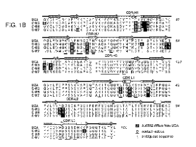
Figure 1. (A) Inferred lineage of clone 860. Left: the unmutated common
ancestor
(UCA) of the three antibodies (shown by their numbers, right) isolated from
the donor. (B)
Alignment of heavy-chain (top) and light-chain (bottom) sequences in the
lineage. (C)
Contact of the Fab from CH65 with HAL Heavy chain in dark blue; light chain in
light blue;
CA 02880791 2015-02-02
WO 2013/020074
PCT/US2012/049573
21
CDRs in colors as labeled in (B); HA in red, with the atomic surface shown as
a partly
transparent overlay. Residues that have mutated from the UCA are in green
stick
representation.
Figure 2. (A) HA trimer with bound CH65 Fab. One HA chain is in red (HA1) and
green (HA2); the other two chains are in gray; glycans are in yellow. The Fab
bound to the
colored HA chain is in dark blue (heavy chain) and light blue (light chain),
with the contacting
CDRs in colors as labeled in Fig. 1B. (B) Blow up of the Fv region and its
contact with HAI_
Colors as in Figure 1. Note that the heavy-chain CDR3 (magenta) projects into
the receptor-
binding pocket on HAI, while the remaining CDRs have more limited surface
contacts. (C) and
(D) Surface representation of the contact between Fab CH65 and HAI, opened up
as shown by
the arrows. The sialic-acid pocket on one HA subunit is in dark red; the rest
of the subunit, in
dull red; the remaining two subunits, in gray; glycans, in yellow.
Fig. 3. Comparison of interactions from CH65 (A) and sa-2,6-sia1y1 lactose
(B).
Fig. 4. Enzyme-linked immunoabsorbent assay (ELISA) of reactivity of CH65-CH67
lineage members to H1 and H3 influenza strains. 293 T cells were transfected
with full- length
HA from strain X31 (H3) (top panel, A-C) or with cell-surface expressed
globular head from
A/Solomon Islands/3/2006 [H1] (bottom panel, E-G). Cells were fixed with
formaldehyde and
probed with CH65 Fab (b and f) or CH66 full-length antibody (C and G),
followed by a FITC-
conjugated secondary antibody specific for the human Fab. Cells were imaged by
FITC
emission (532nm). As a control, transfected cells were probed with secondary
antibody only (A
and E).
Fig. 5. Sequences at the VDJ recombination site of CH65. The key indicates the
origin
of the heavy-chain coding segments (V, D, J, and n-nucleotide).
Fig. 6. Heavy chain DNA sequences of CH65-CH67 HA antibodies.
Fig. 7. Light chain DNA sequences of CH65-CH67 HA antibodies.
Fig. 8. Heavy chain amino acid sequences of CH65-CH67 HA antibodies.
Fig. 9. Light chain amino acid sequences of CH65-CH67 HA antibodies.
Fig. 10. Alignment of VH DNA sequences of CL860UCA, CH65, CH66 and CH67.
CA 02880791 2015-02-02
WO 2013/020074
PCT/US2012/049573
22
Fig. 11. Alignment of VL DNA sequences of CL860UCA, CH65, CH66 and CH67.
Fig. 12. Alignment of VH amino acid sequences of CL860UCA, CH65, CH66 and
CH67.
Fig. 13. Alignment of VL amino acid sequences of CL860UCA, CH65, CH66 and
CH67.
Fig. 14. Representative receptor binding domains from H1 (A), H2 (B), H3 (C),
and H5
(D) hemagglutinin. The CH65-CH67 antibody binding epitopes are underlined.
Fig. 15. Sequence alignment of representative receptor binding domains from
H1, H2,
H3, and H5 hemagglutinin. The amino acid residues that interact with the CH65-
CH67
antibodies are underlined.
DETAILED DESCRIPTION OF THE INVENTION
The invention features novel antibodies that broadly neutralize influenza
antigenic
variants. The invention also provides compositions and kits containing the
novel antibodies,
as well as methods for using these novel therapeutic molecules to treat or
prevent (e.g.,
vaccinate) influenza infection.
The receptor for influenza virus is sialic acid, attached by terminal a-2,3 or
a-2,6
linkage to glycans on glycoproteins or glycolipids (reviewed in Wiley, D.C.
and Skehel, J.J.
Annu. Rev. Biochem. 56:365-394 (1987)). Most neutralizing antibodies block
cell attachment,
either because their footprint overlaps the receptor-binding site or because
they exert steric
interference when bound elsewhere on the HA surface (Knossow, M. and Skehel,
J.J.
Immunology 119:1-7 (2006)). Two mouse monoclonal neutralizing antibodies, for
which
structures of Fab:HA complexes have been determined, have loops that project
into the sialic-
acid binding pocket on HA and present an aspartic-acid side chain roughly
where the sialic-acid
carboxylate would be (Fleury, D. et al., Nat. Struct. Biol. 5:119-123 (1998);
and Barbey-Martin,
C. et al., Virology 294:70-74 (2002)). But both of these antibodies also have
extensive contacts
with other surface regions, in which escape mutations could occur more readily
than in the
receptor site.
The invention is based, at least in part, on the discovery of novel antibodies
having
principal contacts in the receptor pocket. One such antibody, designated CH65,
was found by
CA 02880791 2015-02-02
WO 2013/020074
PCT/US2012/049573
23
isolating rearranged heavy- and light-chain genes from sorted single plasma
cells, obtained from
a subject who had received the 2007 trivalent vaccine. CH65 neutralizes a
remarkably broad
range of H1 seasonal isolates spanning more than three decades. Its 19-residue
heavy-chain
complementarity-determining region 3 (CDR-H3) inserts into the receptor
pocket, mimicking
many of the interactions made by sialic acid. Both heavy- and light-chain CDRs
participate in
more restricted, additional contacts with the outward-facing surface of HAL
The inferred,
unmutated ancestor of CH65 differs from the affinity matured antibody at 12
positions in the
heavy-chain variable domain, and at 6 in the light-chain variable domain. The
human B-cell
repertoire thus includes the potential to generate antibodies directed
primarily at the receptor
binding site. The large number of seasonal H1 viruses neutralized by antibody
CH65 suggests
that such responses are ordinarily too rare to select for resistance, or that
resistance comes at too
great a fitness cost ¨ as would be the case if potential escape mutations were
to compromise
receptor binding. Thus, it is surprising that the inventors have discovered
that broad
neutralization of influenza virus can be achieved by antibodies with contacts
that mimic those of
the receptor. Accordingly, the invention provides novel antibodies that mimic
the contact
between influenza HA and the sialic acid receptor. These novel antibodies can
effectively treat
and/or prevent infection by drifted strains of influenza. As such, the
invention features
compositions and kits containing the novel antibodies, as well as methods for
using these
therapeutic molecules to treat and/or prevent influenza infection. The
invention also relates to
combination therapies including the novel antibodies.
CH65-CH67 Hemagglutinin Antibodies
The present invention provides novel anti-influenza antibodies that
specifically bind
to an epitope of an influenza hemagglutinin (HA). Binding of the antibodies to
the HA reduces or
inhibits influenza hemagglutinin binding to sialic acid.
In embodiments, the epitope of influenza HA comprises a sialic-acid binding
domain.
In embodiments, the HA is H1 HA, H2 HA, H3 HA, or H5 HA (an HA from a human
adapted H5 strain).
In related embodiments, the influenza HA epitope comprises amino acids
corresponding to
CDR H1 residue 158; CDR H2 residues 158-160; CDR H3 residues 135-136, 190-195,
and 226;
CDR L1 residues 222, 225, and 227; and CDR L3 residues 187 and 189 from
A/Solomon
Islands/3/2006.
CA 02880791 2015-02-02
WO 2013/020074
PCT/US2012/049573
24
In related embodiments, the influenza HA epitope comprises the amino acids set
forth in
any one of SEQ ID NOs:17-44.
In related embodiments, the influenza HA epitope comprises the CH65-CH67
binding
residues in any one of SEQ ID NOs:17-44 (e.g., the CH65-CH67 binding residues
identified in
Figure 15).
In embodiments, the anti-influenza antibody or antibody fragment comprises a
variable heavy (VH) chain, and wherein the VH chain comprises an amino acid
sequence set
forth in SEQ ID NO: 9, SEQ ID NO: 10, SEQ ID NO: 11, or SEQ ID NO: 12.
In embodiments, the anti-influenza antibody or antibody fragment comprises one
or
more heavy chain CDR regions present in a variable heavy (VH) chain amino acid
sequence of
SEQ ID NO: 9, SEQ ID NO: 10, SEQ ID NO: 11, or SEQ ID NO: 12. In related
embodiments,
the one or more heavy chain CDR regions comprises a CDR3 region present in the
variable
heavy (VH) chain amino acid sequence of SEQ ID NO: 9, SEQ ID NO: 10, SEQ ID
NO: 11, or
SEQ ID NO: 12.
In embodiments, the anti-influenza antibody or antibody fragment comprises a
variable light (VL) chain, and wherein the VL chain comprises an amino acid
sequence set forth
in SEQ ID NO: 13, SEQ ID NO: 14, SEQ ID NO: 15, or SEQ ID NO: 16.
In embodiments, the anti-influenza antibody or antibody fragment comprises one
or
more light chain CDR regions present in a variable light (VL) chain amino acid
sequence of
SEQ ID NO: 13, SEQ ID NO: 14, SEQ ID NO: 15, or SEQ ID NO: 16. In related
embodiments, the one or more light chain CDR regions comprises a CDR3 region
present in
the variable heavy (VL) chain amino acid sequence of SEQ ID NO: 13, SEQ ID NO:
14, SEQ
ID NO: 15, or SEQ ID NO: 16.
In embodiments, the anti-influenza antibody or antibody fragment comprises i)
a
variable heavy (VH) chain amino comprising an amino acid sequence set forth in
SEQ ID NO:
10, and ii) a variable light (VL) chain comprising an amino acid sequence set
forth in SEQ ID
NO: 14.
In embodiments, the anti-influenza antibody or antibody fragment comprises a
variable heavy (VH) chain, wherein the CDR3 region of the VH chain comprises
Arg104,
Ser105, Va1106, Asp107, Tyr109, Tyr110, Tyr112, or a combination thereof.
CA 02880791 2015-02-02
WO 2013/020074
PCT/US2012/049573
In embodiments, the anti-influenza antibody is a monoclonal antibody or
antibody
fragment thereof.
In embodiments, the anti-influenza antibody is a humanized antibody.
In embodiments, the antibody fragment is an Fab fragment, an Fab' fragment, an
Fd
5 fragment, a Fd' fragment, an Fv fragment, a dAb fragment, an F(ab')2
fragment, a single
chain fragment, a diabody, or a linear antibody.
In embodiments, the anti-influenza antibody or antibody fragment further
comprises an
agent conjugated to the anti-influenza antibody or antibody fragment thereof.
In related
embodiments, the agent conjugated to the antibody or antibody fragment thereof
is a therapeutic
10 agent or detectable label.
The therapeutic agent can be any therapeutic agent suitable for use with the
novel
antibodies. Such agents are well known in the art and include small molecules,
nanoparticles,
polypeptides, radioisotopes, inhibitory nucleic acids, and the like. In
embodiments, the
therapeutic agent is an antiviral agent or a toxin. In embodiments, the
therapeutic agent is an
15 siRNA, shRNA, or antisense nucleic acid molecule that reduces influenza
virus production.
The detectable label can be any detectable label suitable for use with the
novel
antibodies. Such labels are well known in the art and include labels that are
detected by
spectroscopic, photochemical, biochemical, immunochemical, physical, or
chemical means.
In embodiments, the detectable label is an enzyme, a fluorescent molecule, a
particle label, an
20 electron-dense reagent, a radiolabel, a microbubble, biotin,
digoxigenin, or a hapten or a
protein that has been made detectable.
In any of the above embodiments, the influenza can be H1N1, H2N2, H3N2, or a
human adapted H5 strain.
The antibodies of the invention can be prepared by any conventional means
known in
25 the art. For example, polyclonal antibodies can be prepared by
immunizing an animal (e.g., a
rabbit, rat, mouse, donkey, goat, hamster, guinea pig, sheep, ungulate, cow,
camel, fowl,
chicken, and the like) by multiple subcutaneous or intraperitoneal injections
of the relevant
antigen (e.g., a purified peptide fragment, full-length recombinant protein,
fusion protein, and
the like) optionally conjugated to suitable hapten (e.g., keyhole limpet
hemocyanin (KLH),
serum albumin, and the like). The antigens can be diluted in any suitable
vehicle (e.g., sterile
saline) and combined with an adjuvant (e.g., Complete or Incomplete Freund' s
Adjuvant) to
CA 02880791 2015-02-02
WO 2013/020074
PCT/US2012/049573
26
form a stable emulsion. The polyclonal antibody is then recovered from blood,
ascites and
the like, of an animal so immunized. Collected blood is clotted, and the serum
decanted,
clarified by centrifugation, and assayed for antibody titer. The polyclonal
antibodies can be
purified from serum or ascites according to standard methods in the art
including affinity
chromatography, ion-exchange chromatography, gel electrophoresis, dialysis,
and the like.
Monoclonal antibodies can be prepared using hybridoma methods, such as those
described by Kohler and Milstein, Nature 256:495 (1975). Using the hybridoma
method, an
appropriate host animal is immunized as described above to elicit the
production by
lymphocytes of antibodies that will specifically bind to an immunizing
antigen.
Lymphocytes can also be immunized in vitro. Following immunization, the
lymphocytes are
isolated and fused with a suitable myeloma cell line using, for example,
polyethylene glycol,
to form hybridoma cells that can then be selected away from unfused
lymphocytes and
myeloma cells. Hybridomas that produce monoclonal antibodies directed
specifically against
a chosen antigen as determined by immunoprecipitation, immunoblotting, or by
an in vitro
binding assay (e.g., radioimmunoassay (RIA), enzyme-linked immunosorbent assay
(ELISA),
and the like) can then be propagated either in vitro culture using standard
methods (see
Goding, Monoclonal Antibodies: Principles and Practice, Academic Press, 1986)
or in vivo
as ascites tumors in an animal. The monoclonal antibodies can then be purified
from the
culture medium or ascites fluid as described for polyclonal antibodies above.
Alternatively monoclonal antibodies can also be made using recombinant DNA
methods as described in U.S. Patent 4,816,567. The polynucleotides encoding a
monoclonal
antibody are isolated from mature B-cells or hybridoma cell, such as by RT-PCR
using
oligonucleotide primers that specifically amplify the genes encoding the heavy
and light
chains of the antibody, and their sequence is determined using conventional
procedures. The
isolated polynucleotides (including the isolated polynucleotides described
herein) encoding
the heavy and light chains are then cloned into suitable expression vectors,
which when
transfected into host cells such as E. coli cells, simian COS cells, Chinese
hamster ovary
(CHO) cells, or myeloma cells that do not otherwise produce immunoglobulin
protein,
monoclonal antibodies are generated by the host cells. Also, recombinant
monoclonal
antibodies or fragments thereof of the desired species can be isolated from
phage display
libraries expressing CDRs of the desired species as described (see McCafferty
et al., 1990,
Nature, 348:552-554; Clackson et al., 1991, Nature, 352:624-628; and Marks et
al., 1991, J.
Mol. Biol., 222:581-597).
CA 02880791 2015-02-02
WO 2013/020074
PCT/US2012/049573
27
The polynucleotide(s) encoding a monoclonal antibody can further be modified
in a
number of different manners using recombinant DNA technology to generate
alternative
antibodies. In some embodiments, the constant domains of the light and heavy
chains of, for
example, a mouse monoclonal antibody can be substituted 1) for those regions
of, for
example, a human antibody to generate a chimeric antibody or 2) for a non-
immunoglobulin
polypeptide to generate a fusion antibody. In some embodiments, the constant
regions are
truncated or removed to generate the desired antibody fragment of a monoclonal
antibody.
Site-directed or high-density mutagenesis of the variable region can be used
to optimize
specificity, affinity, etc. of a monoclonal antibody.
In some embodiments of the present invention, the monoclonal antibody is a
humanized antibody. Humanized antibodies are antibodies that contain minimal
sequences
from non-human (e.g., rodent) antibodies within the antigen determination or
hypervariable
region that comprise the three complementary determination regions (CDRs)
within each
antibody chain. Such antibodies are used therapeutically to reduce
antigenicity and HAMA
(human anti-mouse antibody) responses when administered to a human subject. In
practice,
humanized antibodies are typically human antibodies with minimum to virtually
no non-
human sequences. A human antibody is an antibody produced by a human or an
antibody
having an amino acid sequence corresponding to an antibody produced by a
human.
Humanized antibodies can be produced using various techniques known in the
art.
An antibody can be humanized by substituting the CDRs of a human antibody with
those of a
non-human antibody (e.g., mouse, rat, rabbit, hamster, and the like.) having
the desired
specificity, affinity, and capability (see, e.g., the methods of Jones et al.,
Nature 321:522-525
(1986); Riechmann et al., Nature 332:323-327 (1988); and Verhoeyen et al.,
Science
239:1534-1536 (1988). The humanized antibody can be further modified by the
substitution
of additional residue either in the variable human framework region and/or
within the
replaced non-human residues to refine and optimize antibody specificity,
affinity, and/or
capability.
The choice of human heavy and/or light chain variable domains to be used in
making
humanized antibodies can be important for reducing antigenicity. According to
the "best-fit"
method, the sequence of the variable domain of a non-human antibody is
screened against the
entire library of known human variable-domain amino acid sequences. Thus in
certain
embodiments, the human amino acid sequence which is most homologous to that of
the non-
CA 02880791 2015-02-02
WO 2013/020074
PCT/US2012/049573
28
human antibody from which the CDRs are taken is used as the human framework
region (FR)
for the humanized antibody (see Sims et al., J. Immunol. 151: 2296 (1993);
Chothia et al., J.
Mol. Biol. 196:901 (1987)). Another method uses a particular FR derived from
the consensus
sequence of all human antibodies of a particular subgroup of light or heavy
chains and can be
used for several difference humanized antibodies (see Carter et al., PNAS 89;
4285 (1992);
Presta et al., J. Immunol. 151: 2623 (1993)). In embodiments, a combination of
methods is
used to pick the human variable FR to use in generation of humanized
antibodies.
It is further understood that antibodies to be humanized must retain high
affinity for
the antigen as well as other favorable biological properties. To achieve this
goal, humanized
antibodies can be prepared by a process of analysis of the parental sequence
from the non-
human antibody to be humanized and the various candidate humanizing sequences.
Three-
dimensional immunoglobulin models are available and familiar to those skilled
in the art.
Computer programs can be used to illustrate and display probable three-
dimensional
conformational structures of selected candidate antibody sequences. Use of
such models
permits analysis of the likely role of the residues in the function of the
antibody to be
humanized, i.e., the analysis of residues that influence the ability of the
candidate antibody to
bind its antigen. In this way, FR residues can be selected and combined from
the parental
antibody to the recipient humanized antibody so that the desired antibody
characteristics are
achieved. In general, the residues in the CDRs of the antigen determination
region (or
hypervariable region) are retained from the parental antibody (e.g. the non-
human antibody
with the desired antigen binding properties) in the humanized antibody for
antigen binding.
In certain embodiments, at least one additional residue within the variable FR
is retained
from the parental antibody in the humanized antibody. In certain embodiments,
up to six
additional residues within the variable FR are retained from the parental
antibody in the
humanized antibody.
Amino acids from the variable regions of the mature heavy and light chains of
immunoglobulins are designated Hx and Lx respectively, where x is a number
designating the
position of an amino acid according to the scheme of Kabat, Sequences of
Proteins of
Immunological Interest, U.S. Department of Health and Human Services, 1987,
1991. Kabat
lists many amino acid sequences for antibodies for each subgroup, and lists
the most
commonly occurring amino acid for each residue position in that subgroup to
generate a
consensus sequence. Kabat uses a method for assigning a residue number to each
amino acid
in a listed sequence, and this method for assigning residue numbers has become
standard in
CA 02880791 2015-02-02
WO 2013/020074
PCT/US2012/049573
29
the field. Kabat' s scheme is extendible to other antibodies not included in
his compendium
by aligning the antibody in question with one of the consensus sequences in
Kabat by
reference to conserved amino acids. The use of the Kabat numbering system
readily
identifies amino acids at equivalent positions in different antibodies. For
example, an amino
acid at the L50 position of a human antibody occupies the equivalent position
to an amino
acid position L50 of a mouse antibody. Moreover, any two antibody sequences
can be
uniquely aligned, for example to determine percent identity, by using the
Kabat numbering
system so that each amino acid in one antibody sequence is aligned with the
amino acid in the
other sequence that has the same Kabat number. After alignment, if a subject
antibody region
(e.g., the entire mature variable region of a heavy or light chain) is being
compared with the
same region of a reference antibody, the percentage sequence identity between
the subject
and reference antibody regions is the number of positions occupied by the same
amino acid in
both the subject and reference antibody region divided by the total number of
aligned
positions of the two regions, with gaps not counted, multiplied by 100 to
convert to
percentage.
In addition to humanized antibodies, fully human antibodies can be directly
prepared
using various techniques known in the art. Immortalized human B lymphocytes
immunized
in vitro or isolated from an immunized individual that produce an antibody
directed against a
target antigen can be generated (See, e.g., Cole et al., Monoclonal Antibodies
and Cancer
Therapy, Alan R. Liss, p. 77 (1985); Boemer et al., J. Immunol. 147:86-95
(1991); and U.S.
Patent No. 5,750,373). Also, the human antibody can be selected from a phage
library, where
that phage library expresses human antibodies (see Vaughan et al., Nat.
Biotech. 14:309-314
(1996); Sheets et al., Proc. Nat'l. Acad. Sci. 95:6157-6162 (1998); Hoogenboom
and Winter,
J. Mol. Biol. 227:381 (1991); and Marks et al., J. Mol. Biol. 222:581 (1991)).
Human
antibodies can also be made in transgenic mice containing human immunoglobulin
loci that
are capable upon immunization of producing the full repertoire of human
antibodies in the
absence of endogenous immunoglobulin production. This approach is described in
U.S.
Patent Nos. 5,545,807; 5,545,806; 5,569,825; 5,625,126; 5,633,425; and
5,661,016.
This invention also encompasses bispecific antibodies. Bispecific antibodies
are
antibodies that are capable of specifically recognizing and binding at least
two different
epitopes. The different epitopes can either be within the same molecule (e.g.,
influenza HA)
or on different molecules such that the bispecific antibody can specifically
recognize and
bind an epitope in an antigen of interest (e.g., influenza HA) as well as, for
example, another
CA 02880791 2015-02-02
WO 2013/020074
PCT/US2012/049573
viral protein (e.g., neurominidase, M2, and the like). Bispecific antibodies
can be intact
antibodies or antibody fragments. Techniques for making bispecific antibodies
are common
in the art (see Millstein et al., Nature 305:537-539 (1983); Brennan et al.,
Science 229:81
(1985); Suresh et al, Methods in Enzymol. 121:120 (1986); Traunecker et al.,
EMBO J.
5
10:3655-3659 (1991); Shalaby et al., J. Exp. Med. 175:217-225 (1992); Kostelny
et al., J.
Immunol. 148:1547-1553 (1992); Gruber et al., J. Immunol. 152:5368 (1994); and
U.S.
Patent No. 5,731,168). Antibodies with more than two valencies are also
contemplated. For
example, trispecific antibodies can be prepared (see Tutt et al., J. Immunol.
147:60 (1991)).
In embodiments, the antibodies of the invention are antibody fragments.
Various
10 techniques are known for the production of antibody fragments:
Traditionally, these
fragments are derived via proteolytic digestion of intact antibodies (see,
e.g., Morimoto et al.,
Journal of Biochemical and Biophysical Methods 24:107-117 (1993); and Brennan
et al.,
Science 229:81 (1985)). Antibody fragments can also be produced recombinantly.
Fab, Fv,
and scFv antibody fragments can all be expressed in and secreted from E. coli
or other host
15
cells, thus allowing the production of large amounts of these fragments. Such
antibody
fragments can also be isolated from antibody phage libraries as discussed
above. The
antibody fragment can also be linear antibodies as described in U.S. Patent
No. 5,641,870, for
example, and can be monospecific or bispecific. Other techniques for the
production of
antibody fragments will be readily apparent to the skilled practitioner.
20
According to the present invention, techniques can be adapted for the
production of
single-chain antibodies specific to a polypeptide of the invention (see U.S.
Pat. No.
4,946,778). In addition, methods can be adapted for the construction of Fab
expression
libraries (see Huse et al., Science 246:1275-1281 (1989)) to allow rapid and
effective
identification of monoclonal Fab fragments with the desired specificity for
influenza HA.
25
Antibody fragments that contain the idiotypes to a polypeptide of the
invention may be
produced by techniques in the art including, but not limited to: (a) an
F(ab')2 fragment
produced by pepsin digestion of an antibody molecule; (b) an Fab fragment
generated by
reducing the disulfide bridges of an F(ab' )2 fragment, (c) an Fab fragment
generated by the
treatment of the antibody molecule with papain and a reducing agent, and (d)
Fv fragments.
30 It
can further be desirable, especially in the case of antibody fragments, to
modify an
antibody in order to increase its serum half-life. This can be achieved, for
example, by
incorporation of a salvage receptor binding epitope into the antibody fragment
by mutation of
CA 02880791 2015-02-02
WO 2013/020074
PCT/US2012/049573
31
the appropriate region in the antibody fragment or by incorporating the
epitope into a peptide
tag that is then fused to the antibody fragment at either end or in the middle
(e.g., by DNA or
peptide synthesis).
Heteroconjugate antibodies are also within the scope of the present invention.
Heteroconjugate antibodies are composed of two covalently joined antibodies.
Such
antibodies have, for example, been proposed to target immune cells to unwanted
cells (U.S.
Pat. No. 4,676,980). It is contemplated that the antibodies can be prepared in
vitro using
known methods in synthetic protein chemistry, including those involving
crosslinking agents.
For example, immunotoxins can be constructed using a disulfide exchange
reaction or by
forming a thioether bond. Examples of suitable reagents for this purpose
include
iminothiolate and methyl-4-mercaptobutyrimidate.
Another alteration contemplated by the present invention is that the variable
domains
in both the heavy and light chains are altered by at least partial replacement
of one or more
CDRs and, if necessary, by partial framework region replacement and sequence
changing.
Although the CDRs may be derived from an antibody of the same class or even
subclass as
the antibody from which the framework regions are derived, it is envisaged
that the CDRs
will be derived from an antibody of different class and preferably from an
antibody from a
different species. It may not be necessary to replace all of the CDRs with the
complete CDRs
from the donor variable region to transfer the antigen binding capacity of one
variable
domain to another. Rather, it may only be necessary to transfer those residues
that are
necessary to maintain the activity of the antigen binding site. Given the
explanations set forth
in U.S. Pat. Nos. 5,585,089, 5,693,761 and 5,693,762, it will be well within
the competence
of those skilled in the art, either by carrying out routine experimentation or
by trial and error
testing to obtain a functional antibody with reduced immunogenicity.
Alterations to the variable region notwithstanding, those skilled in the art
will
appreciate that the modified antibodies of this invention can comprise
antibodies, or
immunoreactive fragments thereof, in which at least a fraction of one or more
of the constant
region domains has been deleted or otherwise altered so as to provide desired
biochemical
characteristics such as increased tumor localization or reduced serum half-
life when
compared with an antibody of approximately the same immunogenicity comprising
a native
or unaltered constant region. In some embodiments, the constant region of the
modified
antibodies will comprise a human constant region. Modifications to the
constant region
CA 02880791 2015-02-02
WO 2013/020074
PCT/US2012/049573
32
compatible with this invention comprise additions, deletions or substitutions
of one or more
amino acids in one or more domains. That is, the modified antibodies disclosed
herein may
comprise alterations or modifications to one or more of the three heavy chain
constant
domains (CH1, CH2 or CH3) and/or to the light chain constant domain (CL). In
some
embodiments of the invention modified constant regions wherein one or more
domains are
partially or entirely deleted are contemplated. In some embodiments the
modified antibodies
will comprise domain deleted constructs or variants wherein the entire CH2
domain has been
removed (ACH2 constructs). In some embodiments the omitted constant region
domain will
be replaced by a short amino acid spacer (e.g., 10 residues) that provides
some of the
molecular flexibility typically imparted by the absent constant region.
The present invention further embraces variants and equivalents which are
substantially homologous to the chimeric, humanized and human antibodies, or
antibody
fragments thereof, set forth herein. These can contain, for example,
conservative substitution
mutations, i.e., the substitution of one or more amino acids by similar amino
acids. For
example, conservative substitution refers to the substitution of an amino acid
with another
within the same general class such as, for example, one acidic amino acid with
another acidic
amino acid, one basic amino acid with another basic amino acid or one neutral
amino acid by
another neutral amino acid. What is intended by a conservative amino acid
substitution is
well known in the art.
The antibodies of the present invention can be assayed for immunospecific
binding by
any method known in the art. The immunoassays which can be used include, but
are not
limited to, competitive and non-competitive assay systems using techniques
such as BIAcore
analysis, FACS analysis, immunofluorescence, immunocytochemistry, Western
blots,
radioimmuno as s ays , ELISA, "sandwich" immuno as says, immunoprecipitation
assays,
precipitin reactions, gel diffusion precipitin reactions, immunodiffusion
assays, agglutination
assays, complement-fixation assays, immunoradiometric assays, fluorescent
immunoassays,
protein A immunoassays, and the like. Such assays are routine and well known
in the art
(see, e.g., Ausubel et al, eds, 1994, Current Protocols in Molecular Biology,
Vol. 1, John
Wiley & Sons, Inc., New York, which is incorporated by reference herein in its
entirety).
In embodiments, the immunospecificity of an antibody against a influenza HA is
determined using ELISA. An ELISA assay comprises preparing antigen, coating
wells of a
microtiter plate with antigen, adding the antibody conjugated to a detectable
compound such
CA 02880791 2015-02-02
WO 2013/020074
PCT/US2012/049573
33
as an enzymatic substrate (e.g., horseradish peroxidase or alkaline
phosphatase) to the well,
incubating for a period of time and detecting the presence of the antigen. In
some
embodiments, the antibody is not conjugated to a detectable compound, but
instead a second
conjugated antibody that recognizes the antibody against the influenza HA
antigen is added
to the well. In some embodiments, instead of coating the well with the
antigen, the antibody
can be coated to the well and a second antibody conjugated to a detectable
compound can be
added following the addition of the antigen to the coated well. One of skill
in the art would
be knowledgeable as to the parameters that can be modified to increase the
signal detected as
well as other variations of ELISAs known in the art (see e.g. Ausubel et al,
eds, 1994,
Current Protocols in Molecular Biology, Vol. 1, John Wiley & Sons, Inc., New
York at
11.2.1).
The binding affinity of an antibody to influenza HA and the off-rate of an
antibody-
antigen interaction can be determined by competitive binding assays. One
example of a
competitive binding assay is a radioimmunoassay comprising the incubation of
labeled
antigen (e.g. 3H or 1251), or fragment or variant thereof, with the antibody
of interest in the
presence of increasing amounts of unlabeled antigen followed by the detection
of the
antibody bound to the labeled antigen. The affinity of the antibody against an
antigen and the
binding off-rates can be determined from the data by scatchard plot analysis.
In some
embodiments, BIAcore kinetic analysis is used to determine the binding on and
off rates of
antibodies against a cancer stem cell marker. BIAcore kinetic analysis
comprises analyzing
the binding and dissociation of antibodies from chips with immobilized cancer
stem cell
marker antigens on their surface.
Influenza Hemagglutinin Antibody Polypeptides and Polynucleotides
The present invention also encompasses isolated polynucleotides that encode a
polypeptide comprising an influenza hemagglutinin (HA) antibody or fragment
thereof.
The term "polynucleotide encoding a polypeptide" encompasses a polynucleotide
which includes only coding sequences for the polypeptide as well as a
polynucleotide which
includes additional coding and/or non-coding sequences. The polynucleotides of
the
invention can be in the form of RNA or in the form of DNA. DNA includes cDNA,
genomic
DNA, and synthetic DNA; and can be double-stranded or single-stranded, and if
single
stranded can be the coding strand or non-coding (anti-sense) strand.
CA 02880791 2015-02-02
WO 2013/020074
PCT/US2012/049573
34
The present invention further relates to variants of the polynucleotides, for
example,
fragments, analogs, and derivatives. The variant of the polynucleotide can be
a naturally
occurring allelic variant of the polynucleotide or a non-naturally occurring
variant of the
polynucleotide. In certain embodiments, the polynucleotide can have a coding
sequence
which is a naturally occurring allelic variant of the coding sequence of the
disclosed
polypeptides. As known in the art, an allelic variant is an alternate form of
a polynucleotide
sequence that have, for example, a substitution, deletion, or addition of one
or more
nucleotides, which does not substantially alter the function of the encoded
polypeptide.
In embodiments, the polynucleotides can comprise the coding sequence for the
mature
polypeptide fused in the same reading frame to a polynucleotide which aids,
for example, in
expression and secretion of a polypeptide from a host cell (e.g., a leader
sequence which
functions as a secretory sequence for controlling transport of a polypeptide
from the cell).
The polypeptide having a leader sequence is a preprotein and can have the
leader sequence
cleaved by the host cell to form the mature form of the polypeptide. The
polynucleotides can
also encode for a proprotein which is the mature protein plus additional 5'
amino acid
residues. A mature protein having a prosequence is a proprotein and is an
inactive form of
the protein. Once the prosequence is cleaved an active mature protein remains.
In embodiments, the polynucleotides can comprise the coding sequence for the
mature
polypeptide fused in the same reading frame to a marker sequence that allows,
for example,
for purification of the encoded polypeptide. For example, the marker sequence
can be a
hexa-histidine tag supplied by a pQE-9 vector to provide for purification of
the mature
polypeptide fused to the marker in the case of a bacterial host, or the marker
sequence can be
a hemagglutinin (HA) tag derived from the influenza hemagglutinin protein when
a
mammalian host (e.g., COS-7 cells) is used. Additional tags include, but are
not limited to,
Calmodulin tags, FLAG tags, Myc tags, S tags, SBP tags, Softag 1, Softag 3, V5
tag, Xpress
tag, Isopeptag, SpyTag, Biotin Carboxyl Carrier Protein (BCCP) tags, GST tags,
fluorescent
protein tags (e.g., green fluorescent protein tags), maltose binding protein
tags, Nus tags,
Strep-tag, thioredoxin tag, TC tag, Ty tag, and the like.
In embodiments, the present invention provides isolated nucleic acid molecules
having a nucleotide sequence at least 80% identical, at least 85% identical,
at least 90%
identical, at least 95% identical, or at least 96%, 97%, 98% or 99% identical
to a
CA 02880791 2015-02-02
WO 2013/020074
PCT/US2012/049573
polynucleotide encoding a polypeptide comprising an influenza HA antibody or
antibody
fragment of the present invention.
By a polynucleotide having a nucleotide sequence at least, for example, 95%
"identical" to a reference nucleotide sequence is intended that the nucleotide
sequence of the
5
polynucleotide is identical to the reference sequence except that the
polynucleotide sequence
can include up to five point mutations per each 100 nucleotides of the
reference nucleotide
sequence. In other words, to obtain a polynucleotide having a nucleotide
sequence at least
95% identical to a reference nucleotide sequence, up to 5% of the nucleotides
in the reference
sequence can be deleted or substituted with another nucleotide, or a number of
nucleotides up
10 to
5% of the total nucleotides in the reference sequence can be inserted into the
reference
sequence. These mutations of the reference sequence can occur at the amino- or
carboxy-
terminal positions of the reference nucleotide sequence or anywhere between
those terminal
positions, interspersed either individually among nucleotides in the reference
sequence or in
one or more contiguous groups within the reference sequence.
15 As
a practical matter, whether any particular nucleic acid molecule is at least
80%
identical, at least 85% identical, at least 90% identical, and in some
embodiments, at least
95%, 96%, 97%, 98%, or 99% identical to a reference sequence can be determined
conventionally using known computer programs such as the Bestfit program
(Wisconsin
Sequence Analysis Package, Version 8 for Unix, Genetics Computer Group,
University
20
Research Park, 575 Science Drive, Madison, WI 53711). Bestfit uses the local
homology
algorithm of Smith and Waterman, Advances in Applied Mathematics 2: 482 489
(1981), to
find the best segment of homology between two sequences. When using Bestfit or
any other
sequence alignment program to determine whether a particular sequence is, for
instance, 95%
identical to a reference sequence according to the present invention, the
parameters are set
25
such that the percentage of identity is calculated over the full length of the
reference
nucleotide sequence and that gaps in homology of up to 5% of the total number
of
nucleotides in the reference sequence are allowed.
The polynucleotide variants can contain alterations in the coding regions, non-
coding
regions, or both. In some embodiments, the polynucleotide variants contain
alterations which
30
produce silent substitutions, additions, or deletions, but do not alter the
properties or activities
of the encoded polypeptide. In some embodiments, nucleotide variants are
produced by
silent substitutions due to the degeneracy of the genetic code. Polynucleotide
variants can be
CA 02880791 2015-02-02
WO 2013/020074
PCT/US2012/049573
36
produced for a variety of reasons, e.g., to optimize codon expression for a
particular host
(change codons in the human mRNA to those preferred by a bacterial host such
as E. coli).
The polypeptides of the present invention can be recombinant polypeptides,
natural
polypeptides, or synthetic polypeptides comprising an antibody, or fragment
thereof, against
influenza HA. It will be recognized in the art that some amino acid sequences
of the
invention can be varied without significant effect of the structure or
function of the protein.
Thus, the invention further includes variations of the polypeptides which show
substantial
activity or which include regions of a humanized antibody, or fragment
thereof, against
influenza HA. Such mutants include deletions, insertions, inversions, repeats,
and type
substitutions.
The polypeptides and analogs can be further modified to contain additional
chemical
moieties not normally part of the protein. Those derivatized moieties can
improve the
solubility, the biological half life or absorption of the protein. The
moieties can also reduce or
eliminate any desirable side effects of the proteins and the like. An overview
for those
moieties can be found in Remington' s Pharmaceutical Sciences, 20th ed., Mack
Publishing
Co., Easton, PA (2000).
The isolated polypeptides described herein can be produced by any suitable
method
known in the art. Such methods range from direct protein synthetic methods to
constructing a
DNA sequence encoding isolated polypeptide sequences and expressing those
sequences in a
suitable transformed host. In some embodiments, a DNA sequence is constructed
using
recombinant technology by isolating or synthesizing a DNA sequence encoding a
wild-type
protein of interest. Optionally, the sequence can be mutagenized by site-
specific mutagenesis
to provide functional analogs thereof. See, e.g. Zoeller et al., Proc. Nat'l.
Acad. Sci. USA
81:5662-5066 (1984) and U.S. Pat. No. 4,588,585.
In embodiments, a DNA sequence encoding a polypeptide of interest would be
constructed by chemical synthesis using an oligonucleotide synthesizer.
Such
oligonucleotides can be designed based on the amino acid sequence of the
desired
polypeptide and selecting those codons that are favored in the host cell in
which the
recombinant polypeptide of interest will be produced. Standard methods can be
applied to
synthesize an isolated polynucleotide sequence encoding an isolated
polypeptide of interest.
For example, a complete amino acid sequence can be used to construct a back-
translated
gene. Further, a DNA oligomer containing a nucleotide sequence coding for the
particular
CA 02880791 2015-02-02
WO 2013/020074
PCT/US2012/049573
37
isolated polypeptide can be synthesized. For example, several small
oligonucleotides coding
for portions of the desired polypeptide can be synthesized and then ligated.
The individual
oligonucleotides typically contain 5' or 3' overhangs for complementary
assembly.
Once assembled (e.g., by synthesis, site-directed mutagenesis, or another
method), the
polynucleotide sequences encoding a particular isolated polypeptide of
interest will be
inserted into an expression vector and optionally operatively linked to an
expression control
sequence appropriate for expression of the protein in a desired host. Proper
assembly can be
confirmed by nucleotide sequencing, restriction mapping, and expression of a
biologically
active polypeptide in a suitable host. As well known in the art, in order to
obtain high
expression levels of a transfected gene in a host, the gene can be operatively
linked to
transcriptional and translational expression control sequences that are
functional in the
chosen expression host.
Recombinant expression vectors are used to amplify and express DNA encoding
the
influenza HA antibodies. Recombinant expression vectors are replicable DNA
constructs
which have synthetic or cDNA-derived DNA fragments encoding an influenza HA
antibody
or a bioequivalent analog operatively linked to suitable transcriptional or
translational
regulatory elements derived from mammalian, microbial, viral or insect genes.
A
transcriptional unit generally comprises an assembly of (1) a genetic element
or elements
having a regulatory role in gene expression, for example, transcriptional
promoters or
enhancers, (2) a structural or coding sequence which is transcribed into mRNA
and translated
into protein, and (3) appropriate transcription and translation initiation and
termination
sequences, as described in detail below. Such regulatory elements can include
an operator
sequence to control transcription. The ability to replicate in a host, usually
conferred by an
origin of replication, and a selection gene to facilitate recognition of
transformants can
additionally be incorporated. DNA regions are operatively linked when they are
functionally
related to each other. For example, DNA for a signal peptide (secretory
leader) is operatively
linked to DNA for a polypeptide if it is expressed as a precursor which
participates in the
secretion of the polypeptide; a promoter is operatively linked to a coding
sequence if it
controls the transcription of the sequence; or a ribosome binding site is
operatively linked to a
coding sequence if it is positioned so as to permit translation. Generally,
operatively linked
means contiguous, and in the case of secretory leaders, means contiguous and
in reading
frame. Structural elements intended for use in yeast expression systems
include a leader
sequence enabling extracellular secretion of translated protein by a host
cell. Alternatively,
CA 02880791 2015-02-02
WO 2013/020074
PCT/US2012/049573
38
where recombinant protein is expressed without a leader or transport sequence,
it can include
an N-terminal methionine residue. This residue can optionally be subsequently
cleaved from
the expressed recombinant protein to provide a final product.
The choice of expression control sequence and expression vector will depend
upon
the choice of host. A wide variety of expression host/vector combinations can
be employed.
Useful expression vectors for eukaryotic hosts, include, for example, vectors
comprising
expression control sequences from SV40, bovine papilloma virus, adenovirus and
cytomegalovirus. Useful expression vectors for bacterial hosts include known
bacterial
plasmids, such as plasmids from Escherichia coli, including pCR 1, pBR322,
pMB9 and their
derivatives, wider host range plasmids, such as M13 and filamentous single-
stranded DNA
phages.
Suitable host cells for expression of a polypeptide include prokaryotes,
yeast, insect or
higher eukaryotic cells under the control of appropriate promoters.
Prokaryotes include gram
negative or gram positive organisms, for example E. coli or bacilli. Higher
eukaryotic cells
include established cell lines of mammalian origin. Cell-free translation
systems could also
be employed. Appropriate cloning and expression vectors for use with
bacterial, fungal,
yeast, and mammalian cellular hosts are well known in the art (see Pouwels et
al., Cloning
Vectors: A Laboratory Manual, Elsevier, N.Y., 1985).
Various mammalian or insect cell culture systems are also advantageously
employed
to express recombinant protein. Expression of recombinant proteins in
mammalian cells can
be performed because such proteins are generally correctly folded,
appropriately modified
and completely functional. Examples of suitable mammalian host cell lines
include the COS-
7 lines of monkey kidney cells, described by Gluzman (Cell 23:175, 1981), and
other cell
lines capable of expressing an appropriate vector including, for example, L
cells, C127, 3T3,
Chinese hamster ovary (CHO), HeLa and BHK cell lines. Mammalian expression
vectors
can comprise nontranscribed elements such as an origin of replication, a
suitable promoter
and enhancer linked to the gene to be expressed, and other 5' or 3' flanking
nontranscribed
sequences, and 5' or 3' nontranslated sequences, such as necessary ribosome
binding sites, a
polyadenylation site, splice donor and acceptor sites, and transcriptional
termination
sequences. Baculovirus systems for production of heterologous proteins in
insect cells are
reviewed by Luckow and Summers, Bio/Technology 6:47 (1988).
CA 02880791 2015-02-02
WO 2013/020074
PCT/US2012/049573
39
The proteins produced by a transformed host can be purified according to any
suitable
method. Such standard methods include chromatography (e.g., ion exchange,
affinity and
sizing column chromatography, and the like), centrifugation, differential
solubility, or by any
other standard technique for protein purification. Affinity tags such as
hexahistidine, maltose
binding domain, influenza coat sequence, glutathione-S-transferase, and the
like can be
attached to the protein to allow easy purification by passage over an
appropriate affinity
column. Isolated proteins can also be physically characterized using such
techniques as
proteolysis, nuclear magnetic resonance and x-ray crystallography.
For example, supernatants from systems which secrete recombinant protein into
culture media can be first concentrated using a commercially available protein
concentration
filter, for example, an Amicon or Millipore Pellicon ultrafiltration unit.
Following the
concentration step, the concentrate can be applied to a suitable purification
matrix.
Alternatively, an anion exchange resin can be employed, for example, a matrix
or substrate
having pendant diethylaminoethyl (DEAE) groups. The matrices can be
acrylamide, agarose,
dextran, cellulose or other types commonly employed in protein purification.
Alternatively, a
cation exchange step can be employed. Suitable cation exchangers include
various insoluble
matrices comprising sulfopropyl or carboxymethyl groups. Finally, one or more
reversed-
phase high performance liquid chromatography (RP-HPLC) steps employing
hydrophobic
RP-HPLC media, e.g., silica gel having pendant methyl or other aliphatic
groups, can be
employed to further purify a cancer stem cell protein-Fc composition. Some or
all of the
foregoing purification steps, in various combinations, can also be employed to
provide a
homogeneous recombinant protein.
Recombinant protein produced in bacterial culture can be isolated, for
example, by
initial extraction from cell pellets, followed by one or more concentration,
salting-out,
aqueous ion exchange or size exclusion chromatography steps. High performance
liquid
chromatography (HPLC) can be employed for final purification steps. Microbial
cells
employed in expression of a recombinant protein can be disrupted by any
convenient method,
including freeze-thaw cycling, sonication, mechanical disruption, or use of
cell lysing agents.
Methods of Treatment
The present invention provides methods for treating or preventing influenza
infection.
In aspects, the invention provides methods for treating or preventing an
influenza virus
infection in a subject in need thereof. The methods involve administering to
the subject an effective
CA 02880791 2015-02-02
WO 2013/020074
PCT/US2012/049573
amount of an anti-influenza antibody or antibody fragment described herein, or
a pharmaceutical
composition containing the antibody or antibody fragment. The methods treat or
prevent
influenza virus infection in the subject, including reducing or alleviating
symptoms associated with
infection.
5 In aspects, the invention provides methods for neutralizing an influenza
virus in a subject in
need thereof. The methods involve administering to the subject an effective
amount of an anti-
influenza antibody or antibody fragment described herein, or a pharmaceutical
composition
containing the antibody or antibody fragment. The methods neutralize the
influenza virus in the
subject, thereby treating or prevent influenza virus infection in the subject,
including reducing or
10 alleviating symptoms associated with infection.
In aspects, the invention provides methods for establishment of influenza
virus infection in
a subject in need thereof. The methods involve administering to the subject an
effective amount of
an anti-influenza antibody or antibody fragment described herein, or a
pharmaceutical
composition containing the antibody or antibody fragment. The methods inhibit
establishment of
15 influenza virus infection in the subject, thereby preventing symptoms
associated with infection.
In aspects, the invention provides methods for inhibiting dissemination of
influenza virus
infection in a subject in need thereof. The methods involve administering to
the subject an effective
amount of an anti-influenza antibody or antibody fragment described herein, or
a pharmaceutical
composition containing the antibody or antibody fragment. The methods inhibit
dissemination of
20 influenza virus infection in the subject, thereby reducing or
alleviating symptoms associated with
infection.
In aspects, the invention provides methods for inhibiting influenza virus
entry into a cell in
a subject. The methods involve administering to the subject an effective
amount of an anti-
influenza antibody or antibody fragment described herein, or a pharmaceutical
composition
25 containing the antibody or antibody fragment. The methods inhibit
influenza virus entry into a cell
in the subject, thereby preventing symptoms associated with infection or
reducing or alleviating
symptoms associated with infection.
In aspects, the invention provides methods for inhibiting influenza virus
entry into a cell.
The methods involve contacting a cell having or at risk of developing
influenza virus infection with
30 an anti-influenza antibody or antibody fragment described herein, or a
pharmaceutical
CA 02880791 2015-02-02
WO 2013/020074
PCT/US2012/049573
41
composition containing the antibody or antibody fragment. The methods inhibit
influenza virus
entry into the cell.
In any of the above aspects, the influenza can be H1N1, H2N2, H3N2, or a human
adapted
H5 strain.
In any of the above aspects, the subject has or is at risk of developing an
influenza
infection. In related embodiments, the subject is a mammal (e.g., human). In
related embodiments,
the subject is susceptible to viral infection (e.g., a pregnant female, a
young subject or an infant
subject, an elderly subject).
In any of the above aspects and embodiments, the anti-influenza antibody or
antibody
fragment, or the pharmaceutical composition is administered by intramuscular
injection,
intravenous injection, subcutaneous injection, or inhalation.
Pharmaceutical compositions
The invention provides for pharmaceutical compositions containing the novel
influenza HA antibodies described herein. In embodiments, the pharmaceutical
compositions
contain a pharmaceutically acceptable carrier, excipient, or diluent, which
includes any
pharmaceutical agent that does not itself induce the production of an immune
response
harmful to a subject receiving the composition, and which may be administered
without
undue toxicity. As used herein, the term "pharmaceutically acceptable" means
being
approved by a regulatory agency of the Federal or a state government or listed
in the U.S.
Pharmacopia, European Pharmacopia or other generally recognized pharmacopia
for use in
mammals, and more particularly in humans. These compositions can be useful for
treating
and/or preventing influenza infection.
A thorough discussion of pharmaceutically acceptable carriers, diluents, and
other
excipients is presented in Remington's Pharmaceutical Sciences (17th ed., Mack
Publishing
Company) and Remington: The Science and Practice of Pharmacy (21st ed.,
Lippincott
Williams & Wilkins), which are hereby incorporated by reference. The
formulation of the
pharmaceutical composition should suit the mode of administration. In
embodiments, the
pharmaceutical composition is suitable for administration to humans, and can
be sterile, non-
particulate and/or non-pyrogenic.
CA 02880791 2015-02-02
WO 2013/020074
PCT/US2012/049573
42
Pharmaceutically acceptable carriers, excipients, or diluents include, but are
not
limited, to saline, buffered saline, dextrose, water, glycerol, ethanol,
sterile isotonic aqueous
buffer, and combinations thereof.
Wetting agents, emulsifiers and lubricants, such as sodium lauryl sulfate and
magnesium stearate, as well as coloring agents, release agents, coating
agents, sweetening,
flavoring and perfuming agents, preservatives, and antioxidants can also be
present in the
compositions.
Examples of pharmaceutically-acceptable antioxidants include, but are not
limited to:
(1) water soluble antioxidants, such as ascorbic acid, cysteine hydrochloride,
sodium
bisulfate, sodium metabisulfite, sodium sulfite and the like; (2) oil-soluble
antioxidants, such
as ascorbyl palmitate, butylated hydroxyanisole (BHA), butylated
hydroxytoluene (BHT),
lecithin, propyl gallate, alpha-tocopherol, and the like; and (3) metal
chelating agents, such as
citric acid, ethylenediamine tetraacetic acid (EDTA), sorbitol, tartaric acid,
phosphoric acid,
and the like.
In embodiments, the pharmaceutical composition is provided in a solid form,
such as
a lyophilized powder suitable for reconstitution, a liquid solution,
suspension, emulsion,
tablet, pill, capsule, sustained release formulation, or powder.
In embodiments, the pharmaceutical composition is supplied in liquid form, for
example, in a sealed container indicating the quantity and concentration of
the active
ingredient in the pharmaceutical composition. In related embodiments, the
liquid form of the
pharmaceutical composition is supplied in a hermetically sealed container.
Methods for formulating the pharmaceutical compositions of the present
invention are
conventional and well-known in the art (see Remington and Remington' s). One
of skill in
the art can readily formulate a pharmaceutical composition having the desired
characteristics
(e.g., route of administration, biosafety, and release profile).
Methods for preparing the pharmaceutical compositions include the step of
bringing
into association the active ingredient with a pharmaceutically acceptable
carrier and,
optionally, one or more accessory ingredients. The pharmaceutical compositions
can be
prepared by uniformly and intimately bringing into association the active
ingredient with
liquid carriers, or finely divided solid carriers, or both, and then, if
necessary, shaping the
CA 02880791 2015-02-02
WO 2013/020074
PCT/US2012/049573
43
product. Additional methodology for preparing the pharmaceutical compositions,
including
the preparation of multilayer dosage forms, are described in Ansel's
Pharmaceutical Dosage
Forms and Drug Delivery Systems (9th ed., Lippincott Williams & Wilkins),
which is hereby
incorporated by reference.
Methods of Delivery
The pharmaceutical compositions of the invention can be administered to a
subject by
oral and non-oral means (e.g., topically, transdermally, or by injection).
Such modes of
administration and the methods for preparing an appropriate pharmaceutical
composition for
use therein are described in Gibaldi's Drug Delivery Systems in Pharmaceutical
Care (1st
ed., American Society of Health-System Pharmacists), which is hereby
incorporated by
reference.
In embodiments, the pharmaceutical compositions are administered orally in a
solid
form.
Pharmaceutical compositions suitable for oral administration can be in the
form of
capsules, cachets, pills, tablets, lozenges (using a flavored basis, usually
sucrose and acacia or
tragacanth), powders, granules, or as a solution or a suspension in an aqueous
or non-aqueous
liquid, or as an oil-in-water or water-in-oil liquid emulsion, or as an elixir
or syrup, or as
pastilles (using an inert base, such as gelatin and glycerin, or sucrose and
acacia) and/or as
mouth washes and the like, each containing a predetermined amount of a
compound(s)
described herein, a derivative thereof, or a pharmaceutically acceptable salt
or prodrug
thereof as the active ingredient(s). The active ingredient can also be
administered as a bolus,
electuary, or paste.
In solid dosage forms for oral administration (e.g., capsules, tablets, pills,
dragees,
powders, granules and the like), the active ingredient is mixed with one or
more
pharmaceutically acceptable carriers, excipients, or diluents, such as sodium
citrate or
dicalcium phosphate, and/or any of the following: (1) fillers or extenders,
such as starches,
lactose, sucrose, glucose, mannitol, and/or silicic acid; (2) binders, such
as, for example,
carboxymethylcellulose, alginates, gelatin, polyvinyl pyrrolidone, sucrose
and/or acacia; (3)
humectants, such as glycerol; (4) disintegrating agents, such as agar-agar,
calcium carbonate,
potato or tapioca starch, alginic acid, certain silicates, and sodium
carbonate; (5) solution
CA 02880791 2015-02-02
WO 2013/020074
PCT/US2012/049573
44
retarding agents, such as paraffin; (6) absorption accelerators, such as
quaternary ammonium
compounds; (7) wetting agents, such as, for example, acetyl alcohol and
glycerol
monostearate; (8) absorbents, such as kaolin and bentonite clay; (9)
lubricants, such a talc,
calcium stearate, magnesium stearate, solid polyethylene glycols, sodium
lauryl sulfate, and
mixtures thereof; and (10) coloring agents. In the case of capsules, tablets,
and pills, the
pharmaceutical compositions can also comprise buffering agents. Solid
compositions of a
similar type can also be prepared using fillers in soft and hard-filled
gelatin capsules, and
excipients such as lactose or milk sugars, as well as high molecular weight
polyethylene
glycols and the like.
A tablet can be made by compression or molding, optionally with one or more
accessory ingredients. Compressed tablets can be prepared using binders (for
example,
gelatin or hydroxypropylmethyl cellulose), lubricants, inert diluents,
preservatives,
disintegrants (for example, sodium starch glycolate or cross-linked sodium
carboxymethyl
cellulose), surface-actives, and/ or dispersing agents. Molded tablets can be
made by
molding in a suitable machine a mixture of the powdered active ingredient
moistened with an
inert liquid diluent.
The tablets and other solid dosage forms, such as dragees, capsules, pills,
and
granules, can optionally be scored or prepared with coatings and shells, such
as enteric
coatings and other coatings well-known in the art.
The pharmaceutical compositions can also be formulated so as to provide slow,
extended, or controlled release of the active ingredient therein using, for
example,
hydroxypropylmethyl cellulose in varying proportions to provide the desired
release profile,
other polymer matrices, liposomes and/or microspheres. The pharmaceutical
compositions
can also optionally contain opacifying agents and may be of a composition that
releases the
active ingredient(s) only, or preferentially, in a certain portion of the
gastrointestinal tract,
optionally, in a delayed manner. Examples of embedding compositions include
polymeric
substances and waxes. The active ingredient can also be in micro-encapsulated
form, if
appropriate, with one or more pharmaceutically acceptable carriers,
excipients, or diluents
well-known in the art (see, e.g., Remington and Remington's).
The pharmaceutical compositions can be sterilized by, for example, filtration
through
a bacteria-retaining filter, or by incorporating sterilizing agents in the
form of sterile solid
CA 02880791 2015-02-02
WO 2013/020074
PCT/US2012/049573
compositions which can be dissolved in sterile water, or some other sterile
injectable medium
immediately before use.
In embodiments, the pharmaceutical compositions are administered orally in a
liquid
form.
5
Liquid dosage forms for oral administration of an active ingredient include
pharmaceutically acceptable emulsions, microemulsions, solutions, suspensions,
syrups and
elixirs. In addition to the active ingredient, the liquid dosage forms can
contain inert diluents
commonly used in the art, such as, for example, water or other solvents,
solubilizing agents
and emulsifiers, such as ethyl alcohol, isopropyl alcohol, ethyl carbonate,
ethyl acetate,
10
benzyl alcohol, benzyl benzoate, propylene glycol, 1,3-butylene glycol, oils
(in particular,
cottonseed, groundnut, corn, germ, olive, castor and sesame oils), glycerol,
tetrahydrofuryl
alcohol, polyethylene glycols and fatty acid esters of sorbitan, and mixtures
thereof. In
addition to inert diluents, the liquid pharmaceutical compositions can include
adjuvants such
as wetting agents, emulsifying and suspending agents, sweetening, flavoring,
coloring,
15 perfuming and preservative agents, and the like.
Suspensions, in addition to the active ingredient(s) can contain suspending
agents
such as, but not limited to, ethoxylated isostearyl alcohols, polyoxyethylene
sorbitol and
sorbitan esters, microcrystalline cellulose, aluminum metahydroxide,
bentonite, agar-agar and
tragacanth, and mixtures thereof.
20 In
embodiments, the pharmaceutical compositions are administered by non-oral
means such as by topical application, transdermal application, injection, and
the like. In
related embodiments, the pharmaceutical compositions are administered
parenterally by
injection, infusion, or implantation (e.g., intravenous, intramuscular,
intraarticular,
subcutaneous, and the like).
25
Compositions for parenteral use can be presented in unit dosage forms, e.g. in
ampoules or in vials containing several doses, and in which a suitable
preservative can be
added. Such compositions can be in form of a solution, a suspension, an
emulsion, an
infusion device, a delivery device for implantation, or it can be presented as
a dry powder to
be reconstituted with water or another suitable vehicle before use. One or
more co-vehicles,
30
such as ethanol, can also be employed. Apart from the active ingredient(s),
the compositions
CA 02880791 2015-02-02
WO 2013/020074
PCT/US2012/049573
46
can contain suitable parenterally acceptable carriers and/or excipients or the
active
ingredient(s) can be incorporated into microspheres, microcapsules,
nanoparticles, liposomes,
or the like for controlled release. Furthermore, the compositions can also
contain suspending,
solubilising, stabilising, pH-adjusting agents, and/or dispersing agents.
The pharmaceutical compositions can be in the form of sterile injections. To
prepare
such a composition, the active ingredient is dissolved or suspended in a
parenterally
acceptable liquid vehicle. Exemplary vehicles and solvents include, but are
not limited to,
water, water adjusted to a suitable pH by addition of an appropriate amount of
hydrochloric
acid, sodium hydroxide or a suitable buffer, 1,3-butanediol, Ringer's solution
and isotonic
sodium chloride solution. The pharmaceutical composition can also contain one
or more
preservatives, for example, methyl, ethyl or n-propyl p-hydroxybenzoate. To
improve
solubility, a dissolution enhancing or solubilising agent can be added or the
solvent can
contain 10-60% w/w of propylene glycol or the like.
The pharmaceutical compositions can contain one or more pharmaceutically
acceptable sterile isotonic aqueous or nonaqueous solutions, dispersions,
suspensions or
emulsions, or sterile powders, which can be reconstituted into sterile
injectable solutions or
dispersions just prior to use. Such pharmaceutical compositions can contain
antioxidants;
buffers; bacteriostats; solutes, which render the formulation isotonic with
the blood of the
intended recipient; suspending agents; thickening agents; preservatives; and
the like.
Examples of suitable aqueous and nonaqueous carriers, which can be employed in
the
pharmaceutical compositions of the invention include water, ethanol, polyols
(such as
glycerol, propylene glycol, polyethylene glycol, and the like), and suitable
mixtures thereof,
vegetable oils, such as olive oil, and injectable organic esters, such as
ethyl oleate. Proper
fluidity can be maintained, for example, by the use of coating materials, such
as lecithin, by
the maintenance of the required particle size in the case of dispersions, and
by the use of
surfactants.
In some embodiments, in order to prolong the effect of an active ingredient,
it is
desirable to slow the absorption of the compound from subcutaneous or
intramuscular
injection. This can be accomplished by the use of a liquid suspension of
crystalline or
amorphous material having poor water solubility. The rate of absorption of the
active
ingredient then depends upon its rate of dissolution which, in turn, can
depend upon crystal
CA 02880791 2015-02-02
WO 2013/020074
PCT/US2012/049573
47
size and crystalline form. Alternatively, delayed absorption of a parenterally-
administered
active ingredient is accomplished by dissolving or suspending the compound in
an oil
vehicle. In addition, prolonged absorption of the injectable pharmaceutical
form can be
brought about by the inclusion of agents that delay absorption such as
aluminum
monostearate and gelatin.
Controlled release parenteral compositions can be in form of aqueous
suspensions,
microspheres, microcapsules, magnetic microspheres, oil solutions, oil
suspensions,
emulsions, or the active ingredient can be incorporated in biocompatible
carrier(s),
liposomes, nanoparticles, implants or infusion devices.
Materials for use in the preparation of microspheres and/or microcapsules
include
biodegradable/bioerodible polymers such as polyglactin, poly-(isobutyl
cyanoacrylate),
poly(2-hydroxyethyl-L-glutamine) and poly(lactic acid).
Biocompatible carriers which can be used when formulating a controlled release
parenteral formulation include carbohydrates such as dextrans, proteins such
as albumin,
lipoproteins or antibodies.
Materials for use in implants can be non-biodegradable, e.g.,
polydimethylsiloxane, or
biodegradable such as, e.g., poly(caprolactone), poly(lactic acid),
poly(glycolic acid) or
poly(ortho esters).
In embodiments, the active ingredient(s) are administered by aerosol. This is
accomplished by preparing an aqueous aerosol, liposomal preparation, or solid
particles
containing the compound. A nonaqueous (e.g., fluorocarbon propellant)
suspension can be
used. The pharmaceutical composition can also be administered using a sonic
nebulizer,
which would minimize exposing the agent to shear, which can result in
degradation of the
compound.
Ordinarily, an aqueous aerosol is made by formulating an aqueous solution or
suspension of the active ingredient(s) together with conventional
pharmaceutically-acceptable
carriers and stabilizers. The carriers and stabilizers vary with the
requirements of the
particular compound, but typically include nonionic surfactants (Tweens,
Pluronics, or
polyethylene glycol), innocuous proteins like serum albumin, sorbitan esters,
oleic acid,
CA 02880791 2015-02-02
WO 2013/020074
PCT/US2012/049573
48
lecithin, amino acids such as glycine, buffers, salts, sugars or sugar
alcohols. Aerosols
generally are prepared from isotonic solutions.
Dosage forms for topical or transdermal administration of an active
ingredient(s)
includes powders, sprays, ointments, pastes, creams, lotions, gels, solutions,
patches and
inhalants. The active ingredient(s) can be mixed under sterile conditions
with a
pharmaceutically acceptable carrier, and with any preservatives, buffers, or
propellants as
appropriate.
Transdermal patches suitable for use in the present invention are disclosed in
Transdermal Drug Delivery: Developmental Issues and Research Initiatives
(Marcel Dekker
Inc., 1989) and U.S. Pat. Nos. 4,743,249, 4,906,169, 5,198,223, 4,816,540,
5,422,119,
5,023,084, which are hereby incorporated by reference. The transdermal patch
can also be
any transdermal patch well-known in the art, including transscrotal patches.
Pharmaceutical
compositions in such transdermal patches can contain one or more absorption
enhancers or
skin permeation enhancers well-known in the art (see, e.g., U.S. Pat. Nos.
4,379,454 and
4,973,468, which are hereby incorporated by reference). Transdermal
therapeutic systems for
use in the present invention can be based on iontophoresis, diffusion, or a
combination of
these two effects.
Transdermal patches have the added advantage of providing controlled delivery
of
active ingredient(s) to the body. Such dosage forms can be made by dissolving
or dispersing
the active ingredient(s) in a proper medium. Absorption enhancers can also be
used to
increase the flux of the active ingredient across the skin. The rate of such
flux can be
controlled by either providing a rate controlling membrane or dispersing the
active
ingredient(s) in a polymer matrix or gel.
Such pharmaceutical compositions can be in the form of creams, ointments,
lotions,
liniments, gels, hydrogels, solutions, suspensions, sticks, sprays, pastes,
plasters and other
kinds of transdermal drug delivery systems.
The compositions can also include
pharmaceutically acceptable carriers or excipients such as emulsifying agents,
antioxidants,
buffering agents, preservatives, humectants, penetration enhancers, chelating
agents, gel-
forming agents, ointment bases, perfumes, and skin protective agents.
CA 02880791 2015-02-02
WO 2013/020074
PCT/US2012/049573
49
Examples of emulsifying agents include, but are not limited to, naturally
occurring
gums, e.g. gum acacia or gum tragacanth, naturally occurring phosphatides,
e.g. soybean
lecithin and sorbitan monooleate derivatives.
Examples of antioxidants include, but are not limited to, butylated hydroxy
anisole
(BHA), ascorbic acid and derivatives thereof, tocopherol and derivatives
thereof, and
cysteine.
Examples of preservatives include, but are not limited to, parabens, such as
methyl or
propyl p-hydroxybenzoate and benzalkonium chloride.
Examples of humectants include, but are not limited to, glycerin, propylene
glycol,
sorbitol and urea.
Examples of penetration enhancers include, but are not limited to, propylene
glycol,
DMSO, triethanolamine, N,N-dimethylacetamide, N,N-dimethylformamide, 2-
pyrrolidone
and derivatives thereof, tetrahydrofurfuryl alcohol, propylene glycol,
diethylene glycol
monoethyl or monomethyl ether with propylene glycol monolaurate or methyl
laurate,
eucalyptol, lecithin, Transcutol , and Azone
Examples of chelating agents include, but are not limited to, sodium EDTA,
citric
acid and phosphoric acid.
Examples of gel forming agents include, but are not limited to, Carbopol,
cellulose
derivatives, bentonite, alginates, gelatin and polyvinylpyrrolidone.
In addition to the active ingredient(s), the ointments, pastes, creams, and
gels of the
present invention can contain excipients, such as animal and vegetable fats,
oils, waxes,
paraffins, starch, tragacanth, cellulose derivatives, polyethylene glycols,
silicones, bentonites,
silicic acid, talc and zinc oxide, or mixtures thereof.
Powders and sprays can contain excipients such as lactose, talc, silicic acid,
aluminum
hydroxide, calcium silicates and polyamide powder, or mixtures of these
substances. Sprays
can additionally contain customary propellants, such as
chlorofluorohydrocarbons, and
volatile unsubstituted hydrocarbons, such as butane and propane.
CA 02880791 2015-02-02
WO 2013/020074
PCT/US2012/049573
Injectable depot forms are made by forming microencapsule matrices of
compound(s)
of the invention in biodegradable polymers such as polylactide-polyglycolide.
Depending on
the ratio of compound to polymer, and the nature of the particular polymer
employed, the rate
of compound release can be controlled. Examples of other biodegradable
polymers include
5
poly(orthoesters) and poly(anhydrides). Depot injectable formulations are also
prepared by
entrapping the drug in liposomes or microemulsions which are compatible with
body tissue.
Subcutaneous implants are well-known in the art and are suitable for use in
the
present invention. Subcutaneous implantation methods are preferably non-
irritating and
mechanically resilient. The implants can be of matrix type, of reservoir type,
or hybrids
10
thereof. In matrix type devices, the carrier material can be porous or non-
porous, solid or
semi-solid, and permeable or impermeable to the active compound or compounds.
The
carrier material can be biodegradable or may slowly erode after
administration. In some
instances, the matrix is non-degradable but instead relies on the diffusion of
the active
compound through the matrix for the carrier material to degrade. Alternative
subcutaneous
15
implant methods utilize reservoir devices where the active compound or
compounds are
surrounded by a rate controlling membrane, e.g., a membrane independent of
component
concentration (possessing zero-order kinetics). Devices consisting of a matrix
surrounded by
a rate controlling membrane also suitable for use.
Both reservoir and matrix type devices can contain materials such as
20
polydimethylsiloxane, such as SilasticTM, or other silicone rubbers. Matrix
materials can be
insoluble polypropylene, polyethylene, polyvinyl chloride, ethylvinyl acetate,
polystyrene
and polymethacrylate, as well as glycerol esters of the glycerol
palmitostearate, glycerol
stearate, and glycerol behenate type. Materials can be hydrophobic or
hydrophilic polymers
and optionally contain solubilising agents.
25
Subcutaneous implant devices can be slow-release capsules made with any
suitable
polymer, e.g., as described in U.S. Pat. Nos. 5,035,891 and 4,210,644, which
are hereby
incorporated by reference.
In general, at least four different approaches are applicable in order to
provide rate
control over the release and transdermal permeation of a drug compound. These
approaches
30
are: membrane-moderated systems, adhesive diffusion-controlled systems, matrix
dispersion-
type systems and microreservoir systems. It is appreciated that a controlled
release
CA 02880791 2015-02-02
WO 2013/020074
PCT/US2012/049573
51
percutaneous and/or topical composition can be obtained by using a suitable
mixture of these
approaches.
In a membrane-moderated system, the active ingredient is present in a
reservoir which
is totally encapsulated in a shallow compartment molded from a drug-
impermeable laminate,
such as a metallic plastic laminate, and a rate-controlling polymeric membrane
such as a
microporous or a non-porous polymeric membrane, e.g., ethylene-vinyl acetate
copolymer.
The active ingredient is released through the rate controlling polymeric
membrane. In the
drug reservoir, the active ingredient can either be dispersed in a solid
polymer matrix or
suspended in an unleachable, viscous liquid medium such as silicone fluid. On
the external
surface of the polymeric membrane, a thin layer of an adhesive polymer is
applied to achieve
an intimate contact of the transdermal system with the skin surface. The
adhesive polymer is
preferably a polymer which is hypoallergenic and compatible with the active
drug substance.
In an adhesive diffusion-controlled system, a reservoir of the active
ingredient is
formed by directly dispersing the active ingredient in an adhesive polymer and
then by, e.g.,
solvent casting, spreading the adhesive containing the active ingredient onto
a flat sheet of
substantially drug-impermeable metallic plastic backing to form a thin drug
reservoir layer.
A matrix dispersion-type system is characterized in that a reservoir of the
active
ingredient is formed by substantially homogeneously dispersing the active
ingredient in a
hydrophilic or lipophilic polymer matrix. The drug-containing polymer is then
molded into
disc with a substantially well-defined surface area and controlled thickness.
The adhesive
polymer is spread along the circumference to form a strip of adhesive around
the disc.
A microreservoir system can be considered as a combination of the reservoir
and
matrix dispersion type systems. In this case, the reservoir of the active
substance is formed
by first suspending the drug solids in an aqueous solution of water-soluble
polymer and then
dispersing the drug suspension in a lipophilic polymer to form a multiplicity
of unleachable,
microscopic spheres of drug reservoirs.
Any of the above-described controlled release, extended release, and sustained
release
compositions can be formulated to release the active ingredient in about 30
minutes to about
1 week, in about 30 minutes to about 72 hours, in about 30 minutes to 24
hours, in about 30
minutes to 12 hours, in about 30 minutes to 6 hours, in about 30 minutes to 4
hours, and in
CA 02880791 2015-02-02
WO 2013/020074
PCT/US2012/049573
52
about 3 hours to 10 hours. In embodiments, an effective concentration of the
active
ingredient(s) is sustained in a subject for 4 hours, 6 hours, 8 hours, 10
hours, 12 hours, 16
hours, 24 hours, 48 hours, 72 hours, or more after administration of the
pharmaceutical
compositions to the subject.
Dosages
When the agents described herein are administered as pharmaceuticals to humans
and
animals, they can be given per se or as a pharmaceutical composition
containing active
ingredient in combination with a pharmaceutically acceptable carrier,
excipient, or diluent.
Actual dosage levels and time course of administration of the active
ingredients in the
pharmaceutical compositions of the invention can be varied so as to obtain an
amount of the
active ingredient which is effective to achieve the desired therapeutic
response for a
particular patient, composition, and mode of administration, without being
toxic to the
patient. Generally, agents or pharmaceutical compositions of the invention are
administered
in an amount sufficient to reduce or eliminate symptoms associated with
influenza infection.
Exemplary dose ranges include 0.01 mg to 250 mg per day, 0.01 mg to 100 mg per
day, 1 mg to 100 mg per day, 10 mg to 100 mg per day, 1 mg to 10 mg per day,
and 0.01 mg
to 10 mg per day. A preferred dose of an agent is the maximum that a patient
can tolerate
and not develop serious or unacceptable side effects. In embodiments, the
agent is
administered at a concentration of about 10 micrograms to about 100 mg per
kilogram of
body weight per day, about 0.1 to about 10 mg/kg per day, or about 1.0 mg to
about 10 mg/kg
of body weight per day.
In embodiments, the pharmaceutical composition comprises an agent in an amount
ranging between 1 and 10 mg, such as 1, 2, 3, 4, 5, 6, 7, 8, 9, or 10 mg.
In embodiments, the therapeutically effective dosage produces a serum
concentration
of an agent of from about 0.1 ng/ml to about 50-100 ig/ml. The pharmaceutical
compositions typically should provide a dosage of from about 0.001 mg to about
2000 mg of
compound per kilogram of body weight per day. For example, dosages for
systemic
administration to a human patient can range from 1-10 jig/kg, 20-80 jig/kg, 5-
50 jig/kg, 75-
150 ig/kg, 100-500 ig/kg, 250-750 ig/kg, 500-1000 ig/kg, 1-10 mg/kg, 5-50
mg/kg, 25-75
mg/kg, 50-100 mg/kg, 100-250 mg/kg, 50-100 mg/kg, 250-500 mg/kg, 500-750
mg/kg, 750-
CA 02880791 2015-02-02
WO 2013/020074
PCT/US2012/049573
53
1000 mg/kg, 1000-1500 mg/kg, 1500-2000 mg/kg, 5 mg/kg, 20 mg/kg, 50 mg/kg, 100
mg/kg,
500 mg/kg, 1000 mg/kg, 1500 mg/kg, or 2000 mg/kg. Pharmaceutical dosage unit
forms are
prepared to provide from about 1 mg to about 5000 mg, for example from about
100 to about
2500 mg of the compound or a combination of essential ingredients per dosage
unit form.
In embodiments, about 50 nM to about 11.iM of an agent is administered to a
subject.
In related embodiments, about 50-100 nM, 50-250 nM, 100-500 nM, 250-500 nM,
250-750
nM, 500-750 nM, 500 nM to 1 1.04, or 750 nM to 11.iM of an agent is
administered to a
subject.
Determination of an effective amount is well within the capability of those
skilled in
the art, especially in light of the detailed disclosure provided herein.
Generally, an
efficacious or effective amount of an agent is determined by first
administering a low dose of
the agent(s) and then incrementally increasing the administered dose or
dosages until a
desired effect (e.g., reduced symptoms associated with influenza infection) is
observed in the
treated subject, with minimal or acceptable toxic side effects. Applicable
methods for
determining an appropriate dose and dosing schedule for administration of a
pharmaceutical
composition of the present invention are described, for example, in Goodman
and Gilman 's
The Pharmacological Basis of Therapeutics, Goodman et al., eds., 11th Edition,
McGraw-
Hill 2005, and Remington: The Science and Practice of Pharmacy, 20th and 21st
Editions,
Gennaro and University of the Sciences in Philadelphia, Eds., Lippencott
Williams &
Wilkins (2003 and 2005), each of which is hereby incorporated by reference.
Combination Therapies
The agents and pharmaceutical compositions described herein can also be
administered in combination with another therapeutic molecule. The therapeutic
molecule
can be any compound used to treat influenza infection. Examples of such
compounds
include, but are not limited to, inhibitory nucleic acids that reduce
influenza virus production,
antiviral agents (e.g., amantadine, rimantadine, zanamivir, oseltamivir, and
the like), toxins,
and agents that reduce the symptoms associated with influenza infection (e.g.,
anti-
inflammatories).
The influenza HA antibody can be administered before, during, or after
administration of
the additional therapeutic agent. In embodiments, the antibody is administered
before the first
administration of the additional therapeutic agent. In embodiments, the
antibody is administered
CA 02880791 2015-02-02
WO 2013/020074
PCT/US2012/049573
54
after the first administration of the additional therapeutic agent (e.g., 1,
2, 3, 4, 5, 6, 7, 8, 9, 10,
11, 12, 13, 14 days or more). In embodiments, the antibody is administered
simultaneously with
the first administration of the additional therapeutic agent.
The amount of therapeutic agent administered to a subject can readily be
determined by
the attending physician or veterinarian. Generally, an efficacious or
effective amount of an
antibody and an additional therapeutic is determined by first administering a
low dose of one or
both active agents and then incrementally increasing the administered dose or
dosages until a
desired effect is observed (e.g., reduced influenza infection symptoms), with
minimal or no
toxic side effects. Applicable methods for determining an appropriate dose and
dosing schedule
for administration of a combination of the present invention are described,
for example, in
Goodman and Gilman 's The Pharmacological Basis of Therapeutics, 11th
Edition., supra, and
in Remington: The Science and Practice of Pharmacy, 20th and 21st Editions,
supra.
Kits
The invention provides for kits for preventing or treating influenza
infection; neutralizing
an influenza virus; inhibiting establishment of influenza virus infection;
inhibiting dissemination of
influenza virus infection; as well as inhibiting influenza virus entry into a
cell. In embodiments, the
kit contains one or more agents or pharmaceutical compositions described
herein. In
embodiments, the kit provides instructions for use. The instructions for use
can pertain to any of
the methods described herein. In related embodiments, the instructions pertain
to using the
agent(s) or pharmaceutical composition(s) for treating or preventing influenza
infection. Kits
according to this aspect of the invention may comprise a carrier means, such
as a box, carton,
tube or the like, having in close confinement therein one or more container
means, such as vials,
tubes, ampules, bottles and the like. In embodiments, the kit provides a
notice in the form
prescribed by a governmental agency regulating the manufacture, use, or sale
of
pharmaceuticals or biological products, which notice reflects approval by the
agency of
manufacture, use or sale of the kit and the components therein for human
administration.
EXAMPLES
It should be appreciated that the invention should not be construed to be
limited to the
examples that are now described; rather, the invention should be construed to
include any and
all applications provided herein and all equivalent variations within the
skill of the ordinary
artisan.
CA 02880791 2015-02-02
WO 2013/020074
PCT/US2012/049573
Example 1: The clonal lineage of a broadly neutralizing antibody
Rearranged Ig VH and VL genes were isolated by RT/PCR from peripheral blood
mononuclear cells, collected from a subject one week after vaccination with
the 2007 trivalent
inactivated vaccine (TIV) (Liao, H.X. et al. J. Virol. Methods 158:171-179
(2009)). Among the
5 clonal lineages detected by sequencing the rearranged genes was the three-
member clone (mAbs
CH65, CH66 and CH67) shown in Figure 1A. The inferred sequence of the
unmutated common
ancestor (UCA) of the clonal lineage of antibodies CH65, CH66 and CH67 is
unambiguous,
except at position 99 of the heavy chain, which might be either glycine or
alanine. Figure 1B
shows an alignment of the amino acid sequences of each antibody to the UCA.
All three mature
10 antibodies bind the H1 hemagglutinin (HA) present in the vaccine
(A/Solomon Islands/3/2006)
with about equal affinity; the UCA binds much more weakly.
Example 2: Breadth of neutralizing activity
The heavy chain of CH65 differs from the UCA at 12 positions in the variable
domain;
and at 6 positions its light chain. CH65 IgG1 and its Fab were expressed in
293T cells by
15 transient transfection and purified as described below. Neutralization
was tested against a large
panel of H1 isolates from the past 30 years, including vaccine strains from
1977, 1991 and 1995,
and observed strikingly broad potency (Table 1). CH67 was also tested against
a subset of this
panel (Table 1).
CA 02880791 2015-02-02
WO 2013/020074
PCT/US2012/049573
56
Table 1. Broad neutralization of seasonal influenza strains A/H1N1 by human
MAb CH65 and
CH67
mAb minimum effective
concentration(ug/m1)
H1N1 virus strain CH65 CH67
A/USSR/90/1977* 100 25
A/Kawasaki/6/1986 0.098 0.39
NTexas/36/1991* Neg Neg
NWellington/47/1992 Neg Neg
A/Flonda/2/1993 0.012 0.012
A/Beijing/262/1995 0.098 0.098
A/Shengzhen/227/1995 0.012 0,024
A/Shanghai/8/1996 Neg Neg
A/Johannesburg/159/1997 0.098 0.39
A/Shanghai/2/1997 0.195 0.195
A/Moscow/13/1998 0.012 0.012
A/Ostrava/801/1998 12.5 Neg
NNew Caledonia/22/1999* 0.391 0.195
A/Bangkok/163/2000 0.195 0.098
A/Fujian/156/2000 0.488 0.049
A/Chile/8885/2001 0.195 0.098
A/Auckland/65/2001 0.195 0.098
A/Neimenggu/52/2002 0.098 0.098
A/Brazil/1403/2003 3.125 0.195
A/Canada/59/2004 0.098 0.098
A/Solomon Islands/03/2006"=+ 0.024 0.098
A/Brisbane/59/2007* 0.098 0.98
A/Caltfornia/07/2009(swine)* Neg 6.25
'Strains that were included in seasonal vaccines.
'Hl component of the vaccine received by the subject.
Originally reported as insensitive to rnAb CH65.
The antibody neutralized H1N1 strains isolated as early as 1986, covering 21
years of antigenic
drift. As expected, it neutralized A/Solomon Islands/3/2006, the H1 component
of the 2007
vaccine. Of the 36 strains tested, it failed to neutralize only six, including
the 2009 pandemic
strain, A/Texas/36/1991 and A/USSR/90/1977. The CH67 antibody has a similar
breadth; it
also neutralizes (weakly) the 2009 pandemic strain. There is also evidence
that CH66 (and
likely CH65) binds HA from an H3 virus (X31, a lab strain derived from the
1968 pandemic.
Too few HA-directed human monoclonal antibodies have been characterized for
systematic
comparison, but neutralization by serum samples does not ordinarily exhibit
this degree of
breadth.
CA 02880791 2015-02-02
WO 2013/020074
PCT/US2012/049573
57
Example 3: Structure of CH65:HA
A complex of the mAb CH65 Fab with the HA ectodomain from A/Solomon
Islands/03/2006 (HAsI) was crystallized, recorded diffraction to a minimum
Bragg spacing of
3.2-A (Table 2), and the structure was determined by molecular replacement as
outlined below.
Table 2: Crystallographic statistics
Data collection
Resolution (last shell), A 30.0-3.20 (3.31-3.20)
Wavelength, A 0.980
Space group 1212121
Unit cell dimensions (a, b, c), A 155.0, 191.8, 332.1
Unit cell angles (a, p, y), 90, 90, 90
I/o- (last shell) 16.7 (2.1)
Rsym (last shell), % 9.8 (63.7)
Completeness (last shell), % 98.9 (94.4)
Number of refections 368947
unique 80377
Redundancy 4.6
Refinement
Resolution, A 30.0-3.20
Number of refections 80336
working 78354
free 1982
Rwork, % 21.1
Rfree, % 24.8
Ramachandran plot, % 88.5/9.3/2.2
(favored/additional/disallowed)
Number of atoms:
protein 21794
other (sulfate ions) 214
rmsd bond lengths, A 0.010
rmsd bond angles, 1.287
The asymmetric unit of the crystal contains a single copy of the HA trimer,
with three bound
Fabs (Figures 2A-2D). The final model includes all HAI_ and HA2 residues in
the expressed
protein, except four disordered residues at the C-terminus of HAI_ The
electron density maps
showed evidence for N-linked glycosylation at all eight potential sites on
each monomer, and
one or more sugar residues at five of these positions could be modeled. The
Fab is well-ordered,
except residue 1 of the light chain and residues 141-147 of the heavy chain;
these residues are
all far from the binding site.
A/Solomon Islands/03/2006 (this work) and A/Puerto Rico/8/1934 (Gamblin, S.J.
et al.,
Science 303:1838-1842 (2004)) are, to the inventors' knowledge, the only
seasonal H1N1
CA 02880791 2015-02-02
WO 2013/020074
PCT/US2012/049573
58
strains for which a structure of the HA has been determined; others are either
pandemic strains
or animal influenza strains. Comparison, using the program DALI, shows that
HAsi is similar to
other H1N1 HAs, such as those of the pandemic isolates from 2009 (Ca RMSD 0.9A
over 495
aligned residues, 79% sequence identity; PDB IDs 3LZG (Xu, R. et al., Science
328:357-360
(2010)) and 3LYJ(Zhang, W. et al., Protein Cell 1:459-467 (2010))) and 1918
(Ca RMSD 1.5A
over 495 aligned residues, 85 % sequence identity; PDB IDs 3LZF(Xu, R. et al.,
Science
328:357-360 (2010)) and 1RUZ (Gamblin, S.J. et al., Science 303:1838-1842
(2004))) and the
seasonal isolate from 1934 (Ca RMSD 1.5A over 482 aligned residues, 86%
sequence identity;
PDB ID 1RVZ (Gamblin, S.J. et al., Science 303:1838-1842 (2004))). The
vestigial esterase
domain of HAsi resembles that of the 2009 HA more closely than it does those
from the 1918
and 1934 HAs.
MAb CH65 binds the globular head of the HA trimer (Figures 1C and 2A). The
epitope
includes both the receptor site and the antigenic site designated Sb in an
early analysis of H1
sequences (Caton, A.J. et al., Cell 31:417-427 (1982)). The contact buries 858
A2 on the
antibody and 748 A2 on HAI_ All three CDRs of the heavy chain, as well as CDR-
L1 and L3 of
the light chain, participate in the interface (Figures 1C and 2B-D). CDR-H3
inserts into the
receptor site. Seven of its nineteen residues contribute 402 A2 of buried
surface area, or 47% of
the complete interface. The other CDRs form flanking interactions. CDR-L3
contacts the N-
terminal end of the short a-helix, site Sb, at the edge of the receptor
pocket, and CDR-H1 and -
H2 contact a loop that protrudes from HAI_ adjacent to the C-terminus of the
short a-helix.
Tables 3 and 4 summarize several of the critical interactions between the
antibody and
HA.
Table 3: Residues in CDR H3 of CH65 that contact HA
Arg104*
Ser105
Val 106
Asp107
Tyr109
Tyr 110
Tyr 112
*for some influenza strains (not Solomon Islands), Arg104 might contact HA
CA 02880791 2015-02-02
WO 2013/020074
PCT/US2012/049573
59
Table 4: Residues in HA that contact CH65
CDR HA residue contacted
CDR H1 158
CDR H2 158-160
CDR H3 135-136; 190-195; 226
CDR L1 222; 225; 227
CDR L2 (none)
CDR L3 187;189
Example 4: CDR-H3 of mAb CH65 compared to the receptor
Because CDR-H3 inserts into the receptor site, this structure was compared to
that of the
human receptor analog LSTc (sialic-acid-a2,6-galactose-I31,4-N-
acteylglucosamine) bound to
1934 HA (PDB ID 1RVZ: reference (Gamblin, S.J. et al., Science 303:1838-1842
(2004)))(Figures 3A and 3B). In CH65, Asp107 at the tip of CDR-H3 accepts
hydrogen bonds
from the backbone amide of HAI_ A1a137, the side chain hydroxyl of Ser136, and
the side chain
NE of Arg226. (Arginine is found only rarely at position 226: glutamine is
more common.
Arg226 adopts a kinked conformation in the crystal structure; a glutamine
would fit readily,
with its NE in the same position as the corresponding atom of the arginine
side chain.) The
backbone amide of Va1106 in the antibody donates a hydrogen bond to the
carboxyl oxygen of
HAI_ Va1135 on HAI, and the nonpolar side chain of Va1106 is in van der Waals
contact with
HAI_ Trp153 and Leu194. In receptor analog LSTc, the carboxylate group of
sialic acid has the
same contacts with HAI_ as does the (chemically analogous) side chain of
Asp107, and the N-
acetyl group interacts with HA in the same way as just described for the amide
and side chain of
Va1106. In short, mAb CH65 mimics most of the chemical groups on the human
receptor that
interact with HA.
Example 5: Glycosylation
Glycosylation at antigenic sites is an important mechanism of immune evasion
by
influenza virus (Knossow, M. and Skehel, J.J. Immunology 119:1-7 (2006);
Wiley, D.C. and
Skehel, J.J. Annu. Rev. Biochem. 56:365-394 (1987); and Wei, C.J. et al., Sci.
Transl. Med.
2:24ra21 (2010)). In HAsi, glycosylation leaves sites Sb and Cb exposed,
partially obscures site
Ca, and entirely masks antigenic site Sa. Site Sa is the epitope recognized by
antibody 2D1, the
prototype for Ig-mediated immunity to 2009 H1N1 in survivors of the 1918
epidemic (Xu, R. et
al., Science 328:357-360 (2010)). Of the side chains in contact with 2D1, 7/16
differ between
CA 02880791 2015-02-02
WO 2013/020074
PCT/US2012/049573
HAsi and 1918 HA; in comparison, only 3/16 differ between 2009 pandemic HA and
1918 HA.
Because the HA of A/Solomon Islands/03/2006 is glycosylated at site Sa,
neither vaccination
with TIV-2007, nor prior infection with an A/Solomon Islands/03/2006-like
strain could have
elicited a 2D1-like immune response.
5 Example 6: Affinity maturation
The amino-acid sequence of CH65 is the result of affinity maturation from its
UCA.
Analysis of the structure in light of its clonal lineage (Figure 1B) shows
that the central
interactions of the antibodies with HA have remained unchanged by affinity
maturation. The
CDR-H3 has not mutated, nor has the contact of the light-chain CDR-L3 with the
N-terminal
10 end of the short a-helix, site Sb. (5er93 of CDR-L3 is Asp in lineage
member CH67;
substitution to Asp may allow CH67 to accept a hydrogen-bond from HA Asn187.)
Elsewhere
on the interaction surface of the antibody, changes to two residues create
additional hydrogen
bonds between the antibody and HAsi. Light-chain residues Asp26 and Arg29 in
CDR-L1 have
mutated from their respective germline counterparts, Asn and Ser. Asp26
accepts a hydrogen-
15 bond from HA Lys222. Arg29 is positioned to donate two hydrogen-bonds to
HA Asp225.
Other changes, including those at position 31 (Gly to Asp) and positions 33-35
(Trp-Met-His to
His-Ile-Asn) may exert subtle effects on the conformation of CDR-H3.
Example 7: CH65-CH67 lineage reactivity to different influenza strains
The ability of CH65-CH67 to react with other influenza strains was assessed.
293 T
20 cells were transfected with full-length HA from strain X31 (H3 influenza
strain) (Figures 4A-
4C, top panel) or with cell-surface expressed globular head from A/Solomon
Islands/3/2006 (H1
influenza strain) (Figures 4E-4G, bottom panel). Cells were fixed with
formaldehyde and
probed with CH65 Fab (Figures 4B and 4F) or CH66 full-length antibody (Figures
4C and 4G),
followed by a FITC-conjugated secondary antibody specific for the human Fab.
Cells were
25 imaged by FITC emission (532nm). As a control, transfected cells were
probed with secondary
antibody only (Figures 4A and 4E). These results indicate that the CH65-CH67
antibodies (e.g.,
CH66) also bind other influenza serotypes (e.g., H3).
As discussed in detail in the above examples, CH65 comes from an adult subject
in the
US, who received the 2007 TIV one week before donating a plasma sample. It was
assumed
30 that the subject had been exposed to H1N1 influenza strains in the past,
so that the antibodies
obtained by screening with a panel of recombinant hemagglutinins (rHAs) were
from a
CA 02880791 2015-02-02
WO 2013/020074
PCT/US2012/049573
61
secondary response. Indeed, the number of mutations (overall frequency about
5%) is too great
to have occurred within just one week of a primary exposure. In the setting of
TIV, a large
fraction of the circulating antibody secreting plasma cells one week post
vaccination are
influenza specific (Wrammert, J. et al., Nature 453:667-671 (2008)). Unlike
other methods (e.g.,
phage display) for high-throughput analysis of human B-cell responses, the
procedure described
herein to isolate CH65 detects paired rearranged VH and VL regions, and hence
reconstructs the
complete antigen combining site of the native antibody (Liao, H.X. et al. J.
Virol. Methods
158:171-179 (2009)).
The CH65 antibody belongs to a relatively small clonal lineage of detected
sequences,
but there were presumably other members not represented among the expressed
antibodies.
Because the plasma cell from which it came probably derived from a vaccine-
stimulated
memory cell, most of the mutations that separate it from the UA probably
occurred during the
earlier primary response. The breadth of infectivity neutralization by CH65
implies that it might
have arisen during nearly any of the seasonal outbreaks of the two decades
preceding 2007, as
only a small number of mutations during the secondary response could have
produced a very
tightly binding antibody from one of somewhat lower affinity.
The antigen combining site of CH65 has no markedly atyptical structural
features. It has
contributions from VH 1-2, DH 1-1, and JH 6 and from Vx3--21 and J12 (Table
5).
Table 5. Gene usage in CH65
VH VL
CDR3 CDR3
Mutation length Mutation
length
ID V D J Isotype ID V J
frequency No. of frequency No.
of
a. a a. a
H0082 1-2 1-1 6 5.0% 19 G1 L0024 3-21 2
4.2% 11
H1226 1-2 1-1 6 4.8% 19 G1 L0408 3-21 2
4.3% 11
H2250 1-2 1-1 6 4.7% 19 G1 L0797 3-21 2
3.3% 11
The 19-residue heavy-chain CDR3 is of roughly average length (Volpe, J.M. and
Kepler, T.B.,
Immunome Res. 4:3 (2008)). Its sequence in the mature antibody is the same as
in the UCA.
The VDJ recombination that gave rise to the coding sequence of the UCA
included 17 n-
CA 02880791 2015-02-02
WO 2013/020074
PCT/US2012/049573
62
nucleotides (Figure 5), so that six of the fourteen CDR3 residues are encoded
by the random
insertions produced by imprecise joining. These six residues include Va1106
and Asp107,
which together make the most critical contacts within the sialic-acid pocket.
The predicted n-
nucleotide additions are somewhat fewer than average at the V-D junction and
somewhat greater
than average at the D-J junction (Volpe, J.M. and Kepler, T.B., Immunome Res.
4:3 (2008)).
The tip of the CH65 heavy-chain CDR3 is a strikingly faithful mimic of the
sialic-acid
surface that contacts HA. Early work on influenza virus antigenic variation
led to discussion of
an apparent conflict between escape from neutralization and conservation of an
exposed receptor
binding site. The HA structure resolved the issue, by showing that the sialic-
acid binding site is
smaller than the footprint of a typical antibody and hence that mutations in
the periphery of the
receptor pocket can interfere with neutralization without blocking receptor
attachment (Wiley,
D.C. et al., Nature 289:373-378 (1981); and Wilson, I.A. et al., Nature
289:366-373 (1981)).
Indeed, variations affecting the susceptibility to neutralization by Ab CH65
map to sites that
flank the receptor pocket but avoid any direct receptor contacts.
Two published structures of murine mAbs bound with H3 HAs show some degree of
penetration into the receptor site ¨ in both cases, by the heavy-chain CDR3.
Neither mAb
mimics the sialic-acid interaction as extensively as does CH65. In one (PDB ID
1KEN (Barbey-
Martin, C. et al., Virology 294:70-74 (2002))), an aspartic acid side chain
approaches the
location of the sialic-acid carboxylate, but in an orientation that can accept
a hydrogen bond
only from the hydroxyl of Ser136 and not from the main-chain NH of Asn 137. In
the other
(2VIR (Fleury, D. et al., Nat. Struct. Biol. 5:119-123 (1998))), a Tyr-Asp
pair at the tip of the
CDR3 has an orientation related to that of the Val-Asp pair in our CH65:HA
complex, and the
aspartic acid side chain has the same hydrogen-bonding pattern, but the
mimicry does not extend
to any of the interactions of the receptor N-acetyl group. H3 HAs have
leucine, rather than
glutamine or arginine at position 226, so that additional polar contact is not
available.
Sites of mutations in naturally occurring, seasonal antigenic variants of HA
are largely
on the outward facing surface of HAL Some relatively rare antibodies that bind
a conserved
site along the "stem" of the HA have come from phage-displayed libraries of
unrelated,
rearranged human VH genes (all from VH1-69). The structure and characteristics
of CH65 show
that it is also possible to elicit broadly neutralizing, receptor-binding site
antibodies. A parallel
can be drawn with the broadly neutralizing, receptor-site antibodies against
HIV-1 (e.g.,
CA 02880791 2015-02-02
WO 2013/020074
PCT/US2012/049573
63
antibody VRC01), which are reasonably close mimics of the functional receptor,
CD4 (Zhou T.
et al., Science 329:811-817 (2010)).
An immunogen with an enhanced probability of eliciting a CH65-like response
may
protect against series of seasonal strains. A strategy for designing such an
immunogen, based on
analysis of both the structure and the lineage, could include (in addition to
the native HA) a
component to induce a UCA-like primary response. Inspection of the differences
between
CH65 and its UCA suggests that the principal changes affecting affinity are in
the light-chain
CDR1, where mutations at positions 26 and 29 have introduced salt bridges with
HA (Table 6).
Table 6: Potential influence of residues in CH65 that have changed from UCA
CH65 UCA potential effect
Heavy chain
El Q distant from contact
D31 G near a contact, but no salt bridge or strong polar
H-bond
H33 Y no obvious likely perturbation
134 M 44
N35 H 44
H52 N might compensate for Y->H at 33
D57 G no obvious likely perturbation
A75 S distant
V83 L 44
N84 S 44
G85 R 44
K87 R 44
Light chain
D26 N adds salt bridge
R29 S adds salt bridge
N35 Y might affect hc:lc interface
C48 Y changes at 48 and 49 would compensate for ech other
Y49 D 44
I 98 V distant
A modified HA, in which the same contacts instead gain stability from
mutations in the antigen,
might have the desired properties.
The lack of common resistance mutations among the many strains tested suggests
that
Ab CH65 will be a useful template for a therapeutic antibody. Oseltamivir-
resistant H1N1,
which emerged rapidly beginning in 2007-8, has become the predominant strain
of seasonal
influenza, and management of severe infection could benefit from a broadly
reacting, immune-
CA 02880791 2015-02-02
WO 2013/020074
PCT/US2012/049573
64
based therapeutic (Dharan, N.J. et al. JAMA 301:1034-1041 (2009)). Previous
studies with a
human mAb targeting the globular head of H5N1 indicate that the effective
neutralizing
concentrations for CH65 will be protective in vivo (Simmons, C.P. et al. PLoS
Med. 4:e178
(2007)).
Accordingly, described herein are novel influenza HA antibodies that will be
extremely
effective in treating and preventing influenza infection. As seasonal
antigenic drift of
circulating influenza virus leads to a requirement for frequent changes in
vaccine composition,
because exposure or vaccination elicits human antibodies with limited cross-
neutralization of
drifted strains, there is a significant unmet need for an effective therapy
that can broadly
neutralize influenza drifted strains. The above results clearly demonstrate
that use of the novel
antibodies provides a solution to this unmet need. Therapy with the novel
antibodies described
herein is therefore a significant advance in the treatment of patients
suffering from influenza
infection.
The results reported herein were obtained using the following methods and
materials.
Clinical Sample
MAbs CH65, CH66 and CH67 were obtained from a subject vaccinated with the 2007
TIV under a Duke Institutional Board approved human subjects protocol. The
subject received
the 2007-2008 Fluzone (Sanofi Pasteur, Swiftwater, PA), which contained
A/Solomon
Islands/3/2006(H1N1), A/Wisconsin/67/2005(H3N2), and B/Malaysia/2506/2004.
Blood was
drawn on day 7 post-vaccination, and PBMC were isolated and cryopreserved on
the same day.
Single plasmablasts were sorted into 96-well plates, using a panel of
antibodies as described
(Moody MA, et al., PLoS One 6:e25797 (2011)). Single-cell RT/PCR was carried
out to obtain
DNA for sequencing (Liao, H.X. et al. J. Virol. Methods 158:171-179 (2009)),
which was done
in both forward and reverse directions using a BigDye sequencing kit on an
ABI 3700 (Ewing,
B. et al., Genome Res. 8:175-185 (1998)) and assembled with a method based on
quality scores
at each position (Kepler, T.B. et al. BMC Genomics 11:444 (2010)). Ig isotype
was determined
by local alignment with known sequences; V,D, and J region genes, CDR3 loop
lengths, and
mutation frequencies were determined by comparison with the inferred unmutated
ancestor.
CA 02880791 2015-02-02
WO 2013/020074
PCT/US2012/049573
Lineage analysis
The UCA was inferred using a Bayesian method by first determining the clonal
tree by
maximum likelihood using DNAML (Felsenstein, J. J. Mol. Evol. 17:368-376
(1981)), and then
computing the posterior joint distribution on gene segments and recombination
sites, conditional
5 on the inferred ML tree. The posterior probability mass function on
nucleotides at each position
was then obtained directly.
Expression and purification of IgG and Fab
The variable regions of immunoglobulin heavy- and light-chain genes were
isolated by
RT/PCR from single plasma cells as described above (Liao, H.X. et al. J.
Virol. Methods
10 158:171-179 (2009)). For production of purified full-length IgG
antibody, the VH and Vic genes
of CH65, CH66 and CH67 were cloned into a pcDNA 3.1 expression vector
containing either
the human IgG1 constant region gene or the ic-chain constant region gene
(Liao, H.X. et al. J.
Virol. Methods 158:171-179 (2009)). To produce the CH65 Fab, a 5' primer,
HV13274-F1 (5' -
AAGCTTACCATGCCGATGGGCTCC-3'), was designed to contain a restriction site (Hind
III)
15 and sequences to anneal to the 5' sequences of the Ig signal peptide,
and a 3' primer,
HV13221H-R474 (5' -GAGCCCAAATCTTGTGACAAATGATCTAGA-3' ) was designed to
contain a restriction site (XbaI) and to introduce a stop codon after the
sequence
(5'TCTTGTGACAAA3'), encoding amino acid residues, SCDK, just before the hinge
of the
human IgG1 constant region. PCR amplification, using these primers and the
full-length IgG1
20 heavy chain gene as template, yielded the Fab gene, which was cloned
into pcDNA3.1/hygro
(Nicely, N.I. et al., Nat. Struct. Mol. Biol. 17:1492-1494 (2010)).
Recombinant, intact, CH65
IgG1 and its Fab were produced in 293T cells by co-transfection with the genes
encoding heavy
and light chains. The intact antibody was purified using anti-human IgG beads
(Sigma, St.
Louis, MO); the Fab, using anti-L chain beads (Sigma, St. Louis, MO) followed
by FPLC gel
25 filtration (Liao, H.X. et al. J. Virol. Methods 158:171-179 (2009); and
Nicely, N.I. et al., Nat.
Struct. Mol. Biol. 17:1492-1494 (2010)).
Infectivity neutralization
Infectivity neutralization was analyzed in a microneutralization assay based
on the
methods of the influenza reference laboratories at the Centers for Disease
Control and
30 Prevention (CDC) (Hancock, K. et al., N. Engl. J. Med. 361:1945-1952
(2009)). H1N1
CA 02880791 2015-02-02
WO 2013/020074
PCT/US2012/049573
66
historical virus stocks were provided by Vladimir Lugovtsev (Div. of Viral
Products, CBER,
FDA). All viruses were titrated on MDCK cells and used at 100 TCID50 per well
(in triplicate).
Two-fold serial dilutions of mAb CH65, starting at 100 g/ml, were mixed with
virus stocks
before addition to MDCK cell monolayers. The minimum concentrations that
completely
inhibited virus replication (EC99) are reported in Table 1.
HA expression and purification
Codon-optimized cDNA of the ectodomain of HA A/Solomon Islands/03/2006 was
synthesized by GeneArt and subcloned into a pET vector modified for ligation-
independent
cloning (LIC). The synthetic gene encoded a secretion signal at the N
terminus, and, in place of
the transmembrane domain, a thrombin cleavage site, a T4-fibritin "foldon" to
promote proper
trimerization, and a His6 tag at the C terminus. Trichoplusia ni (Hi-5) cells
were infected with
recombinant baculovirus. The supernatant was harvested at 48 hours post-
infection by
centrifugation, concentrated and diafiltered against phosphate-buffered saline
with 40 mM
imidazole, and loaded onto Ni-NTA resin. The protein was eluted, dialyzed, and
incubated
overnight with TPCK-treated trypsin at 1:500 mass ratio to remove the
trimerization and His6
tags and to cleave the HAO precursor peptide. The protein was further purified
by gel filtration
chromatography on Superdex 200 (GE Healthcare).
Crystallization
The CH65 Fab and the A/Solomon Islands/03/2006 HA were incubated in 4.5:1
molar
ratio, and the resulting 3:1 complex was separated from excess Fab by gel
filtration
chromatography on Superdex 200 in 10 mM Hepes pH 7.5, 150 mM NaCl. The complex
was
concentrated to an absorbance of 10 at 280 nm (approximately 6 mg/mL).
Crystals were grown
in hanging drops over a reservoir containing 2.2 M ammonium sulfate, 100 mM
Tris pH 7.5,
and 5% PEG-400 at 18 degrees C. Crystallization was improved by microseeding.
After 3-14
days, crystals were cryoprotected by adding reservoir solution supplemented
with 15% glycerol
to the drop, then harvested and flash cooled in liquid nitrogen.
Structure determination and refinement
Diffraction experiments were performed at beamline 24-ID-E at the Advanced
Photon
Source. A dataset at 3.2-A resolution was collected from a single ¨50 x 50 x
300 i.tm rod and
processed using HKL2000 (Table 2). Molecular replacement (MR) calculations
were performed
CA 02880791 2015-02-02
WO 2013/020074
PCT/US2012/049573
67
with PHASER(McCoy, A.J. et al., J. Appl. Crystallogr. 40:658-674 (2007)),
using 1934 H1 HA
(PDB ID 1RVZ (Gamblin, S.J. et al., Science 303:1838-1842 (2004))) as the
starting model.
Initial phases from MR enabled a search for Fab molecules by phased molecular
replacement in
MOLREP, using a library of Fab structures. The model was refined in CNS
(Brunger, A.T.,
Nat. Protoc. 2:2728-2733 (2007)) by simulated annealing using deformable
elastic network
restraints and rebuilt in COOT (Emsley, P. and Cowtan, K., Acta Crystallogr.
D. Biol.
Crystallogr. 60:2126-2132 (2004)). N-linked glycans from a high-resolution
structure were
fitted into experimental electron density maps where appropriate. Strong 3-
fold non-
crystallographic symmetry restraints were applied to HA and to each domain of
the Fab
throughout refinement, allowing variation in the angle between the conserved
domain and the
variable domain of the Fab. Finally, 3 cycles of individual positional and B-
factor refinement in
PHENIX (Adams, P.D. et al., Acta Crystallogr. D. Biol. Crystallogr. 58:1948-
1954 (2002))
resulted in a model in good agreement with observed intensities (R/Rfree =
21.1/24.8 %) (Table
2). Coordinates and diffraction data have been submitted to the PDB, accession
number 35M5.
Other Embodiments
From the foregoing description, it will be apparent that variations and
modifications
may be made to the invention described herein to adopt it to various usages
and conditions.
Such embodiments are also within the scope of the following claims.
The recitation of a listing of elements in any definition of a variable herein
includes
definitions of that variable as any single element or combination (or
subcombination) of
listed elements. The recitation of an embodiment herein includes that
embodiment as any
single embodiment or in combination with any other embodiments or portions
thereof.
Incorporation by Reference
All patents and publications mentioned in this specification are herein
incorporated by
reference to the same extent as if each independent patent and publication was
specifically
and individually indicated to be incorporated by reference.