Note: Descriptions are shown in the official language in which they were submitted.
SHOCKWAVE CATHETER
[0001]
BACKGROUND
[0002] Patients suffering from aortic valve stenosis often have calcified
aortic valve leaflets.
Shockwave therapy for the treatment of aortic valve stenosis has been
previously described in,
for example U.S. Pat. Pub. No. 2010/0114020A1. As described therein, a
valvuloplasty catheter
includes a balloon that is inflatable with a fluid. When the balloon is
inflated, it is configured to
be adjacent valve leaflets, such as the valve leaflets of an aortic valve.
Within the balloon, there
is disposed a shock wave generator. The shock wave generator includes at least
two electrodes.
When a high voltage pulse is applied across the electrodes, an electrical arc
is formed. The
electrical arc creates a shock wave within the fluid that propagates to the
balloon walls to
impinge upon the valve leaflets and the calcification on the valve. Repeated
shock waves cause
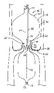
the calcification to break-up.
[0003] The distance between the shock wave generator (the electrodes) and the
valve leaflets
of the catheter described above is variable and not controlled. It has been
found that shock wave
therapy designed to break calcium deposits is most effective at certain
distances from a radiating
shock wave source. This is particularly the case when the source is a point
source without a
reflector. Generally, the effectiveness of the shock waves falls off or
decreases with the square
of the distance from the source.
[0004] When a valvuloplasty balloon and a shock wave generator are combined as
described
above, the distance between the shock wave generator and the balloon walls
generally increases
as the valve is opened by balloon expansion occasioned by effective treatment
and valvuloplasty
pressure. As the distance changes and becomes greater, the effectiveness of
the therapy
decreases. This increases both the time and the number of shock waves required
for complete
1
CA 2881184 2018-12-05
CA 02881184 2015-02-04
WO 2014/025620 PCT/US2013/053292
and effective treatment. Hence, there is a need for a shock wave valvuloplasty
catheter that
maintains therapy effectiveness at a desired level until the valve being
treated is dilated the
desired amount.
SUMMARY
[0005] According to embodiments shown and described herein, a catheter, which
may find
use, for example, in valvuloplasty, includes an elongated body and an
inflatable balloon carried
by the elongated body. The balloon has an inner surface and an outer surface.
The catheter
further includes at least one shock wave source within the inflatable balloon
and a follower
arrangement that maintains the at least one shock wave source a substantially
fixed distance
from the inner surface of the balloon.
[0006] The follower arrangement may be carried by the at least one shock wave
source within
the inflatable balloon. The at least one shock wave source may be an arc
generator including an
electrode pair.
[0007] The follower arrangement may include at least one stand-off extending
from the
electrode pair. The stand-off may be formed of flexible material.
[0008] The arc generator may include an elongated lead. The electrode pair may
be carried by
the elongated lead, and the elongated lead may be biased in a direction
towards the inner surface
of the inflatable balloon. The elongated lead may include at least one bend
that biases the
elongated lead towards the inner surface of the inflatable balloon.
[0009] The catheter may further include a biasing member carried by the
elongated lead that
biases the elongated lead towards the inner surface of the inflatable balloon.
The biasing
member may be a spring.
[0010] The at least one shock wave source may include an arc generator. The
follower
arrangement may include a stand-off carried by the arc generator and the arc
generator may be
biased towards the inner surface of the inflatable balloon.
[0011] The catheter may further include a frame structure that carries the at
least one shock
wave source. The frame structure may be arranged to expand with inflation of
the inflatable
balloon to maintain the at least one shock wave source a substantially fixed
distance from the
2
CA 02881184 2015-02-04
WO 2014/025620 PCT/US2013/053292
inner surface of the balloon. The frame structure may include at least one
stand-off adjacent the
at least one shock wave source to maintain the at least one shock wave source
a substantially
fixed distance from the inner surface of the balloon.
[0012] In other embodiments, a method includes the steps of providing a
catheter including an
elongated body, an inflatable balloon carried by the elongated body and having
an inner surface
and an outer surface, and at least one shock wave source within the inflatable
balloon. The
method further includes the steps of inserting the catheter into a vein or
artery of a patient and
placing the balloon adjacent to an anatomical structure to be treated,
inflating the balloon with a
fluid, causing the shock wave source to provide shock waves within the balloon
that propagate
through the liquid to treat the anatomical structure, and maintaining the at
least one shock wave
source a substantially fixed distance from the inner surface of the balloon
while the shock waves
are provided by the at least one shock wave source.
[0013] The catheter may further include a follower carried by the shock wave
generator, and
the maintaining step may include biasing the follower against the inner wall
of the balloon.
[0014] The catheter may include a frame structure that carries the at least
one shock wave
source. The maintaining step may include expanding the frame structure with
inflation of the
inflatable balloon to maintain the at least one shock wave source the
substantially fixed distance
from the inner surface of the balloon.
BRIEF DESCRIPTION OF THE DRAWINGS
[0015] The features of the present invention which are believed to be novel
are set forth with
particularity in the appended claims. The various described embodiments of the
invention,
together with representative features and advantages thereof, may best be
understood by making
reference to the following description taken in conjunction with the
accompanying drawings, in
the several figures of which like reference numerals identify identical
elements, and wherein:
[0016] FIG. 1 is a partial cut away view of a heart and a catheter embodying
aspects of the
invention within the aortic valve of the heart;
[0017] FIG. 2 is a side view of a shock wave generator which may be used to
advantage
within the catheter of FIG. 1 and which embodies aspects of the invention;
3
CA 02881184 2015-02-04
WO 2014/025620 PCT/US2013/053292
[0018] FIG. 3 is a partial cut away view of the heart of FIG. 1 illustrating
the catheter as it is
delivering therapy to the aortic valve of the heart;
[0019] FIG. 4 is a partial cut away view of the heart of FIG. 1 illustrating
the catheter upon
completion of therapy to the aortic valve of the heart;
[0020] FIG. 5 is a partial cut away view of another heart and another catheter
embodying
further aspects of the invention within the aortic valve of the heart; and
[0021] FIG. 6 is a partial view to an exploded scale illustrating particular
aspects of the
catheter of FIG. 5.
DETAILED DESCRIPTION
[0022] Referring now to FIG. 1, it is a partial cut away view of the aorta 50
of a heart and a
catheter 10 embodying aspects of the invention within the aortic valve 52 of
the heart. The
catheter 10 generally includes an elongated body 12, an inflatable balloon 14
carried by the
elongated body 12, at least one shock wave source 20 within the inflatable
balloon 14, and a
follower arrangement 30. The balloon includes an inner surface 16 and an outer
surface 18. The
follower arrangement 30 is carried by the shock wave source 20. As will be
seen subsequently,
the follower arrangement maintains the at least one shock wave source 20 a
substantially fixed
distance from the inner surface 16 of the balloon.
[0023] The balloon 14 is inflatable through the elongated body 12 with a fluid
such as, for
example, saline. The balloon is configured so that when positioned within the
aortic valve 52,
its outer surface 18 substantially conforms to and is immediately adjacent to
or in contact with
the aortic valve leaflets 54 and the calcification 56 thereon.
[0024] The shock wave source 20 preferably is an arc generator that produces
electrical arcs
that form rapidly expanding and contracting steam bubbles within the balloon
14. The rapidly
expanding and contracting steam bubbles form shock waves within the balloon 14
that propagate
through the fluid within the balloon and impinge upon the inner surface 16 of
the balloon 14 and
the calcification 56. After repeated shock waves, the calcification is broken
up to permit the
aortic valve 52 to function. The follower arrangement 30 maintains the shock
wave source a
substantially fixed distance from the inner surface 16 of the balloon 14 and
hence the valve
4
CA 02881184 2015-02-04
WO 2014/025620 PCT/US2013/053292
leaflets 54 to maintain full effectiveness of the shock waves during the shock
wave application
procedure.
[0025] FIG. 1 also shows that the catheter 10 is arranged to accept a guide
wire 70. The guide
passes through a guide wire lumen 72 and serves to guide the catheter into an
artery or vein to
place the balloon adjacent an anatomical structure to be treated such as an
aortic valve. Once the
balloon is thus positioned, it may be inflated and the shock wave therapy
begun.
[0026] As may be seen in FIG. 2, the shock wave source or generator 20
includes an elongated
lead 22 and an electrode pair 24 carried by the lead 22. The electrode pair 24
is formed by a pair
of coaxially disposed electrodes including a ring electrode 26 and a center
electrode 28. Voltage
pulses are applied across electrodes 26 and 28 through the lead 22 to cause
the arcs which
produce the shock waves.
[0027] The catheter 10 of FIG. 1 includes two shock wave sources 20 and 20'.
The shock
wave source 20' may be identical to the shock wave source 20. Each shock wave
source carries
a follower arrangement. In the embodiment of FIG. 1, a spring 38 is attached
to and in between
the leads 22 and 22' of the shock wave sources 20 and 20', respectively. The
spring 38 serves as
a biasing member to force the electrode pairs of the shock wave sources and
the follower
arrangements off of the center axis 15 of the balloon 14 towards the inner
surface 16 of the
balloon 14.
[0028] Alternatively, or in addition, as may be seen in FIG. 2, the lead 22
may have permanent
bends 34 and 35 formed therein. The bends bias the electrode pair 24 in the
direction indicated
by arrow 36 towards the inner surface 16 of the balloon 14.
[0029] Hence, FIG. 1 shows a valvuloplasty system having a catheter 10
according to some
aspects of the invention that includes a valvuloplasty balloon 14 with two
electrodes (electrode
pair 24) disposed therein. The system is shown within an aortic valve 52 for
treating
calcification 56 on the valve leaflets 54. The electrodes are urged away from
the center axis 15
of the balloon 14 toward the perimeter of the balloon 14 by a spring member
38. As may be
appreciated, the spring member may be replaced by spring loading or biasing
the leads 22 and
22' that carry the electrodes outwardly. The balloon 14 is shown within a
severely stenosed
valve 52. Stand offs 32 carried on electrodes maintain a substantially
constant distance between
CA 02881184 2015-02-04
WO 2014/025620 PCT/US2013/053292
electrodes and the walls of the balloon 14 and hence between the electrodes
and the valve
leaflets 54.
[0030] Further, FIG. 2 shows a detailed view of one electrode pair 24 and its
lead 22. The
standoffs 32 are formed by soft flexible arms that are designed to hold the
electrode pair 24 off
the balloon wall in non-touching relation to the balloon material. They are
also designed to hold
the tip of the electrode pair 24 a substantially constant distance, for
example, 1-2 mm, from the
balloon wall. At the same time, according to this embodiment, the elongated
lead 22 has bends
34 and 35 to provide a predetermined bias toward the outside (away from the
center axis) of the
balloon.
[0031] FIG. 3 is a partial sectional view showing the valvuloplasty balloon 14
placed in an
aortic valve 52 and after providing some treatment to break up or sever the
calcium deposits 56
on the valve leaflets 52. The electrode pairs 24 have been held a
substantially constant distance,
for example about 1-2 mm, from the tissue by the stand offs since the electro-
hydraulic shock
therapy began. As the shock waves break the calcium, the opening 60 in the
valve 52 slowly
widens. Even though the valve is being opened wider, the distance between the
electrode pairs
24 and the tissue of the leaflets 54 remains substantially constant,
controlled by the stand offs 32
and the bends 34 and 35 in the electrode leads.
[0032] FIG. 4 shows a fully opened opening 60 of valve 52 expanded by the
combination
valvuloplasty balloon 14 and the shock wave therapy. The bias in the catheter
and the standoffs
hold the electrode pairs a substantially constant distance from the tissue of
the valve being
treated. For simplicity, only two electrode pairs are shown. However, in
actual practice, as
many as 3-9 electrode pair may typically be used. The electrode pairs 24 can
be fired (provided
with arc forming voltage) alternately or simultaneously. The calcium on the
valve 52 and its
softened valve leaflets 54 (and valve cusps) is now cracked making the valve
much better
prepared for the placement of a TAVI (Transcatheter Aortic-Valve Implantation)
valve. In
addition, the native valve 52 may function on its own without a replacement.
[0033] FIG. 5 shows an alternate embodiment. Here, a catheter 110 includes an
elongated
body 112 and an inflatable balloon 114, as in previous embodiments. Here,
however, the shock
wave sources 120, which may be electrode pairs, are mounted on a basket or
frame structure 122
having basket arms or frame elements 124. The basket arms 124 may be formed of
Nitinol and
6
CA 02881184 2015-02-04
WO 2014/025620 PCT/US2013/053292
may be set to expand with the balloon 114 as the stenosis of the aortic valve
being treated is
softened and expanded by the shock waves.
[0034] FIG.6 shows the Nitinol arms 124 in greater detail with respect to the
shock wave
sources 120. Here it may be seen that the arms 124 may be configured with
bumps or stand offs
132 to hold the shock wave sources 120 away from the balloon and tissue a
substantially fix
distance during the shock wave treatment.
[0035] FIG. 6 also shows that, as in previous embodiments, the catheter 112
may
accommodate a guide wire 170. The guide wire 170 may be received within a
guide wire lumen
172 and used, as previously described, to guide the catheter into proper
position.
[0036] While particular embodiments of the present invention have been shown
and described,
modifications may be made, and it is therefore intended to cover in the
appended claims all such
changes and modifications which fall within the true spirit and scope of the
invention.
7