Note: Descriptions are shown in the official language in which they were submitted.
CA 02881672 2015-02-10
WO 2014/026126
PCT/US2013/054362
1
METHOD AND APPARATUS FOR DERMATOLOGICAL TREATMENT
CROSS-REFERENCE TO RELATED .APPLICATION(S)
(00011 The present application relates to and claims priority from
U.S. Provisional
Patent Application Serial No. 611681,992 filed August 10, 2012, the present
disclosure of
which is incorporated herein by reference in its entirety,
TECHNICAL FIELD
100021 The present disclosure relates to cosmetic methods and
apparatus for fractional
photothermolysis of skin and other tissues, in which a plurality of enemy
pulses having
different properties can he provided onto a single location on the tissue, so
as to form a hole
therein, coagulate tissue within the hole, and then ablate at least a portion
of the coagulated
tissue.
BACKGROUND INFORMATION
(00031 Fractional skin resurfacing relates to a cosmetic procedure
where small
regions of thermal damage are formed in skin, tissue using electromagnetic
energy, e.g.
electromagnetic radiation (EMR), such as a laser beam. Each region is
preferably small, e.g.,
less than 1 mm in diameter or less than 0.5 mm in diameter, and surrounded by
substantially
unaffected, healthy tissue. For example, the areal fraction of skin surface
area covered by
damaged tissue after a conventional fractional resurfacing treatment can be
typically between
about 5% and about 40-50%. Lower areal fractions can result in faster healing
times and/or
less disruption of the epidermis, but the beneficial cosmetic effects may also
be reduced
because of the smaller volume of tissue damaged. Higher areal fractions can
result in
increased Skin tightening, more rejuvenation and/or other desirable cosmetic
effects, but
healing times may be longer because of the larger amount of epidermal tissue
that is
thermally damaged or removed.
100041 The damaged regions of tissue can be generated by heating and/or
ablation.
Ablation can occur when the EMR is of sufficiently high intensity,
sufficiently absorbed. by
the tissue, and applied in a sufficiently short time to vaporize a portion of
the tissue, which
can lead to formation of holes in the tissue and/or removal of further tissue
in an existing
hole. If the energy is applied to the tissue with. sufficient power intensity
in a time that is
CA 02881672 2015-02-10
WO 2014/026126
PCT/US2013/054362
2
shorter than the thermal relaxation time of the tissue, increased localized
energy absorption
and ablation of tissue can occur,
100051 Thermal damage in tissue can also be produced by EMR having a
lower
intensity and/or longer pulse duration, such that the energy absorbed by the
irradiated tissue
is heated but not vaporized. Such milder thermal damage can "cook" the tissue,
e.g.,
coagulate tissue and/or denature proteins such as collagen, which may result
in local death of
some cells and damage to other cells. EMR can also be delivered at
intermediate times and
intensities, such that both tissue ablation and significant coagulation occur
in the irradiated
tissue.
100061 In dermatological applications, EMR. generated by a laser is often
provided in
a form of one or more pulses, where the parameters associated with a pulse of
EMR can
include, e.g., wavelength of the energy, total pulse energy (in J or mJ), beam
width or
diameter, and duration of a pulse. For example, the power of an EMR pulse can
be
determined as the total energy of the pike divided by the duration of the
pulse. Further, the
intensity of a pulse can be determined as the power of a pulse divided by a
cross-sectional
area of the pulse beam. This intensity can be taken as an average intensity,
because the
energy and/or power may not be uniformly disttibuted over the temporal
duration of the pulse
and/or the area of the beam.
100071 Different effects on tissue can be provided by EMR beams having
differing
properties, which is well-recognized in the art. For example, for a single
laser source
emitting EMR at a particular wavelength, one or more beam parameters can be
varied to
produce different effects in tissue. If a beam pulse is provided with a
particular total eneruy,
reducing the pulse duration will result in a higher pulse power, and reducing
the pulse
duration and/or area or width of the pulse can result in a higher intensity.
Similarly, the
intensity of a pulse at. a. fixed beam diameter and duration (or pulse length)
can be increased
by increasing the total energy of the pulse.
100081 Certain types of lasers such as, e.g., an excimer laser, may
produce EMR. that
predominantly ablates biological tissue without generating significant heating
or coagulation
of nearby tissue, even when parameters of a pulse or beam are varied. Other
types of laser
sources, e.g., CO2 lasers, Er:YAG lasers, or Nd:YAG lasers, can be used to
ablate tissue if the
EMR is provided at a sufficiently high intensity, and can also be adapted to
heat or coagulate
tissue with little and/or no ablation when beam parameters are selected to
generate pulses
CA 02881672 2015-02-10
WO 2014/026126
PCT/US2013/054362
3
having lower intensities. These lasers are commonly referred to as "ablative"
lasers. In
contrast, certain low-power laser sources or lasers emitting EMR at certain
weakly-absorbed
wavelengths such as, e.g., some diode lasers, may be better suited for
thermally damaging
tissue and may not be capable of generating sufficient intensity of absorbed
EMR to ablate
tissue under typical operating conditions. Such lasers are often referred to
as "non-ablative"
lasers.
100091 For example, EMR provided by a CO2 laser at a wavelength of
about 10,600
mu is well-absorbed by water, and can ablate skin tissue or other biological
tissue when
provided in a beam of sufficiently high intensity. The pulse duration and
energy of a CO2
laser can be varied, e.g., at a fixed beam width, to generate EMR that can
ablate tissue, cause
thermal damage in tissue without any Ablation or vaporization, or to cause
thermal damage
with varying degrees of ablation. Exemplary parameters for various laser
sources that can be
used to generate ablation, coagulation, or a mix of both effects in tissue,
including general
relationships between such parameters, are known in the art for various types
of lasers. For
example, general parameter limits for producing ablation, coagulation with no
ablation, or a
mix of both effects in skin tissue using a CO2 laser are described, e.g., in
U.S. Publication No.
2006/0149223 Al by Hwang et al., and in U.S. Patent No. 6,159,204 issued to
Hibst.
100101 Regions of thermal damage can generate collagen shrinkage,
coagulation,
and/or a wound healing response that can lead to such effects as an overall
tightening of the
skin tissue and improved appearance in the treated area. Although some
tightening of the
skin occurs over time, it would be desirable to have fractional resurfacing
methods and
apparatus that facilitate faster healing times while generating a desirable
rejuvenation effect
in the treated skin tissue.
100111 Accordingly, there may be a need to address and/or overcome at
least some of
the issues indicated herein above.
SUMMARY OF EXEMPLARY EMBODIMENTS
100121 According to exemplary embodiments of the present disclosure,
method and
apparatus can be provided for ablative fractional skin resurfacing that.
includes forming a
small ablated hole (e.g., less than 1 mm in diameter, or less than about 0.5
min) and then
directing one or more further pulses into the hole sequentially to generate
further tissue
coagulation with little or no further tissue ablation, followed by one or more
pulses to ablate a
portion of the coagulated tissue. Such pulses can be generated using an
ablative laser (e.g., a
CA 02881672 2015-02-10
WO 2014/026126
PCT/US2013/054362
4
CO2 laser, a CO laser, an erbium laser, frir example, an Er:YAG or F.EYSGG
laser, e.g., a
CO2 laser, a CO laser, an erbium laser such as an ErNAG or &NSW laser, and/or
another
type of YAG laser such as a Tm:YAG, "Ho:YAG, or a Nd:YAG laser, or the like),
and
optionally by using an additional non-ablative laser. Parameters of the laser
pulses can be
selected and controlled using, e.g., a control arrangement, to provide a
plurality of pulses
onto a single target location on the skin to achieve these effects.
(0013i For example, the energy pulses can be generally categorized as
one of three
general types. For example, a pulse of highly ablative electromagnetic
radiation or energy
(an "HA pulse") can be used, e.g., to ablate a deep hole or to remove at least
some
to previously-uncoagulated tissue within or proximal to a hole,.without
generating any
-significant coagulation of adjacent tissue. A pulse of mildly or moderately
ablative
electromagnetic energy (an "MA pulse") can be configured generate a
significant. amount of
non-ablative heating of adjacent tissue to produce further coagulation and/or
thermal damage
of tissue whit a relatively small amount of further tissue ablation, e.g., a
portion of coagulated
tissue that may be present within or proximal to an ablated hole. A pulse of
non-ablative
electromagnetic energy (an "NA pulse") can be configured to heat and coagulate
tissue
within or proximal to a hole with no associated tissue ablation or
vaporization.
100141 In certain exemplary embodiments of the present disclosure, a
plurality of
holes can be formed in the tissue, either simultaneously or sequentially
using, e.g., methods
and apparatus similar to those used for conventional ablative fractional
resurfacing. A width
or diameter of an ablative EMR beam used to ablate such holes can be less than
1 mm, e.g.,
between about 0.1 rum and 0.5 nun. A hole formed by ablation using such a beam
can have
a width comparable to the beam width at the tissue surface, e.g., less than
about 1 mm or
between about 0.1 mm and about 0.5 mm.
100151 Individual holes can be further processed by directing one or more
further
pulses of energy into the hole. Various sequences of HA pulses, MA pulses,
and/or NA
pulses can be provided to generate particular effects in tissue. For example,
after a deep hole
is ablated using an HA or predominantly ablative MA pulse, one or more NA
pulses and/or
predominantly coagulative MA pulses can then be directed into the hale to
coagulate more
tissue within the hole. A portion of this coagulated tissue can then be
ablated by directing
one or more MA or HA pulses into the hole. This exemplary procedure can
optionally be
repeated a number of times in a single hole to alternately form coagulated
tissue within the
hole and then remove at least a portion of it by ablation. Such exemplary
pulse sequences
CA 02881672 2015-02-10
WO 2014/026126
PCT/US2013/054362
may reduce healing or recovery times, for example, by directing further pulses
of energy
(EMR) into each ablated hole without a significantly increase in the hole
depth or width.
Rejuvenation effects (e.g. new collagen growth and/or skin tightening)
associated with each
such hole may be comparable or greater than that resulting from, e.g., a
single ablative pulse
5 having the same total energy as the sequence of pulses.
1001(1 According to further exemplary embodiments of the present
disclosure, a
plurality of MA and/or NA pulses can be directed into the ablated hole after
the ablative
pulses to generate more coagulated tissue therein, such that the hole can be
at. least partially
filled with coagulated tissue. In this exemplary manner, a larger amount of
tissue removal
1.0 and shrinkage can be generated in each hole without enlarging the hole
dimensions, which
may facilitate healing and/or reduce healing times, and can also lead to
enhanced tissue
rejuvenation effects for the procedure.
100171 in additional exemplary embodiments of the present disclosure,
system and
apparatus can be provided for directing pulsed electromagnetic energy onto
biological tissue.
Such exemplary system and apparatus can include a source of electromagnetic
radiation
(EMR) and a control arrangement, which can be configured to provide a
plurality of pulses
having predetermined parameters onto a single location of the tissue.
Exemplary parameters
that can be controlled for each pulse can include, e.g., energy, duration,
power, beam width,
etc. Exemplary parameters that can be controlled fin a plurality or sequence
of pulses
include, e.g., the number of pulses and intervals or delays between successive
pulses. The
control arrangement can include a microprocessor and/or other processing
arrangement, and
can be configured to control the exemplary parameters associated with each
pulse, as well as
timing intervals or delays between successive pulses directed onto a. single
location in the
tissue. The exemplary system and apparatus can further include an optical
arrangement
configured to direct a plurality of pulses provided by the laser source(s)
onto each of a
plurality of particular locations on the tissue.
100181 The exemplary system and apparatus can include one or more
sources of
electromagnetic energy, e.g., one or more lasers. For example, the apparatus
can include two
or more lasers, each capable or configured to produce EMR at a different
wavelength, e.g., an
ablative laser and a non-ablative laser. In further embodiments, the system
can include a
single laser such as, e.g., a CO2 laser or an erbium laser. Such single-laser
systems can be
configured to generate Ablative, non-ablative, and/or partially ablative
pulses based on the
parameters selected for each pulse. For example, pulses having higher energies
and/or power
CA 02881672 2015-02-10
WO 2014/026126
PCT/US2013/054362
6
and/or shorter durations may be more ablative, whereas pulses provided at
lower energy or
power and/or longer durations can be non-ablative or only mildly ablative.
100191 According to still further exemplary embodiments of the present
disclosure, a
method can be provided for directing pulsed electromagnetic energy onto
biological tissue.
The exemplary method can direct at least one ablative pulse of EMR onto a
particular
location on the tissue to ablate a hole therein, then direct at least one
further pulse of EMR
onto the same location, e.g., into the ablated hole, to generate tissue
coagulation therein, and
then direct at least one further EMR pulse onto the same location to ablate at
least a portion
of the coagulated tissue formed. In additional exemplary embodiments of the
present
disclosure, the exemplary method. can facilitate directing at least one
further non-ablative or
mildly ablative pulse of EMR onto the same location (e.g., into the hole) to
coagulate further
tissue in the hole and at least partially fill the hole.
100201 These and other objects, features and advantages of the present
disclosure will
become apparent upon reading the following detailed description of exemplary
embodiments
of the present disclosure, when taken in conjunction with the appended
drawings and claims.
BRIEF DESCRIPTION OF UhF DRAWING
[0021] Further objects, features and advantages of the present
disclosure will become
apparent from the following detailed description taken in conjunction with the
accompanying
figures showing illustrative embodiments, results and/or features of the
exemplary
embodiments of the present disclosure, in which:
100221 FIG. I shows two exemplary histograms of a section of skin
tissue that has
been exposed to -five spatially-distinct laser pulses having the same total
energy but different
powers and durations;
100231 FIGS. 2A-2D are exemplary schematic diagrams illustrating the
effect of
ablative and non-ablative energy pulses on a hole made in biological tissue,
in accordance
with exemplary embodiments of the present disclosure;
100241 FIG. 3 is an exemplary apparatus for producing particular
sequences of laser
pulses in accordance with embodiments of the present disclosure;
100251 FIG. 4 shows two exemplary histograms of a section of skin
tissue where
different locations have each been exposed to a sequence of laser pulses, and
the order of the
sequences varied between locations: and
CA 02881672 2015-02-10
WO 2014/026126
PCT/US2013/054362
7
100261 FIG. 5 is a bar graph of exemplary experimental observations of
the effects of
various pulse sequences on irradiated skin tissue.
100271 Throughout the drawings, the same reference numerals and
characters, unless
otherwise stated, are used to denote like features, elements, components, or
portions of the
illustrated embodiments. . Similar features may thus be described by the same
reference
numerals, which indicate to the skilled reader that exchanges of features
between different
embodiments can be done unless otherwise explicitly stated. Moreover, while
the present
disclosure will now be described in detail with reference to the figures, it
is done so in
connection with the illustrative embodiments and is not limited by the
particular
embodiments illustrated in the figures. It is intended that changes and
modifications can be
made to the described embodiments without departing from the true scope and
spirit of the
present disclosure as defined by the appended claims.
DETAILED DESCRIPTION OF THE PREFERRED EMBODIMENTS
[00281 According to exemplary embodiments of the present disclosure,
method and
apparatus can be provided for directing energy onto biological tissue, e.g.,
for fractional skin
resurfacing, that includes ablating a plurality of spaced-apart holes over a
region of the tissue
surface using electromagnetic radiation (EMR) or energy, such as optical
energy produced by
a laser, and then directing one or more further pulses of EMR having different
parameters
into at least some of the holes to generate coagulated tissue therein and/or
ablate at least a
portion of the coagulated tissue that has formed therein. Further pulses can
be directed into
the same hole to coagulate and/or remove additional tissue from the holes.
Further
coagulation can be generated using pulses generated by the ablative energy
source, optionally
at a lower power level and/or longer pulse duration, and/or by a further
source of
electromagnetic energy that is non-ablative.
100291 The energy pulses can be generally categorized by their typical
effects when
directed onto biological tissue such as skin. For example, a highly ablative
electromagnetic
energy pulse (an "HA pulse") can vaporize tissue and thereby ablate a deep
bole, e.g., to
remove at least some previously-uncoagulated tissue, and not for a significant
amount of
coagulation in adjacent tissue. A mildly ablative electromagnetic energy pulse
(an "MA
pulse") can ablate tissue, e.g., a portion of coagulated tissue previously
formed within or
proximal to an ablated hole, while also being absorbed sufficiently by
adjacent tissue to
generate additional coagulation. A non-ablative electromagnetic energy pulse
(an "NA
CA 02881672 2015-02-10
WO 2014/026126
PCT/US2013/054362
8
pulse") can heat tissue locally to coagulate a portion of it within or
proximal to a hole, with
no associated tissue ablation or vaporization.
100301 Exemplary lasers that can be used in embodiments of the present
disclosure
include, e.g., a CO2 laser, a CO laser, an erbium laser such as an Er:YAG or
Er:YSGG laser,
a Im:YAG or Ho:YAG laser, or the like. The exemplary laser can be configured
to provide
EMR in one or more pulses In certain exemplary embodiments of the present
disclosure, a
single laser source such as the ones listed in this paragraph can be used.,
where the laser can
be controlled to provide pulses of EMR that can ablate or vaporize skin
tissue, and further
pulses of EMR having properties that can cause thermal damage in tissue
without ablating it.
1.0 According to further exemplary embodiments of the present disclosure,
at least two laser
-sources can be used, where at least one source is capable of providing pulsed
energy that can
ablate skin tissue, and a second laser source that is capable of providing
EMR. pulses that
cause thermal damage in skin tissue without ablating such tissue.
100311 The exemplary characteristics of an electromagnetic energy
pulse (HA, MA,
NA) can be determined by several parameters or combinations thereof including,
for
example, the wavelength(s) of the radiation (which can affect the absorption
of energy by the
tissue), the type of tissue, the total energy or power of the pulse, the beam
diameter, pulse
length/duration, and the tissue temperature (e.g.. pre-heating). For example,
for particular
total pulse energy, decreasing the pulse duration and/or beam width can lead
to a higher local
power or energy intensity, which may tend to produce more ablation of tissue.
In contrast,
increasing the pulse duration and/or width for a particular total pulse energy
can decrease the
pulse power and intensity, which can lead to more heating or coagulation of
tissue with little
or no removal of tissue by ablation,
100321 As an example, fora CO2 laser emitting pulses of EMR having, a
beam width
less than 1 mm in diameter (e.g., between about 0,1 mm and 0,5 mm), an HA
pulse can have
a total energy of between about 5 in.1 and 500.mi, and duration between about
0,05 ms and 1
ms, with longer pulse durations corresponding to higher pulse energies (for a
particular beam
width) to provide sufficient power intensity to primarily ablate the tissue.
Similarly. NA
pulses can have pulse energy between about 5 m.1 and 500 in.3, and longer
pulse duration
between about 0.5 ms and 100 ms, with higher pulse energies corresponding to
longer pulse
durations to reduce the effective power and intensity, thereby avoiding
vaporization of tissue.
MA. pulses can have properties with intermediate parameter values in these
exemplary ranges
provided for HA and NA pulses, e.g., similar pulse energies and somewhat
longer pulse
CA 02881672 2015-02-10
WO 2014/026126
PCT/US2013/054362
9
durations than HA pulses, e.g., pulse durations up to about 5 ms or 10 ms, or
even longer at
pulse energies in the higher end of the exemplary HA range, which may still be
used to ablate
tissue.
100331 As a further example, for an ErNAG laser emitting pulses of EMR
having a
beam width less than 1 mm in diameter (e.g., between about 0.1 mm and 0.5 mm),
an HA.
pulse can have a total energy of between about 100 rd and 1000 mi, and
duration between
about 0.1 ins and 2 ms, with longer pulse durations corresponding to higher
pulse energies to
provide sufficient power intensity to primarily ablate the tissue. Similarly,
NA pulses can
have pulse energy between about 10 mi and 500 mJ, and pulse duration between
about 5 ins
and 200 ms, with higher pulse energies corresponding to longer pulse durations
to avoid.
vaporization of tissue. Again. MA pulses can have properties with intermediate
parameter
values in these exemplary mum provided for HA and NA palm, or somewhat longer
pulse
durations than HA pulses for similar pulse energies, e.g., pulse durations up
to about 20 ms or
50 ms, or even longer at pulse energies in the higher end. of the exemplary HA
range, which
may still be used to ablate tissue.
100341 In general, for a particular beam width and laser emitting EMR
at a particular
wavelength (and thus a particular absorption value in skin tissue), shorter
pulse durations and
higher pulse energies will result in a higher pulse power and intensity (for a
particular beam
width or diameter), and tend to be more ablative. Conversely, longer pulse
durations with.
lower pulse energies tend to be non-ablative or less ablative, producing more
thermal damage
in the tissue while vaporizing little or no tissue. Because the ablative/no-
ablative
Characteristics of a pulse depend on a plurality of factors, such as pulse
energy, duration and
width (at a particular wavelength having a particular absorption coefficient
in tissue), ranges
of parameter values for HA, MA and NA beams can overlap, as indicated above.
For
example, a 500 nil pulse from a CO z laser that is 0.5 mm wide can be ablative
lithe pulse
duration is short, on the order of a few milliseconds, and it can be mildly
ablative or non-
ablative if the pulse duration is longer, e.g., a few hundred milliseconds.
100351 Specific parameters for a laser emitting EMR at a particular
wavelength,
which correspond to HA, NA, or MA pulses, are known in the art and/or can be
readily
determined by simple calculations based on the known absorptivity of the EMR
at the emitted
wavelength and the known vaporization enthalpy and thermal relaxation time of
skin tissue,
for a particular beam width or diameter. if multiple pulses of EMR are
provided in
succession, with short intervals between them, the pulse energy and/or
duration may be
CA 02881672 2015-02-10
WO 2014/026126
PCT/US2013/054362
decreased somewhat on later pulses to account for local preheating effects in
the tissue,
which can lower the power intensity to ablate tissue.
100361 Further, certain lasers emitting EMR at a wavelength having a
relatively low
absorption coefficient in skin tissue may not be capable of ablating
biological tissue because
5 of limitations on pulse energies and pulse durations. Such non-ablative
lasers can, e.g., be
used with exemplary embodiments of the present disclosure to provide NA
pulses.
100371 Two cross-sectional exemplary images of a histological sample
showing the
effects of several exemplary laser pulses on skin tissue are shown in FIG. I.
The five
spatially distinct pulses were generated by a CO,. laser. The total energy of
each pulse was
10 set to 100 mJ, and each pulse had a power density of 25 W/cm2 and a
fluence of 2,5 I/Q.1n,
(based on the beam width or spot size). However, the power and duration of
each pulse were
varied, as indicated by the parameter labels above each pulse location. The
leftmost pulse
had the highest power level tested, 40 W, corresponding to the shortest pulse
duration o12.5
ins. The rightmost pulse had the 'lowest energy, I W, and the corresponding
largest duration
of 100 ins. The three intermediate pulses had (left to right) a pulse power of
20 W, 10 W,
and. 5 W, and corresponding pulse durations of 5 ms, 10 ms, and 20 ms,
respectively.
100381 As can be seen in FIG, 1, the pulse with the highest energy and
shortest
duration ablated the deepest hole in thetissue,. As pulse power decreases left
to right, the
corresponding depths of the ablated holes become shallower. The. rightmost
pulse barely
ablated any tissue, and instead coagulated a greater volume of tissue. In
terms of the
nomenclature used herein, the 40 W/2,5 ms pulse and the .20 W15 ms pulse may
be
considered to be moderately ablative (MA) pulses that predominantly ablate
tissue with a
relatively small amount of associated coagulation or thermal damage. The 10
Wilt) Ms pulse
and. the 5 Wi20. Ms pulse may be considered to be mildly ablative (MA) pulses.
The 1 WS1.00
ins pulse may be Considered to be a substantially non-ablative(NA) pulse.
100391 The exemplary results shown in FIG. I illustrate that a single
ablative laser
can be used to generate these different types of pulses by varying the
parameters associated
with them, such as pulse power and pulse duration. Each of the pulses directed
onto the
tissue in FIG. 1 had the same total energy of 100 mJ. However, other
parameters such as,
e.g., the spot size and/or energy of individual pulses can also be varied to
produce particular
degrees of ablation and/or coagulation when they interact with tissue.
CA 02881672 2015-02-10
WO 2014/026126
PCT/US2013/054362
11
100401 According to certain exemplary embodiments of the present
disclosure,
methods and apparatus can be provided for ablative fractional skin resurfacing
that includes
forming a small ablated hole using an EMR beam having a width that is, e.g.,
less than 1 mm
in diameter, or between about 0.1 mm and U.S mm, which can ablate holes in the
tissue
having comparable widths. The EMR beam can comprise one or more HA pulses
and/or one
or more MA pulses, e.g., mildly or moderately-ablative pulses that ablate
sufficient tissue to
generate a hole that extends at least into the demi& Each of the ablated holes
can extend
from the skin surface through the epidermal layer and to at least a particular
depth within the
dermis. The fraction of the skin surface in a treatment area that is covered
by such holes can
be, e.g., between about 0,05 and 0.5 (5-50%), for example, between 0.1 and 0.4
(10-40%).
These exemplaiy ranges of hole sizes and surface areal coverage can be
sufficiently small to
allow each damaged region to be surrounded by healthy tissue, which can speed
healing and
reduce or avoid an appearance of visible markings, while being large enough to
generate a
cosmetically desirable effect such as rejuvenation, shrinkage, wrinkle
reduction, and/or a
reduction in appearance of pigmented defects.
100411 After the ablated holes are formed, one or more further pulses
can be directed
onto the same location as the ablative beam, e.g., into the ablated holes, to
generate further
tissue coagulation and thermal damage. Such pulses can be generated using an
Ablative laser
(e.g., a CO2 laser or an erbium laser) with parameters selected to produce
pulses with lower
power and/or intensity (e.g., MA or NA pulses), In certain exemplary
embodiments of the
present disclosure, the further pulses can be provided by a second laser
source that produces
EMR having a different wavelength, e.g., a non-ablative laser.
100421 Exemplary parameters of the laser pulses can be selected and
controlled using,
e.g., a control arrangement, to provide pulses having various characteristics
as described
below. In general, energy pulses having higher power and shorter duration tend
to more
strongly ablate tissue, vaporizing a portion thereof based on the large amount
of energy that is
absorbed by the tissue in a short time (e.g., shorter than a local thermal
relaxation period).
For such ablative pulse, the energy tends to heat up tissue quickly enough to
vaporize a
portion thereof. Such highly ablative pulses tend to have a very thin thermal
affected zone
around the ablated volume. In contrast, pulses having lower power and longer
durations tend
to heat up tissue more slowly, such that some energy can be dissipated to
surrounding tissue
and generate more tissue coagulation, protein denaturation, etc. A single
pulse of certain
types of EMR (e.g. EMR having a wavelength within certain ranges known in the
art) can
CA 02881672 2015-02-10
WO 2014/026126
PCT/US2013/054362
12
both ablate and coagulate tissue, with the relative amount of ablation and
coagulation
depending on the various parameter associated with the energy pulse. A laser
pulse having a
particular set of parameters may also affect different tissues differently,
e.g., based. on the
chromophores present, tissue structures present in the tissue, etc. For
example, the energy
generated by a CO2 laser tends to be strongly absorbed by water and thus its
effect on tissue
may depend more on the amount of water present in the tissue than on the
presence or
absence of visible chromophores such as pigments.
100431 As shown in FIG. 2A, a beam 100 of ablative energy (e.g., one
or more HA or
MA pulses) can be directed onto a particular location on the surface of tissue
110 (e.g., skin)
to to form a bole 120 therein, according to an exemplary embodiment of the
present: disclosure.
213 shows a zone of coagulated tissue 130 that can be formed around a hole 120
that was
ablated in tissue using one or more HA or MA pulses. In certain exemplary
embodiments,
such coagulated tissue 130 can be formed (or the amount of it increased) by
directing one or
more further pulses 100 of NA energy onto the same location and into the hole
120 as shown,
e.g., in FIG. 211.
100441 The coagulation tends to contract maim- shrink, tissue along
portions of the
hole, which may contain collagen or other structural proteins. Additional
tissue adjacent to
the coagulated tissue 130 may also be heated to a lesser degree and respond to
the applied
energy, such that coagulated tissue 130 may partially refill the lower portion
of the hole 120.
The time for coagulation to occur after exposure of tissue to an energy beam
.100 (e.g. a laser)
can be very short, for example, on the order of milliseconds or tens of
milliseconds.
100451 For example, one or more further laser pulses 100, e.g., HA
pulses, can be
directed onto the same location (e.g., into the hole 120), as shown in FIG.
2C, to ablate at
least a portion. .140 of the coagulated tissue130 that has formed within the
hole 120. In FIG.
2C, the boundary of the coagulated tissue 140 that was removed is indicated by
the dashed
line. This procedure facilitates enhanced tissue removal and a greater degree
of shrinkage
within a single ablated micro-hole 120 by directing a plurality of energy
pulses 100 into the
hole 120. Such further tissue coagulation and removal can be achieved, e.g.,
without
enlarging the size of the hole 120 at the surface of the tissue 110.
[00461 The exemplary procedure illustrated in FIGS. 2A-2C, e.g., tissue
ablation to
form a hole 120, .followed by coagulation of tissue in the hole 120 and
subsequent ablation of
some of the coagulated tissue 130 can facilitate ablation of more subsurface
tissue without
CA 02881672 2015-02-10
WO 2014/026126
PCT/US2013/054362
13
deepening the hole 120. The exemplary procedures shown in FIGS. 28 and 2C,
i.e., forming
coagulated. tissue 130 in the hole 120 and then ablating at least some of this
coagulated tissue
130 can be repeated to draw further tissue 110 into the lower portion of the
hole 120 and then
ablating more of it, thereby removing more tissue 110 that is proximal to
lower portions of
the hole .120 without deepening the hole significantly. In contrast, providing
stronger or a
larger number of only ablative pulses 100 of EMR would tend to deepen the hole
120 while
ablating further tissue.
100471 The exemplary coagulative and/or ablative procedures
illustrated in FIGS. 213
and 2C, respectively, can be conducted with one or a plurality of EMR pulses
100. For
example, the tissue coagulation procedure shown in FIG. 213 can be achieved by
directing
either one or a plurality of pulses .100 of NA andior MA energy into the hole
120. If a pulse
100 of MA energy is used, it can predominantly coagulate tissue 110 with only
a moderate
amount of ablation, e.g., such that the volume of coagulated tissue 130
increases after the
tissue is irradiated. Similarly, the tissue ablation shown in FIG. 2C can be
achieved by
directing either one or a plurality of pulses 100 of HA and/or MA energy into
the hole 120. If
a pulse 100 of MA energy is used, it can predominantly ablate tissue 110 with.
a relatively
small amount of coagulation, e.g., such that the volume of coagulated tissue
130 decreases
after the tissue 110 in the hole 120 is irradiated.
100481 In further exemplary embodiments of the present disclosure, one
or more non-
ablative energy pulses 100 can be directed into the hole 120, e.g., after some
coagulated
tissue 130 is ablated as shown in FIG. 2C. For example, one or more non-
ablative (NA)
pulses 100 of EMR can be directed into the hole 120 to at least partially
'fill" the hole with
coagulated tissue 130, as shown in FIG. 2D. In further exemplary embodiments,
coagulated
tissue 130 can be generated within the hole by directing one or more
predominantly non-
ablative pulses 100 (e.g., MA pulses) !Pikes into the hole 120, where the
properties of the
MA pulses can be selected such that additional tissue is coagulated with a
small or negligible
amount of such tissue being ablated. In still further exemplary embodiments of
the present
disclosure, a plurality of MA and NA pulses 100 can. be directed into the hole
120 to "fill" it
with coagulated tissue 130. In such exemplary procedures, the depth of the
hole 120 can be
decreased, by forming additional coagulated tissue 130 (as shown in FIG. 2D)
after forming
and ablating at least a portion of such coagulated tissue 130 in an ablated
hole 120 (as shown
in FIGS. 2B and 2C. Such partial "filling" of the hole 120 after forming a
small hole 120 and
then generating and removing some coagulated tissue 130 therein, can
facilitate .fitster healing
CA 02881672 2015-02-10
WO 2014/026126
PCT/US2013/054362
14
Limes while also providing, a greater amount of tissue damage associated with
the hole .120 as
compared, e.g., to the healing time and extent of tissue damage associated
with just ablating a.
hole 120 as shown, e.g., in FIG. 2A.
100491 The exemplary interval between successive pulses 100 can be
determined
based on several factors. For example, certain ones of the pulses can be
delivered with
substantially no interval between them. in further exemplary embodiments of
the present
disclosure, an interval between certain pulses 100 can be provided, e.g.,
after an NA or MA
pulse, to allow coagulation of tissue 110 to occur betbre irradiation of the
tissue with a
subsequent pulse. Such coagulation can form in several to tens of
milliseconds, e.g., based
on considerations of local thermal relaxation times. Accordingly, intervals
between
-successive pulses 100 applied to the same location can be, for example, tens
of milliseconds,
e.g.,.1 0-30 ms or more, up to 100 ms or greater. Relatively smaller intervals
may lead to
more local preheating as the tissue 110 may have less time to cool off between
pulses 100.
This cumulative preheating can be accounted for, e.g., to generate increased
coagulation
and/or ablation with subsequent pulses 100 that have the same or reduced
intensity, power,
etc. Setting appropriate exemplary pulse intervals can also vary the relative
amounts of
coagulation and ablation that are generated. e.g., by a plurality of MA pulses
having constant
pulse properties.
100501 In still further exemplary embodiments of the present
disclosure, various
combinations of beam parameters can be used to achieve improved cosmetic
effects. For
example, a wider non-ablative beam diameter can be used to generate coagulated
tissue 130
along the hole sides, and then a narrower ablative beam pulse 100 can be
applied to ablate a
portion of this tissue. Other parameters, such as beam pulse energy, pulse
duration, interval
between pulses, etc. can be varied to generate desirable patterns of
coagulation and. tissue
removal. For example, intervals between successive pulses 100 can be selected
to facilitate
formation and shrinkage of coagulated tissue 130, followed by ablation of a
portion of such
tissue.
100511 in certain exemplary embodiments of the present disclosure, the
positioning of
different beam pulses 100 can be shifted slightly relative to the hole center,
Which may
generate more coagulated tissue 130 along the side walls of the micro-holes
120 by
increasing the amount of energy directed onto the hole sides. The shape of the
beam cross-
sections can also be varied; for example, a beam 100 (ablative and/or non-
ablative) having an
ovoid or linear cross-section shape can be used. The long axis of such an
elongated beam
CA 02881672 2015-02-10
WO 2014/026126
PCT/US2013/054362
cross-section shape can be rotated, e.g., between pulses 100, to generate
additional
coagulation and/or ablation along the sides of the micro-holes 120.
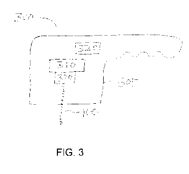
100521 An exemplary apparatus 300 that can be used to perform cosmetic
fractional
resurfacing of skin tissue in accordance with certain exemplary embodiments of
the present
5 disclosure is shown in Fig. 3. The exemplary apparatus 300 can include
an. EMR source 310,
a control arrangement 320 provided in communication with the EMR source 310,
and an
optical arrangement 330. One or more of these components can be provided in a
housing
305, e.g., as shown in FIG. 3. The housing 305 can be provided in a. shape of
a handpiece, or
in another form that can enclose one or more of the components shown in FIG.
3, A power
10 source (not Shown) configured or adapted to provide electrical power to
the EMR source 310
and/or the -Control arrangement 330 can also be provided, e.g., within or
external to the
housing 305.
100531 The EMR source 310 can include at least one source of
electromagnetic
radiation (e.g., optical radiation), such as a laser. For example, the EMR
source 310 can
15 include a CO2 laser or another type of laser, such as an erbium laser.
In certain embodiments,
the ER source 310 can include a plurality of lasers. For example, the EMR
source 310 can
include an ablative laser and a non-ablative laser. In certain exemplary
embodiments of the
present disclosure, the EMR source 310 can be provided external to the housing
305, and
electromagnetic energy provided by the EMR source 310 can be directed to the
optical
arrangement 330, e.g., using one or more waveguides or the like, such as one
or more optical
fibers, a hollow fiber waveguide, etc.
100541 The optical arrangement 330 can include, e.g., one or more
lenses,
.waveguides, reflectors, translators., motors and/or beam splitters. It can be
configured to
direct. EMR from the E.M.R. source 330 onto at least one particular location,
e.g., to direct a
plurality of EMR pulses 100 as described, herein onto a particular location on
a. Skin tissue
that is positioned relative to the apparatus 300. The optical arrangement 330
can be further
configured to direct such pluralities of EMR pulses 100 onto a plurality of
locations, e.g., to
form a plurality of holes 120 in a target region of tissue, with further
pulses 100 directed into
each hole 120 to coagulate and further ablate tissue within the hole 120 as
described herein.
Such holes 1.20 can be formed on a target region of tissue 110, e.g., as a
plurality of spaced
apart holes 1.20 with healthy or substantially undamaged tissue between them,
as can be done
in conventional -fractional resurfacing procedures.
CA 02881672 2015-02-10
WO 2014/026126
PCT/US2013/054362
16
10055j The control arrangement 320 can be specifically configured Co
control and/or
adjust properties of the EMRSOurte 310 to provide predetermined pulse
sequences of EMR
as described herein. For example, the control arrangement 320 can be
configured or adapted
to control or adjust properties of one or more pulses 100 of EMR to form an
ablated hole 120
in tissue 110, as shown in FIG. 2A, then control the EMR source 310 to direct
one or more
further pulses 100 of EMR having different properties into the hole 120 to
coagulate tissue
therein as shown, e.g., in FIG. 213, and then control the EMR source 310 to
direct one or more
still further pulses 100 of E.M.R. having particular properties into the hole
120 to ablate a
portion of the coagulated tissue 130 as shown, e.g.. in FIG, 2C, Optionally,
the EMR source
310 can also be controlled by the control arrangement $20 to then direct one
or more
coagulative beams or pulses 100 (e,g,, MA or NA pulses 100) into the hole 120
to further
coaoullte tissue therein as shown in FIG 21)
100561 Exemplary properties of the energy beams or pulses .100
produced by the
EMR source 310 that can be controlled or adjusted by the control arrangement
330 can
include, e.g., a focal diameter, a pulse duration, a pulse rate, a total pulse
energy, a pulse
power, a number of pulsesõ a frequency of pulses 100 (or, alternatively, an
interval between
successive nukes 100), and/or a pulse intensity. :Some of these parameters can
be related,
4., the pulse energy divided by the pulse duration can determine the pulse
power, the power
and beam width or diameter can determine the pulse intensity, etc.
100571 For example, a smaller focal diameter can generate a deeper ablated
hole 120
for a particular pulse energy and duration, although there may be additional
spreading of the
beam 100 deeper in the tissue 110. Similarly, pulse duration can be varied for
a fixed total
pulse energy and beam width to control the corresponding power and intensity.
100581 In further exemplary embodiments of the present disclosure, the
EMR source
310 can comprise two or more lasers or other components capable of providing
EMR, and the
control arrangement 320 can be configured or adapted to control the timing and
parameters of
EMR provided by each of the lasers. The two or more lasers, if present, can be
selected to
provide EAR. at different wavelengths. For example, one laser can provide EMR
at
Wavelengths that typically ablate biological tissue, whereas a further laser
can provide EMIR
at a *avelength that is more weakly absorbed by -tissue and can generally heat
irradiated
fissile without ablating it
CA 02881672 2015-02-10
WO 2014/026126
PCT/US2013/054362
17
100591 In still further exemplary embodiments of the present
disclosure, the control
arrangement. 320 can be configured or adapted to control the optical
arrangement 330, e.g., to
direct EMR pulses 100 from the EMR source 310 onto one or more particular
locations.
Such control can be adapted, for example, to provide a plurality of pulses
onto a plurality of
particular locations, where the pulses 100 are controlled to perform the
exemplary sequence
of processes illustrated in FIGS. 2A-2C or 2A-2D and described herein at each
location.
100601 The exemplary apparatus and methods described, herein can,
e.g., generate a
greater degree of shrinkage around an ablated micro-hole as a greater amount
of coagulated
tissue is formed around each hole (with portions thereof being ablated) as
compared, for
example, to an ablated hole formed using a single pulse of energy or several
pulses of
ablative energy, with no significant coagulation being produced and then
ablated as described
herein.
100611 Exemplary pulse sequences having a lamer number of pulses 100
can be
directed. onto a single location to ablate more tissue within the same hole
120 and generate
further tissue coagulation. This can lead to greater overall tissue shrinkage
and/or wrinkle
removal as compared to that which may be generated by the same number of
ablated surface
spots in a conventional ablative fractional resurfacing procedure.
Additionally, an ablated
hole 120 may be filled to a substantial degree by increasing the tissue volume
within the
initial hole '120 (with coagulative pulses) such that the volume increase from
coagulation is
greater than the volume removed by ablative pulses.
100621 In a manner similar to that used in conventional fractional
resurfacing
procedures, a plurality of holes 120 can be formed in a target area of skin,
and then treated
with a plurality of EMR pulses 100 as described herein. A local areal fraction
of the skin
surface covered by the holes can be, e.g., between about 0.05 and about 0.5.
Further holes
120 can. be formed in legions proximal to the treated area, and a, plurality
of pulses 100
directed into these additional holes 120. In certain exemplary embodiments of
the present
disclosure., a plurality of pulses 100 as described herein can be directed
onto a single location
on the skin, e.g., to form a hole 120, coagulate tissue therein, and ablate at
least a portion of
the coagulated tissue 130 as shown, e.g., in FIGS. 2A-2C. Optionally, further
pulses 100 can
then be directed into the hole 120 to coagulate further tissue and fill at
least a portion of the
hole 120 before performing the mum or similar procedure to form and irradiate
a second hole
120. The procedure can be repeated. until the entire target area has been
treated. Geometrical
parameters for initial ablative hole sizes and hole patterns/spacings that are
used in
CA 02881672 2015-02-10
WO 2014/026126
PCT/US2013/054362
18
conventional ablative fractional resurfacing, procedures may also be used. in
additional
exemplary embodiments of the present disclosure.
Example 1
10063] Two cross-sectional images of a histological Sample showing the
effects Of
two exemplary laser pulse sequences on ex vivo skin tissue are shown in Flee
4. The skin
sample was irradiated with pulses generated by a. CO2 laser. The holes shown
in FIG. 3. were
generated by sequences of three pulses 'haying the powers and durations
indicated below the
cross-sectional images. For example, the two rightmost holes shown in FIG. 3
were formed
1.0 by first directing an HA pulse onto the tissue (40 W, 2 ms duration),
followed by an MA
pulse (5 W, 2 ms duration), and finally an NA pulse (1 W. 10 ms duration) onto
the same
location. The time interval between pulses was about 10 seconds, although much
shorter
pulse intervals can be used.
1:00641 Similarly, the two leftmost holes shown in FIG. 4 were
generated by the pulse
sequence denoted below these holes, First, an NA pulse with a power of 1 Wand
duration of
10 ms was directed onto the tissue. Next, an MA pulse having 5 W power and 2
ms duration
was directed onto the same location. Finally, an HA. pulse having a higher
power of 40 W
and a duration of 2 ins was directed onto the same spot.
100651 The holes on the left side of FIG. 4 appear deeper than the
holes on the right.
2() This can be attributed to the sequence of pulses, in which the final
pulse is an HA pulse that
would tend to deepen the hole and ablatiesome of the coagulated, tissue that
may he present
therein. In contrast, the two holes on the right of Ha 4 appear somewhat
shallower, With a
larger coagulated zone visible around these holes, The HA pulse in the two
holes on the right
was applied first, followed by an MA pulse and then an NA pulse. The MA pulse
would
ablate a smaller volume of tissue and the NA pulse would ablate little if any
further tissue
while generating more coagulation, thus leading to.a shallower hole with more
coagulated
tissue surrounding it.
190661 The effects of the twoexeMplary pulse sequences shown in FIG. 4
suggest
that the size and coagulatioregeornetry of a hole formed in tissue can be
altered by the
sequential properties of the applied pulses.. For example, characteristics of
the resulting hole
and amount of residual coagulated tissue can be varied, e.g., by changing the
order of a
plurality of pulses having different parameters. Accordingly, pulse sequences
in accordance
CA 02881672 2015-02-10
WO 2014/026126
PCT/US2013/054362
19
with exemplary embodiments of the present disclosure described herein, which
include, for
example, at least. One ablative pulse to form a. hole in tissue followed by at
least one non-
ablative or .mildly ablative pulse to coagulate tissue in the hole, and then
at least one ablative
pulse to remove at least some oldie coagulated tissue, can produce desirable
effects that are
different from an application of similar pulses in a different order.
Elvainp le 2
[0067) A Lumenis UltraPtilse system with AcuScanI20 handpiece (Lumenis
Surgical) that includes a controllable CO2 laser was modified with a
controller arrangement.
The controller arrangement .facilitated programming and control of particular
pulse sequences
in accordance with exemplary embodiments of the present disclosure. 10
sequences of
energy pulses (A242) were directed into different locations on a 2 cm x 2 cm
sample of
previously-froze.n human abdominal skin.
100681 The first three sequences (A2--C2) included only ablative 60W
pulses., with a
total energy of 100 .inS per sequence. Pulse energies and durations for these
ablative
sequences are specified in Table 1 below. The exemplary system was programmed
in
accordance with embodiments of the present disclosure to irradiate the tissue
with five further
sequences of pulses (02-H2), with a different location being irradiated by
each pulse
sequence. Pulse energies and durations for these .five sequences, *Inch
include a mix of
60 W pulses that were primarily ablative (HA/MA) and I W (NA) pulses are also
specified in
Table 1 below, The total energy of each of these five sequences that was
directed onto the ex
vivo skin tissue was also 100 Mi. Finally, two further ablative pulse
sequences (1242), each
with a total energy of 50 nil as specified in Table I below, were also
directed onto distinct
locations on the skin sample.
[0069.1 Pre¨ and post¨ exposure pictures of the irradiated tissue were
obtained, using a
Nikon D80 camera with AF Macro Nikkor 60mm lens, The irradiated skin samples
were
frozen and sectioned, and then stained with the NBTC stain. The lesion sizes:
(1,e width and
depth of both the ablated hole and the coagulation zone) were measured under
light
transmitted microscope (Olympus BH-2),
[00701 Observed ablated hole depths and coagulated tissue depth (at the
bottom of
each hole) are shown in FIG. 5 for each pulse sequence A2-,12. These exemplary
pulse
sequences can be grouped into three main categories. Pulse sequences A2, B2
and C2 each
CA 02881672 2015-02-10
WO 2014/026126
PCT/US2013/054362
provide a total of 100 m..1 of HA energy with differing pulse durations and
intervals. Each of
pulse sequences 1)2-02 delivers a total of 100 nil of HA energy with different
'sequences and.
values for pulse power, durations, and intervals between pulses. Pulse
sequences H2 and 12
each provide a total of 50 nil of HA energy with differing pulse durations and
intervals.
5 These different types of pulse sequences are labeled in FIG. 5 and
separated by vertical
dashed lines.
100711 The purely ablative 100 mil pulse sequences A2-C2 resulted in
the deepest
holes, with some coagulated tissue observed at the bottom of the boles. The
sequences B2
and C2, where the ablative energy was divided into 5 and 10 pulses,
respectively, resulted in
10 somewhat shallower holes than sequence Al, where the 100 nil was
delivered in a single
energy pulse. This may result from a small degree of tissue coagulation that
may occur
between the 60 W pulses followed by subsequent ablation of some of this
coagulated tissue.
In contrast, the single 100 nil ablative pulse of A I could ablate a deeper
hole before any
coagulation could occur.
15 100721 Pulse sequences D2-H2 include different sequences of non-
ablative 1 W
pulses and 60 W pulses that are predominantly ablative (e.g., HA or M.A
pulses). In general,
these "mixed" pulse sequences with 100 mj total energy result in shallower
hole depths than
those resulting from ablative pulse sequences A2-C2, each of which also had a
total energy of
100 mi. The hole depths resulting from pulse sequences D2-H2 are also
shallower than those
20 resulting from the pulse sequences 12 and 32, each of which only
delivered a total of 50 mJ of
energy (half as much) into the tissue. The shallower holes resulting from the
"mixed!' pulse
sequences may, e.g., facilitate faster healing times when formed as part of a
fractional
resurfacing procedure as compared to deeper holes.
10073.1 Pulse sequences E2, F2, And 62 Maude at: least one exemplary
coagulative
pulse (1 W )that is preceded by and followed by at least one exemplary
ablative pulse (60W).
Pulse sequence 132 includes one coagulative (NA) pulse followed by five
ablative (HA/MA)
pulses, and pulse sequence 02 includes five ablative (HA/MA) pulses followed
by one
coagulative (NA) pulse. Although not conclusive, it has been observed that the
hole depths
resulting from pulse sequences E2. F2 and H2, which are in accordance with
exemplary
embodiments of the present disclosure, are slightly shallower than the hole
depths observed
for pulse sequences 1)2 and 02.
CA 02881672 2015-02-10
WO 2014/026126
PCT/US2013/054362
21
100741 The total volume of tissue removed by the various pulse
sequences could not
be measured directly in this study, because of the integrated effects of the
energy pulses such
as tissue ablation and coagulation. Nevertheless, it is likely that a greater
amount of tissue
was removed by embodiments of the present disclosure as expressed in the
exemplary pulse
sequences 02, E2, F2 and H2 than by the other pulse sequences, because of the
presence of
Ablative pulses following coagulative or non-ablative pulses. The resulting
hole depths of
these exemplary pulse sequences are still somewhat shallower than the hole
depths observed
by other pulse sequences tested. The hole depth. could be reduced further,
e.g., by using pulse
sequences that include further non-Ablative pulses at the end of the sequence,
e.g., to provide
a "hole-filling" effect such as that illustrated in FIG. 20.
[00751 A general shrinkage of the irradiated ex vivo tissue samples
was observed for
the various pulse sequences tested. Such shrinkage can result from, e.g..,
tissue coagulation
and denaturation of some tissue components such as collagen. However, the
desirable
cosmetic effects of fractional resurfacing procedures in living tissue (e.g.
skin tightening or
wrinkle reduction) result from stimulated healing responses in the tissue as
well as direct
thermal effects an the tissue components. Accordingly, although not directly
observed, the
enhanced thermal damage and tissue removal, together with shallower hole
depths, provided
by embodiments of the present disclosure may likely lead to more effective
skin tightening
and/or faster healing times as compared to conventional resurfacing procedures
performed
using comparable energy and geometry parameters, e.g., those that deliver the
same amount
of energy to ablate holes in the tissue at the same surface coverage.
CA 02881672 2015-02-10
WO 2014/026126 PCT/US2013/054362
22
100761 TABLE 1
Exemplary Ablative and Non-Ablative Puke Sequences
Pulse sequence: 1 2 3 4 5 5 7 8 9 10
Power (W) 60
A2 Puke duration cnis) 1.67
Energy (m.1) 100
------------------------------
Power (W). 60 0 60 0 60 t1 .. 0 60 0 .
60
_
62 Pulse duration (ms) 0,33 50 0,33 50 0.33 50
0.33 50 0,33
Energy (mi) 20 0 20 0 20 0 20 0 20
_
Power (W) 60
C2 Puke duration (ms) 0.17 1C2 puke repeated 10
times]
Energy (ml) 10
Power (W) 1 60 0 60 0 60 0 60 0
60
0 ,
2 Puke duration (ms) 50 0.17 , 50 0.17 , 50 0.17 50
0.17 50 0,17
Energy (ml) 50 10 0 10 0 10 0 10 0
10
Power (W) 60 1 60 0 60 1 0 60 0 60
E2 Puke duration (ms) 0,17 50 , 0,17 , 500.11 50
0.17 50 0,17
Energy (m.1) 10 50 10 , 0 . 10 1 0 10 0 i
10
Power (W) 60 0 60 0 60 0 60 1 60
F2 Puke duration (ms) , 017 SO 0.17 50 , 0.17
SO 0.17 50 0,17
Energy (m)) 10 0 , 10 0 10 0 10 50 10
, ..
Power (W) 60 0 60 0 60 0 60 .. 0. 60
1
G ,
2 Puke duration (ms) 0.17 50 0.17 50 0.17 50 0.17 50 0.17
50
Energy (ml) 10 0 . 10 0 10 0 10 0 10
50
Power (W) 60 : 1 60 1 60 1 60 1 60
_
H
Puke duration (ms) 0.17 12.5 0.17 12.5 0,17 12.5 0,17
12.5 0.11
2
Energy (rni) 10_ 12.5 10 12.5 10 12.5 10
12.5 10
Power (W) 60
12 Puke duration (ms) 0.83
, Energy (mi) 50 ,
Power (W) 60 0 60 0 60 0 60 0 60
12 Puke duration (ms) . 0.17 50 0.17 50 0.17 SO
0.17 50 0.17
Energy (ml) 10 0 10 0 10 0 10 0 10 '
10077j It will
thus be appreciated that those skilled in the art will be able to devise
numerous systemsõ an-angements and methods witich, although not explicitly
shown or
described herein, embody the principles of the present disclosure and are thus
within the
spirit and scope of the present disclosure, In addition, all publications,
patents, and patent
applications referenced herein are incorporated herein by reference in their
entireties,