Note: Descriptions are shown in the official language in which they were submitted.
CA 02882319 2015-02-17
WO 2014/036555
PCT/US2013/057836
DRUG DELIVERY SYSTEMS AND METHODS FOR TREATMENT OF BLADDER
CANCER COMPRISINGIOXALIPLATIN
Cross-Reference to Related Applications
This application claims priority to U.S. Provisional Patent Application No.
61/696,027, filed August 31, 2012.
Background
This disclosure is generally in the field of pharmaceutical agents for use in
treating the
bladder, and more particularly to drug delivery systems, methods, and drug
formulations for
targeted treatment of urinary bladder cancer.
Delivery of therapeutic agents to the urinary bladder is difficult. Current
practice
requires systemic administration using doses which result in significant
exposure to healthy
tissues and relatively low exposure within the bladder. Frequently the
systemic exposure
leads to unwanted or harmful side effects which limit the usefulness of the
agent in treating
bladder disease.
To avoid systemic effects, drugs may be delivered locally onto tissues at or
near the
target tissue. However, such local administration may not be well tolerated by
the tissue at
the delivery site and/or may not be sufficiently permeable to the particular
drug being
delivered. Accordingly, there is a need to provide a therapeutic agent that is
well tolerated by
the bladder when the agent is applied at concentrations effective to achieve
sufficient
therapeutic (i.e., cytotoxic) concentrations within the target tissues.
Accordingly, there remains a need for improved drug delivery methods and
systems
for treating the bladder, such as in the treatment of bladder cancer, whether
as neoadjuvant
therapy, adjuvant therapy, or palliative therapy.
Summary
In one aspect, a medicament is provided that includes oxaliplatin for use in
the treatment of
bladder cancer by locally administering oxaliplatin into the bladder of a
patient to achieve a
sustained concentration of oxaliplatin in urine in the bladder sufficient to
produce a
therapeutic concentration of oxaliplatin in bladder tissue. The locally
administering into the
patient's bladder may be continuous or intermittent. In one
1
CA 2882319 2019-12-13
CA 02882319 2015-02-17
WO 2014/036555 PCT/US2013/057836
embodiment, the oxaliplatin is delivered into the bladder from an intravesical
drug delivery
device inserted into the bladder, and the device continuously releases the
oxaliplatin into the
urine in the bladder over a sustained period. In an alternative embodiment,
the oxaliplatin is
delivered into the bladder from a coating substance applied to the bladder,
which coating
substance continuously releases the oxaliplatin into the urine in the bladder
over a sustained
period. The coating substance may include a mucoadhesive formulation. In a
further
alternative embodiment, a liquid form of the oxaliplatin is pumped into the
bladder through a
urethral catheter inserted into the bladder. In various embodiments, the
oxaliplatin is released
into the patient's bladder continuously over a period of at least 2 hours,
such as from 1 day to
14 days. In an embodiment, the oxaliplatin is released into the patient's
bladder at a mean
average amount of from 1 mg to about 100 mg oxaliplatin per day for 1 day to
14 days. In an
embodiment, the oxaliplatin is released into the patient's bladder at a mean
average amount of
from 1 mg to about 100 mg oxaliplatin per day for up to 7 days.
In another aspect, a medical device is provided for intravesical
administration of
oxaliplatin. In an embodiment, the device includes a housing configured for
intravesical
insertion, and a dosage form comprising oxaliplatin, wherein the housing holds
the dosage
form and is configured to controllably release the oxaliplatin into the
bladder in amount
therapeutically effective for the treatment of bladder cancer. In an
embodiment, the device
comprises is elastically deformable between a retention shape configured to
retain the device
in a patient's bladder and a deployment shape for passage of the device
through the patient's
urethra. In an embodiment, the device is configured to release from 1 mg/day
to 100 mg/day
of oxaliplatin for up to 7 days.
In still another aspect, a method is provided for administering oxaliplatin to
a
patient in need of treatment of bladder cancer. The method includes locally
administering
oxaliplatin into the bladder of a patient to achieve a sustained concentration
of oxaliplatin in
urine in the bladder sufficient to produce a therapeutic concentration of
oxaliplatin in bladder
tissue. The method may further include administering at least a second
therapeutic agent to
the patient. Non-limiting examples of second therapeutic agents include
gemcitabine or
another cytotoxic agent; an analgesic agent; an anti-inflammatory agent; or a
combination
thereof. The second therapeutic agent may be administered intravesically
and/or by other
routes of administration.
2
CA 2882319 2019-12-13
CA 02882319 2015-02-17
WO 2014/036555
PCT/US2013/057836
Brief Description of the Drawings
FIGS. 1A-1B illustrate one embodiment of an intravesical drug delivery device
that
may be used for administering oxaliplatin as described herein.
FIGS. 2A-2B illustrate another embodiment of an intravesical drug delivery
device
that may be used for administering oxaliplatin as described herein.
FIGS. 3A-3C illustrate still another embodiment of an intravesical drug
delivery
device that may be used for administering oxaliplatin as described herein.
FIGS. 4A-4B illustrate a method of inserting an intravesical drug delivery
device
into the bladder of a patient for local administration of oxaliplatin as
described herein.
FIG. 5A illustrates a material applied to the inner surface of the bladder
wall for
local administration of oxaliplatin as described herein.
FIG. 5B illustrates a method of applying a coating material onto to the inner
surface
of the bladder wall for local administration of oxaliplatin as described
herein.
FIG. 6 is a graph of cisplatin blood profile from a study administering the
drug by
IV bolus or intra-bladder perfusion in rats.
FIGS. 7A-7C are graphs of cisplatin terminal concentrations in blood, urine,
and
tissue samples from a study in rats.
FIG. 8 is a graph of carboplatin blood profile from a study administering the
drug by
IV bolus or intra-bladder perfusion in rats.
FIG. 9 is a graph of carboplatin terminal concentrations in blood, urine, and
tissue
samples from a study in rats.
FIG. 10 is a graph showing cisplatin, carboplatin, and oxaliplatin blood
levels
following 72 hour bladder perfusion in rat study.
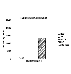
FIG. 11 is a graph showing oxaliplatin terminal concentrations in tissues
following
72 hour bladder perfusion in rat study
FIGS. 12A-12B are graphs showing cisplatin, carboplatin, and oxaliplatin
bladder
permeability following 72 hour bladder perfusion in rat study.
Detailed Description
It has been discovered that intravesical administration of oxaliplatin can be
used to
achieve therapeutically effective amount of the drug in the tissues where
needed and also is
well tolerated by the bladder tissue. That is, oxaliplatin was unexpectedly
shown to meet
3
22229038.1
CA 02882319 2015-02-17
WO 2014/036555
PCT/US2013/057836
both the tissue permeability criteria and the urothelium tolerability criteria
when
administered into the bladder. Several other drugs tested failed to achieve
both.
Furthermore, by local, intravesical administration of the oxaliplatin,
systemic exposure to
the drug is advantageously minimized.
Accordingly, the present methods and devices for treating bladder cancer
include
locally administering oxaliplatin into the bladder of a patient to achieve a
sustained
concentration of oxaliplatin in urine in the bladder sufficient to produce a
therapeutic
concentration of oxaliplatin in bladder tissue.
As used herein, the term "bladder tissue" refers to the bladder wall or one or
more
layers thereof (e.g., mucosa, muscle, and submucosa).
The term "patient" as used herein refers to humans or other mammals, such as
in
veterinary or livestock applications, in need of treatment. In a particular
embodiment, the
patient is an adult human.
Oxaliplatin is platinum based antineoplastic agent. It is known for use in
chemotherapy, for example in the treatment of colorectal cancer, where it is
formulated for
intravenous administration, e.g., EloxatinTM (Sanofi-Aventis). In the present
invention, the
oxaliplatin is formulated for local delivery. It may be provided in solid or
semi-solid form
or in a liquid form, depending on the delivery mechanism employed, as
described herein.
Oxaliplatin and methods of manufacture thereof are described, for example, in
U.S. Patent
5,338,874; U.S. Patent 5,420,319; U.S. Patent 5,716,988; and U.S. Patent
5,290,961.
A variety of methods can be used to achieve the required urine (and thus
tissue)
concentrations of the oxaliplatin. In one embodiment, the oxaliplatin can be
provided by
direct instillation of a simple solution into the bladder. For example, a
solution of the
oxaliplatin may be pumped into the bladder through a urethral or suprapubic
catheter in a
continuous or pulsatile manner over the treatment period. In another
embodiment, the
oxaliplatin is released from a device or composition deployed in the bladder,
wherein the
device or composition releases the oxaliplatin (continuously or
intermittently) at a rate
effective to produce the desired concentration of drug in the urine over a
specified treatment
period. At the end of the treatment period, the device may be retrieved from
the bladder, or
it may be eliminated by being resorbed, dissolved, excreted, or a combination
thereof.
In a preferred embodiment, the oxaliplatin is administered to the bladder from
an
intravesical device. A preferred embodiment of an intravesical drug delivery
device and
4
22229038.1
CA 02882319 2015-02-17
WO 2014/036555
PCT/US2013/057836
methods for deploying those devices into the bladder are described in the
following U.S.
Patent Application Publications: US 2012/0203203 (Lee et al.); US 2012/0089122
(Lee et
al.); US 2012/0089121 (Lee et al.); US 2011/0218488 (Boyko et al.); US
2011/0202036
(Boyko et al.); US 2011/0152839 (Cima et al.); US 2011/0060309 (Lee et al.);
US
.. 2010/0331770 (Lee etal.); US 2010/0330149 (Daniel et al.); US 2010/0003297
(Tobias et
al.); US 2009/0149833 (Cima et al.); and US 2007/0202151 (Lee etal.).
In embodiments in which the oxaliplatin is delivered from an intravesical drug
delivery device, the oxaliplatin may be housed in the device in various forms,
which may
depend on the particular mechanism by which the device controllably releases
the oxaliplatin
into fluid (e.g., urine) in the bladder. In some embodiments, the oxaliplatin
is provided in a
solid, semi-solid, or other non-liquid form, which advantageously may
facilitate stable
storage of the drug before the device is used and advantageously may enable
the drug
payload of the device to be stored in smaller volume than would be possible if
the drug were
housed in the form of a liquid solution. In an embodiment the non-liquid form
is selected
from tablets, granules, semisolids, capsules, and combinations thereof In one
embodiment,
the oxaliplatin is in the form of a plurality of tablets, such as mini-tablets
described in U.S.
Patent No. 8,343,516. In other embodiments, the oxaliplatin may be housed in a
liquid form,
such as in a solution with a pharmaceutically acceptable excipient.
An embodiment of a drug delivery device 100 is illustrated in FIG. 1A. The
device
100 includes a device body having a drug reservoir portion 102 and a retention
frame portion
104. In FIG. 1, the device 100 is shown in a relatively expanded shape suited
for retention in
the body. Following deployment into the body, the device 100 may assume the
relatively
expanded shape to retain the drug delivery device in the body cavity or lumen.
For the purposes of this disclosure, terms such as "relatively expanded
shape",
"relatively higher-profile shape", or "retention shape" generally denote any
shape suited for
retaining the device in the intended implantation location, including but not
limited to the
pretzel shape shown in FIG. 1 that is suited for retaining the device in the
bladder.
Similarly, terms such as "relatively lower-profile shape" or "deployment
shape" generally
denote any shape suited for deploying the drug delivery device into the body,
including a
linear or elongated shape that is suited for deploying the device through the
working channel
of catheter, cystoscope, or other deployment instrument positioned in the
urethra. In
5
CA 2882319 2019-12-13
CA 02882319 2015-02-17
WO 2014/036555
PCT/US2013/057836
embodiments, the drug delivery device may naturally assume the relatively
expanded shape
and may be deformed, either manually or with the aid of an external apparatus,
into the
relatively lower-profile shape for insertion into the body. Once deployed the
device may
spontaneously or naturally return to the initial, relatively expanded shape
for retention in the
body.
In the illustrated embodiment, the drug reservoir and retention frame portions
102,
104 of the drug delivery device 100 are longitudinally aligned and are coupled
to each other
along their length, although other configurations are possible. The drug
delivery device 100
includes an elastic or flexible device body 106 that defines a drug reservoir
lumen 108 (i.e.,
the drug housing) and a retention frame lumen 110. The drug reservoir lumen
108 is
designed to house a drug formulation that comprises the oxaliplatin. In the
illustrated
embodiment, the drug formulation in the form of a number of solid drug tablets
112. The
retention frame lumen 110 is designed to house a retention frame 114 to form
the retention
frame portion 104. The illustrated lumens 108, 110 are discrete from each
other, although
other configurations are possible.
As shown in the cross-sectional view of FIG. 1B, the device body 106 includes
a
tube or wall 122 that defines the drug reservoir lumen 108 and a tube or wall
124 that
defines the retention frame lumen 110. The tubes 122, 124 and lumens 108, 110
can be
substantially cylindrical, with the drug reservoir lumen 108 having a
relatively larger
diameter than the retention frame lumen 110, although other configurations can
be selected
based on, for example, the amount of drug to be delivered, the diameter of the
retention
frame, and deployment considerations such as the inner diameter of the
deployment
instrument. The wall 124 that defines the retention frame lumen 110 may extend
along the
entire length of the wall 122 that defines the drug reservoir lumen 108, so
that the retention
frame lumen 110 has the same length as the drug reservoir lumen 108 as shown,
although
one wall may be shorter than the other wall in other embodiments. The two
walls 122, 124
are attached along the entire length of the device in the illustrated
embodiment, although
intermittent attachment can be employed.
As shown in FIG. 1A, the drug reservoir lumen 108 is loaded with a number of
drug
units 112 in a serial arrangement. Essentially any number of drug units may be
used, for
example, depending upon the sizes of the reservoir and the drug units. The
drug reservoir
lumen 108 includes a first end opening 130 and an opposed second end opening
132. Once
6
22229038.1
the drug units 112 are loaded, restraining plugs 120 are disposed in the
openings 130 and
132. The restraining plugs 120, in this embodiment, are cylindrical plugs
secured into the
entry 130 and the exit 132. In other embodiments, the openings 130 and 132 are
closed off
with other structures or materials, which may, depending on the particular
embodiments,
include an aperture or a water- or drug-permeable wall to facilitate ingress
or egress of water
or drug during use.
The retention frame lumen 110 is loaded with the retention frame 114, which
may be
an elastic wire. The retention frame 110 may be configured to return
spontaneously to a
retention shape, such as the illustrated example "pretzel" shape or another
coiled shape. In
particular, the retention frame 114 may retain the device 100 in the body,
such as in the
bladder. For example, the retention frame 114 may have an elastic limit and
modulus that
allows the device 100 to be introduced into the body in a relatively lower-
profile shape,
permits the device 100 to return to the relatively expanded shape once inside
the body, and
impedes the device from assuming the relatively lower-profile shape within the
body in
response to expected forces, such as the hydrodynamic forces associated with
contraction of
the detrusor muscle and urination. Thus, the device 100 may be retained in the
body once
implanted, limiting or prevent accidental expulsion.
The material used to form the device body 106, at least in part, may be
elastic or
flexible to permit moving the device 100 between deployment and retention
shapes. When
the device is in the retention shape, the retention frame portion 104 may tend
to lie inside the
drug reservoir portion 102 as shown, although the retention frame portion 104
can be
positioned inside, outside, above, or below the drug reservoir portion 102 in
other cases.
The material used to form the device body 106 may be water permeable so that
solubilizing fluid (e.g., urine or other bodily fluid) can enter the drug
reservoir portion 102 to
solubilize the drug units 112 once the device is implanted. For example,
silicone or another
biocompatible elastomeric material may be used. In other embodiments, the
device body may
be formed, at least in part, of a water-impermeable material.
FIG. 2A illustrates an implantable drug delivery device 200, which includes a
drug reservoir
202 loaded with drug 212 and a retention structure that includes two filaments
220, 222
associated with a fastener 230. As shown, the drug reservoir 202 is an
elongated tube that can
be deformed between a relatively linear deployment shape, such as the shape
shown
7
CA 2882319 2019-12-13
in FIG. 2A, and a relatively circular retention shape, such as the shape shown
in FIG. 2B.
The drug 212 may be loaded in the tube in a flexible form, so that the drug
reservoir 202 can
be moved between the two shapes. For example, the drug 212 may be a number of
solid drug
tablets, a liquid, or a gel. The filaments 220, 222 may be attached to
opposite ends of the drug
reservoir 202 and joined by the fastener 230. The fastener 230 can be adjusted
to adjust the
position of one filament 220 with reference to the other 222, thereby
adjusting the position of
one end of the drug reservoir 202 with reference to the other end. The device
200 can assume
the retention shape by adjusting the filaments 220, 222 to draw the ends of
the drug reservoir
202 doser together, and thereafter the device 200 can be retained in the
retention shape by
preventing adjustment of the filaments 220, 222 with the fastener 230. In
such an embodiment, the device 200 is manually adjusted into the retention
shape by
manually adjusting the filaments 220, 222 after the device 200 is inserted
into the bladder.
In the illustrated embodiment, the fastener 230 is a cinch nut that permits
shortening the
portion of the filaments 220, 222 between the drug reservoir ends and the
cinch nut, but
prevents lengthening of these portions of the filaments 220, 222. Thus, the
ends of the drug
reservoir 202 can be drawn doser together by pulling one or both of the
filaments 220, 222
through the cinch nut, causing the device 200 to assume the retention shape.
Once the
filaments 220, 222 have been so adjusted, the cinch nut prevents lengthening
of the
filaments 220, 222, retaining the device in the retention shape. Thus,
manually adjusting the
device 200 into the retention shape once implanted merely requires pulling one
or both of
the filaments 220, 222, although other fasteners 230 that require separate
manipulation can be
employed. Other fasteners may also be used.
Another embodiment of an intravesical drug delivery device is illustrated in
FIGS.
3A-3C. In this embodiment, the device includes a housing 300 having a single,
continuous
structure with multiple, discrete drug reservoir lumens 320 and optionally
having at least
one retention frame lumen 330 in which a retention frame 360 is disposed. Each
drug
reservoir lumen 320 has two defined openings, as shown in FIG. 3B, and is
dimensioned to
hold at least one solid drug unit 340. Solid drug unit 340 may be a drug
tablet or capsule. In
other embodiments not shown, each drug reservoir lumen has one defined
opening. The
housing may be formed of a flexible polymer, such as silicone. FIG. 3B is a
cross-sectional
view of the plane that bisects one of the drug reservoir lumens 320 of the
housing shown in
FIG. 3A along line 3B-3B. As shown in FIG. 3B, the monolithic housing 300 has
two
8
CA 2882319 2019-12-13
CA 02882319 2015-02-17
WO 2014/036555
PCT/US2013/057836
defined openings (350a, 350b) in its drug reservoir lumen 320 that expose both
ends of the
solid drug unit 340. The retention frame lumen 330, in this embodiment, is
aligned parallel
to the longitudinal axis of the housing and perpendicular to the drug
reservoir lumen 320.
FIG. 3C is a perspective view of a portion of the embodiment of the device 300
shown in
FIG. 3A when the device is in its retention shape, which is taken when the
retention frame
360 is disposed in the retention frame lumen 330. The drug reservoir lumens
320 and the
retention frame 360 in the housing of this embodiment are oriented so that the
drug reservoir
lumens 320 are outside the retention frame's 360 arc. Alternatively, the
housing in FIG. 3C
can be rotated 180 degrees about the retention frame 360 to yield a
configuration in which
.. the drug reservoir lumens 320 are arranged within the retention frame's 360
arc. With this
embodiment, the devices provide sufficient direct contact between solid drug
units and with
urine surrounding the device when deployed and retained in the bladder. In
embodiments,
release of the drug from the device is controlled by erosion of an exposed
portion of the
surface of a solid drug unit, such that the rate of drug release from the drug
delivery device
.. may be directly proportional to and limited by the total exposed surface
area of the solid
drug units.
One embodiment of inserting an intravesical device 400 for subsequent
controlled
release of the oxaliplatin into the bladder is shown in FIGS. 4A and 4B. Here,
the device
400 is shown assuming a retention shape as the device exits a deployment
instrument 402.
The deployment instrument 402 may be any suitable device. It may be a lumenal
device,
such as a catheter, urethral catheter, or cystoscope. The deployment
instrument 402 may be
a commercially available device or a device specially adapted for the present
drug delivery
devices. FIG. 4B illustrates the insertion of the device 400 into the bladder,
wherein the
adult male anatomy is shown by way of example. The deployment instrument 402
is
inserted through the urethra to the bladder, and the device 400 may be passed
from/through
the deployment instrument 402, driven by a stylet or flow of lubricant or
combination
thereof until the device 400 exits into the bladder, and as shown is in a
retention shape.
In various embodiments, the oxaliplatin may be released from the intravesical
drug
delivery device by diffusion to through a wall of the drug housing, by
diffusion to through
one or more defined apertures in a wall of the drug housing, by osmotic
pressure through an
aperture in the drug housing, by erosion of a drug formulation in contact with
urine in the
bladder, or by a combination thereof.
9
22229038.1
CA 02882319 2015-02-17
WO 2014/036555
PCT/US2013/057836
In some embodiments in which the device comprises a drug in a solid form,
elution
of drug from the device occurs following dissolution of the drug within the
device. Bodily
fluid enters the device, contacts the drug and solubilizes the drug, and
thereafter the
dissolved drug diffuses from the device or flows from the device under osmotic
pressure or
via diffusion. For example, the drug may be solubilized upon contact with
urine in cases in
which the device is implanted in the bladder.
In various embodiments, the intravesical device may release oxaliplatin
continuously
or intermittent to achieve a therapeutically effective concentration of
oxaliplatin in the
bladder tissue over a sustained period, e.g., from 1 hour to 1 month, for
example from 2
.. hours to 2 weeks, from 6 hours to 1 week, from 24 hours to 72 hours, etc.
Subsequently, the device may be retrieved from the body, such as in cases in
which
the device is non-resorbable or otherwise needs to be removed. Retrieval
devices for this
purpose are known in the art or can be specially produced. The device also may
be
completely or partially bioresorbable, such that retrieval is unnecessary, as
either the entire
device is resorbed or the device sufficiently degrades for expulsion from the
bladder during
urination. The device may not be retrieved or resorbed until some of the drug,
or preferably
most or all of the drug, has been released. If needed, a new drug-loaded
device may
subsequently be implanted, during the same procedure as the retrieval or at a
later time.
In another embodiment, a coating substance may be intravesically applied to
the
bladder wall, wherein the coating substance includes oxaliplatin and one or
more excipient
materials that promote adherance of the coating substance to the bladder wall
and provides
continuous controlled release of the drug over the treatment period. The
coating substance
may be a mucoadhesive formulation, such as gels, ointments, creams, films,
emulsion gels,
tablets, polymers, or a combination thereof. Mucoadhesive formulation polymers
may
include hydrogels or hydrophilic polymers, polycarbophil (i.e. Carbopols,
etc.), chitosan,
polyvinylpyrrolidone (PVP), lectin, polyethyleneglycolated polymers,
celluloses, or a
combination thereof. Suitable celluloses include methyl cellulose (MC),
carboxymethyl
cellulose (CMC), hydroxypropyl cellulose (HPC), or combinations thereof. The
coating
substance may include a permeation enhancer. Non-limiting examples of
permeation
enhancers include dimethyl sulfoxide (DMSO), sodium carboxymethyl cellulose
(NaCMC),
lipids, surfactants, or combinations thereof.
22229038.1
CA 02882319 2015-02-17
WO 2014/036555
PCT/US2013/057836
As shown in FIG. 5A, a coating substance 500 may be deployed in the bladder
550
so that the coating substance 500 engages/adheres to the bladder wall 552. The
coating
substance 500 may be deployed in the bladder using a deployment instrument.
FIG. 5B is a
sagittal view of a male genitourinary system, illustrating a coating substance
500 being
deployed through a deployment instrument 502 into the bladder 550. The coating
substance
500 may be an embodiment of one of the coating substances described herein.
The
deployment instrument 502 is sized and shaped for passing through a urethra
560 of a
patient to a bladder 550 as shown. The deployment instrument 502 may be a
known device,
such as a catheter or cystoscope, or a specially designed device. The
deployment instrument
502 is used to deploy the coating substance 500 into the bladder and is
subsequently
removed from the body, leaving the coating substance 500 in the bladder. Once
so inserted,
the coating substance 500 releases the oxaliplatin into urine and the bladder
wall.
The present invention may be further understood with reference to the
following
non-limiting examples.
Example 1: Testing of Platin Drugs for Bladder Tolerability and Tissue
Permeability
Two studies were conducted in male Sprague Dawley rats administering cisplatin
or
carboplatin by intra-urinary bladder cannula, over a 72-hour continuous
perfusion, or by a
single IV bolus. Blood, urine, and tissue samples were collected and analyzed
for drug
content. Details of the study design and results are set forth in the tables
and description
below.
The study protocol was as follows:
Cisplatin Carboplatin
Group 1 24-hr perfusion via cannula 24-hr perfusion via
cannula to
to bladder dome bladder dome
Group 2 72-hr perfusion via cannula 72-hr perfusion via
cannula to
to bladder dome bladder dome
Group 3 Negative control ¨ 72-hr Negative control ¨ 72-hr
perfusion via cannual to perfusion via cannual to
bladder dome bladder dome
Group 4 IV bolus with saline IV bolus with saline
perfusion
perfusion via cannula via cannula
For each drug, each test group included three male rats. The perfusate drug
concentration was set to 0.3mg/mL and the perfusion rate used was 300
1.1L/hour over the
test periods.
11
22229038.1
CA 02882319 2015-02-17
WO 2014/036555 PCT/US2013/057836
Details of the study design and results are set forth in the tables and
descriptions
below.
Perfusion solutions were prepared by dissolving each drug substance into an
appropriate volume of saline. The finals doses administered are summarized
below.
Animal # Compound Administration Amount Actual Dose
Route Compound Administered
Administered via (mg/kg)
Perfusion Wt. (g)
1 (Group 1) Cisplatin Bladder Perf. 6.95 2.14
2 (Group 1) Cisplatin Bladder Perf. 6.88 2.12
3 (Group 1) Cisplatin Bladder Perf. 7.02 2.16
4 (Group 2) Cisplatin Bladder Perf. 20.44 6.30
5 (Group 2) Cisplatin Bladder Perf. 21.20 6.53
6 (Group 2) Cisplatin Bladder Perf. 20.59 6.34
10 (Group 4) Cisplatin IV Bolus 0.9820 0.74
11 (Group 4) Cisplatin IV Bolus 1.0319 0.77
12 (Group 4) Cisplatin IV Bolus 1.1210 0.84
22 (Group 1) Carboplatin Bladder Perf. 7.08 2.18
23 (Group 1) Carboplatin Bladder Perf. 6.87 2.12
24 (Group 1) Carboplatin Bladder Perf. 7.02 2.16
25 (Group 2) Carboplatin Bladder Perf. 20.89 6.43
26 (Group 2) Carboplatin Bladder Perf. 21.22 6.54
27 (Group 2) Carboplatin Bladder Peril 20.70 6.38
31 (Group 4) Carboplatin IV Bolus 1.1155 0.84
32 (Group 4) Carboplatin IV Bolus 1.1507 0.86
33 (Group 4) Carboplatin IV Bolus 1.1195 0.84
Whole blood samples were collected at various time points following the start
of perfusion,
including times 0, 12, 24, 48 and 72 hours as applicable. Urine was collected
pre-dose and
for 0-24, 24-48, and 48-72-hour periods post dose.
Following the planned infusion periods the animals, terminal blood samples
were
taken via the abdominal aorta, and the bladder, prostate, ureter, and kidney
tissues were
collected, weighed, and visually inspected for evidence of drug tolerability.
12
22229038.1
CA 02882319 2015-02-17
WO 2014/036555 PCT/US2013/057836
For animals dosed with cisplatin (Groups 1, 2, and 4), all animals appeared
normal
during perfusion period except as noted below. Tissue observations at necropsy
are also
summarized.
Group Numbers Clinical Observations of note during Tissue Observations
at Necropsy
Perfusion
Group 1 Normal Bladder lumen: slight to mild
erythemic
(Animals 1,2,3) discoloration, 30 ¨ 50 % of
lumen, mild to
moderate severity, mild edema/ thickened
bladder walls
Group 2 Red tinted urine at 72 hrs , all animals Bladder lumen:
generalized erythemic
(Animals 4,5,6) discoloration, 30 ¨ 50 % of
lumen, mild to
moderate severity, blood clots, moderate
edema/thickened bladder walls
Group 3-CONTROL Dark colored urine (one animal @ 46 Slight to mild focal
erythemia
(Animals 7,8,9) hr)
Group 4 Normal No observations
(Animals 10,11,12)
For animals dosed with carboplatin, all animals appeared normal during
perfusion
period. Tissue observations at necropsy are also summarized.
Group Numbers Clinical Observations of note during Tissue Observations
at Necropsy
Perfusion
Group 1 Normal Bladder lumen: slight to mild
generalized
(Animals 22,23,24) erythemic discoloration, 10 - 30
% of lumen,
no evidence of tissue edema
Group 2 Normal Bladder lumen: slight to mild
generalized
(Animals 25,26,27) erythemic discoloration, 10 - 30
% of lumen,
no evidence of tissue edema
Group 3-CONTROL Red tinted urine (one animal) Bladder lumen: slight
generalized erythemic
(Animals 28,29,30) discoloration, 5 - 10 % of lumen,
mild tissue
edema (one animal)
Group 4 Red tinted urine (one animal) Bladder lumen: slight
generalized erythemic
(Animals 31,32,33) discoloration, 5 - 10 'A of
lumen, no evidence
of tissue edema
Gross pathology observations were substantiated by tissue histology.
ICP-MS for platinum was used to test (i) serial whole blood, (ii) daily urines
in 24- hr
collections, and (iii) terminal tissues, including bladder, kidney, and
prostate. FIG. 6 is a
graph showing the blood profile for cisplatin. The 72 h group show rising
blood levels
suggesting degradation of the bladder lumen permitting increased tissue
cisplatin uptake.
FIGS. 7A and 7B are graphs showing cisplatin terminal concentrations in the
various tissue
and fluid samples. Significantly higher and more variable bladder platinum
concentrations
13
22229038.1
CA 02882319 2015-02-17
WO 2014/036555
PCT/US2013/057836
were observed following 72 hr perfusions when compared to 24 hr perfusions and
were
associated with the pronounced bladder tissue toxicities observed at necropsy.
Individual 72
hr bladder concentration values were 12,000 ng/g, 60,000 ng/g and 160,000
ng/g.
IV bolus administration resulted in measurable kidney and bladder tissue
platinum
levels at 72 hrs despite low urine concentrations. In the IV dosing group
kidney to bladder
platinum concentration ratio was the inverse of that observed following
bladder perfusion.
Kidney tissue concentration was highest, followed by the bladder concentration
both of
which were achieved at approximately half the plasma concentrations observed
at 72 h.
Increased bladder concentration observed following perfusion may reflect
absorption by
bladder from both systemic (blood) and urine (urinary clearance) of platinum
(which is also
supported by elevated kidney levels).
FIG. 7C is another graph showing cisplatin terminal concentrations. The
bladder:urine ratio was near 100% for the 72 h perfusion (tox). The
bladder:urine ratio was
5% for the 24 h perfusion, which reflects cisplatnin partitioning when the
urothelium is less
damaged, exhibiting only mild to moderate erythema as observed in the 24hr
necropsy
results (Group 1). For whole blood, the bladder ratio was 66% at 72 hr for the
IV bolus
administration due to the long half-life of platinum compounds when
administered
systemically. These results confirm a significant advantage of intravescular
bladder
perfusion when the urothelium is largely intact. Significant bladder levels
can be attained
.. without meaningful systemic exposure.
FIG. 8 is a graph showing the blood profile for carboplatin. Observed plasma
levels
were near the limit of the assay detection (twice the limits of detection) to
below the
quantitation limit were observed for the perfusion groups. The IV bolus shows
significant
peak systemic platinum exposure followed by a sharp decay (faster clearance
than observed
with cisplatin). There was one quarter less carboplatin in the IV bolus
terminal phase as
compared with cisplatin.
FIG. 9 is a graph showing carboplatin terminal concentrations in the various
tissue
and fluid samples. Note the scale difference compared to FIG. 6. Carboplatin
tissue levels
were observed to be consistently less than those observed following cisplatin
bladder
perfusion. In the bladder, tissue concentrations were below the IC50 of
carboplatin. The
findings suggest intravesical perfusion of carboplatin does not achieve
therapeutic tissue
platinum concentrations.
14
22229038.1
CA 02882319 2015-02-17
WO 2014/036555
PCT/US2013/057836
Example 2: Oxaliplatin Screening for Bladder Tolerability and Tissue
Permeability
A study was conducted in male Sprague Dawley rats administering oxaliplatin,
oxybutynin, trospium, or tolterodine by intra-urinary bladder cannula, over a
72-hour
continuous perfusion. Blood, urine, and tissue samples were collected and
analyzed for drug
content. Details of the study design and results are set forth in the tables
and descriptions
below.
Animal # Compound Administration Amount Actual Dose
Route Compound Administered per
Administered via animal based on
Syringe Wt. (g) syringe Wt. (mg/kg)
47 Oxaliplatin Bladder Perf. 21.28 6.55
48 Oxaliplatin Bladder Perf. 21.06 6.49
49 Oxaliplatin Bladder Perf. 22.29 6.37
Clear solutions of oxaliplatin were prepared in saline vehicle. The perfusate
formulation
concentration was 0.308 mg/mL. Dose (mg/kg) was calculated as (Dose
administered (g) x
formulation concentration (mg/mL))/Animal Wt. (kg). The drug solutions were
dosed over
a 72-hour period into the non-fasted animal's bladder by intra-urinary bladder
cannula using
an infusion pump. This dose was selected based results observed with
carboplatin and
cisplatin.
Whole blood samples were taken via tailnick or jugular vein cannula at the
following
time points following the start of perfusion: 0, 4, 8, 24, and 48 hours. Urine
was collected
pre-dose and for 0-24, 24-48, and 48-72-hour periods post dose. All animals
appeared
normal throughout the study.
Following the 72-hour infusion period the animals were sacrificed, terminal
blood
samples were taken via the abdominal aorta, and bladder, prostate, ureter, and
kidney tissues
were collected, weighed, and visually inspected for evidence of
tolerability/reaction from
exposure to the drug. All tissues appeared normal except as noted below:
Animal # Observations
47 Slight erythemia 20% of surface, on inside wall of
bladder associated with
the bladder cannula mild erythemia noted
48 Slight crythcmia 20% of surface, on inside wall of
bladder associated with
the bladder cannula moderate erythemia and edema noted
49 Slight erythemic <5% of surface, otherwise normal
urothelium
22229038.1
FIG. 10 compares the blood profiles for cisplatin, carboplatin, and
oxaliplatin.
Comparing these graphs, it was observed that oxaliplatin concentrations fell
between
cisplatin and carboplatin.
FIGS. 11 graphs of the terminal bladder concentrations for oxaliplatin.
Oxaliplatin
data showed a bladder:urine ratio of 10%. No appreciable platinum
concentration was
observed in the kidney or prostate.
FIGS. 12A and 12B compare bladder platinum concentrations following cisplatin,
carboplatin and oxaliplatin bladder perfusion. Surprisingly, oxaliplatin
exhibited significant
platinum bladder concentrations compared to the trends observed following
cisplatin and
.. carboplatin. Comparatively low blood and kidney platinum concentrations
were observed in
contrast to cisplatin. In comparison to carboplatin, high bladder platinum
concentrations were
associated with comparably low platinum levels in the blood.
The results surprisingly show both bladder tolerability and tissue
permeability for
oxaliplatin, but that cisplatin and carboplatin meet only one or other of
these criteria (see
Example 1).
Modifications and variations of the methods and devices described herein will
be
obvious to those skilled in the art from the foregoing detailed description.
16
CA 2882319 2019-12-13