Note: Descriptions are shown in the official language in which they were submitted.
CA 02882479 2015-02-06
WO 2014/023007
PCT/CN2012/079897
1
USE OF PEDF-DERIVED POLYPEPTIDES FOR PROMOTING MUSCLE OR
TENDON REGENERATION OR ARTERIOGENESIS
BACKGROUND OF THE INVENTION
[0001] 1. FIELD OF THE INVENTION
[0002] The present disclosure relates to the treatment of tissue damages. In
particular, the disclosed invention relates to the use of PEDF-derived
polypeptides for
promoting muscle or tendon regeneration or arteriogenesis in the treatment of
tissue
damages.
[0003] 2. DESCRIPTION OF RELATED ART
[0004] Muscle tissues are classified as skeletal, cardiac or smooth muscles.
Muscle is capable of repairing its damage. After injury, skeletal muscle is
repaired by
a spontaneous process to remove damaged myofibers and synthesizing new muscle
fibers. However, such spontaneous tissue repair mechanism is absent in some
tissue
damage or inadequate to effect a full recovery of the tissue. For example,
some
pathologic conditions (such as severe injury, advanced age, muscle disuse,
cancer, and
tissue ischemia) or genetic defects (such as muscular dystrophy) may lead to
impaired
healing. Failure of repair may lead to permanent loss of muscle mass, disease
progression, and functional deficiency.
[0005] A tendon is a tough band of fibrous connective tissue that usually
connects muscle to bone. Tendon injuries generally result in inflammation and
degeneration or weakening of the tendons, which may eventually lead to tendon
rupture. Tendon healing is a long and intricate process that typically takes
months,
CA 02882479 2015-02-06
WO 2014/023007
PCT/CN2012/079897
2
and over a time period of about one year, the tissue will gradually turn from
fibrous to
scar-like. Such scar tissue may result in reduced elasticity and mobility of
the tendon
and increased propensity for recurrence of injury. Tendon-derived stem cells
(TSCs)
and bone marrow-derived mesenchymal stem cells (BM-MSCs) offer limited
autologous healing of tendonitis lesions.
[0006] Episodes of ischemia are another cause of considerable tissue damage.
Ischemic episodes leading to tissue damage result in myocardial infarctions,
stroke,
and other disorders. Short episodes of ischemia cause mild damage from which a
cell
can recover, while longer periods of ischemia cause irreversible cell damage,
leading to
cell death. In the latter case, even if blood circulation is reestablished,
total
functional recovery of the damaged cell is impossible. Furthermore, loss of
function
always precedes cell death.
[0007] No present treatment for these conditions offers a cure or facilitates
regeneration of the damaged, nonfunctional tissue. Thus, there exists a need
in the
art for means that promotes regeneration of tissue. In particular, it would be
desirable to provide a composition and method for promoting arteriogenesis so
as to
promote blood flow in or adjacent to the damaged tissue region and to permit
quasi-normal function to the tissue.
SUMMARY
[0008] The following presents a simplified summary of the disclosure in order
to provide a basic understanding to the reader. This summary is not an
extensive
overview of the disclosure and it does not identify key/critical elements of
the present
CA 02882479 2015-02-06
WO 2014/023007
PCT/CN2012/079897
3
invention or delineate the scope of the present invention. Its sole purpose is
to
present some concepts disclosed herein in a simplified form as a prelude to
the more
detailed description that is presented later.
[0009] The present disclosure is based, at least, on the finding that
synthetic
peptides derived from pigment epithelium-derived factor (PEDF) may promote the
muscle regeneration or tendon regeneration as well as arteriogenesis in a
subject.
The PEDF-derived synthetic peptides of this invention are, therefore, useful
as an
agent or a medicament for treating tissue damages (in particular, those
associated
with ischemia).
[0010] Accordingly, in one aspect, the present disclosure is directed to a
synthetic peptide for promoting muscle or tendon regeneration in a subject.
[0011] According to embodiments of the present disclosure, the synthetic
peptide is 20-39 amino acid residues in length, and has an amino acid sequence
that is
at least 80% identical to SEQ ID NO: 1. Also, the amino acid sequence
comprises at
least 20 consecutive residues, which is at least 90% identical to residues 11-
30 of SEQ
ID NO: 1, such that the synthetic peptide is useful in promoting the muscle or
tendon
regeneration in a subject.
[0012] According to optional embodiments of the present disclosure, at least 4
consecutive residues of the synthetic peptide are identical to residues 11-14
of SEQ ID
NO: 1. Non-limiting examples of such synthetic peptides include those
respectively
having an amino acid sequence of SEQ ID NO: 1 (39-mer), SEQ ID NO: 2 (34-mer),
SEQ
ID NO: 3 (29-mer), SEQ ID NO: 5 (24-mer), SEQ ID NO: 6 (20-mer), SEQ ID NO: 8
(MO
29-mer), and SEQ ID NO: 9 (MO 20-mer). In some embodiments of the present
CA 02882479 2015-02-06
WO 2014/023007
PCT/CN2012/079897
4
disclosure, the amino acid sequence of the synthetic peptide is any of SEQ ID
NO: 3
(29-mer), SEQ ID NO: 5 (24-mer),or SEQ ID NO: 6 (20-mer).
[0013] In another aspect, the present disclosure is directed to a
pharmaceutical
composition for promoting muscle or tendon regeneration in a subject. The
subject
may be any animal classified as a mammal, including human.
[0014] According to one embodiment of the present disclosure, the
pharmaceutical composition comprises a synthetic peptide according to any of
the
above-mentioned aspect/embodiments, and the synthetic peptide is present in an
effective amount sufficient to promote muscle or tendon regeneration in the
subject.
The pharmaceutical composition also comprises a pharmaceutically acceptable
carrier
for the synthetic peptide.
[0015] According to optional embodiments of the present disclosure, the
pharmaceutically acceptable carrier is a polymeric material, which may be any
of
alginate, gelatin, collagen, or poly(lactide-co-glycolide).
[0016] According to optional embodiments of the present disclosure, the
synthetic peptide is present in the pharmaceutical composition in an amount of
about
1-100 M, and preferably, about 10 M.
[0017] In yet another aspect, the present invention is directed to a method
for
promoting muscle or tendon regeneration in or adjacent to a damaged region of
a
subject. The subject may be any animal classified as a mammal, including
human.
[0018] In one embodiment, the method comprises administering, to a
treatment region of the subject, a therapeutically effective amount of the
synthetic
peptide according to the above-mentioned aspect/embodiments of the present
CA 02882479 2015-02-06
WO 2014/023007
PCT/CN2012/079897
disclosure, wherein the treatment region is adjacent to the damaged region so
as to
promote muscle or tendon regeneration in or adjacent to the damaged region of
the
subject.
[0019] According to optional embodiments, the synthetic peptide is formulated
5 into a pharmaceutical composition according to the above-mentioned
aspect/embodiments of the present disclosure. In practice, the pharmaceutical
composition may be administered via intramuscular injection.
[0020] According to some embodiments, the subject may be suffering from
muscle injury, muscle disuse, muscular dystrophy, amyotrophic lateral
sclerosis,
tendon injury, tissue ischemia, cerebral ischemia, peripheral arterial
diseases, or
myocardial infarction, which causes the muscle or tendon damage in the damaged
region.
[0021] Also, in another aspect, the present disclosure is directed to a
synthetic
peptide for promoting arteriogenesis in a subject. The subject may be any
animal
classified as a mammal, including human.
[0022] According to embodiments of the present disclosure, the synthetic
peptide is 20-39 amino acid residues in length, and has an amino acid sequence
that is
at least 80% identical to SEQ ID NO: 1. Also, the amino acid sequence
comprises at
least 20 consecutive residues, which is at least 90% identical to residues 11-
30 of SEQ
ID NO: 1, such that the synthetic peptide is useful in promoting the
arteriogenesis in a
subject.
[0023] According to optional embodiments of the present disclosure, at least 4
consecutive residues of the synthetic peptide are identical to residues 11-14
of SEQ ID
CA 02882479 2015-02-06
WO 2014/023007
PCT/CN2012/079897
6
NO: 1. Non-limiting examples of such synthetic peptides include those
respectively
having an amino acid sequence of SEQ ID NO: 1 (39-mer), SEQ ID NO: 2 (34-mer),
SEQ
ID NO: 3 (29-mer), SEQ ID NO: 5 (24-mer), SEQ ID NO: 6 (20-mer), SEQ ID NO: 8
(MO
29-mer), and SEQ ID NO: 9 (MO 20-mer). In some embodiments of the present
-- disclosure, the amino acid sequence of the synthetic peptide is any of SEQ
ID NO: 3
(29-mer), SEQ ID NO: 5 (24-mer), or SEQ ID NO: 6 (20-mer).
[0024] In another aspect, the present disclosure is directed to a
pharmaceutical
composition for promoting arteriogenesis in a subject. The subject may be any
animal classified as a mammal, including human.
[0025] According to one embodiment of the present disclosure, the
pharmaceutical composition comprises a synthetic peptide according to any of
the
above-mentioned aspect/embodiments, and the synthetic peptide is present in an
effective amount sufficient to promote arteriogenesis in the subject. The
pharmaceutical composition also comprises a pharmaceutically acceptable
carrier for
-- the synthetic peptide.
[0026] According to optional embodiments of the present disclosure, the
pharmaceutically acceptable carrier is a polymeric material, which may be any
of
alginate, gelatin, collagen, or poly(lactide-co-glycolide).
[0027] According to optional embodiments of the present disclosure, the
-- synthetic peptide is present in the pharmaceutical composition in an amount
of about
1-100 M, and preferably, about 10 M.
CA 02882479 2015-02-06
WO 2014/023007
PCT/CN2012/079897
7
[0028] In yet another aspect, the present invention is directed to a method
for
promoting arteriogenesis in or adjacent to an ischemic region of a subject.
The
subject may be any animal classified as a mammal, including human.
[0029] In one embodiment, the method comprises administering, to a
treatment region of the subject, a therapeutically effective amount of the
synthetic
peptide according to the above-mentioned aspect/embodiments of the present
disclosure, wherein the treatment region is adjacent to the ischemic region so
as to
promote arteriogenesis in or adjacent to the ischemic region of the subject.
[0030] According to optional embodiments, the synthetic peptide is formulated
into a pharmaceutical composition according to the above-mentioned
aspect/embodiments of the present disclosure. In practice, the pharmaceutical
composition may be administered via intramuscular injection.
[0031] According to some embodiments, the subject may be suffered from
muscle injury, muscle disuse, muscular dystrophy, amyotrophic lateral
sclerosis,
tendon injury, tissue ischemia, cerebral ischemia, peripheral arterial
diseases, or
myocardial infarction, which causes the blood flow at the ischemic region to
be
hindered or blocked.
[0032] Many of the attendant features and advantages of the present
disclosure will becomes better understood with reference to the following
detailed
description considered in connection with the accompanying drawings.
CA 02882479 2015-02-06
WO 2014/023007
PCT/CN2012/079897
8
BRIEF DESCRIPTION OF THE DRAWINGS
[0033] The patent or application file contains at least one drawing executed
in
color. Copies of this patent or patent application publication with color
drawings will
be provided by the Office upon request and payment of the necessary fee.
[0034] The present description will be better understood from the following
detailed description read in light of the accompanying drawings.
[0035] Figure 1 illustrates the cumulative in vitro release of PEDF peptides
from
alginate gel in PBS at 37 C. The results are presented as the means standard
deviation for three separate experiments.
[0036] Figure 2 provides representative LDPI images illustrating the blood
perfusion of ischemic hindlimbs over a time period of 4 weeks.
[0037] Figure 3 illustrates the blood perfusion analysis of mice hindlimbs
treated with blank alginate gel, sustained-release formulation containing 29-
mer,
24-mer, 20-mer, or 18-mer, and bolus formulation containing 29-mer. The
results are
presented as the means standard deviation for three separate experiments; n
6.
*P < 0.05 versus blank control.
[0038] Figure 4A provides representative photographs from tibialis muscle
specimens stained by Masson trichrome (original magnification, x40), and
Figure 4B
provides representative photographs from the same specimens at higher
magnification
to highlight the extent of necrosis after surgical induction of hindlimb
ischemia for 2
and 7 weeks (original magnification, x200).
CA 02882479 2015-02-06
WO 2014/023007
PCT/CN2012/079897
9
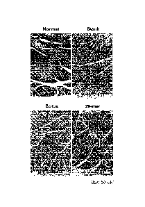
[0039] Figure 5 provides representative immunostained images of arterioles in
adductor magnus muscle after 2 weeks of ischemia. Arterioles were labeled with
anti-a-SMA (brown) and nuclei were labeled with hematoxylin.
[0040] Figure 6 provides representative photographs of aortic ring explants
cultured for 4 days in either basal MCDB131 medium (untreated control) or
medium
supplemented with known angiogenic factors (FGF2 or VEGF), the control PEDF
peptides (25-mer or 18-mer), or the PEDF peptides according to embodiments of
the
present disclosure (29-mer, 24-mer, 20-mer, Mo 29-mer, or Mo 20-mer).
[0041] Figure 7 provides representative dual-immunostained images
illustrating vascular smooth muscle cells (vSMCs) outgrowth from aortic rings
cultured
in medium supplemented with PEDF peptide (29-mer, 20-mer and 18-mer), in which
endothelial cells were detected by Alexa Fluor 594¨labeled isolectin B4 (184;
red; left
panel) and vSMCs were labeled with anti-a-SMA (green; middle panel). Merged
images are located on the right (yellow). Nuclei were visualized with Hoechst
33258
staining. Original magnification, x400.
Images are representative of four
independent experiments.
[0042] Figure 8 provides representative photographs from soleus muscle
specimens stained by H&E at day 14 following bupivacaine injection.
[0043] Figure 9 is a diagram illustrating muscle fiber size distributions of
muscles from animals in various experimental conditions.
[0044] Figure 10 provides representative photographs illustrating regenerating
tissue ( t ) at the inner part of tendon at week 3 post-injury. Original
magnification,
x100.
CA 02882479 2015-02-06
WO 2014/023007
PCT/CN2012/079897
[0045] Figure 11 provides representative photographs of H&E-stained sections
of Achillis tendon at 3 weeks after injury. Original magnification, x400;
scale bar = 50
M. Images are representative of three independent experiments.
[0046] Figure 12 provides representative photographs of tissue sections
5 stained by Masson's trichrome to highlight the collagen fibers at 3 weeks
post-injury.
Stars (*) represent the uninjured area in tendon. Original magnification,
x400; scale
bar= 50 M. Images are representative of three independent experiments.
[0047] Figure 13 provides representative immunostained photographs of newly
formed type 1 collagen (brown color) in regenerating tendon at 3 weeks after
surgery.
10 Nuclei labeled with hematoxylin. Boxed regions are shown at higher
magnification
below. Scale bar = 50 M. Images are representative of three independent
experiments.
[0048] Figure 14 is a representative gel electrophoresis image illustrating
enhanced expression level of tenomodulin (TNMD) gene by the present PEDF
peptides
(29-mer and 20-mer) according to one working example of the present
disclosure.
Expression of tenomodulin (TNMD) gene is indicative of BM-MSC differentiation
into
tenocyte. The image is representative of three independent experiments.
DESCRIPTION
[0049] The detailed description provided below in connection with the
appended drawings is intended as a description of the present examples and is
not
intended to represent the only forms in which the present example may be
constructed or utilized. The description sets forth the functions of the
example and
CA 02882479 2015-02-06
WO 2014/023007
PCT/CN2012/079897
11
the sequence of steps for constructing and operating the example. However, the
same or equivalent functions and sequences may be accomplished by different
examples.
[0050] For convenience, certain terms employed in the entire application
(including the specification, examples, and appended claims) are collected
here.
Unless otherwise defined herein, scientific and technical terminologies
employed in
the present disclosure shall have the meanings that are commonly understood
and
used by one of ordinary skill in the related art. Unless otherwise required by
context,
it will be understood that singular terms shall include plural forms of the
same and
plural terms shall include the singular. Specifically, as used herein and in
the claims,
the singular forms "a" and "an" include the plural reference unless the
context clearly
indicates otherwise.
[0051] Notwithstanding that the numerical ranges and parameters setting
forth the broad scope of the invention are approximations, the numerical
values set
forth in the specific examples are reported as precisely as possible. Any
numerical
value, however, inherently contains certain errors necessarily resulting from
the
standard deviation found in the respective testing measurements. Also, as used
herein, the term "about" generally means within 10%, 5%, 1%, or 0.5% of a
given value
or range. Alternatively, the term "about" means within an acceptable standard
error
of the mean when considered by one of ordinary skill in the art. Other than in
the
operating/working examples, or unless otherwise expressly specified, all of
the
numerical ranges, amounts, values and percentages such as those for quantities
of
materials, durations of times, temperatures, operating conditions, ratios of
amounts,
CA 02882479 2015-02-06
WO 2014/023007
PCT/CN2012/079897
12
and the likes thereof disclosed herein should be understood as modified in all
instances by the term "about." Accordingly, unless indicated to the contrary,
the
numerical parameters set forth in the present disclosure and attached claims
are
approximations that can vary as desired. At the very least, each numerical
parameter
should at least be construed in light of the number of reported significant
digits and by
applying ordinary rounding techniques.
[0052] As used herein, the term "peptide" denotes a polymer of amino acid
residues. By the term "synthetic peptide" as used herein, it is meant a
peptide which
does not comprise an entire naturally occurring protein molecule. The peptide
is
"synthetic" in that it may be produced by human intervention using such
techniques as
chemical synthesis, recombinant genetic techniques, or fragmentation of the
whole
protein or the like. Throughout the present disclosure, the positions of any
specified
amino acid residues within a peptide are numbered starting from the N terminus
of
the peptide.
[0053] The term "stem cell" as used herein, refers to a cell that retains the
capacity, under certain circumstances, to proliferate without substantially
differentiating; as well as the capacity or potential, under particular
circumstances, to
differentiate to a more specialized or differentiated phenotype.
[0054] As used herein, "proliferating" and "proliferation" refers to an
increase
in the number of cells in a population by means of cell division.
[0055] As used herein, the term "muscle cell" refers to any cell which
contributes to muscle tissue, and encompasses myoblasts, satellite cells,
myotubes,
and myofibril tissues. "Muscle regeneration" as used herein refers to the
process by
CA 02882479 2015-02-06
WO 2014/023007
PCT/CN2012/079897
13
which new muscle fibers form from muscle progenitor cells. The regeneration of
muscle in or adjacent to the damaged region may be evidenced by the increase
in the
number, diameter (size), wet weight, and/or the protein content of the muscle
fibers
in or adjacent to the damaged region. Also, the muscle regeneration may be
monitored by the proliferative activity of muscle cells and/or satellite cells
in or
adjacent to the damaged region.
[0056] As used herein, the term "tendon" refers to a fibrous tissue composed
of parallel arrays of closely packed collagen fibers that connects muscle to
bone. The
healing of damaged tendon is a slow process and usually associated with scar
formation which may result in a defective tendon that cannot resume normal or
original tendon function. As used herein, the term "tendon regeneration"
refers to a
tendon healing process in which type I collagen is formed, and the newly
formed
collagen fibers align parallel to the direction of load application, whereby
resulting in
minimal scar formation. The regeneration of tendon in or adjacent to the
damaged
region may be evidenced by the increase in the number of the collagen fibrils
with an
organized orientation in or adjacent to the damaged region. Also, the tendon
regeneration may be monitored by the proliferative activity of tendon stem
cells in or
adjacent to the damaged region.
[0057] As used herein, the term "arteriogenesis" is to be distinguished from
"angiogenesis." Angiogenesis is a process by which new capillary blood vessels
sprout from a pre-existing blood vessel. It is important to recognize that
these newly
formed capillary tubes lack vascular smooth muscle cells. Accordingly, they
are
fragile and prone to rupture. These capillary tubes would not go through
vasculature
CA 02882479 2015-02-06
WO 2014/023007
PCT/CN2012/079897
14
remodeling process, and hence are unable to sustain and/or restore proper
circulation
in and/or adjacent to the damaged region. In contrast to the capillary
sprouting,
arteriogenesis refers to the in situ recruitment and expansion of arteries or
collateral
arteries by proliferation of endothelial and smooth muscle cells from pre-
existing
arteriolar connections. These newly formed arteries or collateral arteries
would
develop into a functional network of arteries (or collateral arteries) which
constitute
natural bypasses capable of supplying sufficient blood to the damaged or
ischemic
tissue or site of inflammation.
[0058] The term "ischemia" as used herein relates to a condition that may
occur in any tissue and/or organ that suffers from a lack of oxygen supply
and/or from
abnormal accumulation of metabolites, which occurs when there is an imbalance
between oxygen supply and demand, due to inadequate perfusion, e.g., caused by
atherosclerosis, restenotic lesions, anemia, stroke or clogged arteries just
to name a
few, that leads to insufficient oxygen to tissues such as, for example, the
muscle, heart
or brain. However, ischemia is not limited to the aforementioned organs or
tissues,
since it may occur in any organ/tissue.
[0059] The term "promote" or "promoting" is meant to refer to a positive
alteration; in particular a statistically significant positive alteration. The
positive
alteration means an increase of at least 10% as compared to a reference level.
[0060] "Percentage (%) amino acid sequence identity" with respect to the
synthetic polypeptide sequences identified herein is defined as the percentage
of
amino acid residues in a candidate sequence that are identical with the amino
acid
residues in the specific polypeptide sequence, after aligning the sequences
and
CA 02882479 2015-02-06
WO 2014/023007
PCT/CN2012/079897
introducing gaps, if necessary, to achieve the maximum percent sequence
identity, and
not considering any conservative substitutions as part of the sequence
identity.
Alignment for purposes of determining percentage sequence identity can be
achieved
in various ways that are within the skill in the art, for instance, using
publicly available
5 computer software such as BLAST, BLAST-2, ALIGN or Megalign (DNASTAR)
software.
Those skilled in the art can determine appropriate parameters for measuring
alignment, including any algorithms needed to achieve maximal alignment over
the full
length of the sequences being compared. For purposes herein, sequence
comparison
between two amino acid sequences was carried out by computer program Blastp
10 (protein-protein BLAST) provided online by Nation Center for Biotechnology
Information (NCB!). The percentage amino acid sequence identity of a given
amino
acid sequence A to a given amino acid sequence B (which can alternatively be
phrased
as a given amino acid sequence A that has a certain % amino acid sequence
identity to
a given amino acid sequence B) is calculated by the formula as follows:
15 -X
x100%
Y
where X is the number of amino acid residues scored as identical matches by
the
sequence alignment program BLAST in that program's alignment of A and B, and
where
Y is the total number of amino acid residues in A or B, whichever is shorter.
[0061] The phrase "pharmaceutically acceptable carrier" as used herein means
a pharmaceutically acceptable material, composition or vehicle, such as a
liquid or
solid filler, diluent, excipient, solvent or encapsulating material, involved
in carrying or
transporting the subject agents from one organ, or a portion of the body, to
another
organ, or another portion of the body. Each carrier must be "acceptable" in
the
CA 02882479 2015-02-06
WO 2014/023007
PCT/CN2012/079897
16
sense of being compatible with the other ingredients of the formulation. The
carrier
can be in the form of a solid, semi-solid, or liquid diluent, cream or a
capsule.
[0062] The terms "treatment" and "treating" are used herein to generally
mean obtaining a desired pharmaceutical and/or physiological effect.
Preferably, the
effect is therapeutic in terms of partially or completely curing the muscle
damage,
tendon damage, or ischemia. The term "treating" as used herein refers to
application
or administration of the synthetic peptide or pharmaceutical composition of
the
present disclosure to a subject, who has a medical condition, a symptom of the
condition, a disease or disorder secondary to the condition, or a
predisposition toward
the condition, with the purpose to partially or completely alleviate,
ameliorate, relieve,
delay onset of, inhibit progression of, reduce severity of, and/or reduce
incidence of
one or more symptoms or features of a particular disease, disorder, and/or
condition.
Treatment may be administered to a subject who does not exhibit signs of a
disease,
disorder, and/or condition and/or to a subject who exhibits only early signs
of a
disease, disorder, and/or condition for the purpose of decreasing the risk of
developing pathology associated with the disease, disorder, and/or condition.
Treatment is generally "effective" if one or more symptoms or clinical markers
are
reduced as that term is defined herein. Alternatively, a treatment is
"effective" if the
progression of a disease is reduced or halted. That is, "treatment" includes
not just
the improvement of symptoms or decrease of markers of the disease, but also a
cessation or slowing of progress or worsening of a symptom that would be
expected in
absence of treatment. Beneficial or desired clinical results include, but are
not
limited to, alleviation of one or more symptom(s), diminishment of extent of
disease,
CA 02882479 2015-02-06
WO 2014/023007
PCT/CN2012/079897
17
stabilized (i.e., not worsening) state of disease, delay or slowing of disease
progression,
amelioration or palliation of the disease state, and remission (whether
partial or total),
whether detectable or undetectable.
[0063] The term "effective amount" as used herein refers to the quantity of a
component which is sufficient to yield a desired response. The term
"therapeutically
effective amount" as used herein refers to the amount of therapeutically agent
of
pharmaceutical composition to result in a desired "effective treatment" as
defined
hereinabove. The specific therapeutically effective amount will vary with such
factors
as the particular condition being treated, the physical condition of the
patient (e.g., the
patient's body mass, age, or gender), the type of mammal or animal being
treated, the
duration of the treatment, the nature of concurrent therapy (if any), and the
specific
formulations employed. A therapeutically effective amount is also one in which
any
toxic or detrimental effects of the compound or composition are outweighed by
the
therapeutically beneficial effects.
[0064] The term "subject" refers to a mammal including the human species
that is treatable with the synthetic peptides, compositions, and/or methods of
the
present invention. The term "subject" is intended to refer to both the male
and
female gender unless one gender is specifically indicated.
[0065] Pigment epithelium-derived factor (PEDF) is a multifunctional secreted
protein that has anti-angiogenic, anti-tumorigenic, and neurotrophic
functions.
Human PEDF protein (SEQ ID No: 14) is a secreted protein of roughly 50 kDa
size and
418 amino acids in length. A 34-mer fragment (residues 44-77) and a 44-mer
CA 02882479 2015-02-06
WO 2014/023007
PCT/CN2012/079897
18
fragment (residues 78-121) of PEDF have been identified to have anti-
angiogenic and
neurotrophic properties, respectively.
[0066] The present disclosure is based, at least, on the finding that
synthetic
peptides derived from PEDF may promote the regeneration of muscle or tendon
tissue
and arteriogenesis in a subject. In particular, the present disclosure is the
first to
identify a link between the local delivery of PEDF-derived peptides and the
healing of
muscle or tendon tissues suffering from damage and/or ischemia or the
formation of
(collateral) arteries in or adjacent to the ischemic region. Another inventive
feature
of the present invention lies in that the synthetic peptides are much shorter
(39 amino
acid residues at most) than the full-length PEDF and thus overcomes the
limitations
associated with the clinical use of conventional protein drugs, including high
manufacturing cost, low bioavailability, and poor pharmacokinetics.
Accordingly, the
present synthetic peptides are useful for treating muscle or tendon damages as
well as
tissues or organs suffering from ischemia.
[0067] Thus, in one aspect, the present disclosure is directed to a synthetic
peptide for promoting muscle or tendon regeneration in a subject. In another
aspect,
the present disclosure is directed to a synthetic peptide for promoting
arteriogenesis
in a subject. Embodiments applicable to either or both of these two aspects
are
discussed below.
[0068] According to embodiments of the present disclosure, the synthetic
peptide has 20-39 amino acid residues in length, and has at least 80% amino
acid
sequence identity with the amino acid sequence of
LSVATALSALSLGAEQRTESIIHRALYYDLISSPDIHGT (SEQ ID NO: 1). For example, the
CA 02882479 2016-05-30
19
synthetic peptide may have an amino acid sequence identity of about 80, 81,
82, 83,
84, 85, 86, 87, 88, 89, 90, 91, 92, 93, 94, 95, 96, 97, 98, 99, or 100 percent
with SEQ ID
NO: 1. Also, the synthetic peptide comprises at least 20 consecutive
residues that
are at least 90% identical to residues 11-30 of SEQ ID NO: 1. Specifically,
the 20
consecutive amino acid residues may have about 90, 91, 92, 93, 94, 95, 96, 97,
98, 99,
or 100 percent amino acid sequence identity with residues 11-30 of SEQ ID NO:
1.
[0069] In one embodiment, the synthetic peptide has the sequence of SEQ ID
NO: 1, which has 39 amino acids in length. This synthetic peptide is referred
to as
39-mer in the description hereinbelow. This 39-mer peptide corresponds to
residues
83-121 of human PEDF and hence is a short variant derived from the known PEDF
44-mer (corresponding to residues 78-121 of PEDF).
[0070] Prior experiments conducted by the present inventors, such as those
disclosed in US Patent No. 9,051,547
and experiments provided hereinbelow, reveal that
several short, synthetic PEDF peptides derived from the 39-mer, are capable of
promoting muscle or tendon regeneration and/or arteriogenesis in a subject.
[0071] For example, based on experiments disclosed in both the prior
application and the present application, a 34-mer synthetic peptide having the
sequence of ALSALSLGAEQRTESIIHRALYYDLISSPDIHGT (SEQ ID NO: 2) is effective in
promoting muscle or tendon regeneration and/or arteriogenesis in a subject.
This
34-mer peptide corresponds to residues 88-121 of human PEDF. According to the
process for estimating percentage of sequence identity between any two given
sequences provided above, the 34-mer has a 100% amino acid sequence identity
to
CA 02882479 2015-02-06
WO 2014/023007
PCT/CN2012/079897
the 39-mer, and the 6th-25th amino acid residues of the 34-mer has a 100%
amino acid
sequence identity to the amino acid residues 11-30 of the 39-mer.
[0072] Additionally, according to various examples hereinbelow, a 29-mer
synthetic peptide having the sequence of SLGAEQRTESIIHRALYYDLISSPDIHGT (SEQ ID
5 NO: 3) has been confirmed to be effective in promoting muscle or tendon
regeneration
as well as arteriogenesis in a subject. This 29-mer peptide corresponds to
residues
93-121 of human PEDF with a 100% amino acid sequence identity to the 39-mer.
Also, the 1st ¨20th amino acid residues of the 29-mer has a 100% amino acid
sequence
identity to the amino acid residues 11-30 of the 39-mer.
10 [0073]
In some examples, a 24-mer has been confirmed to be effective in
promoting tendon regeneration and arteriogenesis in a subject. The 24-mer has
the
sequence of SLGAEQRTESIIHRALYYDLISSP (SEQ ID NO: 5), which corresponds to
residues 93-116 of human PEDF. This 24-mer peptide has a 100% amino acid
sequence identity to the 39-mer in which the first twenty amino acid residues
thereof
15 has a 100% amino acid sequence identity to the amino acid residues 11-30
of the
39-mer.
[0074] In other examples, it has been established that a 20-mer may promote
muscle or tendon regeneration as well as arteriogenesis in a subject. The 20-
mer has
the sequence of SLGAEQRTESIIHRALYYDL (SEQ ID NO: 6), which corresponds to
20 residues 93-112 of human PEDF. This 20-mer peptide is completely
identical to the
amino acid residues 11-30 of the 39-mer (100% amino acid sequence identity),
and
has a 100% amino acid sequence identity to the 39-mer.
CA 02882479 2015-02-06
WO 2014/023007
PCT/CN2012/079897
21
[0075] Two synthetic peptides derived from mouse PEDF may also promote
muscle or tendon regeneration and/or arteriogenesis in a subject based on
experiments disclosed in both the prior application and the present
application. The
first mouse-derived peptide is referred to as "Mo 29-mer" in the present
disclosure.
The Mo 29-mer has a sequence of SLGAEHRTESVIHRALYYDLITNPDIHST (SEQ ID NO: 8),
which has a 83% amino acid sequence identity to 39-mer, and the first 20 amino
acid
residues thereof has a 90% amino acid sequence identity to the 11-30 amino
acid
residues of the 39-mer. Another mouse-derived peptide, Mo 20-mer has a
sequence
of SLGAEHRTESVIHRALYYDL (SEQ ID NO: 9). The Mo 20-mer has a 90% amino acid
sequence identity to either the 39-mer or the 11-30 amino acid residues of the
39-mer.
[0076] Optionally, the synthetic peptide comprises 4 consecutive residues
identical to residues 11-14 of SEQ ID NO: 1. It is believed that residues 11-
14 (i.e.,
SLGA) of SEQ ID NO: 1 play an important role in maintaining the biological
function of
the short PEDF peptides. For example, according to various examples provided
below,
a 18-mer peptide (EQRTESIIHRALYYDLIS; SEQ ID NO: 7) without the SLGA residues
fail
to elicit any arteriogenesis in a subject. Also, based on experiments
disclosed in both
the prior application and the present application, it is suggested that a 25-
mer peptide
(EQRTESIIHRALYYDLISSPDIHGT; SEQ ID NO: 4) is ineffective in promoting muscle
or
tendon regeneration and/or arteriogenesis in a subject.
[0077] The synthetic Peptides of the invention can be synthesized by
commonly used methods such as t-BOC or FMOC protection of alpha-amino groups.
Both methods involve stepwise syntheses whereby a single amino acid is added
at
CA 02882479 2015-02-06
WO 2014/023007
PCT/CN2012/079897
22
each step starting from the C terminus of the peptide. Peptides of the present
invention can also be synthesized by the well-known solid phase peptide
synthesis
methods.
[0078] Other synthetic peptides with conservative variation with respect to
the
39-mer are also contemplated. The term "conservative variation" as used herein
denotes the replacement of an amino acid residue by another, biologically
similar
residue. Examples of conservative variations include the substitution of
one
hydrophobic residue such as isoleucine, valine, leucine or methionine for one
another,
or the substitution of one polar residue for another, such as the substitution
of
arginine for lysine, glutamic for aspartic acids, or glutamine for asparagine,
and the like.
The term "conservative variation" also includes the use of a substituted amino
acid in
place of an unsubstituted parent amino acid provided that antibodies raised to
the
substituted polypeptide also immunoreact with the unsubstituted polypeptide.
[0079] The synthetic peptides according to above-mentioned embodiments
may be formulated into pharmaceutical compositions for promoting muscle or
tendon
regeneration and/or arteriogenesis in a subject, which falls within other
aspects of the
present disclosure.
[0080] According to one embodiment of the present disclosure, the
pharmaceutically composition comprises a synthetic peptide according to any of
the
above-mentioned aspects/embodiments, and the synthetic peptide is present in
an
effective amount sufficient to promote the muscle or tendon regeneration
and/or
arteriogenesis in the subject. The pharmaceutical composition also comprises a
pharmaceutically acceptable carrier for the synthetic peptide.
CA 02882479 2015-02-06
WO 2014/023007
PCT/CN2012/079897
23
[0081] The choice of a pharmaceutically acceptable carrier to be used in
conjunction with a synthetic peptide is basically determined by the way the
pharmaceutical composition is to be administered. According to one optional
embodiment of the present disclosure, the pharmaceutical composition may be
administered locally via intramuscular injection. In this case, the synthetic
peptide
may be formulated with a pharmaceutically acceptable carrier such as a sterile
aqueous solution, which is preferably isotonic with the blood of the
recipient. Such
formulations may be prepared by dissolving or suspending the solid active
ingredient
in water containing physiologically compatible substances such as sodium
chloride,
glycine, and the like, and having a buffered pH compatible with physiological
conditions to produce an aqueous solution, and rendering said solution
sterile.
[0082] Still optionally, the synthetic peptide may be formulated in a
sustained-release dosage form so as to ensure a more prolonged therapeutic
action of
the treatment. There are several polymeric materials suitable for prolonging
drug
release, examples of which include, but are not limited to, alginate, gelatin,
collagen,
and poly(lactide-co-glycolide).
[0083] According to some working examples of the present disclosure, the
present synthetic peptides are embedded in a matrix of cross-linked alginate
gel, and
the final concentration of the synthetic peptides is about 1-100 1.1.M, and
preferably,
about 10 M. For example, the concentration of the synthetic peptides may be
about 1, 2, 3, 4, 5, 6, 7, 8, 9, 10, 15, 20, 25, 30, 35, 40, 45, 50, 55, 60,
65, 70, 75, 80, 85,
90, 95, or 100 M.
CA 02882479 2015-02-06
WO 2014/023007
PCT/CN2012/079897
24
[0084] Pharmaceutical compositions of the invention can also comprise various
additives known to those skilled in the art. For example, solvents, including
relatively
small amounts of alcohol, may be used to solubilize certain drug substances.
Other
optional pharmaceutically acceptable additives include opacifiers,
antioxidants,
fragrance, colorant, gelling agents, thickening agents, stabilizers,
surfactants, and the
like. Other agents may also be added, such as antimicrobial agents, to prevent
spoilage upon storage, i.e., to inhibit growth of microbes such as yeasts and
molds.
Permeation enhancers and/or irritation-mitigating additives may also be
included in
the composition of the present invention.
[0085] In yet another aspect, the present invention is directed to a method
for
promoting muscle or tendon regeneration in or adjacent to a damaged region of
a
subject; and in still another aspect, the present invention is directed to a
method for
promoting arteriogenesis in or adjacent to an ischemic region of a subject. In
either
embodiment, the subject may be any animal classified as a mammal, including
human.
Embodiments applicable to either or both of these two aspects are discussed
below.
[0086] In one embodiment, the method for promoting muscle or tendon
regeneration in or adjacent to a damaged region of a subject comprises
administering,
to a treatment region of the subject, a therapeutically effective amount of
the
synthetic peptide of the present disclosure, wherein the treatment region is
adjacent
to the damaged region so as to promote the muscle or tendon to regenerate in
or
adjacent to the damaged region of the subject to regenerate.
[0087] In another embodiment, the method for promoting arteriogenesis in or
adjacent to an ischemic region of a subject comprises administering, to a
treatment
CA 02882479 2016-05-30
region of the subject, a therapeutically effective amount of the synthetic
peptide of
the present disclosure, wherein the treatment region is adjacent to the
ischemic
region, so as to promote a rteriogenesis in or adjacent to the ischemic region
of the
subject.
5 [0088] According to optional embodiments, the synthetic peptide is
formulated
in a pharmaceutical composition according to the above-mentioned
aspect/embodiments of the present disclosure. In practice, the pharmaceutical
composition may be administered via intramuscular injection.
[0089] According to some embodiments, the subject may be suffering from
10 muscle injury, muscle disuse, muscular dystrophy, amyotrophic
lateral sclerosis,
tendon injury, tissue ischemia, cerebral ischemia, peripheral arterial
diseases, or
myocardial infarction, which causes the blood flow at the ischemic region to
be
hindered or blocked.
[0090] The following Examples are provided to elucidate certain aspects of the
15 present invention and to aid those of skilled in the art in
practicing this invention.
These Examples are in no way to be considered to limit the scope of the
invention in
any manner. Without further elaboration, it is believed that one skilled in
the art can,
based on the description herein, utilize the present invention to its fullest
extent.
EXAMPLES
[0091] Materials and Methods
[0092] Materials
CA 02882479 2015-02-06
WO 2014/023007
PCT/CN2012/079897
26
[0093] Dulbecco's modified Eagle's medium (DMEM), fetal bovine serum (FBS),
0.25% trypsin, anti-BrdU antibody, MCDB131 medium, TRIzol, and Dynabeads were
purchased from Invitrogen (Carlsbad, CA). Ultrapure alginate (6000 Da),
dimethyl
sulfoxide (DMSO), bovine serum albumin (BSA), 5-bromo-2'-deoxyuridine (BrdU),
-- Hoechst 33258 dye, and Masson's Trichrome were all from Sigma-Aldrich (St.
Louis,
MO). Collagenase type I and dispase II were obtained from Roche (Indianapolis,
IN).
All the fluorescent dye-conjugated secondary antibodies were purchased from
BioLegend (San Diego, CA). Hematoxylin and eosin (H&E) dyes were purchased
from
Merck (Rayway, NJ, USA). Anti-collagen 1A1 antibody was obtained from Santa
Cruz
-- Biotechnology (Santa Cruz, CA). Matrigel was purchased from BD Biosciences
(Bedford, MA). Anti-alpha-smooth muscle actin (anti-a-SMA) antibody (ab5694)
and
anti- nucleostemin antibody were from Abcam (Cambridge, MA). Anti-Pax7
antibody
(GTX62311) was from GeneTex (Taipei, Taiwan). Isolectin B4 (164)-Alexa Fluor
568
was from Molecular Probes (Eugene, OR).
[0094] Short synthetic PEDF peptides, including 29-mer (SEQ ID No: 3), 25-mer
(SEQ ID No: 4), 24-mer (SEQ ID No: 5), 20-mer (SEQ ID No: 6), 18-mer (SEQ ID
No: 7),
MO 29-mer (SEQ ID No: 8), and MO 20-mer (SEQ ID No: 9) were synthesized and
modified with acetylated at the NH2 termini and amidated at the COOH termini
for
stability and characterized by mass spectrometry (>95% purity) to order at
GenScript
-- (Piscataway, NJ).
[0095] All animals used in embodiments of the present disclosure were housed
in an animal room under temperature control (24-25 C) and 12:12 light-dark
cycle.
Standard laboratory chow and tap water were available ad libitum. The
experiments
CA 02882479 2015-02-06
WO 2014/023007
PCT/CN2012/079897
27
procedures were approved by the Mackay Memorial Hospital Review Board (New
Taipei City, Taiwan, R.O.C.) and were performed in compliance with national
animal
welfare regulations.
[0096] PEDF peptide/alginate gel formulation and bolus formulation
[0097] Each PEDF-derived short synthetic peptide (the 29-mer, 25-mer, 24-mer,
20-mer, 18-mer, MO 29-mer, or MO 20-mer; hereinbelow, PEDF peptide) was
reconstituted in DIVISO as stock (5 mM). Then, ultrapure alginate was mixed
with the
stock to obtain a 2% wt/vol alginate solution with PEDF peptide at a final
concentration of 10 p.M. The alginate solution was then filtered by membrane
filter
(pore size, 0.22 p.m) and mixed with filtered calcium sulfate (0.21 g CaSO4/mL
of dH20)
at a ratio of 25:1 (40 pL of CaSO4 per 1 mL of the filtered alginate
solution). The
mixture was let standing at RT for about 1 hour to allow for the cross-linking
of the
alginate. The resultant sustained-release formulation was then used in the
treatment
of muscle or tendon damage and ischemia.
[0098] For bolus delivery, a final PEDF concentration of 10 p.M was used by
performing serial dilutions from the 5 mM stock solution.
[0099] Histology, lmmunohistochemistry and Quantification
[0100] The gracilis, adductor magnus, soleus, and tibia lis muscles were fixed
in
4% paraformaldehyde, dehydrated with graded ethanol series, and paraffinized.
Fixed samples were de-paraffinized in xylene and rehydrated in a graded series
of
ethanol. Tissues were sliced into 5-pm sections. General histology was
performed
using H&E dye.
CA 02882479 2015-02-06
WO 2014/023007
PCT/CN2012/079897
28
[0101] De-paraffinized tissue sections were blocked with 10% goat serum for 1
hour. Staining was done using primary antibodies against BrdU (1:50 dilution;
GTX42641) or type I collagen 1A1 (1:50 dilution) overnight at 4 C, followed by
incubation with the appropriate peroxidase-labeled donkey immunoglobulin for
30
min and then with chromogen substrate (3,3'-diaminobenzidine) for 2 min before
counterstaining with hematoxylin. Quantification was estimated based on high
quality images (1208 X 960 pixels) captured using a Nikon Eclipse 80i light
microscope.
[0102] The muscle fiber size was determined on H&E-stained muscle cross
section and quantified using the minimum distance of parallel tangents at
opposing
particle borders (minimal "Feret's diameter"). Pictures were captured using a
Nikon
Eclipse 80i light microscope, and the minimal Feret's diameter was measured
using the
Image-Pro Plus 4.5.1 software (Media Cybernetics). Normalization of the number
of
fibers in each fiber Feret class of 51.1.m was based on the total number of
muscle fibers
in each picture.
[0103] To ascertain the number of centrally nucleated muscle fibers, sections
were stained with H&E and then photographed as described above. At least 100
stained fibers were randomly chosen from each photo. Muscle fibers were judged
centrally nucleated if one or more nuclei were not located at the periphery of
the fiber.
The data were expressed as a % of the total number of muscle fibers counted.
Results were evaluated from 6 sections per muscle section, and 10 mice at each
group.
[0104] De-paraffinized tendon tissue sections were stained using Masson's
Trichrome according to the manufacturer's instructions.
For semi-quantitative
analysis of collagen area, 10 fields from each slide were randomly selected
under a
CA 02882479 2015-02-06
WO 2014/023007
PCT/CN2012/079897
29
light microscope, and the repairing area per intact tendon area of the cross
section
(mm2/mm2) was measured using the Image-Pro Plus 4.5.1 system.
[0105] Isolation and culture of tendon stem cells
[0106] New Zealand White rabbits (6-8 months old, 3.0-4.0 kg) were used in
this study. Achilles tendons were removed from the rabbits by cutting through
their
bony attachments. The tendon sheath was stripped away and the core portion of
the
tendons was minced into small fragments. Each 100 mg of fragment was then
digested in a solution containing 3 mg/mL of type I collagenase and 4 mg/mL of
dispase in 1 ml Dulbecco's Modified Eagle Medium (DMEM-high glucose) at 37 C
for 2
hours. The resultant cell suspension was centrifuged at 1,000 rpm for 15
minutes to
obtain a cell pellet which was then resuspended in a growth medium consisting
of
DMEM supplemented with 10% heat inactivated fetal bovine serum (FBS), 100
1.1.M
2-mercaptoethanol, and 100 U/m1 penicillin and 100 1.1.g/m1 streptomycin. For
passage, near-confluent cells were harvested with 0.25% trypsin and then 1 X
105
subcultured cells were further cultured in medium.
[0107] TSCs proliferation assay
[0108] The TSCs at passage 4 were seeded at gelatin-coated slide in a 6-well
plate at a density of 2 x 10⁵ cells per well and cultured in growth medium
(DMEM +
10% FBS) for 24 hours before being replaced by a basal growth medium with 5%
FBS
only (control group) or with 5% FBS plus an additional 50 nM of PEDF-derived
peptide
(i.e., 29-mer, 24-mer, 20-mer, 18-mer, Mo 29-mer, or Mo 20-mer) for 24 hours.
For
BrdU labeling assay, BrdU (final concentration, 10 1.1.M) was added to the
culture for 4
hours. After fixing with 4% paraformaldehyde, cells were exposed to cold
methanol
CA 02882479 2015-02-06
WO 2014/023007
PCT/CN2012/079897
for 2 minutes, and then treated with 1 N HCI at RT for 1 hour before
performing
immunofluorescence. The phenotype of passage 4 TSC was determined by
immunocytochemistry of nucleostemin and type I collagen. Almost all of
expanded
TSCs were nucleostemin and type I collagen-double positive cells.
5 [0109] In vivo detection of DNA synthesis
[0110] For the detection of cell expansion, BrdU was reconstituted in DMSO as
stock (80 mM). 10 pi of BrdU mixed with 90 pi of PBS was intraperitoneally
injected
into the mouse 16 hours prior to euthanasia. Also, 150 pi of BrdU mixed with
350 pi
of PBS was intraperitoneally injected into the rat 16 hours prior to
euthanasia. DNA
10 synthesis was assessed by BrdU labeling with anti-BrdU antibodies.
[0111] lmmunofluorescence Analysis
[0112] De-paraffinized tissue sections or 4% paraformaldehyde fixed rabbit
tendon stem cells (TSCs) were blocked with 10% goat serum and 5% BSA for 1
hour.
Double staining was done using primary antibodies against a-SMA (1:100
dilution), 1B4
15 (5 p.g/m1), Pax7 (1:100 dilution), nucleostemin (1:100 dilution) and
type! collagen 1A1
(1:50) at 37 C for 2 hours, followed by incubation with the appropriate
rhodamine- or
FITC-conjugated donkey IgG for 1 hour at RT. Nuclei were located by
counterstaining
with Hoechst 33258 for 7 minutes.
Images were captured using a Zeiss
epifluorescence microscope with a CCD camera.
20 [0113]
The small artery densities (a-SMA positive cells surrounding the whole
circumference of the vessel) were measured and images were taken from 10
randomly-selected areas of adductor magnus muscle (200x magnification) in each
sample, and blinded quantification was performed in triplicate by manually
counting
CA 02882479 2015-02-06
WO 2014/023007
PCT/CN2012/079897
31
within each section; values from five sections were then averaged and
expressed as
arteriole density per mm2.
[0114] Bone-marrow-derived Mesenchymal Stem Cells (BM-MSCs) Isolation,
Cell Culture, and Treatments
[0115] Primary rat BM-MSCs were isolated from femur of male
Sprague-Dawley rats (300-450 g). Femora were aseptically removed and dissected
free of adhering tissues, and then the marrow cavities were flushed by
injection of
DMEM medium. Collected bone marrow cells were incubated in a 100 X 15-mm Petri
dish in DMEM medium supplemented with 10% FBS, 100 U/m1 penicillin, and 100
ig/m1 streptomycin for 2 weeks in 5% CO2 at 37 C. The medium was replaced
every
2 to 3 days. For passage, near-confluent cells were detached by 0.25% trypsin
and
then 2 x 10⁵ subcultured cells were seeded in a well of 6-well plate and
further
cultured in the 10% FBS-DMEM. Before treatment, cells were starved for 12
hours in
DMEM supplemented with 1% FBS followed by treatment with 50 nM PEDF-derived
peptide (29-mer or 20-mer) in fresh 1% FBS-DMEM for either 24 or 48 hours.
[0116] RNA Extraction and Reverse Transcription¨Polymerase Chain Reaction
[0117] The total RNA was extracted from cells using TRIzol and treated with
RNase-free DNase l (Qiagen, Santa Clarita, CA) to remove genomic DNA and then
purified with an RNA purification kit (Dynabeads). 1 lig of total RNA
retrieved from
BM-MSCs was reverse-transcribed into cDNA by 200 units of expand
Reverse-Transcriptase (Roche, Mannheim, Germany) in 20 11.1 of reaction buffer
containing 0.25 1..ig of random primers and 0.8 mM dNTPs at 42 C for 1 hour. 2
'al of
the cDNA was used as templates in subsequent PCR reaction.
CA 02882479 2015-02-06
WO 2014/023007
PCT/CN2012/079897
32
[01 18] PCR was performed using a reaction volume of 30 III containing 15 I
of
EconoTaq PLUS GREEN 2x Master Mix (Lucigen Corp.), 11.1.M of each primer and
2 pi
of template DNA. cDNA was synthesized in an 18-22 cycle amplification reaction
(denaturation, 20s, 94 C; annealing, 30s, 57 C; and polymerization, 40s, 72
C). Cycle
number for each primer set was established to be in the linear range of
amplification.
The primer set for the amplification of rat Tenomodulin gene (TNMD; accession
number: NM_022290) included a forward primer of AGAATGAGCAATGGGTGGTC (SEQ
ID No: 10) and a reverse primer of CTCGACCTCCTTGGTAGCAG (SEQ ID No: 11), and
PCR
products of about 240 bp were observed. Analysis of rat glyceraldehyde 3-
phosphate
dehydrogenase (GAPDH; accession number: X02231.1) gene was used as a
housekeeping gene for the normalization of the expression level. For
the
amplification of GAPDH gene, the primer set including a forward primer of
AGACAGCCGCATCTICTIGT (SEQ ID No: 12) and a reverse primer of
CTTGCCGTGGGTAGAGTCAT (SEQ ID No: 13) was used, and PCR products of about 207
bp were observed.
[0119] The PCR products were electrophoresed in a 2% agarose gel containing
ethidium bromide and visualized by UV illumination. The intensities of the PCR
products were quantified densitometrically using a FUJI LAS-3000 system and
Multi
Gauge Ver. 1.01 software (Fujifilm, Tokyo, Japan).
[0120] Statistics
[0121] Results were expressed as the mean standard error of the mean (SEM).
One-way ANOVA was used for statistical comparisons. P < 0.05 was considered
significant, unless otherwise specified.
CA 02882479 2015-02-06
WO 2014/023007
PCT/CN2012/079897
33
[0122] Example 1
[0123] Sustained Release of PEDF Peptides from Alginate Gel
[0124] To determine the release kinetics of 29-mer and 20mer, 100 pig of
FITC-conjugated PEDF peptide was mixed with 100p1 alginate solution, and then
hydrogels were prepared as set forth in the "Materials and Methods" section.
Thereafter, 100 mg hydrogel was incubated in 1.5 ml of PBS (pH 7.4) in
microcentrifuge
tube and placed in an orbital shaking incubator over a 6-day period at 37 C.
The tube
was centrifuged at each predetermined time point and then 2004 of supernatant
was
removed and stored at -80 C for further analysis, and 2004 of fresh PBS was
added to
the tube to replace the supernatant withdrawn. The concentration of
FITC-conjugated PEDF peptide present in the collected supernatants was
determined
using a fluorimeter in 96-well format. A known non-encapsulated FITC-peptide
was
used to generate a standard curve. Triplicate data were used for analysis.
[0125] The results of the assay, as summarized in Figure 1, revealed that the
embedded PEDF peptides were released in a sustained manner over a 6-day
period.
Specifically, approximately 48% of 29-mer and 35% of 20-mer peptide remained
in the
alginate gel matrix after 24 hours. Most of the 29-mer peptides (90%) were
released
within the first 4 days, after which time the release rate decreased
significantly
thereby resulting in a plateau of the cumulative release curve. The 20-mer
peptides
were released in a slightly faster rate in which about 90% of the loaded 20-
mer was
released in the first 3 days.
CA 02882479 2015-02-06
WO 2014/023007
PCT/CN2012/079897
34
[0126] Example 2
[0127] Sustained Release of PEDF Peptides Reduces Ischemic Damages
[0128] Ischemic muscle injury typically leads to necrosis and loss of tissue
and
function. Hence, ischemic animal model was employed in the present examples to
investigate the possibility that the local delivery of the PEDF
peptide/alginate gel
formulation (herein "the sustained-release formulation") may promote the
recovery of
tissue or organ functions in the case of tissue or organ damages. Various
conditions
associated with ischemic damages, such as, limb perfusion, tissue necrosis,
arteriogenesis, and neovessel sprouting, were analyzed in the examples as
follows.
[0129] 6-week-old female C57BL/6 wild-type mice were anesthetized by an
intraperitonea I injection of a mixture of zoletil (6 mg/kg) and xylazine (3
mg/kg). Hair
was removed from the hindquarter with a depilating cream. To establish
hindlimb
ischemia, unilateral external iliac and femoral arteries and veins were
ligated, cut, and
excised. After surgery, the mice were randomly assigned to several
experimental
groups (n = 6, each group) and treated as follows. In the blank control group,
the
mice were treated with 50 pi of blank alginate gel, whereas in the bolus
control group,
the mice received the bolus formulation containing 29-mer. In
the PEDF
peptide/alginate gel treatment groups, the mice received 50 pi of the
sustained-release formulation, which comprised either 29-mer, 24-mer, or 20-
mer.
Additionally, in a PEDF 18-mer control group, mice were treated with a
sustained-release formulation containing a PEDF 18-mer peptide. Treatments
were
applied by way of a single intramuscular injection to the gracilis muscle
immediately
CA 02882479 2015-02-06
WO 2014/023007
PCT/CN2012/079897
after femoral artery and vein excision operation. The incision was closed
after the
wound was irrigated with sterile saline.
[0130] Example 2.1
[0131] Sustained Release of PEDF Peptides Enhances Limb Perfusion
5 [0132]
A laser Doppler perfusion imaging (LDPI) analyzer (Moor Instruments,
USA) was used to quantify hindlimb blood perfusion before surgery (pre OP),
immediately after surgery (post OP), and over time after surgery. To minimize
vasoconstriction by anesthetic heat loss, animals were kept on a heating plate
at 37 C
for 5 min before measurement. Representative LDPI images illustrating the
blood
10 perfusion of ischemic hind limbs over a time period of 4 weeks were
provided in Figure
2 in which dark blue color represents low blood flow. Blood perfusion is
expressed as
LDPI index representing the ratio of operated (ischemic) versus non-operated
(non-ischemic) limb blood flow of the same mouse, and the results were
summarized
in Figure 3 and Table 1 (n 6).
Blood flow was displayed as changes in the laser
15 frequency, represented by different color pixels.
Table 1
Treatment Ischemic/Non-ischemic Perfusion Ratio (%)
Pre OP Post OP 7 days 14days 21 days 28 days
Blank 99.4 1.5 8.1 0.87 30.6 1.9 28.4 3.9 46.313.8 50.0 6.5
Bolus 112.9 6.2 9.0 0.80 22.9 4.6 31.6 2.1 44.1 8.4 55.3++2.8
18-mer 104.5 2.5 7.5 0.67 23.8 4.5 30.3 0.94 46.8 4.3 52.8 7.4
29-mer 108.2 8.8 7.0 3.1 44.8 2.0* 77.6 6.8* 101 7.0* 105 4.8*
24-mer 91.5 5.6 8.0 0.03 53.1 0.37* 67.2 5.8* 86.1 5.9* 91.81-5.5*
20-mer 98.0 7.8 4.6 0.64 43.3 7.2* 60.0 9.0* 75.5 5.2* 92.7 3.0*
*P < 0.05 versus blank control.
CA 02882479 2015-02-06
WO 2014/023007
PCT/CN2012/079897
36
[0133] As illustrated in Figure 3, after the surgery, the regional blood flow
(post
OP) was immediately reduced to about 8% of the non-ischemic limb of the same
animal in all groups, as expected. Blank (alginate gel¨only) control led to a
slow
increase in reperfusion over time. It should be noted that results from the
bolus
delivery was similar to that of the blank control, indicating that sustained
release of
PEDF peptides is essential for exerting its protective effect. Also, mice
treated with
sustained-release formulation containing the control 18-mer peptide did not
exhibit
improved blood perfusion as compared with that of the blank control or the
bolus
control; suggesting that the 18-mer peptide is ineffective in treating
ischemia. In
contrast, the present PEDF treatments significantly improved blood perfusion
over that
of the blank, bolus, and PEDF 18-mer control groups. In particular, animals
treated
with sustained formulations containing 29-mer, 24-mer, or 20-mer exhibited a
marked
increase in blood flow (at least about 60% of normal limbs) starting around
the second
weeks after the surgery. By four weeks after the surgery, the perfusion in
animals
treated with 29-mer, 24-mer, and 20-mer delivered with the sustained-release
formulations lead to a final recovery of, respectively, 105%, 92%, and 93% of
normal
limbs, as compared with 50% in the blank control and 55% in the bolus control.
[0134] Example 2.2
[0135] Sustained Release of PEDF Peptides Prevents Ischemia-induced Tissue
Necrosis
[0136] In most hindlimb ischemia models, tissue necrosis generally occurs in
the muscles below the knee. For example, tibialis anterior muscle, which is
distant to
the gracilis muscle where the treatment was administered, often undergoes
extensive
CA 02882479 2015-02-06
WO 2014/023007
PCT/CN2012/079897
37
necrosis with regeneration after femoral artery excision. The intensity of
Masson's
trichrome blue color staining depended on the content of collagen fibers in
the
investigated tissue, and fibrosis is the result of necrosis. Hence, two weeks
and seven
weeks after the surgery and the treatment, samples from the tibialis anterior
muscle
were analyzed by Masson's trichrome staining to assess the degree of fibrosis
and
hence necrosis. Results from representative samples are illustrated in Figures
4A and
4B.
[0137] As depicted in Figure 4A, at the second week after the surgery, muscle
tissue from the blank control groups exhibited extensive fibrosis (indicated
by the blue
stain), whereas muscle tissue treated with the present sustained-release
formulation
exhibited a relatively smaller fibrosis region. Note also in Figure 4A, at
week 7
post-surgery, the treatment with the present sustained-release formulation
effectively
reduced the areas of necrosis and fibrosis thereby achieved a complete
recovery of
muscle tissue.
[0138] After ischemic injury, muscle fiber regeneration is achieved by
proliferation of satellite cells. The newly formed muscle fiber is marked by
centrally
located nuclei. Also, the necrotic area is evidenced by necrotic myofibers
exhibiting a
pale eosinophilic cytoplasm with oedema and a loss of peripheral nuclei. As
revealed
in Figure 4B, two weeks after the surgery, the regeneration of myofibers with
centrally
located nuclei was more significant in mice treated with the present sustained-
release
formulation than that in mice treated with the blank control. Still referring
to the
upper panels of Figure 4B, the large pale red area in the sample from the
blank control
group, as compared with the sample from the 20-mer treatment group, also
suggested
CA 02882479 2015-02-06
WO 2014/023007
PCT/CN2012/079897
38
that the present sustained-release formulation was effective in preventing
necrosis.
At week 7 post-surgery, small bundles of muscle fiber with intersperse fat
droplet
remained in 15% of muscle area in tibialis anterior muscle in groups treated
with blank
control (Figure 413; lower left panel).
[0139] Statistical analyses regarding injured area (necrotic area + fibrotic
area)
and numbers of centrally nucleated fibers were also performed at two weeks
after the
surgery, and the results were summarized in Table 2. The injured area is
expressed as
the percent of total stained area (%), and the centrally nucleated fibers is
expressed as
the total number of muscle fibers counted (%).
[0140] The data summarized in Table 2 revealed that the injection of the
present sustained-release formulation may substantially reduce tissue injury
as
compared with that of blank or bolus controls. Specifically, the injured areas
of the
PEDF treatment groups were reduced to about 45-48% of those of the blank or
bolus
control group. Also, these data suggested that the treatment with 29-mer, 24-
mer,
or 20-mer formulation resulted in an increase (about 3-3.7-fold) in the number
of
centrally nucleated fibers in tibialis anterior muscle, as compared with that
of blank or
bolus control.
Table 2
Treatment Injured Area (%) Centrally nucleated fibers
(%)
Blank 81.0 3.3 19.5 2.2
Bolus 80.1 4.1 20.2. 3.2
18-mer 78.5 5.1 20.5 3.3
29-mer 39.5 4.2* 72.5 5.2t
24-mer 36.8 5.5* 67.8 5.3t
20-mer 38.0 5.2* 61.2 5.8t
*P < 0.001 versus blank control.
tP < 0.02 versus blank control.
CA 02882479 2015-02-06
WO 2014/023007
PCT/CN2012/079897
39
[0141] In sum, results in Example 2.2 suggested that the treatment with the
present sustained formulation containing either 29-mer, 24-mer, or 20-mer may
prevent necrosis and fibrosis induced by ischemia, and thereby may improve the
recovery of muscle tissue. Also, the increase of recovery of tibialis muscle
in mice
treated with the present sustained-release formulation provides additional
evidence to
support its effect on the promotion of blood perfusion in ischemic limb
(Example 2.1
above).
[0142] Example 2.3
[0143] Sustained Release of PEDF Peptides Stimulates Arteriogenesis That
Supplements Ischemic Tissue with Collateral Circulation
[0144] In the case of acute occlusion of a major artery (such as coronary
artery
or femoral artery), pre-existing arteriolar connections can be recruited to
bypass the
site of occlusion. This process is termed arteriogenesis which differs in many
aspects
from angiogenesis. From the anatomical aspect, these pre-existing collateral
arteries,
unlike capillaries formed during angiogenesis, are microvascular, thin-walled
conduits
that are composed of an endothelial lining, an internal elastic lamina, and
one or two
layers of smooth muscle cells.
Under normal conditions, these endogenous
pre-existing thin-walled arterioles may not be utilized to provide perfusion.
However,
following occlusion of a major artery, these vessels can dramatically increase
their
lumen by growth, bypassing the site of occlusion so as to provide enhanced
perfusion
to the jeopardized ischemic regions. During chronic or acute occlusion of a
major
artery, collateral arteries may ameliorate the ensuing detrimental effects in
many
CA 02882479 2015-02-06
WO 2014/023007
PCT/CN2012/079897
regions of the body (hindlimb, heart, brain, kidney, etc.). It is important to
recognize
that arteriogenesis is not a simple process of passive dilatation of pre-
existing
collateral arteries; rather, it is associated with active proliferation and
remodeling by
growth of pre-existing arteriolar connections into true collateral arteries.
It is
5 established that vessel radius is the dominant influence on blood flow,
and hence, the
collateral arteries, after adaptive growth, are capable of conducting
relatively large
blood volumes per unit of time. Therefore, stimulation of arteriogenesis is
probably
the more efficient mechanism for the survival of ischemic limbs or internal
organs such
as heart and brain, in comparison to angiogenesis. In contrast, angiogenesis
is the
10 formation of capillaries composed of endothelial cells from the pre-
existing vessels;
these capillaries are fruitless in proving higher profusion to the damaged
ischemic
region. Thus, the increase in blood flow to the potentially ischemic tissue,
as caused
by the development of two or three large collateral arteries, cannot be
equaled by
newly formed capillaries, however numerous.
15 [0145]
To investigate the arteriogenic effect of the present sustained-release
formulation, adductor magnus muscles (located at the same level as femoral
artery
excision and in which arteriogenesis responsible for establishing collateral
circulation is
expected to be found) were harvested from animals in each experimental
condition,
two weeks after the surgery. Arterioles in muscle cross sections were
identified by
20 immunohistological staining for vascular smooth muscle cells (a-SMA;
brown), and
nuclei were labeled with hematoxylin; representative photographs were provided
in
Figure 5. Quantitative analysis was also performed and the results were
summarized
CA 02882479 2015-02-06
WO 2014/023007
PCT/CN2012/079897
41
in Table 3, and the data were expressed as a-SMA-positive arterioles per mm2
in the
peri-injury region.
Table 3
Treatment Arteriole Density per mm2
Blank 3.3 0.88
Bolus 3.7 1.2
18-mer 4.0 0.58
29-mer 11.7 1.5*
24-mer 10.0 0.58*
20-mer 10.7 1.2*
*O < 0.001 versus blank control.
[0146] These data revealed that the administration of the present
sustained-release formulation increased arteriole density in adductor magnus
muscle
adjacent to the femoral artery excision, as compared with that of the blank
and bolus
control groups. Therefore, the sustained release of PEDF peptides provides
arteriogenic activity to establish collateral circulation after the acute
disruption of
blood supply. The dramatically increase the lumen of these vessels by growth
provides enhanced perfusion to the jeopardized ischemic regions. This
well-developed collateral network leads to the recovery from ischemic events.
[0147] Example 2.4
[0148] PEDF Peptide Stimulate Ex Vivo Neovessel Sprouting
[0149] To further confirm the neovessel development promoted by PEDF
peptides, rat aortic ring sprouting assay was performed. Thoracic aortas were
removed from euthanized rats and gently stripped of peri-aortic fibroadipose
tissue.
Aortas were sectioned into about 2-mm length rings, which were then embedded
in a
growth factor-reduced Matrigel. Gels containing the aortic rings were
polymerized in
CA 02882479 2015-02-06
WO 2014/023007
PCT/CN2012/079897
42
12-well plates incubated at 37 C for 30 minutes. 1 ml of MCDB131 medium
supplemented with 100 units/ml penicillin and 100 ng/ml streptomycin, 1% FBS,
and a
supplemental agent (50 ng/ml VEGF-A, 20 ng/ml FGF-2, or 50 ng/ml 29-mer, 24-
mer,
20-mer, Mo 29-mer, Mo 20-mer, 25-mer, or 18-mer) were added to the
Matrigel-containing explants. The cultures were propagated at 37 C in a
humidified
incubator for up to 4 days, with media changes every other day. Neovessel
sprouting
was assessed until day 4 using an inverted microscope platform (Leica) with
bright-field optics; representative photographs were provided in Figure 6.
Quantification of neovessel sprouting was assessed using Image- Pro Plus 6.0
software
(Dendrites program). Results were expressed as a fold of untreated aortic
ring, as
summarized in Table 4. The experiment was repeated in triplicate.
[0150] In the untreated control (UT) in which no supplemental factor was
administered, minimal neovessel sprouting was observed at day 4. It is also
noted
that the control PEDF peptides (i.e., 25-mer and 18-mer) did not substantially
enhance
the neovessel sprouting, compared with the untreated control.
Table 4
Treatment Neovessel Sprouting Fold
Untreated 1
VEGF 3.4*
FGF2 3.5*
18-mer 0.97
25-mer 0.89
29-mer 5.6*
24-mer 6.2*
20-mer 6.5*
Mo 29-mer 6.1*
Mo 20-mer 6.4*
*P < 0.02 versus untreated control.
CA 02882479 2015-02-06
WO 2014/023007
PCT/CN2012/079897
43
[0151] As expected, the well-known angiogenic factors, VEGF and FGF2,
induced substantial neovessel sprouting. The neovessel sprouting in samples
treated
with VEGF and FGF-2 increased for about 3.4-fold and 3.5-fold, respectively;
as
compared to that of the UT control.
[0152] The data in Table 4 also indicated that the present PEDF peptides
(including 29-mer, 24-mer, 20-mer, Mo 29-mer, and Mo 20-mer) stimulated more
neovessel sprouting than either VEGF or FGF2. These neovessels were examined
by
dual-staining immunofluorescence assay for a-smooth muscle actin (a marker of
arteriole wall smooth muscle cell (SMC)) and isolectin B4 (1134, a marker of
endothelial
cells), and representative photographs were provided in Figure 7. As could be
seen in
Figure 7, samples treated with the present PEDF peptide (29-mer or 20-mer)
displayed
arteriole phenotype with a SMC coating. In contrast, the formation of
endothelial
tube and SMC proliferation was barely detected upon treatment with PEDF 18-
mer.
This result indicated that PEDF peptides according to embodiments of the
present
disclosure can stimulate neovessel formation beyond the angiogenesis of
capillary
which only contains endothelial cells in culture. It thus supports the notion
that the
present PEDF peptides stimulate arteriogenesis in vivo.
[0153] In conclusion, data presented in Example 2 (including Examples 2.1 to
2.4) demonstrated that the present PEDF peptides were effective in enhancing
limb
perfusion, reducing tissue necrosis and fibrosis, and promoting arteriogenesis
and
neovessel sprouting, and hence, the administration of the PEDF peptides (in
particular,
the sustained-release formulation containing either of the PEDF peptides)
would
reduce ischemic damages and facilitate the structural and functional
recoveries of the
CA 02882479 2015-02-06
WO 2014/023007
PCT/CN2012/079897
44
tissue or organ. It should be noted that it has been established that a 34-mer
fragment of PEDF (residues 44-77) has anti-angiogenic properties, and a 44-mer
fragment of PEDF (residues 78-121) has neurotrophic properties. However, the
present disclosure is the first to confirm that short PEDF fragments (at least
the 29-mer,
24-mer, 20-mer, Mo 29-mer, and Mo 20-mer) exhibit an arteriogenic activity.
[0154] Example 3
[0155] Sustained Release of PEDF Peptides Promotes Muscle Regeneration
[0156] To investigate the effects of the present PEDF peptides on muscle
regeneration, a rat myonecrosis model of a single injection of bupivacaine
into the
soleus muscle was employed. Adult 10-week-old male Sprague-Dawley rats
(initial
body weight = 312 11 g) were anesthetized by an intra peritoneal injection
of a
xylazine (10 mg/kg). Then, the soleus muscle was injured by unilaterally
injecting 0.5
ml bupivacaine (AstraZeneca) with a disposable syringe with a 26-gauge needle.
Briefly, the needle was inserted into the distal portion of the soleus muscle
and then
receded longitudinally to the proximal portion accompanying evenly bupivacaine
solution injection. The solution was then injected throughout the entire
length of the
muscle as the needle was slowly withdrawn.
[0157] After bupivacaine injection, rats were divided equally (n = 10/group)
into four experimental groups and treated as follows. In the blank control
group, the
mice were treated with 50 pi of blank alginate gel. In the treatments groups,
the
mice received 50 pi of the sustained-release formulation (29-mer or 20-mer).
The
mice in the bolus control group received the bolus formulation (29-mer).
Treatments
CA 02882479 2015-02-06
WO 2014/023007
PCT/CN2012/079897
were applied by way of a single intramuscular injection to the soleus muscle
immediately after the bupivacaine perfusion.
[0158] At day 4 after bupivacaine injection, histology of soleus muscle cross
section consisted of general necrosis with disintegrating myofibers and
abundant
5 infiltrating inflammatory cells occupying a great major part of the
soleus muscle
(photographs not shown). Only in the peripheral were some muscle fibers with
relative normal structure remained. The degrees of muscle fiber necrosis were
the
same in blank control group and peptide treatment group. This result indicated
that
the necrosis levels induced by bupivacaine in different groups were
substantially the
10 same.
[0159] Example 3.1
[0160] Sustained Release of PEDF Peptides Promotes Cell Proliferation
[0161] Muscle regeneration involves proliferation of muscle fibers, or muscle
cells. The muscle fiber proliferation was assayed by the incorporation of BrdU
in the
15 proliferating nuclei. Satellite cells proliferation is the key step of
muscle regeneration.
Hence, the soleus muscle specimens were also stained for satellite cell
marker, Pax7,
so as to investigate the muscle regeneration activity. Detailed assay
procedures are
as described in "Materials and Methods." The level of BrdU-positive cells was
expressed as labeling index (%), which was computed as the number of labeled
cells
20 divided by the total number of cells. The labeling index (%) of Pax7-
positive cells was
computed as the number of labeled cells divided by the total number of cells
with
nuclei. Quantitative results were evaluated from 6 sections per muscle section
and
10 mice at each group, and were summarized in Table 5.
CA 02882479 2015-02-06
WO 2014/023007
PCT/CN2012/079897
46
Table 5
Treatment BrdU Labeling Index (%) Pax7 Labeling Index (%)
Blank 4.2 0.9 2.0 0.71
Bolus 4.4 1.4 2.6 0.68
29-mer 15.4 1.7 16.2 2.0
20-mer 13.6 3.0 14.6 1.9
[0162] These results revealed that the numbers of BrdU-positive cells in
wounds treated with the 29-mer- or 20-mer-containing sustained-release
formulation
were significantly increased, as compared with wounds treated with the blank
or bolus
control. Regarding the proliferative activity of satellite cells, the data
revealed that
the present sustained-release formulations lead to higher percentages of Pax7-
positive
cells as compared with blank and bolus controls. Together, these data
suggested that
the administration of the present sustained-release formulations enhances the
proliferative activities of muscle fibers and/or satellite cells, which in
turn may
promote the muscle to regenerate.
[0163] Example 3.2
[0164] Sustained Release of PEDF Peptides Promotes Muscle Fiber
Regeneration
[0165] In the process of muscle regeneration, newly generated muscle fibers
typically contain central located nuclei. Hence, the percentage of such
centrally
nucleated muscle fibers is also an indicator of the regenerative activity of
muscle.
Statistical analysis regarding the percentage of centrally nucleated fibers
was
performed at day 7 after the bupivacaine injection, and the results were
summarized
in Table 6.
CA 02882479 2015-02-06
WO 2014/023007
PCT/CN2012/079897
47
Table 6
Treatment Centrally nucleated fibers (%)
Blank 11.0 1.7
Bolus 13.8 2.2
29-mer 75.2 7.0
20-mer 63.3 7.4
[0166] As could be seen in Table 6, there were higher percentages of muscle
fibers containing centrally located nuclei in animals treated with the 29-mer-
or
20-mer-containing sustained-release formulation, as compared with that of
blank or
bolus control groups. These results indicated that the administration of the
present
sustained-release formulations was effective in promoting muscle regeneration.
[0167] Also, at 14 days after the bupivacaine injection, necrotic myofibers
were
replaced by newly formed myotubes in soleus muscle in all experimental groups.
However, a number of centrally nucleated fibers remained in regenerating
muscles
treated with blank or bolus control (Figure 8), suggesting an incomplete
muscle
regeneration. In contrast, muscle sections from animals treated with the
sustained-release formulation containing 29-mer or 20-mer exhibited much less
centrally nucleated fibers. Together, these data indicated that the muscle
fiber
regeneration was facilitated by the sustained release of the PEDF peptides.
[0168] Example 3.3
[0169] Sustained Release of PEDF Peptides Promotes Maturation of
Regenerated Muscle Fiber
[0170] In the later stage of muscle regeneration, newly generated muscle
fibers start to gain size. Muscle specimens were collected at 14 days after
injury, and
CA 02882479 2015-02-06
WO 2014/023007
PCT/CN2012/079897
48
respective fiber diameters were measured in accordance with the procedure set
forth
in the section of "Materials and Methods." Results were summarized in Figure
9.
[0171] On average, diameters of muscle fibers from animals treated with the
sustained-release formulation containing 29-mer or 20-mer were larger than
those
from animals in the blank or bolus control group. In addition, the size
distribution of
the 20-mer-treated muscles was in close resemblance to that of uninjured,
intact
muscles. Specifically, about 56.6% of muscle fibers from animals treated with
20-mer
and about 53.2% of intact muscle fibers had a minimal Feret's diameter between
15-25 1.1.m, whereas about 59.6% and about 56.2% of the regenerated fibers
from the
blank and bolus control groups had minimal Feret's diameters between about 10-
20
1.1.m. These data indicated that the administration of the present sustained-
release
formulation was effective in increasing the mass of the regenerated muscles.
[0172] In conclusion, data presented in Example 3 (including Examples 3.1 to
3.3) demonstrated that the present PEDF peptides are effective in promoting
the
proliferations of muscle fibers and satellite cells, regeneration of muscle
fibers, and the
maturation of regenerated muscle fibers, and hence, the administration of the
PEDF
peptides (in particular, the sustained-release formulation containing either
of the PEDF
peptides) would promote the muscle regeneration process and facilitate the
structural
and functional recoveries of the muscle tissue. The present disclosure is the
first to
discover that short PEDF fragments (at least the 29-mer and 20-mer) are
capable of
promoting muscle regeneration.
[0173] Example 4
CA 02882479 2015-02-06
WO 2014/023007
PCT/CN2012/079897
49
[0174] Sustained Release of PEDF Peptides Promotes Tendon Regeneration
[0175] To investigate the effects of the present PEDF peptides on tendon
regeneration, a rat model with tendon injury was established as follows. Adult
10-week-old male Sprague-Dawley rats (total n = 50; initial body weight = 312
11 g)
were anesthetized by an intraperitoneal injection of a xylazine (10 mg/kg).
Then, the
left tendo Achilles injury was created by full-thickness insertion of an 18-
gauge needle
through tendo Achilles 1 cm proximal to its insertion into the calcaneum. This
created a horizontal (transaction) wound which was flanked by intact tendon
tissue on
both sides to prevent the retraction of severed ends.
[0176] The rats were randomly assigned (n = 10/group) to five experimental
groups and treated as follows. In the blank control group, the mice were
treated with
150 p.1 of blank alginate gel. For the bolus control group, 150 p.l of the
bolus
formulation (29-mer) was administered. In the treatments groups, the mice
received
150 pi of the sustained-release formulation (29-mer, 24-mer, or 20-mer).
Treatments
were injected subcutaneously near tendon lesion immediately after the injury,
and the
incision was closed after the wound was irrigated with sterile saline.
[0177] Example 4.1
[0178] Sustained Release of PEDF Peptides Promotes Tendon Healing
[0179] Three weeks after tendon injury, histologic analysis was performed to
observe the healing of the tendon. Representative photographs were provided in
Figure 10. As could be seen in the upper panel of Figure 10, in animals
treated with
blank control, a broad band of disorganized fibrous scar was formed between
the two
cut ends, and the pink stained collagen bundle was minimal in the scar tissue.
In
CA 02882479 2015-02-06
WO 2014/023007
PCT/CN2012/079897
contrast, the cut ends of tendons treated with sustained-release formulation
containing 29-mer were healed with much lesser scar tissue as compared with
that of
the blank control specimen, and the pinkish stain of mature collagen bundle
extended
into the damaged region of the tendon; also, tendon fibers from the cut ends
seemed
5 to joint together in some area (Figure 10; lower panel). Also, the
fibrous tissue in the
scar was more organized and in parallel direction in the 29-mer treatment
group.
[0180] Figure 11 provides representative photographs of histologic analysis at
higher magnification. Normal tendon had a relative scarcity of cells among the
collagen fibers, and the nuclei were mostly elongated. In the blank and bolus
control
10 groups, after healing for three weeks, the more abundant presence of
fibroblasts
(characterized by the presence of round- or spindle-shaped fibroblast-like
nuclei) was
observed in the tendon, and the newly formed collagen fibers were structurally
disorganized (the undamaged tissue was indicated by *). These morphological
changes suggested poor healing of the tendon wounds in the blank and bolus
control
15 groups.
[0181] In contrast, still referring to Figure 11, in tendons treated with the
present sustained-release formulations, the healing regions had thin,
elongated nuclei
which were morphologically similar to nuclei of mature tenocyte, and the
collagen
fibers were well organized and parallel to the native tendon (deep pink);
suggesting a
20 better healing of the tendon wounds. These results indicated that the
tendon wound
healing process may benefit from the present sustained-release formulation.
[0182] Further, Masson trichrome staining was performed to evaluate the
structure and organization of collagen fibers, and representative photographs
of the
CA 02882479 2015-02-06
WO 2014/023007
PCT/CN2012/079897
51
specimens were provided in Figure 12. In the uninjured tendon, collagens
fibers were
substantially parallel to one another (Figure 12; left panel). By contrast,
the injured
tendon treated with blank control had disorganized collagen fibers in the
healed region
(Figure 12; middle panel; wound margin marked by *). The injured tendon
treated
with the present sustained-release formulation, however, had well-organized
collagen
fibers that were aligned in substantially the same orientation as the
uninjured tendon
tissue beyond the wound margin (Figure 12; right panel; wound margin marked by
*).
These highly oriented and organized collagen fibers suggested a better tendon
wound
healing effect in animals treated with the present sustained-release
formulation.
[0183] Quantitative analysis was also performed to assess the percentage of
collagen (%) in the regenerated area, and results were summarized in Table 7.
Table 7
Treatment Collagen in Regenerated Area (%)
Blank 56.0 6.87
Bolus 58.25 8.84
29-mer 88.75 1.89
24-mer 85.75 2.56
20-mer 82.01 6.55
[0184] As could be seen in Table 7, animals in the PEDF treatment groups
(29-mer, 24-mer or 20-mer) had higher collagen contents in the regenerated
area, as
compared with those in the blank and bolus control groups. These data
suggested
that collagen synthesis in the wound region may be promoted by the
administration of
the present sustained-release formulation.
[0185] The specimens were also subjected to immunostainning of type I
collagen, and nuclei were labeled with hematoxylin. Representative photographs
CA 02882479 2015-02-06
WO 2014/023007
PCT/CN2012/079897
52
were provided in Figure 13, in which lower panels are photographs of the
magnified
regions respectively enclosed by dash lines in the upper panels. As could be
appreciated, the collagen fibrils in the uninjured tendon were well cross-
linked with
one another, and hence, they were unlikely to be recognized by the anti-
collagen 1A1
antibody. Therefore, only minimal amount of type I collagen (brown stain) was
observed in the left panel of Figure 13. By comparing the photographs of the
blank
control group (Figure 13; middle panel) and the PEDF treatment group (Figure
13; right
panel), it was certain that type I collagen (brown) was more abundant in the
PEDF
treatment groups than in the blank control group.
[0186] Collectively, these results suggested that the administration of
sustained-release formulation containing the present PEDF peptide would
stimulate
type 1 collagen synthesis in cells in injured tendon tissues, facilitate
collagen
deposition in healed tissues, and promote a more organized alignment of
collagen
fibers, and thereby promote tendon regeneration.
[0187] Example 4.2
[0188] PEDF Peptides Induces In Vitro TSC Proliferation
[0189] It has been reported that during the tendon healing process, tendon
stem cells (TSCs) would proliferate and differentiate into tenocytes. To
investigate
whether present PEDF peptides would induce the proliferation of TSCs in vitro,
tendons stem cells were isolated and cultured as described in the "Materials
and
Methods" section. The purity of TSCs was confirmed by a TSC marker,
nucleostemin,
as well as the expression of type I collagen by TSCs; together, these analyses
indicated
a TSC purity of near 100% (data not shown). The proliferation of TSCs was
confirmed
CA 02882479 2015-02-06
WO 2014/023007
PCT/CN2012/079897
53
by BrdU pulse-labeling for 2 hours. Quantitative analysis of the level of BrdU-
positive
cells was performed as described above, and the results were summarized in
Table 8.
Table 8
Treatment BrdU Labeling Index (%)
Control 9.6 2.1
29-mer 31.8 3.6*
24-mer 29.0 4.6*
20-mer 33.2 6.6*
Mo 29-mer 33.2 6.6*
Mo 20-mer 31.8 3.1*
*P < 0.002 versus untreated control cells.
[0190] These data revealed that, as compared with TSCs cultured in control
medium, TSCs cultured in media containing the present PEDF peptide (29-mer, 24-
mer,
20-mer, Mo 29-mer, or Mo 20-mer) were more proliferative. Also, it should be
noted
that Mo 29-mer and Mo 20-mer are derived from the mouse PEDF peptide, and does
not have 100% amino acid sequence identities to the 11-30 amino acid residues
of the
39-mer. However, they respectively exhibited similar mitogenic activity to the
short
PEDF peptides (e.g., 29-mer, 24-mer, and 20-mer) derived from human PEDF.
[0191] Example 4.3
[0192] Sustained Release of PEDF Peptides Promotes In Vivo TSC Proliferation
After Tendon Injury
[0193] Specimens obtained from animals in different experimental groups of
Example 4.1 were stained for nucleostemin (green) to investigate whether in
vivo TSC
proliferation would be promoted by the present sustained-release formulation
during
the tendon wound healing process. In quantitative analysis, ten randomly
selected
CA 02882479 2015-02-06
WO 2014/023007
PCT/CN2012/079897
54
microscopic fields in each experimental group were photographed, and the
percentage
of nucleostemin-positive cells per total cells (counterstained by Hoechst
33258; blue)
was calculated. Quantitative results were summarized in Table 9.
Table 9
Treatment Nucleostemin Labeling Index (%)
Blank 5.8 1.8
Bolus 5.6 1.4
29-mer 16.4 2.9*
24-mer 16.8 4.2*
20-mer 15.0 3.9*
*P < 0.001 versus blank control.
[0194] These data revealed that levels of nucleostemin-positive TSC cells in
animals treated with 29-mer, 24-mer, or 20-mer were elevated, as compared with
those in the blank and bolus control groups. Taken together, results from
Examples
4.1 and 4.3 suggested that the in vivo expansion of TSCs promoted by the
administration of the present sustained-release formulation was coincident
with the
more prominent tendon healing effect, as compared with the native healing
process.
[0195] Example 4.4
[0196] PEDF Peptide Induces Tenocyte-like Cell Generation from Bone
Marrow-Derived Mesenchymal Stem Cells (BM-MSCs)
[0197] Recently, it is established that adult mesenchymal stem cells (MSCs)
could be used to regenerate functional tendons. In this example, BM-MSCs were
cultured in a control medium or a medium containing either PEDF 29-mer or 20-
mer to
investigate the ability of the present PEDF peptides in promoting BM-MSC
differentiation into tenocytes. Tenomodulin gene (TNMD) is a gene
predominantly
CA 02882479 2015-02-06
WO 2014/023007
PCT/CN2012/079897
expressed in tendons, and is considered as the most reliable phenotypic marker
of the
tenocytic lineage. Hence, tenocyte differentiation was evaluated based on the
expression of TNMD. Representative image from RT-PCR analysis was provided in
Figure 14.
5 [0198] The results revealed that the present PEDF peptide (29-mer or 20-
mer)
is an effective inducer of tenocyte-like cell differentiation in cultured BM-
MSCs. Since
the mobilization and differentiation of BM-MSCs is a proposed mechanism of
tendon
repair in vivo, this observation suggested that the present PEDF peptide may
repair
tendon damage by promoting the differentiation of BM-MSCs into tenocytes. It
also
10 indicated the potential of the present PEDF peptide to facilitate the
synthesis of
artificial tendons from scaffold matrix culture of BM-MSCs.
[0199] In conclusion, data presented in Example 4 (including Examples 4.1 to
4.4) demonstrated that the present PEDF peptides were effective in promoting
the
synthesis of well-organized collagen (in particular, type one collagen)
fibrils and
15 proliferation of tendon stem cells, and hence, the administration of the
present PEDF
peptides (in particular, the sustained-release formulation containing either
of the
present PEDF peptides) would promote the tendon regeneration process and
facilitate
the structural and functional recoveries of the tendon tissue. The present
disclosure
is the first to discover that short PEDF fragments (at least the 29-mer and 20-
mer) are
20 capable of promoting tendon regeneration and BM-MSCs differentiation into
tenocytes.
[0200] Collectively, results from the preceding examples established that the
present synthetic PEDF peptides (such as the 29-mer, 24-mer, 20-mer, Mo 29-
mer, and
CA 02882479 2016-05-30
56
Mo 20-mer) may promote a rteriogenesis in or adjacent to the ischemic region,
muscle
and tendon regeneration in or adjacent to the injured region. Accordingly, the
present synthetic PEDF peptides are suitable for use as a therapeutic agent to
promote
muscle and tendon wound-healing and reduce ischemic damages.
[0201] It will be understood that the above description of embodiments is
given by way of example only and that various modifications may be made by
those
with ordinary skill in the art. The above specification, examples, and data
provide a
complete description of the structure and use of exemplary embodiments of the
invention. Although various embodiments of the invention have been described
above with a certain degree of particularity, or with reference to one or more
individual embodiments, those with ordinary skill in the art could make
numerous
alterations to the disclosed embodiments without departing from the scope of
this invention.