Note: Descriptions are shown in the official language in which they were submitted.
CA 02882533 2015-02-19
WO 2014/049447 PCT/1B2013/002848
1
A WHOLE, LEECH SALIVA EXTRACT
AHMED MERZOUK
ABBAS MOHAMMAD GHAWI
ABDUALRAH MAN M. ABDUALKADER
MOHAMED ALAAMA
CROSS-REFERENCE TO RELATED APPLICATIONS
[0001] This application claims priority to U.S. Application No. 13/624,847,
filed September 21,
2012, which claims the benefit of U.S. Provisional Application No. 61/701,735,
filed September
17, 2012, each application of which is hereby incorporated by reference in its
entirety.
BACKGROUND
Field of the Invention
[0002] The teachings provided herein are generally directed to methods of
isolating and using a
whole-saliva leech extract in the treatment of a subject.
Description of the Related Art
[0003] The history of humans using leeches goes back several thousands of
years, and
practically all human civilizations described the use of leeches to treat
different diseases.
Unfortunately, due at least to a lack of understanding of the chemistries and
mechanisms
associated with such uses, the current state-of-the-art has not been able to
successfully
commercialize the use of leech saliva extracts in treating disease.
[0004] There have been attempts at sacrificing leeches to extract active
compounds from the
whole body of leeches, from the heads of leeches, or from their salivary
glands. Much research
has been directed to identifying proteins from leech saliva extracts. None of
these efforts,
however, have been able to reproduce the effect of using a whole, live leech,
with the exception
of, perhaps, the isolation and use of hirudin as an anticoagulant.
[0005] There have been attempts at not sacrificing leeches but, rather,
extracting a much
diluted saliva solution from a live leech. Unfortunately, these efforts have
been faced with two
major problems: (i) the saliva removal requires a manual squeezing of the
leech and, as such,
is not easily scalable; and (ii) the saliva remains dilute, which can only be
used fresh, and any
lyophilization attempts will reduce or completely abolish the therapeutic
activity of the leech
CA 02882533 2015-02-19
WO 2014/049447 PCT/1B2013/002848
2
saliva extract. As such, a dose-dependent treatment, or a treatment at
elevated concentrations,
is not available for testing.
[0006] One of skill will appreciate (i) a method of isolating an active,
refined leech saliva extract
(LSE) that can be successfully stored for months, or even years; (ii) a method
of re-using
leeches to produce the LSE; (iii) a method of commercializing the isolation
and re-use of the
leeches to a scalable amount that is practical for commercialization; (iv) a
method of treating a
solid tumor with the LSE; (v) a method of treating a liquid tumor with the
LSE; (vi) a method of
treating diabetes with the LSE; (vii) a method of treating a virus with the
LSE; (viii) a method of
treating a parasitic disease with the LSE; (ix) a method of using the LSE as
an antioxidant; and
(x) a method of using the LSE as an antibacterial.
SUMMARY
[0007] The teachings provided herein are generally directed to methods of
isolating and using a
whole-saliva leech extract in the treatment of a subject. Pharmaceutical
formulations
comprising the leech extracts and a pharmaceutically acceptable carrier are
provided.
[0008] The teachings include a method of removing a whole saliva from a leech.
In these
embodiments, the methods can include feeding a phagostimulatory agent to a
leech; inducing a
regurgitation in the leech, the inducing including placing the leech in an
environment having a
temperature of less than about 0 C; and, collecting an unrefined, whole saliva
in the
regurgitation of the cooled leech.
[0009] The teachings include a method of creating a lyophilized, whole saliva
extract of a leech
having an improved stability. In these embodiments, the method can include
feeding a
phagostimulatory agent to a leech; inducing regurgitation in the leech, the
inducing including
placing the leech in an environment having a temperature ranging from about -
500 to about 1500;
collecting an unrefined, whole saliva in the regurgitation of the cooled
leech; removing solid
components from the unrefined, whole saliva to create a refined, whole saliva;
and, lyophilizing
separate volumes of the refined, whole saliva extract, the volumes not
exceeding about 2m1
each.
[00010] In some embodiments, the collecting includes squeezing the leech
to increase
the amount of unrefined, whole saliva collected. In some embodiments, the
methods further
comprise revitalizing the leech by warming the leech in a water bath having a
temperature
ranging from about 5 C to about 40 C. In some embodiments, the methods further
comprise
CA 02882533 2015-02-19
WO 2014/049447 PCT/1B2013/002848
3
creating a refined, whole-saliva extract; the creating including removing
solid components from
the unrefined, whole saliva. In some embodiments, the methods further comprise
lyophilizing
separate volumes of the refined, whole saliva extract, the volumes not
exceeding about 2m1
each. And, in some embodiments, the leech is Hirudinaria manillensis.
[00011] The teachings include a stable, lyophilized, whole-saliva extract
of a leech. In
these embodiments, the extract comprises a refined, whole-saliva extract of a
leech lyophilized
in volumes not exceeding about 2m1 each, the extract refined by removing solid
components
from an unrefined, whole saliva to create the refined, whole saliva; wherein,
the extract has a
stable activity when stored for use at a temperature below about -20 C , the
extract maintaining
at least 70% of the activity for at least 6 months. And, the leech can be
Hirudinaria manillensis.
[00012] Methods of treating a subject by administering an effect amount of
the leech
extracts are provided. In some embodiments, the method includes treating a
solid tumor,
treating a liquid tumor, treating diabetes, treating a viral disease, treating
a parasitic disease,
treating a bacterial disease, or administering an anti-oxidant therapy. It
should be appreciated
that each of the treatments also relate to other conditions that may be
desirable to treat in the
subject.
BRIEF DESCRIPTION OF THE FIGURES
[00013] FIG. 1 illustrates a method of feeding a phagostimulatory agent to
a leech using
a membrane, according to some embodiments.
[00014] FIGs. 2A-2C illustrate the collection of unrefined, whole saliva
extract, according
to some embodiments.
[00015] FIG. 3 illustrates a UV spectra of the refined, leech saliva
extract, according to
some embodiments.
[00016] FIG. 4 illustrates a standard curve for a colorimetric Bradford
protein assay,
according to some embodiments.
[00017] FIG. 5 shows the LSE protein molecular weight distribution results
of a Malaysian
leech, Hirudinaria manillensis, using the Laemmli SDS-PAGE 15% gel
electrophoresis,
according to some embodiments.
[00018] FIG. 6 shows the LSE protein molecular weight distribution results
of a Malaysian
leech, Hirudinaria manillensis, using the Laemmli SDS-PAGE 15% gel
electrophoresis, wherein
the LSE was concentrated using acetone precipitation, according to some
embodiments.
CA 02882533 2015-02-19
WO 2014/049447 PCT/1B2013/002848
4
[00019] FIG. 7 shows the LSE protein molecular weight distribution results
of a Malaysian
leech, Hirudinaria manillensis, using the Laemmli SDS-PAGE 15% gel
electrophoresis, wherein
the LSE was precipitated from solution using a trichloroacetic acid (TCA)
precipitation,
according to some embodiments.
[00020] FIG. 8 shows the LSE protein molecular weight distribution results
of a Malaysian
leech, Hirudinaria manillensis, using the Non-Urea SDS-PAGE gel
electrophoresis of Okajima,
according to some embodiments.
[00021] FIG. 9 shows the LSE protein molecular weight distribution results
of a Malaysian
leech, Hirudinaria manillensis, using the Tricine SDS-PAGE gel electrophoresis
method,
according to some embodiments.
[00022] FIGs 10A and 10B show the results of RP-HPLC in the analysis of
LSE,
according to some embodiments.
[00023] FIG. 11 shows isolation of LSE proteins using RP-HPLC, according
to some
embodiments.
[00024] FIG. 12 shows the molecular weights of the two isolated proteins
using Tricine
SDS-PAGE gel electrophoresis, according to some embodiments.
[00025] FIG. 13 illustrates 1050 of LSE with respect to antithrombin
activity, according to
some embodiments.
[00026] FIG. 14 shows the relationship between thrombin time and the
concentration of
LSE protein, according to some embodiments.
[00027] FIG. 15 shows effects of lyophilization conditions and storage
conditions on the
activity and stability of the LSE, according to some embodiments.
[00028] FIG. 16 shows the effect of lyophilization time on antithrombin
activity of LSE,
according to some embodiments.
[00029] FIG. 17 shows the effect of light, and container on antithrombin
activity of LSE
samples (lyophilized and non-lyophilized) stored at room temperature for up to
7 days,
according to some embodiments.
[00030] FIG. 18 shows the effect of storage temperature, light, and
container on
antithrombin activity of LSE samples (lyophilized and non-lyophilized) for up
to 180 days at 4 C,
according to some embodiments.
CA 02882533 2015-02-19
WO 2014/049447 PCT/1B2013/002848
[00031] FIG. 19 shows the effect of container and lyophilization on
antithrombin activity of
LSE samples for up to 180 days at -20 C, according to some embodiments.
[00032] FIG. 20 shows that the LSE showed remarkable anti-proliferation
activity against
human small cell lung cancer (SW1271 cell line), according to some
embodiments.
[00033] FIGs. 21 and 22 show the cytotoxic effect of mixtures of LSE with
irinotecan or
carboplatin, according to some embodiments.
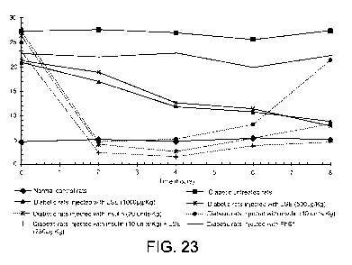
[00034] FIGs. 23 and 24 show the effect of different doses of LSE and
insulin on fasting
blood glucose (mmol/L) in normal and diabetic rats at various time intervals
(h), according to
some embodiments.
[00035] FIG. 25 shows that the LSE has a prophylactic effect on the onset
of diabetes,
according to some embodiments.
[00036] FIGs. 26 and 27 compare the free radical scavenging activity of
LSE to L-
ascorbic acid (vitamin C), according to some embodiments.
DETAILED DESCRIPTION
[00037] The teachings provided herein are generally directed to methods of
isolating and
using a whole-saliva leech extract in the treatment of a subject.
Pharmaceutical formulations
comprising the leech extracts and a pharmaceutically acceptable carrier are
provided.
[00038] It should be appreciated that the term "extract" can be used to
refer to a powder
form of the compounds of interest, a liquid form of the compounds of interest,
or any one or any
combination of the compounds of interest in powder or liquid form. One of
skill will appreciate
that the term "extract" can be used to refer to the compounds of interest
before, during, or after
their removal from the leech. In some embodiments, the compounds of interest
can be
synthesized chemically using standard methods known to one of skill, such that
they can be
synthesized and used alone, or in any combination, by those of skill without
use of the
extraction methods taught herein. The compositions provided herein can be
referred to as
extracts, compositions, compounds, agents, active agents, bioactive agents,
supplements,
drugs, and the like. In some embodiments, the terms "LSE," "extract," "LSE
composition,"
"composition," "compound," "agent," "active", "active agent", "bioactive
agent," "supplement,"
and "drug" can be used interchangeably and, it should be appreciated that, a
"formulation" can
comprise any one or any combination of these. Likewise, in some embodiments,
the
composition can also be in a liquid or dry form, where a dry form can be a
powder form in some
CA 02882533 2015-02-19
WO 2014/049447 PCT/1B2013/002848
6
embodiments, and a liquid form can include an aqueous or non-aqueous
component.
Moreover, the terms "activity" or "bioactivity" can refer to the function of
the compound in vitro, in
an assay for example, or in vivo when administered to a subject.
[00039] It should be appreciated that the leech extracts can be isolated
or purified. In
some embodiments, the terms "isolated" and "purified" can be used
interchangeably. In some
embodiments, the term "isolated" can be used to refer to the extract being
removed from the
natural chemical environment of the leech, such that the extract is not in the
form in which it
exists in nature. It should be appreciated that the term "purified" can be
used to refer to an
extract from a Hirudinaria manillensis leech, in some embodiments, such that
the compounds of
interest are isolated from the remainder of the leech in a form that can be
administered to a
subject, such as a soluble form, or a form that can go into aqueous solution.
As such, one of
skill will appreciate that the compounds of interest can sometimes be
accompanied by other
components that are carried along with the extract. For example, such other
components can
include any one or any combination of proteins found to be active in the
leech.. In some
embodiments, the term "purified" can be used to refer to an extract consisting
of, or consisting
essentially of, any one or any combination of the compounds of interest. In
some embodiments,
the extract includes a phagostimulatory solution or a component from the
phagostimulatory
solution. In some embodiments, an extract "consists essentially of" any one or
any combination
of the compounds of interest, where the presence of any other component from
the leech or
extraction procedure has a negligible effect on the activity of the compounds
of interest. The
term "negligible effect" can be used to mean that the activity does not
increase or decrease
more than about 10% when compared to any one or any combination of the
compounds of
interest, respectively, without the other components. In some embodiments, the
term "negligible
effect" can be used to refer to a change of less that 10%, less than 9%, less
than 8%, less than
7%, less than 6%, less than 5%, less than 4%, and less than 3%. In some
embodiments, the
term "negligible effect" can be used to refer to a change ranging from about
3% to about 10%, in
increments of 1%. For example, the activity of the compounds of interest can
be enhanced by
an amount ranging from about 10% to about 300%, from about 20% to about 200%,
from about
25% to about 250%, from about 30% to about 300%, from about 35% to about 275%,
from
about 40% to about 225%, from about 15% to about 100%, or any range therein in
increments
of 1%.
[00040] Methods of removing a whole saliva from a leech are provided. In
these
embodiments, the methods can include feeding a phagostimulatory agent to a
leech; inducing a
CA 02882533 2015-02-19
WO 2014/049447 PCT/1B2013/002848
7
regurgitation in the leech, the inducing including placing the leech in an
environment having a
temperature of less than about 0 C; and, collecting an unrefined, whole saliva
in the
regurgitation of the cooled leech.
[00041] One of skill will appreciate that any leech having a therapeutic
saliva can be used
in the teachings provided herein. In some embodiments, the leech can belong to
the family of
hirudinidae, to the sub-family hirudinariiae, or it can belong to a genus
selected from the group
consisting of hirudo; hirudinaria; aliolimantis; limantis; asiaticobdella;
goddardobdella;
limnobdella; macrobdella; oxyptychus; philobdella. In some embodiments, the
leech can be
selected from a species selected from the group consisting of hirudo
medicinalis; hirudo
troctina, hirudo nipponia; hirudo orientalis; hirudo verbana; hirudinaria
manillensis; hirudinaria
javanica; aliolimantis africana; aliolimantis michaelseni; aliolimantis
oligodonta; aliolimantis
buntonesis; limantis nilotica; limantis cf. nilotica; limantis paluda;
asiaticobdella fenestrata;
goddardobdella elegans; limnobdella mexicana; macrobdella decora; macrobdella
diploteria;
macrobdella diletra; oxyptychus brasiliensis; oxyptychus striatus; philobdella
floridana;
philobdella gracilis.
[00042] In some embodiments, the leech can belong to the family of
haemadipsidae, or it
can belong to a genus selected from the group consisting of chtonobdella;
haemadipsa;
idiobdella; malagdbdella; nesophilaemon. In these embodiments, the leech can
be selected
from a species selected from the group consisting of chtonobdella bilineata;
chtonobdella
whitmani; haemadipsa interrupta; haemadipsa sylvestris; haemadipsa sumatrana;
idiobdella
seychellensis; malagdbdella fallax; nesophilaemon skottsbergi.
[00043] In some embodiments, the leech can belong to the family of
xerobdellidae, or it
can belong to a genus selected from the group consisting of diestecostoma;
mesobdella;
xerobdella. In these embodiments, the leech can be selected from a species
selected from the
group consisting of diestecostoma magna; diestecostoma mexicana; diestecostoma
trujillensis;
mesobdella gemmata; xerobdella lecomtei.
[00044] In some embodiments, the leech can belong to the family of
haemopidae, or it
can belong to a genus selected from the group consisting of haemopis;
whitmania. In these
embodiments, the leech can be selected from a species selected from the group
consisting of
haemopis grandis; haemopis kingi; haemopis sanguisuga; haemopis terrestris;
whitmania
laevis.
CA 02882533 2015-02-19
WO 2014/049447 PCT/1B2013/002848
8
[00045] In some embodiments, the leech can belong to the family of
semiscolecidae, or it
can belong to a genus selected from the group consisting of patagoniobdella;
semiscolex. In
these embodiments, the leech can be selected from a species selected from the
group
consisting of patagoniobdella fraternal; patagoniobdella variabilis;
semiscolex intermedius;
semiscolex lamothei; semiscolex similis.
[00046] In some embodiments, the leech can belong to the family of
americobdellidae, or
it can belong to a genus selected from the group consisting of americobdella.
In these
embodiments, the leech can be selected from a species selected from the group
consisting of
americobdella valdiviana.
[00047] In some embodiments, the leech can belong to the family of
cylicobdellidae, or it
can belong to a genus selected from the group consisting of cylicobdella. In
these
embodiments, the leech can be selected from a species selected from the group
consisting of
cylicobdella coccinea.
[00048] In some embodiments, the leech can belong to the family of
erpobdellidae. In
these embodiments, the leech can be selected from a species selected from the
group
consisting of erpobdella mentezuma.
[00049] The leeches can be classified according to Table 1, in some
embodiments.
[00050] Table 1.
Family Sub family Genus Species
Hirudinidae Hirudinariinae Hirudo Hirudo medicinalis
Hirudo nipponia
Hirudo orientalis
Hirudo troctina
Hirudo verbana
Aliolimantis Aliolimantis Africana
Aliomantis michaelseni
Aliomantis oligodonta
Aliomantis buntonesis
Asiaticobdella Asiaticobdella fenestrate
Goddardobdella Goddardobdella elegans
Hirudinaria Hirudinaria javanica
Hirudinaria manillensis
CA 02882533 2015-02-19
WO 2014/049447
PCT/1B2013/002848
9
Limantis Limantis nilotica
Limantis cf. nilotica
Limantis paluda
Limnobdella Limnobdella mexicana
Macrobdella Macrobdella decora
Macrobdella diploteria
Macrobdella diletra
Oxyptychus Oxyptychus brasiliensis
Oxyptychus striatus
Philobdella Philobdella floridana
Philobdella gracilis
Haemadipsidae Not applicable Chtonobdella Chtonobdella bilineata
Chtonobdella whitmani
Haemadipsa Haemadipsa interrupta
Haemadipsa sylvestris
Haemadipsa sumatrana
ldiobdella ldiobdella seychellensis
Malagadbdella Malagadbdella fallax
Nesophilaemon Nesophilaemon skottsbergi
Xerobdellidae Not applicable Diestecostoma Diestecostoma magna
Diestecostoma Mexicana
Diestecostoma trujillensis
Mesobdella Mesobdella gemmata
Xerobdella Xerobdella lecomtei
Haemopidae Not applicable Haemopis haemopis grandis
Haemopis kingi
Haemopis sanguisuga
Haemopis terrestris
Whitmania Whitmania laevis
Semiscolecidae Not applicable Patagoniobdella Patagoniobdella
fraternal
Patagoniobdella variabilis
Semiscolex Semiscolex intermedius
Semiscolex lamothei
Semiscolex similis
Americobdellidae Not applicable Americobdella Americobdella
valdiviana
CA 02882533 2015-02-19
WO 2014/049447 PCT/1B2013/002848
Cylicobdellidae Not applicable Cylicobdella Cylicobdella coccinea
Erpobdellidae Not applicable Erpobdella Erpobdella montezuma
[00051] Any phagostimulatory agent known to one of skill can be used. In
some
embodiments, the phagostimulatory agent can include a protein, a polypeptide,
an oligopeptide,
or an amino acid. In some embodiments, the amino acid is an L-amino acid
selected from the
group consisting of arginine, alanine, leucine, aspartic acid, serine,
threonine, isoleucine,
histidine, lysine, tryptophan, glycine, phenylalanine, tyrosine, valine,
glutamic acid, asparagine,
glutamine, cysteine, methionine, and proline. In some embodiments, the
phagostimulatory
agent is arginine. In some embodiments, the phagostimulatory agent is glycine.
In some
embodiments, the phagostimulatory agent is proline. In some embodiments, the
phagostimulatory agent is a sugar. In some embodiments, the phagostimulatory
agent is a
sugar selected from the group consisting of fructose, glucose, sucrose,
maltose, raffinose,
trehalose, robose, and galactose. In some embodiments, the phagostimulatory
agent is corn
oil. In some embodiments, the phagostimulatory agent comprises any one or any
combination
of amino acids and/or sugars taught herein. Any suitable solvent for carrying
the
phagostimulatory can be used, polar or non-polar, as long as the solvent does
not substantially
affect the activity or stability of the leech saliva extract.
[00052] The temperature of the leech that induces the regurgitation can
range from about
-5 C to about 15 C, from about -4 C to about 14 C, from about -3 C to about 13
C, from about -
2 C to about 12 C, from about -1 C to about 11 C, from about 0 C to about 10
C, from about -
2 C to about 2 C, from about -3 C to about 3 C, from about -4 C to about 4 C,
from about -5 C
to about 5 C, or any temperature or range of temperatures therein in
increments of 1 C. The
temperature can be established using any method known to one of skill. In some
embodiments,
the temperature is established to 0 C or about 0 C using an ice water bath. In
some
embodiments, a salt water bath can be used to lower the temperature below 0 C,
and in some
embodiments, other liquids can be used to obtain other temperatures. Any
method of cooling
know to one of skill can be used to induce the leeches to vomit. The rate of
freezing can be 0.1
to 2 C per minute and, in some embodiments, 1 C to 1.5 C per minute. The time
at the cool
temperature can vary and can be, for example, from about 5 minutes to about 45
minutes, from
about 15 minutes to about 40 minutes, from about 15 minutes to about 20
minutes, from about
10 minutes to about 30 minutes, from about 5 minutes to about 25 minutes, from
about 3
minutes to about 35 minutes, from about 2 minutes to about 12 minutes, or any
time or range
times therein in increments of 1 minute.
CA 02882533 2015-02-19
WO 2014/049447 PCT/1B2013/002848
11
[00053] Methods of creating a lyophilized, whole saliva extract of a leech
having an
improved stability are provided by the teachings herein. In these embodiments,
the method can
include feeding a phagostimulatory agent to a leech; inducing regurgitation in
the leech, the
inducing including placing the leech in an environment having a temperature
ranging from about
-500 to about 1500; collecting an unrefined, whole saliva in the regurgitation
of the cooled leech;
removing solid components from the unrefined, whole saliva to create a
refined, whole saliva;
and, lyophilizing separate volumes of the refined, whole saliva extract, the
volumes not
exceeding about 2m1 each.
[00054] In some embodiments, the collecting includes squeezing the leech
to increase
the amount of unrefined, whole saliva collected. In some embodiments, the
methods further
comprise revitalizing the leech by warming the leech in a water bath having a
temperature
ranging from about 5 C to about 40 C. In some embodiments, the methods further
comprise
creating a refined, whole-saliva extract; the creating including removing
solid components from
the unrefined, whole saliva. In some embodiments, the methods further comprise
lyophilizing
separate volumes of the refined, whole saliva extract, the volumes not
exceeding about 2m1
each. And, in some embodiments, the leech is Hirudinaria manillensis.
[00055] Stable, lyophilized, whole-saliva extracts of a leech are provided
by the teachings
herein. In these embodiments, the extract comprises a refined, whole-saliva
extract of a leech
lyophilized in volumes not exceeding about 2m1 each, the extract refined by
removing solid
components from an unrefined, whole saliva to create the refined, whole
saliva; wherein, the
extract has a stable activity when stored for use at a temperature below about
-20 C , the
extract maintaining at least 70% of the activity for at least 6 months. And,
the leech can be
Hirudinaria manillensis.
[00056] Storage temperature has been shown in some embodiments herein to
have a
large effect on the stability of the extracts. In some embodiments, for
example, the refined,
whole saliva can be stored at a temperature ranging from 0 C to -80 C,from -20
C to -270 C,
from -20 C to -196 C, from -20 C to -80 C, from -80 C to -196 C, or any
temperature, or any
range therein in increments of 1 C.
[00057] One of skill will appreciate that the extracts can vary in
stability, but that the
teachings provided herein show extracts with increased stabilities when
compared to the current
state-of-the-art. One of skill will appreciate that the compositions or
formulations should remain
stable, or at least substantially stable, until used or activated, and this
can relate to a shelf life,
or a time between creation and administration of the composition, or some
combination thereof.
CA 02882533 2015-02-19
WO 2014/049447 PCT/1B2013/002848
12
In some embodiments, the composition is stable, or substantially stable, when
usable as
intended within a reasonable amount of time, a time that is considered
reasonable by one of
skill for the applications taught herein. In some embodiments, the composition
should be usable
within a reasonable time from the making to the administration of the
composition and, in some
embodiments, the composition should have a reasonable commercial shelf life, a
shelf life that
is considered reasonable to one of skill. A reasonable shelf life can be at
least 6 months, at
least 1 year, at least 18 months, at least 2 years, at least 3 years, or any
time in-between in
increments of about 1 month, in some embodiments.
[00058] In some embodiments, a composition or formulation can be
considered as
"stable" if it loses less than 10%, less than 7%, less than 6%, less than 5%,
less than 3%, less
than 2%, or less than 1% of its original activity. In some embodiments, a
composition or
formulation can be considered as "substantially stable" if it loses greater
than about 10% of its
original activity, as long as the composition can perform it's intended use to
a reasonable
degree of efficacy. In some embodiments, the composition can be considered as
substantially
stable if it loses activity at an amount greater than about 12%, about 15%,
about 25%, about
35%, about 45%, about 50%, about 60%, or even about 70%. The activity loss can
be
measured by comparing activity at the time of packaging to the activity at the
time of
administration, and this can include a reasonable shelf life. In some
embodiments, the
composition is stable or substantially stable, if it remains useful for a
period ranging from 3
months to 3 years, 6 months to 2 years, 1 year, or any time period therein in
increments of
about 1 month.
Methods of treatment
[00059] Methods of treating a subject by administering an effect amount of
the leech
extracts are provided by the teachings herein. The extracts taught herein can
be used for a
variety of treatments, preventative, ameliorative, or otherwise, as well as
for use as a dietary
supplement. The uses can include medicinal purposes, as a health supplement, a
nutritional
composition, a prophylactic, or a treatment of an existing condition. In some
embodiments, any
tissue that can make contact with one or more active components of an extract
taught herein
can be treated. In some embodiments, a tissue can have a desirable secondary
effect from one
or more of the active components of an extract taught herein making contact
elsewhere in the
subject, such that one or more of the active components can contact a first
tissue, whereas a
second tissue realizes a beneficial effect. For example, the first tissue can
be a stomach lining,
and the second tissue can realize the desirable effect of a release of a
neurotransmitter or a
CA 02882533 2015-02-19
WO 2014/049447 PCT/1B2013/002848
13
neuroimpulse. The tissue can be, for example, connective, muscle, nervous,
and/or epithelial
tissue. In some embodiments, the tissue is a dermal tissue. In some
embodiments, the tissue
is a mucosa! tissue. And, in some embodiments, the tissue is gastrointestinal
tissue. In some
embodiments, the method includes treating a solid tumor, treating a liquid
tumor, treating
diabetes, treating a viral disease, treating a parasitic disease,
administering an anti-oxidant
therapy, or administering an antibacterial therapy.
[00060] As such, the subject can have a target tissue that is the focus of
the treatment in
which the extracts are applied directly or systemically. In some embodiments,
the term "target
site" can be used to refer to a select location on or in a subject that could
benefit from an
administration of a compound taught herein, either parenterally or non-
parenterally, whether
injected or administered topically or orally, for example. In some
embodiments, a target can
include any site of action in which the agent's activity can serve a benefit
to the subject. The
target site can be a healthy or damaged tissue of a subject. As such, the
teachings include a
method of administering one or more compounds taught herein to a healthy or
damaged tissue,
dermal, mucosa!, gastrointestinal or otherwise.
[00061] The terms "treat," "treating," and "treatment" can be used
interchangeably in
some embodiments and refer to the administering or application of the
compositions and
formulations taught herein, including such administration as a health or
nutritional supplement,
and all administrations directed to the prevention, inhibition, amelioration
of the symptoms, or
even a cure of a condition taught herein. The terms "disease," "condition,"
"disorder," and
"ailment" can be used interchangeably in some embodiments.
[00062] The term "subject" and "patient" can be used interchangeably in
some
embodiments and refer to an animal such as a mammal including, but not limited
to, non-
primates such as, for example, a cow, pig, horse, cat, dog, rat and mouse; and
primates such
as, for example, a monkey or a human. As such, the terms "subject" and
"patient" can also be
applied to non-human biologic applications including, but not limited to,
veterinary, companion
animals, commercial livestock, and the like.
Treatment of cancer
[00063] The LSE taught herein can be used in the treatment of cancer. In
some
embodiments, the methods include treating a solid tumor and, in some
embodiments, the
methods include treating a liquid tumor. One of skill will appreciate that the
cancers that can be
treated using the methods taught herein can include any hyperproliferative
tissue. In some
CA 02882533 2015-02-19
WO 2014/049447 PCT/1B2013/002848
14
embodiments, for example, any cancer listed in Table 2 can be treated using
the methods
taught herein.
[00064] Table 2.
Cell line Cancer type Cancer Sub-type
CCRF-CEM Leukemia Acute Lymphoblastic Leukemia
(ALL)
HL-60 (TB) Leukemia Acute Myelogenous Leukemia (AML)
K-562 Leukemia Chronic Myelogenous leukemia
(CML)
MOLT-4 Leukemia Acute Lymphoblastic Leukemia
(ALL)
RPMI-8226 Multiple Myeloma Plasmacytoma, myeloma
SR Leukemia Acute Lymphoblastic Leukemia
(ALL)
A549/ATCC Non-small cell lung Adinocarcinoma
EKVX Non-small cell lung Adinocarcinoma
HOP-62 Non-small cell lung Adinocarcinoma
HOP-92 Non-small cell lung Adinocarcinoma
NCI-H226 Non-small cell lung Squamous Carcinoma
NCI-H23 Non-small cell lung Adinocarcinoma
NCI-H322M Non-small cell lung Bronchioloalveolar Carcinoma
NCI-H460 Non-small cell lung Adinocarcinoma
NCI-H522 Non-small cell lung Adinocarcinoma
COLO 205 Colon Adinocarcinoma
HCC-2998 Colon Adinocarcinoma
HCT-116 Colon Carcinoma
HCT-15 Colon Adinocarcinoma
HT-29 Colon Adinocarcinoma
KM12 Colon Colorectal
SW-620 Colon Adinocarcinoma
SN-268 CNS Glioblastoma
SF-295 CNS Glioblastoma
SF-539 CNS Gliosarcoma
SNB-19 CNS Glioblastoma
SNB-75 CNS Glioblastoma
LOX IMVI Skin Cancer Melanoma
MALME-3M Skin Cancer Melanoma
M14 Skin Cancer Melanoma, amelanotic
SK-MEL-2 Skin Cancer Melanoma, malignant
SK-MEL-28 Skin Cancer Melanoma, malignant
SK-MEL-5 Skin Cancer Melanoma, malignant
UACC-257 Skin Cancer Melanoma
UACC-62 Skin Cancer Melanoma
IGROVI Ovarian Adinocarcinoma
OVCAR-3 Ovarian Adinocarcinoma
OVCAR-4 Ovarian Carcinoma
OVCAR-5 Ovarian Carcinoma
OVCAR-8 Ovarian Carcinoma
SK-OV-3 Ovarian Adinocarcinoma
786-0 Renal Carcinoma
CA 02882533 2015-02-19
WO 2014/049447 PCT/1B2013/002848
A498 Renal Carcinoma
ACHN Renal Adinocarcinoma
CAKI-1 Renal Carcinoma
RXF-393 Renal Carcinoma
SN12C Renal Carcinoma
TK-10 Renal Carcinoma
U0-31 Renal Carcinoma
PC-3 Prostate Adinocarcinoma
DU-145 Prostate Carcinoma
MCF7 Breast Adinocarcinoma
NCl/ADR-RES Breast Adinocarcinoma
MDA-MB-231/ATCC Breast Adinocarcinoma
HS 578T Breast Carcinosarcoma
MDA-MB-435 Breast Carcinoma, ductal
MDA-MB-468 Breast Adinocarcinoma
BT-549 Breast Carcinoma
T-47D Breast Carcinoma, ductal
Treatment of diabetes
[00065] The LSE taught herein can be used in the treatment of diabetes.
Examples of
diabetes include Type 1-, Type 2-, and gestational diabetes. As such, one of
skill will appreciate
that the LSE taught herein can be used in treating and preventing metabolic
imbalances,
diabetes mellitus, a pre-diabetic state, metabolic syndrome, and other related
disorders, such as
Latent Autoimmune Diabetes in adults (referred to as Type 1.5 diabetes). As
such, secondary
medical conditions related to diabetes can also be treated using the LSE
taught herein,
indirectly or directly, including heart disease, stroke, high blood pressure,
eye complications
(retinopathy, cataracts), kidney disease (nephropathy), nervous system disease
(neuropathy),
peripheral vascular disease, dental disease, gastroparesis, sexual
dysfunction, and
complications during pregnancy.
[00066] The term "diabetic" in a rat can refer to a random blood glucose
>225 mg/di or
fasting blood glucose level of >110 mg/dL. The term "diabetic" in a human can
refer to a
random plasma or blood glucose concentration of 200 mg/dL (11.1 mmol/L) or a
fasting
plasma glucose 126 mg/dL (7.0 mmol/L) or a 2 hour post-load glucose200 mg/dL
(11.1
mmol/L) during an oral glucose tolerance test. The term "non-diabetic" in a
rat generally means
a fasting plasma glucose level of 80 mg/dL or a random plasma glucose level
<200 mg/dL.
The term "non-diabetic" in a human can refer to a fasting plasma glucose level
of <100 mg/dL
(5.6 mmol/dL) or a 2 hour post-load glucose<140 mg/dL (<7.8 mmol/dL) during an
oral glucose
tolerance test. The term "pre-diabetic" in a rat can refer to a fasting plasma
glucose level of
about 80 to about 110 mg/dL. The term "pre-diabetic" in a human can refer to a
fasting plasma
CA 02882533 2015-02-19
WO 2014/049447 PCT/1B2013/002848
16
glucose level of 100-125 mg/dL (5.6-6.9 mmol/L) or a 2 hour post-load glucose
140-199 mg/L
(7.8-11.1 mmol/L) during an oral glucose tolerance test. The terms "random"
and "nonfasting"
can be used in reference to any time of day or night without regard to time
since the last meal,
and the term "fasting" generally means no caloric intake for at least 12
hours. The term
"metabolic imbalance" can refer any condition associated with an elevated
plasma glucose. A
metabolic imbalance, for example, comprises diabetes mellitus, gestational
diabetes, genetic
defects of .beta.-cell function, genetic defects in insulin action, diseases
of the exocrine
pancreas, endocrinopathies, drug or chemical-induced, infections, other
genetic syndromes
associated with diabetes, a pre-diabetic state, and metabolic syndrome. The
term "metabolic
syndrome" can refer to a group of metabolic risk factors in one person
including, but not limited
to, abdominal obesity, atherogenic dyslipidemia, hypertension, insulin
resistance or glucose
intolerance, prothrombotic state (high fibrinogen or plasminogen activator
inhibitor-1), and
proinflammatory state (elevated C-reactive protein). In some embodiments,
metabolic
syndrome be the presence of three or more of the following components:
elevated waist
circumference (males: .40 inches, females 35 inches), fasting triglycerides
50 mg/dL,
reduced HDL (males: <40 mg/dL, females<50 mg/dL), blood pressure 130/85 mm Hg,
and
fasting glucose -100 mg/dL.
[00067] The above definitions for diabetes follow standards of the
American Diabetes
Association (ADA), the American Heart Association (AHA) and the National
Heart, Lung, and
Blood Institute. Other definitions can be used and may vary by region or
country, and may
depend upon the group or institution (e.g. ADA, World Health Organization
(WHO), National
Institute of Diabetes and Digestive and Kidney Diseases (NIDDK/NIH), Center
for Disease
Control (CDC), etc.) providing other guidelines. Physicians may also use
clinical experience, a
patient's past medical history, and the like when deciding on a diagnosis and
treatment. As
such, one of skill will appreciate that the particular ranges and measures are
merely relative
rather than critical to making a diagnosis or planning a treatment. In some
embodiments, for
example, any of the above measures can vary by about 1%, about 2%, about 3%,
about 5%,
about 7%, about 10%, about 15%, about 20%, about 25%, about 30%, 40%, 50%, or
any range
or amount therein in increments of 0.1%.
Treatment of a viral disease
[00068] The LSE taught herein can be used in the treatment of several
different types of
viral diseases. In some embodiments, the virus can be a species of
Adenoviridae,
Herpesviridae, Papillomaviridae, Polyomaviridae, Poxviridae, Hepadnaviridae,
Parvoviridae,
CA 02882533 2015-02-19
WO 2014/049447 PCT/1B2013/002848
17
Astroviridae, Caliciviridae, Picornaviridae, Coronaviridae, Flaviviridae,
Togaviridae, Retroviridae,
Orthomyxoviridae, Arenaviridae, Bunyaviridaem, Filoviridae, Paramyxoviridae,
Rhabdoviridae,
or Reoviridae.
[00069] In some embodiments, the species of virus treated can be selected
from the
group consisting of Adenovirus, Herpes simplex, type 1, Herpes simplex, type
2, Varicella-
zoster virus, Epstein-barr virus, Human cytomegalovirus, Human herpesvirus,
type 8, Human
papillomavirus, BK virus, JO virus, Smallpox, Hepatitis B virus, Human
bocavirus, Parvovirus
B19, Human astrovirus, Norwalk virus, coxsackievirus, hepatitis A virus,
poliovirus, rhinovirus,
Severe acute respiratory syndrome virus, Hepatitis C virus, yellow fever
virus, dengue virus,
West Nile virus, Rubella virus, Hepatitis E virus, and Human immunodeficiency
virus (HIV).
[00070] In some embodiments, the viral condition can be a regionally
identified condition
selected from the viral conditions in Table 3:
[00071] Table 3.
United
Australia Hong Kong Malaysia United States
Kingdom
Acquired Acquired
Immunodeficiency immunodeficiency
Syndrome (AIDS) syndrome
Arbovirus
Arbovirus infections:
infections:
California serogroup
Barmah Forest,
virus, Eastern equine
Dengue fever, Arbovirus
encephalitis virus,
Japanese infections:
Powassan virus, St.
encephalitis, West Nile
Louis encephalitis
Kunjin virus, virus
virus, West Nile virus,
Murray Valley
Western equine
encephalitis virus,
encephalitis virus
Ross River virus
Chickenpox (i.e.,
Chickenpox varicella) -
morbidity
and deaths only
Chikungunya
fever
Dengue
Dengue fever Dengue fever
fever
Enterovirus
71 infection
Hantavirus Hantavirus
CA 02882533 2015-02-19
WO 2014/049447 PCT/1B2013/002848
18
infection
Hepatitis Hepatitis Hepatitis
Hepatitis A Hepatitis A Hepatitis A
Hepatitis B Hepatitis B Hepatitis B
Hepatitis C Hepatitis C Hepatitis C
Hepatitis D Hepatitis D
Hepatitis E Hepatitis E
Human Human
immunodeficiency immunodeficiency
HIV infection
virus (HIV) virus (HIV)
infection infection
Influenza A
(H2),
Influenza A Influenza-associated
(H5), pediatric mortality
and
Influenza
Influenza A novel influenza A
(H7) or infection
Influenza A
(H9)
Japanese
encephalitis
Lyssavirus
Measles Measles Measles Measles Measles
Mumps Mumps Mumps Mumps
Poliomyelitis,
Acute
Poliomyelitis
poliomyelitis Poliomyelitis Poliomyelitis paralytic and non-
paralytic
Rabies Rabies Rabies Rabies
Rubella and
congenital
Rubella Rubella Rubella
rubella
syndrome
Severe Severe
Severe Acute
Acute Acute
Respiratory
Respiratory Respiratory
Syndrome
Syndrome Syndrome
Smallpox Smallpox Smallpox Smallpox
Yellow fever Yellow fever Yellow fever Yellow fever Yellow fever
Viral Viral hemorrhagic
Viral haemorrhagic Viral fever, including
hemorrhagic fever, including hemorrhagic Arenavirus (new
fever Lassa fever, fever world), Crimean-
Marburg virus, Congo hemorrhagic
CA 02882533 2015-02-19
WO 2014/049447 PCT/1B2013/002848
19
and Ebola virus fever, Dengue
hemorraghic fever,
Ebola virus, Lassa
virus, Marburg virus
[00072] In some embodiments, the compositions taught herein can be
administered with
a second agent, such as abacavir, aciclovir, acyclovir, adefovir, amantadine,
amprenavir,
ampligen, arbidol, atazanavir, atripla , aoceprevir, cidofovir, combivir,
darunavir, delavirdine,
didanosine, docosanol, edoxudine, efavirenz, emtricitabine, enfuvirtide,
entecavir, entry
inhibitors, famciclovir, fomivirsen, fosamprenavir, foscarnet, fosfonet,
fusion inhibitor,
ganciclovir, ibacitabine, imunovir, idoxuridine, imiquimod, indinavir,
inosine, integrase inhibitor,
interferon type III, interferon type II, interferon type I, interferon,
lamivudine, lopinavir, loviride,
maraviroc, moroxydine, methisazone, nelfinavir, nevirapine, nexavir,
oseltamivir, peginterferon
alfa-2a, penciclovir, peramivir, pleconaril, podophyllotoxin, protease
inhibitor. Raltegravir,
reverse transcriptase inhibitor, ribavirin, rimantadine, ritonavir,
pyramidine, saquinavir,
stavudine, synergistic enhancer (antiretroviral), tea tree oil, telaprevir,
tenofovir, tenofovir
disoproxil, tipranavir, trifluridine, trizivir, tromantadine, truvada,
valaciclovir, valganciclovir,
vicriviroc, vidarabine, viramidine, zalcitabine, zanamivir, and zidovudine.
Treating a parasitic disease
[00073] The LSE taught herein can be used in the treatment of several
different types of
parasitic diseases. In some embodiments, the parasitic disease treated can be
classed as a
condition caused by protozoa (causing protozoan infection), helminths
(helminthiasis), and
ectoparasites.
[00074] In some embodiments, the parasitic disease can be selected from
the group
consisting of Acanthamoeba keratitism, Amoebiasis, Ascariasis, Babesiosis,
Balantidiasis,
Baylisascariasis, Chagas disease, Clonorchiasis, Cochliomyia,
Cryptosporidiosis,
Diphyllobothriasis, Dracunculiasis (caused by the Guinea worm),
Echinococcosis, Elephantiasis,
Enterobiasis, Fascioliasis, Fasciolopsiasis, Filariasis, Giardiasis,
Gnathostomiasis,
Hymenolepiasis, lsosporiasis, Katayama fever, Leishmaniasis, Lyme disease,
Malaria,
Metagonimiasis, Myiasis, Onchocerciasis, Pediculosis, Scabies,
Schistosomiasis, Sleeping
sickness, Strongyloidiasis, Taeniasis(cause of Cysticercosis), Toxocariasis,
Toxoplasmosis,
Trichinosis, and Trichuriasis.
CA 02882533 2015-02-19
WO 2014/049447 PCT/1B2013/002848
[00075] In some embodiments, the compositions taught herein can be
administered with
a second agent, such as thiabendazole, pyrantel pamoate, mebendazole,
praziquantel,
niclosamide, bithionol, oxamniquine, metrifonate, lvermectin, albendazole,
benznidazole,
nifurtimox, and nitroimidazole.
Treatment of a bacterial disease
[00076] The LSE taught herein can be used in the treatment of several
different types of
bacterial diseases. In some embodiments, the bacterial disease can include,
for example,
tuberculosis from Mycobacterium tuberculosis; pneumonia from Streptococcus and
Pseudomonas; a foodborne illness from Shigella, Campylobacter, or Salmonella;
and, either
tetanus, typhoid fever, diphtheria, syphilis, or leprosy. In some embodiments,
the bacterial
disease can be a bacterial vaginosis; bacterial meningitis; bacterial
pneumonia; urinary tract
infection, including E. coli. Infections; bacterial gastroenteritis, also
including E. coli; and,
bacterial skin infections, including impetigo from S. aureus and S. pyogenes,
Erysipelas from
Streptococcus, and cellulitis which can include connective tissue. In some
embodiments, the
bacterial disease can be selected from the group consisting of the diseases in
Table 4.
[00077] Table 4.
United
Australia Hong Kong Malaysia Kingdom
United States
Anaplasmosis
Anthrax Anthrax Anthrax
Botulism Botulism Botulism Botulism
Brucellosis Brucellosis Brucellosis
Campylobacteriosis
Chancroid
Chlamydia
Chlamydia
trachomatis
Cholera Cholera Cholera Cholera Cholera
Diphtheria Diphtheria Diphtheria Diphtheria Diphtheria
Donovanosis
Ehrlichiosis
CA 02882533 2015-02-19
WO 2014/049447 PCT/1B2013/002848
21
Shiga toxin- and Escherichia
verocytotoxin- Escherichia coli coli 0157:H7
producing 0157:H7 or Shiga-toxin
Escherichia coli infection producing
(STEC/VTEC) Escherichia
coli
Encephalitis Encephalitis
Gonorrhea
Haemolytic
Gonococcal
infection/Gonorrhea
infection
Haemolytic Haemolytic Hemolytic
uraemic syndrome uraemic uremic
syndrome syndrome,
(HUS) (HUS) post-diarrheal
HaemophilusHaemophilus
Haemophilus
influenzae serotype influenzae type influenzae,
b (Hib) b infection invasive
(invasive) disease
Legionellosis
Legionnaire's Legionnaire's
Disease Disease
Legionellosis
Hansen's
Leprosy Leprosy Leprosy Leprosy disease
(Leprosy)
Leptospirosis Leptospirosis
Listeriosis Listeriosis Listeriosis
Lyme disease
Meningococcal Meningococcal
septicaemia/ Men ingococcal
Men ingococcal
infection
disease
(invasive) Acute disease
Meningitis
CA 02882533 2015-02-19
WO 2014/049447 PCT/1B2013/002848
22
MSRA:
Community-
associated
methicillin-
resistant
Staphylococcus
aureus
infection
Paratyphoid Paratyphoid
Paratyphoid fever
fever fever
Pertussis Pertussis
Pertussis Pertussis
(Whooping (Whooping
(Whooping cough) (Whooping cough)
cough) cough)
Plague
Plague
(bubonic,
(bubonic,
Plague septicemic, Plague Plague septicemic,
pneumonic
pneumonic and
and
pharyngeal)
pharyngeal)
Psittacosis Psittacosis Psittacosis
Q fever Q fever Q Fever, acute
and chronic
Relapsing fever Relapsing fever
Rickettsiosis Rickettsiosis,
spotted fever
Scarlet fever Scarlet fever
Salmonellosis Salmonellosis
Bacillary Shigellosis
Shigellosis
dysentery
Group A Group A
Streptococcal Streptococcal
disease disease
Streptococcus
Pneumococcal pneumoniae,
disease invasive
disease
CA 02882533 2015-02-19
WO 2014/049447 PCT/1B2013/002848
23
Streptococcus
suis infection
Syphilis Syphilis Syphilis
Tetanus Tetanus Tetanus Tetanus Tetanus
Toxic shock
syndrome
(Streptococcal
and other than
Streptococcal)
Tuberculosis,
Tuberculosis Tuberculosis Tuberculosis Tuberculosis
Mycobacterium
tuberculosis
Tularemia Tularemia
Typhoid fever Typhoid fever Typhoid fever Typhoid fever Typhoid
fever
Typhus and
other rickettsia! Typhus Typhus
diseases
Vancomycin
Intermediate
Staph Aureus
(VISA),
Vancomycin
Resistant
Staph Aureus
(VRSA)
Administering an anti-oxidant therapy
[00078] The LSE taught herein can be used in antioxidant therapy. One of
skill will
appreciate that reactive oxygen species (ROS) are widely believed to cause or
aggravate
several human pathologies such as arthritis, neurodegenerative diseases,
cancer, heart
disease, stroke and many other ailments. Antioxidants can be used to
counteract the harmful
effects of ROS and therefore prevent or treat oxidative stress-related
diseases. In some
embodiments, the LES taught herein can be used as a free radical scavenger, or
to prevent
oxidation in the body. In some embodiments, the LES taught herein can be used
to treat
inflammatory disorders, endocrine disorders, cardiovascular disease, aging, as
well as to serve
CA 02882533 2015-02-19
WO 2014/049447 PCT/1B2013/002848
24
as a neuroprotective agent. In some embodiments, the LES taught herein can be
used to treat
atherosclerosis. And, in some embodiments, the LES can be administered in
combination with
a cholesterol medication such as an absorption blocker, a synthesis inhibitors
and a niacin-
based drug. In some embodiments, a non-drug alternative can be used, such as
beta-glucan
from whole oats or barley; psyllium from wheat bran; or, phytosterols and/or
phytostanols.
[00079] In some embodiments, the absorption blocker can be cholestyramine
or ZETIA.
In some embodiments, the synthesis inhibitor can be a statin including, but
not limited to,
MEVACOR, PRAVACHOL, ZOCOR, LIPITOR, LESCOL, CRESTOR, or LIVALO. In some
embodiments, the synthesis inhibitor can be LOVASTATIN, PRAVASTATIN, or
SIMVASTATIN.
In some embodiments, the niacin-based medication can be NIASPAN or NIACOR. In
some
embodiments, the cholesterol medication can be a combination product such as
MEVACOR
with NIASPAN, or ZETIA with ZOCOR.
Methods of administration
[00080] Any administration vehicle known to one of skill to be suitable
for administration
of the compounds, compositions, and formulations taught herein can be used. A
"vehicle" can
refer to, for example, a diluent, excipient or carrier with which a compound
is administered to a
subject.
[00081] The terms "administration" or "administering" can be used to refer
to a method of
incorporating a composition into or onto the cells or tissues of a subject,
either in vivo or ex vivo
to test the activity of a system, as well as to diagnose, prevent, treat, or
ameliorate a symptom
of a disease or condition. In one example, a compound can be administered to a
subject in vivo
using any means of administration taught herein. In another example, a
compound can be
administered ex vivo by combining the compound with cell tissue from the
subject for purposes
that include, but are not limited to, assays for determining utility and
efficacy of a composition.
And, of course, the compositions can be used in vitro to test their stability,
activity, toxicity,
efficacy, and the like. When the compound is incorporated in the subject in
combination with
one or active agents, the terms "administration" or "administering" can
include sequential or
concurrent incorporation of the compound with the other agents such as, for
example, any agent
described above. A composition can be formulated, in some embodiments, to be
compatible
merely with its intended route of administration.
[00082] Any dosage form known to one of skill can be used for
administrations that
include, for example, parenteral and non-parenteral administrations. In some
embodiments, the
CA 02882533 2015-02-19
WO 2014/049447 PCT/1B2013/002848
composition is in a dosage form for administration topically. And, in some
embodiments, the
composition is in a dosage form for administration orally. In some
embodiments, the dosage
form can be a capsule or an injectable fluid. The composition can also be used
as a dietary
supplement. The term "dosage unit" can refer to discrete, predetermined
quantities of a
compound that can be administered as unitary dosages to a subject. A
predetermined quantity
of active compound can be selected to produce a desired therapeutic effect and
can be
administered with a pharmaceutically acceptable carrier. The predetermined
quantity in each
unit dosage can depend on factors that include, but are not limited to, (a)
the unique
characteristics of the active compound and the particular therapeutic effect
to be achieved, and
(b) the limitations inherent in the art of creating and administering such
dosage units.
[00083] A "pharmaceutically acceptable carrier" is a diluent, adjuvant,
excipient, or
vehicle with which the composition is administered. A carrier is
pharmaceutically acceptable
after approval by a state or federal regulatory agency or listing in the U.S.
Pharmacopeia!
Convention or other generally recognized sources for use in subjects. The
pharmaceutical
carriers include any and all physiologically compatible solvents, dispersion
media, coatings,
antibacterial and antifungal agents, isotonic and absorption delaying agents,
and the like.
Examples of pharmaceutical carriers include, but are not limited to, sterile
liquids, such as
water, oils and lipids such as, for example, phospholipids and glycolipids.
These sterile liquids
include, but are not limited to, those derived from petroleum, animal,
vegetable or synthetic
origin such as, for example, peanut oil, soybean oil, mineral oil, sesame oil,
and the like.
[00084] Suitable pharmaceutical excipients include, but are not limited
to, starch, sugars,
inert polymers, glucose, lactose, sucrose, gelatin, malt, rice, flour, chalk,
silica gel, sodium
stearate, glycerol monostearate, talc, sodium chloride, dried skim milk,
glycerol, propylene
glycol, water, ethanol, and the like. In some embodiments, the composition can
also contain
minor amounts of wetting agents, emulsifying agents, pH buffering agents, or a
combination
thereof. Oral formulations, for example, can include standard carriers such
as, for example,
pharmaceutical grades man nitol, lactose, starch, magnesium stearate, sodium
saccharine,
cellulose, magnesium carbonate, and the like. See Martin, E.W. Remington's
Pharmaceutical
Sciences.
[00085] As described herein, the compositions can take the form of
lotions, creams,
suspensions, emulsions, tablets, pills, capsules, powders, sustained-release
formulations and
the like. In some embodiments, the compositions or formulations can be
administered to a
subject in any non-parenteral manner known to one of skill whereas, in
contrast, a parenteral
CA 02882533 2015-02-19
WO 2014/049447 PCT/1B2013/002848
26
administration involves piercing the skin or a mucous membrane. Depending on
the target
tissue, the administration can be topical, oral, ocular, otologic, nasal,
urogenital, rectal, dermal,
vaginal or otherwise to a mucous membrane. Oral administration, for example,
can include
digestive tract, buccal, and sublingual administration, and a solid or liquid
carrier can be used.
One of skill will appreciate that the therapeutic program selected, the agents
administered, the
condition of the subject, and the effects desired, can affect the
administration schedule and
program used.
[00086] The compositions or formulations can be contained in forms that
include tablets,
troches, capsules, elixirs, beverages, suspensions, syrups, wafers, chewing
gums, gels,
hydrogels, and the like. Tablets, pills, capsules, troches liquids and the
like may also contain
binders, excipients, disintegrating agent, lubricants, glidants, chelating
agents, buffers, tonicity
modifiers, surfactants, sweetening agents, and flavoring agents. Some examples
of binders
include microcrystalline cellulose, gum tragacanth or gelatin. Some examples
of excipients
include starch or maltodextrin. Some examples of disintegrating agents include
alginic acid,
corn starch and the like. Some examples of lubricants include magnesium
stearate or
potassium stearate. An example of a chelating agent is EDTA. Some examples of
buffers are
acetates, citrates or phosphates. Some examples of tonicity modifiers include
sodium chloride
and dextrose. Some examples of surfactants for micellation or increasing cell
permeation
include coconut soap, anionic, cationic or ethoxylate detergents. An example
of a glidant is
colloidal silicon dioxide. Some examples of sweetening agents include sucrose,
saccharin and
the like. Some examples of flavoring agents include peppermint, chamomile,
orange flavoring
and the like.
[00087] In the digestive tract, for example, a solid can include a pill,
capsule, tablet, or
time-release technology in some embodiments; and, a liquid can include a
solution, soft gel,
suspension, emulsion, syrup, elixir, tincture, or a hydrogel. Digestive tract
administration can
include oral or rectal administration using any method known to one of skill.
For buccal,
sublingual, and sublabial administration, a solid can include an orally
disintegrating tablet, a film,
a lollipop, a lozenge, or chewing gum; and, a liquid can include a mouthwash,
a toothpaste, an
ointment, or an oral spray.
[00088] One of skill understands that the amount of the agents
administered can vary
according to factors such as, for example, the type of disease, age, sex, and
weight of the
subject, as well as the method of administration. Dosage regimens may also be
adjusted to
optimize a therapeutic response. In some embodiments, a single bolus may be
administered;
CA 02882533 2015-02-19
WO 2014/049447 PCT/1B2013/002848
27
several divided doses may be administered over time; the dose may be
proportionally reduced
or increased; or, any combination thereof, as indicated by the exigencies of
the therapeutic
situation and factors known to one of skill in the art. It is to be noted that
dosage values may
vary with the severity of the condition to be alleviated, as well as whether
the administration is
prophylactic, such that the condition has not actually onset or produced
symptoms. Dosage
regimens may be adjusted over time according to the individual need and the
professional
judgment of the person administering or supervising the administration of the
compositions, and
any dosage ranges set forth herein are exemplary only and do not limit the
dosage ranges that
may be selected.
[00089] An "effective amount" of a compound can be used to describe a
therapeutically
effective amount or a prophylactically effective amount. An effective amount
can also be an
amount that ameliorates the symptoms of a disease. A "therapeutically
effective amount" can
refer to an amount that is effective at the dosages and periods of time
necessary to achieve a
desired therapeutic result and may also refer to an amount of active compound,
prodrug or
pharmaceutical agent that elicits any biological or medicinal response in a
tissue, system, or
subject that is sought by a researcher, veterinarian, medical doctor or other
clinician that may be
part of a treatment plan leading to a desired effect. In some embodiments, the
therapeutically
effective amount should be administered in an amount sufficient to result in
amelioration of one
or more symptoms of a disorder, prevention of the advancement of a disorder,
or regression of
a disorder. In some embodiments, for example, a therapeutically effective
amount can refer to
the amount of an agent that provides a measurable response of at least 5%, at
least 10%, at
least 15%, at least 20%, at least 25%, at least 30%, at least 35%, at least
40%, at least 45%, at
least 50%, at least 55%, at least 60%, at least 65%, at least 70%, at least
75%, at least 80%, at
least 85%, at least 90%, at least 95%, or at least 100% of a desired action of
the composition.
[00090] In cases of the prevention or inhibition of the onset of a disease
or disorder, or
where an administration is considered prophylactic, a prophylactically
effective amount of a
composition or formulation taught herein can be used. A "prophylactically
effective amount" can
refer to an amount that is effective at the dosages and periods of time
necessary to achieve a
desired prophylactic result, such as prevent the onset of a sunburn, an
inflammation, allergy,
nausea, diarrhea, infection, and the like. Typically, a prophylactic dose is
used in a subject prior
to the onset of a disease, or at an early stage of the onset of a disease, to
prevent or inhibit
onset of the disease or symptoms of the disease. A prophylactically effective
amount may be
less than, greater than, or equal to a therapeutically effective amount.
CA 02882533 2015-02-19
WO 2014/049447 PCT/1B2013/002848
28
[00091] In some embodiments, a therapeutically or prophylactically
effective amount of a
composition may range in concentration from about 0.01 nM to about 0.10 M;
from about 0.01
nM to about 0.5 M; from about 0.1 nM to about 150 nM; from about 0.1 nM to
about 500 M;
from about 0.1 nM to about 1000 nM, 0.001 1..1M to about 0.10 M; from about
0.001 1..1M to about
0.5 M; from about 0.01 1..1M to about 150 M; from about 0.01 1..1M to about
500 M; from about
0.01 1..1M to about 1000 nM, or any range therein. In some embodiments, the
compositions may
be administered in an amount ranging from about 0.005 mg/kg to about 100
mg/kg; from about
0.005 mg/kg to about 400 mg/kg; from about 0.01 mg/kg to about 300 mg/kg; from
about 0.01
mg/kg to about 250 mg/kg; from about 0.1 mg/kg to about 200 mg/kg; from about
0.2 mg/kg to
about 150 mg/kg; from about 0.4 mg/kg to about 120 mg/kg; from about 0.15
mg/kg to about
100 mg/kg, from about 0.15 mg/kg to about 50 mg/kg, from about 0.5 mg/kg to
about 10 mg/kg,
or any range therein, wherein a human subject is often assumed to average
about 70 kg.
[00092] In some embodiments, the compositions or formulations can be
administered in
conjunction with at least one other therapeutic agent for the condition being
treated. The
amounts of the agents can be reduced, even substantially, such that the amount
of the agent or
agents desired is reduced to the extent that a significant response is
observed from the subject.
A "significant response" can include, but is not limited to, a reduction or
elimination of a
symptom, a visible increase in a desirable therapeutic effect, a faster
response to the treatment,
a more selective response to the treatment, or a combination thereof. In some
embodiments,
the other therapeutic agent can be administered, for example, in an amount
ranging from about
0.1 rig/kg to about 1 mg/kg, from about 0.5 rig/kg to about 500 rig/kg, from
about 1 rig/kg to
about 250 rig/kg, from about 1 rig/kg to about 100 rig/kg from about 1 rig/kg
to about 50 rig/kg,
or any range therein. Combination therapies can be administered, for example,
for 30 minutes,
1 hour, 2 hours, 4 hours, 8 hours, 12 hours, 18 hours, 1 day, 2 days, 3 days,
4 days, 5 days, 6
days, 7 days, 8 days, 9 days, 10 days, 2 weeks, 3 weeks, 4 weeks, 6 weeks, 3
months, 6
months, 1 year, 2 years. any combination thereof, or any amount of time
considered desirable
by one of skill. The agents can be administered concomitantly, sequentially,
or cyclically to a
subject. Cycling therapy involves the administering a first agent for a
predetermined period of
time, administering a second agent or therapy for a second predetermined
period of time, and
repeating this cycling for any desired purpose such as, for example, to
enhance the efficacy of
the treatment. The agents can also be administered concurrently. The term
"concurrently" is
not limited to the administration of agents at exactly the same time, but
rather means that the
agents can be administered in a sequence and time interval such that the
agents can work
together to provide additional benefit. Each agent can be administered
separately or together in
CA 02882533 2015-02-19
WO 2014/049447 PCT/1B2013/002848
29
any appropriate form using any appropriate means of administering the agent or
agents. One of
skill can readily select the frequency, duration, and perhaps cycling of each
concurrent
administration.
[00093] Each of the agents described herein can be administered to a
subject in
combination therapy. In some embodiments, the agents can be administered at
points in time
that vary by about 15 minutes, 30 minutes, 1 hour, 2 hours, 4 hours, 8 hours,
12 hours, 18
hours, 24 hours, 48 hours or 1 week in time. In some embodiments, at least one
of the agents is
an immunomodulatory agent. In other embodiments, the agents can include
antiproliferatives,
antineoplastics, antimitotics, anti-inflammatories, antiplatelets,
anticoagulants, antifibrins,
antithrombins, antibiotics, antiallergics, antioxidants, and any prodrugs,
codrugs, metabolites,
analogs, homologues, congeners, derivatives, salts and combinations thereof.
[00094] Without intending to be limited to any theory or mechanism of
action, the
following examples are provided to further illustrate the teachings presented
herein. It should
be appreciated that there are several variations contemplated within the skill
in the art, and that
the examples are not intended to be construed as providing limitations to the
claims.
Example 1. A method of removing a whole saliva from a leech.
[00095] This example shows that leeches can be fed a phagostimulatory
agent, induced
to regurgitate the agent to collect the whole saliva as an unrefined, whole
saliva in the
regurgitation, and then be revitalized for reprocessing to collect more
saliva. The regurgitation
can be induced, for example, by significantly lowering the leeches body
temperature to a state
of paralysis or near-paralysis to induce a vomiting. The leeches can then be
warmed to re-
animate, or revitalize, the leeches for storage and/or the reprocessing to
collect more saliva.
[00096] The leeches were collected by a local supplier from the natural
lake, Cheneh,
located in Terengganu, Malaysia. The leeches were maintained at room
temperature under
12h:12h light and dark cycle in well-aerated plastic containers filled with un-
chlorinated tap
water which was regularly changed every 2-3 days.
[00097] FIG. 1 illustrates a method of feeding a phagostimulatory agent to
a leech using
a membrane, according to some embodiments. As shown in FIG. 1, the leeches 105
were fed a
solution of the phagostimulatory agent 110 comprising 0.001M arginine in
normal saline. The
leeches 105 were fed using the feeding device having the parafilm membrane 120
stretched
across the glass funnel 100 filled with the phagostimulatory solution 110
warmed at a
CA 02882533 2015-02-19
WO 2014/049447 PCT/1B2013/002848
temperature of 37 C. The starved leeches 105 attach to the membrane 120, feed
by sucking
the phagostimulatory solution 110 through the membrane 120 until satiated, and
drop
spontaneously.
[00098] FIGs. 2A-2C illustrate the collection of unrefined, whole saliva
extract, according
to some embodiments. The engorged leeches 105 that were fed the
phagostimulatory solution
110 were transferred to polypropylene containers 205 as shown in FIG. 2A,
immersed in an ice
bath 210 for about 15 to about 20 minutes as shown in FIG. 2B, and induced to
vomit an
unrefined, whole saliva 215 as shown in FIG. 2C.
[00099] The low temperature induced a regurgitation of the
phagostimulatory solution
110, as well as a sort of paralysis or near-paralysis of the leech 105. The
paralyzed leeches
105 were squeezed to remove additional unrefined whole saliva 215 without
harming the
leeches 105. A valuable process consideration is that the leeches 105 were
found to readily
regain their activity by immersing them in a warm water bath at 37 C for about
15 to about
30min, after which they are revitalized and can be stored for re-use.
[000100] The unrefined whole saliva was a colorless fluid that was pooled
and centrifuged
at 4 C and 9000 rpm for 15 min to remove solids and refine the whole saliva.
To further refine
the whole saliva, the supernatant was filtered using a 0.45 m filter paper.
The refined leech
saliva extract was aliquoted in amber flat-bottom glass tubes in amounts that
did not exceed 2
ml for a 24-hour lyophilization cycle. Before lyophilization, the refined
extracts were frozen at -
80 C for 30 min. After lyophilizations, the refined extracts were kept at -80
C in the closed,
amber flat-bottom glass tubes.
Example 2. Chemical characterization of the leech saliva extract.
[000101] This example provides a chemical characterization of the refined,
leech saliva
extract (LSE).
[000102] Standard procedures known to those of skill were used to produce
UV spectra of
the LSE. The spectra were obtained by scanning and measuring the A max,
showing an optimum
protein spectrum with 2 A max values at 199 nm and 207 nm.
[000103] FIG. 3 illustrates a UV spectra of the refined, leech saliva
extract, according to
some embodiments. The spectra of leeches' saliva extract were determined using
UV
spectrophotometer in the following steps: a) UV lamp was warmed up for about
15 min, b) the
CA 02882533 2015-02-19
WO 2014/049447 PCT/1B2013/002848
31
instrument was adjusted to spectrum mode, c) wavelengths were adjusted to a
Ann = 190nm,
and a Amõ = 800 nm, d) a blank (the phagostimulatory solution) was used to
calibrate to zero.
[000104] Standard procedures known to those of skill were used to produce a
quantitative
colorimetic proten assay, in which a reagent kit having bovine serum albumin
(BSA) as a
standard protein was used. Bradford, M.M. Anal. Biochem. 72: 248-254(1976). A
phagostimulatory solution having 0.001M arginine in 0.15M NaCI was used as a
blank, and a
series of known-concentrations of BSA (10 g/mIto 250 pg/m1) were prepared in
the
phagostimulatory solution. Three dilutions of the LSE were prepared in the
phagostimulatory
solution, and 1004 volumes of the BSA, LSE and blank were aliquoted in
EPPENDROF tubes
with an equal volume of Bradford reagent and mixed well. The absorbance at
595nm (A595) were
measured using a microplate reader. The A595 values of the blank were
subtracted from those
of BSA and LSE, and a standard curve of the known concentrations of BSA
against their A595
values was prepared to determine total protein concentration of the leech
saliva extract from the
plot.
[000105] FIG. 4 illustrates a standard curve for a colorimetric Bradford
protein assay,
according to some embodiments. The standard curve was Y=0.001X-0.011, where:
X= BSA
concentration (g/ml) and Y= absorbance at 595nm, R2=0.993. It was found that
the total
protein concentration of the colorless LSE collected from leeches starved for
16 weeks was
119.691 8.690 g/ml, whereas leeches starved for 22-weeks yielded LSE with a
total protein
concentration of 62.682 2.459 g/ml. Table 2 describes the total protein
concentration results of
LSE collected from leeches starved for 16 and 22 weeks as the mean of
triplicates, expressed
as the mean standard deviation SD (n=3).
CA 02882533 2015-02-19
WO 2014/049447 PCT/1B2013/002848
32
[000106] Table 2.
Absorbance A595
Replicate Replicate Replicate
BSA conc. ( g/m1) A595
1 2 3
12.5 0.007 0.006 0.000 0.004 0.004
25 0.017 0.024 0.021 0.021 0.003
50 0.074 0.067 0.069 0.070 0.003
100 0.120 0.123 0.127 0.124 0.003
125 0.170 0.172 0.173 0.171 0.002
150 0.194 0.205 0.204 0.201 0.006
Blank Arg/NaCI
(III)
100 0.287 0.285 0.277 0.283 0.005
LSE volume (ill)
Starvation 80 0.080 0.087 0.099 0.088 0.010
period 16 90 0.095 0.112 0.098 0.102 0.009
weeks 100 0.098 0.099 0.099 0.099 0.000
Starvation 80 0.037 0.045 0.040 0.041 0.004
period 22 90 0.044 0.042 0.043 0.043 0.001
weeks 100 0.053 0.056 0.048 0.052 0.004
Total protein concentration in LSE( g/m1)
Starvation period
16 weeks 124.333 125.074 109.667 119.691
8.690
(November)
Starvation period
22 weeks 64.750 59.963 63.333 62.682
2.459
(December)
[000107] Standard gel electrophoresis procedures known to those of skill
were used to
produce molecular weight distributions of the LSE. The separation of molecules
within a gel is
determined by the relative size of the pores formed within the gel. The pore
size of a gel is
determined by two factors, the total amount of acrylamide present (designated
as %T) and the
amount of cross-linker (%C). As the total amount of acrylamide increases, the
pore size
decreases.
Laemmli SDS-PAGE gel electrophoresis of LSE
[000108] The Laemmli SDS-PAGE gel electrophoresis method is commonly used
and
known to one of skill in the art. The method is widely-used to separate
proteins based on
electrophoretic mobility.
[000109] STOCK SOLUTIONS AND BUFFERS: Stock solutions and buffers were
prepared for a Laemmli SDS-PAGE (sodium dodecyl sulfate polyacrylamide gel)
gel
electrophoresis as follows:
CA 02882533 2015-02-19
WO 2014/049447 PCT/1B2013/002848
33
[000110] In preparing stock solutions, an acrylamide/bisacrylamide (30%T,
2.67%C) (AB
30) was prepared, calculating %C and %T according to (Hjerten, 1962):
g aciyiamide g bisaciylamide
%T =
100 ml solution
; and,
g acrilamide
= 1X100
acTylamide g bisacrylamide.,
[000111] such that 29.2g acrylamide 29.2g and 0.8g bisacrylamide were
dissolved in
distilled water and the volume was brought to 100 ml in a volumetric flask.
The solution was
filtered by using WHATMAN filter paper grade 1 under vacuum. The solution was
kept in a dark
container at 4 C. A 10% (w/v) SDS was prepared by dissolving lOg of SDS in
90m1 water with
gentle stirring and the volume was brought to 100mlwith distilled water in a
volumetric flask.
The solution was kept in room temperature.
[000112] In preparing the 10% APS (ammonium persulfate; prepared and used
fresh daily)
stock solution as a polymerization initiator, 100mg of APS was dissolved in
lml of distilled water
and used immediately.
[000113] In preparing a 1.5M tris-HCI, pH 8.8 buffer, 18.15g of Tris base
(tris(hydroxymethyl)aminomethane) was dissolved in 80 ml distilled water, and
the pH was
adjusted to 8.8 with 6N HCI. The total volume was brought to 100mlwith
distilled water and
stored at 4 C.
[000114] In preparing a 0.5 M tris-HCI buffer, pH 6.8, 6g of the Tris base
was dissolved in
60 ml distilled water. The pH was adjusted to 6.8 with 6 N HCI. The total
volume was brought
to 100mlwith distilled water and store at 4 C.
[000115] In preparing an SDS reducing buffer (sample buffer), 1.25m1 of
0.5M tris-HCI was
mixed with 2.5m1 glycerol, 2m1 of the 10% SDS, and 0.2 ml of 0.5% (w/v)
bromophenol blue.
The total volume was brought to 10m1 with distilled water in a volumetric
flask. The buffer was
stored at room temperature. 50 pl 13-mercaptoethanol were added to 950 pl
sample buffer at the
time of use.
[000116] In preparing a 10x electrode (running) buffer, pH 8.3, 30.3g of
the Tris base,
144.0g glycine, and 10.0g SDS were dissolved in distilled water under gentle
stirring and the
CA 02882533 2015-02-19
WO 2014/049447 PCT/1B2013/002848
34
last volume was brought to 1 liter with distilled water. The buffer was kept
at room temperature.
When running the gel, 100 ml of this buffer were taken and the volume was
brought to 1 liter.
[000117] MAKING THE GEL: The gel electrophoresis procedure was run using a
mini
protein tetra cell BIO RAD instrument. The gel (6X8 cm X1 mm) was prepared
using glass
plates, a gel caster, a resolving gel, and a stacking gel as follows:
[000118] In preparing a resolving gel 15%, 5m1 of the
acrylamide/bisacrylamide stock
solution, 2.4m1 distilled water, 2.5m1 of the pH 8.8 tris buffer and 0.1m1 of
the SDS stock solution
were mixed and degassed for about 15 min, and 50 1 of the APS stock solution
and 5 1 of
TEMED (N, N, N', N'-tetramethylethylenediamine) were added.
[000119] In preparing the stacking ge1,1.7m1 of the
acrylamide/bisacrylamide stock
solution, 5.7m1 distilled water, 2.5 ml of the pH 6.8 tris buffer, and 0.1m1
SDS were mixed and
degassed for about 15 min. 50 1 of the APS stock solution and 100 of TEMED
were added.
[000120] The resolving gel was poured into the gel slabs using a plastic
syringe and 1.5cm
over the separating gel was left empty for the stacking gel. 100plisopropanol
were laid on the
surface of the gel for smoothness and to avoid dehydration, and the gel was
allowed to
polymerize for about 45 minutes. The isopropanol was removed after
polymerization of the
resolving gel, and stacking gel was added after washing the surface of the
resolving gel with a
separating gel buffer. A comb was added to form cells, and the stacking gel
was allowed to
polymerize for about 30 minutes, and the comb was removed from the gel. The
cells were
washed with the electrophoresis buffer, and the gel slab was placed in the
electrophoresis tank,
and the tank was filled with the electrophoresis buffer.
[000121] PREPARING THE LSE: the LSE was lyophilized as described herein,
and the
LSE powder was dissolved in a sample buffer and heated at 95 C for 5 min in a
water bath.
SDS was added to the sample buffer to help in the denaturation of proteins,
masking the
surface of proteins with negative charges to balance the charge/size ratio for
all proteins, such
that the separation will be based only on the size of the protein. Heating the
protein samples
before loading helps in completely denaturing all proteins, increases
solubility and reduction of
disulfide reduction without degradation of proteins (Voerman, 1998).
[000122] RUNNING THE GEL: the sample was applied to the cells using a
micropipette,
and a peptide marker was applied to one cell. The electrophoresis lid was
placed carefully, the
electrodes were attached to a power source, and the electrophoresis was run
for 35 minutes at
200V.
CA 02882533 2015-02-19
WO 2014/049447 PCT/1B2013/002848
[000123] Coomassie Brilliant Blue dye was used to visualize proteins and
determine
molecular weights from the polyacrylamide gels. A 1L stock dye solution was
prepared by
dissolving 1g Coomassie Brilliant Blue R-250 in 450m1 methanol and
100mIglacial acetic acid.
Distilled water was added to increase total volume to 1L. The stock dye
solution was filtered
using WHATMAN filter paper grade 1 and kept at room temperature. A 1L
destaining solution
was prepared by mixing 100m1 methanol with 100mIglacial acetic acid and adding
distilled
water to increase the total volume to 1L.
[000124] After electrophoresis, the gel was transferred to a plastic
container containing
stock dye solution and left there for 30 minutes. The staining solution was
discarded and the
gel was incubated in the destaining solution for 30 minutes with agitation.
This step was
repeated three to four times with fresh destaining solution, and the gel was
incubated in
destaining solution overnight. The gel was imaged and documented using a BIO
RAD gel
imager.
Non-Urea SDS-PAGE gel electrophoresis of LSE for peptides
[000125] This portion of the gel electrophoresis analysis was performed
according to the
Okajima method, which is considered to give better results for peptides.
(Okajima, et al., 1993).
The method generally uses the same stock solutions and buffers as the Laemmli
SDS-PAGE
method, an exception being the separating gel buffer.
[000126] SEPARATING/STACKING GEL BUFFER: a 3M tris-HCI, pH 8.45 buffer was
made by dissolving 36.3g of the Tris base in distilled water, pH was adjusted
to 8.45 with 6N
HCI, and the total volume was brought to 100mlwith distilled water.
[000127] MAKING THE GEL: In preparing the resolving (separating) gel 19.2%,
10m1 of
the AB 30 stock solution was mixed with 3.75m1 of the separating buffer,
0.15m1 SDS, and lml
water. The mixture was degassed for 15 min using the sonicator, and 500 of the
APS stock
solution and 10 1 of the TEMED stock solution were added. The mixture was
poured into the
gel slabs using a plastic syringe and allowed to polymerize for 45 minutes. In
preparing the
stacking gel 4%, 1.3ml of the AB 30 stock solution was mixed with 2.5m1
stacking gel buffer,
0.1m1 SDS, and 6m1 water. The mixture was degassed for 15 minutes before
adding APS 500
and TEMED 100. The sample buffer used for the Laemmli method above was used
for this
method.
[000128] PREPARING THE LSE AND RUNNING THE GEL: the LSE was lyophilized as
described herein, and the LSE powder was dissolved in a sample buffer and
heated at 95 C for
CA 02882533 2015-02-19
WO 2014/049447 PCT/1B2013/002848
36
min in a water bath. SDS was added to the sample buffer to help in the
denaturation of
proteins, masking the surface of proteins with negative charges to balance the
charge/size ratio
for all proteins, such that the separation will be based only on the size of
the protein. 20 I of
the sample was applied for the gel. The gel was run for 100 minutes at 100V.
Commassie blue
staining was used to stain the gel.
Tricine SDS-PAGE gel electrophoresis of LSE for peptides in the range of 1-
100kDa
[000129] This portion of the gel electrophoresis analysis was performed
according to a
tricine SDS-PAGE method commonly used to separate proteins in the smaller
molecular weight
range of 1-100 kDa, and preferably used for resolving proteins smaller than 30
kDa. The use of
tricine instead of glycine as a reduction agent provides a better separation
of peptides having
such low molecular weights.
[000130] STOCK SOLUTIONS AND BUFFERS: The AB 30 stock solution, 10% (w/v)
SDS, 10% (w/v) APS, and the sample buffer (SDS reducing buffer) is the same as
that used in
the Laemmli SDS-PAGE method described herein; and, the separating(stacking)
gel buffer of
Okajima method is used. Otherwise, this method generally uses the same stock
solutions and
buffers as the Laemmli SDS-PAGE method.
[000131] A 10x cathode buffer was prepared by dissolving 12.1g Tris base,
tricine, and 1g
SDS in distilled water. The total volume was brought to 100 ml, and the
solution was kept at
room temperature. The buffer was diluted 10 times before use. In addition, a
10x anode buffer
was prepared by dissolving 12.1g Tris base in water and adjusting pH to 8.9
with HCI 6 N. The
total volume was brought to 100 ml, and the solution was kept at room
temperature. The buffer
was diluted 10 times before use.
[000132] A fixation solution of 5% glutaraldehyde was prepared by add 10m1
of a 50%
glutaraldehyde to distilled water and bringing the total volume to 100 ml. The
solution was
filtered using WHATMAN filter paper grade 1 under a fume hood and used fresh.
[000133] MAKING THE GEL: The gels were made according to methods known in
the art.
(Schagger & von Jagow, 1987). In preparing the resolving (separating) gel 16%,
5m1 of AB 30
was mixed with 5m1 of the separating buffer, 1.5m1 glycerol, and 1.5 ml
distilled water. The
mixture was degassed for 15 minutes, and 50 I of the APS stock solution and 5
1 of the TEMED
stock solution were added. The gel was poured to the gel slab without delay.
The surface of
gel was covered by 1000 isopropanol and allowed to polymerize for 45 minutes.
In preparing
the stacking gel 4%, 1ml of the AB 30 was mixed with 3m1 gel buffer, and 11 ml
distilled water.
CA 02882533 2015-02-19
WO 2014/049447 PCT/1B2013/002848
37
The mixture was degassed for 15 minutes, and 100 I of the APS stock solution
and 10 I of the
TEMED stock solution were added. Without delay, the gel was poured to the gel
slab. The
comb was positioned, and the gel was allowed to polymerize for 30 minutes.
[000134] PREPARING THE LSE AND RUNNING THE GEL: the LSE was lyophilized as
described herein, and the LSE powder was dissolved in a sample buffer and
heated at 95 C for
min in a water bath. SDS was added to the sample buffer to help in the
denaturation of
proteins, masking the surface of proteins with negative charges to balance the
charge/size ratio
for all proteins, such that the separation will be based only on the size of
the protein. 20 I of
the sample was applied for the gel. The gel was run for 5 hours, running at
40V for the first 3
hours and increasing voltage by 10V every 30 minutes. After electrophoresis,
the gel was
washed with distilled water for 5 minutes and repeated three times. The washed
gel was
transferred to a container of the fixer solution for 1 hour and washed with
distilled water to
remove the glutaraldehyde. The fixed gel was placed in Commassie blue staining
solution for
30 min with gentle agitation. The destaining solution was applied for 30 min
with agitation, and
this step was repeated several times until the band became clear.
The gel electrophoresis results
[000135] FIG. 5 shows the LSE protein molecular weight distribution results
of a Malaysian
leech, Hirudinaria manillensis, using the Laemmli SDS-PAGE 15% gel
electrophoresis,
according to some embodiments. Lane 1 is the peptide marker, and lanes 1-4
represent the
week number at which the saliva was extracted in duplicate; wherein, lanes 1-
1' are week 2,
lanes 2-2' are week 3, lanes 3-3' are week 4, and lanes 4-4' are week 0. As
can be seen, the
method works well, as the results showed good resolution with highly isolated
bands.
[000136] FIG. 6 shows the LSE protein molecular weight distribution results
of a Malaysian
leech, Hirudinaria manillensis, using the Laemmli SDS-PAGE 15% gel
electrophoresis, wherein
the LSE was concentrated using acetone precipitation, according to some
embodiments. This
method showed a high resolution and clear bands, with a protein molecular
weight distribution
ranging from 10812 Da to 88210 Da. Lanes 1 and 2 are LSE, and lane 4 is the
peptide marker.
[000137] FIG. 7 shows the LSE protein molecular weight distribution results
of a Malaysian
leech, Hirudinaria manillensis, using the Laemmli SDS-PAGE 15% gel
electrophoresis, wherein
the LSE was precipitated from solution using a trichloroacetic acid (TCA)
precipitation,
according to some embodiments. The results show clear bands with a high
resolution, although
CA 02882533 2015-02-19
WO 2014/049447 PCT/1B2013/002848
38
acetone precipitation gave better resolution for the proteins bands. Lanes 2
and 3 are LSE, and
lane 1 is the peptide marker.
[000138] FIG. 8 shows the LSE protein molecular weight distribution results
of a Malaysian
leech, Hirudinaria manillensis, using the Non-Urea SDS-PAGE gel
electrophoresis of Okajima,
according to some embodiments. Smaller molecular weight peptides and proteins
were shown
having good resolution with clear bands. The molecular weight range is wider
compared to the
classic Laemmli SDS-PAGE method, as proteins as small as 6.5 kDa were
detected. Lanes 2
and 3 are LSE, and lane 1 is the peptide marker.
[000139] FIG. 9 shows the LSE protein molecular weight distribution results
of a Malaysian
leech, Hirudinaria manillensis, using the Tricine SDS-PAGE gel electrophoresis
method,
according to some embodiments. The results showed more than 20 proteins and
peptides
ranging in molecular weight from 4276 Da to 44386 Da. Lanes 2 and 3 are LSE,
and lane 1 is
the peptide marker.
[000140] The data compared well to known literature values of Hirudinaria
species, such
as bufridin (7 kDa), manillase (58 kDa), hirullin P18 (6.8 kDa) and gelin (8.2
kDa). The data
suggested other proteins may be shared with other species, such as Calin (65
kDa),
Destabilase lysozym (12 kDa), lefaxin (30 kDa), Hirudin (7 kDa) and
hyaloronidase (28.5 kDa).
Reverse-Phase HPLC of LSE
[000141] This example shows how to use analytical chromatography (Buffer
(A), 0.1 %
TFA in water and Buffer (B), 0.1 % TFA in acetonitrile) of the crude saliva
extract to identify
more than 30 peaks with high resolution in the LSE. In particular, reverse-
phase HPLC (RP-
HPLC) can be used.
[000142] MATERIALS AND METHODS: An Agilent 018 RP column, buffer (A) 0.1%
TFA
in water, buffer (B) 0.1 %TFA in acetonitrile, a lml/min flow rate, and a 5%
gradient: 5% (B) over
min, 5-90% (B) over 40 min wavelength 214nm, A lyophilized saliva, B fresh
saliva. The
lyophilized saliva extract after reconstitution in distilled was applied to
the 018 RP column at a
flow rate of lml/min, and a gradient of 5% of (B) over 5 min, followed by 5%-
90% (B) over 40
min, and then 90% of (B) over 5 min, and finally 90%-5% of (B) over 5 min. The
UV detector
was set at 214nm, and a volume 100 I was injected in the loop. A blank (0.15M
saline +
0.001M arginine) was run before each analysis.
CA 02882533 2015-02-19
WO 2014/049447 PCT/1B2013/002848
39
[000143] FIGs 10A and 10B show the results of RP-HPLC in the analysis of
LSE,
according to some embodiments. As shown, the results were the same, or at
least substantially
similar, for lyophilized (FIG. 10A) and fresh (FIG. 10B) saliva extracts.
[000144] FIG. 11 shows isolation of LSE proteins using RP-HPLC, according
to some
embodiments. As can be seen, 30 peaks were isolated from the LSE. Examples of
two isolated
proteins from the LSE are indicated by arrows.
[000145] FIG. 12 shows the molecular weights of the two isolated proteins
using Tricine
SDS-PAGE gel electrophoresis, according to some embodiments. Lane 1 is the
peptide
marker, lane 2 is protein 2, and lane 3 is protein 1. The molecular weights of
the two isolated
proteins, protein 1 and protein 2, were 6289.799 Da and 14244.58 Da,
respectively.
Anticoagulant activity of LSE
[000146] This example shows that (i) the lyophilized LSE retains
anticoagulant activity,
and (ii) active components of the LSE can be identified using known methods.
The LSE was
frozen at -40 C, lyophilized, dissolved in 60 I distilled water, and used to
assess the
anticoagulant activity of isolated portions of the LSE. Isolated proteins were
identified and
assessed for anticoagulant activity, and the results revealed two active
proteins that extend
thrombin time. They were given the names. "protein 1" and "protein 2" and
prolonged thrombin
time by 26.23% and 31.65%, respectively. The isolated proteins were also
assessed for
inhibition of amidolytic activity of thrombin, and the results show that they
inhibited the
amidolytic activity of thrombin by 30.61% and 41.22% for protein 1 and protein
2, respectively,
confirming the results obtained regarding thrombin time.
[000147] The determination of the amidolytic activity of LSE was based on
its inhibitory
effect on thrombin-induced release of p-nitroanilide from the synthetic
substrate of thrombin 5-
2238 using known methods. (Mao et al., 1987; Schmied, Hoeffken, Hornberger, &
Bernard,
1995).
[000148] MATERIALS AND METHODS
1. Preparation of the reaction buffer: All reagents used for this
experiment were prepared in
phosphate buffered saline- bovine serum albumin buffer (PBS-BSA, pH 7.4) which
contains
0.12M NaCI, 0.01M sodium phosphate, 0.01% NaN3and 0.1% bovine serum albumin.
2. Thrombin reagent and thrombin substrate S-2238: were prepared in PBS-BSA
to a final
concentration of 0.6NIHU thrombin/ml and 100 M, respectively. Thrombin
substrate
CA 02882533 2015-02-19
WO 2014/049447 PCT/1B2013/002848
solution was preserved at -20 C to be used within one month according to
storage
conditions provided by the manufacturer.
3. Amidolytic assay procedures: Volumes of 50 I of thrombin reagent were
mixed with equal
volumes of different dilutions of LSE in the 96-well plate. The plate was
shaken gently and
incubated for 10min at 25 C in the microplate reader. Thereafter, 100 I of the
substrate was
pipetted and the mixture was agitated. The absorbance at 405nm (A405) was
monitored for
eight hours at 5-minute intervals. Same procedures were done using the
phagostimulatory
solution (PhS) as a negative control. Reaction buffer PBS-BSA was considered
as a
control.
4. Calculations: All measurements were repeated in triplicates and the
means were
considered. The percentage inhibition (%inhibition) was calculated from the
equation:
(Absorbance of control ¨Absorbance of LSE \
9,/oinhibition ¨ __________________________________________ ) 100
Absorbance of control
[000149] FIG. 13 illustrates 1050 of LSE with respect to antithrombin
activity, according to
some embodiments. The LSE effectively inhibited thrombin-mediated release of
the p-
nitroanilide from the synthetic substrate (S-2238). The protein concentration
that inhibits 50% of
thrombin activity (IC50) was determined by plotting the %inhibition against
total protein
concentration in the LSE, and it was found to be 49.391 2.219 g/ml. The dose
responsive
curve of the amidolytic activity of leech saliva extract. Y=2.28X+38.26,
where: Y=%inhibition
and X= protein concentration ( g/m1), R2=0.878.
[000150] Antithrombin activity was determined using a thrombin time (TT)
assay in vitro.
The following standard protocols were used as provided with THROMBOCLOTIN
reagent and a
SYSMIX CA 50 COAGULOMETER:
1. Citrated plasma preparation: prepared from fresh human blood taken by
venipuncture immediately prior to the experiment. Fresh human blood (4.5m1)
was
mixed with sodium citrate in a citrate tube containing 0.5m1 of 0.11mo1/1
sodium
citrate (9 parts of blood: 1 part of sodium citrate). The mixture was
centrifuged at
room temperature (25 C) for 10min at 3000rpm. The supernatant citrated plasma
was kept at room temperature (+25C) to be used within four hours of
preparation.
2. Thrombin reagent preparation: Each vial of THROMBOCLOTIN was
reconstituted
with 10.0mIdistilled water. The resulted solution contains 2.5NIHU thrombin/ml
and
was stable for one week when stored at 2 C-8 C.
CA 02882533 2015-02-19
WO 2014/049447 PCT/1B2013/002848
41
3. Control plasma preparation: a control plasma test was used before each
experiment to evaluate the precision and accuracy of the reagents used and the
coagulometer. One vial of CONTROL N (a control plasma used to test the
instrument) was dissolved in 1.0mIdistilled water, shaken gently and let to
stand for
15 minutes at room temperature. The reconstituted control plasma was kept at -
20 C for a maximum period of four weeks.
4. Thrombin time assay: An aliquot 100 1 of the prepared citrated plasma
was pipetted
into the pre-warmed coagulation tube provided with the coagulometer and
subsequently incubated at 37 C in the coagulation analyzer well for 3minutes.
100 1
of the reconstituted thrombin reagent (2.5NIHU thrombin/ml) was added and the
time until coagulation started was measured by the coagulometer. Different
dilutions of the fresh LSE were mixed with the freshly prepared citrated
plasma to
yield a final volume of 1000 and TT values of the mixtures were measured. The
phagostimulatory solution was used as a negative control.
5. Calculations: All measurements were repeated in triplicates and the
means were
considered. The percentage increase of thrombin time (%TT) was calculated from
the equation:
(TT of the sample ¨ TT of the citrated plasma\
%TT = ______________________________________________________ IxiOO
TT of the citrated plasma
[000151] Fresh LSE collected from leeches starved for 16 weeks prolonged
thrombin time
(TT) of the citrated plasma in a dose dependent manner. Leech saliva protein
concentration
which can increase TT two-fold (IC100) was estimated by plotting %TT values
against saliva
protein concentrations that were mixed with the citrated plasma.
[000152] FIG. 14 shows the relationship between thrombin time and the
concentration of
LSE protein, according to some embodiments. The concentration of LSE protein
which
increased TT two-fold (IC100) was estimated from the curve of saliva protein
concentration
(pg/mIplasma) versus percentage increase of TT (%TT). Consequently, it was
found that IC100
was 22.558 g/mIplasma. The results show that the antithrombotic activity of
LSE was a linear
function with the protein concentration in plasma, Y=4.953X-11.73, where: Y=
/oTT and X=
protein concentration Wimp, R2= 0.984.
CA 02882533 2015-02-19
WO 2014/049447 PCT/1B2013/002848
42
Example 3. A method of creating a stable, lyophilized, whole-saliva extract of
a leech.
[000153] This example shows the substantial effect of lyophilization
conditions and storage
conditions on the activity and stability of the LSE. Antithrombin activity was
used as a measure
of the activity and stability of the LSE under the different conditions.
Lyophilization conditions
such as vessel type, pre-freezing temperature, lyophilization time, and
storage conditions were
all varied to determine their effects on LSE activity.
[000154] The LSE was aliquoted in separate glass and polypropylene tubes
each
containing 1mI. The samples were then frozen at -20 C or -40 C, and the frozen
samples were
lyophilized for 12, 24, 48 or 72 hours. The antithrombin activity (%TT) of
each lyophilized
sample was determined and compared with that of the fresh LSE. In addition,
glass or
polypropylene tubes, each containing 1 ml of lyophilized or non-lyophilized
LSE were stored at
room temperature, 4 C, and -20 C. Some tubes at room temperature were
protected from light
by wrapping them with aluminum foil. The antithrombin activity (%TT) of each
sample was
monitored for a period of six months and compared with that of the fresh LSE.
[000155] FIG. 15 shows effects of lyophilization conditions and storage
conditions on the
activity and stability of the LSE, according to some embodiments. The results
are the mean of
triplicates the standard error of the mean SEM (n=3), analyzed using one-way
ANOVA and
Tukey's HSD post hoc test; p<0.05 was considered statistically significant.
Freezing at -40 C
before lyophilization significantly (p<0.05) decreased the antithrombotic
activity of LSE by 31-
34% when compared to the activity of fresh LSE. Freezing at -20 C before
lyophilization
provided an antithrombin activity (%TT= 60-65%) similar to that of fresh LSE
(%TT= 62%),
regardless the vessel type. The container had no significant effect on LSE
activity during
lyophilization.
[000156] FIG. 16 shows the effect of lyophilization time on antithrombin
activity of LSE,
according to some embodiments. All samples were lyophilized for 24h in glass
tube. a p<0.001
when compared with fresh LSE. The results are the mean of triplicates the
standard error of
the mean SEM (n=3), analyzed using one-way ANOVA and Tukey's HSD post hoc
test; p<0.05
was considered statistically significant. Lyophilization for more than 24
hours led to a dramatic
decrease 67-80% (p<0.001) in antithrombin activity. Lyophilization for 12-24
hours, on the other
hand, retained about 95% of its original activity.
CA 02882533 2015-02-19
WO 2014/049447 PCT/1B2013/002848
43
Storage at room temperature
[000157] After one day of storage, all samples (lyophilized or non-
lyophilized) stored at
room temperature over time lost activity compared to the initial activity of
fresh LSE.
[000158] Fresh samples stored in glass tubes exposed to light lost more
than 90% activity
after one day. Non-lyophilized LSE kept in glass tubes protected from light
lost 62.2% activity
after one day of storage, and more than 90% after 3 days of storage. Non-
lyophilized samples
kept in polypropylene tubes showed more than 90% loss of activity after a
storage period of one
day, regardless of protection from light. Lyophilized LSE kept in glass tubes
protected from light
for one, three, and seven days lost about 26.5%, 75% and 95% activity,
respectively.
Lyophilized LSE exposed to light for one day lost about 48% activity, and
about 90% after 3-7
days. Lyophilized LSE kept in polypropylene tubes in the dark for one day lost
57% activity, and
more than 90% activity after 3 days. Lyophilized LSE kept in polypropylene
tubes and exposed
to light lost 80%-99% activity over 7 days.
[000159] Light significantly affected LSE activity at room temperature.
Lyophilized LSE
kept in glass tubes protected from light lost 26.5% activity in one day
compared to a 48%
(p<0.05) activity loss for samples exposed to light. Non-lyophilized samples
kept in
polypropylene tubes protected from light lost 62.2% activity after one day,
and lost about 92%
(p<0.001) of their activity when exposed to light.
[000160] The type of the container affected activity of LSE when stored at
room
temperature. Lyophilized samples stored in glass tubes lost 26.5%-47.8%
activity, whereas
those stored in polypropylene tubes lost 57.1%-84.5% (p<0.05) activity. Non-
lyophilized
samples kept in glass tubes protected from light lost 62% activity, whereas
such samples stored
in polypropylene tubes lost 92% (p<0.001) activity in one day protected from
light.
[000161] Lyophilization provided stability to the LSE at room temperature.
Non-lyophilized
samples show a substantial loss of activity when compared to lyophilized
samples when stored
under the same conditions. Non-lyophilized LSE stored in glass tubes protected
from light lost
62.2% activity after one day, whereas the lyophilized LSE lost 26.5% activity
after one day
(p<0.001). After 3-7 days of storage, significant differences between samples
were not
observed because samples lost a great part of their biological activity (75-
95%).
[000162] FIG. 17 shows the effect of light, and container on antithrombin
activity of LSE
samples (lyophilized and non-lyophilized) stored at room temperature for up to
7 days,
according to some embodiments. The results are the mean standard error of
the mean SEM
CA 02882533 2015-02-19
WO 2014/049447 PCT/1B2013/002848
44
(n=3) and analyzed by General Linear Model (GLM), repeated measure ANOVA,
using SPSS
18.0 software, and p<0.05 was considered statistically significant. a is
significant when
compared with fresh LSE (reference control); 13 is significant when compared
with lyophilized
LSE stored in glass tubes and protected from light; y is significant when
compared with
lyophilized LSE stored in polypropylene tubes and protected from light; E is
significant when
compared with non-lyophilized LSE stored in glass tubes and protected from
light; and 6 is
significant when compared with lyophilized LSE in glass tubes in light.
Storage at 4 C
[000163] At a reduced temperature of 4 C, no significant loss of activity
occurred in seven
days, regardless of sample type or storage conditions. However, all samples
showed a
significant decrease (p<0.001) in activity after 15 days when compared to the
initial activity of
fresh saliva (control reference).
[000164] Non-lyophilized samples kept in glass tubes retained 100%-97%
activity during
the seven days. A sharp decline (45%) in activity occurred after 15 days, and
longer storage
times showed more than a 90% loss of activity. Lyophilized samples kept in
glass tubes
retained about 100% activity after seven days, lost about 27% of activity
after 15 days, and lost
about 80-90% activity after 30 days.
[000165] Non-lyophilized samples kept in polypropylene containers retained
100-95%
activity during the seven days, lost about 47% activity after 15 days, and
more than 90% activity
after 30 days. Lyophilized saliva samples kept in polypropylene containers
lost about 0-9%
activity after 3 days, about 13%after 7 days, about 32% after 15 days, and
about 85-95% after
30 days.
[000166] FIG. 18 shows the effect of storage temperature, light, and
container on
antithrombin activity of LSE samples (lyophilized and non-lyophilized) for up
to 180 days at 4 C,
according to some embodiments. After 30 days-180 days, all samples lost 81-98%
of their
activity. The results are the mean standard error of the mean SEM (n=3) and
analyzed by
General Linear Model (GLM), repeated measure ANOVA, using SPSS 18.0 software,
and
p<0.05 was considered statistically significant. a is significant when
compared with fresh LSE
(reference control); 13 is significant when compared with lyophilized LSE
stored in glass tubes; y
is significant when compared with lyophilized LSE stored in polypropylene
tubes. The type of
container only had a minor effect, whereas lyophilization had a significant
effect on activity after
15 days of storage. Non-lyophilized samples showed much more activity loss
than lyophilized
CA 02882533 2015-02-19
WO 2014/049447 PCT/1B2013/002848
samples. Non-lyophilized samples kept in glass tubes lost 45% activity, while
lyophilized
counterparts lost 27% after 15 days of storage (p<0.05-0.001). Non-lyophilized
samples kept in
polypropylene tubes lost 47% compared to a loss of 32% in lyophilized samples
(p<0.05) for the
same period of 15 days.
Storage at -20 C
[000167] At the storage temperature of -20 C, the type of container and
state of the extract
were not statistically significant. Non-lyophilized LSE stored in glass tubes
lost from 0-6%
activity (statistically insignificant) in 15 days at -20 C. After 30 days,
about 10% activity was
lost. After 90-180 days, a significant loss of about 12-15% (p<0.05) activity
was observed. Non-
lyophilized LSE kept in polypropylene tubes lost 0-5% (statistically
insignificant) activity in 15
days, and about 13-16% (p<0.05) after 30-180 days. Lyophilized LSE stored in
glass tubes lost
only 0-5% activity in 180 days (statistically insignificant). Lyophilized
samples stored in
polypropylene tubes lost about 3-6% activity in 15 days and about 13%-20%
(statistically
significant) after 30-180 days.
[000168] FIG. 19 shows the effect of container and lyophilization on
antithrombin activity of
LSE samples for up to 180 days at -20 C, according to some embodiments. The
results are the
mean standard error of the mean SEM (n=3) and analyzed by General Linear
Model (GLM),
repeated measure ANOVA, using SPSS 18.0 software, and p<0.05 was considered
statistically
significant.
Example 4. A method of treating a solid tumor.
[000169] This example shows the cytotoxic activity of the LSE prepared
according to
Example 1 in the treatment of a solid tumor.
[000170] REAGENTS: all reagents prepared as desired under strict sterile
conditions per
ESCO Class ll Biological Safety Cabinet; Leibovitz's L-15 medium (from Sigma-
Aldrich);
phosphate buffered saline (PBS), 1X sterile solution (from Amresco); L-
glutamine (L-Glu, liquid,
200mM), penicillin/streptomycin (pen/strep, 100X), fetal bovine serum FBS
mycoplex and
ACCUTASE (a combination of protease and collagen in PBS with 0.5mM EDTA) (from
The Cell
Culture Company PAA); trypan blue dye (from Merck); and CELLTITER-GLO
luminescent cell
viability assay (from Promega); bovine serum albumin and arginine
hydrochloride (from Sigma-
Aldrich); sodium chloride (from Merck); Bradford reagent kit (from Amresco);
carboplatin (cis-
CA 02882533 2015-02-19
WO 2014/049447
PCT/1B2013/002848
46
Diamine [1,1-cyclobutanedicarboxylato]platinum II) (from Calbiochem);
lrinotecan hydrochloride
(USP reference standard from Rockville, MD).
[000171] EQUIPMENT: a Jouan CR22 refrigerated centrifuge (Jouan, France); a
Memmert incubator type BE-400 (Memmert, Germany); an inverted microscope (from
Olympus
model CK30); a TECAN microplate luminometer (TECAN, USA); an Infinite M200,
NanoQuant
TECAN multi detection microplate reader (from TECAN (USA)); and a Christ
freeze-drier model
Alpha 1-4LD (Germany).
[000172] METHODS
[000173] A human small cell lung cancer (5W1271 cell line) was obtained
from the
American Type Cell Collection ATCC. According to ATCC standard protocols, the
anchorage
dependent cell line was cultivated at an initial inoculums cell concentration
of 104cells/cm2 in
15m1 complete growth media (CGM) which consists of Leibovitz's L-15 medium
supplemented
with 10%FBS(v/v), 0.3g/L of L-Glu, and 1%(v/v) pen/strep in a CORNING 75cm2
canted neck
cell culture flask. The cultivated cells were incubated at 37 C in CO2-free
humidified
atmosphere. The CGM was stored at +4 C and warmed (37 C) for 15min in a water
bath prior
to usage (ATCC, 2007). Flasks containing the cultivated cells were checked at
24h intervals for
cell viability, adherence, morphology and confluence state using the inverted
microscope.
Cultures were examined for any macroscopic evidence of microbial contamination
by the
inverted microscope. Media was changed as needed when media color turns to
yellow, as
Leibovitz's L-15 medium contains red phenol which becomes yellow at low pH
levels and bright
red at pH 7.4 which is suitable for cell culture (ATCC, 2007).
[000174] When the monolayer of anchorage-dependent cell line SW1271 is near
90%
confluent, they were subcultured according to protocols provided by the ATCC.
After aspirating
the CGM from the flasks, the adherent cells were dissociated from the cell
culture flask walls by
pipetting 3 ml ACCUTASE. After an incubation period of 15 minutes with
ACCUTASE at 37 C,
cells were examined under the inverted microscope to be sure that most (95%)
cells were
detached and dispersed into a single-cell suspension (ATCC, 2007).
[000175] Counting the viable cells was done using trypan blue dye exclusion
which
depends on counting the unstained cells that have not uptake the dye appearing
rounded with
halos following the below protocol (NSF, 2006):
1. Trypan blue
solution was prepared in sterile BPS to a final concentration of 0.4%
(w/v).
CA 02882533 2015-02-19
WO 2014/049447 PCT/1B2013/002848
47
2. Cell suspension was diluted by a sterile BPS to a total volume of 4m1 so
that cell do
not overlay on each other making counting difficult and inaccurate.
3. Both hemocytometer and coverslip, were cleaned, dried and assembled.
4. Cell suspension and trypan blue was mixed thoroughly at a ratio of 1:1
(creating a
dilution factor of 2). Thence, 10 1 of the mixture was pipetted into the
counting
chamber of the hemocytometer. Touching the tips with the edge of the coverslip
is
sufficient to fill the chamber because of the capillary action.
5. Cell number in the square on each corner was counted and the average was
considered.
6. The total cell number was estimated using the following equation:
Total cell number
= average count per square x dilution factor x104
the total volume of the diluted cell suspension
[000176] Cell suspension was homogenized by gentle pipetting and then
dispensed at a
final density of 104cells/cm2into new cell culture flasks containing 15 ml of
CGM. The flasks
were regularly monitored to check for cell viability and microbial
contamination (ATCC, 2007).
[000177] When cells reached roughly 90% confluence, they were harvested as
described
above using ACCUTASE as a dissociating agent. The ACCUTASE was removed by
gentle
centrifugation (10min, at +4 C and 125xg) with the refrigerated centrifuge,
the supernatant was
discarded, and cells were re-suspended in 4m1 of CGM. The cells were counted
using the
trypan blue dye exclusion, and 104cells were seeded into a CORNING COSTAR 96-
well flat
bottom cell culture microplate containing 2000 of CGM using 8-channel
EPPENDORF
micropipettor. The microplates were incubated at 37 C in a free-002 humidified
environment
for 24 hours (ATCC, 2007).
[000178] After the 24-hour incubation, the medium was discarded and
replaced by new
180 1 of CGM. A series of double dilutions of the concentrated lyophilized
leech saliva extract
(10x LSE) was prepared. The 10x LSE was filtered through 0.2pm SARTORIUS
sterile filter
paper and 20 1 aliquots were added to the first three rows of the microplate
with the higher
concentration in the first row and so forth making the total volume 200 1 (180
1 of CGM + 20 1
of 10xLSE). To the next three rows, 200 volumes of another double dilution
series of a ten-time
concentrated of the phagostimulatory solution were added. Another negative
control plate was
prepared containing untreated cells (104cells/well) cultivated in 2000 of CGM.
CA 02882533 2015-02-19
WO 2014/049447 PCT/1B2013/002848
48
[000179] Other plates were prepared following the same protocols by
replacing 10xLSE
by carboplatin and irinotecan as positive controls with a serial two-fold
dilution of both starting
from 100 M in the first column. Two plates were prepared using 20 I volumes of
a double
dilution series of mixtures consisting of:
1. 10 1 of 10xLSE mixed with 10 1 of 100 M carboplatin.
2. 100 of 10xLSE mixed with 10 1 of 100 M irinotecan.
[000180] All plates were incubated at 37 C in free-0O2 humidified
atmosphere for 5 days.
The antiproliferative or the cytotoxic effect of leech saliva extract was
performed using a
CELLTITER-GLO luminescent cell viability assay based on measuring the
luminescence signal
from the reaction between the ULTRA-GLO recombinant lucif erase and the ATP
molecules
produced by the metabolically viable cells in the presence of Mg+2 and
molecular oxygen (from
Promega, 2009). A CELLTITER-GLO assay was performed according to standard
protocols:
1. CELLTITER-GLO reagent was prepared by mixing CELLTITER-GLO buffer and
the
substrate which were previously equilibrated to room temperature.
2. The 5-day incubated 96-well plates containing the experimental cells
were allowed
to be equilibrated to room temperature prior the assay. The medium was
aspirated
from all wells and replaced by 1000 of new CGM.
3. An equal volume of the prepared CELLTITER-GLO reagent (100 1) was
pipetted
into the well, and then mixed for 2min using the orbital plate shaker and let
to stand
at room temperature for 10min to stabilize the luminescent signal.
4. The reaction medium (CGM + CELLTITER-GLO reagent) was transferred into
new
white 96-well plate suitable for the luminometer used.
5. Luminescence was recorded by the luminometer.
6. Cell inhibition was calculated from the equation (Xu, Guo, Li, Wei, &
Zhao, 2008):
COntrOi.sina ¨ m
Sap.e ,! signal
%inhibitio ¨ ________________________________________ x100
Co Tirr
7. The concentration of the test sample (LSE or the negative control) which
inhibits
50% of cell growth (1050) was averaged from three replicates and estimated
from
plotting the percentage of cell growth inhibition against test sample
concentration.
(Hsu et al., 2011). Plots were carried out using a Four Parametric Logistic
Equation
using Sigma Plot 11.0 software.
CA 02882533 2015-02-19
WO 2014/049447 PCT/1B2013/002848
49
[000181] After an incubation period of 5 days at 37 C in CO2-free
humidified environment,
the cells reached almost 90% confluence. The cells were harvested by detaching
them from the
cell culture flask walls with ACCUTASE and centrifuged at 4 C and 125xg for
10minutes. Cell
counting with a trypan blue method revealed that one flask contains
approximately 5.550-5.740
x106 viable cells at near 90% confluence.
[000182] FIG. 20 shows that the LSE showed remarkable anti-proliferation
activity against
human small cell lung cancer (SW1271 cell line), according to some
embodiments. The
concentration of total LSE protein that inhibits growth of 50% of the cells
after 5 days incubation
(IC50) was 119.844 g/ml, estimated by plotting percent inhibition against
total protein
concentration. The phagostimulatory solution alone had no effect on cell
proliferation.
[000183] FIGs. 21 and 22 show the cytotoxic effect of mixtures of LSE with
irinotecan or
carboplatin, according to some embodiments. The IC50 of irinotecan and
carboplatin were
5.813 g/m1 and 18.754 g/ml, respectively. All measurements were repeated in
triplicate, and
the mean the standard error of the mean SEM (n=3) were considered. Plots
were generated
using Four Parametric Logistic Equation with Sigma Plot 11.0 software.
[000184] A combination of LSE and irinotecan showed an IC5ocomb of 51.463
g/m1which is
about 57.1% less than the IC50 of LSE used alone. A combination of LSE and
carboplatin show
an IC5ocomb of 114.261 g/ml, a 4.6% reduction in IC50. Carboplatin showed a
dramatic decline in
IC50 value by 65%, such that IC5ocomb of carboplatin and LSE was 6.449 g/ml.
[000185] The results suggest that LSE, alone or in combination with other
agents such as
irinotecan or carboplatin could be useful in treating other forms of cancer,
such as prostate,
breast, and liquid cancers such as leukemias and lymphomas. Acute myeloid
leukemia is of
particular interest.
Example 5. A method of treating a diabetes.
[000186] This example shows the effectiveness of LSE in treating diabetes.
The LSE
isolation and total protein measurement was done according to the methods
taught herein.
[000187] MATERIALS AND METHODS:
[000188] Sodium chloride, arginine hydrochloride, absolute ethanol and
formaldehyde
37% (from Merck); Bradford reagent kit (Amresco Inc.); Parafilm membrane (from
American
Can Company); anhydrous glucose (from Fisher Scientific); alloxan monohydrate
used to
induce diabetes (from Sigma Aldrich); bovine serum albumin (from Sigma
Aldrich); insulin from
CA 02882533 2015-02-19
WO 2014/049447 PCT/1B2013/002848
bovine pancreas (27units/mg)(from Sigma Aldrich); the method of preparing
Alloxan solution in
ice-cold normal saline immediately prior to injection (from Lenzen, 2008);.
[000189] Centrifugation was done using Universal 32R centrifuge (from
Hettich
ZenTrifugen, Germany); microplate reader model INFINITE M200, NANOQUANT TECAN
(from
TECAN USA); lyophilization performed using a CHRIST freeze-drier model Alpha 1-
4LD
(Germany); a ONE TOUCH ULTRA glucometer and test strips used for the
determination of
blood glucose concentration (from LifeScan Inc., USA); and, a NIKON ECLIPSE
80i
microscope.
[000190] Male rats of Sprague Dawley strain (SD) (from Mikro Makmur
Enterprise,
Kuantan, Pahang Darul Makmur, Malaysia) were grouped randomly and kept at an
animal post-
graduate laboratory in Kulliyyah of Pharmacy, International Islamic University
Malaysia (IIUM),
maintained with an air conditioning system and exhaust fans. The rats were
under a 12h/12h
dark and light cycle at room temperature (25 C). They were acclimatized to
these conditions for
one week prior to the experiment and housed in polypropylene cages lined with
pine wood husk
changed every two days. The rats were given free access to tap water and a
commercial dry
pellet diet, Gold Coin. The experimental procedures were conducted according
to Principles
and Guide to Ethical Use of Laboratory Animals approved by Ministry of Health
Malaysia
(Sinniah & Hussein, 2000). The experimental protocols were approved by Ethics
Committee
Meeting (No. 1/2011 on 22nd April 2011) of Kulliyyah of Medicine, IIUM (Ref.
No.
I I UM/305/20/4/10).
[000191] The rats were fasted overnight and a type-1-like diabetes was
induced by a
single-dose intraperitoneal (i.p) administration of freshly prepared alloxan
solution 160 mg/kg
body weight (b.w.) (Rajakopal & Sasikala, 2008). In order to prevent fatal
alloxan-induced
hypoglycemia, the rats were administered a 20% glucose solution
intraperitoneally followed by a
5% glucose solution orally for the next 12 hours (Lenzen, 2008). The rats were
then fed a
commercial pellet diet ad libitium and given free access to tap water. All
experimental animals
were injected three times with alloxan at 24 hours intervals.
[000192] After 24 hours of alloxanisation, the fasting blood glucose
concentration (FBG)
was measured every morning to check the diabetic state of the injected rats.
All FBG values
were taken from fresh capillary blood from a tail vein puncture, and
measurements were taken
using a ONE TOUCH ULTRA glucometer. After three days of alloxan injection, the
rats showed
FBG levels of more than 11.1mmol/L, a level considered diabetic for the study
(Abeeleh et al.,
2009).
CA 02882533 2015-02-19
WO 2014/049447 PCT/1B2013/002848
51
[000193] Forty male rats were divided randomly into eight groups, each
comprising five
rats as detailed below:
= Group I: normal control rats, neither alloxan nor LSE was injected into
this group.
= Group II: induced-diabetic control rats injected only with alloxan
solution i.p
= Group III: induced-diabetic rats injected subcutaneously (s.c) with LSE
500 rig/kg
b.w which corresponds to the protein amount/dose given by one leech.
= Group IV: induced-diabetic rats injected s.c with LSE 1000 rig/kg b.w
which
corresponds to the protein amount/dose given by two leeches.
= Group V: induced-diabetic rats injected s.c with the phagostimulatory
solution PhS1
(0.001M arginine in normal saline) in a dose of 20 ml/kg b.w which contains
the
amount of the arginine and sodium chloride that is supposed to accompany the
higher dose of LSE injected to the group IV.
= Group VI: induced-diabetic rats injected s.c with 20 units/kg b.w bovine
pancreas
insulin suspension in distilled water (Booth & Brookover, 1968).
= Group VII: induced-diabetic rats injected s.c with 10 units/kg b.w bovine
pancreas
insulin suspension in distilled water (Booth & Brookover, 1968).
= Group VIII: induced-diabetic rats injected s.c with 250 rig/kg b.w LSE +
10 units/kg
b.w bovine pancreas insulin.
[000194] The antihyperglycemic activity of LSE was assessed by the fall in
FBG values
within eight hours. Fasting blood glucose (FBG) at two-hour intervals was
recorded during the
experiment period for all the experimental animals. The percentage decrease in
fasting blood
glucose concentration was calculated from the following equation (Madubunyi,
Onoja, & Asuzu,
2010):
'FBC; before treat-mew:41 ¨FBG after treatn, etst (c h'
1
Percentage decrease in FBG = ___________________________________ < 100
FEG before. treatment Z.q.,.r;
[000195] Thirty male rats were divided randomly into six groups, each
comprising five rats
as detailed below:
= Group A-I: rats injected i.p with a single dose of alloxan (160 mg/kg
b.w).
= Group A-II: rats injected i.p with two doses of alloxan (160 mg/kg b.w)
at 24-hour
interval.
= Group A-III: rats injected i.p with three doses of alloxan (160 mg/kg
b.w) at 24-hour
interval.
CA 02882533 2015-02-19
WO 2014/049447 PCT/1B2013/002848
52
= Group A-IV: rats injected s.c with a single dose of LSE (250 rig/kg b.w)
followed
after one hour by a single dose i.p of alloxan (160 mg/kg b.w).
= Group A-V: rats injected s.c with two doses of LSE (250 rig/kg b.w)
followed after
one hour by two i.p doses of alloxan (160 mg/kg b.w) at 24-hour interval.
= Group A-VI: rats injected s.c with three doses of LSE (250 rig/kg b.w)
followed after
one hour by three i.p doses of alloxan (160 mg/kg b.w) at 24-hour interval.
[000196] The prophylactic activity of LSE was assessed by measuring FBG
after 24 hours
of each injection. Rats that exhibited FBG values between 8.3 and 13.9mmol/L
were
considered as mild diabetic and those with FBG of more than 13.9mmol/L were
considered as
severe diabetic (Gupta et al., 2009).
[000197] FIGs. 23 and 24 show the effect of different doses of LSE and
insulin on fasting
blood glucose (mmol/L) in normal and diabetic rats at various time intervals
(h), according to
some embodiments. After three days of intraperitoneal alloxan injection (160
mg/kg b.w), the
FBG increased significantly (p<0.001) in the FBG in the diabetic control rats
when compared
with the normal control ones. The FBG levels were significantly reduced after
injecting rats with
LSE subcutaneously at both doses of 1000 and 500 rig/kg b.w. The LSE at a dose
of 1000
rig/kg b.w resulted in a higher significant decline (p<0.001) in FBG than that
of the dose 500
rig/kg (p<0.05). In addition, a significant reduction in FBG (p<0.05) occurred
after two hours of
injection with LSE, and a higher significant decline was noticed after four,
six and eight hours
(p<0.001).
[000198] All insulin-injected rats experienced a sharp significant decrease
(p<0.001) in
FBG after two hours of injection. Rats injected with the lower dose of insulin
(10 units/kg b.w)
returned to the diabetic state with rapid increasing FBG values after 8 hours
of treatment. Rats
that received insulin (10 units/kg) and LSE (250 rig/kg) exhibited a
significant drop in FBG
during the whole 8-hour study period. Diabetic rats injected with the
phagostimulatory solution
showed no significant reduction in FBG compared with the diabetic control
group. No significant
difference was seen between the normal rats and the diabetic rats treated with
LSE or insulin at
either dose.
[000199] Neither mortality nor a behavioural change was observed amongst
the all saliva-
injected animals until the end of the study. All these animals exhibited
typical locomotion and
physical activity, such as no signs of weakness or aggressiveness. No toxicity
reaction were
noticed for example no anorexia, ataxia, piloerection, loss of weight,
diarrhoea, urination,
breathing difficulty and noisy breathing. Simultaneous administration of LSE
(250 g/m1b.w)
CA 02882533 2015-02-19
WO 2014/049447 PCT/1B2013/002848
53
and insulin (10 units/kg b.w) induced hypoglycemia with no mortality. No signs
of acute toxicity
were observed in rats injected subcutaneously with LSE at both doses of 1000
and 500 rig/kg
b.w, On the other hand, injection insulin at a dose of 20 units/kg b.w
resulted in a
hypoglycemic condition in all rats leading to the death of one rat. The other
rats which stayed
alive showed less physical activity especially during the first two hours of
injection.
[000200] FIG. 25 shows that the LSE has a prophylactic effect on the onset
of diabetes,
according to some embodiments. For example, without use of LSE as a
prophylactic, one dose
of alloxan (160 mg/kg b.w) was not enough to induce diabetes in the rats, two
doses made all
rats mildly diabetic (10.1 2.0 mmol/L), and three doses induced severe
diabetes (27.3 2.1
mmol/L). None of the rats injected with one dose of LSE (250 rig/kg b.w)
before alloxanisation
got diabetic at all. Two doses of LSE were able to prevent diabetic induction
in all rats when
injected one hour before the alloxan injection. Only rats which received three
doses of alloxan
after three doses of LSE became mild diabetic (11.5 0.6 mmol/L). The results
are the mean of
triplicates the standard error of the mean SEM (n=5), analyzed using one-way
ANOVA and
Tukey's HSD post hoc test; p<0.05 was considered statistically significant.
P<0.05 when
compared with rats injected with two doses of alloxan and LSE. p<0.001 when
compared with
rats injected with three doses of alloxan and LSE.
Example 6. A method of treating a viral disease.
[000201] This example will be used to show the effectiveness of LSE at
treating a viral
disease. The LSE isolation and total protein measurement will be done
according to the
methods taught herein.
[000202] A tissue culture generally a chicken embryo 3 -6 days old will be
infected in side
the embryo membrane by a dose of live viruses (hepatitis C, HIV, Dengue, West
Nile, and
Influenza Hi Ni, H5N1) if necessary a multiple infections will be used until
the infection takes
place.
[000203] After a period of incubation, embryo growth and changes in their
tissues will be
monitored according to established procedures. This infected tissue culture
will serve as a
control. Three other cultured tissues will be prepared by the same method. In
the first one, four
doses of LSE (100/250/500/1000 ug/b.w) will be injected before the viral
infection. In the
second tissue culture, four doses of LSE (100/250500/1000 ug/b.w) will be
injected at 1,2,3.4
hours after the viral infection. In third a tissue, four doses of inactivated
LSE (100/250/500/1000
CA 02882533 2015-02-19
WO 2014/049447 PCT/1B2013/002848
54
ug/b.w), will be injected after and before infection. The aim in the third
experiment is to analyze
any change in signal pathways and comparison with the active LSE will be made
in order to
have an indication of the mechanism of action. The three cultured tissues:
treated after and
before and with inactivated LSE will be monitored and compared to the control
tissue.
[000204] The effect LSE on viral infection will be then deduced according
to established
procedures and will be compared to the conventional drugs like interferon.
Example 7. A method of treating a parasitic disease.
[000205] This example will be used to show the effectiveness of LSE at
treating a parasitic
disease. The LSE isolation and total protein measurement will be done
according to the
methods taught herein.
[000206] In the case of malaria, an animal model, generally a mouse model
will be injected
by a dose of a malaria parasite (the dose will be determined according to the
virulence of the
parasite Spp) until the animals develop the desired symptoms of malaria. The
sick animals will
be injected (SC or IV mode) by two doses (500/1000ug/K b.w LSE). After
treatment, blood
drops will be taken from the mice tail on glass slides for examination under
the microscope. or
for ELIZA testing.
[000207] We will use eight groups of ten mice each as follows:
1. Group A - control infective positive
2. Group B - Infected & treated with LSE (500 ug/K bw) after 1,2,3,4.hours
3. Group C - Infected & treated with LSE (1000 ug/K bw) after 1,2,3,4.hours
4. Group D - 1/2 hour before the infection with the parasite we inject it with
LSE (500 ug/K
bw) as prophylactic measures.
5. Group E - 1/2 hour before the infection with the parasite we inject it with
LSE (1000 ug/K
bw) as prophylactic measures.
6. Group F & G - Infected mice injected with 500 & 1000 ug/K bw of
phagostimulatory
solution.
[000208] Blood smears will be taken from the tail of the mice daily and
examined under a
light microscope for seven days to monitor the activity of the LSE. The last
(H) infected group
CA 02882533 2015-02-19
WO 2014/049447 PCT/1B2013/002848
will be treated with a standard antimalaria drug to monitor the relative
activity and potency of
LSE on the parasite.
Example 8. A method of administering an antioxidant therapy.
[000209] This example shows the antioxidant activity of LSE. The LSE
isolation and total
protein measurement was done according to the methods taught herein.
[000210] MATERIALS AND METHODS
[000211] Methanol (Me0H) (from Fischer Scientific); L-Ascorbic acid (from
Calbiochem);
arginine hydrochloride, bovine serum albumin (BSA), and DPPH (2,2-Dipheny1-1-
picrylhydrazyl)
(from Sigma Aldrich); sodium chloride (NaCI) (from Merck).
[000212] An Infinite M200, NanoQuant TECAN multi detection microplate
reader (from
TECAN (USA)); a Hettich ZenTrifugen Universal 32R centrifuge (Germany); a
CHRIST freeze-
drier model Alpha 1-4LD (Germany).
[000213] Antioxidant activity was determined using known methods (Blois,
1958) of
measuring radical scavenging ability using 2,2-dipheny1-1-picrylhydrazyl
(DPPH) (Althunibat et
al., 2009; Blois, 1958; Sanja, Sheth, Patel, Patel, & Patel, 2009):
1. Preparation of DPPH solution: a DPPH solution (0.002M) was prepared by
dissolving
4.3mg DPPH in 3.3m1 Me0H. The resultant solution was protected from light by
covering the container with an aluminum foil.
2. Preparation of test sample: The lyophilized LSE was dissolved in the
minimum amount
of distilled water (2m1). The volume was brought to a final volume of 6.6m1 by
Me0H
yielding a 3-time concentrated LSE, and the resultant methanol solution was
termed as
3xmL5E. Volumes of 100 1 of serial double-fold dilutions of the 3xmLSE were
pipetted
into a 96-well plate. All volumes were brought to a final volume of 300 1 by
Me0H.
3. Preparation of standard solution: A stock solution of ascorbic acid (50
g/m1) was
prepared by dissolving 500pg of ascorbic acid in 10m1 Me0H with vigorous
shaking.
Serial step-wise dilutions were prepared in the 96-well plate by taking
different volumes
of the stock solution and diluting them with Me0H up to 300 1, corresponding
to final
concentrations of 50, 40, 30, 20, 10, 5 and 2.5 g/ml.
4. Experimental protocols: A volume of 15 1 of DPPH solution (0.002M) was
added to
300 1 of Me0H. Immediately, the absorbance at 516nm (A516) was measured as a
control reading. 15 1 of DPPH solution was added to the test samples and
standard
CA 02882533 2015-02-19
WO 2014/049447 PCT/1B2013/002848
56
solutions. The A516 of the test samples were taken after 15min, and the same
procedures
were used with PhS1 as a negative control.
5. Calculations: The free radical scavenging activity (%antiradical activity)
was estimated
from the equation:
Control absorbance ¨ Sample absorbance
% antiradical actvity = ____________________________________ x 100
Control absorbance
[000214] FIGs. 26 and 27 compares the free radical scavenging activity of
LSE to L-
ascorbic acid (vitamin C), according to some embodiments. All measurements
were repeated in
triplicate, and the mean SEM was considered. The concentration of ascorbic
acid and the
LSE proteins (..ig/m1) required for scavenging 50% of DPPH (IC50) was
estimated from the curve
resulted from plotting % antiradical activity against concentrations Wimp.
Plots were carried
out using Four Parametric Logistic Equation using Sigma Plot 11.0 software.
[000215] A dose-dependent free radical scavenging activity was shown by the
LSE having
an IC50 of 7.282 g/ml. Similarly, L-ascorbic acid was found to be a free
radical scavenger with
IC50 of 5.803 g/ml.
Example 10. A method of administering an antibacterial therapy.
[000216] This example shows the effectiveness of LSE as an antibacterial.
[000217] METHODS AND MATERIALS
[000218] Mueller-Hinton agar (MHA) and Mueller-Hinton broth (MHB) (from
Oxoid Ltd.);
potato dextrose agar (PDA) and potato dextrose broth (PDB) (from Liofilchem);
antimicrobial
susceptibility test discs containing 51..ig/disc ciprofloxacin and
100units/disc nystatin (from Oxoid
Ltd); bovine serum albumin and arginine hydrochloride (from Sigma-Aldrich);
sodium chloride
(NaCI) (from Merck); Bradford reagent kit (from Amresco).
[000219] a Laminar Flow Hood Jouan (Jouan SA, France); an incubator
Memmert/INB 400
and water bath MemmertNVNB 22 (from Memmert GmbH, Germany); a HIRAYAMA/HV-85
Autoclave (HIRAYAMA Corporation, Japan); a HITACHI U-1900 Spectrophotometer
(from
HITACHI High-Tech (Japan)); sterile 96-well plates (from Greiner Bio-One
Corporation): a
centrifuge Hettich ZenTrifugen Universal 32R (Germany).
[000220] Reference strains of human pathogens were used including Gram-
positive
bacterial spp. (Bacillus cereus ATCC25923 and Staphylococcus aureus
ATCC25923), Gram-
negative bacterial strains (Pseudomonas aeruginosa ATCC27853, Escherichia coli
ATCC35218
CA 02882533 2015-02-19
WO 2014/049447 PCT/1B2013/002848
57
and Salmonella typhi from Institute of Medical Research Health Ministry IMR)
and two fungal
strains (Candida albicans ATCC10231 and Cryptococcus neoformans ATCC90112).
[000221] All media used during the experimental procedures were prepared
according to
the manufacturer instructions, as the following:
1. Muller-Hinton agar (MHA): was prepared by suspending 38g of MHA in 1L
distilled water
with boiling and frequent vigorous agitation until completely dissolved. Then,
it was
sterilized by autoclaving at 121 C for 15 minutes.
2. Muller-Hinton broth (MHB): was prepared by suspending 21g of MHB in 1L
distilled
water with boiling and frequent vigorous agitation until completely dissolved.
Then, it
was sterilized by autoclaving at 121 C for 15 minutes. The resultant stock
sterile broth
was kept in a well closed 1000-ml screw-cap bottle, wrapped with parafilm
membrane at
the cap and stored at +4 C for further usage. Before usage, MHB was warmed (37
C) in
the incubator for 15 minutes.
3. Potato dextrose agar (PDA): was prepared by suspending 39g of PDA in 1L
distilled
water with boiling and frequent vigorous agitation until completely dissolved.
Then, it
was sterilized by autoclaving at 121 C for 15 minutes.
4. Potato dextrose broth (PDB): was prepared by suspending 26.5g in PDB 1L
distilled
water with boiling and frequent vigorous agitation until completely dissolved.
Then, it was
sterilized by autoclaving at 121 C for 15min. The resultant stock sterile
broth was kept
in a well closed 1000-ml screw-cap bottle, wrapped with parafilm membrane at
the cap
and stored at +4 C for further usage. Before usage, PDB was warmed (37 C) in
the
incubator for 15 minutes.
[000222] Before pouring of agar, the freshly prepared sterilized agar
medium MHA/PDA
for bacterial/fungal strains, was allowed to cool in water bath adjusted at 50
C for 15-30 minutes
in order to prevent the formation of moisture droplets by condensation
phenomenon.
Thereafter, a volume of about 20-25m1 was poured into disposable flat-bottom
sterile (gamma-
irradiated) Petri dishes to a height of 4mm avoiding trapping any air bubbles.
The plates with
lids ajar were left to equilibrate at room temperature for about 15 minutes
under the laminar flow
to get rid of excess surface moisture and temperature. Finally, the plates
were covered,
inverted downside upwards and stored in the refrigerator (+4 C) to be used
within a maximum
period of one week. Before inoculation of the agar-containing plates, they
were equilibrated to
CA 02882533 2015-02-19
WO 2014/049447 PCT/1B2013/002848
58
room temperature for about one hour in order to minimize condensation (Coyle,
2005; Goldman
& Green, 2009; Lalitha, 2004).
[000223] A turbid-metric assay was carried out to standardize the
microorganism number
used for inoculation. The Direct Colony Suspension method was used to prepare
inoculation
suspension (Coyle, 2005; Goldman & Green, 2009; Lalitha, 2004; Rex, Pfaller,
Rinaldi, Polak, &
Galgiani, 1993). Experimental procedures include the following steps:
1. Preparation of Barium Sulfate (0.5 McFarland) standard suspension: It was
prepared by
adding 0.5 part of 0.048M BaCl2 to 99.5 parts of 0.18M H2504 and agitated
vigorously until a
homogenous suspension was obtained. The turbidity of the suspension was
verified by
measuring the optical density at 625nm (0D625) by the spectrophotometer.
Proper dilutions
were done to get an absorbance value of 0.008-0.10 which corresponds to 0.5
McFarland
standards. The prepared standard suspension was aliquoted into small screw-
caps glass
bottles, stored at room temperature and protected from light. Before
utilization, the bottles
were stirred well by vortex to maintain a uniform suspension.
2. Direct Colony Suspension method:
a) Under aseptic condition, five colonies isolated by ignition-sterilized
inoculation loop from
18-24-hour cultivated agar plates of each microbe were suspended separately in
20 mL
pre-warmed (37 C) broth medium (MHB for bacterial strains or PDB for fungi
stains) kept
in screw-cap bottles and incubated at 37 C.
b) During the incubation period, aliquots of 1 mL were taken from the culture
at hourly
intervals and the optical density (0D625 for bacterial suspension and 0D530
for fugal
suspension) were measured using the spectrophotometer.
c) Proper dilutions by MHB/PDB were done in order to adjust the microorganism
suspension to match the 0.5 McFarland turbidity standards. Broth suspension
and the
Barium Sulfate 0.5 McFarland standard were compared by the naked eye against a
card
with a white background and black lines. Finally, the resultant broth
suspensions
contained 107 CFU/ml for bacterial spp. and 104 CFU/ml for fungal spp. which
were used
for all experiments performed.
d) Suspensions were always agitated thoroughly before OD measurement and
inoculation.
[000224] The Kirby-Bauer Disc Diffusion method was used for determining
antimicrobial
activity of LSE:
CA 02882533 2015-02-19
WO 2014/049447 PCT/1B2013/002848
59
1. Inoculation of test plates: a sterile cotton swab was immersed and rotated
many times in
the adjusted microorganism suspension. The immersed swab was pressed strongly
till all
excess fluids were removed. The swab was passed over the dried sterile agar
surface of
MHA-containing plates for bacterial strains or PDA-containing plates for
fungal strains.
These steps were repeated three times by spreading the broth suspension using
glass rod
in order to get evenly inoculated plates. The plates were left open under the
laminar flow
hood for 5 minutes to allow the surface to absorb the extra moisture.
2. Preparation of dried filter paper discs: WHATMAN filter paper No. 1 was
used to prepare
discs approximately 6mm in diameter. These discs were dipped in test solution
(LSE or
PhS1, steriled by filtration through 0.2pm sterile SARTORIUS filter paper).
Thereafter, they
were allowed to dry for 5 minutes in the incubator (37 C) before application
onto the Petri
dishes. The discs were left at room temperature for about 15 minutes before
placement to
the inoculated agar Petri dishes.
3. Discs placement to the inoculated agar Petri dishes: The filter discs
loaded with test solution
(LSE/PhS1) and reference antibiotic-containing discs (41g/disc ciprofloxacin
or 100units/disc
nystatin) were laid down on the inoculated agar plates using sterile forceps
with gentle
pressing to ensure a good adherence to the agar surface. The discs were
distributed to be
at least 15mm from the edge of the plate and no closer than 24mm from center
to center.
Lastly, the plates were inverted upside downward and incubated at 37 C for 24h
and 48h for
bacterial and fungal spp., respectively.
[000225] After the incubation period, the zone of inhibition (mm) around
each disc was
measured using a ruler and compared with the reference antibiotics used.
[000226] Microdilution was used to determine the minimal inhibitory
concentration (MIC),
which is the minimal concentration of LSE protein content that can inhibit the
growth of the test
organism. Serial double-fold dilutions of LSE were carried out in a sterile 96-
well plate. 100 1 of
sterile Mueller-Hinton broth was pipetted into the first five columns wells of
the plate. 1000 of
LSE was mixed with the broth in the first three wells of the first row of the
plate. Dilutions were
made by transferring 1004 aliquots of the mixture from the first three wells
into the next ones
vertically, and so forth. 100 of the test organism suspension broth containing
(107CFU/m1) was
pipetted into each well of the first four columns, but no inoculum was added
to the fifth column.
The fourth and the fifth columns were considered as positive (broth with
inoculum) and negative
control (broth only), respectively. The plate was covered, wrapped with
parafilm sheets around
CA 02882533 2015-02-19
WO 2014/049447 PCT/1B2013/002848
the edges to avoid dehydrating, and incubated for 24hours at 37 C. After the
incubation period,
the MIC endpoint was determined by a lack of turbidity in the well.
[000227] The antibacterial and antifungal activities of the fresh LSE and
the lyophilized
samples are shown in Table 3 with ciproflaxin and nystatin, where the LSE is
shown to have
desired activity.
[000228] Table 3.
Bacterial spp. Fungal spp.
Starvation S. P. Sal. E. B. C. C.
Sample period aure
auroginosa typhi coli cereus albicans neoformance
(weeks) us
Zone of inhibition (mm)
-
Fresh
-
LSE
-
Lyoph. 22a 11 0 10 0 0 0 0
LSE 26b 0 0 0 0 0 0 0
Arginine
- - 0 0 0 0 0 0
+NaClc
Cipro
- -
24 24 35 26 28 -
opal
Nystatin
(100unit)- - - - - - 10 9
e
a five folds concentrated, b ten folds concentrated, c the phagostimulatory
solution as
negative control, de reference antibiotics, 0: no inhibition, -: not
determined
Example 11. A method to test, solid and liquid tumor cell types.
[000229] This example discusses an in vitro assessment of the activity of
LSE as applied
to cancer and non-cancer cell lines.
[000230] In order to test the LSE for it's anticancer efficacy, it can be
applied to additional
cell lines that include, for example, MCF-7 (breast), PC-3 (prostate), K562
(leukemia), MeWo
(skin melanoma), Mia PaCa-2 (pancreatic carcinoma), A549 (lung cancer), U87MG
(brain
tumor, glioblastoma), MCF10A (normal epithelial cells), HT-29 (colon
carcinoma), CaCo-2
(normal intestinal epithelial cells), HEP 3B (human hepatoma liver cancer), ES-
2 (ovarian
carcinoma), HBEpC (normal human epithelial cells), CCRF-CEM (leukemia), HL-
60(TB)
(leukemia), MOLT-4 (leukemia), RPMI-8226 (leukemia), SR (leukemia), EKVX (non-
small cell
CA 02882533 2015-02-19
WO 2014/049447 PCT/1B2013/002848
61
lung), HOP-62 (non-small cell lung), HOP-92 (non-small cell lung). NCI-H226
(non-small cell
lung), NCI-H23 (non-small cell lung), NCI-H322M (non-small cell lung), NCI-
H460 (non-small
cell lung), NCI-H522 (non-small cell lung), COLO 205 (colon), HCC-2998
(colon), HOT-116
(colon), HCT-15 (colon), KM12 (colon), SW-620 (colon), SF-268 (CNS), SF-295
(CNS), SF-539
(CNS), SNB-19 (CNS), SNB-75 (CNS), U251 (CNS), LOX IMVI (melanoma), MALME-3M
(melanoma), M14 (melanoma), SK-MEL-2 (melanoma), SK-MEL-28 (melanoma), SK-MEL-
5
(melanoma), UACC-257 (melanoma), UACC-62 (melanoma), IGR-OVI (ovarian), OVCAR-
3
(ovarian), OVCAR-4 (ovarian), OVCAR-5 (ovarian), OVCAR-8 (ovarian), SK-OV-3
(ovarian),
786-0 (renal), A498 (renal), ACHN (renal), CAKI-1 (renal), RXF-393 (renal),
SN12C (renal), TK-
(renal), U0-31 (renal), DU-145 (prostate), NCl/ADR-RES (breast), MDA-MB-
231/ATCC
(breast), HS 578T (breast), MDA-MB-435 (breast), MDA-MB-468 (breast), BT-549
(breast), T-
47D (breast), Saos-2 (bone cancer).
[000231] MATERIALS AND METHODS
[000232] a 96 well flat bottom plate; MTT (3-(4,5-dimethylthiazol-2-y1)-2,5-
diphenyl
tetrazolium bromide) (Sigma catalogue #M2128); reagent reservoirs; a multi-
channel pipette; a
multichannel repeater pipette; a set of single pipettors, 10 pt, 200 pt, 1000
pt; and various
pipette tips.
[000233] Growth media, reagents and serum including Dulbecco's Modified
Eagle Media
(DMEM), high glucose; RPMI-1640 media; lscove's; Hank's Buffered Salt
Solution; L-
glutamine; Fetal Bovine Serum; and Trypsin/EDTA.
[000234] To perform the MTT cytotoxicity assay, produce a stock solution of
5 mg MTT/ml
PBS (phosphate buffered saline); use a sterile filter with .22 pM syringe
filter; and store at 4 C in
the dark. Produce a working solution of 1 mg/ml dilute MTT stock solution with
1:4 (v/v) in pre-
warmed culture medium.
[000235] Day 1, plate cells; Day 2, add drugs; Days 3-5, Read plates.
CA 02882533 2015-02-19
WO 2014/049447 PCT/1B2013/002848
62
[000236] An example of a plate array design
is as follows:
1 2 3 4 5 6 7 8 9 10 11 12
A X X X X X X X X X X X X
B X media control 1433 717 358 179 90 45 22 11 X
C X media control 1433 717 358 179 90 45 22 11 X
D X media control 1433 717
358 179 90 45 22 11 X
E X media control 1433 717 358 179 90 45 22 11 X
F X media control 1433 717 358 179 90 45 22 11 X
G X media control 1433 717
358 179 90 45 22 11 X
H X X X X X X X X X X X X
X: H20; final volume all wells: 200 A
Day 1: Plating Cells
[000237] Two cell lines can be plated on one 96-well plate such that
tests are completed in
triplicate for each (K562 is non-adherent, for example, and so should be on
separate plate).
Adherent cell lines must be plated one day prior to adding drugs to allow the
cells to adhere to
the plate, whereas non-adherent cells (suspension cells) can be plated on the
same day (Day 2)
prior to adding drugs
[000238] Cells are first counted on the hemocytometer to observe the
viability of the cells
which should be greater than or equal to 90%. If this is not the case, the
live cells should be
separated from the dead cells using Ficoll-Paque (3 mL per 4 mL of cells,
centrifuge for 25 min
at 1500 rpm, wash cells once with culture medium 5 min at 1500 rpm). The
number of cells to
be plated per well varies with the growth rate of the cell line. The ideal
optical density (0.D.) of
the control cells should be between 1.00 and 2.00 by the end of the incubation
time. Due to
increased rate of evaporation along the border of the wells of the plate, it
is not used for the
assay, but is filled with 200 I of sterile water.
[000239] Column B2-G2 is used as the blank and is filled with 200 I of
medium. Column
B3-G3 is the control column with 100 I of cells only. Drug is added to the
cells in triplicate,
therefore columns B4-D4, B5-D5, B6-D6 etc. to B11-D11 allows for 8 different
concentrations of
drug to be tested. This applied to the bottom half of the plate as well.
CA 02882533 2015-02-19
WO 2014/049447 PCT/1B2013/002848
63
[000240] When adding cells to the plate to make sure that the cells are
well suspended so
that the same number of cells will be added to each well. Leave the plate in
an incubator
overnight for adherent cell lines to settle on the plate.
Day 2: Adding Drugs
[000241] If there are adherent cells, aspirate the used medium and add 100
I of fresh
medium. Add 100 I of drug of desired concentration in each well. Since the
total volume of the
medium per well is 200 pi, dilute the drug accordingly. If you want the final
concentration of
drug to be 10 M in the well, for example, you will have to make a 20 M
solution (100 I of this
plus 100 I of your medium will give you a 10 Imol solution in a total of 200
I). Add 100 I of
medium to wells in the control column (B3-G3). Return plate to incubator 72
hours
Reading the Plate
[000242] At the end of the incubation period, add 50 I per well of MTT
working solution to
all of the wells. Return plate to incubator for 3-4 hours and keep incubation
times constant for
repeat experiments. Set up your assay template on the plate reader. After 3-4
hours, remove
plates from incubator. If the cells are non-adherent, they must be spun down
for 10 minutes at
1800 rpm. Tip the 96 well plate on a 45 angle towards yourself. Using a 10-
100 I pipet tip
attached to the vacuum, aspirate supernatant from each well. Add 150 I DMSO
per well.
[000243] Resuspend cells by placing plate on the plate shaker (5¨ 10 min
should be long
enough). If needed, resuspend tough cells by hand using the multichannel
pipette, being
careful not to create bubbles. The best way to get rid of any bubbles in a 96
well plate is to
direct a gentle stream of air onto the plate. Place plate into
spectrophotometer and read at 570
nm.
Data Collection
[000244] The following records will be collected: growth cell optimization
for each cell line;
cytotoxicity of LSE; and cell viability graphs.