Note: Descriptions are shown in the official language in which they were submitted.
CA 02882802 2015-02-23
DESCRIPTION
Title of the Invention: METHOD FOR PRODUCING RETINAL PIGMENT
EPITHELIAL CELL SHEET
Technical Field
[0001]
The present invention relates to a production method of a
cell sheet, comprising laminating a retinal pigment epithelial
cell layer and a layer of cells constituting blood vessels
after transplantation. The present invention also relates to a
lo cell sheet for transplantation, comprising a cell layer formed
from retinal pigment epithelial cells, a basement membrane, and
a layer of cells that constitute a blood vessel after
transplantation.
Background Art
[0002]
A method of treating retinal degeneration diseases by
transplanting retinal pigment epithelial cells in the form of a
= cell sheet, which is close to in vivo form. For example, an
autologous tissue transplantation including transplanting a
cell sheet of retinal pigment epithelial cells cut out (as a
layer accompanying choroid) from a retinal tissue of an age-
related macular degeneration patient to a damaged macular area
is being practiced (e.g., non-patent documents 1 - 3). The
cell sheet derived from the patient's tissue is problematic in
that an invasion risk due to an excision surgery on patient's
retina, in addition to the transplant surgery, is created, the
incidence rate of complication is high, the efficient rate of
improvement and stable maintenance of the macular function
after transplantation are low and the like.
[0003]
Asa method of utilizing retinal pigment epithelial cells
cultured ex vivo, without relying on collection of patient's
retina, a method using a cell sheet obtained by culturing
retinal pigment epithelial cells on an artificial membrane or
amniotic membrane for transplantation, in order to cover a
1
CA 02882802 2015-02-23
shortage of stiffness of very fragile monolayer epithelium, is
known. However, artificial membranes are not suitable for
transplantation since it is different from the basement
membrane produced in vivo by the retinal pigment epithelial
cell itself in the composition, properties, stiffness and the
like, and easily induces inflammation and rejection associated
therewith. In relation thereto, the present inventors reported
a method of easily forming a cell sheet composed of retinal
pigment epithelial cells cultured ex vivo and a basement
lo membrane produced by the cells themselves (e.g., patent
document 1 and the like). Since a cell sheet obtained by this
method has a basement membrane composed of similar components
as those of the living body, it is easily engrafted, has
stiffness, is superior in handling property and is preferable
/5 for transplantation treatments.
[0004]
Disorders of retinal pigment epithelium sometimes develop
= choroidal fibrillization and atrophy as complications, and
deficiency of choroidal microvessels, and show unavailable
20 supply of nutrients to retinal pigment epithelium and visual
cells. Transplantation of a retinal pigment epithelial cell
sheet having a basement membrane in such symptoms poses a
problem that a desired treatment effect is difficult to achieve
due to the absence of choroidal microvessels, which prevents
25 sufficient supply of nutrients and oxygen to the retinal
pigment epithelium after transplantation and sufficient
exhibition of in vivo function of the transplanted cells.
[0005]
On the other hand, as a treatment method utilizing
30 vascular regeneration, a method including transplanting
endothelial progenitor cells, forming blood vessels in vivo and
treating a retinal disease is known. For example, patent
document 2 reports that bone marrow-derived endothelial
progenitor cells injected into the vitreous body are localized
35 in the retinal astrocytes, vascularly incorporated to form
2
CA 02882802 2015-02-23
normal retinal blood vessels. However, by the method of patent
document 2 that regenerates a retinal blood vessel by utilizing
localization of endothelial progenitor cells with the astrocyte,
it was impossible to foLm a choroidal blood vessel that
protects separately-located retinal pigment epithelial cells
and visual cells.
[Document List]
[patent documents]
[0006]
lo patent document 1: W02011/142364
patent document 2: JP-A- 2005-538742
[non-patent documents]
[0007]
non-patent document 1: Am J Ophthalmol. 2012 Jan; 153(1):120-7
non-patent document 2: Acta Ophthalmol. 2011 Sep; 89(6):e490-5
non-patent document 3: Br J Ophthalmol. 2011 Mar; 95(3):370-5
SUMMARY OF THE INVENTION
Problems to be Solved by the Invention
[0008]
The problem of the present invention is to develop a new
method of producing a retinal pigment epithelial cell sheet
conveniently and stably without using an artificial membrane,
thus providing a retinal pigment epithelial cell sheet for
transplantation, which shows a high engraftment rate and is
superior in functionality even for patients with diseases such
as chorioretinal degeneration diseases, particularly, high
myopia, severe uveitis and the like associated with
chorioretinal atrophy.
Means of Solving the Problems
[0009]
The present inventors have conducted intensive studies
and developed a production method of a cell sheet, comprising
laminating a retinal pigment epithelial cell layer and cells
having an ability to form blood vessel after transplantation.
3
CA 02882802 2015-02-23
They have found that a cell sheet obtained by such method
contains both a retinal pigment epithelial cell layer and a
vascular forming cell layer, and therefore, when transplanted
to a patient, it reconstructs not only retinal tissue but also
choroid through vascular formation, and is useful for the
treatment of chorioretinal degeneration diseases, particularly
- retinal degenerative diseases associated with choroidal
disorders. Furthermore, when the retinal pigment epithelial
cell layer was prepared by seeding retinal pigment epithelial
_to cells on a collagen gel layer and cultivating same, the
obtained retinal pigment epithelial cell layer maintained a
basement membrane between the collagen gel and the retinal
pigment epithelial cell sheet, had cytokine secretion ability
and adhesiveness between cells similar to those of retinal
pigment epithelial cells in vivo, the retinal pigment
epithelial cell layer could be easily detached from the cell
culture substratum by decomposing the collagen gel with
collagenase, while maintaining the basement membrane. In
addition, the cells constituting the retinal pigment epithelial
cell layer maintained the expression of a retinal pigment
epithelial cell specific marker. Based on these findings, they
have conducted further studies and completed the present
invention. Accordingly, the present invention provides the
following:
[1] A method of producing a cell sheet comprising a retinal
pigment epithelial cell layer and a vascular forming cell layer,
comprising a step of laminating the retinal pigment epithelial
cell layer and the vascular forming cell layer.
[2] The production method of [1], wherein the retinal pigment
epithelial cell layer and the vascular foiming cell layer are
laminated such that the vascular forming cell layer contacts a
basal surface of the retinal pigment epithelial cell layer.
[3] The production method of [1] or [2], wherein the vascular
folming cell layer is composed of at least one cell selected
from the group consisting of hemangioblast, vascular
4
= = CA 02882802 2015-02-23,
endothelial progenitor cell, and vascular endothelial cell.
[4] The production method of [1] or [2], wherein the vascular
forming cell layer is composed of a tissue or cell derived from
a patient to be transplanted with the cell sheet, or a cell
derived from a donor having an HLA type matched with the
patient's HLA type.
[5] The production method of any of [1] - [4], wherein the
retinal pigment epithelial cell layer is a cell sheet produced
by a method comprising the following steps:
/o (1) seeding and culturing retinal pigment epithelial cells on a
collagen gel to form a cell sheet composed of the retinal
pigment epithelial cells, and
(2) decomposing =the collagen gel with collagenase to detach the
cell sheet composed of the retinal pigment epithelial cells.
[6] The production method of any of [1] - [5], wherein the
retinal pigment epithelial cell is obtained by inducing
differentiation of ES cell, iPS cell or progenitor cell.
[7] A cell sheet produced by the method of any of [1] - [6].
[8] A cell sheet for transplantation, comprising a cell layer
formed with retinal pigment epithelial cells obtained by
inducing differentiation of stem cells or progenitor cells ex
vivo, a basement membrane secreted from said cells, and a
vascular forming cell layer.
Effect of the Invention
[0010]
According to the present invention, it is possible to
easily and stably produce a laminated sheet of retinal pigment
epithelial cells, which has a vascular constituting cell layer
capable of complementing a deficient choroidal blood vessel in
the living body and supplying oxygen and nutrients to retina
after transplantation. The cell sheet of the present invention
is extremely useful, since it is superior in the engraftment
rate and functionality, and can also treat severe chorioretinal
degeneration diseases, for which simple retinal pigment
5
CA 02882802 2015-02-23
epithelial cell transplantation cannot easily afford a
sufficient treatment effect, such as chorioretinal degeneration
diseases, particularly, high myopia and severe uveitis and the
like, which are associated with chorioretinal atrophy.
Brief Description of the Drawings
[0011]
Fig. 1 shows immunohistochemical staining of a tissue
section of a host transplanted with the cell sheet of the
lo present invention.
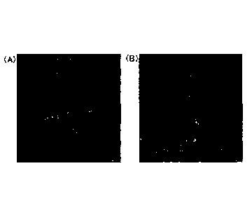
Fig. 2 shows (A) a graph showing the number of vessels
formed by vascular endothelial progenitor cells in each medium,
and (B) a graph showing the number of vessels formed by
vascular endothelial progenitor cells in each medium using
matrigel.
Fig. 3 shows the results of the test of the cytokine
secretion ability of the retinal pigment epithelial cell sheet.
Description of Embodiments
[0012]
The present invention is explained in detail in the
following.
The present invention provides a method of producing a
cell sheet comprising a retinal pigment epithelial cell layer
and a vascular forming cell layer, comprising a step of
laminating the retinal pigment epithelial cell layer and the
vascular forming cell layer (the production method of the
present invention).
[0013]
1. Vascular forming cell layer
The vascular forming cell layer in the present invention
is composed of cells having vascular formation ability
(vascular forming cells). When the cell sheet obtained by the
production method of the present invention is transplanted into
a defect site in the choroid of a retinal degeneration patient,
6
CA 02882802 2015-02-23
the vascular forming cells contained in the cell sheet
reconstitute a blood vessel (preferably, choroidal blood
vessel) in the transplanted site, which supplies oxygen and
nutrients to retinal pigment epithelial cells, and the like.
Therefore, the cell sheet obtained by the production method of
the present invention can exhibit a superior treatment effect
by being transplanted into, particularly, a defect site in the
choroid of a patient with a retinal degenerative disease
associated with a choroidal defect.
lo [0014]
While the vascular forming cell in the present invention
may be a cell derived from any mammal as long as it is derived
from a mammal (e.g., human, monkey, mouse, rat, dog, bovine,
horse, swine, sheep, goat, cat, rabbit, hamster, guinea pig
etc.), it is preferably a cell derived from human.
[0015]
Examples of the vascular forming cell to be used in the
present invention include hemangioblast, vascular endothelial
progenitor cell, vascular endothelial cell and the like. Among
these, vascular endothelial progenitor cell and the like are
preferable as the vascular forming cell, since the cells are
considered to be easily incorporated into the existing blood
vascular network, in the process of vascular formation in vivo
after transplantation. The vascular forming cell layer may
contain cells other than the vascular forming cell and
components other than cells, and may be composed of a cell
population or tissue containing the vascular forming cells.
Generally, not less than 70% (preferably not less than 80%,
more preferably not less than 90%, most preferably 100%), of
the cells constituting the vascular forming cell layer are
vascular forming cells (preferably, vascular endothelial
progenitor cells).
[0016]
The vascular endothelial progenitor cell refers to a cell
having an ability to differentiate into a vascular endothelial
7
CA 02882802 2015-02-23,
cell and committed to differentiate into a vascular endothelial
cell. Plural expression patterns of cellular surface markers
have been reported for vascular endothelial progenitor cell,
and it is known that at least a unified definition based on the
expression pattern of cell surface marker is difficult.
Examples of the expression pattern of surface marker of
vascular endothelial progenitor cells reported in the past
include CD34, CD44+, VEGFR2+ (KDR) for peripheral blood
mononuclear cell-derived CD34 positive vascular endothelial
/0 cell (Science. 1997 Feb 14; 275 (5302): 964-7); CD31+, VEGFR2+,
eN0s+, CD105+, CD34, CD133-, CD45-, CD14-, CD117- for cord
blood mononuclear cell-derived vascular endothelial progenitor
cell (human endothelial colony forming cell (ECFCs (registered
trade mark), manufactured by Takara Bio)); and the like. While
they are considered to result from the difference in the
tissues from which they are derived, differentiation stage,
collection method and the like, they are common in that all of
them have an ability to differentiate into vascular endothelial
cells. As used herein, therefore, the vascular endothelial
progenitor cell is defined as "a cell having an ability to
differentiate into a vascular endothelial cell, and committed
to differentiate into a vascular endothelial cell", and the
expression pattern of cellular surface marker tolerates the
presence of plural combinations.
(0017]
It is known that vascular endothelial progenitor cells
are contained in yolk sac, peripheral blood, bone marrow, cord
blood, mononuclear cells of these and the like, and can be
Prepared from these tissues or cells by a known isolation
method. Examples of the isolation method include an isolation
method using expression of cellular surface markers such as
CD34, VEGF receptor 2 (KDR) and the like as an index and using
magnetic beads and FACS; a method utilizing commercially
available endothelial cell colony-forming units (CFU-ECs) (N
Engl J Med. 2003; 348: 593-600) and the like. As specific
8
CA 02882802 2015-02-23
examples of the production method of peripheral blood
mononuclear cell- or bone marrow mononuclear cell-derived
vascular endothelial progenitor cells, a method including
culturing mononuclear cells separated from peripheral blood or
bone marrow by a conventionally-used method in a vascular
endothelial differentiation promoting medium containing
cytokines such as VEGF and the like, and recovering vascular
endothelial progenitor cells as adhered cells; a method of
separating and recovering vascular endothelial progenitor cells,
/o from the peripheral blood as a CD34 positive cell, which have
been recruited from bone marrow by using G-CSF (Yakugaku Zasshi
2007 125(5) 841-845 etc.) and the like are known.
[0018]
In addition, differentiation of the vascular endothelial
progenitor cell from various cells can be induced. For example,
a method of inducing differentiation from fibroblast through
dedifferentiation; a method of inducing differentiation of
pluripotent stem cell such as ES cell, iPS cell and the like
into vascular endothelial progenitor cell (WO 2008/056779, WO
2009/035217 and the like) and the like are known. These
vascular endothelial progenitor cells can be used alone or
plural kinds thereof can be used in combination. As the
vascular endothelial progenitor cell in the present invention,
a cell mixture containing other cells can be used. For example,
bone marrow cells, peripheral blood mononuclear cell, bone
marrow mononuclear cell and the like containing vascular
endothelial progenitor cell can also be used directly.
[0019]
Vascular endothelial cells can be prepared by a known
method such as a method including separating the cells from a
vascular tissue in the living body by using expression of a
cellular surface marker such as CD31 and the like as an index
and using magnetic beads and FACS; a method of inducing
differentiation by culturing the above-mentioned vascular
endothelial progenitor cell in the presence of an inducer such
9
CA 02882802 2015-02-23
as VEGF and the like, and the like. In addition,
differentiation of various cells into the vascular endothelial
cells can also be induced, and it is known, for example,
differentiation into the vascular endothelial cells can be
induced from somatic stem cells such as mesenchymal stem cells,
adipose tissue derived-stem cells and the like; progenitor
cells such as cardiac muscle progenitor cells, neuronal
precursor cells and the like; pluripotent stem cells such as ES
cells, iPS cells and the like; and the like. Furthermore, as a
commercially available product of vascular endothelial cells,
human microvascular endothelial cells (HMVEC), human umbilical
cord vascular endothelial cells (HUVEC), human aortic
endothelial cells (HAEC, HAOEC) and the like can be obtained.
[0020]
Hemangioblast is a common ancestor cell of vascular
endothelial progenitor cell and hematopoietic stem cell, and
can be prepared from a vascular tissue in the living body by
using expression of cellular surface marker such as CD133,
CD144, CD45 and the like as an index by a known method such as
a separation method using magnetic beads and FACS and the like.
As expression patterns of cellular surface marker of
hemangioblast, for example, a combination of CD1334, CD144%
CD45+, CD344-, VEGFR2+, CD31-has been reported (Stem Cells Dev.
2004 Jun;13(3):229-42.).
[0021]
As the vascular forming cell to be used in the present
invention, a tissue or cell derived from a patient to be
transplanted with the cell sheet obtained by the production
method of the present invention, or a cell derived from a donor
having HLA type matched with the patient's HLA type and the
like can be utilized. As the vascular forming cell to be used
in the present invention, a vascular endothelial progenitor
cell is particularly preferable.
[0022]
As a vascular forming cell preferable for autologous
CA 02882802 2015-02-23
transplantation use, for example, a patient's tissue, a cell
collected therefrom, and a cell derived from iPS cell
established from patient's somatic cell (patient's iPS cell)
are preferably used, since a burden on the patient is small.
Being less invasive, patient's peripheral blood, a mononuclear
cell collected therefrom, a cell derived from patient's
peripheral blood mononuclear cell, a cell derived from
patient's iPS cell and the like are preferably used. These can
be prepared using a cell derived from the patient by the
/o aforementioned method.
[0023]
As a vascular forming cell preferable for
allotransplantation, for example, a cell derived from a donor
having HLA type matched with the patient's HLA type is
preferably used to suppress rejection. The cell derived from a
donor having HLA type matched with the patient's HLA type
includes a donor tissue matching the patient's HLA type, a cell
collected therefrom, cells derived from iPS cell established
from donor having HLA type matched with the patient's HLA type
(HLA-matched donor iPS cell) and the like. The tissue and cell
with matched HLA type can also be obtained from bone marrow
bank, cell bank and the like. In particular, a cell having 3
locus (HLA-A, HLA-B, HLA-DR) homozygous showing low rejection
with other HLA types is preferable as a donor cell since it
matches with many patients' HLA types.
[0024]
The vascular forming cell layer is preferably laminated
on the retinal pigment epithelial cell layer such that the
vascular forming cell layer contacts a basal surface of the
retinal pigment epithelial cell layer. The vascular forming
cell layer only needs to be laminated on at least one part of
the retinal pigment epithelial cell layer. The density of the
vascular forming cells relative to the retinal pigment
epithelial cell layer is not particularly limited, and can be
determined as appropriate in consideration of the choroidal
11
CA 02882802 2015-02-23
disorder state in the transplanted site, affinity for existing
blood vascular network and the like. The density of the
vascular forming cell relative to the retinal pigment
epithelial cell layer is, for example, about 1x10² - 1x10⁶
cells/cm, preferably about 1x10³ - lx105 cells/cm2, since
vascular forming cells are easily incorporated into the
existing blood vessels in the living body when the density of
the vascular forming cells is low. When transplantation into a
patient with large damage on choroid and markedly small number
lo of remaining blood vessels is desired, a vascular forming cell
layer having a high density of the vascular forming cells is
preferable.
[0025]
As a step of laminating a retinal pigment epithelial cell
layer and a vascular forming cell layer (laminating step), a
known method can be utilized as a method of laminating plural
cell layers. Examples of such method include a method of
laminating plural sheet-like cell layers, a method including
seeding cells which constitute one cell layer, on the other
sheet-like cell layer, a method including placing one sheet-
like cell layer on the other cell layer cultured in a culture
container, a method including seeding cells which constitute
one cell layer, on the other cell layer cultured in a culture
container and the like. In the present invention, it is
preferable to laminate two cell layers such that the vascular
forming cell layer contacts a basal surface of the retinal
pigment epithelial cell layer. For example, a sheet-like
retinal pigment epithelial cell layer is placed on a vascular
forming cell layer cultured in a culture container, whereby the
vascular forming cell layer contacts a basal surface of the
retinal pigment epithelial cell layer. A cell sheet obtained
by laminating layers in a culture container can be directly put
to use.
[0026]
In a preferable embodiment of the laminating step in the
12
CA 02882802 2015-02-23
present invention, vascular forming cells are seeded using a
medium in a culture container and cultured to form a vascular
forming cell layer in the culture container, a retinal pigment
epithelial cell sheet formed separately is placed on the
vascular forming cell layer, and the medium is aspirated,
whereby the both cell layers are laminated such that the
vascular forming cell layer contacts a basal surface of the
retinal pigment epithelial cell layer. The aforementioned
medium is not particularly limited as long as it is a
lo composition capable of maintenance culture of vascular forming
cells and retinal pigment epithelial cells. Generally, both a
medium for vascular forming cell culture and a medium for
vascular endothelial progenitor cell culture can be used. For
example, as a medium for vascular endothelial progenitor cell
/5 culture, commercially available products such as EGM-2 medium
(manufactured by Takara Bio) and the like can be used. After
seeding of vascular forming cells, it is preferable to stand
the cells for at least the time necessary for the cells-to
adhere to the surface of the culture container and form a
20 vascular forming cell layer (e.g., about 10 hr - 24 hr) and,
where necessary, a culture period of about 1 day - 3 days may
be set to achieve growth to reach a desired cell number.
[0027]
The production method of the present invention may
25 further contain a step of recovering a cell sheet wherein a
retinal pigment epithelial cell layer and a vascular forming
cell layer are laminated (recovery step). A method of
recovering the cell sheet is not particularly limited as long
as it can recover, the sheet while maintaining the sheet
30 structure, and a known method can be used. Examples of such
method include a method of detaching a cell sheet from a
culture container by an enzyme treatment, a method using a cell
non-adhesive culture container, a method including laminating
cell layers by using a culture container surface-treated to be
35 cell-adhesive, and detaching the formed cell sheet by treating
13
CA 02882802 2015-02-23
with an enzyme etc., and the like. In the present invention,
when vascular forming cells adhere to a surface of a culture
container and fixed in the laminating step, the retinal pigment
epithelial cell layer is easily laminated on the vascular
forming cell layer. Therefore, a method including laminating =
cell layers by using a culture container surface-treated to be
cell-adhesive and detaching the formed cell sheet from the
culture container is preferable. In one embodiment, a cell
sheet is formed in a culture container surface-treated with a
temperature-responsive polymer, and the cell sheet is detached
by a treatment of the temperature change. The temperature-
responsive polymer refers to a polymer having a,hydration force
that changes in a temperature-dependent manner and, for example,
a temperature-responsive polymer having a hydration force that
changes in a temperature range of 0 - 80 C is described in JP-
A-2-211865. To be specific, for example, it can be obtained by
homopolymerization or copolymerization of the following
monomers. Examples of the usable monomer include
(meth)acrylamide compound, N-(or N,N-di)alkyl substituted
(meth)acrylamide derivative, and vinylether derivative. In the
case of a copolymer, any two or more kinds of these can be used.
Furthermore, monomers other than the above-mentioned monomers,
copolymerization with ionic monomer to improve adhesiveness and
growth of cells, graft or copolymerization of polymers, or a
mixture of polymer and copolymer may be used. The temperature-
responsive polymer undergoes hydration and dehydration in
response to temperature change, and the temperature range
thereof is 0 C - 80 C, preferably 10 C - 50 C, more preferably
20 C - 45 C. A preferable temperature-responsive polymer is,
for example, poly(N-isopropylacrylamide). Poly(N-
isopropylacrylamide) is a polymer having a lower critical
solution temperature of 31 C. When it is in a free form, it
undergoes dehydration in water at not less than 31 C, at which
the polymer chain coagulates and the polymer is clouded.
Conversely, at a temperature of less than 31 C, the polymer
14
CA 02882802 2015-02-23
chain is hydrated and the polymer is dissolved in water. When
poly(N-isopropylacrylamide) is fixed on the surface of a
culture container, poly(N-isopropylacrylamide) is dehydrated at
not less than 31 C, and the surface of the culture container
acquires hydrophobicity and shows adhesiveness to cells (e.g.,
vascular forming cell, retinal pigment epithelial cell). At a
temperature of less than 31 C, poly(N-isopropylacrylamide) is
hydrated, and the surface of the culture container acquires
hydrophilicity and shows non-adhesiveness to cells. Utilizing
lo such temperature responsiveness, cells are cultured at a
temperature (e.g., 37 C) not less than the lower critical
solution temperature (31 C for poly(N-isopropylacrylamide)) in
the laminating step to achieve adhesion of the cell sheet to
the culture container, a temperature less than the lower
/5 critical solution temperature (e.g., 20 C) is provided in the
recovery step to enable detachment and isolation of the cell
sheet from the culture container without applying an enzyme
treatment. Culture containers coated with such temperature-
responsive polymer are described in JP-A-2-211865, JP-A-05-
20 192138, JP-A-2008-220354 and the like. In addition, such
culture container is commercially available as a temperature-
sensitive culture container (manufactured by Cellseed, UpCell
(registered trade mark)). Vascular forming cells seeded in a
culture container are preferably adhered onto the culture
25 container so that they will be certainly transferred to the
retinal pigment epithelial cell layer.
[0028]
In a preferable embodiment, vascular forming cells are
adhesion-cultured in a temperature responsive culture container
30 coated with poly(N-isopropylacrylamide) at a temperature (e.g.,
37 C) not less than the lower critical solution temperature
(31 C) to form a vascular forming cell layer. Then, a
separately-prepared retinal pigment epithelial cell layer
(retinal pigment epithelial cell sheet) is laminated on the
35 vascular forming cell layer while maintaining a temperature
CA 02882802 2015-02-23
(e.g., 37 C) not less than the lower critical solution
temperature, such that the vascular forming cell layer contacts
a basal surface of the retinal pigment epithelial cell layer.
After incubation at a temperature (e.g., 37 C) not less than
the lower critical solution temperature for a time sufficient
for the vascular forming cell layer and the retinal pigment
epithelial cell layer to be adhered to each other, the culture
is cooled to a temperature (e.g., 20 C) less than the lower
critical solution temperature, whereby the formed cell sheet is
_to detached from the culture container. Cooling and detachment
are performed, for example, by aspirating the medium, adding a
medium with a temperature (e.g., 20 C) less than the lower
critical solution temperature to the culture container,
standing same for a time necessary for detaching the cell sheet
is from the culture container (e.g., not less than 30 min), and
recovering the laminated cell sheet. The medium is the same as
those recited as examples in the preferable embodiment of the
laminating step. When the standing time after addition of the
cooling medium is too long, detachment of the cell sheet
20 becomes difficult. Thus, it is preferable to recover the sheet
within one day from the addition of the medium.
[0029]
The production method of the present invention may also
comprise a step of applying a vascular formation treatment to
25 the vascular forming cell layer. The vascular formation
treatment step may be a pre-step or a subsequent step of the
step of laminating a retinal pigment epithelial cell layer and
a vascular forming cell layer. The vascular formation
treatment can be performed by a known method and, for example,
30 a known method of inducing tube formation such as a method of
culturing vascular forming cells in a collagen gel in the
presence of a factor such as VEGF, IGF-1, PDGF and the like, a
method of contacting a vascular forming cell layer with
matrigel and the like can be applied. Since retinal pigment
35 epithelial cell secretes VEGF, the supernatant of retinal
16
CA 02882802 2015-02-23
pigment epithelial cell culture can also be used for vascular
formation.
[0030]
When a vascular formation treatment is applied, all
s vascular forming cells constituting the vascular forming cell
layer may have a vascular structure, or only a part thereof may
have a vascular structure. In the vascular forming cell layer,
it is desirable to constitute a blood vessel having a structure
suitable for the environment of the transplantation site in
vivo. For example, when the cell sheet obtained by the
production method of the present invention is transplanted
without any vascular formation treatment ex vivo and without a
vascular structure, transplanted vascular forming cells
spontaneously form a blood vessel in vivo, during which process
/5 the blood vessel is linked to existing blood vessels to easily
form a functional blood vascular network. On the other hand,
when the damage on choroid is large and the remaining blood
vessels are markedly small in number, reconstruction of a blood
vascular network based on the transplanted vascular structure
can be preferably promoted by applying the vascular formation
treatment to the vascular forming cell layer.
[0031]
The vascular forming cell layer may contain one or plural
kinds of cells other than the vascular forming cells, for
example, cells supporting vascular formation or angiogenesis
such as hematopoietic stem cell and the like, blood vessel
constituting cells other than the vascular endothelial cell
such as vascular smooth muscle cell, blood cell and the like,
and the like. The vascular forming cell layer may further
contain components other than cell, for example, a factor
promoting angiogenesis, and the like.
[0032]
2. Retinal pigment epithelial cell layer
The retinal pigment epithelial cell to be used in the
present invention may be a primary cell directly collected from
17
CA 02882802 2015-02-23
an eyeball, or a cell after several passages. The primary
retinal pigment epithelial cells can be isolated by a known
method. For example, in the case of eyeball-derived retinal
pigment epithelial cells, a cadaveric eyeball is isolated,
rapidly divided at the equatorial segment, the vitreous body
and the retina are removed and treated with collagenase,
hyaluronidase and the like as necessary, the cells are
collected by scratching with a cell scraper, or treatment with
trypsin or EDTA solution to liberate the cells from the Bruch's
/o membrane, stood in a culture medium to induce adhesion to the
culture dish and growth, and the cells grown in the required
number are appropriately passaged with a trypsin treatment etc.
to sufficiently secure the cell number.
[0033]
Furthermore, these cells may also be the cells obtained
by inducing differentiation of undifferentiated pluripotent
stem cells such as embryonic stem cell (ES cell), induced
pluripotent stem cell (iPS cell) and the like, stem cells
including somatic stem cells such as neural stem cell and the
like, or progenitor cells including neural progenitor cell and
retinal progenitor cell. The ES cell may also be an ES cell
produced by nuclear reprogramming of a somatic cell. In
addition, as the stem cell, the object cell may be prepared by
inducing differentiation of induced pluripotent stem cell (iPS
cell) reported in recent years. The iPS cell is a somatic
cell-derived induced stem cell having properties equivalent to
those of ES cell, which can be produced by introducing a
particular nuclear reprogramming substance (nucleic acid,
protein, low-molecular-weight compound etc.) into a somatic
cell [Takahashi, K. and Yamanaka, S., Cell, 126: 663-676
(2006); Takahashi, K. et al., Cell, 131: 861-872 (2007)]. The
conditions and medium used for differentiation of the
aforementioned stem cell into the object differentiated cell
may follow conventionally-known conditions and medium, or may
be appropriately determined by those of ordinary skill in the
18
CA 02882802 2015-02-23
4
art. In the present invention, a cell obtained by inducing
differentiation of stem cell or progenitor cell, preferably
pluripotent stem cell, is preferably used as the retinal
pigment epithelial cell to be used for cell sheet, since a
retinal pigment epithelial cell at an appropriate maturation
stage can be prepared, and particularly, comparatively immature
retinal pigment epithelial cells can be prepared and a cell
sheet can be advantageously formed. In addition, when the cell
sheet to be produced by the present invention is for
transplantation, use of an iPS cell is preferable since a cell
sheet obtained using a somatic cell of the subject, who
receives transplantation, as a source of iPS cell does not have
antigenicity against the subject. When a stem cell is induced
to differentiate, for example, human ES cell or pluripotent
/5 stem cell such as iPS cell and the like is cultured in an ES
cell differentiation medium added with Wnt antagonist such as
Dkk-1, CKI-7 and the like and Nodal antagonist such as Lefty A,
SB-431542 and the like. When cultured for a given period, Rx,
Pax6 and Mitf, which are retinal progenitor cell markers, are
expressed, and human retinal pigment epithelial cells can be
obtained by morphological observation with an optical
microscope, by confirming cells having a polygonal form and
pigment [Neuroscience Letters 2009 Jul 24 458(3) 126-31,
Journal of Cell Science 2009 Sep 1 122(Pt 17) 3169-79].
[0034]
The retinal pigment epithelial cell layer in the present
invention is composed of a layer of retinal pigment epithelial
cells arranged on a flat plane and, for example, can be
composed as a cell sheet comprising retinal pigment epithelial
cells produced by a known method. As a production method of
such cell sheet (retinal pigment epithelial cell layer), for
example, the method described in WO 2011/142364 is known.
[0035]
A preferable embodiment of the cell sheet of the present
invention is a cell sheet wherein the retinal pigment
19
CA 02882802 2015-02-23
epithelial cell layer is produced by a method including the
following steps (hereinafter to be referred to as "the collagen
method"):
(1) seeding and culturing retinal pigment epithelial cells on a
collagen gel to foLm a cell sheet composed of the retinal
pigment epithelial cells, and
(2) decomposing the collagen gel with collagenase to detach the
cell sheet composed of the retinal pigment epithelial cells.
[0036]
While the retinal pigment epithelial cell to be seeded in
step (1) may be a cell derived from any mammal as long as it is
derived from a mammal (e.g., human, monkey, mouse, rat, dog,
bovine, horse, swine, sheep, goat, cat, rabbit, hamster, guinea
pig etc.), it is preferably a cell derived from human.
/5 [0037]
In the collagen method, the retinal pigment epithelial
cells are cultured by seeding on a collagen gel. The collagen
used for the collagen gel may be any as long as it is derived
from a mammal (e.g., human, monkey, mouse, rat, dog, bovine,
20 horse, swine, sheep, goat, cat, rabbit, hamster, guinea pig
etc.) and, for example, human- or swine-derived collagen is
used. Examples of the tissue from which collagen is derived
include tendon, skin and the like. While the kind of the
collagen may be any, one other than the collagen constituting
25 the human basement membrane is preferable, one other than type-
IV collagen is specifically preferable. Of these, type
collagen is preferably used. While a collagen gel can be
produced by, for example, a conventionally-known production
method, in the present invention, a gel composed of a collagen
30 fiber network is produced by inducing fibrogenesis of collagen,
as described in the below-mentioned Example. Since the
fibrotic collagen has strength and flexibility in combination,
it is easy to handle, shows good maintenance of cell
proliferation and cell differentiation, and is preferable as
35 the collagen gel to be used in the present invention. In
CA 02882802 2015-02-23
addition, the collagen to be used in the present invention is
required to maintain cells, which are seeded on the collagen
gel, on the gel surface without allowing them to sink into the
gel layer. As the collagen, therefore, preferred is one
wherein the gel has the strength necessary for cell
proliferation and, for example, collagen having a large amount
of intermolecular crosslinking is preferable. As such collagen,
tendon-derived collagen can be mentioned.
[0038]
ao While the collagen concentration of the aforementioned
collagen gel may be in any range as long as it can afford a gel
having strength permitting engraftment and growth of retinal
pigment epithelial cells, and satisfying solubility
facilitating decomposition by collagenase, viscosity enabling
is easy handling and the like, it is preferably 0.1% (W/V) - 0.5%
(W/V), more preferably 0.2% (W/V) - 0.3% (W/V). When the
collagen concentration of the collagen gel is less than 0.1%
(W/V), the strength of the collagen gel becomes insufficient,
and therefore, the colonization rate and cell proliferation
20 rate of retinal pigment epithelial cells decrease. When the
collagen concentration of the collagen gel exceeds 0.5% (W/V),
the time of a collagenase treatment to decompose the collagen
gel becomes long, which is feared to exert an adverse influence
on the cells.
25 [0039]
While the volume of a collagen gel mixed solution used
for the production of the aforementioned collagen gel varies
depending on the culture area and shape of a culture substratum
to be used for the cell culture, it is preferably about 100 pl
30 - about 250 pl, more preferably about 150 pl - about 200 pl,
per unit area (ce). When the amount of the collagen gel mixed
solution is too small, a collagen gel layer having a thin
center part due to the influence of a surface tension applied
to the gel surface is formed, and the sheet tends to be damaged
.3.5 during cutting out of the cell sheet composed of the retinal
21
CA 02882802 2015-02-23
piyment epithelial cells, since the cells directly contact with
a culture substratum for when the retinal pigment epithelial
cells are cultured. When the amount of the collagen gel mixed
solution is in excess, a thick collagen gel layer is formed on
a culture substratum, which relatively reduces the amount of
the culture medium, and therefore, maintenance culture is not
easy to perform, collagenase treatment takes time, and damages
on the cell sheet composed of the retinal pigment epithelial
cells are feared.
/o [0040]
In step (1), a cell sheet composed of the retinal pigment
epithelial cells can be produced by seeding and culturing the
aforementioned retinal pigment epithelial cells on the collagen
gel of a cell culture substratum. The cell culture substratum
in the present invention is not particularly limited as long as
it is for cell culture. Examples thereof include culture
containers having a porous membrane such as transwell and the
like, flask, tissue culture flask, dish, petri dish, tissue
culture dish, multi dish, microplate, microwell plate,
muitiplate, multiwell plate, chamber slide, petri dish, tube,
tray, culture bag and roller bottle. Culture containers having
a porous membrane are preferable, since a collagenase treatment
and a cutting operation of the cell sheet are conveniently
performed. For example, a commercially available transwell is
preferably used. Examples of the material of the cell culture
substratum in the present specification include, but are not
limited to, inorganic materials such as metal, glass, ceramic,
silicon and the like, organic materials represented by
elastomer, plastic (e.g., polyester resin, polyethylene resin,
polypropylene resin, ABS resin, nylon, acrylic resin,
fluororesin, polycarbonate resin, polyurethane resin,
methylpentene resin, phenol resin, melamine resin, epoxy resin,
vinyl chloride resin).
[0041]
The number of¨the retinal pigment epithelial cells to be
22
CA 02882802 2015-02-23
seeded may be in any range as long as it is a cell density
capable of forming a cell sheet. However, when the cell
density is too low, the cell shape is bad, the culture time
before reaching confluence is long, and further, the time
necessary for cell maturation and coloring is long. When the
cell density is too high, similarly, cell proliferation is
suppressed, the culture time before reaching confluence tends
to be long, and the cells may die from being overcrowded.
Therefore, the density of the cells to be seeded is preferably
/0 about 4.5x10⁴ cells/cm2 - about 8.5x10⁵ cells/cm2, more
preferably about 8.5x10⁴ cells/cm2 - about 8.5x10⁵ cells/cm?,
most preferably about 4.5x10⁵ cells/cm2.
[0042]
A monolayer cell population (cell sheet) composed of
/5 retinal pigment epithelial cells can be formed by culturing the
retinal pigment epithelial cells seeded on collagen gel in a
culture medium. A culture medium can be used without
particular limitation as long as it is a cell culture medium
generally used in the pertinent field. For example, basal
20 media described in "Japan tissue culture conference ed.,
Technique of Tissue Culture 3rd edition" page 581, published by
Asakura Shoten, such as F-10 medium, F12 medium, MEM, BME
medium, DMEM, aMEM, IMD medium, ES medium, DM-160 medium,
Fisher medium, WE medium, RPMI1640 medium and the like, can be
25 used. Furthermore, serum (fetal bovine serum etc.), various
growth factors (EGF, FGF, HGF, PDGF etc.), antibiotic, amino
acid and the like may be added to the basal medium. The pH of
the medium is preferably about 6 - about 8. As for culture,
for exaMiole, a primary culture is performed generally at about
3o 30 - about 40 C for about 15 - about 60 hr until the retinal
pigment epithelial cells become confluent. Thereafter, a
secondary culture is performed for about 1 week - about 2
months while changing the medium, after which the culture is
performed while aerating and stirring where necessary until
35 foLmation of a cell sheet. Cells constituting the cell sheet
23
CA 02882802 2015-02-23,
obtained by such culture are maintained as retinal pigment
epithelial cells. Maintenance of the cells as retinal pigment
epithelial cells can be confiimed by detecting BEST1, RPE65,
MERTK, CRALBP or the like as a specific differentiation marker.
[0043]
Since the cell sheet formed in step (1) is adhered to
collagen gel, for example, when it is directly used for
transplantation and the like, the collagen gel is feared to
prevent engraftment in a transplant recipient. In addition, it
lo is feared that collagen gel may prevent binding and adhesion of
a vascular foLming cell layer and a retinal pigment epithelial
cell layer. If the collagen gel can be removed in advance, it
is conducible to the solution of such problem. In step (2) of
the present invention, the collagen gel adhering to the cell
sheet formed in step (1) is decomposed by collagenase. Those
of ordinary skill in the art can select appropriate collagenase
according to the kind of the collagen used for preparing the
collagen gel. While the collagenase to be used for the
decomposition of the collagen gel is not particularly limited
as long as it has an activity to digest collagen gel, one that
does not easily decompose collagen constituting the human
basement membrane (e.g., Type-IV collagen etc.) is preferable.
For example, collagenase derived from a microorganism induced
from Clostridium (Clostridium histolyticum) or Streptomyces
(Streptomyces parvulus), which are available at a commercial
level, safe and have a high enzyme activity, can be used.
[0044]
As the activity of the above-mentioned collagenase, the
specific activity relative to the collagen weight in the
collagen gel is important rather than the activity per unit
weight of collagenase and the activity per unit volume of an
aqueous collagenase solution. The specific activity of the
collagenase to be used for dissolving collagen gel (collagenase
activity/collagen weight) is preferably not less than 0.1 U/mg.
When the specific activity of the collagenase is less than 0.1
24
CA 02882802 2015-02-23
U/mg, dissolution of the collagen gel may unpreferably take too
long or the gel may unpreferably be dissolved insufficiently.
It is more preferably within the range of 0.1 - 10,000 U/mg,
further preferably 1 - 3,000 U/mg.
[0045]
In the collagen method, a method of acting collagenase on
collagen gel is not particularly limited. A collagenase
solution prepared using, as a solvent, a medium or an isotonic
solution having a buffering capacity may be added to a medium,
119 or a cell-attached collagen gel detached from a cell culture
dish may be immersed in the aforementioned collagenase solution.
Since a transwell is used as a cell culture substratum in the
present invention, a collagen gel layer can be exposed by
recovering an insert and removing the membrane on the bottom of
the insert, and the exposed collagen gel is preferably immersed
directly in the above-mentioned collagenase solution.
[0046]
In the collagen method, the time of dissolving the
collagen gel by collagenase is not particularly limited. When
the time of acting the collagenase is too long, cell functions
such as adhesion ability, proliferative capacity and the like
may unpreferably decomposed. While the time of dissolution by
collagenase is subject to change due to specific activity of
collagenase, temperature, the shape of collagen gel,
collagenase treatment method and the like, it is generally 15
min - 60 min. The collagenase treatment may be a single
treatment or performed plural times.
[0047]
The temperature during the treatment of collagen gel by
collagenase in the collagen method is preferably set within the
range of 10 - 42 C, more preferably 30 - 40 C, further
preferably 36 - 38 C, since flowability of the cytoplasm of the
cell generally decreases and the metabolic capacity decreases
when the temperature inside living organisms becomes lower by
not less than 10 C (about 30 C in human), the protein is
CA 02882802 2015-02-23
denatured and the cell function decreases when the temperature
exceeds 42 C, and the optimal temperature of collagenase is
mostly 37 C and a temperature below this level prolongs the
dissolution time.
[0048]
In the collagen method, when the dissolution of collagen
gel proceeds, the cell sheet is gradually detached from the gel,
and finally liberated in the collagenase solution. To recover
the cell sheet, the cell sheet may be mechanically detached
from the remaining gel, or may be recovered after complete
dissolution of the gel. While the mechanical detachment
shortens the time until recovery of the cell sheet, since the
cell sheet may be destroyed, it is preferably recovered after
complete dissolution of the gel.
[0049]
While the cell sheet recovered as mentioned above can be
directly used for the laminating step with a vascular forming
cell layer, since the residual collagenase may inhibit
adhesiveness to the vascular forming cell layer, it is
preferably washed with a medium or an isotonic solution having
a buffering capacity. The temperature during cleansing can be
determined according to the collagen gel dissolution treatment
by collagenase. To sufficiently remove residual collagenase,
the sheet is preferably washed one or more times with a medium
or an isotonic solution having a buffering capacity.
In the cell sheet composed of retinal pigment epithelial
cells obtained by the collagen method, cytokine specific to a
retinal pigment epithelial cell is secreted with the polarity
similar to that in living organisms, and transepithelial
electric resistance (TER) to be an index of close adhesionic
bond between cells elevated as in living organisms. Therefore,
it has a cell layer barrier function similar to that in living
organisms. According to the collagen method, a cell sheet
composed of retinal pigment epithelial cells and having
functions similar to those in living organisms can be obtained.
26
CA 02882802 2015-02-23 .
[0050]
In the cell sheet obtained by the collagen method, a
tight junction is form between retinal pigment epithelial cells,
and a basement membrane is formed on a contact surface with the
collagen gel. In the present specification, the "basement
membrane" is a membrane formed from the components produced
from retinal pigment epithelial cells, and means a membrane
containing at least a part of the basement membrane component
(hereinafter to be referred to as a "basement membrane of
io retinal pigment epithelial cells"). The basement membrane of
the retinal pigment epithelial cell in living organisms is
present as a thin film between a retinal pigment epithelial
cell layer and an inner collagen layer constituting the Bruch's
membrane, and is an extracellular matrix having Type-IV
collagen, laminin, heparan sulfate proteoglycan (perlecan),
nidogen and the like as representative components. The Bruch's
membrane is a thin film between the retinal pigment epithelial
cell layer and choroid, and has a 5-layer structure of a
basement membrane of retinal pigment epithelial cells, an inner
collagen layer, an elastin layer, an outer collagen layer, and
a basement membrane of capillary lamina of choroid. A cell
sheet composed of retinal pigment epithelial cells obtained by
the collagen method contains a part (basement membrane of
retinal pigment epithelial cell) of the structure of the
Bruch's membrane. The formation of tight junction can be
confirmed by observing hexagonally-shaped closely-adhered cell
form, and expression of occludin, ZO-1 and the like between
cells by immunostaining. The formation of basement membrane
can be confirmed by observing expression of basement membrane
markers such as laminin, heparan sulfate proteoglycan
(perlecan), nidogen, or Type-IV collagen and the like on a cell
surface by immunostaining, or observation with a scanning
electron microscope.
Generally, retinal pigment epithelial cells cultured on a
culture dish produce basement membrane components, but it is
27
CA 02882802 2015-02-23
extremely difficult to detach the cells in the form of a usable
retinal pigment epithelial cell sheet detached from a culture
dish (Invest. Ophthalmol. Vis. Sc., 36(2), 1995, 381-390).
According to the collagen method, retinal pigment epithelial
cells together with a basement membrane produced from retinal
pigment epithelial cells can be recovered as a sheet without
utilizing an artificial membrane. Since retinal pigment
epithelial cells form a monolayer structure, when they are
handled singly, the sheet structure is disintegrated and the
lo cells are scattered into cell units. Thus, transplantation
thereof as a sheet is extremely difficult. On the other hand,
since a cell sheet composed of retinal pigment epithelial cells
obtained by the collagen method accompanies a basement membrane
and has sufficient stiffness, it is not easily wrinkled during
/5 recovery, which makes the handling thereof extremely easy.
Consequently, since laminating operation of vascular forming
cell layers can be performed smoothly, and mounting on a cell
transplantation device and a transplantation operation can be
performed smoothly, cell transplantation can be performed with
20 minimum invasion, and both the effect and the prognosis are
expected to be improved. In addition, since a cell sheet
composed of retinal pigment epithelial cells obtained by the
collagen method accompanies a basement membrane, it is
extremely advantageous for transplantation in a disease wherein
25 the basement membrane is simultaneously disordered. For
example, age-related macular degeneration sometimes accompanies
disorder of Bruch's membrane. When a retinal pigment
epithelial cell sheet obtained by the collagen method is used
as a retinal pigment epithelial cell layer in the above-
30 mentioned production method of the present invention, a
basement membrane of the cell sheet produced by the method
compensates for the disordered part, whereby the engrafting
rate of the cell sheet can be improved, and a treatment effect
thereof can also be expected. Hence, in the production method
35 of the present invention, when a retinal pigment epithelial
28
CA 02882802 2015-02-23
.
cell sheet obtained by the collagen method is used as a retinal
pigment epithelial cell layer, the produced cell sheet is
preferable as a sheet for transplantation recipienting a
disease with a disordered basement membrane, and can be
s preferably utilized as a sheet for transplantation particularly
targeting age-related macular degeneration.
[0051]
The collagen method may further contain the following
step (3):
lo (3) confirming the presence or absence of a basement membrane
on the contact surface between the detached cell sheet and the
collagen gel.
[0052]
In step (3), formation of a cell sheet having a cell
15 layer composed of retinal pigment epithelial cells and a
basement membrane can be determined by confirming the presence
or absence of the basement membrane of the cell sheet. The
presence or absence of the basement membrane can be confirmed
by a method similar to the aforementioned confirmation of the
20 formation of the basement membrane, for example, expression of
a basement membrane marker, observation with a scanning
electron microscope and the like. For detection of the
basement membrane, expression of a basement membrane marker may
be confirmed at any site of the cell (e.g., cytoplasm, cellular
25 membrane, nuclear membrane and the like). Preferably, a marker
expressed on a contact surface with collagen gel is targeted.
[0053]
The basement membrane marker in the present specification
includes a transcription product, a translation product or a
30 decomposition product of a gene specifically expressed in the
basement membrane. Examples of such gene include laminin,
heparan sulfate proteoglycan (perlecan), nidogen, Type-IV
collagen and the like. Of these, laminin, Type-IV collagen and
the like, which are main components of the basement membrane,
3,5 are preferably used.
29
CA 02882802 2015-02-23
[0054]
A sample to =be used for "confirming the presence or
absence of a basement membrane on the contact surface between
the detached cell sheet and the collagen gel" is not
particularly limited as long as it contains a basement membrane
marker (e.g., RNA, protein, decomposition product thereof and
the like) derived from the cell sheet (or cell) detached in
step (2).
[0055]
io The expression of the basement membrane marker gene when
the above-mentioned sample is RNA can be examined by preparing
an RNA (e.g., total RNA, mRNA) fraction from the cell of the
cell sheet detached in step (2) and detecting a transcription
product of the marker gene contained in the fraction, or
is directly detecting a marker gene product in the cell without
extracting RNA from the cell.
[0056]
When an RNA (e.g., total RNA, mRNA) fraction is prepared
from the cell, it can be prepared using a known method such as
20 guanidine-CsC1 ultracentrifugation method, AGPC method and the
like. Using a commercially available RNA extraction kit (e.g.,
RNeasy Mini Kit; manufactured by QIAGEN etc.), total RNA with
high purity can be prepared rapidly and conveniently from a
trace amount of a sample. Examples of the method for detecting
25 a transcription product of a basement membrane marker gene in
an RNA fraction include a method using hybridization
(Northernblot, dot blot, DNA chip analysis etc.), a method
using PCR (RT-PCR, competitive PCR, real-time PCR etc.) and the
like. Quantitative PCR methods such as competitive PCR, real-
30 time FOR and the like are preferable since expression variation
of a basement membrane marker gene can be detected rapidly and
conveniently from a trace amount of a sample, and DNA chip
analysis is preferable since expression variation of plural
marker genes can be collectively detected and quantification
35 performance can also be also improved by selecting a detection
CA 02832802 2015-02-23,
method and the like.
[0057]
When Northernblot or dot blot hybridization is employed,
the basement membrane marker gene can be detected using a
nucleic acid (probe) capable of hybridizing with a
transcription product of the gene. Examples of such nucleic
acid include nucleic acid capable of hybridizing with a
transcription product of a basement membrane marker gene under
high stringent conditions. Examples of the "high stringent
conditions" include hybridization reaction at 45 C in 6xSSq
(sodium chloride/sodium citrate), followed by washing once or
more at 65 C in 0.2xSSC/0.1% SDS and the like. Those of
ordinary skill in the art can easily adjust to a desired
stringency by appropriately changing the salt concentration of
/5 a hybridization solution, temperature of hybridization reaction,
probe concentration, probe length, number of mismatch,
hybridization reaction time, salt concentration of washing,
washing temperature and the like. The nucleic acid may be DNA,
RNA or DNA/RNA chimera, with preference given to DNA.
[0058]
The nucleic acid to be used as a probe may be double
stranded or single stranded. When double stranded, it may be
double stranded DNA, double stranded RNA or DNA:RNA hybrid.
When single stranded, an antisense strand can be used. While
the length of the nucleic acid is not particularly limited as
long as it can specifically hybridize with the target nucleic
acid, it is, for example, not less than about 15 bases,
preferably not less than about 30 bases. To enable detection
and quantification of the target nucleic acid, the nucleic acid
to be used as a probe is preferably labeled. Examples of the
labeling agent include radioisotope, enzyme, fluorescent
substance, luminescence substance and the like. Examples of
the radioisotope include [32P], [3E], [14C] and the like. As
the enzyme, a stable enzyme having a high specific activity is
preferable, for example, p-galactosidase, p-glucosidase,
31
CA 02882802 2015-02-23.
alkaline phosphatase, peroxidase, malic acid dehydrogenase and
the like. Examples of the fluorescent substance include
fluorescamine, fluorescein isothiocyanate and the like.
Examples of the luminescence substance include luminol, luminol
s derivative, luciferin, lucigenin and the like. Furthermore,
biotin-(strept)avidin can also be used for binding a probe and
a label.
[0059]
When Northern hybridization is employed, an RNA fraction
io prepared as mentioned above is separated by gel electrophoresis,
transferred to a membrane of nitrocellulose, nylon,
polyvinylidene difluoride and the like, hybridized under the
above-mentioned 'high stringent conditions" in a hybridization
buffer containing a labeling probe prepared as mentioned above,
15 and the amount of the label bound to the membrane is measured
for each band by a suitable method, whereby the expression
level of each basement membrane marker gene can be measured.
Also in the case of dot blot, a membrane spotted with an RNA
fraction is subjected to a similar hybridization reaction
20 (perfoLmed for each marker gene), and the amount of the label
at the spot is measured, whereby the expression level of each
marker gene can be measured.
[0060]
When DNA chip analysis is employed, for example, cDNA
25 introduced with a suitable promoter such as T7 promoter and the
like by a reverse transcription reaction is synthesized from an
RNA fraction prepared as mentioned above, cRNA is synthesized
using RNA polymerase (in this case, labeled cRNA is obtained by
using a mononucleotide labeled with biotin and the like as a
30 substrate). The labeled cRNA is contacted with a chip having
the above-mentioned probe immobilized thereon to perform a
hybridization reaction, and the amount of the label bound with
each probe on the solid phase is measured, whereby the
expression level of each basement membrane marker gene can be
35 measured. This method is advantageous in terms of rapidness
32
CA 02632802 2015-02-23
and convenience as the number of the detected differentiated
marker genes (therefore, probes to be solid phased) increases.
[006l]
On the other hand, when a marker gene is detected without
extracting RNA from the cell, in situ hybridization can be used
as the detection means. In this method, the cell is
immobilized by treating the cell with a fixing agent,
preferably a precipitation fixing agent, for example, acetone,
or incubating the cell for a short time in a buffering
/o formaldehyde solution, instead of extracting RNA from the cell.
After imrecruitment, the cell is embedded in paraffin to form a
block, and a slice cut out therefrom can be used as a sample.
A well-prepared paraffin-embedded sample can be preserved at
room temperature for many years. As nucleic acid to be used as
/5 a probe, those similar to the above-mentioned examples can be
used. In situ hybridization is preferably used in the present
invention since expression of a basement membrane marker on the
contact surface between the cell and collagen gel can be
directly confirmed.
20 [0062]
Alternatively, expression of a basement membrane marker
in the detached cell sheet in step (2) can be confirmed by
preparing a protein fraction from the cell sheet (or cell), and
detecting a translation product (i.e., marker protein) of the
2.5 marker gene contained in the fraction, or directly detecting a
translation product of the marker gene in the cell sheet (or
cell), without extracting the protein from the cell sheet (or
cell). A marker protein can be detected by an immunological
measurement method (e.g., ELISA, FIA, RIA, Western blot etc.)
30 using an antibody to each protein and, in the case of a protein
showing a measurable physiological activity such as an enzyme
and the like, it can be detected by measuring the physiological
activity of each marker protein by a known method.
Alternatively, a marker protein can also be detected by a mass
35 spectrometry method such as MALDI-TOFMS and the like.
33
CA 02882802 2015-02-23 ,
An antibody to each marker protein can be obtained
according to a generally-used polyclonal antibody or monoclonal
antibody production technique and using a marker protein or
protein, or a partial peptide thereof as an immunization
antigen.
[0063]
When respective immunological measurement methods are
applied to the present invention, setting of special conditions,
operations and the like is not necessary. A measurement system
_to of the basement membrane marker protein can be constructed by
adding general technical consideration of those of ordinary
skill in the art to general conditions and operation methods in
each method. As for the detail of these general technical
means, compendia, books and the like can be referred to. For
/5 example, "Radioimmunoassay" edited by Hiroshi Irie (Kodansha,
published in 1974), "cont. Radioimmunoassay" edited by Hiroshi
Irie (Kodansha, published in 1979), "Enzyme Immunoassay" edited
by Eiji Ishikawa et al. (Igaku-Shoin, published in 1978),
"Enzyme Immunoassay" edited by Eiji Ishikawa et al. (2nd
20 edition) (Igaku-Shoin, published in 1982), "Enzyme Immunoassay"
edited by Eiji Ishikawa et al. (3rd edition) (Igaku-Shoin,
published in 1987), "Methods in ENZYMOLOGY", Vol. 70
(Immunochemical Techniques (Part A)), ibidem, Vol. 73
(Immunochemical Techniques (Part B)), ibidem, Vol. 74
25 (Immunochemical Techniques (Part C)), ibidem, Vol. 84
(Immunochemical Techniques (Part D: Selected Immunoassays)),
ibidem, Vol. 92 (Immunochemical Techniques (Part E: Monoclonal
Antibodies and General Immunoassay Methods)), ibidem, Vol. 121
(Immunochemical Techniques (Part I: Hybridoma Technology and
30 Monoclonal Antibodies)) (all published by Academic Press) and
the like can be referred to.
[0064]
A vascular forming cell layer may be directly laminated
on the aforementioned retinal pigment epithelial cell layer, or
35 a vascular forming cell layer may be laminated via other layer.
34
CA 02882802 2015-02-23
In the present invention, a retinal pigment epithelial cell
layer and a vascular forming cell layer are preferably
laminated directly.
[0065]
The present invention also relates to a cell sheet
comprising a retinal pigment epithelial cell layer and a
vascular forming cell layer, obtained by the above-mentioned
production method of the present invention. The cell sheet of
the present invention preferably contains a cell layer formed
io from retinal pigment epithelial cells obtained by ex vivo
differentiation induction of stem cells or progenitor cells and
a vascular forming cell layer. When the retinal pigment
epithelial cell layer is produced by the above-mentioned
collagen method, the cell sheet of the present invention
further contains basement membrane secreted from the retinal
pigment epithelial cell layer. The cell sheet of the present
invention is preferable as a transplantation material for the
retinal treatment of patients with ophthalmic diseases.
Examples of the ophthalmic disease include chorioretinal
degeneration diseases such as age-related macular degeneration,
retinitis pigmentosa, diabetic retinopathy, retinal detachment,
central retinal artery occlusion, central retinal vein
occlusion, chorioretinal atrophy, retinal pigment epithelial
detachment, uveitis (Behcet's disease, Harada disease etc.),
excessive myopia (pathologic myopia) and the like.
[0066]
Since the cell sheet of the present invention contains a
vascular forming cell layer, it can be transplanted with a high
engraftment rate for a disease involving simultaneously
disordered choroid. Therefore, the cell sheet obtained by the
production method of the present invention is preferably used
for the treatment of, among the chorioretinal degeneration
diseases recited above as examples, particularly, ophthalmic
diseases associated with chorioretinal atrophy, for which
exclusive transplantation of retinal piyment epithelial cells
CA 02882802 2015-02-23
could not afford a treatment effect with ease (age-related
macular degeneration, retinitis pigmentosa, chorioretinal
atrophy, retinal pigment epithelial detachment, uveitis
(Behcet's disease, Harada disease etc.) and excessive myopia
(pathologic myopia) etc.).
[0067]
In addition, since the cell sheet of the present
invention has a basement membrane made from components similar
to those in living organisms, it can also be utilized for
m various screening purposes such as efficacy screening, toxicity
evaluation and the like in the aforementioned ophthalmic
diseases. For the efficacy screening for the aforementioned
ophthalmic diseases, for example, the cell sheet of the present
invention can be applied to screening for a substance having
efficacy for the aforementioned ophthalmic diseases, according
to the method described in JP-A- 2007-500509. To be specific,
the cell sheet of the present invention is cultured in the
presence or absence of a candidate substance having efficacy
under the stress conditions possibly causing the aforementioned
ophthalmic diseases (e.g., light (e.g., white light, blue
light; light induces death of retinal cells, particularly
photoreceptor cells, and can be a macular degeneration inciting
factor), A2E [retinoid N-retinylidene-N-retinyl-ethanolamine]
(accumulation of A2E is considered to contribute to age-related
neurodegeneration of retinal cells, particularly expression of
macular degeneration), cigarette smoke aggregate (smoking is
considered to be a risk factor of macular degeneration),
external pressure (e.g., hydrostatic pressure; increase in the
intraocular pressure is suspected to be involved in glaucoma)),
and evaluation can be performed based on the number of
photoreceptor that expresses rhodopsin, and by immunostaining
using anti-caspase 3 antibody. For toxicity evaluation, the
cell sheet of the present invention can be applied to screening
for a toxic substance according to the method described in JP-
A- 2007-517210. To be specific, the cell sheet of the present
36
CA 02882802 2015-02-23
invention is cultured in the presence or absence of a toxicity
- candidate substance and using the integrin marker peptide
described in JP-A- 2007-517210, excited with a laser at a
wavelength of 488 nm, and the fluorescence at 520 nm is
detected for evaluation. Moreover, the cell sheet of the
present invention can also be utilized as an in-vitro model for
the evaluation of various in vivo functions of retinal pigment
epithelial cell such as the function relating to the
maintenance of visual cells such as phagocytic capacity of
/o photoreceptor outer segment, neuroprotective action and the
like, retinal blood vessel barrier function such as pumping
action, tight junction, and the like.
[0068]
The cell sheet for transplantation of the present
/5 invention can be used for the treatment of the above-mentioned
diseases in human and mammals other than human (e.g., monkey,
mouse, rat, dog, bovine, horse, swine, sheep, goat, cat, rabbit,
hamster, guinea pig etc.).
[0069]
20 The range of the disease area to which the cell sheet for
transplantation of the present invention can be applied is
appropriately determined depending on the target disease, the
animal species, age, sex, body weight and symptom of
administration subject, and the like.
25 [0070]
The cell sheet for transplantation of the present
invention can be transplanted at once or in several portions.
The application number of transplantation is deterwined by
health-care professionals according to the disease and the
30 guideline. For example, when the disease is age-related
macular degeneration disease, the cell sheet for
transplantation of the present invention may be transplanted
two or more times depending on the severity thereof. When
transplantation is performed plural times, the interval is not
35 particularly limited, and a period of several days to several
37
CA 02882802 2015-02-23
weeks may be placed.
[0071]
The cell sheet for transplantation of the present
invention is transplanted by health-care professionals
according to an appropriate transplantation method in
accordance with the guideline. When the cell sheet for
transplantation of the present invention is transplanted under
the retina, a transplantation method including delivering the
sheet on a water flow from a punctured injection needle, up to
io the transplantation site under the retina of the eyeball, may
be employed or a therapeutic apparatus exclusive for
transplantation may also be used.
Examples
[0072]
The present invention is explained in more detail in the
following by referring to Examples, which are mere
exemplifications and do not limit the scope of the present
invention in any way.
[0073]
Production Example 1. Preparation of retinal pigment
epithelial cells
As the retinal pigment epithelial cells to be used in the
following Production Example 2, used were mature retinal
pigment epithelial cells (253G1, K11PD2, 59M8, 59SV2, 59SV3,
59SV9, 46a, K21EV15, 101EV3, K1lEV9, 454E2) obtained by
inducing differentiation of human iPS cell, and retinal pigment
epithelial cells (hES, CMK6) obtained by inducing
differentiation of ES cell, according to the method described
in Neuroscience Letters 458 (2009) 126-131.
<Human iPS-derived retinal pigment epithelial cells>
253G1 is a retinal pigment epithelial cell obtained by
differentiation induction of human iPS cell (253G1) derived
from healthy human as described in Nature Biotechnology 26,
101-106, 2008.
38
CA 02882802 2015-02-23
59SV2, 59SV3 and 59SV9 are retinal pigment epithelial
cells obtained by inducing differentiation of human iPS cells
derived from the same retinitis pigmentosa patient. The iPS
cells were established by a method including introducing 0ct3/4,
Sox2, Klf4 and c-Myc into human skin-derived fibroblasts by
using Sendai virus, according to the method described in Proc.
Jpn. Acad., Ser. B 85 (2009) 348-362.
K21EV15, 101EV3, K11EV9 and 454E2 are retinal pigment
epithelial cells obtained by inducing differentiation of human
lo iPS cells derived from retinitis pigmentosa patients different
from each other. The iPS cells were established by a method
including introducing human 0ct3/4, Sox2, Klf4, L-Myc and LIN2B
into human skin-derived fibroblasts by using episomal vector,
according to the method described in Nat Methods. 2011 May;
8(5): 409-12).
<Monkey iPS-derived retinal pigment epithelial cells>
46a is a retinal pigment epithelial cell obtained by
inducing differentiation of monkey (cynomolgus monkey) iPS cell,
according to the method described in Jpn. J. Transplant. 44
(2009) 231-235.
<ES-derived retinal pigment epithelial cells>
hES is a retinal pigment epithelial cell obtained by
inducing differentiation of human ES cell line khES-1. CMK6 is
a retinal pigment epithelial cell obtained by inducing
differentiation of monkey ES cell, according to the method
described in Neuroscience Letters 458 (2009) 126-131.
[0074]
Production Example 2. Production method of retinal pigment
epithelial cell sheet
<Preparation of collagen gel mixed solution>
The following SOLUTION A, SOLUTION B and SOLUTION C were
prepared.
solution A: Swine tendon-derived acid-soluble Type-I collagen
Cellmatrix I-A (Nitta Gelatin, 3.0 mg/ml),
-
solution B: concentrated culture medium at 5-fold concentration
39
CA 02882802 2015-02-23
[OMEM/F12 (Invitrogen, 12500-062, 3 g) was dissolved in MilliQ
water, and total volume (50 ml) was filter-treated], and
solution C: buffer for reconstitution
[1N NaOH (50 mM, 5 ml), NaHCO3 (260 mM, 2.2 g) and HEPES (200
mM, 4.77 g) were dissolved in MilliQ water, and total volume
(100 ml) was filter-treated]
Under cooling, solution B (2 vol) was mixed (pale-yellow)
with solution A (7 vol) without bubbling. Then, solution C (1
vol) was added and the mixture was mixed (pale-pink) to give a
0.21% collagen gel mixed solution.
<Preparation of retinal pigment epithelial cell sheet>
The 0.21% collagen gel mixed solution (200 pl) was added
into the insert of a 12 mm transwell insert (0.4 pm Pore
Polyester membrane; Corning, 3460), and the mixture was
/5 incubated at 37 C for 30 min. Then, F10-10% FBS [F-10 (Sigma,
N6908, 445 ml), FBS (50 ml), Penicilin-Streptomycin (Invitrogen,
15140-122, 5 ml)] was added by 1500 pl to the outside of the
insert and 500 pl to the inside of the insert, and the
transwell was incubated at 37 C for 24 hr. Thereafter, the
inside and outside of the insert were washed once with F10-10%
FBS, the retinal pigment epithelial cells obtained in
Production Example 1 were seeded to 5x10⁵ cells (F10-10% FBS,
500 pl) inside the insert, and F10-10% FBS (1500 pl) was added
to the outside of the insert. The retinal pigment epithelial
cells were cultured in F10-10% FBS until confluence. After
reaching confluent, the medium was changed to SFRM-B27 [DMEM
(Sigma, D6046, 350 ml), F12 HAM (Sigma, N6658, 150 ml), B27
(Invitrogen, 17504-044, 10 ml), 200 mM L-Glutamine (Sigma,
G7513, 5 ml), Penicilin-Streptomycin (Invitrogen, 15140-122, 5
ml), bFGF (wako, 060-04543, 10 ng/ml)] (1500 pl to the outside
of the insert, 500 pl to the inside of the insert, medium
change was 3 times/week), and the retinal pigment epithelial
cells were cultured until they showed suitable color and shape.
<Cutting out>
After progress for 6 weeks from the start of the culture,
CA 02832802 2015-02-23
the membrane of the insert was removed, collagenase L (Nitta
Gelatin, PBS(+): Sigma, 2600 U/ml, 100 pl) was added under the
insert, and the insert was incubated at 37 C for 60 min and
washed 3 times with PBS(+). SFRM-527 was added dropwise so
that the retinal pigment epithelial cell sheet would not get
dry and cut into a desired size with PALM MicroBeam (ZEISS).
<Property>
By immunohistochemistry of the tissue section, it was
confirmed that the prepared cell sheet had a structure wherein
/o a retinal pigment epithelial cell sheet, in which tight
junction (Z0-1 positive) is formed, is undercoated by basement
membrane (laminin, type-IV collagen positive), and type-I
collagen used for sheet formation did not remain (type-I
collagen negative).
/5 [0075]
Example 1 Production of laminated cell sheet of vascular
endothelial progenitor cell layer and retinal pigment
epithelial cell layer
<Preparation of vascular endothelial progenitor cell>
20 Using endothelial cell culture kit-2 (EGM-2 medium
(containing 2% FBS); manufactured by Takara Bio, B3162), human
vascular endothelial progenitor cells (ECFCs; manufactured by
Takara Bio, PT056) were seeded at 1.3x10⁴ cells/cm2 in a
temperature responsive culture dish (3.5 cm dish; manufactured
25 by Cellseed, CS3007).
<Laminating of vascular endothelial progenitor cells on retinal
pigment epithelial cell sheet>
After lapse of 15 hr, the human iPS cell-derived retinal
pigment epithelial cell sheet obtained in Production Example 1
30 was placed on the vascular endothelial progenitor cells in the
temperature responsive culture dish. The medium was gently
aspirated, and the cell sheet was arranged to be placed in the
center of the temperature responsive culture dish. Thereafter,
for preventing drying, 100 pl of EGM-2 medium at 20 C was added,
35 and the mixture was left standing for 30 min to convert the
41
CA 02832802 2015-02-23
.
temperature-responsive polymer on the surface of the culture
dish to be hydrophilic, whereby the cell sheet was detached.
The cell sheet was washed once with EGM-2 medium, and the
obtained cell sheet was transferred to a culture dish for
adherent cells (Lumox dish 35; manufactured by Greiner, 077331;
bottom surface is removable by cutting with a scalpel). Using
laser microdissection (PALM MicroBeam; manufactured by ZEISS),
and the cell sheet was cut into a desired size to give a cell
sheet wherein human vascular endothelial progenitor cells and
/o human retinal pigment epithelial cells were laminated.
[0076]
Example 2 Cell sheet transplantation
The laminated cell sheet obtained in Example I was
subcutaneously transplanted to the latissimus dorsi muscle of
/5 NOD/SCID mouse. Tissue sections were prepared one week later.
By immunohistochemistry using anti-CD31 antibody (endothelial
cell), anti-HLA-1 antibody (transplanted human cell), and DAPI,
engraftment of the transplanted cell sheet and formation of a
vascular structure derived from the transplanted cells were
20 observed (in Fig. 1, arrow 'capillary (donor)"). From the
results, it was confirmed that vascular endothelial progenitor
cells matured into endothelial cells after transplantation, and
could form a blood vessel.
[0077]
25 Example 3 Cell sheet transplantation
The laminated cell sheet obtained in Example 1 is
transplanted to the subretina of a rabbit with partially
deleted retinal pigment epithelial cells and choroid. Tissue
sections are prepared one week later. By immunohistochemistry
30 using anti-0D31 antibody (endothelial cell), anti-HLA-1
antibody (transplanted human cell), and DAPI, engraftment of
the transplanted cell sheet and vascular formation derived from
the transplanted cells can be confirmed.
[0078]
35 Comparative Example 1
42
CA 02632802 2015-02-23
The human retinal pigment epithelial cell sheet obtained
in Production Example 2 (with no human vascular endothelial
progenitor cell) is transplanted to the subretina of a rabbit
with partially deleted retinal pigment epithelial cells and
choroid. Tissue sections are prepared one week later. By
immunohistochemistry using anti-CD31 antibody (endothelial
cell), anti-HLA-1 antibody (transplanted human cell), and DAPI,
it is found that the sheet structure is destroyed, a small
number of the transplanted cells are detected in a dispersed
io manner and, as compared to Example 3, a phenomenon of markedly
decreased engrafting rate of the transplanted cells is observed.
[0079]
Reference Example 1. Vascular formation
Formation of vessels by vascular endothelial progenitor
is cells cultured in a medium containing VEGF was confirmed by the
following method.
(medium)
"EM": EGM-2 medium (containing 2% FBS; manufactured by
Takara Bio, 53162)
20 "F10": F10-10% PBS (F-10 (Sigma, N6908) 445 ml, PBS 50 ml,
Penicilin-Streptomycin (Invitrogen, 15140-122) 5 mL). The
results obtained by using this medium as a vascular formation
medium are shown as "F10" in Fig. 2.
"F10-1": The retinal pigment epithelial cell sheet
25 produced in Production Example 2 was placed in the insert of a
12 mm transwell insert (0.4 pm Pore Polyester membrane; Corning,
3460), and F10-10% PBS was added into and outside the insert by
500 pl, 1500 pl, respectively. The cell sheet was cultured for
one day, and the culture supernatant was recovered. The
30 results obtained by using the culture supernatant as a vascular
formation medium are shown as "F10-1" in Fig. 2.
"F10-2": The retinal piyment epithelial cell sheet
produced in Production Example 2 was placed in the insert of a
12 mm transwell insert (0.4 pm Pore Polyester membrane;
35 Corning, 3460), and F10-10% PBS was added into and outside the
43
= CA 02882802 2015-02-23
insert by 500 pl, 1500 pl, respectively. The cell sheet was
cultured for 2 days, and the culture supernatant was recovered.
The results obtained by using the culture supernatant as a
vascular formation medium are shown as "F1b-2" in Fig. 2.
(Vascular formation)
Using the above-mentioned 4 kinds of media, human
vascular endothelial progenitor cells (ECFCs; manufactured by
Takara Bio, PT056) were seeded in each culture dish at 1.3x10⁴
cells/cm2. The cells were incubated at 37 C, 5% CO2 and 4 hr
lo later, the number of formed vasculars was counted under a
microscope. The results of the number of formed vasculars
obtain in 3 repeats of the experiment are shown in Fig. 2(A)
for each medium used (*P<0.001; ANOVA, Scheffe test). Vascular
formation could be morphologically confirmed for each of them
/5 by optical microscope photograph and fluorescence microscope
photograph.
The concentration of VEGF in 4 kinds of media used was
measured. As a result, EM was 1.44 ng/ml, F10 was 0 ng/ml,
F10-1 was 2.64 ng/ml, and F10-2 was 2.80 ng/ml. F10 free of
20 VEGF showed a markedly small number of vascular formation. The
media other than F10 showed many vascular formation, which
confirms that VEGF promotes vascular formation by vascular
endothelial progenitor cells. Also, from the results, it could
be confirmed that a vascular formation treatment can be applied
25 to the form of a cell sheet wherein a vascular endothelial
progenitor cell layer and a retinal pigment epithelial cell
layer are laminated, since a culture supernatant of retinal
pigment epithelial cells could promote vascular formation.
[0080]
30 Reference Example 2. Use of matrigel
By the same method as that in Reference Example 1 except
that a matrigel-coated culture dish was used, 4 kinds of media
were used to culture human vascular endothelial progenitor
cells and the number of formed vasculars was counted. The
35 results of the number of formed vasculars obtain in 3 repeats
44
CA 02832802 2015-02-23
=
of the experiment are shown in Fig. 2(B) for each medium used
(*P<0.01 "P<0.05; ANOVA, Scheffe test). Vascular formation
could be morphologically confirmed for each of them by optical
microscope photograph and fluorescence microscope photograph.
As compared to Reference Example 1, the vascular
formation number was markedly improved in all media using
matrigel. Particularly, F10 free of VEGF showed a markedly
small number of vascular formation (Reference Example 1), but
vascular formation was promoted by using matrigel. These
lo results confirm that vascular formation can be promoted by
utilizing matrigel, irrespective of the presence of VEGF.
[0081]
Production conditions of retinal pigment epithelial cell sheet
Reference Example 3. Production method of retinal pigment
epithelial cell sheet (kind of collagen)
In the same manner as in Production Example 2 except that,
in the step of producing a cell sheet using 253G1 (iPS-retinal
pigment epithelial cells) in Production Example 2, (A) swine
skin-derived Type-I collagen TE (special order product: mainly
containing Type-I collagen, a small quantity of Type-III
collagen, Nitta Gelatin, 5 mg/ml) was used as 0.35% collagen
mixed solution/well, (B) swine tendon-derived Type-I collagen
T-1002 (special order product: Type-I collagen, Nitta Gelatin,
5.1 mg/ml) was used as 0.35% collagen mixed solution/well, (C)
FITC-labeled collagen I (Chondrex, 1 mg/ml) was used as 0.07%
collagen mixed solution/well, (D) FITC-labeled collagen I
(special order, Chondrex, 3 mg/ml) was used as 0.21% collagen
mixed solution/well, (E) atelocollagen (KOKEN, 3 mg/ml) was
used as 0.21% collagen mixed solution/well, and (F)
permeability collagen membrane for cell culture (KOKEN) was
used, respectively, instead of swine tendon-derived acid-
soluble Type-I collagen Cellmatrix I-A (Nitta Gelatin, 3 mg/ml)
as 0.21% collagen mixed solution/well, cell sheets were
produced and cut out to give retinal pigment epithelial cell
sheets.
CA 02882802 2015-02-23
The test results of Production Example 2 and the cases
using each of the aforementioned collagens were compared and
evaluated in terms of 4 items [1. gel strength; 2. cell
adhesion; 3. cell proliferation; 4. safety]. As a result, (A)
(1. inferior; 2. equivalent; 3. inferior; 4. good), (B) 11.
good (5.1 mg/ml); 2. equivalent; 3. inferior; 4. good}, (C) 11.
inferior (1 mg/ml); 2. inferior; 3. unknown; 4. unknown}, (D)
{1. equivalent (3 mg/ml); 2. equivalent; 3.inferior; 4.
unknown}, (E) {1. equivalent (3 mg/ml); 2. inferior; 3.
lo unknown; 4. good}, and (F) {was not lysed by collagenase, thus
unusable}. As for the gel strength, a certain level of
strength is required to enable growth of retinal pigment
epithelial cells. From such aspect, particularly preferable
kind and concentration of collagen were the swine tendon-
derived acid-soluble Type-I collagen Cellmatrix I-A of
Production Example 2 and (B) swine tendon-derived Type-I
collagen T-1002 used at the above-mentioned concentration.
When the substrate does not have a certain level of strength,
retinal pigment epithelium does not grow and cannot be used for
the present invention.
[0082]
Reference Example 4. Production method of retinal pigment
, epithelial cell sheet (collagen amount)
In the same manner as in Production Example 2 except that,
in the step of producing a cell sheet using 253G1 (iPS-retinal
pigment epithelial cell) of Production Example 2, the amount of
the collagen gel mixed solution to be used was changed to 100
pl or 300 pl from 200 pl, cell sheets were produced and cut out,
whereby retinal pigment epithelial cell sheets were recovered.
As compared to Production Example 2, when the amount of
the collagen gel mixed solution used was 100 pl, a thin
collagen gel layer was formed in the center part due to an
influence of the surface tension caused by the small amount of
the collagen gel mixed solution and, as the culture proceeded,
the seeded retinal pigment epithelial cells directly contacted
46
CA 02882802 2015-02-23
the bottom membrane with ease, which caused breakage of the
retinal pigment epithelial cell sheet during an operation to
cut out the sheet. When the amount of the collagen gel mixed
solution used was 300 pl, since the amount of the collagen gel
mixed solution was high, a thick collagen gel layer was formed,
which relatively reduced the amount of the medium that could be
retained in the insert, and therefore, maintenance culture was
not easy to perform, collagenase treatment took time, and
damages on the cell sheet were feared to become greater.
lo [0083]
Reference Example 5. Production method of retinal pigment
epithelial cell sheet (amount of collagenase and treatment
time)
In the same manner as in Production Example 2 except that,
is in the step of producing a cell sheet using 253G1 (iPS-retinal
pigment epithelial cell) of Production Example 2, 1%
Collagenase L (Nitta Gelatin) or Type I collagenase (Roche) was
contacted with the retinal pigment epithelial cell sheet for 10
min in an amount of 10 pl, 20 min in an amount of 10 pl, 30 min
20 in an amount of 10 pl, 60 min in an amount of 10 pl, 20 min in
an amount of 20 pl, 60 min in an amount of 20 pl, and 50 min in
an amount of 30 pl, instead of 30 min in an amount of 30 pl,
cell sheets were produced and cut out, whereby retinal pigment
epithelial cell sheets were recovered.
25 As a result, when a collagenase treatment was performed
for 60 min in an amount of 10 pl or 60 min in an amount of 20
pl, collagen decomposition of the same level as with 30 pl for
30 min was observed.
[0084]
30 Reference Example 6. Production method of retinal pigment
epithelial cell sheet (number of seeded cells)
In the same manner as in Production Example 2 except that,
in the step of producing a cell sheet using 253G1 (iPS-retinal
pigment epithelial cell) of Example 1, the number of the cells
3.5 to be seeded inside the insert was changed to (A) 5104
47
CA 02882802 2015-02-23
cells/500 pl, (B) 1x10⁵/500 pl or (C) 1x10⁶/500 pl from 5x10⁵
cells/500 pl, cell sheets were produced and cut out, whereby
retinal pigment epithelial cell sheets were recovered.
As compared to Production Example 2, (A) and (B) required
a longer time to reach cell confluence due to the small number
of cells, and (C) showed slow growth and also tended to require
a longer time to reach cell confluence.
[0085]
Reference Example 7. Basement membrane formed on retinal
lo pigment epithelial cell sheet
A cryo section (frozen section) was produced from the
cell sheet produced from 253G1 (IFS-retinal pigment epithelial
cell) in Production Example 2, and subjected to
immunohistochemical staining. Formation of a tight junction
was confirmed by the expression of 20-1, and formation of a
basement membrane was confirmed by the expression of laminin
and Type-IV collagen. For detection of each protein,
respective antibodies of rabbit anti-Z0-1 manufactured by Zymed
(1:100 dilution), rabbit laminin manufactured by Abcam (1:200
dilution), and mouse anti-human collagen type IV antibody
manufactured by Calbiochem (1:40) were used. Furthermore, the
retinal pigment epithelial cell sheet was confirmed to have a
monolayer epithelial form from the state of nuclear staining
using 4',6-diamidino-2-phenylindole manufactured by Molecular
Probes (DAPI; 1 pg/m1).
[0086]
Evaluation of retinal pigment epithelial cell sheet
Evaluation 1. Retinal pigment epithelial specific gene
expression profile of cell sheet
In the step of producing a cell sheet from 59SV3, 59SV9
(iPS-retinal_pigment epithelial cells) in Production Example 2,
the expression of BEST1, RPE65, MERTK, CRALBP in the cells
constituting the sheets after lapse of 1 week, 4 weeks, 2
months, wherein the day when the medium was changed to SFRM-327
after cell confluence was day 0, was confirmed by RT-PCR. As a
48
CA 02882802 2015-02-23
0
result, expression of the same level as that of the positive
control (human retinal pigment epithelial cell total RNA
(manufactured by ScienCell, Cat NO. 6545)) was observed. Here,
BEST1, RPE65, MERTK are genes specifically expressed in retinal
pigment epithelial cells. CRALBP is a gene expressed in
retinal pigment epithelial cells and Muller cells.
[0087]
Evaluation 2. Measurement of residual collagen in retinal
pigment epithelial sheet
/o Cryo sections (frozen section) were produced by cutting
out, before and after collagenase treatment, from respective
cell sheets produced from 253G1 (iPS-retinal pigment epithelial
cell) in Production Example 2, and subjected to
immunohistochemical staining. The nucleus was stained with
/5 4',6-diamidino-2-phenylindole (DAPI; 1 pg/ml) manufactured by
Molecular Probes, and Collagen type 1 was stained with rabbit
anti-human collagen type I antibody (1:40 dilution)
manufactured by Calbiochem. As a result, collagen was not
detected from the sheets after the collagenase treatment, and
20 it was confirmed that collagenase removed collagen coated on
the culture dish. On the other hand, collagen was detected
from the sheets cut out before the collagenase treatment.
[0088]
Evaluation 3. Cytokine secretion capability of retinal pigment
25 epithelial cell sheet
The culture media on the Apical side and the Basal side
in the transwell were recovered before the step of cutting out
retinal pigment epithelial cell sheets from the cell sheets
produced from 253G1 (iPS-retinal pigment epithelial cell) and
30 454E2 (iPS-retinal pigment epithelial cell) in Production
Example 2, and the production amounts of VEGF and PEDF were
detected by ELISA according to the method described in Arvydas
M, IOVS. 2006; 47: 3612-3624. As a result, it was confirmed
that, similar to the human embryo-derived retinal pigment
35 epithelium reported in Arvydas M, IOVS. 2006; 47: 3612-3624,
49
CA 02882802 2015-02-23
VEGF was mainly secreted on the Basal side, and PEDF was mainly
secreted on the Apical side (Fig. 4). It was shown that the
retinal pigment epithelial cell sheet produced from 253G1 and
454E2 in Production Example 2 has cytokine secretory capability
similar to that in living organisms, and is superior in
functionality.
[0089]
Evaluation 4. Transepithelial electric resistance of retinal
pigment epithelial cell sheet
A strong correlation is seen between the barrier function
of a cell layer and impedance, namely,
transepithelial/transendothelial electric resistance (TER). A
probe was placed in the media inside and outside the insert
according to the method described by MILLIPORE (using Millicell
ERS-2), before the step of cutting out the retinal pigment
epithelial cell sheet produced from 454E2 (iPS-retinal pigment
epithelial cell) in Production Example 2, and TER was
electrically measured. As a result, TER was 6400.cm2, and
showed a high TER value like the human embryo-derived retinal
pigment epithelium reported in Nature Protocols vol 4, No 5
662-673 (2009), Fig 10. It was shown that the retinal pigment
epithelial cell sheet of the present invention produced in
Production Example 2 has a high barrier function similar to
that in living organisms.
[0090]
Evaluation 5. Transplantation of retinal pigment epithelial
cell sheet derived from monkey ES cell
A monkey retinal pigment epithelial cell sheet produced
from monkey ES cell-derived retinal pigment epithelial cells,
CMK6 in Production Example 2 was transplanted into one eye of a
monkey according to the method described in Invest Ophthalmol
Vis Sci. 1995 Feb; 36(2): 381-90. Before the transplantation,
retinal photocoagulation was performed to disorder the retina
of the eye to be subjected to transplantation. On day 28 from
the transplantation into the one eye of the monkey having
- CA 02882802 2015-02-23
retinal photocoagulation macula formed therein, eye fundus
photographs were taken, and images of the ocular fundus
sections were produced as histological sections by using OCT
(Optical coherence tomograph), based on which the condition of
the retina was confirmed. As a result, no leakage of
fluorescence was found by fluorescein angiography, the graft
survived, and a disorder such as thinning of sensory retina and
the like were not found.
[0091]
/o Evaluation 6. Transplantation of retinal pigment epithelial
cell sheet derived from monkey iPS cell
A monkey retinal pigment epithelial cell sheet produced
from monkey iPS cell-derived retinal pigment epithelial cells,
46a in Production Example 2 was transplanted under the retina
of one eye for autologous transplantation and three eyes for
cross transplantation according to the method described in
Invest Ophthalmol Vis Sci. 1995 Feb; 36(2): 381-90. Up to one
year post-transplantation, eye fundus photographs were taken,
and images of the ocular fundus sections were produced as
histological sections by using OCT (Optical coherence
tomograph), based on which the condition of the retina was
observed with the lapse of time. In cross transplantation,
clear rejection reactions such as fibrous changes on the
periphery of the graft, leakage of fluorescence by fluorescein
angiography, and high brightness lesion under the retina by OCT
were found. On the other hand, in autologous transplantation,
such clear rejection was not observed, no leakage of
fluorescence was found by fluorescein angiography, the graft
survived, and a disorder such as thinning of sensory retina and
the like were not found.
Industrial Applicability
[0092]
According to the present invention, it is possible to
easily and stably produce a laminated sheet of retinal pigment
51
81786113
epithelial cells, whio4 has a vascular constituting cell layer
capable of complementing a deficient choroidal blood vessel in
the living body and supplying oxygen and nutrients to retina
after transplantation. The cell sheet of the present invention
is extremely useful, since it is superior in the engraftment
rate and functionality, and can also treat severe chorioretinal
degeneration diseases, for which simple retinal pigment
epithelial cell transplantation cannot easily afford a
sufficient treatment effect, such as chorioretinal degeneration
/0 diseases, particularly, high myopia and severe uveitis and the
like, which are associated with chorioretinal atrophy.
[0093]
This application is based on a patent application
No. 2012-185932 filed in Japan (filing date: August 24, 2012).
52
CA 2882802 2020-01-08