Note: Descriptions are shown in the official language in which they were submitted.
CA 02882976 2016-07-26
IMPLANT SUITABLE FOR CALCANEAL OSTEOTOMY
[0001]
FIELD
[0002] This disclosure relates to an implant for an osteotomy, and
tools for inserting an
implant.
BACKGROUND
[0003] A calcaneal osteotomy is a form of surgery for correction of
severe hind foot mis-
alignment. During the procedure, the heel bone (calcaneus) is cut, and the
tuberosity is moved
medially toward the inside or laterally toward the outside, depending upon the
direction of the
misalignment that is to be corrected. For example, if the patient has flat
feet, the heel may be
offset medially to shift the hind foot toward the inside to improve the weight
distribution on the
foot. On the other hand, if the patient has a high arched foot the calcaneal
osteotomy may be
perfoluted to shift the hind foot laterally, to improve stability and reduce
risk of sprain. This
procedure has been performed by cutting the bone, moving the tuberosity
medially or laterally,
and driving screws through the tuberosity into the anterior calcaneus. Some of
the challenges
associated with this approach are determining the amount of intra-operative
offset that is
achievable, the capability of fluoroscopy techniques for targeting and placing
of screws, and
post-placement screw head prominence.
[0004] Implants are known for insertion during calcaneal osteotomy.
For example, the
assignee of this patent application, Wright Medical Technologies, has
developed the DARCO
DPS plate, which provides support. This implant includes an anterior plate, a
posterior plate, and
1
CA 02882976 2015-02-04
WO 2015/026375
PCT/US2013/056942
an offset segment connecting the anterior and posterior plates. The DARCO(g)
DPS plate is
available with different amounts of offset between the anterior and posterior
plates.
[0005] U.S. Patent Application Publication No. 2011/0009866 describes
an osteotomy
plate having a top side and a bottom side, with a first end and a second end
aligned along a
longitudinal axis and joined by a middle section. The first end includes a
cutting edge having a
chamfer of between about 5 and 30 . As a screw hole in the first end forms an
angle of from
about 10 to about 45 with respect to the longitudinal axis of the plate. The
screw hole is not
threaded, but does include an arcuate shroud on the top side of the plate. The
second end has a
locking hole which includes internal threads. The first hole and the second
hole are aligned
along the longitudinal axis. One or more of additional screw holes,
compression holes,
fenestrations or guide wire holes are provided.
[0006] Improved osteotomy plates are desired.
SUMMARY
[0007] In some embodiments, an implant comprises an elongated plate
having a first
major face and at least one locking screw hole to receive a locking fastener
oriented normal to
the major face. The elongated plate has a wall having a flat surface normal to
the first major
face. A non-locking screw hole is located between the locking screw hole and
the wall. The non-
locking screw hole is configured to receive a non-locking fastener oriented at
an acute angle
relative to the locking fastener.
[0008] In some embodiments, an implant may comprise an elongated plate
having a first
major face and at least one locking screw hole normal to the major face, to
receive a locking
fastener. The elongated plate has a non-locking screw hole configured to
receive a non-locking
fastener oriented at an acute angle relative to the locking fastener. At least
one insertion member
extends in an anterior direction, away from the locking screw hole, the at
least one insertion
member having an edge with barbs.
[0009] A method is also provided, which in some embodiments,
comprises: (a) fastening
an implant to a first portion of a bone, so that a face of the implant abuts
the bone, the implant
having a hole configured to receive a fastener oriented at an obtuse angle
relative to the face, the
implant having a flat surface normal to the face; (b) cutting the bone along a
plane of the flat
surface, so as to separate a second portion of the bone from the first portion
of the bone; (c) off-
2
CA 02882976 2015-02-04
WO 2015/026375
PCT/US2013/056942
setting the second portions of the bone relative to the first portion of the
bone, such that the flat
surface of the implant abuts the second portion of the bone; and (d) fastening
the implant to the
second portion of the bone using the fastener.
BRIEF DESCRIPTION OF THE DRAWINGS
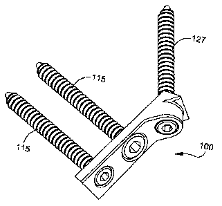
[0010] FIG. 1 is a perspective view of an embodiment of an implant, with
insertion
screws.
[0011] FIG. 2 is a diagram of the implant of FIG. 1, inserted in a
calcaneus.
[0012] FIG. 3 is a perspective view of the implant of FIG. 1.
[0013] FIG. 3A is a top plan view of a variation of the implant of
FIG. 3.
[0014] FIG. 3B is a 45 degree plan view looking directly into the non-
locking fastener
hole of the implant of FIG. 3A.
[0015] FIG. 3C is a side elevation view of the implant of FIG. 3A.
[0016] FIG. 3D is a perspective view of the implant of FIG. 3A.
[0017] FIG. 4 is a perspective view of a second embodiment of an
implant, inserted in
the calcaneus.
[0018] FIG. 4A is a perspective view of the implant of FIG. 4.
[0019] FIG. 4B is a side view of the implant of FIG. 4.
[0020] FIG. 5 is a perspective view of a third embodiment of an
implant, inserted in the
calcaneus.
[0021] FIG. 6 is a perspective view of a fourth embodiment of an implant,
inserted in the
calcaneus.
[0022] FIG. 7 is a perspective view of a fifth embodiment of an
implant.
[0023] FIG. 8 is a perspective view of a sixth embodiment of an
implant.
[0024] FIG. 9A is a perspective view of a seventh embodiment of an
implant.
[0025] FIG. 9B is a perspective view showing a double-wide version of the
implant of
FIG. 9A, inserted into the calcaneus.
[0026] FIG. 9C is a side elevational view of the a double-wide version
of the implant of
FIGS. 9A and 9B, inserted into the calcaneus.
[0027] FIG. 10 is a perspective view of a eighth embodiment of an
implant.
[0028] FIG. 11 is a perspective view of a ninth embodiment of an implant.
3
CA 02882976 2015-02-04
WO 2015/026375
PCT/US2013/056942
[0029] FIG. 12A is a perspective view of a tenth embodiment of an
implant.
[0030] FIG. 12B is a perspective view of an eleventh embodiment of an
implant.
[0031] FIG. 13 is a perspective view of a twelfth embodiment of an
implant.
[0032] FIG. 14 is a perspective view of a thirteenth embodiment of an
implant.
[0033] FIG. 15 is a perspective view of a fourteenth embodiment of an
implant.
[0034] FIG. 16 is a perspective view of a fifteenth embodiment of an
implant.
[0035] FIG. 17 is a perspective view of a sixteenth embodiment of an
implant.
[0036] FIG. 18 is a perspective view of a seventeenth embodiment of an
implant.
[0037] FIG. 19 is a perspective view of a tool for installing the
implant.
DETAILED DESCRIPTION
[0038] This description of the exemplary embodiments is intended to be
read in
connection with the accompanying drawings, which are to be considered part of
the entire
written description. In the description, relative terms such as "lower,"
"upper," "horizontal,"
"vertical,", "above," "below," "up," "down," "top" and "bottom" as well as
derivative thereof
-- (e.g., "horizontally," "downwardly," "upwardly," "anterior," "posterior,"
etc.) should be
construed to refer to the orientation as then described or as shown in the
drawing under
discussion. These relative terms are for convenience of description and do not
require that the
apparatus be constructed or operated in a particular orientation. Terms
concerning attachments,
coupling and the like, such as "connected" and "interconnected," refer to a
relationship wherein
-- structures are secured or attached to one another either directly or
indirectly through intervening
structures, as well as both movable or rigid attachments or relationships,
unless expressly
described otherwise. In the various drawings, like reference numerals indicate
like items, unless
expressly indicated otherwise.
[0039] Figures 1-3 show a first embodiment of an implant 100 for an
osteotomy, such as
-- a calcaneal osteotomy. The implant 100 allows polyaxial screw placement,
with both locking
and non-locking screw holes. The implant 100 includes an elongated plate 110
having a first
major face 111 and at least one locking screw hole 114 that is defined normal
to the major face
111, to receive a locking fastener 115. In some embodiments, the locking screw
hole 114 is
located at an end of the implant 100, distal from the joint line where the
calcaneus 130 is cut, and
-- the tuberosity 131 is to be rejoined to the anterior calcaneus 132. The
locking screw hole 114
4
CA 02882976 2015-02-04
WO 2015/026375
PCT/US2013/056942
has a female thread for locking the plate 100 against the lateral or medial
side of the tuberosity
131 and tightening the implant 100 down against the tuberosity.
[0040] The elongated plate 110 includes a non-locking screw hole 126
configured to
receive a non-locking fastener 127 at an end of implant 100 opposite locking
screw hole 114.
The non-locking screw hole 126 is oriented at an acute angle 0 relative to the
locking fastener
and locking screw hole 114. The implant 100 includes an abutting end 120 with
a wall having a
flat surface 124, which may be normal to the first major face 111. The non-
locking screw hole
126 penetrates the flat surface 124 of the wall. The non-locking screw hole
126 is oriented at an
angle 0 of about 30 degrees to about 60 degrees from the locking screw hole
114. In some
embodiments, the non-locking screw hole 126 is oriented at an angle 0 of about
40 degrees to
about 50 degrees from the locking screw hole 114. In some embodiments, the non-
locking screw
hole 126 is oriented at an angle 0 of about 45 degrees from the locking screw
hole 112.
[0041] Referring to FIG. 2 , implant 100 may have the first major face
111 of elongated
plate 110 affixed to a lateral or medial side of the tuberosity 131 of the
calcaneus 130, with the
normal flat surface 124 abutting the posterior facing cut surface of the
anterior calcaneus 132. In
some embodiments, the implant is affixed to the tuberosity 131 such that the
flat anterior surface
124 of implant 100 is recessed, about 0.127 mm to 0.381 mm in the posterior
direction relative to
the cut surface of the tuberosity 131. Thus, when the implant plate 100 is
fastened to the anterior
calcaneus 132, and the fasteners 115, 127 are tightened, there is a 0.127 mm
to 0.381 ram
translation of the implant plate 100 resulting in advantageous compression of
the calcaneus
against the tuberosity. When the non-locking screw 127 is inserted through the
hole 126 and
tightened, the anterior calcaneus 132 is pulled towards the tuberosity 131 by
up to 0.127 to 0.381
mm, until the anterior surface 124 of the implant 100 abuts the cut surface of
the anterior
calcaneus 132. This provides compression of the two abutting cut surfaces of
the tuberosity 131
and anterior calcaneus 132, enhancing rotational stability about the
anatomical axes resulting in
solid fixation. Since face 124 abuts the cut surface of the anterior calcaneus
132, there is no
requirement to drive the implant 100 into the anterior calcaneus 132 before
inserting either of the
fasteners 115, 127. This arrangement simplifies the surgical procedure and
reduces the duration
of the surgery. Also, of the implant 100 fitting in a corner formed between
the lateral (or medial)
surface of the tuberosity 131 and the cut surface of the anterior calcaneus
132, the implant 100
does not extend along the lateral (or medial) surface of the anterior
calcaneus beyond the cut
5
CA 02882976 2015-02-04
WO 2015/026375
PCT/US2013/056942
line. In some embodiments, elongated plate 110 further comprises a compression
slot 112
between locking screw hole 114 and non-locking screw hole 126. The compression
slot 112 has
a ramped surface 112R on its anterior side, toward flat anterior surface 124.
When a ramped
compression screw is inserted into compression slot 112, subsequent tightening
of the screw
causes implant 100 to translate in the anterior direction away from the
locking screw hole 114.
[0042] The implant 100 is affixed to the calcaneus 130 by first
inserting locking screw
115 though locking screw hole 114 distal from the joint-line (where the
calcaneus has been cut).
A compression screw is then inserted in compression slot 112, which forces
implant 100 to
translate in the anterior direction away from locking screw hole 114 and
compress the joint-line
between the tuberosity 131 and anterior calcaneus 132. Then, a non-locking
screw is inserted
through the angled non-locking screw hole 126. The head of this non-locking
screw hole 126 is
on the same side of the joint-line as compression slot 112, and fixes implant
100 in its
compressed state. The screw advances into the anterior calcaneus 132 on the
opposite side of the
joint line.
[0043] In some embodiments, the locking screw is first inserted into hole
114 to affix the
posterior portion of implant 100 to tuberosity 131. Then, the bone of the
calcaneus 130 is cut
about 0.127 to 0.381 mm beyond the flat anterior surface 124 of implant 100.
In other
embodiments, a cutting guide is attached to the implant to guide the location
of the cut in the
bone along a plane that is substantially parallel to anterior flat face 124 of
implant 100. Once in
this position, the guide may be removed from implant 100. In further
embodiments, a second
tool is attached to the implant for drilling the non-locking screw hole 126 so
that it is sized to
receive the non-locking screw. This tool may include a drill guide which is
inserted into non-
locking screw hole 126 for correctly aligning non-locking screw 126. In some
embodiments, this
tool has an offset medial displacement shelf, which is temporarily fixed to
the lateral or medial
surface of the anterior calcaneus 132 so as to positively locate bottom
surface 111 of implant 100
in the medial-lateral direction relative to the anterior calcaneus. This
allows control of the offset
between the anterior calcaneus 132 and tuberosity 131. Because flat face 124
of implant 100
abuts the cut surface of the anterior calcaneus 132 and is directly fastened
to the cut surface, a
single size of implant 100 may be used for osteotomies involving a variety of
different offsets
between the tuberosity and anterior calcaneus. In other embodiments, the tool
provides the
capability of continuously varying the position of the medial displacement
shelf. This
6
CA 02882976 2015-02-04
WO 2015/026375
PCT/US2013/056942
arrangement allows the surgeon to select the size of the offset, which can be
varied throughout
the range of offsets used for calcaneal osteotomies.
[0044] In many embodiments, the offset between bottom surface 111 of
implant 100 and
the lateral/medial surface of the anterior calcaneus 132 is determined using a
fixed offset
insertion tool. A set of such tools may be provided to the surgeon, with each
tool having a
respective offset. In some embodiments, the implant is formed of a
biocompatible material, such
as a titanium alloy or stainless steel of the type known for use in surgical
procedures. In some
embodiments (e.g., FIG. 1), implant 100 defines a single row of screw holes
114, 112, 126, and
has a width sufficient to secure implant 100 with a single row of holes. In
other embodiments,
the implant defines additional holes, and may be wider, to improve stability.
[0045] In the various drawings referred to in the following
description of alternative
embodiments, the implant plate may be shown alone or positioned on the
calcaneus without
fasteners, such as screws. This is solely to provide a clear and simple
illustration of the implant
plates. One of ordinary skill would understand that each of the implants is
intended for use with
at least one anterior fastener and at least one posterior fastener. Some of
the embodiments are
configured with at least one compression slot and are intended for use with at
least one
compression screw.
[0046] FIGS. 3A to 3D show an implant 100', which illustrates a
variation of the implant
shown in FIG. 3, in which the square corners of the abutting end 120 are
replaced by smooth
edges in abutting end 120'. The posterior portion of implant 100' includes the
same locking
fastener hole 114 and compression hole 112 (with ramped surface 112R), and non-
locking
fastener hole 126 as in implant 100, and descriptions of these like features
are not repeated here.
The structure of implant 100' also provides a flat abutting surface 124' for
interfacing with the
cut surface of the anterior calcaneus 132, however, the corners of abutting
end 120' are smooth
rounded curves. The elimination of square cornered edges on implant 100' may
be more
comfortable for some patients.
[0047] FIGS. 4, 4A and 4B show an embodiment of an implant 200 having
a generally T-
shaped configuration. Note that in FIG. 4, the tuberosity 131 and anterior
calcaneus 132 are only
shown in outline, and the details of the bones (shown in FIG. 2) are omitted
for ease of viewing
the implant. The implant 200 of FIG. 4 can be used for osteotomy involving the
same bone as
the implant shown in FIG. 2. The same is also true of the views of the
calcaneus 130 in FIGS. 5,
7
CA 02882976 2015-02-04
WO 2015/026375
PCT/US2013/056942
6, 9B and 9C. More particularly, anterior portion 220 of implant 200 may be
the same as
described above with reference to anterior portion 120 of implant 100 shown in
FIG. 3. This
includes a flat anterior surface 124, a non-locking screw hole 126, and a
compression slot 112,
which may all be the same as described above with reference to implant 100 of
FIG. 3, and
descriptions thereof are not repeated here. The anterior portion of plate 210
may also be the
same as the anterior portion of plate 110 of implant 100.
[0048] Implant 200 has a posterior portion 228 including a plurality
of posterior locking
screw holes 114 which may be arranged symmetrically about a longitudinal axis
of implant 200.
Two posterior locking screws may be inserted into holes 114 so as to provide
additional stability
and resistance to twisting of implant 200. Implant 200 is affixed to the
calcaneus 130 by first
inserting the locking screws (not shown) through locking screw holes 114
distal from the joint-
line. A compression screw is then inserted into compression slot 112, which
forces implant 200
to translate toward the anterior direction and away from locking screw holes
114 thereby
compressing the joint-line between the tuberosity 131 and anterior calcaneus
132. Then, a non-
locking screw is inserted through the angled non-locking screw hole 126.
[0049] FIG. 5 shows an embodiment of the invention utilizing an
implant 300 having two
rows of fastener holes 114, 126. Implant 300 includes a wider flat anterior
surface 324, having
two non-locking screw holes 126, which may be the same as described above with
reference to
implant 100 of FIG. 3, and descriptions thereof are not repeated here. Implant
300 also has two
posterior locking screw holes 114, which may be the same as described above
with reference to
implant 100 of FIG. 3. Two non-locking screws are inserted into non-locking
screw holes 126
and two posterior locking screws are inserted into holes 114 to provide
additional stability and
resist twisting of implant 300. The implant 300 is affixed to the calcaneus
130 by first inserting
locking screws 115 (FIG. 1) through locking screw holes 114 distal from the
joint-line. Then,
the non-locking screws are inserted through the angled non-locking screw hole
126.
[0050] FIG. 6 shows an embodiment of the invention utilizing an
implant 400 having two
rows of fastener holes 114, 126. Implant 400 includes an anterior portion 410
having a wider flat
anterior surface 324, and two non-locking screw holes 126, which may be the
same as described
above with reference to implant 300 of FIG. 5, and descriptions thereof are
not repeated here.
Implant 400 also has two posterior locking screw holes 114, which may be the
same as described
above with reference to implant 300 of FIG. 5. In addition, implant 400
includes two
8
CA 02882976 2015-02-04
WO 2015/026375
PCT/US2013/056942
compression screw slots 112 for receiving compression screws and positioning
the implant 400.
Implant 300 is affixed to the calcaneus 130 by first inserting the locking
screws (not shown)
through locking screw holes 114 distal from the joint-line. Then, the
compression screws are
inserted in compression screw holes 112, and the non-locking screws are
inserted through angled
non-locking screw hole 126.
[0051] FIG. 7 shows another embodiment of the invention utilizing an
implant 500,
having at least one insertion member 550 that extends beyond a wall 540 in an
anterior direction,
away from an elongated plate 510. The at least one insertion member 550 has a
top face 551
with ridges or barbs 552. The top face 551 confronts face 511, which abuts the
lateral or medial
surface of the tuberosity 131. The thickness of the insertion member generally
decreases toward
an anterior end 554 of insertion member 550. The insertion member 550 with
ridges or barbs
552 is configured to be driven into the cut surface of the anterior calcaneus
132. Ridges or barbs
552 allow implant 500 to provide additional resistance to pulling out from the
anterior calcaneus
132. Wall 540 is configured so that anterior surface 542 of wall 540 abuts the
cut surface of the
anterior calcaneus 132 when implant 500 is driven into the bone 132 to a
desired depth. Wall
540 also helps prevent the surgeon from inadvertently driving implant 500 too
far into the bone.
In some embodiments, wall 540 includes a fillet to provide additional strength
to implant 500.
[0052] FIG. 8 shows another embodiment of the invention utilizing an
implant 700,
which is similar to the implant 500 of FIG. 7, except that implant 700 does
not include wall 540
of implant 500. Implant 700 has at least one insertion member 550 extending
beyond the
anterior non-locking screw hole 112 in an anterior direction, away from the
elongated plate 510.
The at least one insertion member 550 has a top face 551 with ridges or barbs
552. The top face
551 is opposite the face 511, which abuts the lateral or medial surface of the
tuberosity 131. The
thickness of the insertion member generally decreases toward anterior end 554
of insertion
member 550. Insertion member 550 with ridges or barbs 552 is configured to be
driven into a
cut surface of the anterior calcaneus 132. The ridges or barbs 552 give the
implant 500
additional resistance to pulling out from the anterior calcaneus 132.
[0053] FIG. 9A shows another embodiment of the invention utilizing an
implant 600,
having at least one insertion member 650 extending beyond wall 540 in an
anterior direction,
away from the elongated plate 510. The at least one insertion member 650 has
ridges or barbs
652 on the side edges of the insertion member 650. The top face 651 of the
insertion member
9
CA 02882976 2015-02-04
WO 2015/026375
PCT/US2013/056942
650 is a ramped planar surface. The thickness of insertion member 650
generally decreases
toward anterior end 654 of insertion member 650. The width of insertion member
650 also
gradually decreases toward anterior end. The insertion member 650 with ridges
or barbs 652 is
configured to be driven into the cut surface of the anterior calcaneus 132.
The ridges or barbs
652 give the implant 600 additional resistance to pulling out from the
anterior calcaneus 132.
The posterior portion of implant 600, extending from wall 540 to locking screw
hole 114 can be
the same as discussed above with reference to implant 500, and descriptions of
the individual
structures are not repeated here.
[0054] FIGS. 9B and 9C show an implant 680, which is a double-wide
version of th
implant 600 of FIG. 9A. The insertion members 650 of implant 680 or inserted
into the anterior
calcaneus 132, and the implant is securely fastened to the tuberosity using
locking screws (not
shown) inserted in holes 114. The non-locking screws (not shown) are inserted
into the anterior
calcaneus, through the holes 126, to provide compression between the
tuberosity 131 and the
anterior calcaneus 132.
[0055] FIG. 10 shows another embodiment of the invention utilizing an
implant 800,
which is similar to the implant 600 of FIG. 9A, except that implant 800 does
not include wall
540 of implant 600. Implant 800 has at least one insertion member 650
extending beyond wall
540 in an anterior direction, away from elongated plate 510. The at least one
insertion member
650 has ridges or barbs 652 on the side edges of insertion member 650. The top
face 651 of
insertion member 650 is a ramped planar surface. The thickness of insertion
member 650
generally decreases toward the anterior end 654 of insertion member 650. The
width of insertion
member 650 also gradually decreases toward anterior end 654. The insertion
member 650 with
ridges or barbs 652 is configured to be driven into the cut surface of the
anterior calcaneus 132.
Each barb 652 has an anterior edge 657 and a posterior edge 658. The anterior
edge 657 has a
first angle a relative to a longitudinal axis of the implant. The posterior
edge 658 has a second
angle f3 relative to the longitudinal axis of the implant. The first angle a
is smaller than the
second angle 13. In some embodiments, the first angle a is less than 50
degrees, and the second
angle 13 is less than 90 degrees.
[0056] FIG. 11 shows another embodiment of the invention utilizing an
implant 900,
which is similar to the implant 600 of FIG. 9A, except that the at least one
insertion member 950
has a top face 951 which includes ridges or barbs 952. The top face 951 is
opposite face 911,
CA 02882976 2015-02-04
WO 2015/026375
PCT/US2013/056942
which abuts the lateral or medial surface of the tuberosity 131. The thickness
of the insertion
member 950 generally decreases toward anterior end 954 of insertion member
950. Like implant
800, insertion member 950 of implant 900 includes ridges or barbs 953 on its
side edges. The
insertion member 950 with ridges or barbs 952 and 953 is configured to be
driven into the cut
surface of the anterior calcaneus 132. The ridges or barbs 952, 953 improve
implant 900's
resistance to being pulled out from the anterior calcaneus 132.
[0057] FIG. 12A shows another embodiment of the invention utilizing an
implant 1000,
which is similar to the implant 600 of FIG. 11, except that implant 1000 does
not include wall
540 of implant 900. Implant 1000 has at least one insertion member 950
extending beyond plate
1010 in an anterior direction. The top face 951 of insertion member 950 has
ridges or barbs 952.
The at least one insertion member 950 has ridges or barbs 953 on the side
edges of the insertion
member 950. The thickness of insertion member 950 generally decreases toward
anterior end
954. The width of insertion member 950 also gradually decreases toward the
anterior end. The
insertion member 950 with ridges or barbs 952 is configured to be driven into
the cut surface of
the anterior calcaneus 132.
[0058] FIG. 12B shows another embodiment of the invention utilizing an
implant 1001.
Implant 1001 has at least one insertion member 1050 extending beyond plate
1003 in an anterior
direction. The top face 1051 of insertion member 1050 includes ridges or barbs
952. The at
least one insertion member 1050 has barbs 1053 on the side edges of the
insertion member 1050.
The thickness of insertion member 1050 generally decreases toward anterior end
1054 of
insertion member 1050. The width of the insertion member 1050 also gradually
decreases
toward anterior end 1054. Thus, implant 1001 is similar to the implant 1000 of
FIG. 12A, except
that the side edges have barbs 1053 aligned with the ridges 1052 on the top
face 1051 of the
implant.
[0059] FIG. 13 shows another embodiment of the invention utilizing an
implant 1100,
having at least two insertion members 1150 extending beyond wall 540 in an
anterior direction,
away from the elongated plate 1110. The insertion members 1150 are separated
from each other
by at least one slot 1160. The at least two insertion members 1150 each have
ridges or barbs
1152 on their outer side edges. The barbs 1152 face outwardly, away from an
axis of symmetry
of implant 1100. In some embodiments, inside edges 1161 of insertion members
1160 are
smooth. In other embodiments (not shown), the inside edges of insertion
members 1160 are
11
CA 02882976 2015-02-04
WO 2015/026375
PCT/US2013/056942
barbed. The top face 1151 of insertion member 1150 is a ramped planar surface.
The thickness
of insertion member 1150 generally decreases towards the anterior end 1154 of
insertion member
1150. The width of insertion member 1150 also gradually decreases toward its
anterior end. The
insertion member 1150 with ridges or barbs 1152 is configured to be driven
into the cut surface
of the anterior calcaneus 132. The ridges or barbs 1152 allow implant 1100 to
provide additional
resistance to pulling out from the anterior calcaneus 132. The posterior
portion of the implant
1100, extending from wall 540 to locking screw hole 114 can be the same as
discussed above
with reference to implant 500 (FIG. 7), and descriptions of the individual
structures are not
repeated here.
[0060] FIG. 14 shows another embodiment of the invention utilizing an
implant 1200,
which is similar to the implant 1100 of FIG. 13, except that implant 1200 does
not include wall
540 of implant 900. The implant 1200 can be the same as implant 1100 of FIG.
13 in all other
respects.
[0061] FIG. 15 shows another embodiment of the invention utilizing an
implant 1300,
having at least two insertion members 1350 extending beyond wall 540 in an
anterior direction,
away from elongated plate 1310. The insertion members 1350 are separated from
each other by
at least one slot 1360. The at least two insertion members 1350 have ridges or
barbs 1353 on the
outer side edges of insertion member 1350 that face outwardly, away from an
axis of symmetry
of the implant 1300. In some embodiments, the inside edges 1361 of insertion
members 1360
are smooth. In other embodiments (not shown), the inside edges of insertion
members 1360 are
barbed. The top face 1351 of insertion members 1350 has ridges or barbs 1352
extending
upwardly, away from a plane containing the first major face 1311 of the
implant. The thickness
of insertion members 1350 generally decreases toward anterior end 1354 of
insertion members
1350. The width of insertion members 1350 also gradually decreases toward its
anterior end.
The insertion members 1350 with ridges or barbs 1352 and 1353 are configured
to be driven into
the cut surface of the anterior calcaneus 132. The ridges or barbs 1352, 1353
allow implant 1300
to provide additional resistance to pulling out from the anterior calcaneus
132. The posterior
portion of the implant 1300, extending from the wall 540 to the locking screw
hole 114 can be
the same as discussed above with reference to implant 500 (FIG. 7), and
descriptions of the
individual structures are not repeated here.
12
CA 02882976 2015-02-04
WO 2015/026375
PCT/US2013/056942
[0062] FIG. 16 shows another embodiment of the invention utilizing an
implant 1400,
which is similar to the implant 1300 of FIG. 15, except that implant 1400 does
not include wall
540 of implant 1300. The implant 1400 can be the same as implant 1300 of FIG.
15 in all other
respects.
[0063] FIG. 17 shows an embodiment of the invention utilizing an implant
1500 having a
major face 1411 for interfacing with the medial or lateral surface of the
tuberosity 131. A
locking screw hole 114 and a non-locking screw 126 are provided. The implant
1500 has an
anterior wall 1540 that is similar in function to anterior wall 540 of implant
500, and provides a
stop to limit the insertion depth of implant 1500 into anterior calcaneus 132.
The anterior wall
542 of anterior wall 1540 has an expanding anchor punch 1550 extending in the
anterior
direction. In some embodiments, expanding anchor punch 1550 has slots 1560 in
the vertical
and horizontal planes, dividing punch 1550 into four quadrants. The anterior
wall 150 has a pin
drive hole 1544, which penetrates the wall 1540 and extends through to the
intersection of the
two slots. After anchor punch 1550 is driven into anterior calcaneus 132, a
pin (not shown) is
inserted into pin-drive-hole 1544 to expand the anchor punch 1550 by bending
the four quadrants
outward. Although anchor punch 1550 has four sections, in other embodiments,
the anchor
punch may include fewer or more than four separately bendable cantilevered
segments with a
head for retaining the anchor punch within the bone. Also, hole 1544 can serve
to positively
position and seat a tool for driving implant 1500 into bone. The configuration
of an implant
having at least one anchor punch can be varied as would be understood by those
skilled in the
art.
[0064] FIG. 18 shows another example of the invention utilizing an
implant 1600 having
at least one anchor punch 1550. The implant 1600 has all the features of
implant 1500 replicated
symmetrically about the anterior-posterior axis. The implant 1600 has two rows
of holes,
including two locking screw holes 114 and two non-locking screw holes 126. Two
anchor
punches 1550 are included, with two pin-drive-holes 1544. Each of these
components in implant
1600 performs the same function as in the implant 1500.
[0065] The embodiments described above are only examples. One of
ordinary skill can
readily configure an implant in accordance with the teachings as described
above, with a variety
of hole configurations, ridge and / or barb configurations, with or without an
a stop wall.
13
CA 02882976 2015-02-04
WO 2015/026375
PCT/US2013/056942
[0066] FIG. 19 shows a tool 1900 and method for inserting one of the
example implants
900 of FIG. 11. The same tool 1900 or similar tool may be used to insert any
of the single wide
implants shown in FIGS. 1-3 or 7-17. Further, a similar tool with a wider set
of jaws may be
used for inserting any of the double wide implants of FIGS. 5, 6 or 18. To
insert implant 900,
-- bone 130 is first cut. The implant 900 is placed in a tool 1900, shown in
FIG. 19. The tool 1900
has a shelf 1901 which is temporarily fastened to the anterior calcaneus 132
by a pin or screw
(not shown), inserted through hole 1902. The tool 1900 has a shaft 1904 which
engages shelf
1901, and a pair of arms 1906 at the end of shaft 1904, for holding implant
900 during insertion.
In some embodiments, shaft 1904 is slidable in the anterior direction along a
slot 1908 with
-- respect to shelf 1901 during insertion, and arms 1906 firmly clamp the
implant 900. The arms
1906 have an adjustable clamping mechanism (not shown) to permit tightening,
for example by a
thumbscrew or knob, or a latch to adjust the clamping force on implant 900. Of
course, slot
1908 may be omitted, with shaft 1904 having a fixed anterior displacement
relative to shelf
1901. The arms 1906 define grooves 1909 for slidably receiving implant 1900,
as the implant is
-- driven into bone 132, so that implant 900 is restricted to move in the
anterior direction while
being driven into the bone.
[0067] In some embodiments, where the anterior calcaneus 132 is
shifted laterally or
medially, implant 900 is driven into the cut face of the anterior calcaneus
132 until anterior
surface 542 of wall 540 abuts the bone, implant 900 is placed on the lateral
or medial surface of
-- the tuberosity 131, a locking screw is inserted through hole 114 into the
tuberosity, and a non-
locking screw is inserted through hole 126, into the cut surface of the
anterior calcaneus.
[0068] In various embodiments, a method for installing the implant
comprises: (a)
fastening an implant to a first portion of a bone, so that a face of the
implant abuts the bone, the
implant having a hole configured to receive a fastener oriented at an obtuse
angle relative to the
-- face, the implant having a flat surface normal to the face; (b) cutting the
bone along a plane of
the flat surface, so as to separate a second portion of the bone from the
first portion of the bone;
(c) offsetting the second portion of the bone relative to the first portion of
the bone, such that the
flat surface of the implant abuts the second portion of the bone; and (d)
fastening the implant to
the second portion of the bone using the fastener. In some embodiments (e.g.,
to install the
-- implant 100 of FIG. 3), steps (a) to (d) are performed in that order. In
other embodiments, the
steps are performed in a different sequence.
14
CA 02882976 2015-02-04
WO 2015/026375
PCT/US2013/056942
[0069] In other embodiments, a method for installing the implant
comprises: (a) cutting
the bone along a plane of the flat surface, so as to separate a second portion
of the bone from the
first portion of the bone; (b) offsetting the second portion of the bone
relative to the first portion
of the bone, such that the flat surface of the implant abuts the second
portion of the bone; (c)
-- inserting an insertion member of the implant into a cut surface of the bone
until a stop wall of the
implant abuts the cut surface, and (d) fastening the implant to the first
portion of a bone, so that a
face of the implant abuts the bone. In some embodiments (e.g., to install the
implant 100 of FIG.
3), steps (a) to (d) are performed in that order. In other embodiments, the
steps are performed in
a different sequence.
[0070] Although the examples are described with reference to an exemplary
use for a
calcaneal osteotomy, one of ordinary skill can apply the implants and methods
described herein
to treat other bones. Also, even though the subject matter has been described
in terms of
exemplary embodiments, it is not limited thereto. Rather, the appended claims
should be
construed broadly to include other variants and embodiments which may be
obvious to those
-- skilled in the art.