Note: Descriptions are shown in the official language in which they were submitted.
WO 2011/033516 PCT/1132013/002501
METHODS FOR PRODUCING CELLS HAVING A PHENOTYPE OF A PRIMARY HUMAN
HEPATOCYTES AND COMPOSITIONS
CROSS-REFERENCE TO RELATED APPLICATIONS
[001] This application claims priority benefit of U.S. provisional
application serial no.
61/696,059, filed August 31, 2012 and U.S. provisional application serial no.
61/761,588, filed
February 6, 2013.
INTRODUCTION
[002] Hepatitis C virus (HCV) is small enveloped, positive-strand RNA virus
of the
family of Flaviviridae that causes acute and chronic hepatitis. It can cause
cirrhosis,
hepatocellular carcinoma and steatosis in affected individuals. The 9.6 kb
genome of HCV
consists of a single open reading frame, encoding an about 3,000 amino acid
polyprotein that is
cleaved co- and post-translationally. Several studies have reported human
factors that support
HCV infection (Li et al. (2009) Proc Natl Acad Sci USA 106: 16410-16415; Reiss
et al. (2011)
Cell Host Microbe 9: 32-45). A significant number of these factors play a role
in vesicle
organization, or membrane and lipid related genes. On the other hand, HCV
proteins are also
known to induce transcriptional changes in infected cells (Blais et al. (2010)
J Proteome Res 9:
912-923; Blais et al. (2010) J Biol Chem 285: 25602-25612; Joyce et al. (2009)
PLoS pathogens
5: el000291; Singaravelu et al. (2010) Proteome Sci 8:5; Walters et al. (2006)
Virology Journal
3:37; Walters et al. (2006) PLoS pathogens 2: e59), for example by the
formation of the
membranous web, modulation of innate immunity pathways and induction of lipid
synthesis
pathways.
[003] Subgenomic, full-length replicon systems and JFH-1 infection models
have
yielded insight into HCV translation and RNA replication, entry and egress.
Most of these
models are based on HuH-7 or HuH-7 derived cells. The use of HuH-7 (or -
derived) cells has
many advantages for the in vitro study of HCV. They are readily available, are
rapidly dividing
and therefore enable large-scale experiments. However, these systems do not
necessarily
accurately represent the events that occur during a natural HCV infection in
vivo, since
hepatocytes are normally non-dividing and fully differentiated. Efforts have
been taken to
1
CA 2883146 2020-03-23
CA 02883146 2015-02-25
WO 2014/033546 PCT/IB2013/002501
circumvent this, by growth arrest of cells by adding 1-2% dimethyl sulfoxide
(DMSO, a polar
aprotic solvent) to the cell culture medium (Sainz et al. (2006) J Virol 80:
10253-10257),
resulting in the induction of expression of hepatocyte-specific genes.
[004] Freshly isolated primary human hepatocytes are logically a more
representative in
vitro model to study HCV infectivity. However, the amount of virus produced in
these cells is
low (typcially less than 103 RNA copies/m1), and for long term experiments
(more than a few
days) these cells have to be co-cultured with other cell types (Banaudha et al
(2010) Hepatology,
51: 1922-1932; Ploss et al. Proc. Natl. Acad. Sciences 2010 vol. 107 no. 73141-
3145). Primary
hepatocytes are thus not suitable for large scale virus production.
[005] HCV viral titers have been achieved in HuH-7 or HuH-7-derived cells
of
approximately 106 to 107 RNA copies per ml (about 1 virus per cell). However,
these viral titers
are generally too low to provide for commercial scale production of viral
particles. Additionally,
infection in HuH7.5 cells has only been possible with an atypical HCV variant,
JFH-1.
[006] There is a need in the field for a culture system that can serve as
an in vitro model
of primary human hepatocytes.
SUMMARY
[007] The present disclosure provides methods and compositions relating to
in vitro
cultures of human hepatocyte cell lines which exhibit a primary human
hepatocyte phenotype.
Such cell lines are susceptible to infection by a hepatotrophic virus, such as
HCVor HBV, and
support both viral replication and high levels of viral particle production.
Such in vitro cultures
find use in production and study of hepatotrophic virus, as well as methods of
screening (e.g., for
antiviral drugs, assessing drug metabolism), and study of primary human
hepatocytes.
[008] The present disclosure provides methods of producing a cell culture
comprising
cells having a primary human hepatocyte phenotype, which method comprises
culturing a human
hepatocellular carcinoma (hHCC) cell line in a culture medium comprising human
scrum for
more than 11 days, wherein said culturing induces differentiation of the hHCC
cell line into a
cell having a primary human hepatocyte phenotype. In some embodiments, the
culture medium
comprises from about 1% to 20% human serum, optionally from about 2% to 10%
human serum.
In some embodiments, the hHCC cell line is a HuH-7 or HuH-7-derived cell line.
In some
embodiments, the culturing is conducting without subculturing after 10 days of
culturing in the
2
CA 02883146 2015-02-25
WO 2014/033546 PCT/IB2013/002501
culture medium comprising human serum. The present disclosure further provides
cell cultures
comprising cells produced by such culturing methods.
[009] The present disclosure provides cell cultures comprising cells
having a phenotype
of a human primary hepatocyte, wherein the cells are the differentiated
progeny of an hHCC cell
line; and a culture medium comprising human serum. In some embodiments, the
cell culture has
been continuously maintained for at least 7 days, at least 8 days, at least 10
days, at least 11 days,
at least 12 days, at least 13 days, at least 14 days, at least 15 days, at
least 16 days, at least 17,
days at least 18 days, at least 19 days, at least 20 days, or at least 21
days, or more. In some
embodiments, the culture medium comprises from about 1% to 20% human serum,
optionally
from about 2% to 10% human serum. In some embodiments, the hHCC cell line is a
HuH-7 or
HuH-7-derived cell line.
[0010] The present disclosure provides methods for assessing an effect of a
candidate
agent on a cell having a phenotype of a human primary hepatocyte comprising
contacting a
differentiated cell culture of the present disclosure with a candidate agent;
and assaying for the
presence of absence of an effect of the candidate agent on a phenotype of the
cell having a
phenotype of a human primary hepatocyte. In some embodiments, assaying is for
an effect of the
candidate agent on lipid metabolism by the cell having a phenotype of a
primary human
hepatocyte. In other embodiments, assaying is for an effect of the candidate
agent on very low
density lipoprotein (VLDL), low density lipoprotein (LDL), and/or high density
lipoprotein
(HDL) secretion by the cell having a phenotype of a primary human hepatocyte.
[0011] The present disclosure provides methods for assessing metabolism of
an agent by
a cell having a phenotype of a human primary hepatocyte comprising contacting
a differentiated
cell culture of the present disclosure with an agent; and assaying for the
presence of absence of a
metabolite of the agent and/or the agent. In some embodiments, the agent is a
drug.
[0012] The present disclosure provides methods for assessing toxicity of an
agent on a
cell having a phenotype of a human primary hepatocyte comprising contacting a
differentiated
cell culture of the present disclosure with an agent; and assaying for the
presence of absence of a
change in a phenotype of the cell which is indicative of toxicity of the agent
for the cell. In some
embodiments, the phenotype assayed is an increase in transaminase in culture
medium and/or
assaying for a marker of cell death.
3
CA 02883146 2015-02-25
WO 2014/033546 PCT/IB2013/002501
[0013] The present disclosure provides methods of producing viral particles
comprising
incubating a cell culture comprising a human hepatocellular carcinoma (hHCC)
cell line in a
culture medium comprising human serum for more than 11 days, wherein said
incubating
induces differentiation of the hHCC cell line into a cell having a primary
human hepatocyte
phenotype; introducing a genome of a hepatotrophic virus into at least one of
the hHCC cell line
or the cell having primary human hepatocyte phenotype; and maintaining the
cell culture under
conditions suitable for production of viral particles. In some embodiments,
the viral genome is
introduced by adding infectious viral particles to the culture medium. In some
embodiments, the
infectious viral particles are added at day 1 of said culturing. In some
embodiments, the viral
genome is introduced into the hHCC cell line prior to said incubating. In some
embodiments of
these methods, the method comprises isolating viral particles from the culture
medium.
[0014] The present disclosure provides virally-infected cell cultures
comprising cells
produced by a method comprising incubating a cell culture comprising a human
hepatocellular
carcinoma (hHCC) cell line in a culture medium comprising human serum for more
than 11
days, wherein said culturing induces differentiation of the hHCC cell line
into a cell having a
primary human hepatocyte phenotype; and introducing a genome of a
hepatotrophic virus into at
least one of the hHCC cell line or the cell having primary human hepatocyte
phenotype.
[0015] The present disclosure provides methods for screening a candidate
agent for
antiviral activity comprising incubating a cell culture comprising a human
hepatocellular
carcinoma (hHCC) cell line in a culture medium comprising human serum for more
than 11
days, wherein said incubating induces differentiation of the hHCC cell line
into a cell having a
primary human hepatocyte phenotype; introducing a genome of a hepatotrophic
virus into at
least one of the hHCC cell line or the cell having primary human hepatocyte
phenotype;
contacting the cell culture with a candidate antiviral agent; maintaining the
cell culture under
conditions suitable for viral replication; and detecting the presence or
absence of an effect of the
candidate agent upon viral replication; wherein a decrease in viral particle
production in the
presence of the candidate agent as compared to the absence of the candidate
agent indicates the
candidate agent have antiviral activity. In some embodiments, the viral genome
is introduced by
adding infectious viral particles to the culture medium. In some embodiments,
the infectious
viral particles are added at day 1 of said culturing. In some embodiments, the
viral genome is
introduced into the hHCC cell line prior to said incubating.
4
CA 02883146 2015-02-25
WO 2014/033546 PCT/IB2013/002501
[0016] The present disclosure provides methods for screening a sample
suspected of
containing an antibody for antiviral activity comprising: incubating a cell
culture comprising a
human hepatocellular carcinoma (hHCC) cell line in a culture medium comprising
human serum
for more than 11 days, wherein said incubating induces differentiation of the
hHCC cell line into
a cell having a primary human hepatocyte phenotype; introducing a genome of a
hepatotrophic
virus into at least one of the hHCC cell line or the cell having primary human
hepatocyte
phenotype; contacting the cell culture with a sample of suspected of
containing an antibody;
maintaining the cell culture under conditions suitable for viral replication;
and detecting the
presence or absence of an effect of the sample upon viral replication; wherein
a decrease in viral
particle production in the presence of the sample as compared to the absence
of the sample
indicates the sample contains an antibody having antiviral activity. In some
embodiments, the
viral genome is introduced by adding infectious viral particles to the culture
medium. In some
embodiments, the infectious viral particles are added at day 1 of said
culturing. In some
embodiments, the viral genome is introduced into the hHCC cell line prior to
said incubating.
[0017] The present disclosure provides methods for screening a candidate
agent for the
treatment of a lipoprotein mediated disease, the method comprising: incubating
a cell culture
comprising a human hepatocellular carcinoma (hHCC) cell line in a culture
medium comprising
human serum for at least 3 days, at least 5 days, and, in some embodiments, at
least 14 days,
wherein said incubating induces differentiation of the hHCC cell line into a
cell having a primary
human hepatocyte phenotype; contacting the cell culture with the candidate
agent; and assaying
the cell culture for the presence or absence of an effect of the candidate
agent on the levels of
lipoprotein secreted by the differentiated hHCC cell line as compared to a
control sample;
wherein an effect of the candidate agent on the levels of lipoprotein secreted
by the differentiated
hHCC cell line indicates that the candidate agent can be used for the
treatment of the lipoprotein
mediated disease. In certain embodiments, the cell culture is assayed for the
presence or absence
of an effect of the candidate agent on the levels of very low density
lipoprotein (VLDL), low
density lipoprotein (LDL), and/or high density lipoprotein (HDL) in the
culture medium as
compared to a control sample. In specific embodiments, the method is for the
screening for a
candidate agent for the treatment of atherosclerosis, wherein a decrease in
VLDL or LDL or an
increase in HDL indicates that the candidate agent can be used for the
prevention or treatment of
atherosclerosis.
CA 02883146 2015-02-25
WO 2014/033546 PCT/IB2013/002501
[0018] The present disclosure provides methods for produced a cultured
hepatocytic cell
infected with a hepatotrophic microorganism (e.g., virus), the method
comprising contacting a
cultured hepatocyte cell with an infectious hepatotrophic microorganism in a
culture medium
comprising serum depleted of LDL-receptor binding lipoproteins, wherein
contacting is for a
time sufficient to provide for infection of the cultured hepatocyte cell with
the hepatotrophic
microorganism. In some embodiments, the cultured cells are primary hepatocytes
or an
immortalized hepatocyte cell line. In some embodiments, the cultured cells are
differentiated
hHCC cells. In some embodiments, the serum depleted of LDL-receptor binding
lipoproteins is
human serum depleted of LDL-receptor binding lipoproteins or fetal bovine
serum depleted of
LDL-receptor binding lipoproteins. In some embodiments, the infectious
hepatotrophic
microorganism is a hepatotrophic virus. In some embodiments, the hepatotrophic
virus is
hepatitis C virus or hepatitis B virus. In some embodiments, the hepatotrophic
virus is a clinical
isolate.
[0019] These and other features will be apparent to the ordinarily skilled
artisan upon
reviewing the present specification.
BRIEF DESCRIPTION OF THE DRAWINGS
[0020] Figure 1 is a flow chart showing an example of a time-line of
culture conditions.
[0021] Figures 2A-2B illustrate the differences in appearance and growth of
cells in
culture medium containing fetal bovine serum (FBS) versus human serum (HS).
Figure 2A is a
set of photographs showing cells grown in media supplemented with FBS (left
panel) and HS
(middle panel) as compared to human primary hepatocytes in culture (right
panel). Figure 2B is a
graph showing the cell number for cell cultures maintained in FBS-containing
media (closed
circles) versus HS-containing media (open circles).
[0022] Figure 3A is a set of graphs showing expression of the hepatocyte
differentiation
markers LDL-receptor, Albumin, and Alphal-antiTrypsin, in cells maintained in
FBS or in HS
for 7 or 21 days, as well as HuH7.5 cells that were maintained in primary
hepatocyte medium
(PHM) and human primary hepatocytes in culture (prim. hep.) Also included is
the expression
of claudin-1 and occludin, two tight junction proteins that are highly
expressed in liver
[0023] Figure 3B is a set of graphs showing that key lipid metabolism
regulators (Liver
X receptor alpha (LXRa), Peroxisome Proliferator-Activated Receptor alpha and
gamma
6
CA 02883146 2015-02-25
WO 2014/033546 PCT/IB2013/002501
(PPARa, PPARy), are all highly expressed in cells that are cultured in HS.
Cytoskeleton proteins
vimentin and E-Cadherin arc also increased in cells grown in human serum.
[0024] Figure 4 is a graph showing the effect on above mentioned
differentiation markers
on cells grown in the presence of 2% DMSO, or 2% adult bovine serum (ABS),
compared to
FBS and cultured primary hepatocytes (prim. hep.). ABS and DMSO both induce
contact
inhibition/ growth arrest, but do not induce increased expression of
differentiation markers
[0025] Figure 5A is a set of photographs illustrating that cellular lipid
droplets are
increased in cells cultured in HS (right panels) as compared to FBS.
[0026] Figure 5B is a graph showing quantitation of Bodipy fluorescence of
cells
maintained in FBS or HS (n>4).
[0027] Figures 6A-6E are a set of graphs showing the results of infection
of cells with
the HCV strain JFH-1 ("JFH") where the cells are cultured in FBS or HS. Figure
6A: Viral titers
from cells that were cultured in FBS, following infection with JFH virus
produced from cells
cultured in human serum or fetal bovine serum (JFH-HS, circles; JFH-FBS,
squares).
Figure 6B: Viral titers from cells grown under different conditions (FBS, open
circles, HS,
closed circles) were infected with the same virus (JFH-HS). Figure 6C shows
the 1000 fold
difference in viral titers between production of JFH-FBS in FBS cultured cells
as compared to
JFH-HS in HS cultured cells. Figure 6D: Long term production of high viral
titers in cells grown
in human serum with infection at the time of transfer of cells from FBS-
containing media to HS-
containing media (open circles) or at 14 days after transfer from FBS-
containing media to HS-
containing media (closed circles). Figure 6E: Illustrates viral titers of
cells grown in primary
hepatocyte medium with 2% HS (PHM, 2% HS) or DMEM with 2% HS (DMEM, 2% HS).
[0028] Figure 7 is a graph showing the results of infection of a chimeric
SCID/Alb-uPA
mouse model with HCV-infected patient serum (circles), JFH virus produced by
electroporation
of HuH7.5 cells followed culture in HS-containing medium (JFH-HS, squares), or
the tissue-
cultured virus HCVcc (triangles),
[0029] Figure 8 is a set of graphs showing the effects of human-serum
containing
cultures on HCV. Panel A: Viral density of viruses produced from cells
cultured in FBS (circles)
or human serum (squares) as determined by sucrose gradient centrifugation;
Panel B:
Quantitation of viral density distribution of viruses produced from cells
cultured in FBS or
human serum (HS); and Panel C: Apolipoprotein B association of different viral
variants.
7
CA 02883146 2015-02-25
WO 2014/033546 PCT/IB2013/002501
JFH FBS: JFH-1 produced from cells cultured in FBS; JFH HS: JFH-1 produced
from cells
cultured in HS; patient sera.
[0030] Figure 9 is a set of graphs showing the effect of addition of ApoB-
containing
lipoproteins to differentiated cells. Left panel: Effect of addition of human
VLDL to
differentiated cells on viral production; Right panel: Effect of addition of
human LDL to
differentiated cells on viral production.
[0031] Figure 10 is a graph showing infection of cells maintained in human
serum with
HCV positive scrum from 2 different patients (patient 1: circles, patient 2 :
squares, both
genotype 1A).
[0032] Figure 11, Panels A and B are graphs showing triacylglyceride-based
lipoprotein
profiles of human blood (A) and media from Huh7.5 cells cultured in human
serum (HS) for
various lengths of time (B). Lipoprotein separation was performed using size
exclusion fast-
protein liquid chromatograph (FPLC).
[0033] Figure 12 is a graph showing a cholesterol-based lipoprotein profile
of media
taken from Huh7.5 cells cultured in human serum (HS) for various lengths of
time. Lipoprotein
separation was performed using size exclusion fast-protein liquid
chromatograph (FPLC).
[0034] Figure 13 is a graph showing HBV production following infection of
Huh7.5 cells
cultured in human serum (HS).
[0035] Figure 14 is a graph showing NTCP expression in Huh7.5 cells
cultured in FBS or
in human serum for different time periods.
[0036] Figure 15 is a set of graphs showing a comparison of HCV viral
production in
different culture media (FBS, HS, and heparin treated FBS and HS (HepHS and
HepFBS). Panel
A: Viral titers in cells cultured in HS (squares) or FBS (circles). Panel B:
Viral titers in cells
cultured in HepFBS or HepHS. Panel C: Enlargement of the first 8 days of Panel
B.
[0037] Figure 16 is a graph showing purification of HCV using a heparin
column. HCV
was detected by quantitation of HCV core protein in each fraction.
[0038] Figure 17 is an image showing infection of cells cultured in FBS, or
in HepHS for
2 days, with virus that was purified using a heparin column.
[0039] Figure 18 provides graphs showing infection of HS-cultured Huh7.5
cells with
mouse passaged HCV genotype la (Panel A) or with supernatant from cells at the
timepoint
8
WO 2014/033546 PCT/IB2013/002501
indicated by the asterisk (*) in Panel A (Panel B). Cells in Panel B were
differentiated in HS,
placed in HepHS for 2 days, and then infected.
[0040] Figures 19 and 20 show gene expression analysis of Phase I
metabolism genes in
HS-cultured Huh7.5 cells.
[0041] Figures 21 and 22 show gene expression analysis of Phase II
metabolism genes in
HS-cultured Huh7.5 cells.
DETAILED DESCRIPTION OF EMBODIMENTS
[0042] Before the present invention is further described, it is to be
understood that this
invention is not limited to particular embodiments described, as such may, of
course, vary. It is
also to be understood that the terminology used herein is for the purpose of
describing particular
embodiments only, and is not intended to be limiting, since the scope of the
present invention
will be limited only by the appended claims.
[0043] Where a range of values is provided, it is understood that each
intervening value,
to the tenth of the unit of the lower limit unless the context clearly
dictates otherwise, between
the upper and lower limit of that range and any other stated or intervening
value in that stated
range, is encompassed within the invention. The upper and lower limits of
these smaller ranges
may independently be included in the smaller ranges, and are also encompassed
within the
invention, subject to any specifically excluded limit in the stated range.
Where the stated range
includes one or both of the limits, ranges excluding either or both of those
included limits are
also included in the invention.
[0044] Unless defined otherwise, all technical and scientific terms
used herein have the
same meaning as commonly understood by one of ordinary skill in the art to
which this invention
belongs. Although any methods and materials similar or equivalent to those
described herein can
also be used in the practice or testing of the present invention, the
preferred methods and
materials are now described.
100451 It must be noted that as used herein and in the appended claims,
the singular
forms "a", "and", and "the" include plural referents unless the context
clearly dictates otherwise.
Thus, for example, reference to "a cell" includes a plurality of such cells
and reference to "the
9
CA 2883146 2020-03-23
CA 02883146 2015-02-25
WO 2014/033546 PCT/IB2013/002501
virus" includes reference to one or more viruses and equivalents thereof known
to those skilled
in the art, and so forth.
100461 The publications discussed herein are provided solely for their
disclosure prior to
the priority date of the present application. Nothing herein is to be
construed as an admission that
the present invention is not entitled to antedate such publication by virtue
of prior invention.
Further, the dates of publication provided may be different from the actual
publication dates
which may need to be independently confirmed.
[0047] Examples are put forth so as to provide those of ordinary skill in
the art with a
complete disclosure and description of how to make and use the present
invention, and are not
intended to limit the scope of what the inventors regard as their invention
nor are they intended
to represent that the experiments below are all or the only experiments
performed. Efforts have
been made to ensure accuracy with respect to numbers used (e.g., amounts,
temperature, etc.) but
some experimental errors and deviations should be accounted for. Unless
indicated otherwise,
parts are parts by weight, molecular weight is weight average molecular
weight, temperature is in
degrees Centigrade, and pressure is at or near atmospheric.
[0048] It is appreciated that certain features of the invention, which are,
for clarity,
described in the context of separate embodiments, may also be provided in
combination in a
single embodiment. Conversely, various features of the invention, which are,
for brevity,
described in the context of a single embodiment, may also be provided
separately or in any
suitable sub-combination. All combinations of the embodiments pertaining to
the invention are
specifically embraced by the present invention and are disclosed herein just
as if each and every
combination was individually and explicitly disclosed, to the extent that such
combinations
embrace subject matter that are, for example, compounds that are stable
compounds (i.e.,
compounds that can be made, isolated, characterized, and tested for biological
activity). In
addition, all sub-combinations of the various embodiments and elements thereof
(e.g., elements
of the chemical groups listed in the embodiments describing such variables)
are also specifically
embraced by the present invention and are disclosed herein just as if each and
every such sub-
combination was individually and explicitly disclosed herein.
CA 02883146 2015-02-25
WO 2014/033546 PCT/IB2013/002501
DEFINITIONS
[0049] A cell or cell culture is described as "undifferentiated" when a
cell, or substantial
proportion of cells and their progeny in a cell population, display
morphological characteristics
of undifferentiated parental cells, distinguishing them from differentiated
cells having a desired
phenotype different from the undifferentiated parental cell, e.g., a phenotype
of a primary
hepatocyte as described herein. It is understood that colonies of
undifferentiated cells within the
population will often be surrounded by neighboring cells that are
differentiated.
[0050] A "growth environment" is an environment in which cells of interest
will
proliferate, differentiate, and/or mature in vitro. Features of the
environment include the medium
in which the cells are cultured, any growth factors or differentiation-
inducing factors that may be
present, and a supporting structure (such as a substrate on a solid surface)
if present.
[0051] A "phenotypic marker" refers to an observable characteristic of a
cell that is an
indicator of a cell type. Phenotypic markers include biomarkers, morphological
features, and
physiological functions of a cell.
[0052] A "biomarker" as used herein generally refers to an organic
biomolecule (e.g., a
polypeptide) which is differentially present in cells of different phenotypic
status (e.g., an
undifferentiated hHCC cell as compared to a primary hepatocyte) or which is
similar in a first
cell to that of a second cell of having a known phenotypic status (e.g., a
differentiated hHCC cell
as compared to a primary hepatocyte). A biomarker is differentially present
between different
phenotypic statuses if the mean or median level of the biomarker in a first
phenotypic status
relative to a second phenotypic status is calculated to represent
statistically significant
differences. Common tests for statistical significance include, among others,
t-test, ANOVA,
Kruskal-Wallis, Wilcoxon, Mann-Whitney and odds ratio. Biomarkers, alone or in
combination,
provide measures of relative likelihood that a cell belongs to a phenotypic
status of interest.
[0053] In assessment of phenotypic biomarkers on individual cells or cell
populations,
unless stated otherwise, the cell is said to be "positive" for a biomarker if
the cell exhibits a
detectable level of the biomarker significantly above a background or negative
control level.
Unless stated otherwise, a cell is said to be "negative" for a biomarker if
expression of the
biomarker is not significantly above a background or control level.
[0054] A cell is referred to as "genetically altered" or "genetically
modified" when a
polynucleotide has been transferred into the cell by any suitable means of
artificial manipulation,
11
CA 02883146 2015-02-25
WO 2014/033546 PCT/IB2013/002501
or where the cell is a progeny of the originally genetically altered cell that
has inherited the
polynucleotide. The genetic alteration is said to be "inheritable" if progeny
of the altered cell
have the same alteration.
[0055] By "tissue culture adapted virus" is meant a virus that has been
previously
cultured in an in vitro cell culture so as to be selected for improved
replication in an in vitro cell
line, infectivity of an in vitro cell line, or both relative to the parent
virus prior to culturing.
[0056] As used herein, an "adaptive mutation" in the context of an adapted
virus refers to
a genetic change that increases the ability of a virus to replicate, infect or
both replicate and
infect a target cell as compared to a replication competent virus that does
not have the adaptive
mutation.
[0057] By "genetically modified virus" is meant a virus produced from a
viral genome
genetically modified relative to a naturally occurring virus.
[0058] By "pseudotyped virus" is meant a virus having at least one viral
protein (e.g., a
coat protein) that is from a different origin (e.g., a different virus) than
the viral genome
contained in the virus.
[0059] "Primary cell culture" refers to an in vitro culture of cells
obtained directly from a
tissue (e.g., from liver) and are not immortalized (e.g., are not from a
cancerous tissue or have
not been transformed through culture to become immortalized).
[0060] The terms "polypeptide," "peptide" and "protein", used
interchangeably herein,
refer to a polymeric form of amino acids of any length, which can include
biochemically
modified or derivatized amino acids. The term includes fusion proteins,
including, but not
limited to, fusion proteins with a heterologous amino acid sequence, fusions
with heterologous
and homologous leader sequences, with or without N-terminal methionine
residues;
immunologically tagged proteins; and the like. "NH2" refers to the free amino
group present at
the amino terminus of a polypeptide and "COOH" refers to the free carboxyl
group present at the
carboxyl terminus of a polypeptide.
[0061] "Conservative amino acid substitution" refers to a substitution of
one amino acid
residue for another sharing chemical and physical properties of the amino acid
side chain (e.g.,
charge, size, hydrophobicity/hydrophilicity). "Conservative substitutions" are
intended to include
substitution within the following groups of amino acid residues: gly, ala;
val, ile, leu; asp, glu;
12
CA 02883146 2015-02-25
WO 2014/033546 PCT/IB2013/002501
asn, gin; ser, thr; lys, arg; and phe, tyr. Guidance for such substitutions
can be drawn from
alignments of amino acid sequences of polypeptides.
[0062] The term "polynucleotide" as used herein refers to a polymeric form
of
nucleotides of any length, either ribonucleotides or deoxyribonucleotides.
This term includes
double- and single-stranded DNA and RNA. It also includes known types of
modifications, for
example, naturally-occurring modifications such as methylation. Where the
polynucleotide is
non-naturally occurring, the polynucleotide can include substitution of one or
more of the
naturally occurring nucleotides with an analog, internucleotide modifications
such as, for
example, those with uncharged linkages (e.g., methyl phosphonates,
phosphotriesters,
phosphoamidates, carbamates, etc.) and with charged linkages (e.g.,
phosphorothioates,
phosphorodithioates, etc.), those containing pendant moieties, such as, for
example proteins
(including for e.g., nucleases, toxins, antibodies, signal peptides, poly-L-
lysine, etc.), those with
intercalators (e.g., acridine, psoralen, etc.), those containing chelators
(e.g., metals, radioactive
metals, boron, oxidative metals, etc.), those containing alkylators, those
with modified linkages
(e.g., alpha anomeric nucleic acids, etc.), as well as unmodified forms of the
polynucleotide.
[0063] "Recombinant host cells", "host cells", "cells", "cell lines", "cell
cultures", and
other such terms denoting microorganisms or higher eukaryotic cell lines
cultured as unicellular
entities refer to cells which can be, or have been, used as recipients for
recombinant vector or
other transfer DNA, and include the progeny of the original cell which has
been transfected. It is
understood that the progeny of a single parental cell may not necessarily be
completely identical
in morphology or in genomic or total DNA complement as the original parent,
due to natural,
accidental, or deliberate mutation.
[0064] "Replicon" refers to any genetic element, e.g., a plasmid, a
chromosome, a virus,
a cosmid, etc., that behaves as an autonomous unit of polynucleotide
replication within a cell,
i.e., capable of replication under its own control.
[0065] A "vector" refers to a replicon into which a selected polynucleotide
can be
inserted so as to bring about the replication and/or expression of the
selected polynucleotide.
[0066] "Operably linked" refers to a juxtaposition wherein the components
so described
are in a relationship permitting them to function in their intended manner. A
control sequence,
such as a promoter, "operably linked" to a coding sequence is joined in such a
way that
13
CA 02883146 2015-02-25
WO 2014/033546 PCT/IB2013/002501
expression of the coding sequence is achieved under conditions compatible with
the control
sequence.
[0067] "Transformation", as used herein, refers to the introduction of an
exogenous
polynucleotide into a host cell, irrespective of the method used for the
insertion, for example,
transduction, transfection, or electroporation. The exogenous polynucleotide
may be maintained
as a non-integrated vector, for example, a plasmid, or alternatively, may be
integrated into the
host genome.
[0068] "Isolated" refers to an entity of interest that is in an environment
different from
that in which the compound may naturally occur. "Isolated" is meant to include
compounds that
are within samples that are substantially enriched for the compound of
interest and/or in which
the compound of interest is partially or substantially purified.
[0069] By "purified" refers to a compound of interest (e.g., a polypeptide)
that has been
separated from components that accompany it in nature. "Purified" can also be
used to refer to a
compound of interest separated from components that can accompany it during
manufacture
(e.g., in chemical synthesis). In some embodiments, a compound is
substantially pure when it is
at least 50% to 60%, by weight, free from organic molecules with which it is
naturally associated
or with which it is associated during manufacture. In some embodiments, the
preparation is at
least 75%, at least 90%, at least 95%, or at least 99%, by weight, of the
compound of interest. A
substantially pure compound can be obtained, for example, by extraction from a
natural source
(e.g., bacteria), by chemically synthesizing a compound, or by a combination
of purification and
chemical modification. A substantially pure compound can also be obtained by,
for example,
enriching a sample that contains the compound. A substantially pure compound
can also be
obtained by recombinant or chemical synthetic production. Purity can be
measured by any
appropriate method, e.g., chromatography, mass spectroscopy, high performance
liquid
chromatography analysis, etc.
[0070] As used herein, the terms "determining", "assessing", "assaying",
"measuring"
and "detecting" refer to quantitative, semi-quantitative and qualitative
determinations and as
such, the term "determining" is used interchangeably herein with "assaying,"
"measuring," and
the like. Where a quantitative determination is intended, the phrase
"determining an amount" of
an analyte and the like is used. Where either a quantitative and semi-
quantitative determination is
intended, the phrase "determining a level" of an analyte or "detecting" an
analyte is used.
14
CA 02883146 2015-02-25
WO 2014/033546 PCT/IB2013/002501
IN VITRO HEPATOCYTE CELL CULTURE METHODS AND COMPOSITIONS
[00711 The present disclosure provides methods of culturing a human
hepatocellular
carcinoma (hHCC) cell line under conditions sufficient to differentiate the
hHCC cell line into a
cell having a phenotype of a primary human hepatocyte. Examples of cells,
culture medium and
culture methods are described in more detail below.
Cells for use in culture methods
[0072] Cells suitable for use the methods of the present disclosure to
produce a cell
having a primary human hepatocyte phenotype include human hepatocellular
carcinoma (hHCC)
cell lines. A "hHCC cell line" (also referred to as a human hepatoma cell
line) refers to an
immortalized cell line, and progeny thereof, where the immortalized cell line
is of human
hepatocyte origin (e.g., a cell line obtained by culturing a naturally-
occurring cancerous human
liver cell, e.g. hepatocellular carcinomas or hepatoblastomas). hHCC cell
lines include, but are
not necessarily limited to, HuH-7 cells (JCRB0403), a cell line derived from
HuH-7 ("HuH-7-
derived cells", e.g., Huh7.5; ATCC PTA-8561; US 7,455,969), HuH-6 (JCRB0401)
HepG2
(ATCC No. HB-8065); HepG2-derived cells (e.g., C3A (ATCC No. CRL-10741); and
HepaRGTM cells (available from Life Technologes, Grand Island, NY, USA). By
"derived cell"
refers to a cell that is a subpopulation (or "subline") of a referenced parent
cell, which has been
isolated by selection of a desired phenotype (e.g., improved support of viral
replication of a virus
(e.g., a tissue culture-adapted virus (e.g., HCV)) relative to the parent
cell). In one example, the
hHCC cell is a HuH-7 cell or a HuH-7-derived cells (e.g., Huh7.5).
[0073] Cells suitable for use in the methods of the present disclosure can
be used without
the need for recombinant genetic modification, e.g., transfection with a
construct, e.g., to
facilitate increased viral titer production, or to facilitate differentiation
to a phenotype of a
primary human hepatocyte.
Culture media
[0074] Culture media for maintenance and propagation of hHCC cells,
including virally
infected hHCC cells, can be any suitable culture media containing non-human
serum, e.g., fetal
bovine serum (FBS), also referred to as fetal calf serum (FCS), or synthetic
serum (e.g., Nu-
CA 02883146 2015-02-25
WO 2014/033546 PCT/IB2013/002501
Serum ). In general, culture media for maintenance and propagation of hHCC
cell lines can be
selected so as to be compatible with the cell line.
[0075] Culture media for use in the methods of the present disclosure to
induce
differentiation of hHCC cells to have a phenotype of a primary human
hepatocyte may be
selected to as to be most compatible with the starting cell line used, but
with substitution of
human serum (HS) for non-human serum (e.g., in lieu of fetal bovine serum
(FBS)).
[0076] In general, the culture medium comprises a carbon source, a nitrogen
source,
inorganic salts, trace nutrients, buffers and, optionally, antibiotics. The
carbon source can be
various sugar alcohols, polyols, aldol sugars or keto sugars including but not
limited to
arabinose, cellobiose, fructose, glucose, glycerol, inositol, lactose,
maltose, mannitol, mannose,
rhamnose, raffinose, sorbitol, sorbose, sucrose, trehalose, pyruvate,
succinate or methylarnine or
other substrates which may be determined by one skilled in the art.
[0077] The medium can contain a polyol or aldol sugar. In a more specific
embodiment,
the media comprises mannitol, inositol, sorbose, glycerol, sorbitol, lactose
and arabinose as the
carbon source at a concentration of about 0.1 hHCC to about 20.0% by weight.
All of the carbon
source(s) may be added to the medium before the start of culturing, or it may
be added step by
step or continuously during culturing. The culture media may also comprise a
nitrogen source,
suitable inorganic salts, and, as appropriate, various trace nutrients, growth
factors and the like.
[0078] Examples of suitable supplemental carbon sources include, but are
not limited to:
other carbohydrates, such as glucose, fructose, mannitol, starch or starch
hydrolysate, cellulose
hydrolysate and molasses; organic acids, such as acetic acid, propionic acid,
lactic acid, formic
acid, malic acid, citric acid, and fumaric acid; and alcohols, such as
glycerol, inositol, mannitol
and sorbitol.
[0079] Examples of suitable nitrogen sources include, but are not limited
to: ammonia,
including ammonia gas and aqueous ammonia; ammonium salts of inorganic or
organic acids,
such as ammonium chloride, ammonium nitrate, ammonium phosphate, ammonium
sulfate and
ammonium acetate; urea; nitrate or nitrite salts, and other nitrogen-
containing materials,
including amino acids as either pure or crude preparations, meat extract,
peptone, fish meal, fish
hydrolysate, corn steep liquor, casein hydrolysate, soybean cake hydrolysate,
yeast extract, dried
yeast, ethanol-yeast distillate, soybean flour, cottonseed meal, and the like.
16
CA 02883146 2015-02-25
WO 2014/033546 PCT/IB2013/002501
[0080] Examples of suitable inorganic salts include, but are not limited
to: salts of
potassium, calcium, sodium, magnesium, manganese, iron, cobalt, zinc, copper,
molybdenum,
tungsten and other trace elements, and phosphoric acid.
[0081] Examples of appropriate trace nutrients, growth factors, and the
like include, but
are not limited to: coenzyme A, pantothenic acid, pyridoxine-HC1, biotin,
thiamine, riboflavin,
flavine mononucleotide, flavine adenine dinucleotide, DL-6,8-thioctic acid,
folic acid, Vitamin
Bi2, other vitamins, amino acids such as cysteine and hydroxyproline, bases
such as adenine,
uracil, uridine, guanine, thymine and cytosine, sodium thiosulfatc, p- or r-
aminobenzoic acid,
niacinamide, nitriloacetate, and the like, either as pure or partially
purified chemical compounds
or as present in natural materials. In some embodiments, the culture medium
contains uridine
and/or cytidine in concentrations of less than 50 juM - 200WVI each.
[0082] Examples of antibiotics used in cell cultures include, but are not
limited to
penicillin, neomycin, tetracycline, gentamicin, kanamycin, streptomycin and
mixtures thereof.
[0083] Examples of suitable culture media include, but are not limited to,
DMEM,
DMEM/F-12, Leibovitz L- 15 media, RPMI 1640, and primary hepatocyte medium
(referred to
herein as PHM). Suitability of additional culture media can be easily
assessed, and the methods
of the present invention are not limited to the specific type of cell culture
media used in the
culture, provided the media is suitable to support growth of the hHCC cell
line and suitable for
addition of human serum. The culture medium can be provided so as to contain
no added non-
human serum (e.g., no added fetal bovine serum).
[0084] In general, the human serum used in the methods and compositions of
the present
disclosure is obtained from a human subject by conventional methods. "Human
serum"
contemplates use of serum from an individual as well as pooled serum from
multiple individuals.
"Human serum" encompasses complete human serum, as well as subfractions
thereof. Where the
differentiated hHCC are to be used in production viral particles at high
levels, the human serum
subfraction contains human low density lipoproteins (LDL), or is supplemented
with human
LDL. Human serum can be used fresh or after storage (e.g., at -20 degrees
Celsius). Human
serum can be heat-inactivated prior to use.
[0085] The human serum may be obtained from a healthy human subject, e.g.,
a human
subject who does not have a detectable infection by a pathogen (e.g., a
pathogen to be cultured in
the cells) and/or is naïve with respect to the pathogen (e.g., no anti-
pathogen antibodies are
17
CA 02883146 2015-02-25
WO 2014/033546 PCT/IB2013/002501
present in the human serum). Use of human serum obtained from a healthy
subject may be of
particular interest where the culture of differentiated hHCC cells having a
phenotype of a
primary human hepatocyte is to be used in methods involving infection of the
cells with a
pathogen and/or propagation of a pathogen in cells (e.g., with HCV). .
[0086] Human serum can be present in the culture media at any concentration
suitable for
culturing of a hHCC cell line and its differentiation to have a phenotype of a
primary human
hepatocyte. For example, human serum can be present at a concentration of from
at least
1% (v/v), at least 2% (v/v), at least 5% (v/v), at least 8% (v/v), at least
10% (v/v) or more, and
can have from about 1% (v/v) to 20% (v/v), or from about 2% (v/v) to 10%
(v/v).
Methods of Culturing to Promote hHCC Cell Differentiation
[0087] In general, the culturing method of the present disclosure involves
culturing a
hHCC cell line in a culture medium comprising human serum under conditions
sufficient to
provide for induction of differentiation of the hHCC into a cell having a
phenotype of a primary
human hepatocyte. Figure 1 provides an example of a culturing method of the
present
disclosure.
[0088] As noted above, prior to culturing in HS, the hHCC cells may be
cultured in any
suitable culture medium, which normally contains non-human serum or a non-
human serum
substitute (e.g., NuSerum0). hHCC cell cultures in FBS-containing medium can
be subcultured
(e.g., by trypsinization) as needed, e.g., every 3-4 days. Whether maintained
n FBS-containing
medium or during culture in HS-containing medium, cells are generally cultured
under
incubation conditions suitable for the hHCC cell line, e.g., 37 C, 5% CO,.
[0089] Transfer of FBS-cultured hHCC cells to HS-containing medium can be
accomplished by dissociation of FBS-cultured hHCC monolayers, and resuspension
and plating
of hHCC cells in HS-containing medium. Cultures of FBS-cultured hHCC cell
monolayers can
be dissociated using any suitable method, e.g., by treatment with trypsin).
Where an enzyme
such as trypsin is used to facilitate monolayer dissociation, the enzyme can
be inactivated (e.g.,
with culture medium containing serum, e.g., FBS or HS). Dissociated cells are
then centrifuged
and cell pellets resuspended in HS-containing culture medium. Cells are then
plated onto tissue
culture plates at a suitable density, e.g., about 30% to 50%. Such can be
achieved by, for
example, plating 3-5 mls of a suspension of 106 cells/ml on to a 75 cm2 plate.
18
CA 02883146 2015-02-25
WO 2014/033546 PCT/IB2013/002501
[0090] After culturing in HS-containing medium is initiated, cells need not
be
subcultured for the remainder of the life of the cell culture. Cell culture
media is generally
changed every 3 to 4 days, or every 2 to 4 days.
[0091] Optionally, within about 2 to 7 days, about 2 to 6 days, about 2 to
5 days, about 2
to 4 days, or about 2 to 3 days after plating in HS-containing medium (e.g.,
when cells cultured
in HS-containing medium are at or near confluency), cell cultures can be
subcultured ("split"),
e.g., using standard techniques of monolayer dissociation (e.g., by treatment
with trypsin
followed by trypsin inactivation), centrifugation, resuspension in HS-
containing culture medium,
and plating onto tissue culture dishes. In general at this stage, if this
optional subculture best
results are achieved if the cells are diluted at no more than about 1:2 (i.e.,
no more than about
50% dilution) prior to replating. Although the methods of the present
disclosure provide that HS-
cultured cells can be divided in this manner for up to approximately 1 week
after transition to
HS-containing culture medium (again, with best results achieved if cells are
not divided more
than 1:2), trypsinization after about 10 days of culturing in human serum and
replating did not
provide best results, and was associated with cell culture death. The adverse
impact of
trypsinization after about 10 days may be reduced by plating of cells on to
collagen-coated
plates.
[0092] Without being held to theory, at about 7 days of culturing in HS-
containing
medium, the cells appear to enter growth arrest, at which point division of
cells slows and
generally stops as compared to growth in FBS -containing medium. (Figure 1)
The culture
method can thus take this observation into account, and avoid subculturing of
cells following
initiation of growth arrest during culturing in HS-containing medium, e.g., no
subculturing after
about 7 days, about 8 days, about 9 days, or about 10 days of culturing in HS-
containing
medium.
[0093] As set out in the example of a method of the present disclosure in
Figure 1, and
without being held to theory, following apparent growth arrest of cells at
about 7 days of culture
in HS-containing medium, hHCC cells begin to show evidence of differentiation
toward the
phenotype of a primary human hepatocyte. Within about 14 days, 15 days, 16
days, 17 days, or
18 days or more of culture in HS-containing medium, the culture contains cells
that have
completed differentiation into the phenotype of a primary human hepatocyte. In
general, such
19
CA 02883146 2015-02-25
WO 2014/033546 PCT/IB2013/002501
differentiated cells cultures contain at least 75%, at least 80%, at least
85%, at least 90%, or at
least 95% or more differentiated cells.
100941 The culture conditions suitable for induction of the phenotype of a
primary human
hepatocyte in a hHCC generally involve culturing the hHCC in a suitable
culture medium
containing human serum (HS) (e.g., at least 2% vol/vol. HS) for a period of at
least 7 days,
usually more than 11 days, usually at least 14 days, at least 15 days, at
least 16 days, at least 17,
days at least 18 days, at least 19 days, at least 20 days, or at least 21
days, or more.
[0095] In general, the culture of hHCC cells in HS-containing culture
medium can be
performed without subculturing of cells more than one time or without
subculturing within the
first 7 days or within the first 10 days of culturing in HS-containing medium.
"Subculturing"
refers to dividing ("splitting") the cultured cell population so as to reduce
cell density in the
culture.
[0096] hHCC cells differentiated into the phenotype of a primary human
hepatocyte can
be sustained in culture for at least 1 month, or at least 2 months or more.
Such long term cultures
can be maintained without subculturing.
[0097] An example of a method of the present disclosure is described
schematically in
Figure 1. For example, hHCC cells can be maintained in FBS-containing culture
media, with
subculturing (e.g., by trypsinization) every 3-4 days. hHCC cells are
subcultured and transferred
into culture medium containing HS. At day 2-4 after transfer to HS-containing
media, the cells
are optionally subcultured, with dilution at no more than 1:2. At day 7 after
transfer to HS-
containing medium, hHCC cells begin to show signs of growth arrest, e.g.,
arrest of cell division
(i.e., cell number in the population does not significantly increase), cells
do not aggregate (e.g.,
cells do not pile up on top of each other), can be maintained long term
without the need to
subculture). After this point the cells are not subcultured and the media is
changed to fresh HS-
containing media every 3-4 days. At about days 14-18 after transfer to HS-
containing media,
the hHCC cells appear to have completed differentiation into cells having the
phenotype of a
primary human hepatocyte. These cells are maintained in culture, with media
changes to fresh
HS-containing media, for at least 1 month, 2 months or more.
Compositions Comprising Differentiated hHCC Cells
[0098] The present disclosure provides cell populations comprising a
differentiated
hHCC cell having a phenotype of a primary human hepatocyte. For sake of
brevity, cells
CA 02883146 2015-02-25
WO 2014/033546 PCT/IB2013/002501
differentiated from hHCC cells and having a phenotype of a human primary
hepatocyte may also
be referred to herein as "differentiated hHCC cells". By "phenotype of a
primary human
hepatocyte" is meant a cell characterized according to whether it expresses a
phenotypic marker
(e.g., biomarkers, physiological functions of the cell, and morphological
features) characteristic
of a primary human hepatocyte. The appearance and number of phenotypic markers
(e.g., 2 or
more, 3 or more, 4 or more or all of the biomarkers and/or morphological
features and/or
physiological features such as described below) depends upon the stage of
differentiation of an
hHCC cell toward a fully differentiated hHCC having a phenotype of a primary
human
hepatocyte.
[0099] Phenotypic markers of a phenotype of a primary human hepatocyte as
provided
below generally begin appear in hHCC after culturing in HS-containing medium
for at least 7
days. Additional phenotypic markers, as well as increasing expression levels
of a phenotypic
biomarkers, are present with continued culturing of the hHCC in HS-containing
medium (e.g.,
for at least 8 days, at least 9 days, at least 10 days, at least 11 days, at
least 12 days, at least 13
days, or at least 14 days or more) as the hHCC progresses toward becoming a
fully differentiated
hHCC.
[00100] Phenotypic biomarkers characteristic of a primary human hepatocyte
include
expression of 2, 3, 4 or all of the biomarkers albumin, alphal-antiTrypsin,
and LxRa,NR1H3, as
well as expression of claudin-1 and occludin, which are associated with
formation of tight
junctions. A phenotype of a primary human hepatocyte can be defined by being
positive for
expression of at least albumin and alpha] -antiTrypsin, where expression of
each of these
biomarkers at a level above a negative control level is indicative of a
phenotype of a primary
human hepatocyte (e.g., above a level of expression of albumin and alphal-
antiTrypsin when the
hHCC is cultured in the absence of human serum, with or without DMSO, as
discussed below).
Additional phenotypic biomarkers characteristic of a primary human hepatocyte
include
LxRa/NR1H3, claudin-1, and occludin. Differentiated hHCC may be described as
being positive
for expression of at least 1, 2, or all 3 of LxRa/NR1 H3, claudin-1, and
occludin above a negative
control level (e.g., above a level of expression of the hHCC cultured in in
the absence of human
serum, with or without DMSO, as discussed below).
[00101] Differentiated hHCCs having a characteristic of a primary human
hepatocyte can
be described in terms of differential expression of one or more phenotypic
biomarker(s) relative
21
CA 02883146 2015-02-25
WO 2014/033546 PCT/IB2013/002501
that expressed by a negative control, e.g., the hHCC cultured in the absence
of human serum
(e.g., in the presence of a non-human serum such as FBS or adult bovine serum
(ABS), which
culturing may be with or without DMSO). Thus, for example, a level of
expression of a
phenotypic biomarker in hHCC cells cultured in non-human serum (e.g., FBS or
ABS, with or
without DMSO) are considered to be negative for expression of phenotypic
markers of a primary
human hepatocyte of albumin, alphal-antiTrypsin, LxRa/NR1H3, occludin and
claudinl at a
level characteristic of a primary human hepatocyte.
[00102] Differentiated hHCC cells having a phenotype characteristic of a
primary human
hepatocyte may be described as exhibiting differential expression of 1, 2, 3,
4 or more of
albumin, alphal-antiTrypsin, LxRa/NR1H3, claudin-1, and occludin, where the
expression level
of the biomarker in the differentiated hHCC is significantly greater than the
expression level of
the biomarker of the hHCC cultured in the absence of human scrum, and may be
at least 1.5-fold,
2-fold, 2.5-fold or greater in differentiated hHCC as compared to the hHCC
cultured in the
absence of human serum.
[00103] Phenotypic markers of a phenotype of a primary human hepatocyte
include
morphological features. Morphological features of primary human hepatocytes
include granular
appearance, polygonal (e.g., cuboidal) shape of cells, formation of tight
junctions, and pavement
like organization of cell population, with multinucleation also often a
typical feature of a primary
human hepatocytes.
[00104] Phenotypic marker of a phenotype of a primary human hepatocyte
include cellular
physiological features, e.g., cellular activities associated with a primary
human hepatocyte.
Physiological features of a phenotype of primary human hepatocyte include
secretion of
albumin, as well as uptake and utilization of lipids of low density
lipoprotein (LDL).
[00105] Fully differentiated hHCC having a phenotype of a primary human
hepatocyte
can be described by any suitable combination of the phenotypic markers
described herein, where
the combination is sufficient to distinguish a primary human hepatocyte from
cells that is not a
primary human hepatocyte. In general, fully differentiated hHCC having a
phenotype of a
primary hepatocyte can be identified by being positive for expression of
alphal-antiTrypsin and
albumin, and polygonal (e.g., cuboidal) cell shape. In fully differentiated
hHCC, albumin and
alphal-antiTrypsin may be each expressed at a level that is not significantly
lower than, and may
22
CA 02883146 2015-02-25
WO 2014/033546 PCT/IB2013/002501
about the same or greater than, a level of albumin and alphal -antiTrypsin
expression in a
primary human hepatocyte cultured under the same conditions.
[00106] Fully differentiated hHCC having a phenotype of a primary human
hepatocyte
may also be optionally characterized by expression of one or both of the tight
junction proteins
claudin-1 and occludin. For example, fully differentiated hHCC having a
phenotype of a primary
human hepatocyte may express each of claudin-1 and occludin at a level that is
not significantly
lower than, and may about the same or greater than, that of a primary human
hepatocyte cultured
under the same conditions. Fully differentiated hHCC having a phenotype of a
primary human
hepatocyte may also be characterized by the morphological feature of the
presence of tight
junctions and/or by the physiological feature of the ability to uptake and
utilize lipid of LDL.
[00107] Markers of a primary human hepatocyte phenotype can be detected
using any
suitable method. For example, markers can be detected using any suitable
immunological
technique ¨ such as flow immunocytochemistry for cell-surface markers, or
immunohistochemistry (for example, of fixed cells or tissue sections) for
intracellular or cell-
surface markers. For example, expression of a cell-surface antigen can be
detected by binding of
a specific antibody to the antigen in a standard immunocytochemistry or flow
cytometry assay,
optionally after fixation of the cells, and optionally using a labeled
secondary antibody or other
conjugate to amplify labeling.
[00108] The expression of gene product markers can also be detected at the
mRNA level
by Northern blot analysis, dot-blot hybridization analysis, or by reverse
transcriptase initiated
polymerase chain reaction (RT-PCR) using sequence-specific primers in standard
amplification
methods. Sequence data for particular markers can be obtained from public
databases such as
GenBank.
[00109] The present disclosure can provide a culture cell population
comprising both
undifferentiated and differentiated hHCC cells such that a proportion of cells
in the population
have the characteristics of a primary human hepatocyte. For example, such cell
cultures can
include those in which at least about 20%, at least 30%, at least 40%, at
least 50%, at least 60%,
at least 70%, at least 80%, at least 90%, at least 98% or more of the cells in
the population are
differentiated hHCC cells, e.g., which are positive for one, two, three, or
more of any of the
phenotypic markers and/or morphological markers characteristic of a primary
human hepatocyte
as described above. It may also be desirable to minimize the proportion
undifferentiated hHCC
23
CA 02883146 2015-02-25
WO 2014/033546 PCT/IB2013/002501
cells in a cell population. In certain embodiments, differentiated hHCC cell
populations have less
than 15%, less than 10%, or less than 5% undifferentiated hHCC cells.
[00110] The methods of the present application can provide for large
populations of hHCC
differentiated cells having a phenotype of a primary human hepatocyte.
Populations of at least
108, 1010, or 1012 cells having a phenotype of a primary human hepatocyte can
be produced.
[00111] Compositions of the present disclosure include cultured cell
populations
comprising cells having a phenotype of a human primary hepatocyte, wherein the
cells are the
differentiated progeny of an hHCC cell line; and a culture medium comprising
human scrum. In
some embodiments, the cell culture has been continuously maintained for at
least 7 days, at least
8 days, at least 9 days, at least 10 days, at least 11 days, at least 12 days,
at least 13 days, at least
14 days, at least 15 days, at least 16 days, at least 17, days at least 18
days, at least 19 days, at
least 20 days, or at least 21 days, or more. In some embodiments, the culture
medium comprises
from about 1% to 20% human serum, optionally from about 2% to 10% human serum.
In some
embodiments, the hHCC cell line is a HuH-7 or HuH-7-derived cell line. In some
embodiments
the cultured cells are adhered on a solid support (e.g., a well of a culture
plate, a bead, and the
like), where the solid support optionally comprises one or more extracellular
matrix components,
e .g., collagen (e.g., collagen Type I), to facilitate adherence of the
cultured cells to the solid
support.
[00112] Compositions of the present disclosure include cultured cell
populations
comprising differentiated hHCC cells which have been maintained in culture for
at least 1 week,
2 weeks, 3 weeks, 4 weeks, 5 weeks, 6 weeks, 7 weeks, or 8 weeks or more.
[00113] Cells differentiated from hHCC cell lines exhibit a phenotype of a
primary human
hepatocyte, but, because they are derived from an hHCC cell line, can also be
characterized as
being differentiated progeny of the originating cell or cell line.
Accordingly, the differentiated
hHCC will have the same genome as the cells from which they are derived. This
means that over
and above any karyotype changes, the chromosomal DNA will be over 90%
identical between
the original parental hHCC cell line and the differentiated hHCC cells
produced therefrom and
having the phenotype of a primary human hepatocyte. Differentiated hHCC cells
that have
undergone genetic changes normally associated with culturing of the parental
hHCC cell line
under conventional culture methods, or which have been treated by recombinant
methods to
introduce a transgene or knock out an endogenous gene, are still considered to
have the same
24
CA 02883146 2015-02-25
WO 2014/033546 PCT/IB2013/002501
genome as the line from which they are derived, since all non-recombinantly
manipulated
genetic elements are preserved. Differentiated hHCC cells and their parental
hHCC cells can be
identified as having the same genome by standard genetic fingerprinting
techniques.
[00114] This characteristic can be valuable feature of the differentiated
hHCC cells of the
present disclosure. For example, use of the same hHCC cell line to generate
differentiated hHCC
cells can reduce variation between the populations of differentiated hHCC
cells generated at
different times.
[00115] Certain embodiments of the compositions of the disclosure include
originating
cells (such as a undifferentiated hHCC cell line, or an intermediate
population) in combination
with differentiated cells bearing characteristics of primary human
hepatocytes. The two
populations may either be in the same container (e.g., in coculture), in
separate containers in the
same facility, or in two different locations. The undifferentiated and
differentiated cells may be
present simultaneously or at a different time, such as when a culture of
undifferentiated cells is
caused to differentiate it its entirety into differentiated hHCC.
METHODS AND COMPOSITIONS USING CULTURE MEDIUM CONTAINING SERUM DEPLETED OF
LDL-RECEPTOR BINDING LIPOPROTEINS
[00116] The present disclosure also provides methods and compositions for
use in
infection of cultured cells with a hepatotrophic microorganism (e.g.,
hepatotrophic virus,
hepatotrophic parasite (e.g., malaria), and the like). In general, the method
involves exposing the
cultured cells to a hepatotrophic microorganism in the presence of culture
medium containing
scrum depleted of LDL-receptor binding lipoproteins, including LDL and VLDL,
to facilitate
infection of the cultured cells by the hepatotrophic microorganism such as a
hepatitis virus, e.g.,
hepatitis A, B, C, D or E virus (HAV, HBV, HCV, HDV, HEV), and the like,
cytomegalovirus
(CMV). Where the hepatotrophic microorganism is a hepatitis virus, the virus
can be on any
genotype (e.g., HCV genotype 1 (e.g., genotype la, lb, 1c), 2, 3, 4, 5, 6, and
7) and may be a
naturally-occurring virus (e.g., a virus obtained from an infected primate,
e.g., a clinical isolate
obtained from an infected human), a tissue culture-adapted virus, a
genetically modified virus,
chimeric virus, or pseudotyped virus.
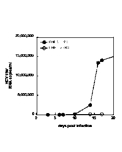
[00117] The cells for use in methods using serum depleted of LDL-receptor
binding
lipoproteins can be any suitable hepatocyte cell, such as primary hepatocytes,
an immortalized
CA 02883146 2015-02-25
WO 2014/033546 PCT/IB2013/002501
hepatocyte cell line (e.g,. an hHCC cells), and hHCC cells differentiated
according to the
methods disclosed herein to exhibit a phenotype of a primary human
hepatocyte). The present
method finds particular use in providing for infection of cultured hepatocyte
cells (e.g., primary
hepatocytes or immortalized hepatocyte cell lines, including hHCC cells
differentiated to exhibit
a phenotype of a primary human hepatocyte) with a clinical isolate of a
hepatitis virus, e.g., a
clinical isolate of HCV.
[00118] Serum depleted of LDL-receptor binding lipoproteins for use in the
culture
medium in these methods of the present disclosure can be prepared by any
suitable method
available in the art. Serum for use in the methods can be of human, bovine
(e.g., fetal bovine) or
any other suitable source. In one example, serum depleted of LDL-receptor
binding lipoproteins
is prepared by contacting the serum with a binding agent for lipoproteins that
bind the LDL
receptor, e.g., the human LDL receptor (e.g., an anti-LDL receptor binding
lipoprotein antibody
(e.g., a polyclonal or monoclonal antibody, an anti-VLDL antibody, an anti-LDL
antibody, an
anti-Apolipoprotein B antibody) or heparin) for a period of time sufficient to
provide for binding
to the binding agent, followed by recovering serum components that are not
bound by the
binding agent for use in the culture medium. By "serum depleted of LDL-
receptor binding
lipoproteins" is meant serum that is depleted of LDL-receptor binding
lipoproteins relative to
serum prior to treatment ("untreated serum"), and encompasses serum that is
depleted of LDL-
receptor binding lipoproteins by at least 20%, 30%, 40%, 50%, 60%, 70%, 80%,
90% or more
relative to serum prior to depletion. In one embodiment, the serum depleted of
LDL-receptor
binding lipoproteins is not substantially depleted of HDL (which does not
significantly bind the
LDL receptor) relative to the scrum prior to treatment.
[00119] In one embodiment, serum depleted of LDL-receptor binding
lipoproteins is
prepared by contacting the serum with heparin as a binding agent, where
contacting is for a
period of time sufficient to provide for binding of heparin-binding components
in serum (e.g.,
LDL, VDL) to heparin, followed by recovering serum components that are not
bound by heparin
for use in the culture medium.
[00120] Sera for use in the present methods includes serum depleted of at
least 25%, 30%,
40%, 50%, 60%, 70%, 80%, 90% or more of lipoproteins compared to serum prior
to depletion,
serum depleted of at least 25%, 30%, 40%, 50%, 60%, 70%, 80%, 90% of heparin-
binding
26
CA 02883146 2015-02-25
WO 2014/033546 PCT/IB2013/002501
lipoproteins compared to serum prior to depletion, and/or serum depleted of at
least 25%, 30%,
40%, 50%, 60%, 70%, 80%, 90% or more of ApoB compared to serum prior to
depletion.
[00121] Methods of the present disclosure using lipoprotein-depleted serum
(e.g., heparin-
treated serum) involve infecting cells (e.g., an hHCC cell, primary human
hepatocytes, or other
cells susceptible to infection by a hepatotrophic microorganism) with a
hepatotrophic
microorganism, where the cells are cultured in medium containing lipoprotein-
depleted serum.
Following infection, cells can be transitioned to any suitable culture medium
(e.g., HS-
containing medium for hHCC cells where differentiation is desired or
maintenance of
differentiated hHCC cells).
USES
[00122] The methods and compositions of the present disclosure can be used
in a variety
of ways such as, but not limited to, production of viral particles (e.g., as
in viral vaccine
production), study of hepatocyte function, and screening methods. Examples of
uses are
described below in more detail.
Viral Particle Production
[00123] The methods and compositions of the present disclosure can be used
in production
of viral particles, particularly viral particles of a hepatotrophic virus such
as a hepatitis virus,
e.g., hepatitis A, B, C, D or E virus (HAV, HBV,HCV, HDV, HEV), and the like,
cytomegalovirus (CMV). The virus can be on any genotype (e.g., HCV genotype 1
(e.g.,
genotype la, lb, lc), 2, 3, 4, 5, 6, and 7) and may be a naturally-occurring
virus (e.g., a virus
obtained from an infected primate, e.g., an infected human, often referred to
as a "clinical
isolate"), a tissue culture-adapted virus, a genetically modified virus, or
pseudotyped virus.
[00124] Virus particle-producing, differentiated hHCC cells of the present
disclosure can
be accomplished using any suitable method. For example, a viral genome can be
introduced into
cells by infection, electroporation, transfection, and the like. In some
embodiments, the hHCC
cells and/or the differentiated hHCC cell are not modified by recombinant
techniques so as to
provide a genomically integrated viral genome. In some embodiments, the hHCC
cells and/or the
differentiated hHCC cells are not genetically modified to include a
genomically integrated viral
genome operably linked to a promoter that is not native to the viral genome.
27
CA 02883146 2015-02-25
WO 2014/033546 PCT/IB2013/002501
[00125] Viral genomes can be introduced into hHCC cells prior to
differentiation in HS-
containing medium, at the time of transition from FBS-containing medium to HS-
containing
medium, during differentiation by culturing in HS-containing medium (e.g., at
1, 2, 3, 4, 5, or 6
days after transition to HS-containing medium), or after growth arrest and/or
differentiation to a
phenotype of a primary human hepatocyte (e.g., at 7, 8, 9, 10, 11, 12, 13, 14
days or more after
transition to HS-containing medium).
[00126] Where viral genomes are to be introduced by infection, infection
can be
conducted in culture medium containing lipoprotein-depleted (e.g., heparin-
treated) scrum (e.g.,
human serum, fetal bovine serum) as described above. Optionally, following
infection cells can
be transitioned to HS-containing medium, e.g., to provide for differentiation
of hHCC cells
and/or to facilitate cell viability. In one embodiment, undifferentiated or
differentiated hHCC
cells are cultured in medium containing lipoprotein-depleted serum for a
period of time to allow
for infection of viral particles added to the cells. Following the infection
period, the culture
medium is replaced with HS-containing medium and the cells cultured for a
period of time to
provide for differentiation of hHCC cells, continue differentiation of hHCC
cells, and/or provide
for maintenance of differentiated hHCC cells and provide for viral particle
production.
[00127] The present disclosure provides cultures of differentiated hHCC
cells comprising
at least 105, at least 106, at least 107 at least 108, or at least 109 or more
viral particles per
milliliter of culture medium. The present disclosure further provides such
viral particle-
producing cultures that can be maintained for at least 1 week, 2 weeks, 3
weeks, 4 weeks, 5
weeks, 6 weeks, 7 weeks, or 8 weeks or more.
[00128] Viral particles produced from hHCC cells cultured in HS exhibit
structural
features that distinguish such from viral particles produced in hHCC cells
cultured in non-human
serum (e.g., in FBS). For example, when produced in differentiated hHCC cells
cultured in HS,
viral particles have an average lower density than when produced from the hHCC
cells cultured
in FBS. For example, when HCV particles are produced in FBS-cultured hHCC
cells, about 75%
of the viral particles have a density greater than about 1.16 g/ml. In
contrast, when HCV particles
are produced in HS-cultured hHCC cells, the HCV particles are of a lower
average density, with
only about 25% of the HCV particles in the population having a density of
greater than 1.16g/ml.
In addition, HCV particles produced from HS-cultured hHCC cells are associated
with ApoB,
with usually at least about 50%, at least about 60%, at least about 70%, at
least about 80%, or at
28
CA 02883146 2015-02-25
WO 2014/033546 PCT/IB2013/002501
least about 90% or more of the HCV particles in the viral particle population
produced being
ApoB-associated.
[00129] Viral particles produced from the differentiated hHCC cells can be
used in a
variety of ways. For example, the viral particles produced from the
differentiated hHCC cells can
be isolated from the cell culture medium, and formulated so as to be suitable
for administration
as a vaccine. The viral particles can be used in research settings, e.g., for
infection of animal
models, such as a human chimeric mouse model. The differentiated hHCC cell
populations also
provide a research tool for use in study of viruses, especially hcpatotrophic
viruses.
Assays
[00130] The differentiated hHCCs of the present disclosure find use in a
variety of assays.
For example, the differentiated hHCCs can serve as a model of human
hepatocytes, and thus a
model of liver function and disease (for example, carcinogenesis, steatosis
(fatty liver disease)).
Other uses include studies of lipoprotein metabolism and secretion, and
metabolic studies, both
of biological intermediates as well as xenobiotic compounds (e.g., oxidation
by cytochrome
P450).
[00131] Assays using the differentiated hHCCs of the present disclosure can
involve, for
example, assessing the effect of an agent on cells having a phenotype of a
primary human
hepatocyte, e.g., to assess the effect of an agent on human hepatocyte
function (e.g., to assess
liver toxicity of an agent); to assess metabolism of an agent by a human
hepatocyte; study
mechanisms involved in hepatocyte function; and the like. The differentiated
hHCC cells can be
used to screen for agents (such as solvents, small molecule drugs, peptides,
polynucleotides)
and/or environmental conditions (such as culture conditions or manipulation)
that affect the
characteristics of cells having a phenotype of a human primary hepatocyte.
[00132] Assessment of activity of an agent (e.g., a candidate agent)
generally involves
contacting a differentiated hHCC cells with the agent, either alone or in
combination with other
agents (e.g., drugs having a known activity). After incubation for a
sufficient amount of time, an
appropriate assay is conducted to detect the effect, if any, of the candidate
agent on the
differentiated hHCC cells (e.g., by assessing a change in cell morphology,
cell function, change
in a marker of a phenotype of a human primary hepatocyte). The presence or
absence of an effect
of the agent on the phenotype is detected and analyzed, e.g., by comparing to
a control (e.g., by
29
CA 02883146 2015-02-25
WO 2014/033546 PCT/IB2013/002501
comparison to the phenotype in the absence of the agent and/or by comparison
to the phenotype
in the presence of an agent having a known effect on a liver cell).
[00133] For example, where the assay is to assess an effect of a candidate
agent on
lipoprotein metabolism, the assay can involve detecting a change in a
phenotypic marker, e.g., a
morphological change (e.g., the presence or absence of a change in lipid
organization in the
differentiated hHCC cell (e.g., as indicated by the presence or absence of
lipid droplets and/or a
change in the pattern of such lipid droplets)) or a change in a biomarker,
such as a lipoprotein or
lipid (e.g., a change in a level of a lipoprotein or a lipid), in the presence
of the candidate agent
compared to the absence of the candidate agent. A change in the phenotypic
marker in the
presence of the candidate agent as compared to the absence of the agent
indicates the candidate
agent has an effect on lipoprotein metabolism.
[00134] In one aspect, provided herein is a method for assessing the effect
of a candidate
agent on lipoprotein secretion of any of the differentiated hHCC cells
provided herein. As
discussed in the Examples below, human hepatocellular carcinoma (hHCC) cell
line cultured in a
culture medium comprising human serum for at least 3-5 days or more secrete
lipoprotein (e.g.,
VLDL, LDLs, HDLs). At 14 days or more of culturing in culture medium
comprising human
serum, hHCC cells secrete lipoproteins at levels that closely resemble those
secreted by primary
human hepatocytes in culture, and also closely resemble lipoprotein levels
found in human
serum. Thus, the secreted lipoprotein profile of hHCC cells cultured in human
serum is similar to
that of the profile of lipoproteins secreted by primary human hepatocytes and
to that of the
lipoprotein profile found in human serum. As such, a change in the levels of
lipoprotein secretion
in the presence of a candidate agent as compared to the absence of the agent
indicates the
candidate agent has an effect on lipoprotein secretion.
[00135] In certain embodiments, the method includes the steps of incubating
a cell culture
comprising a human hepatocellular carcinoma (hHCC) cell line in HS-containing
media for more
than 14 days. The hHCC cell line can be cultured in any HS-containing media
described herein
for 3 days or more. In certain embodiments the human hepatocellular carcinoma
(hHCC) cell
line is cultured in the HS-containing media for 3, 4, 5, 6, 7, 8, 9, 10,11,
12, 13, 14, 15, 16, 17, 18,
19, 20, 21, 22, 23, 24, 25, 26, 27, 28, 29, or 30 or more days prior to
contact with the candidate
agent.
CA 02883146 2015-02-25
WO 2014/033546 PCT/IB2013/002501
[00136] After culturing the hHCC cell line for a selected time period
(e.g., to provide for a
desired secreted lipoprotein profile), the cell line is then contacted with
the candidate agent.
Candidate agents include, but are not limited to, synthetic, naturally
occurring, or recombinantly
produced molecules (e.g., small molecule; drugs; polynucleotides;
polypeptides; peptides;
antibodies; endogenous factors present in eukaryotic or prokaryotic cells
(e.g., polypeptides,
plant extracts, and the like)); etc.). Candidate agents include agents of
numerous chemical
classes, though typically they are organic molecules, preferably small organic
compounds having
a molecular weight of more than 50 and less than about 2,500 daltons.
Candidate agents include
biomolecules such as, but not limited to: antibodies, peptides, saccharides,
fatty acids, steroids,
purines, pyrimidines, derivatives, structural analogs, nucleic acid inhibitors
or combinations
thereof.
[00137] The method can involve administering varying amounts of the
candidate agent
(from no agent to an amount of agent that approaches an upper limit of the
amount that can be
delivered within toxicity limits for cultured cells), and may include delivery
of the agent in
different formulations. The agents can be administered singly or can be
combined in
combinations of two or more, e.g., where the effect of a candidate combination
drug therapy is to
be assessed.
[00138] After incubation for a sufficient amount of time with the candidate
agent, an
appropriate assay is conducted to detect the effect, if any, of the candidate
agent on the
differentiated hHCC cells to secrete lipoproteins. In certain embodiments, the
method includes
the step of assessing the level of lipoproteins (e.g., VLDL, LDLs, and/or
HDLs) in the HS-
containing media. Levels of lipoproteins that are secreted by the hHCC cells
can be assessed by
any suitable method. For example, the levels of secreted lipoproteins can be
assessed by
assaying the media collected from the cultured hHCC cells to obtain a
lipoprotein profile (a
measurement of HDL, LDL, and/or VLDL cholesterol and triglyceride levels).
Lipoprotein
profiles can be obtained, for example, using ultra-centrifugation or by liquid
chromatography
techniques. See, e.g., Brousseau et al. (1993) Clinical Chemistry 39(6): 960-
964. In certain
embodiments, the lipoprotein profile is obtained by isolating the media used
to culture the hHCC
cells and separating the lipoproteins from the media using size exclusion fast
protein liquid
chromatography. Lipid content (e.g., cholesterol or triacylglycerol) can be
subsequently
31
CA 02883146 2015-02-25
WO 2014/033546 PCT/IB2013/002501
measured by any suitable lipid detection method, including fluorescence-based
lipid detection
methods. See, e.g., Yang et al. (2012) Chem Phys Lipids 165(2): 133-41.
[00139] The presence or absence of an effect of the candidate agent on
lipoprotein
secretion is determined by comparison to a control sample (e.g., a control
lipoprotein profile
obtained from media of hHCC cells not contacted with the agent). In such
embodiments, a
difference in the lipoprotein levels in the presence of a candidate agent as
compared to the
control sample indicates the candidate agent has an effect on lipoprotein
secretion. Any suitable
sample can serve as a control sample for comparison to the levels of
lipoprotein obtained from
the hHCC cell line contacted with the candidate agent. In specific
embodiments, the control
sample is a lipoprotein profile obtained from a human hepatocellular carcinoma
(hHCC) cell line
cultured in the HS-containing media under the same conditions as the sample
that is tested and in
the absence of the candidate agent. In other embodiments, a comparison is made
to a lipoprotein
profile from a sample contacted with an agent having a known effect on
hepatocyte lipoprotein
secretion.
[00140] Such methods can be useful, for example, in screening for
therapeutic agents for
use in the treatment of lipoprotein mediated diseases and conditions,
including, but not limited to
dyslipidemia, hypolipidemia, hyperlipidemia, hypolipoproteinemia,
hyperlipoproteinenemia,
tangiers disease, hyperalphalipoproteinemia, hypoalphalipoproteinemia,
coronary heart disease
(CHD), cerebrovascular disease (CVD), atherosclerosis, thrombosis, and stroke.
In specific
embodiments, the method is for the prevention or treatment of atherosclerosis.
In such
embodiments, a decrease in VLDL or LDL or an increase in HDL in the culture
medium as
compared to the control sample is indicative that the candidate agent can be
used for the
prevention or treatment of atherosclerosis.
[00141] Where the assays is to assess metabolism of a drug, the method
generally involves
contacting a culture of differentiated hHCC cells having a phenotype of a
human primary
hepatocyte with a drug for a period of time sufficient for production of
metabolites of the drug (if
any), and assaying for metabolites of the drug (e.g., in the culture
supernatant). Drug metabolites
can be detected using methods available in the art. The metabolite can be
subjected to further
analysis, e.g., to identify the structure of the metabolite.
[00142] Where the assay is to assess toxicity of an agent on human
hepatocytes, the
method generally involves contacting a culture of differentiated hHCC cells
having a phenotype
32
CA 02883146 2015-02-25
WO 2014/033546 PCT/IB2013/002501
of a human primary hepatocyte with an agent with an agent, and detecting a
change in a
phenotypic maker (e.g., a morphological change and/or a change in a biomarker
indicative of
toxicity, e.g., a change in a level of transaminase). Detection of a change in
the presence of the
agent as compared to the absence of the agent (e.g., an increase in
transaminase in the cell
medium in the presence of the agent and/or an increase in an phenotypic marker
of cell death
(e.g., a phenotypic marker of apoptotic cell death, necrotic cell death (e.g.,
TUNEL, caspase)) is
an indicator of toxicity of the agent for the differentiated hHCC cells).
[00143] Where the differentiated hHCCs are cultured with a hcpatotrophic
microorganism
(e.g., a hepatotrophic virus such as HCV, HBV), the assays can involve study
of entry,
replication, and, in the context of viruses, viral particle production. The
assays can also provide
for screening for anti-hepatotrophic pathogen agents, e.g., antiviral agents.
[00144] For example, where the assay is to screen for antiviral agents, the
assay can
generally involve incubating the differentiated hHCC cells in the presence of
a viral genome with
at least one test candidate agent, and detecting the presence or absence of an
effect on the virus.
Such can be accomplished by, for example, assaying viral replication. Assaying
viral replication
can be accomplished by, e.g., assessing levels of viral particles (e.g., viral
titers), detecting viral
nucleic acid, and the like. For example, where the differentiated hHCC cells
are infected with
HCV, levels of HCV may be assessed by measuring viral titers, measuring HCV
RNA
quantitatively, measuring HCV viral particles (e.g., by detection of core
proteins, e.g., by
immunoassay), by microscopy, and the like. Inhibition of virus production
viral replication can
be due to, for example, inhibition of viral replication at the nucleic acid
level, inhibition of viral
particle production, and/or inhibition or viral entry into cells. A decrease
in viral replication in
the presence of the candidate agent as compared to the absence of the
candidate agent indicates
the candidate agent has antiviral activity.
[00145] Screening methods for antiviral agents can be adapted to screen for
antiviral
activity of an antibody, e.g., to assay for antibodies that neutralize virus
by, for example,
inhibition viral infectivity of a differentiated hHCC. Such antibodies may be
a polyelonal or
monoclonal antibody. In on embodiment, the antibodies to be screened are
present in a biological
samples obtained from a subject exposed to a virus (e.g., HCV) or to whom a
vaccine (e.g.,
candidate vaccine) has been administered. The biological sample can be, for
example, blood or a
fraction thereof, e.g., suspected of containing antiviral antibodies. In one
embodiment, the
33
CA 02883146 2015-02-25
WO 2014/033546 PCT/IB2013/002501
differentiated hHCC cells are infected with a clinical viral isolate. The
assay can involve
comparing viral replication in the presence of the antibody or sample of
suspected of containing
the antibody, where a decrease in viral replication in the presence of the
antibody or the sample
is an indicator of antiviral activity. Where the biological sample is from a
subject post-
immunization, the antiviral activity of the post-immunization sample may be
compared to
antiviral activity of a pre-immunization biological sample (e.g., from the
same subject).
[00146] Any of a variety of candidate agents can be screened. "Candidate
agents" is meant
to include synthetic, naturally occurring, or recombinantly produced molecules
(e.g., small
molecule; drugs; polynucleotides; polypeptides; peptides; antibodies;
endogenous factors present
in eukaryotic or prokaryotic cells (e.g., polypepti des, plant extracts, and
the like)); etc.).
Candidate agents encompass numerous chemical classes, though typically they
are organic
molecules, preferably small organic compounds having a molecular weight of
more than 50 and
less than about 2,500 daltons. Candidate agents can also include biomolecules
such as, but not
limited to: antibodies, peptides, saccharides, fatty acids, steroids, purines,
pyrimidines,
derivatives, structural analogs or combinations thereof.
[00147] Candidate agents can be obtained from a wide variety of sources
including
libraries of synthetic or natural compounds. For example, numerous methods are
available for
random and directed synthesis of a wide variety of organic compounds and
biomolecules,
including expression of randomized oligonucleotides and oligopeptides.
Libraries of natural
compounds in the form of bacterial, fungal, plant and animal extracts are
available or readily
produced. Libraries of antibodies can be produced by methods available in the
art. Additionally,
natural or synthetically produced libraries and compounds are readily modified
through
conventional chemical, physical and biochemical means, and may be used to
produce
combinatorial libraries. Known pharmacological agents may be subjected to
directed or random
chemical modifications, such as acylation, alkylation, esterification,
amidification, etc. to
produce structural analogs.
[00148] The assays method can involve administering varying amounts of the
candidate
agent (from no agent to an amount of agent that approaches an upper limit of
the amount that can
be delivered within toxicity limits for cultured cells), and may include
delivery of the agent in
different formulations. The agents can be administered singly or can be
combined in
34
CA 02883146 2015-02-25
WO 2014/033546 PCT/1B2013/002501
combinations of two or more, e.g., where the effect of a candidate combination
drug therapy is to
be assessed.
[00149] It will be appreciated that the assay can be conducted in a variety
of formats, and
that the order of contacting the differentiated hHCC cells with the virus and
the candidate agent
can be varied. For example, the differentiated hHCC cells can be infected
virus prior to
contacting with the candidate agent; alternatively, the differentiated hHCC
cells can be contacted
with the candidate agent prior to exposure to infectious viral particles.
[00150] The agent (e.g., candidate agent, drug, and/or microorganism (e.g.,
virus)) can be
added to the culture medium at the time of transition of hHCC cells to HS-
containing medium,
during differentiation of hHCC cells, or after differentiation of hHCC cells
into a phenotype of a
human primary hepatocyte.
KITS
[00151] Kits of the present disclosure can include hHCC cells,
differentiated hHCC cells
having a phenotype of a human primary hepatocyte, or both. The kit can
optionally include
culture medium components, e.g., culture media (e.g., without serum or
containing human
serum), human serum for use in the culture, and the like). For example, the
kit can include
primary hepatocyte medium (such as described in detail in the Examples below),
with or without
human serum. The various components of the kit may be present in separate
containers or certain
compatible components may be pre-combined into a single container, as desired.
[00152] Kits can include instructions for using the components of the kit
to practice a
method of the present disclosure. The instructions are generally recorded on a
suitable recording
medium, such as paper, plastic, electronic storage, medium, and the like. For
example, the
instructions may be present in the kits as a package insert, in the labeling
of the container of the
kit or components thereof (e.g., associated with the packaging), etc. In other
embodiments, the
instructions are present as an electronic storage data file present on a
suitable computer readable
storage medium, e.g. compact disc-read only memory (CD-ROM), digital versatile
disk (DVD),
diskette, etc. In other examples, the instructions provided do not contain
many or all assay
details, but rather provide direction as to a remote source for obtaining
detailed instructions, e.g.
via the intemet.
CA 02883146 2015-02-25
WO 2014/033546 PCT/1B2013/002501
EXAMPLES
[00153] The following examples are put forth so as to provide those of
ordinary skill in
the art with a complete disclosure and description of how to make and use the
present invention,
and are not intended to limit the scope of what the inventors regard as their
invention nor are
they intended to represent that the experiments below are all or the only
experiments performed.
Efforts have been made to ensure accuracy with respect to numbers used (e.g.
amounts,
temperature, etc.) but some experimental errors and deviations should be
accounted for. Unless
indicated otherwise, parts are parts by weight, molecular weight is weight
average molecular
weight, temperature is in degrees Celsius, and pressure is at or near
atmospheric.
MATERIALS AND METHODS
[00154] The following methods and materials are used in the Examples below.
[00155] Standard culture conditions (for cell proliferation prior to
culturing in human
serum). HuH-7 (JCRB403) cells are available from the Japanese Collection of
Research
Resources ¨ Cell bank (JCRB Cell bank). Huh7.5 cells were a kind gift of Dr.
C. Rice. Both cell
line were maintained (prior to culturing in human serum) according to the
protocols described. In
short, Huh7.5 or HuH-7 cells were maintained in culture medium containing
DMEM/10% fetal
bovine serum (FBS) /penicillini streptomycin with trypsinization and splitting
every 3 to 4 days.
After approximately 20-30 passages, cells were discarded, and a new vial was
thawed.
[00156] Since the use of human serum results in growth arrest of cells,
cell cultures were
normally maintained FBS containing media (DMEM/10% FBS /penicillin/
streptomycin, as
described above).
[00157] Infection of cells with JFH-1 virus. JFH-1 virus (JFH) was obtained
from Dr. T.
Wakita. Two days prior to infection, cells were replated at 30% density. After
infection for 4
hours, cells were washed and cultured in either FBS or HS containing as
appropriate..
[00158] Infection of chimeric mice. Chimeric SCID/uPA mice transplanted
with human
hepatocytes (see, e.g., WO 01/67854) were infected as described previously
(Steenbergen et al.
(2010) Am J Physiol Gastrointest Liver Physiol 299: G844-854; Mercer et al.
(Aug 2001) Nat.
Med. 7(8):927-33. Infection was accomplished using 100 I of tissue culture
supernatant from
JFH-infected cells or serum from HCV-infected patients. HCV titers and human
albumin (hAlb)
36
WO 2014/033546 PCT/1112013/002501
in the serum of the mouse model chimeric mice was determined by quantitative
PCR and ELISA
respectively.
1001591 Visualization and ouantitation of lipid droplets by
immunofluorescence. Cells
were grown on coverslips and either cultured in FBS or HS. Cells were stained
with
Bodipy 493/503 (Invitrogen) according to the supplier's instructions in order
to visualize lipid
droplets. The quantity of neutral lipid staining was visualized using a
fluorescence microscope.
Images of the Bodipy 493/503 staining under different cell culture conditions
were taken using
identical microscope and exposure settings. The amount of fluorescence was
quantitated using
ImageJ software (National Institutes of Health). Data were collected in 3
independent
experiments, with 4-8 microscopic fields measured per condition. Background-
and auto-
fluorescence was negligible.
[00160] Distribution and shape of lipid droplets were examined in
separately stained
samples under identical microscopy settings, but with optimized exposure for
each individual
condition.
[00161] Sucrose gradients/density centrifugation. Sucrose density-
gradient
ultracentrifugation analysis was performed as previously described (Zhong et
al. (2005) Proc
Natl Acad Sci U S A 102: 9294-9299). Supernatants from HCV-infected cells were
centrifuged
at 300g for 5 min to remove cellular debris in 1 ml of TNE buffer (50 mM Tris
HC1, pH 8; 100
mM NaCl; 1 mM EDTA) containing protease inhibitors (RocheApplied Science,
Indianapolis),
loaded onto a 20-60% sucrose gradient (12.5-ml total volume), and centrifuged
at 120,000 g for
16 h at 4 C in a SW41Ti rotor (Beckman). Fractions of 0.5 ml each were
collected from the top
of the gradient, and the titer in each fraction was determined by quantitative
RT-PCR as
described below. The density of each fraction was determined by determining
weight of a known
volume.
[00162] Immunoprecipitation of ApoB containing particles.
Immunoprecipitation
experiments were essentially performed as previously described (Steenbergen et
al. (2010) Am J
Physiol Gastrointest Liver Physiol 299: G844-854). Serum or cell culture
supernatant samples
(50 p.1) with titers of at least 105 RNA copies/ml were incubated overnight
with 7.5 I anti ApoB
antibody (Chemicon AB742, goat anti human apolipoprotein B; cross-reacts with
human, mouse
and bovine ApoB), at 4 C while rotating. Protein G slurry (20 I, GE
healthcare, Protein
SepharoseTM 4 fast flow) was added to each sample and incubated for a minimum
of 1 hour on a
37
CA 2883146 2020-03-23
CA 02883146 2015-02-25
WO 2014/033546 PCT/IB2013/002501
rotator. The Protein G complexes were precipitated at 14000g in a microfuge.
The pellets were
washed once with PBS and viral RNA was isolated directly from the pellets
(QiaAmp Viral
RNA kit, Qiagen) and analyzed by quantitative RT-PCR. Samples containing high
amounts of
immunoglobulins (like patient serum) were pre-cleared with protein G beads,
prior to
immunoprecipitation, to ensure quantitative immunoprecipitation of the ApoB
complexes. To
ensure that the Protein G beads did not bind HCV or HCV complexes directly, we
incubated
HCV containing mouse serum with Protein G Sepharose beads in the absence of
anti-ApoB
antibodies. The HCV titers in the precipitate of these samples were
negligible.
[00163] Quantitative RT-PCR of hepatocyte markers. RNA was isolated from
cells using
Trizol, according to the instructions of the manufacturer. cDNA was produced
from the RNA
using Quantitect Reverse Transcription kit (Qiagen). Gene specific primer-
probe sets were
designed by Applied Biosystems. An Applied Biosystems 7900HT Fast Real-Time
PCR system
was used for the quantitation of gene products. Gene expression was calculated
relative to HPRT
according to Pfaffl (Pfaffl MW. Nucleic Acids Res 29: e45, 2001).
[00164] Primary hepatocyte cultures: Frozen human primary hepatocytes were
either
purchased from Invitrogen, or isolated in-house as described previously
(Mercer et al, 2001 Nat
Med. 7 927-933). Isolated cells were pretreated with HypoThermosol HTS-Purge
(BioLife
Solutions), resuspended in Cryostor CS10 (BioLife Solutions) and cooled in a
controlled rate
freezer at the rate of 1 C per minute until -40 C. Cells were then stored in
liquid nitrogen. Cells
were thawed and resuspended in pre-warmed primary hepatocyte medium (DMEM, 1.2
ug/m1
insulin, 11 uM hydrocortisone hemisuccinate, 15 mM Hepes,
penicillin/streptomycin, 200 mmol
glucose, 2% human serum) and plated on Collagen type I coated plates
(Millicoat 6 well plates,
Millipore at a density of 1 million cells per well. After 12-18 hrs the tissue
culture media was
refreshed to remove unattached cells. Cells were used within 3 days after
plating.
[00165] FPLC analysis of secreted lipoproteins: Size-exclusion fast-protein
liquid
chromatography (FPLC) was used to separate lipoprotein particles secreted by
cultured Huh7.5
cells into media. In this system, the largest particles eluted first from the
column (VLDL)
followed by LDL and HDL sized particles. After separation, triacylglycerol
(TG, also referred to
as tryglyceride) or cholesterol content was determined in-line.
[00166] To prepare samples, cells were washed extensively with serum free
OptiMEM
(Gibco/ Invitrogen) to remove lipoproteins present in the serum containing
media. The last wash
38
WO 2014/033546 PCT/1B2013/002501
was collected and served as a baseline measurement. Cells were then placed in
serum free
OptiMEM overnight. The following day, media was collected and cellular debris
was removed
by centrifugation (300g, 10 minutes). Media was then concentrated using a
centrifugal filter unit
(Amicon Ultra-100, Millipore). Concentrated media (65 IA) was injected into an
Agilent 1200
HPLC instrument equipped with a Superose 6 10/300 FPLC column. In-line assays
for total
cholesterol and TG (Infinity Cholesterol and Triglyceride reagents, Thermo
Scientific) were
performed at 37 C using a post-column reaction. Reaction products were
monitored in real-time
at 500 nm and analyzed using Agilent Chemstation software. All lipoproteins
contain both TG
and cholesterol. However, VLDL, is rich in TG and has little cholesterol,
whereas HDL is rich in
cholesterol and has little TG. Therefore, cholesterol profiles are more
suitable to detect
differences in secreted HDL levels, whereas TG profiles are more suitable to
analyze VLDL
levels.
EXAMPLE 1: GROWTH AND APPEARANCE OF HUH7.5 CELLS CULTURED IN HUMAN SERUM
[00167] Figure 1 provides a flowchart showing an example of the steps
used in culturing
of cells in human serum (HS) to provide for differentiated cells. Huh7.5 cells
previously
maintained in FBS-containing culture medium were trypsinized to facilitate
dissociation of
monolayers. Trypsin was inactivated with DMEM/10% FBS. Cells were then
centrifuged at
300g, and cell pellets were resuspended in DMEM/2% HS/ penicillin/
streptomycin and plated at
a density of 30-50%. At confluency (typically after 2 days of incubation)
cells were trypsinized
once more, and then plated at a density of 50%. From this point on, cells were
cultured without
further splitting and allowed to form confluent layers of undividing cells.
Optionally, cells
cultured in human serum can be divided for up to approximately 1 week,
provided they are not
divided more than 1:2. However, repeated trypsinization after 10 days of
culturing in human
serum resulted in loss of viability of the cell cultures, with death of 80-90%
or more of cells.
[00168] Huh7.5 cells grown in media supplemented with human serum (HS),
instead of
fetal bovine serum (FBS), underwent a series of morphological changes, and
were maintained as
confluent layers for over two months, without the further need for splitting.
HS-cultured cells
became organized in a pavement like structure, formed tightly packed
monolayers that were very
strongly attached to the tissue culture substrate. After approximately one
week, cell division
slowed down in cells cultured in HS-containing media, and eventually underwent
contact
39
CA 2883146 2020-03-23
CA 02883146 2015-02-25
WO 2014/033546 PCT/IB2013/002501
inhibition and became growth arrested. Similar to primary hepatocytes in
culture (right panel),
HS-cultured cells were mono-or binucleated and had a granular appearance
(Figure 2A, middle
panel) as compared to FBS-cultured cells (Figure 2A, left panel). After
approximately 14-18
days in HS cell size increased significantly.
[00169] To examine the apparent growth arrest effect of culturing in HS,
cell numbers
where counted in a three-week time span after initial plating in HS-cultured
cells and FBS-
cultured cells. As shown in Figure 2B, growth of cells cultured in HS-
containing medium was
significantly slowed as compared to growth in FBS-containing medium.
[00170] With regular media changes, cells cultured in HS-containing medium
could be
maintained without splitting and replating for at least 2 months.
EXAMPLE 2: CULTURING OF HuH7.5 CELLS IN HS PROMOTES DIFFERENTIATION To A
PRIMARY HEPATOCYTE-LIKE PHENOTYPE
[00171] To further evaluate the effect of culturing cells in HS-containing
medium, and to
assess whether these cells underwent differentiation to a primary hepatocyte-
like cell, level of
expression of hepatocyte differentiation marker were assayed at 7 days and 21
days after transfer
of Huh7.5 cells into HS-supplemented medium. After 7 days only minor changes
in hepatocyte
markers were observed. (Figure 3A) However, after 21 days, the expression of
the hepatic
differentiation markers albumin and alphal-antiTrypsin was increased
approximately 4 fold in
cells grown in human serum compared to FBS. The level of expression of these
proteins is
similar to the level of expression of human primary hepatocytes in culture.
Culturing the Huh7.5
cells in primary hepatocyte medium (PHM) did not have a significant additional
effect on the
expression of albumin or alphal-antitrypsin.Changes in LDL-receptor (LDL-R)
were not
significant in cells grown in HS, however, when Huh7.5 cells were grown in
primary hepatocyte
medium (PHM), LDL-R expression was increased.
[00172] Expression of claudin-1 and occludin, tight junction components,
was also
increased in cells grown in human serum after 21 days, and comparable to
expression in human
primary hepatocytes in culture (Figure 3A). High levels of tight junctions are
present in liver
tissue. Additionally, through immobilization of cells, these proteins are also
known as potent
tumor suppressors, and indicate the transition of a tumorigenic state to a
more differentiated state
of cells. Claudin-1 and occludin also function as entry receptors for HCV.
CA 02883146 2015-02-25
WO 2014/033546 PCT/IB2013/002501
[00173] Expression of key lipid metabolism regulators was increased in
cells that were
grown in HS, compared to cells grown in FBS (Figure 3B). LXRa (Liver X
Receptor alpha) is
highly expressed in liver, and controls transcriptional programs involved in
lipid homeostasis
and inflammation. Peroxisome proliferator-activated receptors (PPARs) are key
regulators of
differentiation and metabolism. PPARa is a major regulator of lipid metabolism
in the liver.
Activation of PPARa promotes uptake and utilization of fatty acids by
upregulation of genes
involved in fatty acid transport and fatty acid oxidation. PPARy regulates
fatty acid uptake and
storage as well as glucose metabolism. Cytoskeleton proteins vimentin and e-
cadherin are also
increased in cells cultured in human serum. Vimentin, is essential for
maintaining shape and
keeping organelles in place, it also controls the transport of low density
lipoprotein inside the
cell. E-Cadherin play an important role in cell to cell adhesion, and is
therefore, like tight
junction proteins, indicative of a non-proliferative/ non-tumorigenic state of
the cell.
EXAMPLE 3: EXAMINATION OF OTHER CULTURE CONDITIONS
[00174] In order to compare the effects of culturing of HuH-7 or HuH-7-
derived cells in
human serum, other culture conditions were examined.
[00175] DMSO, (dimethyl sulfoxidc) has been reported to induce growth
arrest in HuH-7
or ¨derived cells, and other hcpatoma cells. (Sainz et al. (2006) J Virol 80:
10253-10257). In
order to assess the effect of DMSO, the level of expression of hepatocyte-
specific genes in
Huh7.5 cells grown for 3 weeks in the presence of 1% or 2% DMSO in DMEM (DMEM,
10% FBS, 1% or 2% DMSO, penicillin/streptomycin) was examined. Additionally,
Huh7.5 cells
were cultured in 2-10% adult bovine serum (ABS, Invitrogen) in DMEM in lieu of
fetal bovine
serum (DMEM, 2% ABS, penicillin/streptomycin.
[00176] Both culture conditions resulted in growth arrest, within
approximately 7 days.
Cells cultured in medium containing DMSO or ABS did undergo some morphological
changes,
but never to the extent of cells cultured in human serum. In either culture, a
punctate pattern in
the cytoplasm indicative of a primary human hepatocyte phenotype was not
typically observed
(data not shown).
[00177] The differentiation status of cells cultured in ABS or DMSO was
examined by
assessing the level of expression of the same hepatocyte differentiation
markers as described in
above (Figure 4). Whereas culturing in 2% DMSO resulted in a slight increase
in Albumin
41
CA 02883146 2015-02-25
WO 2014/033546 PCT/IB2013/002501
expression, and ABS resulted in a minor increase in expression of tight
junction proteins claudin-
1 and occludin, none of the proteins were expressed at a level comparable to
those in primary
human hepatocytes in culture or at a level comparable to expression in Huh7.5
cells cultured in
human serum. The level of expression of other biomarkers tested was not
significantly increased
relative to culturing in medium containing FBS.
EXAMPLE 4: EFFECT OF CULTURE MEDIUM ON APPEARANCE OF CELLULAR LIPID DROPLETS
[00178] The appearance of cellular lipid droplets following culturing of
Huh7.5 cells in
FBS versus HS was examined. As shown in Figures 5A and 5B, Bodipy fluorescence
indicative
of lipids was much more intense in cells that are cultured in HS, compared to
FBS. When
quantitated using ImageJ software, Bodipy fluorescence intensity was
approximately 4 times
higher in cell cultures in HS than in cells cultured in FBS (Figure 5B).
[00179] Additionally, the distribution of lipid droplets is affected by the
different culturing
conditions. In FBS medium, lipid droplet size in cells is heterogeneous. In HS
medium, lipid
droplets in cells are generally smaller than in FBS-cultured cells, and are
much more uniform in
size.
EXAMPLE 5: PRODUCTION OF VIRAL TITERS OF JFH-1
[00180] JFH-1 was electroporated into FBS-cultured cells. The
electroporated cells were
then maintained in FBS or transferred to HS-containing media, immediately
after
electroporation, according to Example 1. At 4-6 days after electroporation,
virus was harvested
by collection of culture supernatants.
[00181] Virus produced by electroporation of cells maintained in FBS media
is referred to
as "JFH-FBS"; virus produced by electroporation of cells which were
transferred to and
maintained in HS medium immediately after electroporation, is referred to as
"JFH-HS".
[00182] Figure 6A shows infection of cells cultured in FBS with the two
different viruses.
When FBS maintained cells were infected with JFH-FBS that was isolated 4 days
after
electroporation (like JFH-HS), infection was not detected in these cells,
because the RNA titers
of these viral stocks were too low. In contrast, JFH-HS immediately resulted
in high viral titers
in FBS grown cells. Viral titers quickly reached a plateau. For comparison,
JFH-FBS that was
42
CA 02883146 2015-02-25
WO 2014/033546 PCT/IB2013/002501
isolated 15 days after electroporation was used (referred to as JFH-FBS(15).)
Viral titers of JFH-
FBS(15) remained low throughout the infection course compared to JFH-HS.
[00183] Figure 6B shows viral titers of cells that were grown under the
different
conditions (FBS and HS), but that were infected with the same virus (JFH-HS).
Notably, during
the first 10 to 11 days of culture in HS, the benefit of using HS for viral
production was not
apparent. However, after about 11 days viral titers began to increase such
that at about 14 days
post transfer of cells to HS media, viral production rapidly increased in HS
maintained cells.
This increase in viral production was not observed in FBS-cultured cells. This
timing
corresponds to the approximate time at which HS-cultured cells exhibited a
phenotype
differentiated toward a phenotype resembling that of a primary human
hepatocyte. Similar
infections were performed in HuH-7 cells. Trends observed in HuH-7 cells (the
fold increase was
similar) were similar to those observed in Huh7.5 cells.
[00184] Figure 6C shows that when cells grown in HS are infected with JFH-
HS, viral
production (HCV RNA copies per ml) increases about 1,000 fold, compared to the
standard
tissue culture conditions using JFH-FBS in FBS-cultured cells. Culturing of
cells in HS-
containing media allowed production of continuous viral titers approaching 108
RNA copies/m1
or more for at least 45 days, with changes to fresh HS-containing media every
3-4 days (Figure
6D). When cells were first differentiated to the primary hepatocyte-like
phenotype (by culturing
in HS-containing media for about 14 days), and then infected with JFH-HS,
similar viral titers
were accomplished (Figure 6C).
[00185] Figure 6E show the effect of using primary hepatocyte medium on
viral titers.
Cells that were infected, and first differentiated using HS, were transferred
to primary hepatocyte
medium (DMEM, 1.2 ,t.g/m1 insulin, 11 i.tM hydrocortisone hemisuccinate, 15 mM
Hepes,
penIstrep, 200 mmol glucose, 2% human serum) and the effect on viral
production was assessed.
Within 3 day after transfer, the morphological changes seen as a result of
culturing in HS became
more distinct (not shown). Additionally, viral titers increased approximately
5 fold compared to
the same cells that were maintained in DMEM/ 2%HS/ pen/strep.
EXAMPLE 6: INFECTION OF MICE WITH JFH-HS AND JFH-FBS
[00186] In vivo infectivity of both JFH-HS and JFH-FBS were tested in the
chimeric
SCID/Alb-uPA mouse model. JFH-HS stocks collected after electroporation were
used to infect
43
CA 02883146 2015-02-25
WO 2014/033546 PCT/IB2013/002501
new cells, and the supernatant from these infected cells used to infect mice.
Direct comparison to
the same virus produced in FBS was not possible, since viral titers were too
low to expect
detectable infection. Therefore, a tissue culture adapted JFH variant (HCVcc)
was used at a
similar titer solely for purposes of comparison.
[00187] HCVcc, JFH viral stocks were produced by electroporation of cells
cultures in
FBS. Pooled virus was isolated every 5 days, from day 15 till day 30 post-
infection. This pooled
virus was used to infect cells grown in FBS. Subsequently, the supernatant of
infected cells was
used to infect fresh cells. This procedure was repeated for several rounds of
infection. The
resulting virus is thus tissue-culture adapted. Serum from an HCV-infected
patient served as a
positive control.
[00188] Figure 7 shows viral titers in mouse serum for up to 25 days post
infection.
JFHHS caused rapid infection of chimeric mice. JFHHS titers were detectable 4
days post
infection at high titers, and only marginally increased further over the next
3 weeks. In contrast,
viral titers of a tissue culture adapted JFHFBS slowly increased over the
first 2 weeks and then
appeared to reach the same plateau. Similar to JFHHS, viral titers of a highly
infectious patient
serum HCV also are detectable within 4 days.
EXAMPLE 7: EFFECT OF HS-CULTURED CELLS UPON HCV PARTICLE: VIRAL DENSITY,
LIPOPROTEIN ASSOCIATION.
[00189] In order to assess whether the biophysical properties of the virus
were affected by
culturing in HS versus FBS, viral density and ApoB association of the virus
was assessed.
[00190] As shown in Figure 8, Panel A the density of the virus cultured in
HS shifts
towards a lower density. Under standard tissue culture conditions using FBS,
the median density
of JFH was 1.16 g/ml, which is well in line with previous reports. Upon
culturing cells in HS, the
overall density of the viral particles shifts towards a lower density, with a
median density of
1.09 g/ml, and the additional appearance of a very low density peak. Viral
density was both
determined in produced by HuH-7 and Huh7.5 cells, and results were similar in
both cell types.
[00191] An alternative representation is shown in Figure 8, Panel B. When
produced in
FBS-cultured cells, about 75% of the virus had a density higher than 1.16
g/ml, whereas when
produced in HS-cultured cells, only about 25% of the virus has a density of
>1.16g/ml.
44
CA 02883146 2015-02-25
WO 2014/033546 PCT/IB2013/002501
[00192] It has been previously reported that virus produced in tissue
culture is not
associated with ApoB, but is associated with ApoE. In human serum, and in the
HCV infection
mouse model, a significant portion of HCV is associated with ApoB (about 60%).
Thus the
effect of altered tissue culture conditions on HCV association with ApoB was
examined.
[00193] Under standard tissue culture conditions using FBS, only
approximately 5% of
JFH was associated with ApoB. When cells were cultured in human serum,
approximately 90%
of the virus was ApoB associated (Figure 8, Panel C). For comparison, in
patient sera about 30-
80% of HCV has been observed to be associated with ApoB, and about 40-70% of
HCV in
chimeric mouse sera has been observed to be associated with ApoB. ApoB
association was
determined in virus produced by HuH-7 and Huh7.5 cells, and results were
similar.
EXAMPLE 8: EFFECT OF HUMAN Low DENSITY LIPOPROTEIN (HLDL) ON VIRAL PRODUCTION
IN HS-CULTURED CELLS
[00194] To examine the effect of lipoproteins on viral production, human
serum deficient
in Apolipoprotein B containing lipoproteins (VLDL and LDL) was prepared. Low
(LDL)
lipoprotein- and very low density (VLDL) lipoprotein-depleted HS was prepared
by running the
serum over a heparin column according to the instructions provided by the
manufacturer
(HiTRAP Heparin HP, GE Healthcare). Heparin binds ApoB containing
lipoproteins, VLDL and
LDL, at a high affinity, whereas HDL does not bind heparin. The resulting
serum is therefore
depleted of VLDL and LDL only. In short, after equilibration with binding
buffer, serum was
applied to the column, allowing the ApoB containing lipoproteins to bind. ApoB
depleted
fractions were collected and filter sterilized and stored at -20 C.
[00195] When cells were cultured in lipoprotein-depleted HS, much of the
beneficial
effect of human serum on production of high viral titers was lost, although
differentiation of the
Huh7.5 cells into the primary hepatocyte-like phenotype still occurred. Adding
back human
VLDL did not rescue the viral production. However, addition of human LDL did
result in a
1000-fold increase in viral production. This effect of human LDL was dose
dependent, and only
can be achieved in cells that are (partially) differentiated (Figure 9).
CA 02883146 2015-02-25
WO 2014/033546 PCT/1B2013/002501
EXAMPLE 9: INFECTION OF CELLS WITH SERUM FROM HCV INFECTED PATIENTS
[00196] Huh7.5 cells cultured in human serum were tested for susceptibility
to infection
by serum from HCV patients infected with HCV genotype la. Successful infection
was achieved
with viral titers of up to about 105 RNA copies/ml. (Figure 10) In contrast,
Huh7.5 cells cultured
in FBS-containing medium and infected with the same serum did not produce
detectable virus as
assayed by RT-PCR.
[00197] The Examples above illustrate that culturing of hHCC cells, such as
HuH-7-
derived cells, in human serum results in the differentiation to a phenotype of
a primary
hepatocyte. HuH-7 or Huh7.5 cells cultured in medium containing human serum
(rather than
FBS according to conventional tissue culture protocols) become contact
inhibited and show an
increase in expression of several hepatocyte differentiation markers,
including claudin-1 and
occludin, two factors that have been implicated as entry receptors for HCV.
Additionally, HS-
cultured cells show an increase in cellular lipid droplets, the organelle that
has been implicated as
the site of HCV replication and/or assembly.
[00198] Infection of HS-cultured hHCC cells with HCV shows a 1,000 fold
increase in
viral production compared to the standard tissue culturing methods using DMEM
supplemented
with FBS. Shortly after FBS-containing medium of hHCC cell cultures is
replaced with HS-
containing medium, an increase in viral titers of about 10-100 fold is
observed. Around the time
differentiation of HS-cultured hHCC cells to a primary hepatocyte phenotype is
achieved
(around 14 days), viral replication is increased about 1,000-fold compared as
to compared to
hHCC cells maintained in FBS-containing medium. Results are shown in Figure 9.
[00199] Human LDL plays a role in the increase of viral titers. Removal of
VLDL and
LDL from the serum prevented the increase in viral titers associated with HS-
culture conditions.
Selective addition of human LDL, but not human VLDL rescued the viral
production levels of
HS-culture conditions. The differentiation state of the hHCC cells played a
role in the beneficial
effect of LDL on viral titers.
[00200] HCV particles produced from HS-cultured hHCC cells differ
structurally from
HCV produced from FBS-cultured hHCC cells. HCV particles produced from HS-
cultured
hHCC cells have a lower density and are ApoB associated. Lower density of HCV
fractions has
been linked to higher infectivity. Higher infectivity was indeed observed in
the chimeric mouse
46
CA 02883146 2015-02-25
WO 2014/033546 PCT/IB2013/002501
model. Virus produced from HS-cultured cells exhibited a similar infection
time course as a
HCV from highly infectious patient serum, indicative of higher infectivity as
compared to virus
produced from FBS-cultured hHCC cells. The lower density and ApoB association
of HCV
produced in HS-cultured hHCC cells more closely resembles virus circulating in
the blood
stream of patients.
[00201] HS-cultured cells can be used to produce higher titers of virus as
compared to the
same virus produced in FBS-cultured cells. As shown above, JFH-1 was produced
from HS-
cultured cells at much greater titers than in FBS-cultured cells without the
need for genetic
modification of the virus and/or prior tissue culture-adaptation to select
adaptive mutations. HS
cultured cells transfected with JFH-1 and cultured for 13 days produced virus
particles which
were infectious for HS cultured cells and the virus produced by these cells
was highly infectious
in chimeric mice.
EXAMPLE 10: LIPOPROTEIN SECRETION IN HS-CULTURED CELLS
[00202] Hepatocyte-specific functions, such as VLDL secretion, is absent in
hHCC grown
in FBS supplemented serum. VLDL secretion occurs in hHCC cultured in human
serum, Huh7.5
cells cultured in FBS supplement serum were switched to media supplemented in
human serum
(HS) as described in Example 1 and triacylglyceride (Figure 11) and
cholesterol (Figure 12)
based lipoprotein profiles were obtained from the media of the cultured cells
at various time
points, using size exclusion fast-protein liquid chromotography.
[00203] In line with previous observations (Ling et al. (2013) Biochim
Biophys Acta
1831(2): 387-97; and Mccx et al. (2011) J Lipid Res 52:152-158), VLDL
secretion was absent in
Huh7.5 cells that were grown in FBS supplemented scrum (Figure 11, Panel B and
Figure 12),
and only a small LDL-sized peak was observed. After 5 days of culturing in
human serum, there
was a small increase in VLDL secretion, and minor changes were observed in the
LDL and HDL
sized fractions on cholesterol profiles (Figure 12).
[00204] VLDL secretion, however, was present in cells cultured in human
serum upon cell
differentiation (from 14 days on), as indicated by the prominent VLDL peaks
shown in the
lipoprotein profiles (Figure 11, Panel B and Figure 12). Additionally, upon
differentiation of the
cells, the LDL peaks both increased in size (area under the curve is larger)
and larger LDL
particles were present (LDL peaks shifted left (larger particles elute
first)). On day 30 of
47
CA 02883146 2015-02-25
WO 2014/033546 PCT/IB2013/002501
culturing in HS, the lipoprotein profile of the media from the HS cultured
cells closely resembled
the lipoproteins secreted by primary human hepatocytes in culture (Ling et at.
(2013) Biochim
Biophys Acta 1831(2): 387-97) and the lipoprotein profile of human blood serum
(Figure 11,
Panel A, Steenbergen et al. (2010) 299:G844-854). An increase in the HDL peak
was also
observed in the cholesterol based lipoprotein profiles (Figure 12). Taken
altogether, this data
shows that lipoprotein secretion can be restored in HS cultured cells secrete
lipoproteins and
exhibit a secreted lipoprotein profile similar to that found in human serum
and produced by
primary human hepatocytes.
EXAMPLE 11: INFECTION OF CELLS WITH HBV
[00205] The susceptibility of hHCC cells to infection with Hepatitis B
virus (HBV) was
examined. The expression level of a putative HBV entry receptor, the sodium
taurocholate co-
transporting polypeptide (NTCP, SLC10A1), was also monitored.
[00206] Huh7.5 cells were cultured as described above in human serum (HS)
containing
media for 21 days to produce differentiated cells. Following this period, the
cells were infected
with HBV for 24 hours or 48 hours. Infection was accomplished by either adding
50-100 1 of
HBV positive serum from an infected patient to the cell culture, at an
multiplicity of infection
(MOI) of 15-30 viruses per cell (or higher), or by adding 10 pl of a 50X
concentrated sample of
cell supernatant from cultured HepAD38 cells, a cell line that is genetically
modified to express
HBV particles. For HepAD38 cells an MOT of approximately 1000 viruses per cell
was used.
[00207] Cells were washed extensively following infection to remove
unincorporated
virus. Viral secretion into the media was monitored by determining the amount
of viral genomes
in the media, on different days post infection, using quantitative PCR.
[00208] As shown in Figure 13, HS-cultured cells were successfully infected
with two
different clinical isolates of HBV (open circles and closed circles), as well
as with virus
produced by HepAD38 cells (open triangles). After approximately 4 days post
infection, viral
titers were detected that exceeded the viral titers in the wash (where the
wash was at either day 1
or day 2 post-infection (day 0)). Viral titers reached a maximum at
approximately 5-14 days.
After 15 days post-infection viral titers decreased, coinciding with
decreasing entry receptor
levels.
48
CA 02883146 2015-02-25
WO 2014/033546 PCT/1B2013/002501
EXAMPLE 12: EXPRESSION OF AN HBV ENTRY RECEPTOR IN HS- CULTURED CELLS
[00209] The sodium taurocholate cotransporting polypeptide (NTCP) has been
reported to
be an entry receptor for HBV in human cells (Yan et al. (2012) eLife
1:e00049). Expression of
NTCP at the mRNA level was assess in Huh7.5 cells were cultured in HS as
described above for
different lengths of time (0-35 days). Huh7.5 cells cultured in FBS and
isolated at the same
passage number served as a control. At the end of each culturing period, cell
lysates were
prepared in TRIZOL (Invitrogen) according to the instructions by the
manufacturer. Relative
mRNA levels of NTCP were determined compared to HPRT using quantitative PCR.
[00210] As shown in Figure 14, HS-cultured cells exhibited increased levels
of NTCP
shortly after transfer to human serum-containing media. The highest level of
expression of NTCP
was observed 25 days after the transfer to HS, showing a 60-fold increase
compared to FBS. At
about day 28, NTCP levels began to decline.
[00211] Because NTCP is has a function as a bile exchanger, these data
indicate that bile
secretion, a liver specific function, is increased or restored in HS-cultured
cells. In addition, in
view of the NTCP expression levels, these data indicate that shorter
differentiation times of
hHCCs in HS-containing medium (e.g. 10-14 days) are sufficient to provide for
HBV infection.
EXAMPLE 13: USE OF HEPARIN-TREATED SERUM IN THE TISSUE CULTURE MEDIA ENHANCES
INFECTION AND INCREASES VIRAL TITERS.
[00212] Lipoproteins were removed from serum by eluting the serum over a
Heparin
column (according to the instructions of the manufacturer; HiTrap Heparin
column, GE
Healthcare). Heparin binds Apolipoprotein B (ApoB) with a high affinity. Serum
(FBS or HS)
was applied to the columns and the run-through was collected. The run-through
fractions are
essentially free of ApoB containing lipoproteins, such as LDL and VLDL.
[00213] Huh7.5 cells initially cultured in FBS were infected with the same
amount of
virus (HS produced virus, thus ApoB associated virus) and then further
maintained in FBS, HS,
Heparin-treated FBS (HepFBS) or Heparin-treated HS (HepHS) for up to 28 days.
Cell viability
was not affected by culturing in HepFBS or HepHS. Viral titers were monitored
during the 28
days.
[00214] Figure 15, Panel A shows viral titers in cells cultured in HS or in
FBS. As
expected, viral titers in HS (squares) far exceeded viral titers in FBS
(circles). Figure 15, Panel B
49
CA 02883146 2015-02-25
WO 2014/033546 PCT/IB2013/002501
shows viral titers in cells cultured in HepFBS or HepHS. In the first 10-15
days viral titers in
HepHS and HepFBS exceeded titers in FBS, However over time, HepHS, HepFBS and
FBS
resulted in similar titers. Figure 15, Panel C is an enlargement of the first
8 days of Panel B, and
it shows that HS, HepFBS and HepHS culturing results in titers that are
similarly enhanced
relative to cells cultured in FBS during this period.
[00215] These data indicate that enhanced viral titers relative to those
achieved using
untreated FBS can be achieved by culturing cells in using serum depleted of
lipoproteins (e.g., by
heparin treatment) at the time of viral infection. Surprisingly, enhanced
viral titers can be
achieved using either Heparin-treated human serum or Heparin-treated fetal
bovine serum.
EXAMPLE 14: INCREASING INFECTION RATES OF PURIFIED, LIPOPROTEIN ASSOCIATED
VIRUS
(HS PRODUCED VIRUS)
[00216] Virus produced in human serum is associated with human
Apolipoprotein B
(ApoB). This property was exploited to purify virus from sera from an HCV-
infected patient
using the high binding affinity of ApoB to Heparin. Virus produced in HS-
cultured cells was
concentrated using a 300 kDa filter in a Centrimate 500S tangential flow
apparatus to 60 ml.
This was loaded onto HiTrap heparin columns (run through fractions 1-10 [no
HCV present, as
detected by viral core protein ELISA]). The column was washed with 120m1 of
wash buffer
(0.1M NaC1 20mM NaPO4 buffer ph7.4- made by standard techniques) (fractions 11-
17). The
column was then eluted with 0.2 M NaC1, 20 mM NaPO4 buffer ph7.4 (fractions 18-
27), with
0.4 M NaCl, 20 mM NaPO4 buffer ph7.4 (fractions 28-34) and finally with 1M
NaCl, 20 mM
NaPO4 buffer ph7.4 (fractions 35-38). HCV core protein was quantitated in each
fraction using
ELISA. As shown in Figure 16, two peaks were present (pooled fractions 28-30,
and 35-37).
[00217] The purified virus from each fraction was diluted 100 times, and
the infectivity of
the virus tested by 4 hours of infection of cells cultured in either FBS or in
Heparin-depleted HS
(HepHS). The same amount of purified virus was used for both conditions.
Infection rates were
determined by staining for HCV NS5a, 2 days post infection as described
previously
(Lindenbach et al, 2005 Science Vol. 309 pp. 623-626) Infected cells will
develop to a dark
brown color (dark grey in greyscale).
[00218] Fractions 35-37 were relatively poorly infectious (with infectivity
similar to that
of unpurified virus in FBS), and infection rates were not affected by removal
of lipoproteins (not
CA 02883146 2015-02-25
WO 2014/033546 PCT/1B2013/002501
shown). Virus from fractions 28-30 successfully infected cells. As shown in
the example
provided in Figure 17, much higher infection rates can be achieved in serum
where the heparin-
binding lipoproteins were depleted.
EXAMPLE 15: INFECTION OF HuH7.5 CELLS WITH CLINICAL ISOLATES OF HCV (GENOTYPE
IA)
[00219] The susceptibility of HS-cultured Huh7.5 cells to infection by
clinical isolates of
HCV was tested. An infection protocol was developed to facilitate entry of
lipoprotein
associated-HCV, using cells that secrete lipoproteins could result in
successful infection with
clinical isolates of HCV.
[00220] In order to avoid the presence of human antibodies in serum
obtained directly
form patients, HCV clinical isolates were first passaged in SCID/Alb-uPA mice
transplanted
with human liver cells ("chimeric mice") (US 6,509,514; Mercer et al. (2001)
Nat. Med. 7:927-
33). Although humans can generate neutralizing antibodies, chimeric mice do
not. Thus, the
HCV positive serum from HCV-infected chimeric mice is free of HCV neutralizing
antibodies.
[00221] Chimeric mice were infected with an HCV genotype la clinical
isolate. Blood
samples were harvested at different time points, and the amount of virus in
the serum assessed by
quantitative RT PCR. A HCV positive mouse serum sample (mouse A578) with a
viral titer of
107 RNA copies/ ml was used to infect cultured cells.
[00222] Cells were cultured in human serum containing media for up to 10-21
days to
differentiate cells. 2 days prior to infection cells were placed in HepHS as
described above. Cells
were then infected with HCV positive mouse sera, at 1 to 3 viral genomes per
cell.
Simultaneously cells cultured in media containing normal scrum (HS) were
infected with the
same amount of virus. Cells were infected for 24 hours; thereafter the media
was replaced with
serum containing normal human serum (HS). Viral titers were monitored using
quantitative RT
PCR. The samples with the highest viral titers were then used to directly
inoculate fresh cells,
using the same cell infection protocol.
[00223] As shown in Figure 18, cells that were cultured in normal human
serum could not
be detectably infected with HCV positive mouse serum A578 (closed circles,
A578). However,
when cells were cultured in lipoprotein-depleted serum during the infection
stage, viral titers of
up to 105 RNA copies/ml were obtained (A578 HepHS; open circles). Day 15
samples (marked
with an asterisk) from A578 HepHS were then used to infect new cells, using
the same infection
51
CA 02883146 2015-02-25
WO 2014/033546 PCT/1B2013/002501
protocol. After 7 days viral titers were approximately 104 RNA copies per ml,
and after 14 days
viral titers approached 107 RNA copies per ml
EXAMPLE 16: ANALYSIS OF EXPRESSION OF GENES INVOLVED IN DRUG METABOLISM
[00224] As illustrated above, culturing human hepatocellular carcinoma
(hHCC) cells
(e.g., HuH-7 or Huh7.5 cells) in human serum (HS) containing media, instead of
the standard
fetal bovine serum (FBS) containing media, induces these cells to
differentiate. As further shown
above, culturing hHCC cells in human serum restores albumin and VLDL
secretion, indicating
these cells have become more hepatocyte-like. Here the differentiated cells
were tested to assess
whether these cells could serve as a model of primary human hepatocytes with
respect to drug
metabolism.
[00225] A genome wide microarray study comparing cells cultured for various
times in
HS containing media with cells grown in FBS containing media was conducted.
Cells were
cultured as before in HS containing media for 8, 15, or 23 days, and duplicate
wells were
harvested using Trizol (Invitrogen). Cells in FBS media were harvested at
confluency. RNA was
extracted according to the manufacturer's instructions, and cDNA synthesized
using random
primers and MMLV reverse transcriptase. Affymetrix Human PrimeView arrays were
then
performed and the data analyzed using the Robust Multi-Array Analysis method
(RMA).
[00226] Drug metabolism is divided into three phases. In Phase I, enzymes
such as
cytochrome P450 oxidases introduce reactive or polar groups into xenobiotics
(such as small
molecule drugs). These modified compounds are then conjugated to polar
compounds in P,
which are catalyzed by transferase enzymes such as glutathione S-transferases.
In Phase III, the
conjugated xcnobiotics may be further processed, before being recognized by
efflux transporters
and pumped out of cells. The present analysis was focused on Phase I and Phase
11 metabolism
genes, and transporters predicted to be involved in drug metabolism in
previous studies. (Jennen
et al. (2010) Drug Discovery Today 15:851-858). The general families of genes
were
cytochromes (16 genes), alcohol dehydrogenases (8 genes), aldehyde
dehydrogenases (18
genes), methyl transferases (8 genes), glutathione transferases (13 genes),
Sulfotransferases (11
genes), UDP glucuronosyltransferases (11 genes), and ABC transporters (26
genes). The results
are shown in Figures 19-22.
52
CA 02883146 2015-02-25
WO 2014/033546 PCT/IB2013/002501
[00227] Strikingly, a number of Phase I genes were upregulated including a
number of
cytochromes and flavine monooxygenases and aldo-keto reductases (Figures 19-
20). In addition
glucuronosyltransferases and some other Phase II genes were upregulated
(Figures 20-21). Many
of the other genes in the various families were not upregulated, which is
expected since no drugs
were added to the cells.
[00228] SULTE1, an estrogen preferring sulfotransferase involved in drug
metabolism
was upregulated 23 fold, and a Phase II enzyme BAAT, a bile acid acyl
transferase, was
upregulated 13 fold. Many of the mRNAs that are upregulated in these studies
seem to encode
enzymes involved in bile acid metabolism, which is consistent both with
differentiated liver cells
and with increased detoxification function, This indicates that HS cultured
cells better reflect
primary hepatocytes. Such differentiated cells can provide a more consistent
model than primary
hepatocytes, since primary hepatocytes are normally isolated from sub-standard
tissue (since the
best livers are in general destined for transplantation).
[00229] While the present invention has been described with reference to
the specific
embodiments thereof, it should be understood by those skilled in the art that
various changes
may be made and equivalents may be substituted without departing from the true
spirit and scope
of the invention. In addition, many modifications may be made to adapt a
particular situation,
material, composition of matter, process, process step or steps, to the
objective, spirit and scope
of the present invention. All such modifications are intended to be within the
scope of the claims
appended hereto.
53