Note: Descriptions are shown in the official language in which they were submitted.
CA 02883375 2015-02-26
WO 2014/039836 PCT/US2013/058533
1
Vibrating Surgical Device for Removal of Vitreous and Other Tissue
Background
1. Field
[0001] The present embodiment relates to an ophthalmic surgical
device
for the removal of vitreous and other tissue from a patient's eye and, more
particularly,
to an ophthalmic surgical device that vibrates a cannula with at least one
port to disrupt
and aspirate vitreous and other tissue from the eye.
2. Description of the Related Art
[0002] Vitrectomy cutters (or simply vit cutters) are ophthalmic
medical
device accessories indicated for use in removing the vitreous humour (often
referred to
within ophthalmology as "vitreous" or "vit") from the posterior segment of the
eye, which
lies between the lens and the retina. Sometimes vitreous is removed because it
is
contaminated with materials that degrade vision (e.g., blood from ruptured
vessels, or
other cell material, referred to as vitreous floaters, that create spots in
the visual field).
Other times vitreous is removed to provide surgical access to structures on or
near the
retina. Also, vitreous is removed to relieve tension exerted on the retina and
other
structures of the eye.
[0003] Vitreous is about 98% to 99% water, but it is bound together
with
vitrosin. Vitrosin is a "network of collagen type II fibers with the
glycosaminoglycan
hyaluronic acid" (taken from http://en.wikipedia.oro/wikiNitreous humour).
Vitreous has
a soft jelly consistency and a viscosity two to four times higher than water.
The vitreous
fibers or strands are anchored to the vitreous membrane (or hyaloid membrane)
which
rests, in part, next to the retina ¨ pulling on the vitreous membrane can
cause optical
distortions or even damage to the retina as the vitreous membrane pulls away
from the
retina. From a fluid dynamic perspective, vitreous may be treated as being
thixotropic,
exhibiting shear thinning, and as a ground substance, because it is a water
based
substance containing glycosaminoglycans. Thus, vitreous is an extracellular
material in
CA 02883375 2015-02-26
WO 2014/039836 PCT/US2013/058533
2
the body classified as thixotropic (see Wikipedia.org for Ground Substance and
Thixotropy).
[0004] The long collagen fibers create a gelatinous consistency and
prevent the vitreous from being aspirated out of the posterior section
directly (without
prior disruption), in at least three ways. First, the vitreous fibers pull
along enough
material to prevent vitreous from being drawn into a small hole directly via
vacuum
aspiration. That is, a small portion will get drawn into the hole, pulling
along a larger
portion which will not fit through the hole, thereby clogging it. Second, even
if, by using
a large enough hole and a strong enough vacuum, some vitreous was successfully
pulled into the aspirating device, the sticky nature of the vitreous would
grab an inner
wall of the aspirating device, reducing flow rates below surgically desirable
levels.
Third, even if a continuous flow were established for a short period of time,
the ends of
the vitreous strands not yet pulled through the hole will continue to pull
material toward
them, eventually pulling on and damaging other structures such as the hyaloid
membrane or the retina; this is colloquially referred to as "traction" by
retinal surgeons.
A surgeon will not attempt to passively aspirate the vitreous directly without
some form
of dissection or disruption if they feel the risk of injury is high enough.
For instance,
when there is a dropped lens fragment into the posterior segment during
surgery, it is
common that a vitrectomy (removal of the vitreous) will be done before the
lens
fragment is removed via phacoemulsification; this is to eliminate the dangers
of traction
that could occur if phacoemulsification were attempted in vitreous.
[0005] Various amounts of vitreous may be removed depending on the
disease state being treated. Vitreous is typically removed from the center of
the globe
to provide access to various areas around the posterior surface of the eye.
Vitreous is
removed from areas that the surgeon needs to access for therapeutic reasons -
for
instance, to provide safe direct access to membranes that cover and obscure
specific
retinal regions. Vitreous is also removed from areas the surgeon identifies as
necessary for prevention of future damage to the retina from traction or
pulling. In these
last two instances, the surgeon will want to remove as much vitreous as
possible from
specific areas that may be close to the retina.
CA 02883375 2015-02-26
WO 2014/039836 PCT/US2013/058533
3
[0006] In most instances, access to the vitreous is gained through
the
sclera. In some instances, referred to as vitreous prolapse, the posterior
wall of the
capsular bag holding the lens is ruptured during cataract surgery using
ultrasonic
phacoemulsification (phaco). In these cases, vitreous in the anterior segment
and some
vitreous from the anterior portion of the posterior segment may be removed
through a
corneal entry. It is current clinical practice that a separate vitrectomy
device must be
used to remove vitreous, instead of the ultrasonic phacoemulsification (phaco)
device.
If the surgeon attempts to remove the vitreous with either the ultrasonic
device using
the lens removal tip or an irrigation / aspiration handpiece using the capsule
polishing
tip, the handpiece needles become clogged by the sticky vitreous and generate
traction
on the elements in the posterior section of the eye (for the reasons noted
above) and
become ineffective. It is generally acknowledged in the industry that vitreous
cannot be
removed from the anterior chamber using a phaco device with a standard tip.
[0007] Many patents relating to ultrasound describe breaking ocular
tissue
in general and lens tissue specifically into fragments or pieces. When
considering
removal of the lens, describing it as slurry of broken lens fragments mixed
with the
irrigation fluid provides a fairly accurate model. Given the stringy, sticky,
gelatinous
nature of the vitreous, this is a less accurate description of the tissue.
[0008] In light of the above, a primary design objective of devices
for
vitreous removal is to break up the vitreous strands, permitting aspiration
into a cutter,
improving flow through the cutter, and minimizing traction outside the cutter.
An
additional objective is to minimize the distance between the aspiration port
and the end
of the device, so that, as long as the low traction target is achieved,
vitreous can be
removed from regions as close to the retina as possible.
[0009] Clinically, the user wishes to achieve five objectives:
Remove the
vitreous quickly, enter the eye through as small a wound as possible, avoid
mechanical
damage to the retina from traction or direct cutting, minimize the infusion
pressure in the
eye, and maintain a stable and positive pressure in the eye. Slow removal of
vitreous
means longer surgical times, which are stressful for the patient and the
patient's eye.
Large wounds require stitches across the wound for closure, potentially
causing
CA 02883375 2015-02-26
WO 2014/039836 PCT/US2013/058533
4
discomfort and optical distortion. Mechanical retinal damage may result in
blind spots
or chronic vision degradation. High infusion pressures may restrict blood flow
to the
retina, potentially causing permanent damage to the retina. Fluctuations in
intraocular
pressure may cause tissue to move into the mouth of the cutter inadvertently,
or cause
the eye to collapse momentarily. Furthermore, it is possible for bubbles to
form at the
tip of an ultrasonic cannula when brought into contact with vitreous thereby
obscuring
vision of the surgical site and adversely affecting the fluidics within the
eye. These
bubbles, commonly referred to as cavitation, also may damage tissue not
intended to be
damaged.
[0010] These objectives may conflict with each other. In general,
tissue
aspiration paths must get bigger to speed up vitreous removal; larger
aspiration paths,
in turn, require larger wounds to insert a cannula, and require higher
infusion pressures
to support water flow into the eye to keep the intraocular pressure stable.
Low infusion
pressures provide less safety margin for intraocular pressure fluctuation. A
further
complicating factor is that vitreous flow for a given pressure differential is
generally
lower than water flow; and vitreous and water are hard to distinguish visually
during
surgery, as they are both transparent. The infusion pressure must be set high
enough to
keep the chamber stable if a tissue cutter's mouth gets into water, or the
aspiration
vacuum must be set at a low level, minimizing vitreous flow and risking
clogging of the
tissue cutter. Therefore, it would be desirable to provide a surgical device
that allows
use of an infusion pressure near normal physiological intraocular pressure
levels and
still achieve satisfactory vitreous flow through a small lumen while
maintaining stable
pressure in the eye during surgery.
[0011] There have been patents and scientific articles that mention
removing vitreous with an ultrasonic device but none have taught how to safely
and
reliably remove vitreous without traction during surgery.
[0012] US 3,805,787 by Banko, discloses removing vitreous with an
ultrasonic device. The device includes a shield to confine the ultrasonic
energy and
provide a safety factor by keeping tissue not to be removed away from the
ultrasonic
CA 02883375 2015-02-26
WO 2014/039836 PCT/US2013/058533
probe, such as protecting the retina. There is no discussion regarding
traction of
vitreous during removal.
[0013] US 3,941,122 by Jones, teaches removing vitreous gels from a
physically small, high frequency source, preferably pulsed. The frequency of
operation
is "on the order of at least 90-100 MHz", considerably higher than
conventional 20 to 60
kHz frequencies employed in standard ophthalmic microsurgical systems.
Furthermore,
the transducer is identified as being located in the radiating tip itself.
There is no
discussion regarding traction of vitreous during removal.
[0014] US 4,531,934 by Kossovsky et al., teaches fragmenting and
aspirating ocular tissue, including vitreous, using ultrasound and a needle
with a single
opening at one end with a diameter substantially less than the diameter of the
axial bore
of the needle. It includes a "transverse end wall portion ... opening and
bore... joined
together ... to create a vacuum to aspirate the ocular tissue", or aspiration
without
assistance of an aspiration pump, which could result in unacceptably low flow
rates.
There is no discussion regarding traction of vitreous during removal.
[0015] US 4,634,420 by Spinosa et al., relates primarily to an
ultrasonic
system with an improved removable sheath device for delivery of treatment
fluid.
Reference to use on vitreous is mentioned. There is no discussion regarding
traction of
vitreous during removal.
[0016] US 6,126,629 by Perkins, discloses a phaco-emulsification
needle
with multiple ports, including an axial port, i.e. a port on the apex of the
distal tip, which
is safe near the posterior capsule so that vitreous prolapse does not occur.
There is no
discussion regarding traction of vitreous during removal.
[0017] US 6,299,591 by Banko, describes a phacoemulsification
instrument, including several embodiments of needles with different
geometrical tips
and aspiration ports. The different tip designs are for concentrating the
ultrasonic
energy as desired. There is no discussion regarding traction of vitreous
during removal.
[0018] US 2007/0255196 by Wuchinich, describes an ultrasonically
vibrated solid tip surrounded by a stationary sheath for liquefaction of
vitreous. There is
no discussion regarding traction of vitreous during removal.
CA 02883375 2015-02-26
WO 2014/039836 PCT/US2013/058533
6
[0019] Studies have been published on the use of ultrasound in
vitreous,
without simultaneous irrigation and aspiration. For instance, in Ultrasonic
Vitrectomy ¨
an Aftemative Technique to Presently Used Mechanical Procedures (Lietgeb,
Schuy,
and Zirm in Graefes Archives of Clinical and Experimental Ophthalmology,
volume 209,
pages 263-268, 1979) the authors used a 2 mm diameter probe at 60 kHz with an
unknown stroke located in the middle of the posterior chamber to liquefy
bovine
vitreous, and measured the diameter of the liquefied regions around the
probe's distal
tip. However, no attempt was made to aspirate the vitreous out of the chamber
through
the device. There is no discussion of traction of vitreous during removal.
[0020] Mechanical vit cutters having an inner cutter that is
movable
relative to an outer cutter are well known and are essentially the only type
of vit cutter
used. Virtually all mechanical vit cutters are of the guillotine- type with an
axially
reciprocating inner cutter. There are however, examples in the prior art of
inner cutters
that rotate or oscillate back-and-forth across a port on the outer cutter. The
oscillating
cutters are not used because of potential traction problems from uncut
vitreous
("spooling") that could cause damage to the retina. In all cases, mechanical
vit cutters
rely on aspiration to pull vitreous into the cutter port and a reliable
scissors-type contact
between the inner and outer cutters is required to prevent traction.
Typically, pneumatic
drives have been used to create the axial inner needle motion; electric drive
designs
have also been proposed or marketed using motor driven cams, voice coil,
solenoids, or
low frequency non-resonant piezoelectric actuators. David Wuchinich has
proposed a
version on his website where the inner needle is driven by a piezo-electric
element in a
resonant transducer. Despite the differences in drive mechanisms, all of these
devices
consist of a stationary outer needle with a port and a moving inner needle.
[0021] Recently, the frequency of the cutting action of mechanical
vit
cutters has been increased and the period between cuts has decreased to reduce
the
overall size of the pieces of cut strands. Cut rates have advanced from 600
CPM (100
msec per cut cycle) to 5,000 CPM (12 msec per cut cycle) and there are active
efforts to
increase the cut rate to 10,000 CPM (6 msec per cut cycle). The ultimate
maximum cut
rate will be limited at some point, by the reciprocating mass and by the
volumes of air
CA 02883375 2015-02-26
WO 2014/039836 PCT/US2013/058533
7
that must be moved back and forth in the pneumatic devices and the motor
requirements in electrical devices.
[0022] Necessarily, all mechanical vit cutters with needle pair
designs
include two needles, an outer needle and an inner needle. The aspiration path
is routed
through the inner needle, and the geometry of the aspiration path is
determined, in part,
by the inner needle inner diameter (ID). Because the inner needle must move
relatively
freely inside the outer needle, the effective separation between the inner
cutter OD and
the aspiration path OD must be two tube wall thicknesses plus some air gap.
Ophthalmic surgical instrumentation has been getting smaller, to permit use of
smaller
incisions, which leak less, heal faster, do not require sutures, require less
preparation
time, and induce fewer optical aberrations. However, because of this trend,
there is user
interest in making the OD of the outer cutter smaller. Since (within the basic
model for
flow in a tube) resistance is proportional to the fourth power of the tube
diameter, the
use of a second, smaller inner tube to provide the aspiration path limits the
aspiration
rate by increasing the flow resistance and decreasing the flow rate.
[0023] Because the mouth of the outer needle's port must be large
enough
for a reasonable amount of intact vitreous to be pulled in past the outer
needle wall so
that it can get trapped and cut by the inner needle and the outer port edge,
some of the
pieces of vitreous may have a cross sectional area about the same size as or
larger
than the inner diameter of the outer needle. Therefore, the cut pieces of
vitreous are
necessarily larger than the aspiration path defined by the ID of the inner
needle. This
means that the vitreous pieces will drag the inner needle walls and may, from
time to
time, jam together as they flow up the tube. This increases the flow
resistance and the
likelihood of clogs, while also decreasing the effective flow rate.
[0024] In order to cut effectively, the forward edge of the moving
inner
needle must extend past the forward edge of the port in the stationary outer
needle,
while staying pressed hard against it. Because of the desire for both complete
cutting of
the vitreous to minimize traction, and for the forward-most possible position
of the port,
designers and manufacturers find themselves balancing the likelihood of an
occasional
incomplete cut (because the needle end fails to pass the port end) against the
inability
CA 02883375 2015-02-26
WO 2014/039836 PCT/US2013/058533
8
to cut close to the retina (because the port is located further back from the
distal end to
provide more room for the inner needle to drive past the end of the port). All
mechanical vit cutters rely on some level of interference between the inner
needle and
the outer needle due to bending or displacement of the inner needle; this
interference
adds drag, which slows down the inner needle, and makes higher cut rates
harder to
achieve.
[0025] High speed video of vitreous being cut by guillotine cutters
has
shown that, as the inner needle passes over the port and squeezes the vitreous
against
the leading outer port edge, the vit cutter pulls on the vitreous outside the
port, moving it
a distance equal to about the port mouth size, which is typically around
0.015" (381 pm).
This creates traction (pulling on the vitreous outside the port beyond the
natural flow of
vitreous to the port) during each cut, even during perfect cuts.
[0026] Flow measurements have shown that the flow rate of water
through
the current mechanical vit cutters is much higher than the flow rate of
vitreous through
the same cutters at the same vacuum levels and actuation rates. This indicates
that the
flow resistance of the vitreous is higher than the flow resistance of water,
which has two
effects. It makes the overall vitrectomy time longer, and it causes abrupt
changes in
irrigation flow into the eye as the cutter moves between water and vitreous,
and back
again. These abrupt flow changes require higher infusion pressures to manage
the
intraocular pressure, and potentially could cause damage to the structures in
the eye.
[0027] As noted, surgeons would like the port to be located as
close to the
end of the cutter as possible, to facilitate removal of vitreous close to
membranes that
are close to the retina. However, in conventional mechanical vit cutters, the
designer
must leave space between the forward edge of the port and the end of the outer
needle,
so that the inner needle has room to pass by it, accounting for all assembly
variances
and tolerances. This means the forward edge of the cutter port may be located
about
0.008" to 0.015" (200 to 380 pm) from the end of the outer needle.
[0028] Although partially effective, all the prior art vitreous
removal devices
fail to fully realize the end goals of small wound size, high flow, and low
traction.
CA 02883375 2015-02-26
WO 2014/039836 PCT/US2013/058533
9
Brief Description of the Drawings
[0029] The drawings described herein are for illustrative purposes
only of
selected embodiments and not all possible implementations, and are not
intended to
limit the scope of the present disclosure.
[0030] FIG. 1 is an elevation of a device of one example embodiment;
[0031] FIG. 2 is a partial elevation of FIG. 1 of dashed circle 2;
[0032] FIG. 2-2 is an elevation of FIG. 2 taken along line 2-2;
[0033] FIG. 3 is a partial elevation of an alternate example of FIG.
2;
[0034] FIG. 3-3 is an elevation of FIG. 3 taken along line 3-3;
[0035] FIG. 4 is a partial elevation of an another alternate example
of FIG.
2;
[0036] FIG. 4-4 is an elevation of FIG. 4 taken along line 4-4;
[0037] FIG. 5 is a partial elevation of a yet another alternate
example of
FIG. 2;
[0038] FIG. 6 is a partial elevation of a still another alternate
example of
FIG. 2;
[0039] FIG. 6-6 is an elevation of FIG. 6 taken along line 6-6;
[0040] FIG. 7 is a an elevation of a cannula example to be used with
the
example device;
[0041] FIG. 8 is a partial elevation of yet another example of a
cannula of
the example device;
[0042] FIG. 8a is a 90 degree rotated view of FIG. 8;
[0043] FIG. 9 is a partial elevation of still another example of a
cannula of
the example device;
[0044] FIG. 10 is a partial elevation of another example of a
cannula of the
example device;
[0045] FIG. 11 is a partial elevation of another example of a
cannula of the
example device;
[0046] FIG. 12 is a partial elevation of an alternate example of a
cannula
of the example device;
CA 02883375 2015-02-26
WO 2014/039836 PCT/US2013/058533
[0047] FIG. 13 an elevation of an example curved cannula of the
example
device;
[0048] FIG. 14 is a partial perspective view of an example system;
[0049] FIG. 15 is an elevation view of an example kit;
[0050] FIG. 16 is an elevation view of an alternate device included
in the
example kit;
[0051] FIGS. 17A-D are partial cut-away views of a cannula
illustrating
vitreous flow;
[0052] FIG. 18 is a diagram showing the pressure gradient drop as a
function of a distance from the port;
[0053] FIG. 19 is a graph showing vitreous flow rates of a 22 gauge
cannula; and
[0054] FIG. 20 is a graph showing static holding forces of various
ports
sizes.
[0055] Corresponding reference numerals indicate corresponding
parts
throughout the several views of the drawings.
Summary
[0056] This section provides a general summary of the disclosure,
and is
not a comprehensive disclosure of its full scope or all of its features.
[0057] Some example embodiments may include an ophthalmic surgical
device comprising a housing having a distal end and a proximal end. A cannula
is
attached to the housing distal end and has a distal tip with at least one port
in
communication with a lumen extending through the cannula. The lumen is in
communication with an aspiration path in the housing. Also, a cross-sectional
area of
the port is less than a cross-sectional area of the lumen. The ophthalmic
surgical
device further includes a vibration source held within the housing for
vibrating the distal
tip of the cannula for assisting in vitreous and other tissue removal from a
patient's eye.
An aspiration source is connected to the aspiration path for applying a
negative
pressure to the lumen and the at least one port for removing fluids and the
vitreous and
other tissue from the eye. The vibration source and the aspiration source
together
CA 02883375 2015-02-26
WO 2014/039836 PCT/US2013/058533
11
create a periodic bi-directional flow of tissue through the port without
creating cavitation
externally of the distal tip.
[0058] Other example embodiments disclose a cannula for attachment
to a
surgical instrument capable of vibrating the cannula. The surgical instrument
also
includes an aspiration path. The cannula has a shaft with a length sufficient
to extend
across an eye's posterior segment without a proximal portion of the cannula or
a distal
portion of the surgical instrument contacting an entry-site alignment device.
At least
one port is formed adjacent a cannula distal tip and to a side of a central
axis of the
cannula. The port is in communication with a lumen extending through the
cannula for
communication with the aspiration path. A cross-sectional area of the at least
one port
is at least one third or less compared to a cross-sectional area of the lumen.
[0059] Further example embodiments disclose an ophthalmic surgical
kit
comprising a first entry site alignment device, an infusion cannula attached
to a length
of tubing, and a second entry site alignment device for receiving a tissue
extraction
device. The infusion cannula is for insertion into the first entry site
alignment device and
the tubing is for attachment to a source of infusion fluid. The first entry
site alignment
device has a larger diameter lumen than a lumen diameter of the second entry
site
alignment device.
[0060] Another example embodiment discloses an ophthalmic surgical
kit
comprising a plurality of entry site alignment devices and a plurality of
infusion cannulas
attached to a length of tubing. Each of the infusion cannulas are for
insertion into one of
the plurality of entry site alignment devices and the tubing is for attachment
to a source
of infusion fluid. Another of the plurality of entry site alignment devices is
for receiving a
tissue extraction device. The plurality of infusion cannulas provide more
cross-sectional
area for infusion fluid than an aspiration cross-sectional area of a port of
the tissue
extraction device.
[0061] Another example embodiment discloses an ophthalmic surgical
system comprises a vitreous cannula attached to a surgical instrument for
vibrating the
vitreous cannula. The vitreous cannula has a distal tip with at least one port
in
communication with a lumen extending through the vitreous cannula to a
proximal end
CA 02883375 2015-02-26
WO 2014/039836 PCT/US2013/058533
12
of the vitreous cannula. The lumen communicates with an aspiration path in the
surgical instrument and a cross-sectional area of the port is less than a
cross-sectional
area of the lumen. Vitreous and other tissue are removed from an eye when the
vitreous cannula is vibrated such that a periodic bi-directional flow of
tissue is created
through the port. An infusion fluid source is connected to an infusion
cannula. An
aspiration source is attached to the surgical instrument aspiration path for
aspirating the
vitreous and other tissue from the eye. A plurality of entry site alignment
devices for
insertion into the eye are for receiving at least the infusion cannula and the
vitreous
cannula.
[0062] Further areas of applicability will become apparent from the
description provided herein. The description and specific examples in this
summary are
intended for purposes of illustration only and are not intended to limit the
scope of the
present disclosure.
Detailed Description
[0063] Example embodiments will now be described more fully with
reference to the drawings.
[0064] FIG. 1 is an elevation of an ophthalmic surgical device 10
according
to one example embodiment. Device 10 includes a housing 12 having a distal end
14
and a proximal end 16. A cannula 18 is attached to the housing distal end 14.
The
cannula 18 has a distal tip 20 with at least one port 22 in communication with
a lumen
(not shown in FIG. 1) extending through the cannula 18 and in communication
with an
aspiration path 24 in the housing 12. A vibration source 26 is held within the
housing 12
for vibrating the distal tip 20 of the cannula 18 for assisting in vitreous
and other tissue
removal from a patient's eye. An aspiration source (not shown in FIG. 1) is
connected
to aspiration path 24, via tube connector 21, for applying a negative pressure
to the
lumen and the at least one port 22 for removing fluids and the vitreous and
other tissue
from the eye. The vibration source 26 and the aspiration source together
create a
periodic bi-directional flow of tissue through the port 22, without creating
cavitation
externally of the distal tip 20. The motion of the tip can cause a periodic bi-
directional
CA 02883375 2015-02-26
WO 2014/039836 PCT/US2013/058533
13
flow of fluid to pass back and forth through the port or ports, as will be
explained in
further detail below.
[0065] It is noted that device 10 may be cannula 18 attached to a
conventional phacoemulsification or fragmentation surgical device which is
vibrated as
described above. Device 10 may have a vibration source 26 that is piezo-
electric,
magneto-resistive, or any other vibration mechanism that vibrates cannula
sufficiently to
disrupt vitreous and other tissue with little or no traction. Vibration source
26 may cause
cannula distal tip 20 to vibrate ultrasonically or sonically. If a
conventional ultrasonic
surgical device is used vibration frequencies of 20-60 kHz are common.
Similarly,
vibration source 26 may cause the cannula distal tip 20 to vibrate in one or
more of a
longitudinal manner (as indicated by arrow 28), a torsional manner (about a
longitudinal
axis of cannula 18), and a transverse manner (a side-to-side or elliptical
movement of
distal tip 20).
[0066] The cannula distal tip may have any of several embodiments,
depending on the design and desired performance of the device 10. FIG. 2
through
FIG. 6-6 show several examples of cannula distal tips and ports. In addition
to the
examples shown, the ports can be of varying size and of any desired
geometrical shape
(e.g. triangular, rectangular, square, oval, octagonal, etc.). The combined
cross-
sectional area of port 22 or the combine cross-sectional area of multiple
ports preferably
is less than approximately 75000 square microns (pm2). Each port preferably
has a
smaller cross-sectional area than the lumen of cannula 18 (see FIG. 7 below).
More
preferably, each port has a cross-sectional area of 1/3 or less compared to
the lumen
cross-sectional area.
[0067] The cannula 18 preferably has a shaft length of 31 to 33 mm
from a
hub (shown below at 17 in FIG. 7) of the cannula to the distal tip 20, i.e.
sufficient length
to extend across an eye's posterior segment without a posterior portion of the
cannula
(the hub) or a distal portion of a surgical instrument (not shown in FIG. 7)
contacting an
entry-site alignment device (shown in FIG. 14 at 164). The cannula 18 is
longer than
typical fragmentation needles (typically about 17 mm) and phacoemulsification
needles
(typically about 14.5 mm). The cannula length or shaft length is defined as
the part of
CA 02883375 2015-02-26
WO 2014/039836 PCT/US2013/058533
14
the cannula that is generally cylindrical and is the portion of the cannula
that will fit
within the entry-site alignment device but does not include the tapered
portion that is
formed from the hub to the generally cylindrical part of the cannula. The
cannula
preferably has an outer diameter of 20, 23, 25, or even 27 gauge. The port 22
diameters can be formed by plunge EDM (electric discharge machining), laser
cutting,
or other suitable method and have been formed as small as 0.004" (102 pm) with
diameters between 0.006" (152.4 pm) and 0.008" (203.2 pm) presently believed
to be
preferred, resulting in port cross-sectional areas of less than 35,000 pm2 and
less than
20,000 pm2. A port diameter of 127 pm (0.005") results in a cross-sectional
area of
12,667 pm2, a diameter of 152.4 pm (0.006") has a cross-sectional area of
18,241 pm2,
and a diameter of 203.2 pm (0.008") has a cross-sectional area of 32,429 pm2.
Therefore, the port preferably has a diameter that is less than 205 pm, less
than 155
pm, or less than 130 pm.
[0068] FIGS. 2 and 2-2 have one port 22 formed to a side of the
cannula
distal tip 20. This side placement of port 22 assists a surgeon to see the
port 22 during
surgery and to allow a side 29 of the cannula distal tip 20 opposite the side
with the port
22 to contact delicate tissue without damage. If a port were to be formed on
the axial tip
or apex of distal tip 20 it would be impossible for a surgeon to see the port
during
surgery and unwanted tissue could be disrupted and removed from the eye. The
ability
to see the tissue around the port is critical to the surgeon for safe
treatment. Also, an
axial tip port would reduce the effectiveness of creating the desired periodic
bi-
directional flow by reducing the effective moving cross-sectional area in the
distal tip
20,requiring greater vibration power (tip velocity) which, in turn, could
increase the
possibility of harm to retinal tissue compared to that required by ports
formed to the side
of the cannula central axis
[0069] FIGS. 3 and 3-3 show a cannula distal tip 30 with multiple
ports 32
in communication with a lumen of the cannula. The multiple ports 32 are formed
to a
side of a central axis 34 of the cannula.
[0070] FIGs. 4 and 4-4 show a cannula with a generally flat distal
tip 40
with a port 42 formed is a radiused transition portion 44 between the flat
distal tip 40
CA 02883375 2015-02-26
WO 2014/039836 PCT/US2013/058533
and a side wall 46. The geometry or form-factor of the distal tip may be of
any shape,
depending on the method of manufacture and the desired performance of the
cannula
(e.g. pyramid-shape, rounded (like FIG. 2), square, conical, frusto-conical,
etc.).
[0071] FIG. 5 is similar to FIG. 4 except flat distal tip 50 has a
port 52 in a
side wall 56.
[0072] FIGS. 6 and 6-6 show a distal tip 60 with multiple ports 62.
[0073] FIG. 7 shows cannula 18 with port 22 having a smaller cross-
sectional area than lumen 19. Cannula 18 is also shown having a threaded
connection
at a proximal end 27 for attachment to the device 10, which may be a
phacoemulsification surgical instrument. Of course cannula 18 may have other
connections such as frictional-fit, quick-connection, or any suitable
mechanism for
attaching cannula 18 to device 10. In addition, cannula 18 could be machined
as a
single structure with component parts of vibration source 26, such as a horn
(not
shown).
[0074] FIG. 8 through FIG. 11 show alternate examples of a guard
device
attached to the cannula and extending beyond the cannula distal tip. The guard
devices
can be attached by any sufficient method, including adhesive, frictional
contact, over-
molding, or any other suitable technique. The guard devices are preferably
formed of a
soft, compliant material, such as silicone or other suitable material. The
guard devices
serve to protect delicate tissue, such as the retina, from damage.
[0075] FIG. 8 shows a cannula distal tip 80 with ports 82 and a
guard
device 84 attached to the cannula and extending beyond the cannula distal tip
80. The
distance that guard device 84 extends beyond cannula distal tip 80 depends on
the
performance of the device, the surgeon's preference, and the margin of safety
desired.
Guard device 84 may be described as a looped band configuration. The distance
may
be about 1mm or less. FIG. 8a shows FIG. 8 rotated 90 degrees.
[0076] FIG. 9 shows a cannula distal tip 90 with port 92 and a
guard
device 94. Guard device 94 may be described as a plurality of tentacles 96.
[0077] FIG. 10 is the same as FIG. 9 with the addition of a
reinforcement
ring 100 supporting the tentacles 96.
CA 02883375 2015-02-26
WO 2014/039836 PCT/US2013/058533
16
[0078] FIG. 11 shows a cannula distal tip 110 with a port 112 and a
hole
114 for receiving a plug 116 formed of silicone or other suitable soft,
compliant material
for protecting delicate tissue that is not to be removed or damaged during a
vitrectomy.
[0079] FIG. 12 shows a compliant jacket 120 attached to the device
10
(attachment not shown). Jacket 120 surrounds and extends beyond the cannula
distal
tip 122. The jacket 120 also includes a port 124 proximate the cannula distal
tip 22.
This jacket is not stationary with the needle, but moves with it.
[0080] FIG. 13 shows a curved cannula 130 that may assist the
surgeon to
see the port 132 of the distal tip 134.
[0081] Referring back to FIG. 1, vibration source 26 preferably is
capable
of vibrating the cannula distal tip 20 sufficiently to remove a lens fragment
embedded in
the vitreous, which typically will be significantly greater than the vibration
needed to
remove vitreous. To remove a lens fragment may require ultrasonic vibration.
It will be
appreciated that the cannula 18 with its relatively small port 22 will
emulsify a lens
fragment but because the holding force or purchase of the cannula 18 is
significantly
less than a standard phaco or fragmentation needle, device 10 will be less
efficient than
a standard fragmentation device with a standard needle. It is believed that
vibration
source 26 needs to vibrate the cannula distal tip 20 at a velocity amplitude
that depends
on both the inner lumen area and the average aspiration flow rate. For a 20
gauge ETVV
(extra-thin wall) tip and a 0.5 ml/min aspiration rate, this would be at least
0.02 meters
per second (m/sec). Device 10 also includes an aspiration tube connector 21
for
attaching aspiration tubing (not shown) and a power cord 23 for supplying
control
signals and power to vibration source 26. Cannula 18 is attached to surgical
instrument
or device 10 that vibrates cannula 18. Cannula 18 has a distal tip 20 with at
least one
port 22 in communication with lumen 19. Lumen 19 extends through the cannula
18 to
a proximal end 27 of the cannula 18 and the lumen 19 communicates with an
aspiration
path 24 in the surgical instrument 10. Vitreous and other tissue are removed
from an
eye when the cannula is vibrated such that a periodic bi-directional flow of
tissue
through the port 22 is created. Some expected target minimum tip velocities as
a
CA 02883375 2015-02-26
WO 2014/039836 PCT/US2013/058533
17
function of flow and gauge are shown in Table 1 below, as a design example. In
Table
1, under the gauge column, E refers to extra-thin wall and U refers to ultra-
thin wall.
Unidirectional Flow Ceiling / Bidirectional Flow Threshold:
Minimum Tip Velocity, m/sec, to get bi-directional flow, Based on Gauge
VW) > Clasp / ArealnsideTip
Target Aspiration Flow, ml/min
Gauge ID Max (m) 0.5 1 1.5 2 2.5
20E 0.00072 0.020 0.040
0.061 0.081 0.101
23E 0.00051 0.041 0.082
0.123 0.164 0.206
24E 0.00043 0.057 0.114
0.171 0.228 0.285
25E 0.00038 0.073 0.146
0.219 0.292 0.365
26E 0.00037 0.078 0.156
0.235 0.313 0.391
27E 0.00033 0.097 0.195
0.292 0.389 0.487
23U 0.00056 0.034 0.068
0.102 0.136 0.170
24U 0.00048 0.046 0.091
0.137 0.182 0.228
25U 0.00043 0.057 0.114
0.171 0.228 0.285
26U 0.00039 0.068 0.137
0.205 0.274 0.342
27U 0.00036 0.084 0.168
0.252 0.336 0.420
Table 1
[0082] The peak tip velocity is the peak velocity reached by distal
tip 20
caused by the vibration. The peak tip velocity or V-rp, can be expressed as
the tip
harmonic velocity at a frequency, f, of vibration. It is well-known to use a
value of peak
to peak stroke distance (Sp-p) at a vibration frequency as a metric unit for
quantifying
vibration output. Therefore, Vi-p= Sp.p*Tr*f.
[0083] The potential maximum flow velocity of water Vwater through
a port
depends on a pressure differential or pressure drop across the port. So in
this instance
the maximum flow velocity of water through the at least one port 22 can be
expressed
for a pressure differential between the intraocular pressure of the eye and a
pressure
within the cannula lumen 19. For there to be aspiration the pressure in the
lumen 19
must be less than the intraocular pressure. The intraocular pressure includes
the
natural pressure in the eye plus any infused fluid into the eye less any fluid
aspirated or
leaked from the eye. It is noted that in the formulas below the pressure
differential is
stated as intraocular pressure plus aspiration vacuum value because aspiration
is
conventionally expressed in terms of a negative pressure below atmospheric
pressure,
rather than as absolute pressure. For a pressure drop of kip and an aspiration
medium
._
CA 02883375 2015-02-26
WO 2014/039836 PCT/US2013/058533
18
of density p, if there are no other losses, an infinitesimal volume of water
can be
accelerated from static to a velocity of 4(2=1ïrip). Thus, V
water = 4(2=1ïp/p), where Ap =
(intraocular pressure + aspiration vacuum), and p = density of medium, ¨ 1000
kg / m3
for water and vitreous. Vwater .S i expressed in meters per second (m/sec) and
may be
further modified by a coefficient applied to the kip term to compensate for
losses from
the flow through the port. The coefficient is commonly between 0.62 and 0.75.
[0084] An average aspiration fluid velocity through a port VflEwg,
depends
on the volumetric flow rate F and an area size of the port. So F= Vflavg * N *
Aport, where
F may be in m3/sec, Vflavg is in m/sec, Aport is in m2, and N is the number of
ports. For
circular ports the area is of course 1r*r2.
[0085] Port 22 or any combination of multiple ports has a holding
force of
less than 1 gram at 735 mmHg vacuum or less. Because of the small holding
force,
combined with the limited distal tip velocity, there has been found to be
little or no
traction when disrupting and aspirating vitreous and other delicate tissue in
the back of
the eye.
[0086] FIG. 14 shows a system 140 that includes additional devices
beyond ophthalmic surgical device 10. For instance an infusion fluid source
142 is in
communication with the eye 144. A pressure of infusion fluid 146 into the eye
144
forms a part of the intraocular pressure of eye 144. An infusion cannula 148
for
insertion into the eye 144 preferably has a cross-sectional area larger than a
cross-
sectional area of port 22 or any combined cross-sectional area of multiple
cannula
ports. Infusion cannula 148 communicates with source 142 via infusion tubing
150. An
aspiration source 152 is applied to the aspiration path 24, via aspiration
tubing 154.
Aspiration source 152 applies a negative pressure to the lumen 19 and the port
22 for
removing fluids and the vitreous and other tissue from the eye 144. Power cord
23 is
connected to a surgical console 156 for controlling vibration source 26.
Infusion source
142 is shown as a bottle or bag of balanced-salt solution attached to a pole
158 that
moves up and down to increase or decrease the pressure of the fluid 146
flowing into
the eye 144. However, infusion source could take other forms such as a
pressurized
-
CA 02883375 2015-02-26
WO 2014/039836 PCT/US2013/058533
19
infusion source or a bag that is squeezed to apply the proper infusion
pressure, or any
other suitable manner of providing infusion fluid into the eye.
[0087] FIG. 14 shows device 10 removing vitreous 160 from eye 144
in
order to repair retinal rupture 162. Cannula18 and infusion cannula 148 are
shown
inserted into eye 144 through entry site alignment (ESA) devices 164 and166.
ESA
devices 164 and 166 are known and allow for sutureless surgery, as the ESA
devices
make incisions small enough to self-seal without the need for sutures.
Preferably
infusion cannula 148 has a lumen (not shown) that is 23 gauge and the vitreous
cannula
18 lumen 19 is 25 gauge or smaller.
[0088] FIG. 14 can be described as an ophthalmic surgical system
140.
The system 140 includes a vitreous cannula 18 attached to a surgical
instrument or
device 10 for vibrating the vitreous cannula 18. The vitreous cannula 18 has a
distal tip
20 with at least one port (see FIGS. 2 thru 6) in communication with a lumen
19 (see
FIG. 7) extending through the vitreous cannula 18 to a proximal end 27 of the
vitreous
cannula 18. The lumen 19 communicates with an aspiration path 24 in the
surgical
instrument 10. Vitreous 160 and other tissue are removed from the eye 144 when
the
vitreous cannula 18 is vibrated such that the vibration source and the
aspiration source
together create a periodic bi-directional flow of tissue through the port
without creating
cavitation externally of the distal tip. Infusion fluid source 142 is
connected to an
infusion cannula 148 through infusion tubing 150. Aspiration source 152 is
attached to
the surgical instrument 10 and aspiration path 24, via connector 21, for
aspirating the
vitreous 160 and other tissue from the eye 144. A plurality of entry site
alignment
devices 164, 166 are for insertion into the eye 144 and for receiving at least
the infusion
cannula 148 and the vitreous cannula 18.
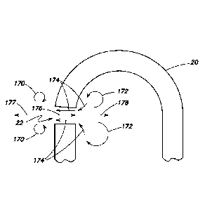
[0089] FIG. 15 shows an ophthalmic surgical kit 200. Kit 200 may be
useful with conventional vitrectomy systems and devices, such as mechanical
vit
cutters, in addition to the present invention. The current trend towards ever
smaller
surgical instruments creates a problem of generating enough flow through the
vit
cutter/removal device. Smaller instrument lumens require higher vacuum levels
to
generate enough flow to prevent clogging and maintain a sufficient volume of
tissue
CA 02883375 2015-02-26
WO 2014/039836 PCT/US2013/058533
removal. If the size of the infusion cannula and the infusion ESA device is
the same as
that used for the vit cutter, the infusion cannula may require excessive
infusion
pressures to maintain the intraocular pressure during surgery. Excessive
infusion
pressure can lead to tissue damage and fluid jets that obscure the surgical
site and
create unwanted turbulence in the liquid in the eye. To avoid these problems
an
infusion cannula with a larger cross-sectional area than the cross-sectional
area of the
inner diameter of the vit cutter/vitreous removal device can be used. This
allows the
infusion cannula to provide sufficient infusion fluid volume at safe, low
infusion
pressures and maintain a stable intraocular pressure.
[0090] Kit 200 includes a package 202 with a first entry site
alignment
device 204 and a second entry site alignment device 206. Kit 200 also includes
an
infusion cannula 188 attached to a length of tubing 210. The cannula 188 is
for
insertion into the first entry site alignment device 204 and the tubing 210 is
for
attachment to a source of infusion fluid (not shown and not part of the kit),
via a
connector 212. The second entry site alignment device 206 is for receiving a
tissue
extraction device, such as the vibrating surgical instrument 10 described
above or a
conventional guillotine-type vit cutter 214, as shown in FIG. 16. The first
entry site
alignment device 204 has a larger diameter lumen than a lumen diameter of the
second
entry site alignment device 206. The kit 200 may further included the tissue
extraction
device 214 or 10 for removal of vitreous and other tissue from a patient's
eye. The kit
200 may also include a cannula 18 for attachment to a vibrating surgical
instrument 10,
the cannula 18 having a distal tip 20 with at least one port 22 in
communication with a
lumen 19 extending through the cannula 18 and in communication with an
aspiration
path 24 in the housing 12 (as shown in FIG. 7). A plurality of trocars 201 may
also be
included in kit 200.
[0091] In the alternative, rather than the infusion cannula having
a lumen
larger than the lumen of the tissue extraction device, one could use multiple
infusion
cannulas of the same or smaller lumen size of the tissue extraction device.
The goal is
to provide more cross-sectional area for the infusion fluid than aspiration
cross-sectional
area of the port(s) or lumen of the tissue extraction device.
AA
CA 02883375 2015-02-26
WO 2014/039836 PCT/US2013/058533
21
[0092] In consideration of the above, a new vitrectomy device
design is
proposed that addresses the shortcomings of existing designs. It consists of a
single,
covered or uncovered, moving outer needle, sans inner needle, with one or more
off-
axis ports. The port(s) preferably has a cross sectional area less than 70% of
the cross
sectional area of the inner diameter of the needle (so that any dimensions of
remaining
intact gelatinous chunks are too small to cause clogging) and a maximum
dimension
across the port between about 0.003" and 0.012" (75 ¨ 305 microns), depending
on the
needle gauge. This could be a single port for to a side of the axis of the
cannula, with a
diameter less than half the inner diameter of the needle and this will achieve
acceptable
results.
[0093] Additional construction preferences are that the
cannula/needle be
small enough to pass through either standard wounds in surgery (for instance,
OD of 1
mm or less) or entry sight alignment system cannulas (for instance, OD of
0.625mm or
less for 23 gauge systems), and that it be long enough to reach across the eye
globe
(for instance, a distance from the taper at the end of the hub to the tip of
the shaft of 30
mm or more).
[0094] As an example of the motion amplitudes and construction
dimensions, the port size might be around 0.005" or 127 microns and the
minimum wall
thickness might be 0.001" or 25 microns; the maximum expected displacement
amplitude might be between 5 and 15 microns. As an example of relative
velocities, a
device with a displacement amplitude of 10 microns and a harmonic operating
frequency of 28,500 Hz would have a harmonic velocity amplitude of about 1.8
m/sec;
the particle velocity through four 0.005" (125 micron) diameter holes in the
end of the
device would be around 1.2 m/sec for flow rates of 3.5 ml/min, consistent with
desirable
flow rates of water through similar 23 gauge devices.
[0095] A number of minor variations on the example embodiments can
be
made. These include the total number of holes, the diameter of the holes, the
inner and
outer contour of the distal tip, the material used to make the needle, the
processes used
to make the needle (which could be machined monolithically or fabricated from
components, and could include drilling or EDM or other processes for forming
the
CA 02883375 2015-02-26
WO 2014/039836 PCT/US2013/058533
22
holes), the overall length, inner and outer diameters, operating frequencies,
and
continuous or pulsed operation.
[0096] As part of the example embodiments, certain drive control
modes
may be envisioned. For instance, some users may want to grasp or peel
anatomical
features such as membranes with the tip. Because the cutter only requires low
vacuum
levels and has small holes, the grasping power of the tip may be considerably
lower
than with conventional cutters. Therefore, one control mode of the device
involves
automatically applying high levels of vacuum when ultrasonic power is not
being
commanded, but dropping the commanded aspiration vacuum level down once
ultrasonic power is applied.
[0097] Alternative directions of tip motion can be contemplated.
For
instance, because the holes in the end of the tip are slightly off center,
torsional action
of the tip may result in the sides of the holes disrupting and liquefying the
vitreous in a
manner similar to the disruption that takes place with longitudinal motion.
Torsional
action of the port may create bi-directional flow by pushing fluid from the
side region of
the port directly to the outside and inside of the needle. Likewise, a slight
lateral or
transverse motion may achieve the same effect. In this case, bidirectional
flow may
result in the alternating pressure and vacuum zones that will be created in
front of the
port as the needle moves along the axis of the port, or induced low pressure
zones
created by high fluid velocities across the face of the port if the needle is
moving parallel
to the face of the port.
[0098] An alternative description of an example embodiment may be
that
the surgical device of the present disclosure may achieve higher flow rates
than a
conventional guillotine vit cutter at the same vacuum levels.
[0099] The example embodiments improve flow through multiple
mechanisms, including increasing the area within the needle available for the
aspiration
path for tissue that has made it through the ports, eliminating blockage of
the aspiration
port by an inner needle during a portion of the aspiration cycle, the
application of high
shear stresses along the wall of the smallest aspiration path diameters,
causing shear
thinning in the thixotropic vitreous, and breaking up the vitreous into
smaller pieces
CA 02883375 2015-02-26
WO 2014/039836 PCT/US2013/058533
23
(through mechanisms that will be discussed shortly). Each of these mechanisms
is
described in greater detail in the paragraphs which follow.
[00100] The example embodiments have only one needle, not two
needles,
as in mechanical vit cutters. By eliminating the inner needle, flow through
the needle at
a given pressure differential across the needle may be increased by a factor
of two to
four for the same outer diameter of the outer needle. Classical analysis of
non-turbulent
flow resistance through a long tube is known to be related to the equation
1/Length*Diameter4. Typical conventional mechanical vitrectomy devices have an
aspiration lumen at least 30 mm, long enough to reach from the entry point at
the side
of the eye across the globe to points on the other side of the eye. Diameters
of some
typical needle material combinations are shown in Table 2, below, in units of
0.001" (for
conversion to metric multiply numbers in table by 25.4 microns).
Outer Needle or Vitrectomy Needle Inner Needle, Guillotine Only
Gauge Wall OD ID Gauge Wall OD ID
23 MTW 1 25.0 - 25.5 22.5 - 24.0 25 MTW 1 20.0 - 20.5 17.5 - 18.5
25 MTW 1 20.0 - 20.5 17.5 - 18.5 27 MTW 1 16.0 - 16.5 14.0 - 15.0
27 MTW 1 16.0 - 16.5 14.0 - 15.0 29 MTW ' 1 13.0 - 13.5 11.0 - 12.0
23 UTVV 1.5 25.0 - 25.5 20.0 - 22.0 26 UTW 1.5 18.0 - 18.5 14.5 - 15.5
25 UTVV ' 1.5 20.0 - 20.5 15.5- 17.0 28 UTVV ' 1.5 14.0- 14.5 11.0- 12.0
27 UTVV 1.5 16.0 - 16.5 13.0 - 14.0 30 UTVV ' 1.5 12.0 - 12.5 9.0 - 10.0
Table 2
where MTW=Micro-Thin Wall; UTVV=Ultra-Thin Wall
[00101] It can be seen that, for outer needles fitting through a
given gauge
cannula, eliminating the inner needle may increase the diameter of the
aspiration path
by 25% to 40%, which translates to decrease in flow resistance of at least (1-
(1/1.25)4
which is greater than) 55%. Use of different production methods for the
needles, such
as machining, may impact this final result.
[00102] In addition, the inner needle of a mechanical vit cutter is
generally
much longer than the minimum distance required to reach across the eye, as it
must be
CA 02883375 2015-02-26
WO 2014/039836 PCT/US2013/058533
24
attached to a drive mechanism after exiting the outer needle. This contributes
to an
additional increase in the flow resistance, due to the inverse of the length
factor.
[00103] In conventional vitrectomy cutters, the inner needle blocks
the
aspiration port during part of the cut cycle, resulting in a decrease in
aspiration. For
instance, at a fixed vacuum level, water will flow through a conventional
cutter at a rate
of about 5 ml/min, with the port continuously open; vitreous flows at a rate
of 0 ml/min in
the same condition. Once cutting is activated, vitreous flow increases
(because the
vitreous is now cut into smaller pieces) but water flow decreases, because the
cutter
aspiration port is now blocked part of the time by the needle. In conventional
cutters,
minimizing the transit time of the inner needle minimize the period of
blockage, thereby
increasing the flow through the eye, but shorter transit times typically
require higher
drive pressures, and as cut rates increase, the relative transit time (the
closed duty
cycle) inevitably rises. Eliminating this blockage time with the example
embodiments
maximizes the total time available during the disruption cycle for aspiration.
[00104] Thixotropic materials, such as the vitreous, become less
viscous
when subjected to high shear stresses. Reciprocating the wall of the
aspiration path
continuously will apply high shear stresses to any vitreous in contact with
the wall,
causing it to stay liquid, dropping the flow resistance. Once the aspirated
tissue moves
into the larger diameter aspiration path downstream of the needle, the fluidic
resistance
of the pathway drops, as does the expected flow velocity, minimizing the
impact of
fluidic resistance of this portion of the path.
[00105] The example embodiments also include features which
facilitate the
disruption and liquefication of the vitreous; the pieces that result will be
smaller than the
pieces that result from conventional vitreous dissection. Even if the inner
diameter of
the aspiration path remained the same, the smaller pieces would result in
reduced flow
resistance.
[00106] Specifically, the hole or pattern of holes at the end of the
needle
permits only smaller pieces through; by selecting holes substantially smaller
than the ID
of the aspiration path helps break the material into pieces smaller than the
aspiration
path cross sectional area. Additionally, hole patterns with multiple holes
create one or
CA 02883375 2015-02-26
WO 2014/039836 PCT/US2013/058533
more webs between the holes that separate individual flow streams (and the
strands in
those flow streams), separating the vitreous as it comes in. Furthermore, when
the end
of the needle is displaced rapidly in a harmonic manner at velocities that
form bi-
directional flow through the port creates local shear stresses that liquefy
the thixotropic
vitreous. Furthermore, once pulled through the small hole, the minimum
aspiration path
area through the 25 gauge tube in the cannula is much greater than the port
area.
Pieces which fit through the port and are disrupted close to the wall will not
clump
together nor clog on each other.
[00107] Moreover, in the process of spreading out in the aspiration
lumen
after passing through the ports, the material will be subjected to high
lateral shear
stresses. Thus, during a time period lasting just a couple of harmonic cycles
(< 0.1
msec), the vitreous tissue will be pulled into the vicinity of the aspiration
port, portions of
the tissue will be pulled back in a direction opposite to the direction of
flow, then pulled
in the direction of aspiration flow at velocities greater than the average
flow velocity,
then spread out thin within the larger aspiration lumen area. The resulting
turbulent mix
of cyclical stresses disrupts and liquefies the tissue, breaking it into
pieces that
eliminate traction outside the needle and minimize flow resistance inside the
needle.
[00108] Furthermore, as inertial jets form at higher port
velocities, high
lateral shear stresses will be encountered in the port from the simultaneous
bidirectional
opposing form that form and by the rotational flows that form in the toroidal
cells, further
breaking up the vitreous and liquefying it.
[00109] The discussion immediately above is shown in Table 3 below
comparing conventional vit cutter performance against the example embodiments
disclosed.
Conventional Conventional Harmonic Vitreous Vitreous
Aspiration Cutter peak displacement harmonic harmonic
Particle velocity velocity amplitude, H20, liquefier
particle liquefier velocity
STP velocity amplitude
0.75 m/sec 0.4 m/sec 0.37 m/sec 1.1 m/sec 3.5 m/sec
Length of Length of
aspirated & cut aspirated & cut
segment segment
¨ 9 mm ¨ 0.0662 mm
Table 3
CA 02883375 2015-02-26
WO 2014/039836 PCT/US2013/058533
26
[00110] In an acoustic medium, harmonic motion is reciprocating or
oscillating motion, where a pressure wave may be transmitted through a medium
through the local displacement of particles. It is important to understand
that, while the
phase front of the pressure wave can travel tremendous distances, the
particles in the
medium move very little, and in a cyclical fashion, returning to previous
locations every
oscillation period. The amplitude of the particle motion is determined by the
magnitude
of the pressure wave passing by the particle in the media; the greater the
pressure
amplitude of the wave, the greater the displacement amplitude of the particle.
The
particle velocity can also be described as a harmonic function, and will be
the first
derivative of the particle displacement amplitude function. This relationship
is well
understood in acoustics, and one simple guiding equation is:
P = zm*u where
P = the harmonic pressure function
u = the harmonic particle velocity function, and
zm = a material characteristic known as the acoustic impedance.
[00111] It may be appreciated that, at sea level, the pressure
amplitude
cannot exceed one atmosphere ¨ at amplitudes above this, the negative half of
the
pressure amplitude, in absolute terms, would drop below absolute vacuum, which
is
physically impossible. Therefore, as the needle tip moves backward away from
the
tissue outside it, it will create a near vacuum for a pressure, but cannot
create an
absolute vacuum. Once the absolute pressure in the vicinity of the tip drops
below the
vapor pressure of the medium, pockets full of saturated vapor will form at
convenient
boundaries, limiting further drops in vacuum.
[00112] In contrast to harmonic particle motion, aspiration particle
velocities
are unidirectional. Particle velocities are a function of the actual
volumetric flow rate
and the aspiration path geometry. Aspiration flow particle velocities can be
estimated
from the equation V = Q/Area, where V is the average flow velocity across a
plane, Q is
the volumetric flow rate, and Area is the cross-sectional area of the flow
path.
[00113] Flow rates between 1.5 and 15 ml/min would be considered
desirable from an ophthalmic clinical perspective. If flow rates are too low,
the time to
CA 02883375 2015-02-26
WO 2014/039836 PCT/US2013/058533
27
remove the vitreous from the globe becomes excessive. If they are too high,
fluidic
balance in the eye may be compromised, and the surgeon may have difficulty
keeping
the surgical site stable.
[00114] Flow velocities will be highest where the path area is
smallest. In
conventional vitrectomy devices, this has been within the inner needle. Flow
path
diameters of devices compatible with standard 23 or 25 gauge entry site
alignment
system cannulas are determined by the ID of the aspiration needle, which, in
conventional devices, will be 0.38 mm at most, down to as small as 0.28 mm in
diameter. This results in a range of particle speeds of 0.29 m/sec to 0.54
m/sec at flow
rates of 2 ml / min, as a minimum. Higher velocities would result from higher
flow rates,
and smaller needle path geometries.
[00115] For a conventional 23 gauge 5000 CPM (cuts per minute)
cutter
with desired water flow rates around 3.5 ml/min, it is worth noting that the
leading edge
of a segment of cut tissue will travel a distance of 6 to 12 mm through the
aspiration
needle for every cut, with an average particle velocity between about 0.5 and
1.0 m/sec,
and a peak particle velocity around twice this. For comparison, the peak inner
needle
velocity for a 5000 CPM cutter with a total stroke of around 1 mm will be
around 0.4
m/sec. Because the needle velocity will be proportional to the stroke, which
is
proportional to the port size, which is, in part, proportional to the needle
gauge, the
smaller cutter inner needles may run slightly slower.
[00116] However, the inner needle achieves this peak velocity for
only a
short period of time. The needle accelerates forward from a dead stop in
response to
increasing air pressure; the resulting velocity function is proportional to
the square of
time, until the needle hits the forward stop.
[00117] For comparison, a prototype 23 monolithic ultrasonic
vitrectomy
needle with an OD of 0.025"(635 microns), an ID of 0.020"(508 microns), and
four
0.005" (127 microns) diameter holes arranged symmetrically at the end for use
at 28.5
kHz and harmonic stroke amplitudes of 5 to 15 microns has been constructed. At
the
same 3.5 ml/min flow rate of a typical mechanical vit cutter, average flow
velocities
through these four holes would be around 1.1 m/sec. At peak to peak
displacement
CA 02883375 2015-02-26
WO 2014/039836 PCT/US2013/058533
28
amplitudes of around 10 microns, the peak exchange flow velocity amplitude is
around
3.7 m/sec, meaning that it exceeds the aspiration flow velocity for a
significant portion of
the cycle (greater than 30%), effectively reversing the direction of flow
through the port,
and, at the same time, moves in the opposite direction for a significant
portion of the
time. An alternative single 0.005" hole device of otherwise similar
construction and drive
conditions will result in both higher aspiration flow velocity (3.5 m/s) and
peak exchange
flow velocity (¨ 10 m/s), still yielding a reversal of flow direction.
[00118] The inventive surgical device and needle of this disclosure
breaks
up vitreous by passing a small volume of fluid (the "exchange bolus") from a
generally
slowly advancing intake flow rapidly back and forth through a small port such
as port 22
of FIG. 1, creating a periodic bi-directional flow of tissue through the port.
The periodic
bi-directional flow of tissue is similar to the harmonic motion referred to
above. The
rapid back and forth motion breaks up the tissue through shear forces that
develop
between the edge of the exchange flow and the center of the exchange flow, and
by
tension forces between the rapidly moving trailing edge of the exchange bolus
and the
slow moving leading edge of the advancing intake flow (when the bolus is
moving from
outside the tip to inside the tip, or normal motion) or trailing edge of the
receding
exhaust flow (when the bolus is moving from inside the tip to outside the tip,
or
retrograde motion.) In theory, the greater the velocity of the exchange flow,
the greater
both types of forces are.
[00119] The present device creates the periodic bi-directional flow
or
exchange bolus by providing a substantially closed tip with a small port,
driven at a
velocity sufficient to cause bi-directional flow through the port, with an
aspiration
vacuum applied. The drive velocity of the tip must be sufficient to create the
bi-
directional flow at the desired aspiration flow level. At some point, the
vacuum created
inside the tip by the tip velocity reaches a maximum level, and the effect of
the tip may
be said to be optimized. Tip velocities above those greater than that creating
a
maximum vacuum will continue to be effective for disrupting the tissue, but
may have
negative side effects, such as turbulence or tissue injury, and at some point,
they begin
CA 02883375 2015-02-26
WO 2014/039836 PCT/US2013/058533
29
to generate cavitation outside of the tip. Maintaining the tip drive velocity
at a point
below a level at which cavitation is created externally of the tip is desired.
[00120] Although the invention is described below using a hollow
spherical
tip, it should be noted that the action of the invention depends only on
areas, and not
shapes. Hollow hemispherical tips, solid hemispherical tips, and flat ended
tips with
equivalent inner, outer, and port areas will have similar minimum drive
velocities and
optimal drive velocities, although the upper cavitation drive velocity limit
may depend
somewhat on the external tip geometry.
[00121] Volumetric flow through the tip consists of two components:
a time
invariant aspiration flow rate Qasp and a time variant acoustic flow (Lest,.
The use of the
capital letter Q to represent volumetric flow (volume per unit time) is well
established in
the literature.
[00122] Qtotal (time) (= QT (t)) = Qasp + Qacstc (t) = QDF + QHF (t)
[00123] The subscript HF denotes bi-directional flow or Harmonic
Flow and
DF for Direct Flow. It is noted that if QHF(t) is symmetric, (for instance,
QHF(t) =
QHFO*Sin(Wt)), QT (t) Will be asymmetric, and the maximum positive value will
be greater
than the minimum negative value.
[00124] QDF should be selected to be a value high enough to permit
the
surgeon to get through surgery in a timely manner, but not so high that the
eye
becomes unstable or requires high static infusion pressures. Surgeons have
generally
been satisfied with products that permit vitreous aspiration rates of about
1.5 ml / min
(between 1 and 2 ml / min) in the center of the eye, and may use lower flow
rates as
they get close to critical or loose structures, such as the retina. SI units
for volumetric
flow are m3/sec; 1.5 ml / min is about 2.5 x 10⁻⁸ m3/sec, or 0.025 ml/sec, or
25 pl/sec, or
25 mm3/sec. Aspiration is always into the port. Therefore, for sign
convention, flows into
the tip through port are designated as positive flow and flows out of the tip
through the
port are negative flow.
[00126] Aspiration Velocity VDF = QDF /Areaport = QDF /Aport
[00126] QHF (t) is the volume displaced back and forth through the
port by
the action of the inner and outer moving surfaces of the tip. Effectively, it
is the area of
CA 02883375 2015-02-26
WO 2014/039836 PCT/US2013/058533
the inner surface normal to the axis of motion multiplied by the velocity of
the surface in
parallel to the axis of motion. The magnitude of the Q depends on the inner
area, the
port area and the velocity of the tip, and is influenced by the angle between
the port and
the axis of motion. The basic equation is:
[00127] QHF (t) = QHFosine(wt)
[00128] where QHFO = Velocity of the tip normal to the axis of
motion
multiplied by the {Inner area of tip normal to the axis of motion ¨ (Area of
port multiplied
by the cosine of the angle between normal to port and the axis of motion)} =
VTIAID-
Aport*Cosine (Cy
[00129] Positive flow is, by the convention we established above,
flow into
the port; negative flow is flow out of the port. Positive flow occurs as the
tip moves
forward, into the external area, as the outer surface pushes fluid from the
front surface
of the tip and pulls fluid with the inside of the tip surface.
[00130] Aspiration Velocity VHF(t) = QHF(t)/Aport, also
[00131] QHF(t) = AlnsideTip*V(t)tip
[00132] In a linear state, QHF(t) = QHFesin(wt); in non-linear
states this may
not be true.
[00133] A needle or device is operating in a unidirectional flow
domain (no
bi-directional fluid flow through the needle port) when the peak negative
value for VHF(t)
is less than VDF. That is, VT(t) = VDF + VHF(t) is always positive. Fluid is
always flowing
into the port, although fluid is speeding up and slowing down. Fluid flow
corresponds
essentially to the tip passing the midpoint in its forward moving stroke, and
fluid flow is a
minimum about when the tip passes the midpoint in its backward moving stroke.
[00134] Flow is in the unidirectional flow domain when QDF /Ainside-
rip > Vtip=
Devices and needles operate in this domain for high aspiration rates, small
displacement areas, and low tip velocities. Typical phaco needles operate in
this
domain, because the area inside the tip is zero, (that is, the tip is not
closed at all)
making the term on the left side of the equation infinitely large. They
typically clog if
operated in vitreous, confirming this domain is not effective in vitreous.
Typical I/A
devices and needles operate in this domain, because the tip velocity is zero,
making the
CA 02883375 2015-02-26
WO 2014/039836 PCT/US2013/058533
31
term on the right side of the equation very small. They typically clog if
operated in
vitreous, confirming this domain is not effective in vitreous.
[00135] A device and needle enters the bi-directional flow domain
above
the threshold identified above, when the harmonic velocity is just slightly
above the
aspiration velocity, or average linear velocity. Changes in flow in this
domain can be
considered linear up to the point where the pressure difference necessary to
achieve
the velocity into the port from either side is greater than about one to two
full
atmospheres of pressure. The action of the inner surface moving forward can
create up
to one atmosphere of vacuum at the outer entrance to the port; the action of
the outer
surface moving forward at the same time can create positive pressure outside
and
adjacent the port. The amount of positive pressure generated depends on the
outer tip
dimensions and the tip velocity.
[00136] Flow through the port is limited at the upper end by static
flow
through an orifice, where Vp0rt2 = 2APressure/Pi which can be extended into
the time
domain: Vp0rt2(t) = 2 APressure(t)/p. In this case, Apressore(t) is from the
forward moving
surface to the outer entrance to the port. It cannot rise much above 1 ATM
(103500 Pa)
as there can be no greater than 1 ATM vacuum inside the needle at the port,
and the
peak pressure generated at the outside of the tip can never be greater than
the peak
vacuum generated inside the needle, and typically less, in linear conditions.
Furthermore, note that flow through the port identified above by Vport is a
combination of
the linear flow and the bi-directional exchange flow. When the needle is
moving back
towards the surgical device, the needle inner surface can generate a pressure
necessary to move fluid out of the port. The forward motion case is the
limiting case.
Furthermore, the maximum bidirectional flow velocity that can be achieved may
be less
than the theoretical maximum aspiration flow, because of the acoustical and
inertial
effects.
[00137] The device and needle fluidics will be limited when the
reactive
acoustic pressures that are created by the tip in the vicinity of the port are
no longer
sufficiently high to pull material through the port at the necessary velocity.
At this point,
cavitation will start inside the needle, and the reciprocating acoustic flow
will start to be
CA 02883375 2015-02-26
WO 2014/039836 PCT/US2013/058533
32
limited. This cross-over point can be predicted, to an extent, and depends
mostly on
the tip velocity, with a weak dependence on the tip dimension. Once in the non-
linear
domain, the bi-directional flow increases more slowly as the tip velocity
increases.
[00138] QDF = Vtip*Atip = Aport* 4(2 *ATM / p) ¨ Qasp
[00139] To find a limiting case, the Qasp term is dropped and the
equation
above simplifies to Vtip = (Apprt/ Atip ) *4(2 *ATM / p). The 4(2 *ATM / p)
term is entirely
physical constants; at ATM = 103500 Pa and p = 1000 kg/m3 for water, it equals
14.4
m/s. Furthermore, in the case of a round inner area and a round port, the
equation for
the maximum linear action tip velocity can be further simplified to Vtip =
(Dport/Dtip10214.4 m/sec. Over a large variation in flow, the limiting
velocity does not
vary much for the needle sizes considered, supporting the use of the
simplified equation
for calculating the limit.
[00140] At reciprocating flow velocities for which the particle
displacement
inside the port in the reciprocating flow over a single cycle exceeds a
significant fraction
of the port diameter, the small amplitude assumption used in linear acoustics
may be
violated, and small inertial jets out of the port and toroidal recirculation
flow cells around
the axis of the port may form. This will occur at some point above the bi-
directional flow
threshold. These do not alter the basic exchange flow quantities, but result
in slightly
different flow distributions. FIGS. 17A-D illustrate the flow of tissue
through the port 22
at different points in the longitudinal vibration of cannula 18. All of the
flows illustrated
disregard any aspiration pressure applied to cannula 18. FIG. 17A shows
cannula 18 at
maximum extension, resulting in simultaneously opposing port flows with little
or no net
flow through port 22. Lines 170 indicate recirculating flow outside of port
22, creating a
small toroid around the port. Lines 172 indicate recirculating flow inside
port 22. Lines
174 indicate flow exiting the port and line 176 indicates flow entering the
port. Lines
177 and 178 are always present and indicate flow away from port 22 both inside
and
outside of the port. FIG. 17B shows cannula 18 at a point pulling back towards
device
12 as indicated by arrow 171 with the net flow moving out of port 22. Lines173
indicate
flow back towards cannula 18. FIG. 17C shows cannula 18 at minimum extension,
resulting in simultaneously opposing port flows with little or no net flow
through port 22.
CA 02883375 2015-02-26
WO 2014/039836 PCT/US2013/058533
33
FIG. 17D shows cannula 18 at a point moving forward away from device 12 as
indicated
by arrow 179 with the net flow moving into the port 22. Line 180 indicates the
flow of
fluid to fill the inside apex of cannula 18. Line 182 indicates the flow of
fluid along the
outer surface of cannula 18 into port 22. Line 184 indicates a toroid of flow
away from
the flow indicated at 182.
[00141] Larger tips have lower non-linear flow tip velocity
thresholds than
smaller tips for the same port area. The non-linear threshold changes rapidly
with
gauge and wall thickness. The non-linear threshold changes more slowly with
changes
in target aspiration rates. Also, the non-linear threshold tip velocity
decreases in
proportion to the port area.
[00142] At very high tip velocities, cavitation begins to develop on
the outer
surface of the tip. Vitreous may be liquefied externally, but the velocity or
volume of the
aspiration flow becomes much less predictable, acoustic field intensities
become larger
and less predictable, the fluid around the tip becomes more turbulent,
acoustic
streaming may develop, and the possibility of damage to adjacent tissues
rises. This,
then, may be considered as an upper limit for safe practice in using the
devices and tips
of the present disclosure. The velocity of the tip should be low enough that
the pressure
amplitude of a simple source equal to the difference between the inside and
outside
areas of the tip, minus the port area, is less than one atmosphere.
[00143] A quantitative understanding of this upper velocity
threshold is as
follow. In acoustics, it is known that for small point sources (of which the
tips of the
present disclosure are variants), P = pl*Q/(2r), where r is the radius from
the acoustic
center to the point under consideration. For the external surface of a tip, Q
= Vtip*A outer
and at the cavitation limit, P is one atmosphere or 103500 Pa. The equation is
then Vtip
= (ATM/p)*(1/0*(2r/A outer). For a linearly moving hemispherical shell, the
acoustic
center is located about 1/3 of a radius, or 1/6 of a diameter behind the apex
of the shell
on the axis of vibration, so that r=router/3 or Douter/6. The equation
simplifies to Vtip =
(2ATM/pf)*((router/3)/A outer). For tip velocities near to or above this,
external caviation is
likely to occur.
CA 02883375 2015-02-26
WO 2014/039836 PCT/US2013/058533
34
[00144] It is important to note that the volume displaced by the
portion of
the outer surface that is matched by the inner area is part of the bi-
directional or
exchange flow, and may not contribute to the external cavitation, as it is
being replaced
by fluid coming through the port. Thus, A....uter Ýs the total outer area
minus the total inner
area (or, in the case of round geometry, Aouter = (Tr/4)*(0D2-1D2). Thus, the
limit is most
strongly influenced by the tube wall thickness. Further note that the
effective location of
the acoustic center may shift because of this exchange, and make geometry
factors
much more difficult to calculate for a closed form equation. The limiting
velocity for
cavitation may be identified through FEA (finite element analysis) modeling of
pressure
fields around the vibrating tip and through direct observation with a high-
speed camera
looking for bubbles and voids. Note from the previous equation, that the
external
cavitation limit is relatively unaffected by the aspiration flow or the port
area or location.
[00145] For some design conditions, reaching the optimal tip
velocity for the
bi-directional flow cannot be accomplished without exceeding the external
cavitation
threshold. In these instances, the tip should be vibrated close to but not
above the
external cavitation threshold for maximum effectiveness. This is particularly
true for
thicker walls, smaller gauges, and larger ports.
[00146] Summarizing the disclosure above: unidirectional flow domain
is
not effective; driving at levels above the unidirectional flow domain
represents a lower
bound of desirable drive levels; use of ultrasonic needles designed for
cataract surgery
(these normally do not have any inside tip surface area) in vitreous falls in
this domain;
these needles generally clog; for vitreous removal some non-zero tip velocity
is
required; this lower velocity limit may be defined by Vtip > QDF/AinsideTip;
bi-directional
flow in the linear domain is effective, with effectiveness increasing as the
amplitude of
the bidirectional flow increases; bi-directional flow in the presence of
internal cavitation
or the is effective, but the increase in effectiveness is plateauing; bi-
directional flow
above the external cavitation threshold is no more effective in disrupting
tissue at the
port than the internally non-linear domain, while adverse effects such as
turbulence and
safety concerns start escalating rapidly. Propagation of shock waves from
cavitation
CA 02883375 2015-02-26
WO 2014/039836 PCT/US2013/058533
may disrupt either vitreous or retinal tissue indiscriminately in areas
outside the
immediate vicinity of the tip.
[00147] All the examples provided assume standard pressure at sea
levels.
At high elevations atmospheric pressure will be lower (for instance, about 80
kPa in
Mexico City at 2200 meters or 7200 feet of elevation) and may alter some of
the
transition points, but not the basic principles. Operation at levels enough
below sea
level to significantly alter the performance is unlikely.
[00148] In addition, the device and needle performance will be
affected by
the distance between the port and the acoustic center of the external surface.
As these
two points move apart, the tip will move from the linear to the non-linear
domain at lower
tip velocities, because the action of the external surface will have less
influence on the
pressure at the port.
[00149] Although reference has been made to diameters such as inner
diameters and outer diameters of the needle and the diameter of the port, the
cross
sectional areas can be non-circular without substantially affecting the basic
principles.
[00150] Cavitation will start at around the vapor pressure of water
for the
ambient temperature; obviously, this is close to but greater than a full
vacuum. We have
assumed a full vacuum for the purposes of simplifying the analysis.
[00151] Some observations follow. The primary impact of the needle
outer
diameter (OD) is on the external cavitation velocity limit. The larger the OD,
the greater
the overall Q, even if wall thickness is maintained, although it is spread
over a larger
area. The most critical impact of the internal diameter(ID) of the needle is
to the
internal cavitation threshold ¨ for a given port size, the larger the area,
the lower the
cavitation velocity threshold. Smaller ports increase the velocities through
the port, for
both aspiration and reciprocating flow. The velocity field outside the port
will be
generally independent of the port size, depending only on Q, at distances
large with
respect to the port. Port diameter has no effect on the lower unidirectional /
bidirectional
threshold or on the external cavitation bound. Wall thickness ((0D-ID)/2)
influences the
acoustic area and, if it is unchanged down the needle shaft, it influences the
stiffness of
the needle. The ratio of the ID to port diameter drives the velocity of the
reciprocating
CA 02883375 2015-02-26
WO 2014/039836 PCT/US2013/058533
36
flow. Large ratios (small ports near large IDs) make the velocity through the
port large,
starting the onset of bi-directional flow at lower strokes.
[00152] Some design considerations follow. Although the major
factors that
influence the balance of aspiration and bi-directional flow have been
disclosed, the
designer is likely to focus on the selection of the port area.
[00153] Absent any other effect, users have a strong preference for
going
through smaller incisions rather than larger incisions. However, they also
want the
highest flow possible, and stiff, reliable instruments, and they may have
varying
preferences for true end cutting action and visibility of tissue in front of
the port.
[00154] The designer will select the largest outer diameter that
will fit
acceptably through the incision or through a cannula inserted through the
incision.
[00155] The designer will select the inner diameter as large as
possible (i.e.
the thinnest wall thickness) without making the instrument unacceptably
flexible or
prone to breakage, in order to minimize flow resistance.
[00156] The designer typically prefers to use off the shelf
materials for cost
effectiveness, so they may further select their inner and outer diameters
based on
available dimensions for commodity medical grade micro-tubes.
[00157] The designer will select the angle between the port and the
tip to
balance the end cutting effect (at zero degrees) and port visibility when the
tip is
inserted (maximized at 90 degrees).
[00158] This leaves (to the designer) the selection of the port area
and the
tip velocity to provide optimal exchange flow according to the above
equations,
balanced against the selection of the tip velocity to minimize external
cavitation, and the
selection of the aspiration vacuum to achieve the desired aspiration flow.
[00159] Needle geometry (round or non-round), and tip geometry
(hemispherical, bullet shaped, conical, flat, chisel, etc.) details may then
be influenced
by other user desires, or to enhance manufacturability or durability.
[00160]
[00161] The substantially closed distal end of the needle
contributes
significantly to the liquefication of the vitreous. Open needles, such as
standard phaco
CA 02883375 2015-02-26
WO 2014/039836 PCT/US2013/058533
37
needles, do not have hydraulic gain and cannot create bi-directional flow.
Furthermore,
the ability to grab, hold onto and move a large chunk of tissue is highly
desirable in
phaco and fragmentation needles; traction is, essentially, desirable in phaco
procedures, and undesirable in vitrectomy procedures. Phaco needles have
evolved
bell structures with large open cross-sectional areas specifically intended to
enhance
purchase (the amount of pulling or holding force available). Since the area of
the single
port in standard phaco needles is equal to or larger than the aspiration cross-
section
area, any pieces that do get broken up barely fit through the subsequent
aspiration
area, and can get bound together, causing the clogs that are seen clinically.
[00162] The effect of vitreous liquefication might be observed by two
separate methods.
[00163] Method la: In this method, a fixed volume of either pure
vitreous or
pure water is aspirated out of an eye with a fixed vacuum, and the time to
aspirate the
tissue is measured. When the times for the two different fluids are
significantly different,
the tissue is much less liquid than the water (i.e. more viscous); where they
approach
each other, the tissue is substantially liquid. This measurement can be made
on a
comparative basis, comparing mechanical cutters and ultrasonic technologies.
[00164] Method lb: A variant of method la aspirates a volume of
either pure
vitreous or pure water with a fixed vacuum for a fixed time; in this case, the
device that
permits the greatest volume or mass of aspiration in the fixed time is
superior, and
aspirating a similar mass or volume as can be aspirated using pure water is an
indication that the tissue is substantially liquid.
[00165] Method 11: Samples of vitreous are cut or liquefied, and the
molecular weight of both the pre and post processed tissue is measured and
compared
to that of water. Molecular weight is a critical mechanical characteristic of
a medium,
and relates to a number of properties, including fluidity. In this case, lower
molecular
weights reflect greater liquidity.
[00166] A variety of approaches may be taken to making the devices of
the
example embodiments, which will affect aspects of the geometry not critical to
the aims
of the vitreous removal.
CA 02883375 2015-02-26
WO 2014/039836 PCT/US2013/058533
38
[00167] For instance, the devices may be machined monolithically out
of a
material such as Titanium. In this case, the inner path is formed by drilling
a blind hole
into a titanium rod. Since the point of the drill will typically not be
hemispherical, the
inner surface of the end will not be hemispherical, so the shape and thickness
of the
closed end may be non-uniform. However, in this case, the holes drilled in the
closed
end will still have a cross-sectional area smaller than the area of the
aspiration path
through the device.
[00168] The holes may be fabricated by a variety of methods,
including but
not limited to punching, drilling, or cut using a wire EDM process.
[00169] The shaft may be formed by deep drawing, machining
monolithically, or sealing preformed, precut micro-tubes with welding, swaging
or other
processes.
[00170] Also, the tip with a port may be formed separately from the
shaft of
the needle and attached by any known method such as adhesive, welding,
frictional
contact, or any other method that secures the tip and the shaft to each other.
In such a
two-part design where the ID of the shaft is smaller than the ID of the tip
portion the ID
of the shaft is the ID to be used in the teachings above.
[00171] The devices may be constructed so that the user receives
both the
cutting needle and the ultrasonic driver, or the needles may be made so that
they can
be installed on a reusable ultrasonic driver by the user.
[00172] The devices may be constructed out of any bio-compatible
material
that is appropriate for the fabrication process, including metals and
plastics.
[00173] One example that was constructed was a 22 gauge needle with
four 0.005" ports, in combination with a 150 mmHg port pressure differential,
driven at
velocities of up to about 3 m/sec (35 microns peak to peak at 28.5 kHz),
resulting in
water flow rates around 10 ml/min and vitreous flow rates around 2.5 to 4.0
ml/min. The
lumen ID was around 0.020", and the nominal port fluid velocity was less than
((20/10)2*3 =) 12 m/sec, well above the 6 m/sec maximum achievable water
aspiration
flow from the pressure differential, or the 0.8 m/sec vitreous aspiration flow
velocity
expected for a 2.5 ml/min flow through the four ports. At the higher
velocities, internal
CA 02883375 2015-02-26
WO 2014/039836 PCT/US2013/058533
39
cavitation was starting to take place. The relative liquidity of the vitreous
is apparent
from the fact that vitreous and water flow rates were less than an order of
magnitude
apart.
[00174] Other configurations can be imagined. As the total port area
gets
larger, total aspiration volume increases for a fixed pressure differential;
ultimately, the
desire to maintain a stable eye (and, to a lesser extent, a firm eye)
dominates the
design decision process. As the infusion cannulas get smaller (for instance,
with 25
gauge or 27 gauge cannulas), the drop in eye pressure from peak water infusion
flow
becomes increasingly important, and the overall optimal design begins to
converge on
the small port/high vacuum design identified earlier.
[00175] In conventional guillotine cutters, the vitreous is cut in
to segments
using a scissor action between two opposing metallic surfaces; the segments
are
relatively large (for instance, approximately 0.2 pl per segment for a 5000
CPM cutter
aspirating 1 ml of processed vitreous per minute).
[00176] In contrast, in the example embodiments, vitreous may be
liquefied
through a combination of highly localized shear and compressive forces by the
needle,
resulting in much smaller segments between individual processing stress points
(for
instance, less than 0.0006 pl per segment for a 28.5 kHz device aspirating 1
ml of
vitreous per minute).
[00177] The following is an outline of the process of liquefying
vitreous
using the example embodiments:
1) Bi-directional flow stresses and breaks the vitreous strands
2) Relatively high velocity flow through small ports results in non-
constant flow
velocity profiles, classically considered to be parabolic, resulting in high
shearing forces through the flow
3) The material passing through the port undergoes cyclical pressure cycles
with
a minimum rarifying vacuum potentially as low as the vapor pressure of water
at body temperature (around 7000 Pa, absolute) and a maximum
compressive pressure several atmospheres, which will cause any trapped
gas to expand and compress quickly, causing local disruption
CA 02883375 2015-02-26
WO 2014/039836 PCT/US2013/058533
4) The material flowing past the edges of the port will be forced to turn
ninety
degrees suddenly, undergoing high vorticity, which will also require large but
highly localized shearing forces, and
5) The material at the external edge of the ports will be subjected to a
shearing
shock wave by the edge of the port at the forward and rearward most extreme
needle position during each cycle of the stroke, creating many points for
tearing, and
6) Toroidal flow cells around the axis of the ports at the inside and
outside at
higher velocities may tear material apart.
[00178] The example embodiments control traction through use of
relatively
small port openings, compared to conventional ultrasonic emulsification
devices. It can
be shown that on a first order basis, pressure gradients drop dramatically as
distance
from the port increases, and the pressure drop between distant points and
points about
one diameter from the port is only around 1.5% of the total pressure drop from
the
distant field to the port itself (see FIG. 18). Guillotine ports of mechanical
vit cutters
must be large ¨ close to size of the OD of the outer needle, or around 0.025"
(635
microns) for a 23 gauge needle ¨ in order to permit the vitreous to enter the
port far
enough to get trapped between the moving inner needle and the front edge of
the outer
port to be cut, and in order to ensure that enough vitreous is cut each cut
cycle to create
useful flow. Because of the localized shearing action of the ultrasonic
vitrectomy tip and
the significantly higher cycle rate, vitreous can still travel far enough into
small ports to
be cut at clinically useful rates.
[00179] Conventional ultrasonic needles generally have a single large
diameter distally located end port, made as large as possible to improve the
traction
(also referred to as "purchase" in cataract surgery) that can be applied to
lens fragments
in order to move them around. Post-phaco Irrigation/Aspiration (I/A) tips are
known with
ports as small as 0.3 mm (0.012").
[00180] The example embodiments also control traction by the basic
vitreous cutting action, which creates small ruptures in the vitreous at the
entrance to
CA 02883375 2015-02-26
WO 2014/039836 PCT/US2013/058533
41
the port and breaks up the fibers in the vitreous near the edge, so that they
cannot
create localize pulling action along the length of the strand.
[00181] The example embodiments also control traction by enabling
the use
of relatively low vacuum levels.
[00182] FIG. 19 is a graph showing the vitreous flow rates (Flow, on
y-axis)
of the 22 gauge cannula with 4 ports of 0.005" diameter (127 microns)
described above,
for a given potential maximum flow velocity of water (Potential Velocity, on x-
axis) at
various ultrasonic power levels of a Ste!lade (available from Bausch & Lomb
Incorporated) phacoemulsification device (the ultrasonic levels are expressed
in terms
of peak tip velocity in m/sec).
[00183] Any flow out of the eye must be balanced by flow into the
eye
through an infusion cannula. The flow device may be aspirating any fluid in
the eye,
including infusion solution (water/BSS). The pressure used to supply this
fluid is
generally kept low (as close to the healthy eye intraocular pressure (10P) of
less than
22 mm HG as possible). For low viscosity flow out of the eye, the pressure
drop across
the infusion cannula causes a pressure drop within the eye; if the pressure
drops too
far, harm can result to the eye. Typical cannulas for 23 gauge surgery can
have 6 to 10
mm of pressure drop from just 10 ml / min of infusion flow. In sample needles,
vitreous
flow rates have been around 33% of water flow rates, some times as high as 40%
of
water flow rates, and vitreous aspiration rates of 1 to 2 ml / min permit bulk
removal of
all the vitreous in the eye in a minute or two, which has been clinically
acceptable.
Therefore, ports designed to permit (for instance) 10 ml / min of water flow
through the
needle at the anticipated pressure differential level may be considered large
enough,
and larger ones do not need to be used.
[00184] Kits with larger infusion cannulas and smaller vitrectomy
cannulas
could help offset this somewhat, but there will always be some upper limit to
flow based
on infusion limits with a passive infusion scheme. An active scheme, which
detects flow
and increases 10P, can also offset this, but carries performance risk and cost
and, given
the acceptable performance achievable with passive systems and the vitrectomy
needle, this may be avoided.
CA 02883375 2015-02-26
WO 2014/039836 PCT/US2013/058533
42
[00185] Ports smaller than those found on known phacoemulsification
or
fragmentation needles perform better for several reasons. First, smaller ports
increase
the velocity of the flow through the port, creating bi-directional flow at
lower velocities
compared to larger ports. Secondly, they significantly reduce the static
traction, (see
FIG. 20 and discussion below). Thirdly, small ports are a boundary protecting
the high
flow field from the tip, permitting the tip to be used closer to the retina.
[00186] FIG. 20 is a graph showing the different gram holding
strengths (in
a log scale on the y-axis) of various port sizes at different velocity
potentials. A
conventional 23 gauge vit cutter might have port on the order of 0.018" to
0.024",
resulting in around 1.5 ml/min of vitreous flow at around 350 mm Hg vacuum,
generating a gram or more of static traction. In contrast, 22 gauge samples
built with
four 0.005" holes (equivalent to one 0.010" hole) provided the same flow at
around 150
mm Hg, and would generate only about 100 milligrams of traction. Orders of
magnitude
less holding force, is most likely why the example embodiments provide
superior
vitreous removal without any notable traction compared to conventional
mechanical vit
cutters.
[00187] Standard phaco or frag needles are not effective for
vitreous
removal for at least a few reasons. Chief among these is that they have no
structure to
generate reciprocating flow into and out of the port at the point of contact
with the
vitreous. This results in large tangles of vitreous dragging on the walls at
the entrance,
clogging the port and creating external traction. Furthermore, the flow
limiting diameter
has typically been located all along the shaft, and, where smaller diameters
have been
used, a bell has been located at the distal end, to provide better traction
for lens
material, further reducing the aspiration fluid velocity at the port where the
vibration is
taking place. Furthermore, the larger port diameters would permit some
aspiration of
vitreous, albeit with traction, encouraging users to use minimal aspiration
vacuum
levels, further limiting aspiration fluid velocities. The present examples
requires some
vibration, to start the fluid through the ports, but the smaller ports result
in higher fluid
velocities, permitting lower velocity tip motion, which limits external
cavitation.
CA 02883375 2015-02-26
WO 2014/039836 PCT/US2013/058533
43
[00188] A chop needle was designed with a constrictive throat near
the hub
of the needle. Although this has a smaller aspiration port that would create
higher flow
rates, it is not located distally, where the vibratory motion first interacts
with the tip. At
the entry port, the diameter is much larger (to enhance purchase, as the chop
needle is
intended for lens removal and manipulation), decreasing the aspiration
velocities.
[00189] The foregoing description of the embodiments has been
provided
for purposes of illustration and description. It is not intended to be
exhaustive or to limit
the disclosure. Individual elements or features of a particular embodiment are
generally
not limited to that particular embodiment, but, where applicable, are
interchangeable
and can be used in a selected embodiment, even if not specifically shown or
described.
The same may also be varied in many ways. Such variations are not to be
regarded as
a departure from the disclosure, and all such modifications are intended to be
included
within the scope of the disclosure.