Note: Descriptions are shown in the official language in which they were submitted.
CA 02883810 2016-06-03
= 72689-224
METHODS FOR ASSESSMENT OF PEPTIDE-SPECIFIC IMMUNITY
RELATED CASES
[0001] This application claims the benefit of U.S. Provisional Application No.
61/697,591,
filed on September 6, 2012.
FIELD OF THE INVENTION
[0002] Several embodiments of the present disclosure relates to methods for
assessment of
the T-cell immune function of a subject. More specifically, several
embodiments of the present
disclosure relate to the ex vivo assessment of a subject's peptide-specific T-
cell immunity and/or
monitoring of peptide vaccine therapy being administered to the subject.
DESCRIPTION OF RELATED ART
[0003] The immune system comprises a set of diverse proteins, cells, tissues,
and related
processes that serve to protect a host from diseases and/or infections by
identifying and eliminating or
otherwise inhibiting pathogens. To accomplish this, a key function of the
immune system is to
distinguish foreign cells or pathogens from endogenous cells, e.g.,
distinguish between "self' and
"non-self." In addition, certain cells of the immune system function to
identify a pathogen to which
the host was previously exposed, thereby improving the response time of the
immune system and the
outcome for the host.
SUMMARY
[0004] While humoral immunity can be assessed by measuring IgG titers in serum
samples
from a patient, up until the methods disclosed herein, cellular immunity has
had no straightforward
diagnostic counterpart. Among the many benefits disclosed herein, an ex vivo
diagnostic for cellular
immunity directed against a particular antigen allows assessment of the
antigen-specific immunity of a
subject, thereby allowing a specifically tailored and informed decision to be
made for the overall
health of the subject (e.g., whether to treat or not, or what treatment is
likely to succeed).
[0005] There are therefore provided herein methods for the identification of a
subject having
cellular immunity against a specific antigen, comprising obtaining a first
blood sample and a second
blood sample from a subject, exposing the first blood sample to
- 1 -
CA 02883810 2015-04-07
72689-224
a peptide derived from the specific antigen and exposing the second blood
sample to the
solvent alone, quantifying the level of expression of one or more T-cell
function associated
markers in the first and the second whole blood samples and identifying the
subject as
having cellular immunity against the specific antigen when the expression of
the one or
more T-cell function associated markers is increased in the first sample as
compared to the
second sample; or identifying the subject as not having cellular immunity
against the
specific antigen when the expression of the one or more T-cell function
associated markers
is substantially similar in the first sample as compared to the second sample.
[0006] In several embodiments, the blood samples are whole blood
samples.
In several embodiments the peptide derived from the specific antigen of
interest is
dissolved in a solvent, in which case the second blood sample is exposed
(under identical
conditions) to the solvent without the peptide.
[0007] In several embodiments, the quantification is performed
by a method
comprising adding a primer and a reverse transcriptase to RNA isolated from
each of the
first blood sample and the second blood sample to generate complementary DNA
(cDNA),
and contacting the cDNA with sense and antisense primers that are specific for
one or
more T-cell function associated markers and a DNA polymerase to generate
amplified DNA.
In several embodiments, the T-cell function associated markers comprise one or
more of
CD25, FoxP3, CTLA4, GARP, IL17, arginase, PD-1, PDL1, and granzyme B.
Additionally, the markers may include one or more of GMCSF, interferon gamma,
TNFSF2, CXCL10, CCL4, IL2, IL4, ILIO, C'TLA4, CCL2, and CXCL3.
[0008] In several embodiments, the method further comprises
treating the
subject according to the subject's having cellular immunity to a particular
antigen (or not).
[0009] There is also a provided herein a method of
characterizing the peptide-
specific T-cell function of a subject, comprising obtaining a first whole
blood sample and a
second whole blood sample from a subject, exposing the first whole blood
sample to a
solvent comprising a peptide derived from an antigen, exposing the second
whole blood
sample to the solvent alone, and quantifying the level of expression of one or
more T-cell
function associated markers in the first and the second blood samples, wherein
a greater
level of expression of the one or more T-cell function associated markers in
the first whole
blood sample as compared to the second whole blood sample indicates that the
subject has
cellular immunity to the antigen, and wherein a level of expression of the one
or more T-
-2-
CA 02883810 2015-03-04
WO 2014/039231
PCT/US2013/055605
cell function associated markers in the first whole blood sample that is not
significantly
different from the level of expression as compared to the second whole blood
sample
indicates that the subject lacks cellular immunity to the antigen.
[0010] In
several embodiments the quantifying is performed by a method
comprising adding a primer and a reverse transcriptase to RNA isolated from
each of the
first whole blood sample and the second whole blood sample to generate
complementary
DNA (cDNA), and contacting the cDNA with sense and antisense primers that are
specific
for one or more T-cell function associated markers selected from the group
consisting of
CD25, FoxP3, CTLA4, GARP, IL17, arginase, PD-1, PDL1, and granzyme B and a DNA
polymerase to generate amplified DNA. Additionally, the method optionally
further
comprises contacting the cDNA with a DNA polymerase and sense and antisense
primers
that are specific for one or more T-cell function associated markers selected
from the
group consisting of GMCSF, interferon gamma, TNFSF2, CXCL10, CCL4, IL2, IL4,
IL10, CCL2, and CXCL3.
[0011] In
several embodiments, the method further comprises treating the
subject based on the characterization of the subject's peptide-specific T-cell
function.
[0012] There
are also provided methods for determining the likelihood of the
efficacy of a peptide-specific therapy comprising obtaining a first and a
second blood
sample from a subject, exposing the first blood sample to a solvent comprising
a peptide
antigen against which the peptide-specific therapy is to be directed, exposing
the second
blood sample to the solvent alone, quantifying the level of expression of one
or more T-
cell function associated markers associated with either (i) cytotoxic T-cells
or cytotoxic T-
cell function or (ii) T-reg and/or MDSC or T-reg and/or MDSC function markers
in the
first and the second blood samples by a method comprising (i) adding a primer
and a
reverse transcriptase to RNA isolated from each of the first whole blood
sample and the
second whole blood sample to generate complementary DNA (cDNA), and contacting
the
cDNA with sense and antisense primers that are specific for one or more T-cell
function
associated markers selected from the group consisting of CD25, FoxP3, CTLA4,
GARP,
IL17, arginase, PD-1, PDL1, and granzyme B and a DNA polymerase to generate
amplified DNA; and identifying an increased likelihood of efficacy of the
peptide-specific
therapy when the T-cell function associated markers are associated with
cytotoxic T-cells
or cytotoxic T-cell function and expression of the T-cell function associated
markers is
-3-
CA 02883810 2015-03-04
WO 2014/039231
PCT/US2013/055605
increased in the first sample as compared to the second sample; or identifying
an decreased
likelihood of efficacy of the peptide-specific therapy when (a) the T-cell
function
associated markers are associated with T-reg and/or MDSC or T-reg and/or MDSC
function and expression of the T-cell function associated markers is increased
in the first
sample as compared to the second sample, or (b) the T-cell function associated
markers
are associated with cytotoxic T-cells or cytotoxic T-cell function and the
expression of the
T-cell function associated markers is substantially similar in the first
sample as compared
to the second sample.
[0013]
Additionally provided is a method for monitoring the ongoing efficacy
of a vaccine, comprising obtaining a first and a second blood sample from a
subject prior
to the subject being exposed to an antigen of interest, exposing the first
blood sample to a
solvent comprising a peptide derived from the antigen of interest, exposing
the second
blood sample to the solvent alone, quantifying the level of expression of one
or more T-
cell function associated markers in the first and the second blood samples by
a method
comprising: (i) adding a primer and a reverse transcriptase to RNA isolated
from each of
the first whole blood sample and the second whole blood sample to generate
complementary DNA (cDNA), and (ii) contacting the cDNA with sense and
antisense
primers that are specific for one or more T-cell function associated markers
selected from
the group consisting of CD25, FoxP3, CTLA4, GARP, IL17, arginase, PD-1, PDL1,
and
granzyme B and a DNA polymerase to generate amplified DNA, obtaining a third
and a
fourth blood sample from the subject after a vaccine directed against the
antigen of
interest has been administered to the subject, exposing the third blood sample
to the
solvent comprising the peptide derived from the antigen of interest, exposing
the fourth
blood sample to the solvent alone, quantifying the level of expression of one
or more T-
cell function associated markers in the third and the fourth blood samples by
a method
comprising: (i) adding a primer and a reverse transcriptase to RNA isolated
from each of
the first whole blood sample and the second whole blood sample to generate
complementary DNA (cDNA), and (ii) contacting the cDNA with sense and
antisense
primers that are specific for one or more T-cell function associated markers
selected from
the group consisting of CD25, FoxP3, CTLA4, GARP, IL17, arginase, PD-1, PDL1,
and
granzyme B and a DNA polymerase to generate amplified DNA, optionally
normalizing
the level of expression of one or more T-cell function associated markers in
the third and
-4-
CA 02883810 2015-03-04
WO 2014/039231
PCT/US2013/055605
the fourth blood samples based on the level of expression of one or more T-
cell function
associated markers in the first and the second blood samples; and identifying
a maintained
or an increased efficacy of the vaccine when the expression of the T-cell
function
associated markers is increased in the third sample as compared to the first
sample; or
identifying a decreased efficacy of vaccine when the expression of the T-cell
function
associated markers is reduced in the third sample as compared to the first
sample.
[0014] Methods
are also provided for identifying a biomarker of cellular
immunity, comprising exposing a first portion of a blood sample to a solvent
comprising a
peptide derived from known antigens, exposing a second portion of the blood
sample to
the solvent alone, quantifying the level of expression of one or more T-cell
function
associated markers in the first and the second portions by a method comprising
(i) adding a
primer and a reverse transcriptase to RNA isolated from each of the first
whole blood
sample and the second whole blood sample to generate complementary DNA (cDNA),
and
(ii) contacting the cDNA with sense and antisense primers that are specific
for one or more
T-cell function associated markers selected from the group consisting of CD25,
FoxP3,
CTLA4, GARP, IL17, arginase, PD-1, PDL1, and granzyme B and a DNA polymerase
to
generate amplified DNA; and identifying a biomarker of cellular immunity when
the
expression of a T-cell function associated marker is increased in the first
sample as
compared to the second sample or when the expression of a T-cell function
associated
marker is decreased in the first sample as compared to the second sample.
[0015]
Additionally, there is provided herein a method for determining the
likelihood of the efficacy of a peptide-specific therapy comprising, obtaining
a first and a
second blood sample from a subject, exposing the first blood sample to a
solvent
comprising a peptide antigen against which the peptide-specific therapy is to
be directed,
exposing the second blood sample to the solvent alone, quantifying the level
of expression
of one or more T-cell function associated markers in the first and the second
blood
samples, wherein the one or more T-cell function associated markers are
associated with
either (i) cytotoxic T-cells or cytotoxic T-cell function or (ii) T-reg and/or
MDSC or T-reg
and/or MDSC function; identifying an increased likelihood of efficacy of the
peptide-
specific therapy when the T-cell function associated markers are associated
with cytotoxic
T-cells or cytotoxic T-cell function and expression of the T-cell function
associated
markers is increased in the first sample as compared to the second sample; or
identifying
-5-
CA 02883810 2015-03-04
WO 2014/039231
PCT/US2013/055605
an decreased likelihood of efficacy of the peptide-specific therapy when (a)
the T-cell
function associated markers are associated with T-reg and/or MDSC or T-reg
and/or
MDSC function and expression of the T-cell function associated markers is
increased in
the first sample as compared to the second sample, or (b) the T-cell function
associated
markers are associated with cytotoxic T-cells or cytotoxic T-cell function and
the
expression of the T-cell function associated markers is substantially similar
in the first
sample as compared to the second sample. In some embodiments, an increased
likelihood
of efficacy is observed when certain T-cell function associated markers are
decreased in
expression. For example, in several embodiments an increased likelihood of
efficacy of a
peptide-specific therapy is identified when T-cell function associated markers
are
associated with cytotoxic T-cells or cytotoxic T-cell function and expression
of said T-cell
function associated markers is decreased in said first sample as compared to
said second
sample.
Similarly, a decreased likelihood of efficacy can be identified, in certain
embodiments, when T-cell function associated markers are associated with T-reg
and/or
MDSC or T-reg and/or MDSC function and expression of said T-cell function
associated
markers is decreased in said first sample as compared to said second sample,
or the T-cell
function associated markers are associated with cytotoxic T-cells or cytotoxic
T-cell
function and the expression of said T-cell function associated markers is
substantially
similar in said first sample as compared to said second sample.
[0016] As used
herein, the term "increased" shall be given its ordinary meaning
and shall also refer to increases in expression of greater than about 5%,
greater than about
10%, greater than about 15%, greater than about 20%, greater than about 25%,
greater
than about 50%, or more. Likewise, As used herein, the term "decreased" shall
be given
its ordinary meaning and shall also refer to decreases in expression of
greater than about
5%, greater than about 10%, greater than about 15%, greater than about 20%,
greater
than about 25%, greater than about 50%, or more. In some embodiments, an
increase
refers to a statistically significant increase in expression (e.g., p<0.05
based on an art-
established statistical analysis). In some embodiments, a decrease refers to a
statistically
significant decrease in expression (e.g., p<0.05 based on an art-established
statistical
analysis.)
[0017] There is
also provided, in several embodiments, a method for
identifying a peptide-specific therapy effective to treat an autoimmune
disorder comprising
-6-
CA 02883810 2015-03-04
WO 2014/039231
PCT/US2013/055605
obtaining a blood sample from the subject at risk for or suffering from an
autoimmune
disorder, exposing a first portion of the blood sample to a solvent comprising
a specific
peptide associated with the peptide-specific therapy, exposing a second
portion of the
blood sample to the solvent alone, quantifying the level of expression of one
or more
mRNA associated with self-limiting immune function in the first and the second
portion of
the blood sample, and determining that the peptide-specific therapy is likely
to be
efficacious when there is a greater level of expression in the first portion
of the blood
sample as compared to the second portion of the blood sample.
[0018] There is
provided in several embodiments, a method for monitoring the
ongoing efficacy of a vaccine, comprising, obtaining a first and a second
blood sample
from a subject prior to the subject being exposed to an antigen of interest,
exposing the
first blood sample to a solvent comprising a peptide derived from the antigen
of interest,
exposing the second blood sample to the solvent alone, quantifying the level
of expression
of one or more T-cell function associated markers in the first and the second
blood
samples, administering to the subject a vaccine directed against the antigen
of interest,
obtaining a third and a fourth blood sample from the subject after the
administering,
exposing the third blood sample to the solvent comprising the peptide derived
from the
antigen of interest, exposing the fourth blood sample to the solvent alone,
quantifying the
level of expression of one or more T-cell function associated markers in the
third and the
fourth blood samples, such as by using a method selected from the group
consisting of
reverse-transcription polymerase chain reaction (RT-PCR), real-time RT-PCR,
northern
blotting, microarray gene analysis, digital PCR, RNA sequencing, nanoplex
hybridization,
fluorescence activated cell sorting, ELISA, mass spectrometry, and western
blotting,
normalizing the level of expression of one or more T-cell function associated
markers in
the third and the fourth blood samples based on the level of expression of one
or more T-
cell function associated markers in the first and the second blood samples,
and identifying a
maintained or an increased efficacy of the vaccine when the expression of the
T-cell
function associated markers is increased in the third sample as compared to
the first
sample, or identifying a decreased efficacy of vaccine when the expression of
the T-cell
function associated markers is reduced in the third sample as compared to the
first sample.
[0019] In
additional embodiments, there is provided a method for identifying a
subject having cellular immunity against a specific antigen, comprising,
obtaining a first
-7-
CA 02883810 2015-03-04
WO 2014/039231
PCT/US2013/055605
and a second blood sample from a subject, exposing the first blood sample to a
solvent
comprising a peptide derived from the specific antigen, exposing the second
blood sample
to the solvent alone, quantifying the level of expression of one or more T-
cell function
associated markers in the first and the second blood samples, and identifying
the subject as
having cellular immunity against the specific antigen when the expression of
the T-cell
function associated markers is increased in the first sample as compared to
the second
sample, or identifying the subject as not having cellular immunity against the
specific
antigen when the expression of the T-cell function associated markers is
substantially
similar in the first sample as compared to the second sample.
[0020]
Moreover, there is provided a method of characterizing the peptide-
specific T-cell function of a subject, comprising, obtaining a first and a
second blood
sample from a subject, exposing the first blood sample to a solvent comprising
a peptide
derived from an antigen, exposing the second blood sample to the solvent
alone,
quantifying the level of expression of one or more T-cell function associated
markers in the
first and the second blood samples, wherein a greater level of expression of
the one or
more T-cell function associated markers in the first sample as compared to the
second
sample indicates that the subject has cellular immunity to the antigen, and
wherein a level
of expression of the one or more T-cell function associated markers in the
first sample that
is not significantly different from the level of expression as compared to the
second sample
indicates that the subject lacks cellular immunity to the antigen.
[0021] In
several embodiments, the methods provided herein allow for
identification of a biomarker of cellular immunity, the methods comprising,
exposing a first
portion of a blood sample to a solvent comprising a peptide derived from known
antigens,
exposing a second portion of the blood sample to the solvent alone,
quantifying the level
of expression of one or more T-cell function associated markers in the first
and the second
portions, and identifying a biomarker of cellular immunity when the expression
of a T-cell
function associated marker is increased in the first sample as compared to the
second
sample.
[0022] In
several embodiments, the quantification are achieved using methods
such as reverse-transcription polymerase chain reaction (RT-PCR), real-time RT-
PCR,
northern blotting, microarray gene analysis, digital PCR, RNA sequencing,
nanoplex
hybridization, fluorescence activated cell sorting, ELISA, mass spectrometry,
and western
-8-
CA 02883810 2015-03-04
WO 2014/039231
PCT/US2013/055605
blotting. Other methods, such as quantitative imaging techniques,
immunohistochemical
methods, immunopreciptation and the like may also be used to quantify the
markers of T-
cell function, depending on the embodiment.
[0023] In
several embodiments, the peptide-specific T-cell function is related to
T-cell activity directed against one or more of a cancerous condition, an
autoimmune
condition, a viral infection, a bacterial infection, a fungal infection, a
yeast infection,
infection due to prions, and infections due to parasites. In some embodiments,
the one or
more T-cell function associated markers is selected from the group consisting
of GMCSF,
interferon gamma, TNFSF2, CXCL10, CCL4, IL2, IL4, IL10, CTLA4, CCL2, CXCL3,
CD25, FoxP3, CTLA4, GARP, IL17, and arginase. Other markers that are
associated with
accessory immune functions are also quantified, either in addition to or in
place of the T-
cell function markers, depending on the embodiment. In addition, evaluation of
various
pathways associated with immune function can also optionally be evaluated
according to
the methods disclosed herein (e.g., a specific pathway can be evaluated, in
whole or in
part) rather than a single marker or panel of markers.
[0024] In
several embodiments, the whole blood samples are untreated prior to
the exposure to the solvent, although in several embodiments, the whole blood
samples are
treated with an anti-coagulant. In several embodiments, the anti-coagulant
comprises
heparin. Other anti-coagulants (e.g., citrate) can also be used, depending on
the
embodiment.
[0025] In
several embodiments, the samples are exposed to the peptides at a
temperature that approximates a physiological temperature. For example, in
several
embodiments, the exposing is performed at a temperature from about 30 C to
about 42 C.
In several embodiments the exposing is performed at a temperature of about 37
C. The
duration of exposure may vary, depending on the embodiment (for example based
on the
relative antigenicity of the peptide). In several embodiments, the exposing is
performed
for an amount of time of less than about 8 hours. In several embodiments, the
amount of
time is from about 1 to about 4 hours. Longer or shorter durations can be used
in other
embodiments.
[0026] In
addition to enabling the determination of the potential efficacy of a
peptide therapy, the identification of a peptide-specific therapy for treating
autoimmune
disorders, monitoring of the ongoing efficacy of a vaccine, identifying a
subject having
-9-
CA 02883810 2017-02-14
, 72689-224
cellular immunity against a specific antigen, characterizing the peptide-
specific T-cell function of a
subject, and/or identifying a biomarker of cellular immunity, the methods
described herein also,
depending on the embodiment, allow for one or more of the following: enabling
a medical professional
to recommend a peptide-based or non-peptide based therapy, enabling
recommendations to be made to
medical professionals on whether a peptide therapy would be appropriate for a
specific patient,
enabling advising a specific peptide-based therapy to be undertaken by a
subject in need of a therapy,
and methods of treating a subject based on the subject's T-cell immune
function.
[0026A] The present invention as claimed relates to:
- a method for identifying a subject having cellular immunity against a
specific antigen,
comprising: obtaining an exogenous major histocompatibility complex (MHC)
class-I restricted
peptide fragment from the specific antigen; exposing a first blood sample from
said subject to a
solvent comprising said peptide fragment, allowing said peptide fragment to
replace or supplement an
endogenous peptide fragment on the MHC molecule on the surface of said
subject's antigen presenting
cell, and allowing said peptide fragment-MHC molecule complex to interact with
a T-cell receptor
present on T-cells from said subject; exposing a second blood sample from said
subject to said solvent
alone; quantifying the level of expression of one or more T-cell function
associated markers in said
first and said second blood samples by a method comprising: (i) adding a
primer and a reverse
transcriptase to RNA isolated from each of the first blood sample and the
second blood sample to
generate complementary DNA (cDNA), and (ii) contacting said cDNA with sense
and antisense
primers that are specific for one or more T-cell function associated markers
selected from the group
consisting of CD25, FoxP3, CTLA4, GARP, IL17, arginase, PD-1, PDL1, and
granzyme B, and a
DNA polymerase, to generate amplified DNA; and identifying the subject as
having cellular immunity
against said specific antigen when the expression of said one or more T-cell
function associated
markers is increased in said first blood sample as compared to said second
blood sample; or
identifying the subject as lacking cellular immunity against said specific
antigen when the expression
of said one or more T-cell function associated markers is not increased in
said first blood sample as
compared to said second blood sample;
- 10-
CA 02883810 2017-02-14
72689-224
- a method of determining a subject's cellular immunity to a specific antigen,
comprising:
obtaining an exogenous major histocompatibility complex (MHC) class-I
restricted peptide fragment
from the specific antigen; exposing a first blood sample from said subject to
a solvent comprising said
peptide fragment, allowing said peptide fragment to replace or supplement an
endogenous peptide
fragment on the MHC molecule on the surface of said subject's antigen
presenting cell, and allowing
said peptide fragment-MHC molecule complex to interact with a T-cell receptor
present on T-cells
from said subject; exposing a second blood sample from said subject to said
solvent alone; quantifying
the level of expression of one or more T-cell function associated markers in
said first and said second
blood samples by a method comprising: (i) adding a primer and a reverse
transcriptase to RNA
isolated from each of the first blood sample and the second blood sample to
generate complementary
DNA (cDNA), and (ii) contacting said cDNA with sense and antisense primers
that are specific for one
or more T-cell function associated markers selected from the group consisting
of CD25, FoxP3,
CTLA4, GARP, IL17, arginase, PD-1, PDL1, and granzyme B, and a DNA polymerase,
to generate
amplified DNA; and wherein a greater level of expression of said one or more T-
cell function
associated markers in said first blood sample as compared to said second blood
sample indicates that
said subject has cellular immunity to said antigen; and wherein a level of
expression of said one or
more T-cell function associated markers in said first blood sample that is not
significantly different
from the level of expression as compared to said second blood sample indicates
that said subject lacks
cellular immunity to said antigen;
- a method for determining the likelihood of efficacy of a peptide-specific
therapy comprising:
obtaining an exogenous major histocompatibility complex (MHC) class-I
restricted peptide fragment
against which the peptide-specific therapy is to be directed, from a specific
antigen; exposing a first
blood sample from a subject to a solvent comprising said peptide fragment,
allowing said peptide
fragment to replace or supplement an endogenous peptide fragment on the MHC
molecule on the
surface of said subject's antigen presenting cell, and allowing said peptide
fragment-MHC molecule
complex to interact with a T-cell receptor present on T-cells from said
subject; exposing a second
blood sample from said subject to said solvent alone; quantifying the level of
expression of one or
more T-cell function associated markers associated with either (I) cytotoxic T-
cells or cytotoxic T-cell
function or (II) T-reg and/or MDSC or T-reg and/or MDSC function markers in
said first and said
second blood samples by a method comprising: (i) adding a primer and a reverse
transcriptase to RNA
isolated from each of the first blood sample and the second blood sample to
generate complementary
DNA (cDNA), and (ii) contacting said cDNA with sense and antisense primers
that are specific for one
or more T-cell function associated markers selected from the group consisting
of CD25, FoxP3,
- 10a -
CA 02883810 2017-02-14
72689-224
CTLA4, GARP, IL17, arginase, PD-1, PDL I, and granzyme B, and a DNA
polymerase, to generate
amplified DNA; and identifying an increased likelihood of efficacy of the
peptide-specific therapy
when said T-cell function associated markers are associated with cytotoxic T-
cells or cytotoxic T-cell
function, and expression of said T-cell function associated markers is
increased in said first blood
sample as compared to said second blood sample; or identifying an decreased
likelihood of efficacy of
the peptide-specific therapy when (a) said T-cell function associated markers
are associated with T-reg
and/or MDSC or T-reg and/or MDSC function, and expression of said T-cell
function associated
markers is increased in said first blood sample as compared to said second
blood sample, or (b) said
T-cell function associated markers are associated with cytotoxic T-cells or
cytotoxic T-cell function,
and the expression of said T-cell function associated markers is not increased
in said first blood sample
as compared to said second blood sample;
- a method for identifying a peptide-specific therapy effective to treat an
autoimmune disorder
comprising: obtaining an exogenous major histocompatibility complex (MHC)
class-I restricted
peptide fragment which is associated with said peptide-specific therapy, from
a specific antigen;
exposing a first portion of a blood sample from a subject at risk for or
suffering from an autoimmune
disorder, to a solvent comprising said peptide fragment, allowing said peptide
fragment to replace or
supplement an endogenous peptide fragment on the MHC molecule on the surface
of said subject's
antigen presenting cell, and allowing said peptide fragment-MHC molecule
complex to interact with a
T-cell receptor present on T-cells from said subject; exposing a second
portion of said blood sample to
said solvent alone; quantifying the level of expression of one or more mRNA
associated with
self-limiting immune function in said first and said second portion of said
blood sample, such as by
using a method selected from the group consisting of reverse-transcription
polymerase chain reaction
(RT-PCR), real-time RT-PCR, northern blotting, fluorescence activated cell
sorting, ELISA, mass
spectrometry, and western blotting, wherein said one or more mRNA associated
with self-limiting
immune function is selected from the group consisting of CD25, FoxP3, CTLA4,
GARP, IL17,
arginase, PD-1, PDL I, and granzyme B; and determining that the peptide-
specific therapy is likely to
be efficacious when there is a greater level of expression in the first
portion of the blood sample as
compared to the second portion of the blood sample;
- 10b -
CA 02883810 2017-02-14
, 72689-224
- a method for monitoring the ongoing efficacy of a vaccine, comprising:
obtaining an
exogenous major histocompatibility complex (MHC) class-I restricted peptide
fragment from an
antigen of interest; exposing a first blood sample from a subject to a solvent
comprising said peptide
fragment, allowing said peptide fragment to replace or supplement an
endogenous peptide fragment on
the MHC molecule on the surface of said subject's antigen presenting cell, and
allowing said peptide
fragment-MHC molecule complex to interact with a T-cell receptor present on T-
cells from said
subject; exposing a second blood sample from the subject to said solvent
alone; wherein the first and
second blood samples are from the subject prior to said subject being exposed
to said antigen of
interest; quantifying the level of expression of one or more T-cell function
associated markers in said
first and said second blood samples by a method comprising: (i) adding a
primer and a reverse
transcriptase to RNA isolated from each of the first blood sample and the
second blood sample to
generate complementary DNA (cDNA), and (ii) contacting said cDNA with sense
and antisense
primers that are specific for one or more T-cell function associated markers
selected from the group
consisting of CD25, FoxP3, CTLA4, GARP, IL17, arginase, PD-1, PDL1, and
granzyme B, and a
DNA polymerase, to generate amplified DNA; and exposing a third blood sample
from said subject to
said solvent comprising said peptide derived from said antigen of interest;
exposing a fourth blood
sample from said subject to said solvent alone; wherein the third and fourth
blood samples are from the
subject after said subject has been administered a vaccine directed against
said antigen of interest;
quantifying the level of expression of one or more T-cell function associated
markers in said third and
said fourth blood samples by a method comprising: (i) adding a primer and a
reverse transcriptase to
RNA isolated from each of the third blood sample and the fourth blood sample
to generate
complementary DNA (cDNA), and (ii) contacting said cDNA with sense and
antisense primers that are
specific for one or more T-cell function associated markers selected from the
group consisting of
CD25, FoxP3, CTLA4, GARP, IL17, arginase, PD-1, PDL1, and granzyme B, and a
DNA polymerase,
to generate amplified DNA; and normalizing the level of expression of the one
or more T-cell function
associated markers in said third and said fourth blood samples based on the
level of expression of the
one or more T-cell function associated markers in said first and said second
blood samples; and
identifying a maintained or an increased efficacy of the vaccine when the
expression of said T-cell
function associated markers is increased in said third blood sample as
compared to said first blood
sample; or identifying a decreased efficacy of vaccine when the expression of
said T-cell function
associated markers is reduced in said third blood sample as compared to said
first blood sample; and
- 10c -
CA 02883810 2017-02-14
72689-224
- a method for identifying a biomarker of cellular immunity, comprising:
obtaining an
exogenous major histocompatibility complex (MHC) class-I restricted peptide
fragment from a known
antigen; exposing a first portion of a blood sample to a solvent comprising
said peptide fragment,
allowing said peptide fragment to replace or supplement an endogenous peptide
fragment on the MHC
molecule on the surface of said subject's antigen presenting cell, and
allowing said peptide fragment-
MHC molecule complex to interact with a T-cell receptor present on T-cells
from said subject;
exposing a second portion of said blood sample to said solvent alone;
quantifying the level of
expression of one or more T-cell function associated markers in said first and
said second portions by a
method comprising: (i) adding a primer and a reverse transcriptase to RNA
isolated from each of the
first portion and the second portion of said blood sample to generate
complementary DNA (cDNA),
and (ii) contacting said cDNA with sense and antisense primers that are
specific for one or more T-cell
function associated markers selected from the group consisting of CD25, FoxP3,
CTLA4, GARP,
IL17, arginase, PD-1, PDL1, and granzyme B, and a DNA polymerase, to generate
amplified DNA;
and wherein, when the expression of a T-cell function associated marker is
increased in the first
portion as compared to the second portion or when the expression of a T-cell
function associated
marker is decreased in the first portion as compared to the second portion,
the T-cell function
associated marker is identified as a biomarker of cellular immunity.
BRIEF DESCRIPTION OF THE DRAWINGS
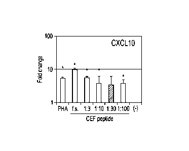
100271 Figures IA ¨ 1L depict induction of various immune related mRNAs in
response to
stimulation by a control agent or by a pool of viral peptides.
[0028] Figures 2A ¨21 depict the kinetics of mRNA induction by a pool of viral
peptides in
comparison to phytohemagglutinin (PHA).
DETAILED DESCRIPTION
[0029] Alterations in immune function, whether function is reduced or
increased, are a source
of a variety of potential health concerns. For example, overactive immune
function, in some cases,
can lead to autoimmune diseases. In other cases, decreased immune function can
result in a propensity
for developing infections, being increasingly at risk for certain diseases,
and/or development of cancer
of various types. As such, knowing the current immune status of a subject
could be a very important
piece of information in order to maintain a subject's health or treat a
subject for a particular ailment.
- 10d-
CA 02883810 2016-06-03
72689-224
Immune Function - General and Peptide Specific
[0030] A variety of cell types, proteins, and pathways that are functionally
interrelated make
up the immune system. The function of the immune system is to protect a host
from disease by
identifying and then eliminating pathogens and/or undesired cells (e.g.,
damaged cells or tumor cells).
As many of the pathogens and undesired cells that cause infections or diseases
are foreign to a host (or
endogenous cells that have lost some "self' aspect and gained some "non-self'
aspect) a first step in
the immune cascade is often identifying particular cells as "non-self."
Endogenous cells are
recognized by the expression of Class I Major Histocompatibility Complex
(MHC). Those cells
without Class I MHC or with reduced levels of expression may be targeted by
the immune system
- 10e -
CA 02883810 2015-03-04
WO 2014/039231
PCT/US2013/055605
as damaged "self' or "non-self' cells. Foreign pathogens are processed by the
immune
system and antigens derived from the foreign cells are complexed with MHC,
thereby
enabling other cells in the immune system to later recognize and target cells
bearing such
foreign antigens.
[0031] While
the immune system is comprised of many different cell types,
white blood cells (WBCs; leukocytes) are one of the key functional classes
immune cells.
Lymphocytes are a subtype of WBC that are further divided in Natural Killer
(NK) cells, T
cells and B cells. Natural killer (NK) cells are specialized, cytotoxic
lymphocytes target
and destroy, among others, tumor cells, virally infected cells, or damaged
"self' cells. T
cells are involved in cell-mediated immunity (discussed more below) whereas B
cells are
primarily responsible for humoral immunity (relating to antibodies). T cells
are
distinguishable from other lymphocyte types by the presence of the T-cell
receptor on the
cell surface. T cells are capable of inducing the death of infected somatic or
tumor cells.
Cytokines (e.g., those released due to inflammation or infection) or
presentation of a
foreign antigens activate NK cells and cytotoxic T cells, which then release
small granules
containing various proteins and proteases. One such released protein,
perforin, induces
pore formation in the membrane of a targeted cell, allowing proteases, such as
granzymes,
to enter the targeted cell and induce programmed cell death (apoptosis). Thus,
T cells,
among other immune cell types, play an important role in the ongoing immune
function
and overall health of a subject.
[0032] As
mentioned above, T cells express T cell receptors on their surface,
which function to recognize specific self MHC molecules expressed on the
surface of
neighboring cells. Antigen Presenting Cells (APC) work in conjunction with MHC
and T
cells to combat infection or foreign bodies. APCs process foreign antigens
(for example,
by phagocytosis and subsequent digestion) and present peptide fragments of the
foreign
antigens in a complex with the MHC molecules on the surface of the subject's
own cells.
Peptide¨MI-IC complexes on APCs then interact with the T-cell receptor on
certain T cells
(e.g., CD4 positive T cells), which is the first step in the establishment of
peptide-specific
immunity. The fraction of T cells which interact with the APC then produce
specific
clones comprising pools of effector T cells and memory T cells.
[0033] Effector
T cells (such as CD8+ T cells) are outfitted to specifically
recognize the particular foreign antigen that was processed by the APC. They
function in
-11-
CA 02883810 2015-03-04
WO 2014/039231
PCT/US2013/055605
the short to mid-term to attack cells expressing the foreign antigen, such as
cancers,
infected cells, and the like. This is known as the primary cell-mediated
immune response.
[0034] Memory T
cells play a more prominent role in the secondary cell-
mediated immune response. The memory cells represent a "pool" of cells that
are primed
to recognize the particular foreign antigen that was initially presented to
the T cells in the
form of the peptide-MI-IC complex. Upon a subsequent exposure to the foreign
antigen,
the memory T cells can rapidly generate additional effector T cells to combat
the cells
expressing the foreign antigen.
[0035] As a
result of the cascades of events outlined above, a subject generates
a first, slower response to an antigen (primary cell-mediated immune
response), and
simultaneously primes their immune system to be prepared to mount a more rapid
attack
upon a subsequent exposure (secondary cell-mediated immune response).
Categories of Immune Function
[0036]
Generally speaking, the immune cascades described above can be
characterized by the various types of immune function involved. The main
categories of
immune activity come together functionally to ensure that the immune system
can
effectively pilot immune cells to an area of the body where they are needed
and, once
there, act to inhibit and/or kill foreign cells or otherwise assist in
mounting an immune
response. These categories include, but are not limited to, recruitment
function, killer
function, suppressor (of killer) function, and helper function. A variety of
other functions,
e.g., antigen presentation, regulation of angiogenesis, pain modulation, etc.
are also
included.
[0037] A
threshold step in the initiation of effective immune function is the
delivery of immune cells from regions of storage to the site of a foreign cell
or antigen.
This recruitment function is essential for the proper function of the immune
system.
Regions from which immune cells are mobilized include, but are not limited to,
whole
blood, bone marrow, the lymphatic system, and other areas. Recruitment of
immune cells
allows recognition of foreign antigens at the location of the foreign antigen
(e.g., a tumor
or infection). Recruitment is often initiated by release of chemokines from
foreign cells or
even from endogenous cells that are in the region of the foreign cell.
Recruitment function
that is compromised or malfunctioning means that immune cells cannot be
properly
instructed on where to go to function. Recruiter function is provided, in some
-12-
CA 02883810 2015-04-07
72689-224
embodiments, by chemokines or other chemotactic molecules. In some
embodiments,
chemokines of a particular motif function to recruit other immune molecules.
For
example, in several embodiments, CCL molecules, such as CCL-2, CCL-4, CCL-8,
or
CCL-20 are involved in recruiting other immune cells. In other embodiments,
CXCL
molecules, such as CXCL-3 or CXCL-10 are involved. In some embodiments, other
chemokine effectors, whether C-C or C-X-C motif or another variety, are
involved.
[0038] After having been recruited to the proper location, the
other types of
immune cells can perform their designated function, which in some embodiments,
is to kill
the target cell(s). In some embodiments, the death of the target cells occurs
via apoptosis.
For example, when the target is a tumor, one or more cells having killer
function are
recruited to the target site. In some embodiments, such killer cells express
one or more of
molecules such as Granzyme B, perforin, TNFSF1 (lymphotoxin), TNFSF2 (TNF-
alpha),
TNFSF 5 (CD40 ligand), TNFSF6 (Fas ligand), TNFSF14 (LIGHT), TNFSF 15 (TL1A),
and/or CD16. As such, the recruitment of these cells to the target site
initiates a cascade
that results in the destruction of the target cells, and thus reali7es one
goal of the immune
system, e.g., destruction and/or removal of a foreign body or cell.
[0039] Another function of the immune system, is to provide a
negative
influence (e.g., a limit) on the killing function of the immune system This
is, at least in
part, to prevent overactive immune function, which could lead to autoimmune
disorders).
Cells that participate in this limiting function can be recognized by markers
including, but
not limited to, IL10, TGF-beta, (forkhead box p3) FoxP3, CD25, arginase, CTLA-
4, and
/or PD-1. These cells help to ensure proper overall immune function by keeping
the
activity of the immune system balanced.
[0040] Additional cells types may be involved, to varying
degrees, in the killing
function of the immune system and/or the self-limiting function of the immune
system.
Helper T-cells (Th cells) are a sub-group of lymphocytes that assist in
maximizing the
capabilities of the immune system. Unlike the cells described above, Th cells
lack cytotoxic
or phagocytic activity. Th cells are, however, involved in activating and
directing other
immune cells such as the cytotoxic T cells (e.g., the killer cells described
above). Th cells
are divided into two main subcategories (Thl or Th2) depending on, among other
factors,
what cell type they primarily activate, what cytokines they produce, and what
type of
immune stimulation is promoted. For example, Thl cells primarily partner with
-13-
i
CA 02883810 2015-03-04
WO 2014/039231
PCT/US2013/055605
macrophages, while Th2 cells primarily partner with B-cells. Thl cells produce
interferon-
gamma, TNF-beta, and IL-2, while Th2 cells product ILL IL5, IL6, IL10 and
IL13.
Markers of the subsets of Th cells are known and can be used to identify the
induction of
certain Th cell subtypes in response to stimulation. For example, the
induction of IL2 or
IFNG represent responses to stimulation by Thl cells, while induction of IL4
or IL10
represent responses to stimulation by Th2 cells. Other subtypes, such as Th17
are
represented by other markers, such as IL17 (see e.g., Tables 5 and 6).
[0041] A
variety of other markers of accessory immune functions also exist.
For example, antigen presentation function can be evaluated by measurement of
GMCSF,
B-cell proliferation can be evaluated by measurement of IGH2, angiogenesis can
be
evaluated by measurement of VEGF (which may be of particular importance with
respect
to possible tumor formation, as many tumors have increased blood flow
demands), pain
can be evaluated by measurement of POMC.
[0042] The
killing function of the immune system such as the function of NK
cells and cytotoxic T cells is important, in several embodiments, for
destruction of
cancerous cells and combating infections and/or inflammation (among other
applications).
Due to their ability to potentially kill both unwanted target cells as well as
normal
endogenous cells, NK cells possess two types of surface receptors, activating
receptors
and inhibitory receptors. Together, these receptors serve to balance the
activity of, and
therefore regulate, the cytotoxic activity of NK cells. Activating signals are
required for
activation of NK cells, and may involve cytokines (such as interferons),
activation of FcR
receptors to target cells against which humoral immune responses have been
mounted,
and/or foreign ligand binding to various activating NK cell surface receptors.
Targeted
cells are then destroyed by the apoptotic mechanism described above.
[0043]
Similarly, cytotoxic T cells also require activation, thought to be
through a two signal process resulting in the presentation of a foreign (e.g.,
non-self)
antigen to the cytotoxic T cells. Once activated, cytotoxic T cells undergo
clonal
expansion, largely in response to interleukin-2 (IL-2), a growth and
differentiation factor
for T cells. Cytotoxic T cells function somewhat similarly to NK cells in the
induction of
pore formation and apoptosis in target cells. In several embodiments, the
identification of
a subject's specific T-cell function is important to determining the ability
of the subject to
mount a response to a particular foreign antigen. In addition, in several
embodiments, the
-14-
CA 02883810 2015-03-04
WO 2014/039231
PCT/US2013/055605
function of the T cells determines, at least in part, the rate of response of
the subject's
immune function.
[0044] The self-
limiting nature of immune function is believed to be moderated
by T-reg and MDSCs. Developing in the thymus, many T-reg express the forkhead
family
transcription factor FoxP3 (forkhead box p3). In many disease states,
particularly cancers,
alterations in T-reg numbers, particularly those T-reg expressing Foxp3, are
found. For
example, patients with tumors have a local relative excess of Foxp3 positive T
cells which
inhibits the body's ability to suppress the formation of cancerous cells.
MDSCs do not
destroy offensive T cells, however, they do alter how cytotoxic T cells
behave. MDSCs
secrete arginase (ARG), a protease that breaks down the amino acid arginine.
Lymphocytes, including cytotoxic T cells and NK cells are indirectly dependent
on arginine
for activation. Secretion of ARG by MDSCs limits the activation of NK cells
and
cytotoxic T cells. Thus, in several embodiments, peptide specific immunity may
be
impacted by the limitation of activation of T cells. In some cases, self-
limiting regulation
by T-reg and MDSCs may lead to an overall limiting of the functionality of the
immune
system in a local tissue environment. This has the potential to lead to
reduced killing
function and which may be insufficient to completely eradicate foreign cells.
[0045] As
discussed in more detail below, the evaluation of peptide-specific
immunity allows assessment of the efficacy of a vaccine, the probability that
a subject will
(or will not) mount an immune response against a certain antigen, and tracking
of immune
function related to a specific antigen or class of antigens over time (among
other
applications). Moreover, by the methods disclosed herein specific antigens (or
classes of
antigens) can be evaluated with respect to how they stimulate immune function
in an
individual.
Diagnostic Measures
[0046] A
subject may receive immunotherapy, or a vaccination, directed to
treat (e.g., eliminate) a particular population of cells in a subject, for
example, a cancerous
tumor. In response to the immunotherapy or vaccination production of a
specific IgG may
be induced in the subject. While the titer of that specific IgG can be
measured by a variety
of immunoassays, these assays are generally not informative with respect to T-
cell function
that is specific the vaccine. Thus, no routine diagnostic test presently
exists to determine
the function of T cells directed against specific targets (e.g., a foreign
antigen or peptide
-15-
CA 02883810 2015-03-04
WO 2014/039231
PCT/US2013/055605
fragment of that antigen as discussed above). Technical difficulties such as
cell isolation,
varying culture conditions, and methods to detect or quantify function have
precluded such
routine diagnostic assays. For example, in order to stimulate the T-cell
receptor on a
subject's T cells, living cells from that subject are required (T cells do not
recognize non-
self MHC); in other words, MHC matched donor cells are necessary. This
presents an
issue with respect to the practical use of diagnostic assays as a subject's
own cells must be
collected and grown in culture prior to assessing peptide specific T-cell
immunity.
[0047] To
address these limitations and provide a more routine diagnostic
assessment of peptide specific T-cell immunity, several embodiments disclosed
herein
enable use of a panel of one or more exogenous peptides (e.g., those for which
an
assessment of a subject's immunity is desired). In several embodiments, the
exogenous
peptides are used to supplement those peptides which have already been
processed by the
APCs, thereby allowing a more complete determination of the T-cell function of
that
particular subject.
[0048] In
several embodiments, the methods disclosed herein are used to
monitor the immune function of a subject over time, with respect to a
particular peptide
target. For example, in some embodiments, a plurality of samples can be
collected from
the subject and the peptide specific T-cell function is assessed. The results
of this
monitoring over time, in some embodiments, enable a determination of whether
that
subject has had or continues to have an increased level of immune activity
specific to that
peptide. In some embodiments, this monitoring over time can be used to assess
whether a
subject has developed in immunodeficiency (e.g., congenital or acquired
immunodeficiency). In several embodiments, this assessment is made by
collecting a
sample from the patient and exposing it to a panel of specific peptides. In
several
embodiments, this exposure will result in induction of certain immune related
mRNAs.
Subsequent samples collected over time and tested in the same fashion, should
an mRNA
that was previously induced show a lack of or a diminished induction, would
demonstrate
a deficient immune response to one or more of the specific peptides in the
panel.
Advantageously, such a determination enables detection of immunocompromised
status in
a subject at early stages, thereby allowing appropriate medical intervention,
if needed. In
some embodiments, rather than a panel of specific peptides, singular peptides
are used.
-16-
CA 02883810 2015-03-04
WO 2014/039231
PCT/US2013/055605
[0049] In
several embodiments, monitoring of the peptide specific T-cell
function can be used to assess the efficacy of a vaccine therapy. Prior to
being exposed to
an antigen, a subject will not have mRNA induced in response to exposure of
their blood
samples to a peptide derived from the antigen. If that subject subsequently
receives a
vaccine comprising that particular antigen, the subject's immune system will
process the
antigen as described herein. Thereafter, exposure of a blood sample from the
subject to a
peptide derived from the antigen would induce mRNA (because the subject has
generated
immune cells that recognize that peptide/antigen). In this manner, the
efficacy of a vaccine
therapy can be monitored in a subject. For example, after an initial
vaccination, the
induction of mRNA after exposure to the peptide can be used as a baseline for
ongoing
monitoring. After collecting future samples and testing them as disclosed
herein, a drop in
the level of induction over time indicates a loss of efficacy of the vaccine.
This suggests,
in several embodiments, that a new "booster" of vaccine, or an alternative
vaccine, may be
necessary. In some embodiments, the determination of induction of mRNA in an
initial
sample is used as a threshold. In other words, if induction of particular mRNA
is not
sufficient to reach a certain level, then, in some embodiments, another dose
of the vaccine
is administered. The testing of the patient's responsiveness is then repeated,
and if the
threshold induction is met, no additional vaccine administrations need be made
(until such
time as a "booster" is required, as described above).
[0050] In some
embodiments, the methods disclosed herein are used to
determine whether a subject has been previously exposed to a particular
peptide. For
example, in several embodiments, a subject had not been previously exposed to
a particular
antigen, induction of immune related mRNA would likely not be detected. This
is due to,
at least in part, a relative lack of memory T cells, as discussed above. In
contrast, if a
subject had in fact been previously exposed to the specific peptide, induction
of immune
related mRNA would result, as the first exposure would have led to production
of a pool
of memory T cells. Thus, in several embodiments, a determination can be made
of
whether the subject is at risk for a hyperactive immune response based on a
subsequent
exposure to that peptide.
[0051] In
several embodiments, assessment of a subject's peptide specific
immunity enable a determination of whether a subject can mount an effective
response
against a particular type of foreign cell, e.g., a particular type of cancer.
For example, if a
-17-
CA 02883810 2015-03-04
WO 2014/039231
PCT/US2013/055605
specific cancer cell produces a marker (e.g., a peptide) that is unique to the
cancer cell (as
compared to normal cells) and exposure of a sample from a subject to that
specific peptide
results in the induction of immune related mRNA associated with the killing
function (e.g.,
cytotoxic T cells), it is likely that the subject is able to mount an immune
response against
that cancer cell. In contrast, exposure to sample from the subject to the
specific peptide of
the cancer cell and a lack of induction of immune function related mRNA
associated with
killing would indicate the subject is less likely to be able to mount an
immune response to
eliminate the cancer cell. In such instances, adjunct therapy (e.g., surgery,
chemical or
radiation therapy) may be advisable.
[0052] In
several embodiments, the methods disclosed herein are used to
identify a subject having cellular immunity against a specific antigen and
treating that
subject accordingly. In several embodiments, such a method comprises obtaining
at least
two biological samples (e.g., blood samples) from a subject, exposing said one
of such
samples to a peptide derived from a specific antigen of interest and treating
a second
sample to identical conditions (without the peptide) and quantifying the level
of expression
of one or more T-cell function associated markers in the samples. As the
expression of the
T-cell function markers is analyzed, a subject can be identified as having
cellular immunity
against the specific antigen when the expression of said one or more T-cell
function
associated markers is increased in said sample to the peptide as compared to
the sample
not exposed to the peptide. Likewise, the subject is identified as not having
cellular
immunity against said specific antigen when the expression of said one or more
T-cell
function associated markers is substantially similar in the two samples
(exposed to peptide
vs. not exposed). Based on that identification, the subject can be treated
accordingly.
Thus, in those embodiments wherein the subject exhibits cellular immunity, an
immune-
based therapy can be administered to the subject. If no cellular immunity is
detected, non-
immune based therapies may prove more effective for that subject. In several
embodiments, the subject can be "vaccinated" with the peptide from the antigen
of interest,
in order to boost the cellular immune response that the subject mounts.
[0053] In
several embodiments, there is also provided a method of treating a
subject based on their peptide-specific T-cell function of a subject. Similar
to the above, a
plurality of blood samples are collected from the subject, at least one of
which is exposed
to a peptide derived from an antigen of interest and one of which is not so
exposed. The
-18-
CA 02883810 2015-03-04
WO 2014/039231
PCT/US2013/055605
level of expression of one or more T-cell function associated markers in the
exposed and
unexposed samples is quantified and when a greater level of expression of the
T-cell
function associated markers is present in the exposed sample as compared the
non-exposed
sample, the subject has cellular immunity to that specific antigen.
Conversely, when the
level of expression is not significantly different in the exposed versus
unexposed samples,
the subject lacks cellular immunity to said antigen. Thereafter,
administration of a
particular therapy is performed; an immune-based therapy if the subject does
have cellular
immunity and a non-immune based therapy if the subject lacks cellular
immunity.
[0054] In
several embodiments, the quantification is performed according to
the methods described herein. For example, in one embodiment, the
quantification
comprises adding a primer and a reverse transcriptase to RNA isolated from
each of
samples (exposed and unexposed) to generate complementary DNA (cDNA) and
contacting said cDNA with sense and antisense primers that are specific for
one or more
T-cell function associated markers and a DNA polymerase to generate amplified
DNA.
[0055]
Additionally, several embodiments are directed to determining the
likelihood of the efficacy of a peptide-specific therapy and then
administering the therapy,
if appropriate. In several embodiments, the methods comprise obtaining a first
and a
second blood sample from a subject, exposing said first blood sample to a
solvent
comprising a peptide antigen against which said peptide-specific therapy is to
be directed
and exposing said second blood sample to said solvent alone. Thereafter the
level of
expression of one or more T-cell function associated markers is quantified.
These markers
may be, depending on the embodiment, markers of cytotoxic T-cells or cytotoxic
T-cell
function or T-reg and/or MDSC function markers. A peptide-specific therapy is
then
identified as having an increased likelihood of efficacy when said T-cell
function associated
markers are associated with cytotoxic T-cells or cytotoxic T-cell function and
expression
of said T-cell function associated markers is increased in said first sample
as compared to
said second sample. Alternatively, the quantification may result in an
identification of a
decreased likelihood of efficacy of the peptide-specific therapy when (a) said
T-cell
function associated markers are associated with T-reg and/or MDSC or T-reg
and/or
MDSC function and expression of said T-cell function associated markers is
increased in
said first sample as compared to said second sample, or (b) said T-cell
function associated
markers are associated with cytotoxic T-cells or cytotoxic T-cell function and
the
-19-
CA 02883810 2016-06-03
= 72689-224
expression of said T-cell function associated markers is substantially similar
in said first sample as
compared to said second sample. Based on the identification of the peptide-
specific therapy being
effective, the therapy can then either be administered to the subject (when
determined likely to be
effective) or administration can be foregone (when determined unlikely to be
effective). In several
embodiments, the peptide-specific therapy is an anti-cancer therapy.
[0056] Also, in one embodiment, there is provided a method for identifying a
peptide-specific
therapy effective to treat an autoimmune disorder in a subject and thereafter
treating the subject. The
method comprises, in several embodiments, obtaining a blood sample from said
subject at risk for or
suffering from an autoimmune disorder, exposing a first portion of said blood
sample to a solvent
comprising a specific peptide associated with said peptide-specific therapy,
exposing a second portion
of said blood sample to said solvent alone, and quantifying the level of
expression of one or more
mRNA associated with self-limiting immune function in said first and said
second portion of said
blood sample, determining that the peptide-specific therapy is likely to be
efficacious when there is a
greater level of expression in the first portion of the blood sample as
compared to the second portion of
the blood sample, and when the peptide-specific therapy is determined to be
likely to be effective,
administering the peptide-specific therapy to the subject.
[0057] In several embodiments, the methods disclosed herein can be used to
determine the
potential efficacy of a particular type of peptide vaccine. For example, in
certain autoimmune
situations, there exist cells or proteins that attack other endogenous cells
within a subject's body
(as occurs with type I diabetes). Several embodiments of the methods disclosed
herein are useful for
determining the potential efficacy of a putative peptide vaccine. In other
words, if the exposure of a
sample from a subject to the putative peptide vaccine results in induction of
mRNA related to the
self-limiting immune function discussed above, then it is likely that that
putative peptide vaccine
would be efficacious to treat the autoimmune situation. This is because the
diagnostic test has
indicated that the peptide will induce a set of cells associated with self-
limitation of the subject
immune function. Moreover, these cells will be specifically directed against
those cells that also bear
the specific peptide and are attacking endogenous cells (e.g., the "culprit"
cells).
Target Conditions
- 20 -
CA 02883810 2015-04-07
72689-224
[0058] In several embodiments, the methods and compositions
disclosed here
are used to assess a subject's ability to mount an immune response against a
variety of
different specific antigens. For example, in several embodiments, the foreign
antigen can
be derived from cancerous cells (or other mutated cells). Markers specific to
a variety-of
cancers can be tested for, depending on the embodiment. For example, in
several
embodiments a subject can be tested for specific immunity to -a variety of
cancers
including, but not limited to lymphoblastic leukemia (ALL), acute myeloid
leukemia
(AML), adrenocortical carcinoma, kaposi sarcoma, lymphoma, gastrointestinal
cancer,
appendix cancer, central nervous system cancer, basal cell carcinoma, bile
duct cancer,
bladder cancer, bone cancer, brain tumors (including but not limited to
astrocytomas,
spinal cord tumors, brain stem glioma, craniopharyngioma, ependymoblastoma,
ependymoma, medulloblastoma, medulloepithelioma), breast cancer, bronchial
tumors,
burkitt lymphoma, cervical cancer, colon cancer, chronic lymphocytic leukemia
(CLL),
chronic myelogenous leukemia (CML), chronic myeloproliferative disorders,
ductal
carcinoma, endometrial cancer, esophageal cancer, gastric cancer, Hodgkin
lymphoma,
non-Hodgkin_lymphoina,hany cell leukemia, renal cell cancer, leukemia, oral
cancer, liver
cancer, lung cancer, lymphoma, melanoma, ocular cancer, ovarian cancer,
pancreatic
cancer, prostate cancer, pituitary cancer, uterine cancer, and vaginal cancer.
[0059] Alternatively, in several embodiments, a subject can be
tested for
specific immunity to infections cells derived from bacteria, viruses, fungi,
and/or parasites.
In some embodiments, T cells responsive to infections of bacterial origin
(e.g., infectious
bacteria is selected the group of genera consisting of Bordetella, Borrelia,
Brucella,
Campylobacter, Chlamydia and Chlamydophila, Clostridium, Corynebacterium,
Enterococcus, Escherichia, Francisella, Haemophilus, Ilelicobacter,
Legionella,
Leptospira, Listeria, Mycobacterium, Mycoplasma, Neisseria, Pseudomonas,
Rickettsia,
Salmonella, Shigella, Staphylococcus, Streptococcus, Treponema, Vibrio, and
Yersinia,
and mutants or combinations thereof) can be identified by several embodiments
of the
methods disclosed herein.
[0060] In some embodiments, the ability of a subject to mount a
specific
response against infectious agents of a viral origin can be assessed. The
viruses can
include, but are not limited to adenovirus, Coxsacicievirus, Epstein-Barr
virus, hepatitis a
virus, hepatitis b virus, hepatitis c virus, herpes simplex virus, type 1,
herpes simplex virus,
-21-
_
CA 02883810 2015-03-04
WO 2014/039231
PCT/US2013/055605
type 2, cytomegalovirus, ebola virus, human herpesvirus, type 8, HIV,
influenza virus,
measles virus, mumps virus, human papillomavirus, parainfluenza virus,
poliovirus, rabies
virus, respiratory syncytial virus, rubella virus, and varicella-zoster virus,
and combinations
thereof . Exosomes can be used to treat a wide variety of cell types as well,
including but
not limited to vascular cells, epithelial cells, interstitial cells,
musculature (skeletal, smooth,
and/or cardiac), skeletal cells (e.g., bone, cartilage, and connective
tissue), nervous cells
(e.g., neurons, glial cells, astrocytes, Schwann cells), liver cells, kidney
cells, gut cells, lung
cells, skin cells or any other cell in the body.
[0061] In
several embodiments, the methods disclosed herein are useful for the
determination of whether a subject can (or has) mount an immune response to
cells having
altered metabolic function. In some embodiments, cells with a metabolic
discrepancy (as
compared to normal cells) express specific identifying markers. A subject may
mount an
immune response against such cells, in an effort to avoid the possibility of
adverse effects
based on the malfunctioning cell. For example, the metabolic disruption of a
cell may
cause a cell to be converted from a normal cell to a pre-cancerous cell. Thus,
the immune
response can eliminate the cell prior to the cell becoming cancerous. In
several
embodiments, a propensity for autoimmunity can be detected. In several
embodiments, the
methods disclosed herein can be used to determine if a subject has in fact
previously
generated a cell with a certain metabolic malfunction. For example, the
methods disclosed
herein, in some embodiments, allow for the detection of peptides specific to a
particular
kind of metabolic dysfunction.
Methods
[0062] In
several embodiments, the samples used in the claimed methods are
whole blood samples. In several embodiments, the blood samples can be
heparinized.
Once collected, the blood samples are exposed to at least one specific
antigen. As
discussed above, the antigen can be derived from any of a variety of sources
(cancer cells,
viruses, bacteria, etc.). In some embodiments, the exposure occurs at a
temperature
approximating a physiological temperature. In
several embodiments, exposure is
performed at a temperature ranging from about 30 C to about 40 C. In several
embodiments, the exposure is performed at approximately 37 C.
Depending on the
embodiment, the duration of the exposure can vary from about one hour to about
eight
hours. In some embodiments, exposure lasts for about 1 to about 2 hours, about
two
-22-
CA 02883810 2016-06-03
72689-224
hours to about three hours, about three hours to about four hours, about four
hours to about five hours,
about five hours to about six hours, or about six hours to about eight hours.
Longer or shorter
durations of exposure are also used, depending on the embodiment. In some
embodiments single
peptides are used, while in other embodiments, a plurality or panel of
peptides is used. In several
embodiments, the peptides that make up the panel are all derived from a common
general source,
e.g., all peptides are from a single type of cancer cell. In some embodiments,
the peptides making up
the panel are derived from different sources, e.g. some peptides from cancer
cells and some peptides
from infectious agents such as bacteria. The flexibility in designing the
panel of peptides allows
customization of the determination of peptides specific T-cell function
depending on the needs of a
particular subject being tested.
[0063] In some embodiments, peptides are diluted with non-reactive solvent
(e.g. phosphate
buffered saline) in order to tailor the amount of induction that is detected,
such that a desired degree of
signal gain is achieved (e.g., signal-to-noise ratio is sufficient to allow
accurate quantification).
Thus, in several embodiments the methods comprise exposing a blood sample
(e.g., a whole blood
sample) to a peptide derived from an antigen of interest, that peptide have
been dispersed
(e.g., diluted) in a solvent. In several embodiments, the blood sample is a
whole blood sample. In
several embodiments, no additional antigen presenting cells are added to the
sample. Despite the use
of a solvent to dilute the peptide in several embodiments, in other
embodiments, a solvent is not used
(e.g., if a peptide has been dried, such as with a freeze-dried peptide).
[0064] In those embodiments in which mRNA levels are determined, erythrocytes
and blood
components other than leukocytes are optionally removed from the whole blood
sample. In other
embodiments, whole blood is used without removal or isolation of any
particular cell type. In
preferred embodiments, the leukocytes are isolated using a device for
isolating and amplifying mRNA.
Embodiments of this device are described in more detail in United States
Patent Nos. 7,745,180,
7,968,288, 7,939,300, 7,981,608, and 8,076,105.
[0065] In brief, certain embodiments of the device comprise a multi-well plate
that contains a
plurality of sample-delivery wells, a leukocyte-capturing filter underneath
the wells, and an mRNA
capture zone underneath the filter which contains immobilized oligo(dT). In
certain embodiments, the
device also contains a vacuum box adapted to receive the filter plate to
create a seal between the plate
and the box, such that when vacuum pressure is applied, the blood is drawn
from the sample-delivery
wells across the leukocyte-capturing filter, thereby capturing the leukocytes
and allowing
non-leukocyte blood components to be removed by washing the filters. In other
embodiments,
-23 -
CA 02883810 2016-06-03
72689-224
other means of drawing the blood samples through out of the sample wells and
through the across the
leukocyte-capturing filter, such as centrifugation or positive pressure, are
used. In preferred
embodiments of the device, leukocytes are captured on a plurality of filter
membranes that are layered
together. In several embodiments, the captured leukocytes are then lysed with
a lysis buffer, thereby
releasing mRNA from the captured leukocytes. The mRNA is then hybridized to
the oligo(dT)-
immobilized in the mRNA capture zone. Further detail regarding the composition
of lysis buffers that
may be used in several embodiments can be found in United States Patent
8,101,344. In several
embodiments, cDNA is synthesized from oligo(dT)-immobilized mRNA. In preferred
embodiments,
the cDNA is then amplified using real time PCR with primers specifically
designed for amplification
of infection-associated markers. In several embodiments, other methods of
quantifying mRNA are
used, including, but not limited to, northern blotting, 2-dimensional RT-qPCR,
RNase protection, and
the like. In several embodiments, other measurement endpoints are used, such
as, for example, protein
levels and/or functional assays.
100661 After the completion of PCR reaction, the various mRNA (as represented
by the
amount of PCR-amplified cDNA detected) for one or more leukocyte-function-
associated markers are
quantified. In certain embodiments, quantification is calculated by comparing
the amount of mRNA
encoding one or more markers to a reference value. In other embodiments, the
reference value is
expression level of a gene that is not induced by the stimulating agent, e.g.,
a house-keeping gene.
In certain such embodiments, beta-actin is used as the reference value.
Numerous other house-keeping
genes that are well known in the art may also be used as a reference value. In
other embodiments, a
house keeping gene is used as a correction factor, such that the ultimate
comparison is the induced
expression level of one or more leukocyte-function-associated markers as
compared to the same
marker from a non-induced (control) sample. In still other embodiments, the
reference value is zero,
such that the quantification of one or more leukocyte-function-associated
markers is represented by an
absolute number. In several
- 24 -
CA 02883810 2015-03-04
WO 2014/039231
PCT/US2013/055605
embodiments, two, three, or more leukocyte-function-associated markers are
quantified.
In several embodiments, the quantification is performed using real-time PCR
and the data
are expressed in terms of fold increase (versus an appropriate control). In
certain
embodiments, the level of expression of one or more T-cell function associated
markers is
quantified using a method selected from the group consisting of reverse-
transcription
polymerase chain reaction (RT-PCR), real-time RT-PCR, northern blotting,
microarray
gene analysis, digital PCR, RNA sequencing, nanoplex hybridization,
fluorescence
activated cell sorting, ELISA, mass spectrometry, and western blotting. In
some
embodiments, an increased likelihood of efficacy is observed when certain T-
cell function
associated markers are decreased in expression. For example, in several
embodiments an
increased likelihood of efficacy of a peptide-specific therapy is identified
when T-cell
function associated markers are associated with cytotoxic T-cells or cytotoxic
T-cell
function and expression of said T-cell function associated markers is
decreased in said first
sample as compared to said second sample. Similarly, a decreased likelihood of
efficacy
can be identified, in certain embodiments, when T-cell function associated
markers are
associated with T-reg and/or MDSC or T-reg and/or MDSC function and expression
of
said T-cell function associated markers is decreased in said first sample as
compared to
said second sample, or the T-cell function associated markers are associated
with cytotoxic
T-cells or cytotoxic T-cell function and the expression of said T-cell
function associated
markers is substantially similar in said first sample as compared to said
second sample.
[0067] As used
herein, the term "increased" shall be given its ordinary meaning
and shall also refer to increases in expression of greater than about 5%,
greater than about
10%, greater than about 15%, greater than about 20%, greater than about 25%,
greater
than about 50%, or more. Likewise, As used herein, the term "decreased" shall
be given
its ordinary meaning and shall also refer to decreases in expression of
greater than about
5%, greater than about 10%, greater than about 15%, greater than about 20%,
greater
than about 25%, greater than about 50%, or more. In some embodiments, an
increase
refers to a statistically significant increase in expression (e.g., p<0.05
based on an art-
established statistical analysis). In some embodiments, a decrease refers to a
statistically
significant decrease in expression (e.g., p<0.05 based on an art-established
statistical
analysis.)
EXAMPLES
-25-
CA 02883810 2016-06-03
72689-224
[0068] A specific embodiment will be described with reference to the following
example,
which should be regarded in an illustrative rather than a restrictive sense.
Example 1 - Induction of Immune-Function-Related mRNA in Response to Peptide
Exposure
[0069] While peptides on MHC are known to be derived from digested proteins in
APC,
however, the present example evaluates the replacement (or supplementation) of
endogenous peptides
with exogenous peptides. A commercially available peptide pool (CEF peptide
pool; Mabtech, 01-1
45227, Cincinnati, U.S.A.) was employed, though as discussed above, single
peptide or customized
panels of peptides are used. This pool contains 23 different class-I
restricted peptides, all defined as
common CD8+ T-cell epitopes derived from cytomegalovirus, Epstein-Barr virus
and influenza virus.
This panel induces IFN-y production by virus-specific CD8+ T-cells in almost
90% of Caucasians and
also elicits Perforin, Granzyme B and MIP-1I3 responses in many individuals.
[0070] The stock peptide (200 ptg/mL) was diluted with 1:3, 1:10, 1:10, and
1:100 in PBS,
and applied to heparinized whole blood at 37 C for 4 hours. No additional
cells were added. Positive
and negative controls leucoagglutinin (PHA-L) and PBS were used, respectively.
[0071] As shown in Figure 1, positive control PHA-L induced GMCSF, IFNG,
TNFSF2,
CXCL10, CCL4, IL4, IL10, CTLA4, and CXCL3, whereas control housekeeping gene
beta actin
(ACTB) was not induced. This confirms the appropriate performance of the
general immune response
assay. The CEF peptide pool induced GMCSF, IFNG, TNFSF2, CXCL10, and CCL4 in a
dose
dependent manner.
[0072] Figure 2 depicts the kinetics of the induction of mRNA in response to
the exposure to
the CEF panel. Exposure was performed as described above for durations of 1,
2, 4, 8, and 24 hours
and mRNA expression was evaluated by real time PCR (closed circles represent
induction by CEF and
open triangles are the PBS control). The similarity of the induction of the
various mRNA suggest that
the exogenous peptides replace (or supplement) existing peptides on MHC,
rather than being taken up
by cells and processed to be complexed with the MHC (which would shift the
kinetic curve for the
CEF exposure to the right).
[0073] These data indicate that the exposure of leukocytes to exogenous
peptides allows for
immune-function-related mRNAs to be induced. As such, this experiment
demonstrates that peptide
specific T-cell immunity can be assessed by the ex vivo methods disclosed
herein.
- 26 -