Note: Descriptions are shown in the official language in which they were submitted.
CA 02883885 2015-03-03
WO 2014/046753 PCT/US2013/046041
METHOD AND SYSTEM FOR TREATMENT
OF BIOLOGICAL TISSUE
FIELD OF THE INVENTION
[0001] The present invention relates to methods for treating biological
tissue. More
particularly, the present invention relates to methods and systems for
treating damaged and
diseased biological tissue; particularly, cardiovascular tissue.
BACKGROUND OF THE INVENTION
[0002] Myocardial infarction is a common presentation of ischemic heart
disease/coronary artery disease. The World Health Organization estimated in
2004 that
12.2% of worldwide deaths occurred as a result of ischemic heart disease.
Ischemic heart
disease was also deemed the leading cause of death in middle to high income
countries and
second only to respiratory infections in lower income countries. The Global
Burden of
Disease: World Health Organization 2004 Update, Geneva (2008). Worldwide more
than 3
million people present with a ST elevation myocardial infarction (STEMI) and 4
million
people present with a non-ST elevation myocardial infarction (NSTEMI) a year.
White, et
al., Acute Myocardial Infarction, Lancet 372 (9638), pp. 570-84 (August 2008).
[0003] Rates of death from ischemic heart disease have slowed or declined
in most high
income countries, although cardiovascular disease still accounted for 1 in 3
of all deaths in the
USA in 2008. Roger, et al., Executive summary: Heart Disease and Stroke
Statistics--2012
update: A report from the American Heart Association, Circulation 125 (1), pp.
188-97
(January 2012).
[0004] In contrast, ischemic heart disease is becoming a more common cause
of death in
the developing world. For example in India, ischemic heart disease had become
the leading
cause of death by 2004; accounting for 1.46 million deaths (14% of total
deaths). Deaths in
India due to ischemic heart disease were also expected to double during 1985-
2015. Gupta,
et al., Epidemiology and Causation of Coronary Heart Disease and Stroke in
India, Heart 94
(1), pp. 16-26 (January 2008).
[0005] Globally, it is predicted that disability adjusted life years
(DALYs) lost to ischemic
heart disease will account for 5.5% of total DALYs in 2030, making it the
second most
CA 02883885 2015-03-03
WO 2014/046753 PCT/US2013/046041
important cause of disability (after unipolar depressive disorder), as well as
the leading cause
of death by this date.
[0006] A myocardial infarction (a common presentation of ischemic heart
disease) often
occurs when a coronary artery becomes occluded and can no longer supply blood
to the
myocardial tissue, thereby resulting in myocardial cell death. When a
myocardial infarction
occurs, the myocardial tissue that is no longer receiving adequate blood flow
ultimately dies
(without effective intervention) and is eventually replaced by scar tissue.
[0007] Within seconds of a myocardial infarction, the under-perfused
myocardial cells no
longer contract, leading to abnormal wall motion, high wall stresses within
and surrounding
the infarct, and depressed ventricular function. The high stresses at the
junction between the
infarcted tissue and the normal tissue lead to expansion of the infarcted area
and remodeling,
i.e. a cascading sequence of myocellular events, over time.
[0008] Various methods for treating a myocardial infarction are often
employed. Such
methods include stabilizing the hemodynamics associated with a myocardial
infarction via
systemic delivery of various phainiacological agents and restoring the patency
of occluded
vessels via thrombolytic therapy or angioplasty and stents.
[0009] Several additional methods for treating a myocardial infarction are
directed to re-
establishing blood flow to the ischemic area through stimulation of
angiogenesis. Re-
establishing blood flow at the ischemic area can, and in many instances will,
reduce
symptoms associated with a myocardial infarction and/or improve cardiac
function.
[00010] Some methods for re-establishing blood flow and rehabilitating the
heart involve
invasive surgery, such as bypass surgery or angioplasty. Other methods employ
lasers to bore
holes through the infarctions and ischemic area(s) to promote blood flow. As
one can readily
appreciate, there are numerous incumbent risks associated with the noted
methods.
[00011] A further method for treating a myocardial infarction is the direct or
selective
delivery of bioactive or phaintacological agents to the infarction and/or
ischemic area (i.e.
effected or damaged cardiovascular tissue). Direct delivery of a bioactive or
pharmacological
agent to the effected cardiovascular tissue is often preferred over the
systemic delivery for
several reasons. A primary reason is that a substantially greater
concentration of such agents
that can be delivered directly into the effected cardiovascular tissue,
compared with the dilute
2
CA 02883885 2015-03-03
WO 2014/046753 PCT/US2013/046041
concentrations possible through systemic delivery. Another reason is the risk
of systemic
toxicity which can, and in many instances will, occur with doses of
pharmacological agents
that are typically required to achieve desired drug concentrations in the
effected
cardiovascular tissue.
[00012] One common method of delivering bioactive or pharmacological agents to
effected
cardiovascular tissue, e.g. damaged myocardial tissue, comprises advancing a
catheter
through the vasculature and into the heart to inject the agents directly into
the effected
cardiovascular tissue from within the heart.
[00013] Another method of delivering bioactive or pharmacological agents to
effected
cardiovascular tissue comprises epicardial, direct injection into the tissue
during an open chest
procedure. The bioactive agents that can be, and have been, administered to
the effected
cardiovascular tissue include various pharmacological agents, such as
antithrombotic agents,
e.g., heparin, hirudin, and ticlopidine, and cells that are capable of
maturing into actively
contracting cardiac muscle cells or regenerating cardiovascular tissue.
Examples of such cells
include myocytes, myoblasts, mesenchymal stem cells, and pluripotent cells.
[00014] However, to date, cell therapy of effected cardiovascular tissue has
not reached its
full potential, due, in part, to the failure of implanted cells to survive and
regenerate the
damaged tissue in ischemic area(s) or regions with inadequate vascularization.
[00015] It would thus be desirable to provide bioactive and pharmacological
agents (and
compositions) that promote tissue survival and induce neovascularization and
regeneration of
effected or damaged cardiovascular tissue, and improved methods for delivering
same to
effected cardiovascular tissue.
[00016] It is therefore an object of the present invention to provide
bioactive and
pharmacological agents (and compositions) that promote tissue survival, and
induce
neovascularization and regeneration of damaged cardiovascular tissue.
[00017] It is another object of the present invention to provide extracellular
matrix (ECM)
compositions, which, when delivered to damaged biological tissue;
particularly,
cardiovascular tissue, induce neovascularization, host tissue proliferation,
bioremodeling, and
regeneration of cardiovascular tissue and associated structures with site-
specific structural and
functional properties.
3
CA 02883885 2015-03-03
WO 2014/046753 PCT/US2013/046041
[00018] It is yet another object of the present invention to provide improved
methods and
systems for administering an ECM composition directly to damaged or diseased
biological
tissue; particularly, cardiovascular tissue.
SUMMARY OF THE INVENTION
[00019] The present invention is directed to methods and systems for treating
damaged and
diseased biological tissue; particularly, cardiovascular tissue. In some
embodiments, the
method comprises direct delivery or administration of at least one
pharmacological
composition of the invention to the damaged or diseased biological tissue.
[00020] In a preferred embodiment, the pharmacological compositions comprise
extracellular matrix (ECM) compositions that include at least one ECM scaffold
component.
[00021] In a preferred embodiment, the ECM scaffold component comprises
mammalian
mesothelium.
[00022] In some embodiments, the ECM compositions further include one or more
additional biologically active components to facilitate the treatment of
damaged tissue
and/or the tissue regenerative process.
[00023] In some embodiments, the ECM compositions thus include at least one
pharmacological agent or composition, which can comprise, without limitation,
antibiotics
or antifungal agents, anti-viral agents, anti-pain agents, anesthetics,
analgesics, steroidal
anti-inflammatories, non-steroidal anti-inflammatories, anti-neoplastics, anti-
spasmodics,
modulators of cell-extracellular matrix interactions, proteins, hormones,
enzymes and
enzyme inhibitors, anticoagulants and/or antithrombic agents, DNA, RNA,
modified DNA
and RNA, NSAIDs, inhibitors of DNA, RNA or protein synthesis, polypeptides,
oligonucleotides, polynucleotides, nucleoproteins, compounds modulating cell
migration,
compounds modulating proliferation and growth of tissue, and vasodilating
agents.
[00024] In some embodiments of the invention, the pharmacological agent
specifically
comprises an anti-inflammatory agent or composition.
[00025] In some embodiments of the invention, the biologically active
component
comprises a statin. According to the invention, suitable statins include,
without limitation,
atorvastatin, cerivastatin, fluvastatin, lovastatin, mevastatin, pitavastatin,
pravastatin,
rosuvastatin, and simvastatin.
4
CA 02883885 2015-03-03
WO 2014/046753 PCT/US2013/046041
[00026] In some embodiments of the invention, the ECM compositions are
formulated to
facilitate injection of the ECM compositions to damaged or diseased tissue
(i.e. injectable
ECM compositions).
[00027] In some embodiments of the invention, one or more ECM compositions of
the
invention are directly administered to damaged cardiovascular tissue via a
multi-needle
injection system. According to the invention, the ECM compositions can be
directly
administered to the heart wall and/or the various cardiovascular structures
associated
therewith, including the epicardium, endocardium and myocardium.
BRIEF DESCRIPTION OF THE DRAWINGS
[00028] Further features and advantages will become apparent from the
following and
more particular description of the preferred embodiments of the invention, as
illustrated in the
accompanying drawings, and in which like referenced characters generally refer
to the same
parts or elements throughout the views, and in which:
[00029] FIGURE 1 is a depiction of a normal heart;
[00030] FIGURE 2 is a of a heart having an ischemic infracted region;
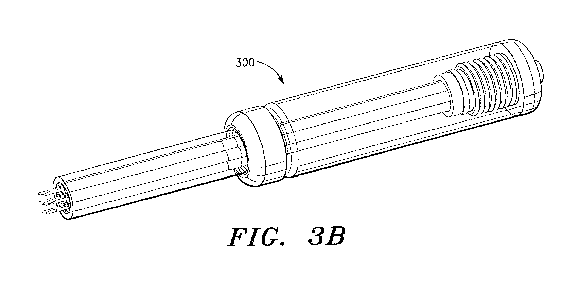
[00031] FIGURE 3A is an exploded perspective view of one embodiment of a multi-
needle
injection apparatus that is suitable for direct administration of ECM
compositions to
biological tissue, e.g. cardiovascular tissue, in accordance with the
invention; and
[00032] FIGURE 3B is an assembled perspective view of the multi-needle
injection
apparatus shown in FIGURE 3A, in accordance with the invention.
DETAILED DESCRIPTION OF THE PREFERRED EMBODIMENT
[00033] Before describing the present invention in detail, it is to be
understood that this
invention is not limited to particularly exemplified apparatus, systems,
compositions or
methods as such may, of course, vary. Thus, although a number of systems,
compositions and
methods similar or equivalent to those described herein can be used in the
practice of the
present invention, the preferred systems, compositions and methods are
described herein.
[00034] It is also to be understood that, although the systems,
pharmacological
compositions and methods of the invention are illustrated and described in
connection with
administration (or delivery) of pharmacological compositions (and bioactive
and
pharmacological agents) to cardiovascular tissue, the systems, compositions
and methods of
CA 02883885 2015-03-03
WO 2014/046753 PCT/US2013/046041
the invention are not limited to such delivery. According to the invention,
the systems and
methods of the invention can be employed to administer pharmacological
compositions (and
bioactive and pharmacological agents) to numerous additional biological
tissue, including,
without limitation, gastrointestinal and respiratory organ tissue.
[00035] It is also to be understood that, although a preferred method of
delivering a
pharmacological composition of the invention to biological tissue comprises
direct injection
into the tissue. The delivery of the pharmacological composition is not
limited to direct
injection. According to the invention, a pharmacological composition of the
invention can be
delivered to biological tissue by other conventional means, including topical
administration.
[00036] It is further to be understood that the terminology used herein is
for the purpose of
describing particular embodiments of the invention only and is not intended to
be limiting.
[00037] Unless defined otherwise, all technical and scientific terms used
herein have the
same meaning as commonly understood by one having ordinary skill in the art to
which the
invention pertains.
[00038] Further, all publications, patents and patent applications cited
herein, whether
supra or infra, are hereby incorporated by reference in their entirety.
[00039] Finally, as used in this specification and the appended claims, the
singular forms
"a, "an" and "the" include plural referents unless the content clearly
dictates otherwise. Thus,
for example, reference to "an anti-inflammatory" includes two or more such
agents and the
like.
Definitions
[00040] The terms "cardiac tissue damage", "cardiac tissue injury" and
"cardiovascular
tissue damage" are used interchangeably herein, and mean and include any area
of abnormal
tissue in the cardiovascular system or heart caused by a disease, disorder,
injury or damage,
including damage to the epicardium, endocardium and/or myocardium. Non-
limiting
examples of causes of cardiovascular tissue damage include acute or chronic
stress (systemic
hypertension, pulmonary hypertension, valve dysfunction, etc.), coronary
artery disease,
ischemia or infarction, inflammatory disease and cardiomyopathies.
6
CA 02883885 2015-03-03
WO 2014/046753 PCT/US2013/046041
[00041] As is well known in the art, cardiovascular tissue damage most often
involves
damage or injury to the myocardium and, therefore, for the purposes of this
disclosure,
myocardial damage or injury is equivalent to cardiovascular tissue damage.
[00042] The term "damaged tissue", as used herein, means and includes
biological tissue;
particularly, cardiovascular tissue damaged or injured by trauma, ischemic
tissue, infarcted
tissue or tissue damaged by any means which results in interruption of normal
blood flow to
the tissue.
[00043] The terms "prevent" and "preventing" are used interchangeably herein,
and mean
and include reducing the frequency or severity of a disease, condition or
disorder. The term
does not require an absolute preclusion of the disease, condition or disorder.
Rather, this term
includes decreasing the chance for disease occurrence.
[00044] The terms "treat" and "treatment" are used interchangeably herein, and
mean and
include medical management of a patient with the intent to cure, ameliorate,
stabilize, or
prevent a disease, pathological condition or disorder. The terms include
"active treatment",
i.e. treatment directed specifically toward the improvement of a disease,
pathological
condition or disorder, and "causal treatment", i.e. treatment directed toward
removal of the
cause of the associated disease, pathological condition or disorder.
[00045] The temis "treat" and "treatment" further include "palliative
treatment", i.e.
treatment designed for the relief of symptoms rather than the curing of the
disease,
pathological condition or disorder, "preventative treatment", i.e. treatment
directed to
minimizing or partially or completely inhibiting the development of the
associated disease,
pathological condition or disorder, and "supportive treatment", i.e. treatment
employed to
supplement another specific therapy directed toward the improvement of the
associated
disease, pathological condition or disorder.
[00046] The term "chamber remodeling", as used herein, means and includes a
series of
events (which may include changes in gene expression, molecular, cellular and
interstitial
changes) that result in changes in size, shape and function of cardiac tissue
following stress or
injury. As is well known in the art, remodeling can occur after a myocardial
infarction,
pressure overload (e.g., aortic stenosis, hypertension), volume overload
(e.g., valvular
7
CA 02883885 2015-03-03
WO 2014/046753 PCT/US2013/046041
regurgitation), inflammatory heart disease (e.g., myocarditis), or in
idiopathic cases (e.g.,
idiopathic dilated cardiomyopathy).
[00047] The tenn "angiogenesis", as used herein, means a physiologic process
involving
the growth of new blood vessels from pre-existing blood vessels.
[00048] The term "neovascularization", as used herein, means and includes the
formation
of functional vascular networks that can be perfused by blood or blood
components.
Neovascularization includes angiogenesis, budding angiogenesis, intussuceptive
angiogenesis,
sprouting angiogenesis, therapeutic angiogenesis and vasculogenesis.
[00049] The terms "extracellular matrix", "extracellular matrix material" and
"ECM
material" are used interchangeably herein, and mean a collagen-rich substance
that is found in
between cells in animal tissue and serves as a structural element in tissues.
It typically
comprises a complex mixture of polysaccharides and proteins secreted by cells.
The
extracellular matrix can be isolated and treated in a variety of ways.
Extracellular matrix
material (ECM) can be isolated from small intestine submucosa, stomach
submucosa, urinary
bladder submucosa, tissue mucosa, dura mater, liver basement membrane,
pericardium or
other tissues. Following isolation and treatment, it is commonly referred to
as extracellular
matrix or ECM material.
[00050] The terms "pharmacological agent", "pharmaceutical agent", "agent",
"active
agent", "drug" and "active agent fonnulation" are used interchangeably herein,
and mean
and include an agent, drug, compound, composition of matter or mixture
thereof, including
its formulation, which provides some therapeutic, often beneficial, effect.
This includes any
physiologically or pharmacologically active substance that produces a
localized or systemic
effect or effects in animals, including wan-n blooded mammals, humans and
primates;
avians; domestic household or farm animals, such as cats, dogs, sheep, goats,
cattle, horses
and pigs; laboratory animals, such as mice, rats and guinea pigs; fish;
reptiles; zoo and wild
animals; and the like.
[00051] The terms "pharmacological agent", "pharmaceutical agent", "agent",
"active
agent", "drug" and "active agent formulation" thus mean and include, without
limitation,
antibiotics, anti-viral agents, analgesics, steroidal anti-inflammatories, non-
steroidal anti-
inflammatories, anti-neoplastics, anti-spasmodics, modulators of cell-
extracellular matrix
8
CA 02883885 2015-03-03
WO 2014/046753
PCT/US2013/046041
interactions, proteins, hormones, enzymes and enzyme inhibitors,
anticoagulants and/or
antithrombic agents, DNA, RNA, modified DNA and RNA, NSAIDs, inhibitors of
DNA,
RNA or protein synthesis, polypeptides, oligonucleotides, polynucleotides,
nucleoproteins,
compounds modulating cell migration, compounds modulating proliferation and
growth of
tissue, and vasodilating agents.
[00052] The terms "anti-inflammatory" and "anti-inflammatory agent" are also
used
interchangeably herein, and mean and include a "pharmacological agent" and/or
"active
agent formulation", which, when a therapeutically effective amount is
administered to a
subject, prevents or treats bodily tissue inflammation i.e. the protective
tissue response to
injury or destruction of tissues, which serves to destroy, dilute, or wall off
both the injurious
agent and the injured tissues. Anti-inflammatory agents thus include, without
limitation,
alclofenac, alclometasone dipropionate, algestone acetonide, alpha amylase,
amcinafal,
amcinafide, amfenac sodium, amiprilose hydrochloride, anakinra, anirolac,
anitrazafen,
apazone, balsalazide disodium, bendazac, benoxaprofen, benzydamine
hydrochloride,
bromelains, broperamole, budesonide, carprofen, cicloprofen, cintazone,
cliprofen,
clobetasol propionate, clobetasone butyrate, clopirac, cloticasone propionate,
con-nethasone
acetate, cortodoxone, decanoate, deflazacort, delatestryl, depo-testosterone,
desonide,
desoximetasone, dexamethasone dipropionate, diclofenac potassium, diclofenac
sodium,
diflorasone diacetate, diflumidone sodium, diflunisal, difluprednate,
diftalone, dimethyl
sulfoxide, drocinonide, endrysone, enlimomab, enolicam sodium, epirizole,
etodolac,
etofenamate, felbinac, fenamole, fenbufen, fenclofenac, fenclorac, fendosal,
fenpipalone,
fentiazac, flazalone, fluazacort, flufenamic acid, flumizole, flunisolide
acetate, flunixin,
flunixin meglumine, fluocortin butyl, fluorometholone acetate, fluquazone,
flurbiprofen,
fluretofen, fluticasone propionate, furaprofen, furobufen, halcinonide,
halobetasol
propionate, halopredone acetate, ibufenac, ibuprofen, ibuprofen aluminum,
ibuprofen
piconol, ilonidap, indomethacin, indomethacin sodium, indoprofen, indoxole,
intrazole,
isoflupredone acetate, isoxepac, isoxicam, ketoprofen, lofemizole
hydrochloride,
lomoxicam, loteprednol etabonate, meclofenamate sodium, meclofenamic acid,
meclorisone
dibutyrate, mefenamic acid, mesalamine, meseclazone, mesterolone,
methandrostenolone,
methenolone, methenolone acetate, methylprednisolone suleptanate, momiflumate,
9
CA 02883885 2015-03-03
WO 2014/046753
PCT/US2013/046041
nabumetone, nandrolone, naproxen, naproxen sodium, naproxol, nimazone,
olsalazine
sodium, orgotein, orpanoxin, oxandrolane, oxaprozin, oxyphenbutazone,
oxymetholone,
paranyline hydrochloride, pentosan polysulfate sodium, phenbutazone sodium
glycerate,
pirfenidone, piroxicam, piroxicam cinnamate, piroxicam olamine, pirprofen,
prednazate,
prifelone, prodolic acid, proquazone, proxazole, proxazole citrate,
rimexolone, romazarit,
salcolex, salnacedin, salsalate, sanguinarium chloride, seclazone, sermetacin,
stanozolol,
sudoxicam, sulindac, suprofen, talmetacin, talniflumate, talosalate,
tebufelone, tenidap,
tenidap sodium, tenoxicam, tesicam, tesimide, testosterone, testosterone
blends,
tetrydamine, tiopinac, tixocortol pivalate, tolmetin, tolmetin sodium,
triclonide, triflumidate,
zidometacin, and zomepirac sodium.
[00053] The term "chitosan", as used herein, means and includes the family of
linear
polysaccharides consisting of varying amounts of 13 (1¨>4) linked residues of
N-acetyl-2
amino-2-deoxy-D-glucose and 2-amino-2-deoxy-Dglucose residues, and all
derivatives
thereof.
[00054] The terms "active agent formulation", "pharmacological agent
formulation" and
"agent formulation", are also used interchangeably herein, and mean and
include an active
agent (and chitosan) optionally in combination with one or more
pharmaceutically
acceptable caniers and/or additional inert ingredients. According to the
invention, the
formulations can be either in solution or in suspension in the canier.
[00055] The term "pharmacological composition", as used herein, means and
includes a
composition comprising a "pharmacological agent" and/or an "extracellular
matrix
material" and/or a "pharmacological agent formulation" and/or any additional
agent or
component identified herein.
[00056] The terin "therapeutically effective", as used herein, means that the
amount of
the "pharmacological composition" and/or "pharmacological agent" and/or
"active agent
formulation" administered is of sufficient quantity to ameliorate one or more
causes,
symptoms, or sequelae of a disease or disorder. Such amelioration only
requires a reduction
or alteration, not necessarily elimination, of the cause, symptom, or sequelae
of a disease or
disorder.
CA 02883885 2015-03-03
WO 2014/046753 PCT/US2013/046041
[00057] The terms " delivery" and "administration" are used interchangeably
herein, and
mean and include providing a "pharmacological composition" or "pharmacological
agent"
or "active agent formulation" to a treatment site, e.g., damaged tissue,
through any method
appropriate to deliver the functional agent or formulation or composition to
the treatment
site. Non-limiting examples of delivery methods include direct injection,
percutaneous
delivery and topical application at the treatment site.
[00058] The term "percutaneous", as used herein, means and includes any
penetration
through the skin of a patient or subject, whether in the form of a small cut,
incision, hole,
cannula, tubular access sleeve or port or the like.
[00059] The terms "patient" and "subject" are used interchangeably herein, and
mean and
include warm blooded mammals, humans and primates; avians; domestic household
or farm
animals, such as cats, dogs, sheep, goats, cattle, horses and pigs; laboratory
animals, such as
mice, rats and guinea pigs; fish; reptiles; zoo and wild animals; and the
like.
[00060] The term "comprise" and variations of the teim, such as "comprising"
and
"comprises," means "including, but not limited to" and is not intended to
exclude, for
example, other additives, components, integers or steps.
[00061] The following disclosure is provided to further explain in an enabling
fashion the
best modes of perfoiming one or more embodiments of the present invention. The
disclosure
is further offered to enhance an understanding and appreciation for the
inventive principles
and advantages thereof, rather than to limit in any manner the invention. The
invention is
defined solely by the appended claims including any amendments made during the
pendency
of this application and all equivalents of those claims as issued.
[00062] As will readily be appreciated by one having ordinary skill in the
art, the present
invention substantially reduces or eliminates the disadvantages and drawbacks
associated with
prior art methods of treating damaged or diseased biological tissue.
[00063] In overview, the present disclosure is directed to methods and systems
for treating
damaged and diseased biological tissue; particularly, cardiovascular tissue,
via the "direct"
delivery of a pharmacological composition (and/or pharmacological agent and/or
fommlation)
to the damaged or diseased tissue. According to the invention, the delivery of
a
therapeutically effective amount of a pharmacological composition of the
invention to
1
CA 02883885 2015-03-03
WO 2014/046753 PCT/US2013/046041
damaged or diseased tissue induces neovascularization, host tissue
proliferation,
bioremodeling and regeneration of new tissue.
[00064] According to the invention, the phaimacological compositions can
comprise mixed
liquids, mixed emulsions, mixed gels, mixed pastes, or mixed solid
particulates.
[00065] In some embodiments, one or more pharmacological compositions of the
invention
are directly administered to the damaged or diseased tissue via a multi-needle
injection
system, such as disclosed in Co-pending Application No. 61/704,634, filed
September 24,
2012 and illustrated in FIGS. 3A and 3B.
[00066] In a preferred embodiment, the pharmacological compositions comprise
extracellular matrix (ECM) compositions that include at least one ECM scaffold
component.
[00067] According to the invention, the ECM scaffold component can be derived
from
various mammalian tissue sources and methods for preparing same, such as
disclosed in
U.S. Pat. Nos. 7,550,004, 7,244,444, 6,379,710, 6,358,284, 6,206,931,
5,733,337 and
4,902,508 and U.S. Application No. 12/707,427; which are incorporated by
reference herein
in their entirety. The mammalian tissue sources include, without limitation,
the small
intestine, large intestine, stomach, lung, liver, kidney, pancreas, placenta,
heart, bladder,
prostate, tissue surrounding growing enamel, tissue surrounding growing bone,
and any fetal
tissue from any mammalian organ.
[00068] In a preferred embodiment, the ECM scaffold component comprises
mammalian
mesothelium.
[00069] According to the invention, the ECM scaffold component (or material)
can be
formed into a particulate and fluidized, as described in U.S. Pat. Nos.
5,275,826, 6,579,538
and 6,933,326, to form an ECM composition of the invention.
[00070] According to the invention, various conventional means can be employed
to
form a particulate ECM scaffold material. In some embodiments, the ECM
scaffold
material is formed into a sheet, fluidized (or hydrated), if necessary, frozen
and ground.
[00071] In some embodiments of the invention, the ground ECM scaffold material
is
subsequently filtered to achieve a desired particulate size. Thus, in some
embodiments, the
ECM scaffold material has a particulate size no greater than 2000 microns. In
some
embodiments, the ECM scaffold material preferably has a particulate size no
greater than
12
CA 02883885 2015-03-03
WO 2014/046753
PCT/US2013/046041
500 microns. In a preferred embodiment, the ECM scaffold material has a
particulate size
in the range of about 20 microns to about 300 microns.
[00072] According to the invention, fluidized or emulsified compositions (the
liquid or
semi-solid forms) can comprise various certain concentrations of ECM scaffold
material. In
some embodiments of the invention, the concentration of the ECM scaffold
material is
greater than about 5%, more preferably, greater than about 20%, even more
preferably,
greater than about 70%.
[00073] According to the invention, the particulate ECM scaffold material can
be
fluidized or hydrated by various conventional buffer materials. Suitable
buffer materials
include, without limitation, water and saline.
[00074] According to the invention, the liquid or semi-solid components of
the ECM
compositions (i.e. liquids, gels, emulsions or pastes) can comprise various
concentrations.
Preferably, the concentration of the liquid or semi-solid components of the
ECM
compositions are in the range of about 0.001 mg/ml to about 200 mg/ml.
Suitable
concentration ranges thus include, without limitation: about 5 mg/ml to about
150 mg/ml,
about 10 mg/ml to about 125 mg/ml, about 25 mg/ml to about 100 mg/ml, about 20
mg/ml
to about 75 mg/ml, about 25 mg/ml to about 60 mg/ml, about 30 mg/ml to about
50 mg/ml,
and about 35 mg/ml to about 45 mg/ml and about 40 mg/ml. to about 42 mg/ml.
[00075] The noted concentration ranges are, however, merely exemplary and not
intended to be exhaustive or limiting. It is understood that any value within
any of the listed
ranges is deemed a reasonable and useful value for a concentration of a liquid
or semi-solid
component of an ECM composition.
[00076] According to the invention, the dry particulate or reconstituted
particulate that
forms a gel emulsion or paste of the two ECM scaffold materials can also be
mixed together
in various proportions. For example, the particulates can comprise 50% of
small intestine
submucosa mixed with 50% of pancreatic basement membrane. The mixture can then
similarly be fluidized by hydrating in a suitable buffer, such as saline.
[00077] As indicated above, in some embodiments of the invention, the ECM
compositions are foimulated to be injected into damaged or cardiovascular
tissue, i.e.
injectable ECM compositions. In some embodiments of the invention, the
injectable ECM
13
CA 02883885 2015-03-03
WO 2014/046753 PCT/US2013/046041
compositions thus comprise approximately 70% particulate ECM scaffold material
and
approximately 30% fully hydrolyzed ECM gel.
[00078] According to the invention, the pharmacological compositions of the
invention
can further include one or more additional bioactive agents or components to
aid in the
treatment of damaged tissue and/or facilitate the tissue regenerative process.
[00079] In some embodiments, the pharmacological compositions of the invention
thus
include at least one pharmacological agent or composition, which can comprise,
without
limitation, antibiotics or antifungal agents, anti-viral agents, anti-pain
agents, anesthetics,
analgesics, steroidal anti-inflammatories, non-steroidal anti-inflammatories,
anti-
neoplastics, anti-spasmodics, modulators of cell-extracellular matrix
interactions, proteins,
hormones, enzymes and enzyme inhibitors, anticoagulants and/or antithrombic
agents,
DNA, RNA, modified DNA and RNA, NSAIDs, inhibitors of DNA, RNA or protein
synthesis, polypeptides, oligonucleotides, polynucleotides, nucleoproteins,
compounds
modulating cell migration, compounds modulating proliferation and growth of
tissue, and
vasodilating agents.
[00080] Suitable pharmacological agents and/or compositions thus include,
without
limitation, atropine, tropicamide, dexamethasone, dexamethasone phosphate,
betamethasone,
betamethasone phosphate, prednisolone, triamcinolone, triamcinolone acetonide,
fluocinolone
acetonide, anecortave acetate, budesonide, cyclosporine, FK-506, rapamycin,
ruboxistaurin,
midostaurin, flurbiprofen, suprofen, ketoprofen, diclofenac, ketorolac,
nepafenac, lidocaine,
neomycin, polyrnyxin b, bacitracin, gramicidin, gentamicin, oyxtetracycline,
ciprofloxacin,
ofloxacin, tobramycin, amikacin, vancomycin, cefazolin, ticarcillin,
chlorarnphenicol,
miconazole, itraconazole, trifluridine, vidarabine, ganciclovir, acyclovir,
cidofovir, ara-amp,
foscamet, idoxuridine, adefovir dipivoxil, methotrexate, carboplatin,
phenylephrine,
epinephrine, dipivefrin, timolol, 6-hydroxydopamine, betaxolol, pilocarpine,
carbachol,
physostigmine, demecarium, dorzolamide, brinzolamide, latanoprost, sodium
hyaluronate,
insulin, verteporfin, pegaptanib, ranibizumab, and other antibodies,
antineoplastics, Anti
VGEFs, ciliary neurotrophic factor, brain-derived neurotrophic factor, bFGF,
Caspase-1
inhibitors, Caspase-3 inhibitors, a-Adrenoceptors agonists, NMDA antagonists,
Glial cell
14
CA 02883885 2015-03-03
WO 2014/046753 PCT/US2013/046041
line-derived neurotrophic factors (GDNF), pigment epithelium-derived factor
(PEDF), and
NT-3, NT-4, NGF, IGF-2.
[00081] In some embodiments of the invention, the pharmacological agent
comprises an
anti-inflammatory agent selected from the group comprising, without
limitation, alclofenac,
alclometasone dipropionate, algestone acetonide, alpha amylase, amcinafal,
amcinafide,
amfenac sodium, amipri lose hydrochloride, anakinra, anirolac, anitrazafen,
apazone,
balsalazide di sodium, bendazac, benoxaprofen, benzydamine hydrochloride,
bromelains,
broperamole, budesonide, carprofen, cicloprofen, cintazone, cliprofen,
clobetasol propionate,
clobetasone butyrate, clopirac, cloticasone propionate, cormethasone acetate,
cortodoxone,
decanoate, deflazacort, delatestryl, depo-testosterone, desonide,
desoximetasone,
dexamethasone dipropionate, diclofenac potassium, diclofenac sodium,
diflorasone diacetate,
diflumidone sodium, diflunisal, difluprednate, diftalone, dimethyl sulfoxide,
drocinonide,
endrysone, enlimomab, enolicam sodium, epirizole, etodolac, etofenamate,
felbinac,
fenamole, fenbufen, fenclofenac, fenclorac, fendosal, fenpipalone, fentiazac,
flazalone,
fluazacort, flufenamic acid, flumizole, flunisolide acetate, flunixin,
flunixin meglumine,
fluocortin butyl, fluorometholone acetate, fluquazone, flurbiprofen,
fluretofen, fluticasone
propionate, furaprofen, furobufen, halcinonide, halobetasol propionate,
halopredone acetate,
ibufenac, ibuprofen, ibuprofen aluminum, ibuprofen piconol, ilonidap,
indomethacin,
indomethacin sodium, indoprofen, indoxole, intrazole, isoflupredone acetate,
isoxepac,
isoxicam, ketoprofen, lofemizole hydrochloride, lomoxicam, loteprednol
etabonate,
meclofenamate sodium, meclofenamic acid, meclorisone dibutyrate, mefenamic
acid,
mesalamine, meseclazone, mesterolone, methandrostenolone, methenolone,
methenolone
acetate, methylprednisolone suleptanate, momiflumate, nabumetone, nandrolone,
naproxen,
naproxen sodium, naproxol, nimazone, olsalazine sodium, orgotein, orpanoxin,
oxandrolane,
oxaprozin, oxyphenbutazone, oxymetholone, paranyline hydrochloride, pentosan
polysulfate
sodium, phenbutazone sodium glycerate, pirfenidone, piroxicam, piroxicam
cinnamate,
piroxicam olamine, pirprofen, prednazate, prifelone, prodolic acid,
proquazone, proxazole,
proxazole citrate, rimexolone, romazarit, salcolex, salnacedin, salsalate,
sanguinarium
chloride, seclazone, seimetacin, stanozolol, sudoxicam, sulindac, suprofen,
talmetacin,
talniflumate, talosalate, tebufelone, tenidap, tenidap sodium, tenoxicam,
tesicam, tesimide,
CA 02883885 2015-03-03
WO 2014/046753 PCT/US2013/046041
testosterone, testosterone blends, tetrydamine, tiopinac, tixocortol pivalate,
tolmetin, tolmetin
sodium, triclonide, triflumidate, zidometacin, and zomepirac sodium.
[00082] According to the invention, the amount of a pharmacological agent
added to an
ECM composition of the invention will, of course, vary from agent to agent.
For example, in
one embodiment, wherein the pharmacological agent comprises ibuprofen (Advil
), i.e. an
anti-inflmmatory, the amount of ibuprofen included in the ECM composition is
preferably in
the range of 100 p.g ¨ 200 mg.
[00083] In some embodiments of the invention, the pharmacological agent
comprises a
statin, i.e. a HMG-CoA reductase inhibitor. According to the invention,
suitable statins
include, without limitation, atorvastatin (LIPITORO), cerivastatin,
fluvastatin (Lesco10),
lovastatin (Mevacort, Altocor0, Altoprev0), mevastatin, pitavastatin (Livalo
0, Pitavae),
pravastatin (Pravachol , Selektine0, Lipostat0), rosuvastatin (Crestor0), and
simvastatin
(Zocore, Lipex0). Several actives comprising a combination of a statin and
another agent,
such as ezetimbe/simvastatin (Vytorin0), are also suitable.
[00084] Applicant has found that statins exhibit numerous beneficial
properties that
provide several beneficial biochemical actions or activities. The properties
and beneficial
actions resulting therefrom are discussed in detail below.
Anticholesterolemic Properties/Actions
[00085] As is well known in the art, statins are a class of drugs that
primarily function to
lower levels of cholesterol production in the liver. Lower levels of
cholesterol are achieved
via the statins limiting the production of mevalonate in the cholestrol
biosynthetic pathway.
Statins competitively inhibit HMG-CoA reductase, which, because the molecules
are so
similar, results in the statins actually taking the place of the HMG-CoA
reductase in the
cholesterol biosynthetic pathway and reducing the rate at which the mevalonate
is produced,
subsequently lowering the rate at which cholesterol is produced in the liver.
Anti-Inflammatory Properties/Actions
[00086] Statins also have numerous additional favorable effects on the
vascular wall cells,
and cardiovascular system. One specific example of this is thromboxane A2
(TXA2). Statins
can aid in the reduction of TX/62, which then lowers the platelet activation
in the
cardiovascular system.
16
CA 02883885 2015-03-03
WO 2014/046753 PCT/US2013/046041
[00087] TXA2 is also known as a vasoconstrictor and is especially important
during tissue
injury and inflammation due to its impact on platelet activation and
aggregation, as well as its
ability to augment the expression of adhesion molecules and chemokines. This
allows statins
to aid in the reduction of inflammation by the reduction of TXA?, which
results in less
vascoconstriction, less platelet activation and aggregation, as well as
reduced augmentation of
adhesion molecules and chemokines.
[00088] Statins further impact vascular wall cells and the cardiovascular
system by
blocking ras homilog gene family, member A (RhoA) activation. Blocking RhoA
activation
further impacts numerous systems, such as macrophage growth, tissue
plasminogen activators
(t-PA), plasminogen activator inhibitor type 1 (PAI-1), smooth muscle cell
(SMC)
proliferation, nitric oxide (NO) production, endothelins, and angiotensin
receptors. When
statins block RhoA activation, the resultant impact can be seen in many
phsyiological
responses of the cardiovascular system, including vascular inflammation,
smooth muscle cell
production and size, and vasconstriction inter alia.
[00089] Macrophage growth reduced by blocking RhoA activation results in the
reduction
of matrix metalloprotinases (MMPs) and tissue factors (TF). MMPs are part of a
larger
family of metalloprotinase enzymes that play on important part in wound
healing and
inflammation. MMPs are produced by activated neutrophils and macrophages
(inflammatory
cells).
[00090] Statins facilitate the reduction of inflammatory factors by lowering
macrophage
growth, which results in reduced production of MMPs. Lowered MMPs also results
in a
lowered presence of thrombi as the MMPs attach to ECM present in thrombi or
damaged
ECM at wound sites. Macrophage growth reduction also results in lowered
presence of tissue
factor (TF).
[00091] TF is a protein necessary for the initiation of thrombin formation.
This factor also
enables cells to initiate the coagulation cascade. Lowered presence of TF
results in lowered
presence of thrombi in the cardiovascular system, especially in conjunction
with reduced
MMPs.
Plaque Stabilizing Properties/Actions
[00092] Reduced MMPs and reduced TF also results in increased plaque stability
which
17
CA 02883885 2015-03-03
WO 2014/046753 PCT/US2013/046041
can help to prevent stroke or myocardial infarction by reducing the
probability of a portion of
the plaque to break off and become lodged within a smaller vessel. Plaque
stability further
aids in reduction of atherosclerosis.
Fibrinolysis Properties/Actions
[00093] Blocking RhoA activation also affects the presence of tissue
plasminogen
activators (t-PA) and plasminogen activator inhibitor type 1 (PAI-1). T-PA is
a protein
involved in the breakdown of blood clots, and is found on endothelial cells.
As an enzyme it
catalyzes the conversion of plasminogen to plasmin, the major enzyme
responsible for clot
breakdown (fibrinolysis).
[00094] PAI-1 is a protein that functions as the principal inhibitor oft-
PA. Thus, PAT-1 is
the principal inhibitor of fibrinolysis. With t-PA presence raised and PAT-1
diminished from
the blocking of RhoA activation caused by statins, a reduced thrombotic effect
is realized due
to reduced opportunity for fibrin to form the polymeric mesh of a hemostatic
plug. The
reduced MMPs and TF that result from the use of statins work in concert with
the increased t-
PA and reduced PAI-1 to further reduce the potential for thrombii.
NO Regulation Properties/Actions
[00095] Blocking RhoA activation also affects the presence of Nitric Oxide
(NO) in the
cardiovascular system. The endothelium uses NO to signal the surrounding
smooth muscles
to relax, resulting in vasodialation and increased blood flow. NO contributes
to vessel
homeostasis by inhibiting vascular smooth muscle contraction and growth,
platelet
aggregation, and leukocyte adhesion to the endothelium. These factors are what
allow NO to
aid in the reduction of endothelial dysfunction when modulated in such a way
as is typical
with the administration of statins. The reduction of leukocyte adhesion is a
specific example
of how the NO production associated with statins aids in the reduction
inflammation desired
when coadministered locally with an extracellular matrix.
RhoA Activation Blocking Properties/Actions
[00096] The administration of statins can affect the presence of endothelins
and agiotensin
receptors. Endothelins and angiotensin receptors can also be affected by the
subsequent
blocking of RhoA activation associated with statin administration.
[00097] Endothelins are proteins that constrict blood vessels and raise
blood pressure.
18
CA 02883885 2015-03-03
WO 2014/046753 PCT/US2013/046041
There are three isofonns; ET-1, ET-2, and ET-3, with ET-1 being the isoform
primarily
affected by statins and RhoA activation blocking. Secretion of ET-1 from the
endothelium
signals vasoconstriction and influences local cellular growth and survival. ET-
1 has been
implicated in the development and progression of vascular disorders, such as
atherosclerosis
and hypertension. The decrease in the presence of ET-1 associated with statins
and RhoA
activation blocking results in decreased vasoconstriction and progression of
the
aforementioned vascular disorders.
[00098] Angiotensin receptors are protein coupled receptors that are
responsible for the
signal transduction of the vasoconstricting stimulus of the main effector
hormone angiotensin
II. Angiotensin Receptor II Type I (AT-1) is the angiotensin receptor
primarily affected by
statin administration and RhoA activation blocking. AT-1 mediates
vasocontraction, cardiac
hypertrophy, vascular smooth muscle cell proliferation, inter cilia. The
reduction in AT-1 that
accompanies statin administration and RhoA activation blocking results in
reduced
vasoconstriction in the cardiovascular system.
C-Reactive Protein Reduction Properties/Actions
[00099] C-Reactive Proteins (CRP) are also influenced by statin
administration. CRP are
found in the blood; the levels of which deviate in response to differing
levels of inflammation.
CRP levels diminish in response to statin administration. This functions as a
result of a
statin's impact on the reduction of inflammation.
Adhesion Molecule Reduction Properties/Actions
[000100] Adhesion molecules are proteins that are located on the cell surface
and are
involved with inflammation and thrombin formation in vascular endothelial
cells. With
higher incidence of inflammation comes higher incidence of cell adhesion
molecules. A statin
functions to reduce the presence of adhesion molecules on the endothelium.
This helps to
reduce inflammation by removing the attachment mechanism for leukocytes and
subsequent
plaque buildup, the result being lowered chance for atherosclerosis.
Rac-1 Reduction Properties/Actions
[000101] Rac-1 is a protein found in human cells. It plays a central role in
endothelial cell
migration, tubulogenesis, adhesion, and permeability. The expression of Rac-1
can be
affected by the administration of statins, specifically such that Rae-1 is
decreased by statins.
19
CA 02883885 2015-03-03
WO 2014/046753 PCT/US2013/046041
The decrease in the presence of Rae-1 also results in the decrease of reactive
oxygen species
(ROS). ROS are chemically reactive molecules that have important roles in cell
signaling
and homeostasis.
[000102] According to the invention, the amount of a statin added to a
pharmacological
composition of the invention is preferably less than 20 mg, more preferably,
less than
approximately 10 mg.
[000103] In some embodiments of the invention, the ECM scaffold material
includes 100 ug
¨ 5 mg of a statin. In some embodiments of the invention, the ECM scaffold
material
includes 500 ug ¨ 2 mg of a statin.
[000104] In some embodiments of the invention, the bioactive agent comprises
chitosan or a
derivative thereof.
[000105] Chitosan exhibits a wide range of favorable biochemical properties
that make it an
outstanding agent for use in the medical field. The biochemical properties of
chitosan include
biocompatibility, biodegradability and non-toxicity. Additional properties,
such as analgesic,
hemostatic, antimicrobial, and antioxidant have also been reported. See
Aranaz, et al.,
Functional Characterization of Chitin and Chitosan, Current Chemical Biology,
vol. 3, pp.
203-230 (2009); and Kumar MNVR, A Review of Chitin and Chitosan Applications,
React.
Funct. Polm., vol. 46, pp. 1-27 (2000).
[000106] In some embodiments of the invention, the bioactive agent comprises a
cell.
According to the invention, the cell can comprise, without limitation, a stem
cell, such as,
for example, a human embryonic stem cell, fetal cell, fetal cardiomyocyte,
myofibroblast,
mesenchymal stem cell, autotransplanted expanded cardiomyocyte, adipocyte,
totipotent
cell, pluripotent cell, blood stem cell, myoblast, adult stem cell, bone
marrow cell,
mesenchymal cell, embryonic stem cell, parenchymal cell, epithelial cell,
endothelial cell,
mesothelial cell, fibroblast, myofibroblast, osteoblast, chondrocyte,
exogenous cell,
endogenous cell, stem cell, hematopoetic stem cell, pluripotent stem cell,
bone marrow-
derived progenitor cell, progenitor cell, myocardial cell, skeletal cell,
undifferentiated cell,
multi-potent progenitor cell, unipotent progenitor cell, monocyte,
cardiomyocyte, cardiac
myoblast, skeletal myoblast, macrophage, capillary endothelial cell, xenogenic
cell, and
allogenic cell.
CA 02883885 2015-03-03
WO 2014/046753
PCT/US2013/046041
[000107] In some embodiments of the invention, the bioactive agent comprises a
protein.
According to the invention, the protein can comprise, without limitation, a
growth factor,
collagen, proteoglycan, glycosaminoglycan (GAG) chain, glycoprotein, cytokine,
cell-
surface associated protein, cell adhesion molecule (CAM), angiogenic growth
factor,
endothelial ligand, matrikine, matrix metalloprotease, cadherin, immunoglobin,
fibril
collagen, non-fibrillar collagen, basement membrane collagen, multiplexin,
small-leucine
rich proteoglycan, decorin, biglycan, fibromodulin, keratocan, lumican,
epiphycan, heparan
sulfate proteoglycan, perlecan, agrin, testican, syndecan, glypican,
serglycin, selectin,
lectican, aggrecan, versican, nuerocan, brevican, cytoplasmic domain-44
(CD44),
macrophage stimulating factor, amyloid precursor protein, heparin, chondroitin
sulfate B
(dermatan sulfate), chondroitin sulfate A, heparan sulfate, hyaluronic acid,
fibronectin (Fn),
tenascin, elastin, fibrillin, laminin, nidogen/entactin, fibulin I, fibulin
II, integrin, a
transmembrane molecule, platelet derived growth factor (PDGF), epidermal
growth factor
(EGF), transforming growth factor alpha (TGF-alpha), transforming growth
factor beta
(TGF-beta), fibroblast growth factor-2 (FGF-2) (also called basic fibroblast
growth factor
(bFGF)), thrombospondin, osteopontin, angiotensin converting enzyme (ACE), and
vascular
epithelial growth factor (VEGF).
[000108] According to the invention, the bioactive agents referenced above can
comprise
any faun. In some embodiments of the invention, the bioactive component or
components,
e.g. simvastatin and/or chitosan, comprise microcapsules that provide delayed
delivery of
the agent contained therein.
[000109] Additional suitable pharmacological compositions that can be
delivered within
the scope of the invention are disclosed in Pat. Pub. Nos. 20070014874,
20070014873,
20070014872, 20070014871, 20070014870, 20070014869, and 20070014868; which are
expressly incorporated by reference herein in its entirety.
[000110] As indicated above, in some embodiments of the invention, one or more
pharmacological compositions of the invention are directly administered or
delivered to
damaged or diseased cardiovascular tissue via a unique multi-needle injection
system. As
will readily be appreciated by one having ordinary skill in the art, the
pharmacological
compositions of the invention can also be delivered to damaged tissue via one
or more
21
CA 02883885 2015-03-03
WO 2014/046753 PCT/US2013/046041
conventional injection apparatus and systems, e.g., syringe. According to the
invention, the
pharmacological compositions can be directly administered to the heart wall
and/or the
various cardiovascular structures associated therewith.
[000111] Referring now to Fig. 1 there is shown a depiction of a normal heart
100. The
heart wall 102 consists of an inner layer of simple squamous epithelium,
referred to as the
endocardium. The endocardium overlays the myocardium (a variably thick heart
muscle) and
is enveloped within a multi-layer tissue structure referred to as the
pericardium. The
innermost layer of the pericardium, referred to as the visceral pericardium or
epicardium,
covers the myocardium. An outermost layer of the pericardium, referred to as
the fibrous
pericardium, attaches the parietal pericardium to the sternum, the great
vessels and the
diaphragm.
[000112] According to the invention, a pharmacological composition can be
delivered to
each of the noted structures; particularly, the myocardium, whereby
neovascularization, host
tissue proliferation, and bioremodeling is induced.
[000113] Referring now to Fig. 2, there is shown a depiction of a heart 200
having an
ischemic infracted region 202, and a pen-infarcted region 204 that is
surrounded by healthy
non-ischemic myocardium tissue 206.
[000114] As indicated above, a myocardial infarction, i.e. irreversible
myocardial injury
resulting in necrosis of a significant portion of myocardium, can result in an
acute depression
in ventricular function and expansion of the infarcted tissue under stress.
This triggers a
cascading sequence of myocellular events. In many cases, this progressive
myocardial infarct
expansion and remodeling leads to deterioration in ventricular function and
heart failure.
[000115] When a myocardial infarction occurs, the myocardial tissue that is no
longer
receiving adequate blood flow dies and is replaced with scar tissue. This
infarcted tissue
cannot contract during systole, and may actually undergo lengthening in
systole and leads to
an immediate depression in ventricular function. This abnormal motion of the
infarcted tissue
can cause delayed or abnormal conduction of electrical activity to the still
surviving peri-
infarct tissue (tissue at the junction between the normal tissue and the
infarcted tissue) and
also places extra structural stress on the pen-infarct tissue.
22
CA 02883885 2015-03-03
WO 2014/046753 PCT/US2013/046041
[000116] In addition to immediate hemodynamic effects, the infarcted heart
tissue and
undergoes three major processes: infarct expansion, infarct extension, and
chamber
remodeling. These factors individually and in combination contribute to the
eventual
dysfimction observed in the cardiac tissue remote from the site of the
infarction.
[000117] Infarct expansion is a fixed, permanent, disproportionate regional
thinning and
dilatation of tissue within the infarct zone. Infarct extension is additional
myocardial necrosis
following myocardial infarction. Infarct extension results in an increase in
total mass of
infarcted tissue.
[000118] However, as indicated above, the noted effects of a myocardial
infarction can be
ameliorated or eliminated by administering a pharmacological composition of
the invention
directly to the infarcted cardiovascular tissue. As also indicated herein, the
pharmacological
compositions of the invention will specifically induce neovascularization,
host tissue
proliferation, bioremodeling, and regeneration of new cardiac tissue
structures with site-
specific structural and functional properties. A preferred means of
administering the
pharmacological compositions to infracted cardiovascular tissue comprises
direct injection via
the multi-needle injection apparatus 300 illustrated in Figs. 3A and 3B and
the associated
control system described in Co-Pending Application No. 61/704,634.
[000119] As will readily be appreciated by one having ordinary skill in the
art, the present
invention provides numerous advantages compared to prior art methods and
systems for
treating damaged cardiac tissue. Among the advantages are the following:
O The provision of pharmacological compositions which, when delivered to
damaged
biological tissue; particularly, cardiovascular tissue, induce
neovascularization, and
promote survival and regeneration of damaged cardiovascular tissue.
= The provision of extracellular matrix (ECM) compositions which, when
delivered to
damaged biological tissue; particularly, cardiovascular tissue, induce host
tissue
proliferation, bioremodeling, and regeneration of cardiovascular tissue
structures with
site-specific structural and functional properties.
= The provision of improved methods and systems for administering
pharmacological
compositions; particularly, ECM compositions directly to damaged or diseased
biological tissue.
23
CA 02883885 2015-03-03
WO 2014/046753 PCT/US2013/046041
[000120] Without departing from the spirit and scope of this invention, one of
ordinary skill
can make various changes and modifications to the invention to adapt it to
various usages and
conditions. As such, these changes and modifications are properly, equitably,
and intended to
be, within the full range of equivalence of the following claims.