Note: Descriptions are shown in the official language in which they were submitted.
CA 02884793 2015-03-11
WO 2014/042840
PCT/US2013/056096
SYSTEM AND METHOD FOR MEASURING
CARDIAC OUTPUT
BACKGROUND
The present disclosure relates generally to medical devices and, more
particularly,
to the use of medical devices to non-invasively measure cardiac output.
This section is intended to introduce the reader to various aspects of art
that may be
related to various aspects of the present disclosure, which are described
and/or claimed
below. This discussion is believed to be helpful in providing the reader with
background
information to facilitate a better understanding of the various aspects of the
present
disclosure. Accordingly, it should be understood that these statements are to
be read in this
light, and not as admissions of prior art.
In the field of medicine, doctors often desire to monitor certain
physiological
characteristics of their patients. Accordingly, a wide variety of devices have
been developed
for monitoring many such characteristics of a patient. Such devices provide
doctors and
other healthcare personnel with the information they need to provide the best
possible
healthcare for their patients. As a result, such monitoring devices have
become an
indispensable part of modern medicine.
For example, clinicians may wish to monitor a patient's blood flow and blood
oxygen
saturation to assess cardiac function. In particular, clinicians may wish to
monitor a patient's
cardiac output. The determination of cardiac output may provide information
useful for the
diagnosis and treatment of various disease states or patient abnormalities.
For example, in
cases of pulmonary hypertension, a clinical response may include a decrease in
cardiac
output.
Accordingly, there are a variety of clinical techniques which may be used for
analyzing cardiac output. The direct Fick method, which calculates cardiac
output as the
quotient of the oxygen uptake and the difference of the arterial and mixed
venous oxygen
content, is generally regarded as the standard technique for determining
cardiac output.
Although the direct Fick method may be highly accurate, it involves the use of
catheters and
may be invasive to a patient. Further, the techniques for measuring various
parameters (e.g.,
oxygen uptake) may be technically demanding. Thus, the direct Fick method is
rarely used
CA 02884793 2015-03-11
WO 2014/042840
PCT/US2013/056096
in a clinical setting to determine cardiac output. Accordingly, it may be
beneficial to develop
systems and methods for non-invasively monitoring cardiac output using the
direct Fick
method.
BRIEF DESCRIPTION OF THE DRAWINGS
Advantages of the disclosed techniques may become apparent upon reading the
following detailed description and upon reference to the drawings in which:
FIG. 1 is a block diagram of a patient monitor and photoacoustic sensor in
accordance with an embodiment;
FIG. 2 is a block diagram of a method of determining cardiac output in
accordance
with an embodiment;
FIG. 3 is a schematic view of a system, including the patient monitor and one
or
more photoacoustic sensors of F1G.1, for determining cardiac output using an
oxygen
uptake estimation in accordance with an embodiment;
FIG. 4 is a flow diagram of a method of determining cardiac output using an
oxygen uptake estimation using the system of FIG. 3 in accordance with an
embodiment;
FIG. 5 is a block diagram of a system, including the patient monitor and one
or
more photoacoustic sensors of FIG. 1 and a gas analysis device, for
determining cardiac
output of a ventilated patient in accordance with an embodiment; and
FIG. 6 is a flow diagram of a method of determining cardiac output of a
patient
using the system of FIG. 5.
DETAILED DESCRIPTION OF SPECIFIC EMBODIMENTS
One or more specific embodiments of the present techniques will be described
below. In an effort to provide a concise description of these embodiments, not
all features
of an actual implementation are described in the specification. It should be
appreciated
that in the development of any such actual implementation, as in any
engineering or design
project, numerous implementation-specific decisions must be made to achieve
the
developers' specific goals, such as compliance with system-related and
business-related
2
CA 02884793 2015-03-11
WO 2014/042840
PCT/US2013/056096
constraints, which may vary from one implementation to another. Moreover, it
should be
appreciated that such a development effort might be complex and time
consuming, but
would nevertheless be a routine undertaking of design, fabrication, and
manufacture for
those of ordinary skill having the benefit of this disclosure.
Current techniques for monitoring a patient's cardiac output using the direct
Fick
method may be complex and invasive. To implement the direct Fick method, the
Fick
equation may be used, in which:
vo
(1) CO =
CV
where CO = cardiac output, V02 = oxygen uptake, Ca = oxygen content of
arterial blood,
and C, = oxygen content of mixed venous blood. To measure Ca and C, a
clinician may
take blood samples, often via a catheter, to analyze the oxygen content.
Specifically, the
clinician may use a catheter to obtain mixed venous blood from the pulmonary
artery of a
patient, as the pulmonary artery may provide the best source of mixed venous
blood.
Alternatively, the oxygen content of mixed venous blood may be derived using
samples
from the femoral and jugular veins. The arterial blood may be sampled from the
pulmonary vein, or any suitable peripheral artery. Oxygen uptake may be
determined by
measuring the inspired and expired oxygen concentrations, as well as the
expired minute
volume. However, measuring the inspired oxygen concentration may be difficult,
and
small errors in the value may lead to highly inaccurate calculations of oxygen
uptake.
Thus, it may be desirable to provide a system and method for non-invasively
and
accurately monitoring the cardiac output of a patient.
Accordingly, the disclosed embodiments include using photoacoustic
spectroscopy
to non-invasively determine the oxygen content of arterial and mixed venous
blood of a
patient. Photoacoustic spectroscopy involves emitting light into a tissue such
that the
emitted light is absorbed by certain components of the tissue and/or blood.
Tissue and/or
blood in an interrogated region may absorb the emitted light and generate
kinetic energy,
which results in pressure fluctuations at the interrogated region. The
pressure fluctuations
may be detected in the form of acoustic radiation (e.g., ultrasound) by a
sensor (e.g., a
photoacoustic transducer). As different absorbers and concentrations of
absorbers at an
interrogated region may have different absorption properties, the amplitude of
the detected
acoustic radiation may be correlated to a density or concentration of a
particular absorber.
3
CA 02884793 2015-03-11
WO 2014/042840
PCT/US2013/056096
As such, photoacoustic spectroscopy may be used to determine the oxygen
saturation of an
interrogated region. The oxygen content may then be calculated from the oxygen
saturation.
Furthermore, the disclosed embodiments provide systems and methods for
determining the oxygen uptake of a patient to facilitate the calculation of
cardiac output.
In certain embodiments, oxygen uptake estimate may be determined by using an
equation
that relates the body surface area of the patient to oxygen uptake.
Additionally, or
alternatively, oxygen uptake may be measured using a mass flow device. In one
embodiment, the mass flow device may be in line with a ventilator circuit of a
patient, and
as such, oxygen uptake may be measured continuously. In another embodiment,
the mass
flow device may be used to measure oxygen uptake intermittently for a
spontaneously
breathing patient.
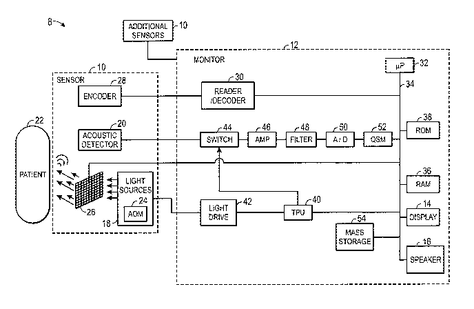
With the foregoing in mind, Fig. 1 depicts a photoacoustic spectroscopy system
8
that may be utilized in determining cardiac ouput. The system 8 includes a
photoacoustic
spectroscopy sensor 10 and a monitor 12. Some photoacoustic spectroscopy
systems 8
may include one or more photoacoustic spectroscopy sensors 10, as illustrated
in FIG. 1,
to generate physiological signals for different regions of a patient. For
example, in certain
embodiments, a single sensor 10 may have sufficient penetration depth to
generate
physiological signals from deep vessels (e.g., pulmonary artery and/or
pulmonary vein).
In other embodiments, more than one (e.g., two sensors) sensor 10 may be used
to monitor
physiological parameters (e.g., oxygen saturation) of more superficial vessels
(e.g., the
jugular vein and the femoral vein).
The sensor 10 may emit spatially modulated light at certain wavelengths into a
patient's tissue and may detect acoustic waves (e.g., ultrasound waves)
generated in
response to the emitted light. The monitor 12 may be capable of calculating
physiological
characteristics based on signals received from the sensor 10 that correspond
to the detected
acoustic waves. The monitor 12 may include a display 14 and/or a speaker 16
which may
be used to convey information about the calculated physiological
characteristics to a user.
The sensor 10 may be communicatively coupled to the monitor 12 via a cable or,
in some
embodiments, via a wireless communication link.
4
CA 02884793 2015-03-11
WO 2014/042840
PCT/US2013/056096
In one embodiment, the sensor 10 may include a light source 18 and an acoustic
detector 20, such as an ultrasound transducer. The present discussion
generally describes
the use of continuous wave (CW) light sources to facilitate explanation.
However, it
should be appreciated that the photoacoustic sensor 10 may also be adapted for
use with
other types of light sources, such as pulsed light sources, in other
embodiments. In certain
embodiments, the light source 18 may be associated with one or more optical
fibers for
conveying light from one or more light generating components to the tissue
site.
The photoacoustic spectroscopy sensor 10 may include a light source 18 and an
acoustic detector 20 that may be of any suitable type. For example, in one
embodiment
the light source 18 may be one, two, or more light emitting components (such
as light
emitting diodes) adapted to transmit light at one or more specified
wavelengths. In certain
embodiments, the light source 18 may include a laser diode or a vertical
cavity surface
emitting laser (VCSEL). The laser diode may be a tunable laser, such that a
single diode
may be tuned to various wavelengths corresponding to a number of different
absorbers of
interest in the tissue and blood. That is, the light may be any suitable
wavelength or
wavelengths (such as a wavelength between about 500 nm to about 1100 nm or
between
about 600 nm to about 900 nm) that is absorbed by a constituent of interest in
the blood or
tissue. For example, wavelengths between about 500 nm to about 600 nm,
corresponding
with green visible light, may be absorbed by deoxyhemoglobin and
oxyhemoglobin. In
other embodiments, red wavelengths (e.g., about 600 nm to about 700 nm) and
infrared or
near infrared wavelengths (e.g., about 800 urn to about 1100 nm) may be used.
In one
embodiment, the selected wavelengths of light may penetrate between 1 mm to 3
cm into
the tissue of the patient 22. In certain embodiments, the selected wavelengths
may
penetrate through bone (e.g., the rib cage) of the patient 22.
One problem that may arise in photoacoustic spectroscopy may be attributed to
the
tendency of the emitted light to diffuse or scatter in the tissue of the
patient 22. As a
result, light emitted toward an internal structure or region, such as a blood
vessel, may be
diffused prior to reaching the region so that amount of light reaching the
region is less than
desired. Therefore, due to the diffusion of the light, less light may be
available to be
absorbed by the constituent of interest in the target region, thus reducing
the ultrasonic
waves generated at the target region of interest, such as a blood vessel. To
increase the
5
CA 02884793 2015-03-11
WO 2014/042840
PCT/US2013/056096
precision of the measurements, the emitted light may be focused on an internal
region of
interest by modulating the intensity and/or phase of the illuminating light.
Accordingly, an acousto-optic modulator (AOM) 24 may modulate the intensity of
the emitted light, for example, by using LFM techniques. The emitted light may
be
intensity modulated by the AOM 24 or by changes in the driving current of the
LED
emitting the light. The intensity modulation may result in any suitable
frequency, such as
from 1 MHz to 10 MHz or more. Accordingly, in one embodiment, the light source
18
may emit LFM chirps at a frequency sweep range approximately from 1 MHz to 5
MHz.
In another embodiment, the frequency sweep range may be of approximately 0.5
MHz to
10 MHz. The frequency of the emitted light may be increasing with time during
the
duration of the chirp. In certain embodiments, the chirp may last
approximately 1 second
or less and have an associated energy of a 10 mJ or less, such as between 11.0
to 2 mJ, 1-5
mJ, 1-10 mJ. In such an embodiment, the limited duration of the light may
prevent
heating of the tissue while still emitting light of sufficient energy into the
region of interest
to generate the desired acoustic waves when absorbed by the constituent of
interest.
Additionally, the light emitted by the light source 18 may be spatially
modulated,
such as via a modulator 26. For example, in one embodiment, the modulator 26
may be a
spatial light modulator, such as a Holoeye LC-R 2500 liquid crystal spatial
light
modulator. In one such embodiment, the spatial light modulator may have a
resolution of
1024 x 768 pixels or any other suitable pixel resolution. During operation,
the pixels of
the modulator 26 may be divided into subgroups (such as square or rectangular
subarrays
or groupings of pixels) and the pixels within a subgroup may generally operate
together.
For example, the pixels of a modulator 26 may be generally divided into square
arrays of
10 x 10, 20 x 20, 40 x 40, or 50 x 50 pixels. In one embodiment, each subgroup
of pixels
of the modulator 26 may be operated independently of the other subgroups. The
pixels
within a subgroup may be operated jointly (i.e., are on or off at the same
time) though the
subgroups themselves may be operated independently of one another. In this
manner,
each subgroup of pixels of the modulator 26 may be operated so as to introduce
phase
differences at different spatial locations within the emitted light. That is,
the modulated
light that has passed through one subgroup of pixels may be at one phase and
that phase
may be the same or different than the modulated light that has passed through
other
subgroups of pixels, i.e., some segments or portions of the modulated light
wavefront may
6
CA 02884793 2015-03-11
WO 2014/042840
PCT/US2013/056096
be ahead of or behind other portions of the wavefront. In one embodiment, the
modulator
26 may be associated with additional optical components (e.g., lenses,
reflectors,
refraction gradients, polarizers, and so forth) through which the spatially
modulated light
passes before reaching the tissue of the patient 22.
In one example, the acoustic detector 20 may be one or more ultrasound
transducers suitable for detecting ultrasound waves emanating from the tissue
in response
to the emitted light and for generating a respective optical or electrical
signal in response
to the ultrasound waves. For example, the acoustic detector 20 may be suitable
for
measuring the frequency and/or amplitude of the ultrasonic waves, the shape of
the
ultrasonic waves, and/or the time delay associated with the ultrasonic waves
with respect
to the light emission that generated the respective waves. In one embodiment
an acoustic
detector 20 may be an ultrasound transducer employing piezoelectric or
capacitive
elements to generate an electrical signal in response to acoustic energy
emanating from the
tissue of the patient 22, i.e., the transducer converts the acoustic energy
into an electrical
signal.
In one implementation, the acoustic detector 20 may be a low finesse Fabry-
Perot
interferometer mounted on an optical fiber. In such an embodiment, the
incident acoustic
waves emanating from the probed tissue modulate the thickness of a thin
polymer film.
This produces a corresponding intensity modulation of light reflected from the
film.
Accordingly, the acoustic waves are converted to optical information, which is
transmitted
through the optical fiber to an upstream optical detector, which may be any
suitable
detector. In some embodiments, a change in phase of the detected light may be
detected
via an appropriate interferometry device which generates an electrical signal
that may be
processed by the monitor 12. The use of a thin film as the acoustic detecting
surface
allows high sensitivity to be achieved, even for films of micrometer or tens
of micrometers
in thickness. In one embodiment, the thin film may be a 0.25 mm diameter disk
of 50
micrometer thickness polyethylene terepthalate with an at least partially
optically
reflective (e.g., 40% reflective) aluminum coating on one side and a mirror
reflective
coating on the other (e.g., 100% reflective) that form the mirrors of the
interferometer.
The optical fiber may be any suitable fiber, such as a 50 micrometer core
silica multimode
fiber of numerical aperture 0.1 and an outer diameter of 0.25 mm.
7
CA 02884793 2015-03-11
WO 2014/042840
PCT/US2013/056096
The photoacoustic sensor 10 may include a memory or other data encoding
component, depicted in FIG. 1 as an encoder 28. For example, the encoder 28
may be a
solid state memory, a resistor, or combination of resistors and/or memory
components that
may be read or decoded by the monitor 12, such as via reader/decoder 30, to
provide the
monitor 12 with information about the attached sensor 10. For example, the
encoder 28
may encode information about the sensor 10 or its components (such as
information about
the light source 18 and/or the acoustic detector 20). Such encoded information
may
include information about the configuration or location of photoacoustic
sensor 10,
information about the type of lights source(s) 18 present on the sensor 10,
information
about the wavelengths, light wave frequencies, chirp durations, and/or light
wave energies
which the light source(s) 18 are capable of emitting, information about the
nature of the
acoustic detector 20, and so forth. In certain embodiments, the information
also includes a
reference linear frequency modulation (LFM) chirp that was used to generate
the actual
LFM emitted light. This information may allow the monitor 12 to select
appropriate
algorithms and/or calibration coefficients for calculating the patient's
physiological
characteristics, such as the amount or concentration of a constituent of
interest in a
localized region, such as a blood vessel.
In one implementation, signals from the acoustic detector 20 (and decoded data
from the encoder 28, if present) may be transmitted to the monitor 12. The
monitor 12
may include data processing circuitry (such as one or more processors 32,
application
specific integrated circuits (AS1CS), or so forth) coupled to an internal bus
34. Also
connected to the bus 34 may be a RAM memory 36, a ROM memory 38, a speaker 16
and/or a display 14. In one embodiment, a time processing unit (TPU) 40 may
provide
timing control signals to light drive circuitry 42, which controls operation
of the light
source 18, such as to control when, for how long, and/or how frequently the
light source
18 is activated, and if multiple light sources are used, the multiplexed
timing for the
different light sources.
The TPU 40 may also control or contribute to operation of the acoustic
detector 20
such that timing information for data acquired using the acoustic detector 20
may be
obtained. Such timing information may be used in interpreting the acoustic
wave data
and/or in generating physiological information of interest from such acoustic
data. For
example, the timing of the acoustic data acquired using the acoustic detector
20 may be
8
CA 02884793 2015-03-11
WO 2014/042840
PCT/US2013/056096
associated with the light emission profile of the light source 18 during data
acquisition.
Likewise, in one embodiment, data acquisition by the acoustic detector 20 may
be gated,
such as via a switching circuit 44, to account for differing aspects of light
emission. For
example, operation of the switching circuit 44 may allow for separate or
discrete
acquisition of data that corresponds to different respective wavelengths of
light emitted at
different times.
The received signal from the acoustic detector 20 may be amplified (such as
via
amplifier 46), may be filtered (such as via filter 48), and/or may be
digitized if initially
analog (such as via an analog-to-digital converter 50). The digital data may
be provided
directly to the processor 32, may be stored in the RAM 36, and/or may be
stored in a
queued serial module (QSM) 52 prior to being downloaded to RAM 36 as QSM 52
fills
up. In one embodiment, there may be separate, parallel paths for separate
amplifiers,
filters, and/or A/D converters provided for different respective light
wavelengths or
spectra used to generate the acoustic data.
In certain embodiments, the data processing circuitry, such as processor 32,
may
perform a signal quality assessment on the digital data. For example, it may
be desirable
to use a single, transthoracic photoacoustic sensor 10 to directly obtain the
arterial and
mixed venous oxygen saturation measurements from the pulmonary vein and
pulmonary
artery, respectively. Signals generated by interrogating the pulmonary artery
may yield
more accurate measurements for the mixed venous oxygen saturation. However,
the depth
of penetration of the photoacoustic sensor 10 may not be sufficient to
interrogate the
pulmonary artery for certain patients 22. Accordingly, it may be desirable to
compare the
digital data received from the sensor 10 to a threshold value (e.g., a signal
quality
threshold value) to determine whether the penetration depth is sufficient for
transthoracic
measurements. Additionally, as will be described in more detail below with
respect to
FIGS. 3 and 5, multiple sensors 10 may be coupled to the monitor 12. As such,
the
processor 32 may compare the digital sigmals received from each sensor 10 to
the
threshold value and based at least in part upon the comparisons, select
digital signals from
one or more sensors 10 to further process and/or use in the calculation of one
or more
physiological characteristics (e.g., oxygen saturation).
The data processing circuitry, such as processor 32, may derive one or more
physiological characteristics based on data generated by the photoacoustic
sensor 10. For
9
CA 02884793 2015-03-11
WO 2014/042840
PCT/US2013/056096
example, based at least in part upon data received from the acoustic detector
20, the
processor 32 may calculate the amount or concentration of a constituent of
interest in a
localized region of tissue or blood using various algorithms. In certain
embodiments, the
processor 32 may calculate arterial and/or mixed venous oxygen saturation
using signals
obtained from one or more sensors 10. In one embodiment, the processor 32 may
calculate both mixed venous and arterial oxygen saturation using signals
obtained from a
signal sensor 10. In certain embodiments, these algorithms may use
coefficients, which
may be empirically determined, that relate the detected acoustic waves
generated in
response to emitted light waves at a particular wavelength or wavelengths to a
given
concentration or quantity of a constituent of interest within a localized
region.
In one embodiment, processor 32 may access and execute coded instructions,
such
as for implementing the algorithms discussed herein, from one or more storage
components of the monitor 12, such as the RAM 36, the ROM 38, and/or a mass
storage
54. Additionally, the RAM 36, ROM 38, and/or the mass storage 54 may serve as
data
repositories for information such as templates for LFM reference chirps,
coefficient
curves, and so forth. For example, code encoding executable algorithms may be
stored in
the ROM 38 or mass storage device 54 (such as a magnetic or solid state hard
drive or
memory or an optical disk or memory) and accessed and operated according to
processor
32 instructions using stored data. Such algorithms, when executed and provided
with data
from the sensor 10, may calculate one or more physiological characteristics as
discussed
herein (such as the type, concentration, and/or amount of a constituent of
interest). Once
calculated, the physiological characteristics may be displayed on the display
14 for a
caregiver to monitor or review. Additionally, the calculated physiological
characteristics,
such as the arterial and the mixed venous oxygen saturation values, may be
sent to a multi-
parameter monitor for further processing (e.g., for the calculation of cardiac
output) and
display. Alternatively, the processor 32 may use the algorithms to calculate
the cardiac
ouput and the cardiac ouput may be displayed on the display 14 of the monitor
12.
With the foregoing in mind, FIG. 2 illustrates a method 70 for monitoring the
cardiac output of a patient using photoacoustic spectroscopy in accordance
with some
embodiments. The method 70 may be performed as an automated procedure by a
system,
as will be described in more detail below with respect to FIGS. 3 and 5. In
addition,
certain steps of the method 70 may be performed by a processor, or a processor-
based
CA 02884793 2015-03-11
WO 2014/042840
PCT/US2013/056096
device such as the monitor 12 that includes instructions for implementing
certain steps of
the method 70.
The method 70 may include non-invasively measuring the arterial and mixed
venous oxygen saturation using one or more photoacoustic sensors 10 (block
72). In some
embodiments, both the arterial and mixed venous oxygen saturation may be
determined
from a single transthoracic sensor 10. In other embodiments, signals from two
or more
photoacoustic sensors 10 may be used. For example, in one embodiment, the
mixed
venous saturation may be derived using signals from a first sensor 10 disposed
about the
jugular vein of the patient 22 and a second sensor 10 disposed about the
femoral vein of
the patient 22. Additionally, a third sensor 10, or alternatively, a pulse
oximetry sensor,
may be disposed about a peripheral artery of the patient 22 to obtain signals
related to the
arterial oxygen saturation. The monitor 12 may perform analysis of the one or
more
signals from the one or more-photoacoustic sensors 10 and calculate the
arterial and mixed
venous oxygen saturation, as previously described.
The method 70 may also include determining oxygen uptake of the patient 22
(block 74). The determining of oxygen uptake (block 74) may be performed
independent
of measuring the arterial and mixed venous oxygen saturation (block 72).
Additionally,
in certain embodiments, oxygen uptake may be an estimate based upon the body
surface
area of the patient 22. In other embodiments, oxygen uptake may be derived
using a mass
flow device that analyzes the inhaled and end-tidal respiratory gases of the
patient 22.
After the values for arterial and mixed oxygen saturation and oxygen uptake
have
been determined, cardiac output may be determined (block 76). In certain
embodiments,
the monitor 12 may receive the oxygen uptake parameter from the mass flow
device or,
alternatively, from another processor or processor-based monitor. As such, the
monitor 12
may be configured to use the determined values to calculate cardiac output
using the Fick
method. In other embodiments, the method 70 may include another processor or
processor-based device, such as a multi-parameter monitor, that may be
configured to
receive the determined values and may apply various algorithms to calculate
cardiac
output using the Fick method. For example, as discussed above, the oxygen
content of
arterial or mixed venous blood may be calculated using the respective oxygen
saturation
value. Specifically, in one embodiment, the oxygen content may be determined
in
accordance with the equation:
11
CA 02884793 2015-03-11
WO 2014/042840
PCT/US2013/056096
(2) C = (MB x k1 x S)/100
where C is the oxygen content of arterial or mixed venous blood (e.g., Ca or
C,), lc/ is a
coefficient, FIB is the hemoglobin, and S is the oxygen saturation of arterial
or mixed
venous blood (e.g., S, or Sy). To determine oxygen content for both arterial
and mixed
venous blood, Equation 2 may be applied once using the determined arterial
oxygen
saturation and once using the determined mixed venous oxygen saturation. The
hemoglobin of the patient 22 may be readily measured using known techniques,
such as
supplying a small sample of blood taken via a finger prick to a hemoglobin
photometer. In
certain embodiments, the monitor 12 may be configured to calculate the oxygen
content
values. Accordingly, the values for arterial and mixed venous oxygen content
and the
value for oxygen uptake may then be applied to Equation 1 to determine cardiac
output.
Furthermore, the method 70 may include displaying the cardiac ouput (block 78)
on a display of the monitor 12 and/or a multi-parameter monitor for a user to
monitor or
review. Additionally, the processor-based device (e.g., monitor 12 and/or
multi-parameter
monitor) may compare the determined cardiac output to a threshold or threshold
range
(e.g., a low threshold and a high threshold) related to a normal and/or
acceptable range of
cardiac output values (block 80). In certain embodiments, a user may input
parameters
related to the patient 22, such as age, gender, and/or weight, to the
processor-based device.
The processor-based device may be configured to apply the parameters to
various
algorithms to determine the threshold or threshold range specific for the
patient 22.
Alternatively, the processor-based device may include various predetermined
threshold
ranges, and a user may simply select the appropriate range for each patient
22.
Furthermore, the processor-based device may be configured to provide an alarm
(block
82) to the user in response to determining that the cardiac output parameter
is outside of
the predetermined threshold range. The alarm may be a visual indication, such
as an error
message, a flashing light, or a change in color and/or size of the displayed
cardiac output
parameter. The alarm may also be an auditory indication, such as a beep.
As described above, the oxygen uptake may be determined using an estimation of
the body surface area of the patient 22. In certain circumstances, it may be
advantageous
to use the body surface area estimation, as body surface area may be easily
and non-
invasively determined. A clinician may be provided with graphs and/or charts
relating to
height and weight, which may allow the clinician to readily determine the body
surface
12
CA 02884793 2015-03-11
WO 2014/042840
PCT/US2013/056096
area of the patient 22. Alternatively, the body surface area may be calculated
using
various algorithms (e.g., the Mosteller formula or the DuBois and DuBois
formula). It is
known that oxygen uptake is linearly related to body surface area, and as
such, the oxygen
uptake may be readily calculated using a known or empirically determined
coefficient.
Accordingly, FIG. 3 illustrates an embodiment of a system 90 that may be
operable to monitor the cardiac output of the patient 22 using a body surface
area
estimation to determine oxygen uptake. The system 90 includes one or more
photoacoustic sensors 10, the monitor 12, and a multi-parameter monitor 92.
The multi-
parameter monitor may be communicatively coupled to the monitor 12 via a cable
94
connected to a sensor input port or a digital communication port.
Alternatively, the
monitor 12 and the multi-parameter monitor 92 may be configured to communicate
wirelessly.
As previously described, based at least in part upon the received signals
corresponding to the acoustic waves received by detector 20 of the sensor 10,
the
microprocessor 32 of the monitor 12 may calculate the oxygen saturation using
various
algorithms. In certain embodiments, the system 90 may include a single,
transthoracic
sensor 10 disposed about the pulmonary artery 96 and the pulmonary vein 98 of
the patient
22 for generating signals corresponding to the mixed venous and arterial
oxygen
saturation, respectively. The transthoracic sensor 10 may be coupled to the
monitor 12 via
a cable 100. As described above, the microprocessor 32 may compare the signals
received
from the transthoracic sensor 10 to a threshold (e.g., signal quality
threshold) and may
determine whether the transthoracic sensor 10 has sufficient penetration depth
for
transthoracic measurements. Accordingly, the system 90 may additionally, or
alternatively, include a sensor 10 disposed about the jugular vein 102 and a
sensor 10
disposed about the femoral vein 104 for deriving the mixed venous oxygen
saturation for
circumstances in which the penetration depth is insufficient for transthoracic
measurements. For illustration purposes, the jugular and the femoral sensor 10
are
displayed with dashed lines to indicate that, in certain embodiments, the
jugular and
femoral sensors 10 may not be applied to the patient 22 and, in other
embodiments, the
jugular and femoral sensors 10 may be applied but not used (e.g., as backup in
case the
transthoracic sensor 10 is insufficient): Again, for illustration purposes,
the jugular and
femoral sensors 10 are depicted without cables 100. However, it should be
appreciated
13
CA 02884793 2015-03-11
WO 2014/042840
PCT/US2013/056096
that they may also be coupled to the monitor 12 via one or more cables 100.
Further, for
embodiments in which the transthoracic sensor 10 is insufficient, the system
90 may
include an additional sensor 10 (not shown) or a pulse oximetry sensor 106
disposed about
a peripheral artery (e.g., disposed about a finger of the patient 22) for
determining the
arterial oxygen saturation. The pulse oximetry sensor 106 may be
communicatively
coupled to the monitor 12 via a cable or wireless communication 108.
After determining the arterial and mixed venous oxygen saturation parameters,
the
monitor 12 may transmit the parameters to the multi-parameter monitor 92 for
further
analysis and calculation of cardiac output using the Fick method. The multi-
parameter
monitor 92 may include a processor 110 configured to execute code to perform
the
techniques as described herein. Alternatively, the processor 32 of the monitor
12 may
process the determined parameters and calculate cardiac output using the Fick
method.
Accordingly, the processor 32, 110 may calculate the arterial and mixed venous
oxygen
content based in part upon the received arterial and mixed venous oxygen
saturation
parameters. The processor 32, 110 may apply various algorithms, such as
Equation 2.
These algorithms may employ certain coefficients, which may be empirically
determined.
The algorithms and coefficients may be stored in any suitable computer-
readable storage
medium and accessed and operating according to processor 32, 110 instructions.
Additionally, the processor 32, 110 may be configured to calculate body
surface
area and/or oxygen uptake using various algorithms based on clinician inputs
entered via
control inputs 112 on the monitor 92, a keyboard, or other device. For
example, a
clinician may input a patient's height, weight, and/or gender, which may be
employed in
various algorithms to determine body surface area and additionally, in certain
embodiments, to determine body mass index (I3M1). Alternatively, the clinician
may input
a determined body surface area of the patient 22 via the control inputs 112.
Furthermore,
the processor 32, 110 may be configured to apply various algorithms to
calculate the
oxygen uptake based on the determined body surface area. In one embodiment,
the
algorithms may employ certain coefficients, which may be empirically
determined, that
correspond to the BMI and/or gender of the patient 22. For example, certain
patients 22,
such as patients 22 with a BM1 greater than 35 kg/m2, or more specifically,
male patients
22 with a BM1 greater than 40 kg/m2, may have a greater oxygen uptake than the
determined oxygen uptake based on the body surface area estimate. Using the
estimation
14
CA 02884793 2015-03-11
WO 2014/042840
PCT/US2013/056096
of oxygen uptake may be advantageous as the system 90 may continuously monitor
cardiac output for patients 22 on a mechanical ventilator, as well as
spontaneously
breathing patients 22. That is, the estimation of oxygen uptake does not
require analyzing
the inhaled and exhaled gas of the patient 22, which may be technically
complex for the
spontaneously breathing patient 22.
After determining the parameters for the Fick method, the processor 32, 110
may
calculate and monitor cardiac output. Specifically, the processor 32, 110 may
apply
Equation 1 for determining cardiac output. To monitor cardiac output over
time, a
baseline measurement of cardiac output may be established by an independent
measurement (e.g., blood samples taken to apply the direct Fick method) and
may be used
to calibrate cardiac output determined using body surface area. Specifically,
the processor
32, 110 may determine a calibration coefficient for the estimation of oxygen
uptake in
accordance with the equation:
(3) k2 = COb x (HB X (Sa02 ¨ Svoz))b
where k2 is the calibration coefficient, COI, is the baseline cardiac output
parameter, HB is
the baseline hemoglobin parameter, Saw is the baseline arterial oxygen
saturation
parameter, and S.2 is the baseline mixed venous oxygen saturation parameter.
The
parameters for arterial and mixed venous oxygen saturation, and accordingly
for arterial
and mixed venous oxygen content, may fluctuate with time. As such, in one
embodiment,
processor 32, 110 may monitor cardiac output over time (e.g., a trend in
cardiac output) in
accordance with the equation:
4) 1r2
( CO(t) =
(t) X (Saoz W-Svo 2 (0))
where k2 is the calibration coefficient, CO is cardiac output as a function of
time, FIB is
hemoglobin as a function of time, Sao. is arterial oxygen saturation as a
function of time,
and Sv02 is mixed venous oxygen saturation as a function of time.
The multi-parameter monitor 92 may provide a display 114 to facilitate the
presentation of patient data, such as the calculated cardiac output and/or
physiological
parameters determined by other patient monitoring systems (e.g.,
electrocardiographic
(ECG) monitoring system, a respiratory monitoring system, a blood pressure
monitoring
CA 02884793 2015-03-11
WO 2014/042840
PCT/US2013/056096
system, etc.). In certain embodiments, the display 114 may provide a graph of
cardiac
output over time (e.g., a cardiac output trend), as well as the current
cardiac output value.
Alternatively, the patient data (e.g., oxygen saturation and cardiac output)
may be
displayed on the display 14 of the monitor 12.
Furthermore, as described above, an alarm may be provided in response to
determining that the cardiac output value falls outside of a predetermined
threshold range.
Accordingly, the processor 32, 110 may be configured to determine the
threshold range for
each patient 22. For example, the clinician may input via control inputs 112 a
patient's
age, weight, height, gender, or information about the patient's clinical
condition that may
be relevant to the determination of a threshold range corresponding to normal
values for
cardiac output. The processor 32, 110 may use the parameters provided by the
clinician to
determine the threshold range using various algorithms. In the event that the
cardiac
output parameter falls outside of the threshold range, the monitor 12 or the
multi-
parameter monitor 92 may provide an alarm, such as an error indication on the
display 14,
114, or a beep or other auditory alarm via speaker 16, 116.
FIG. 4 is a method 130 for calculating and monitoring cardiac output over time
(e.g., a cardiac output trend) using one or more photoacoustic sensors 10 and
an oxygen
uptake estimation using the system 90 of FIG. 3. The method 130 may be
performed as
an automated procedure by a system, such as the system 90. In addition,
certain steps of
the method may be performed by a processor, or a processor-based device such
as the
monitor 12 and/or the multi-parameter monitor 92 that includes instructions
(e.g., code)
for implementing certain steps of the method 130.
According to an embodiment, the method 130 may include establishing a cardiac
output baseline 134 for the patient 22 (block 132). As described above, this
may include
analyzing blood samples taken from the patient 22 by a clinician to implement
the direct
Fick method. In other embodiments, establishing the cardiac output baseline
134 may
include other known clinical techniques such as the thermodilution technique,
echocardiographic technique, or radionuclide imaging technique (block 132). In
certain
embodiments, an additional system may measure the cardiac output baseline 134,
and the
monitor 12, 92 may receive, and accordingly establish the cardiac output
baseline 134
(block 130) from the additional system or from a clinician via the control
inputs 112. As
will be described in more detail below, a CO baseline 134 may be used to
calibrate a
16
CA 02884793 2015-03-11
WO 2014/042840
PCT/US2013/056096
calculation of cardiac output and determine a trending measurement of cardiac
output
(e.g., cardiac output as a function of time).
The method 130 may also include determining an estimation of body surface area
(BSA) 138 for the patient 22 (block 136). In certain embodiments, the
processor 32, 110
may determine an estimation of the BSA 138 (block 136) using algorithms and
patient
parameters inputted by a clinician (e.g., height and weight of the patient
22). The
processor 32, 110 may then use the determined BSA 138 to estimate (block 140)
the
oxygen uptake 142, as described above.
Additionally, the method 130 may include measuring arterial and mixed venous
oxygen saturation using a transthoracic photoacoustic sensor 10 (block 144)
disposed
about the pulmonary artery 96 and the pulmonary vein 98 of the patient 22. As
discussed
above, it may be desirable to measure the mixed venous oxygen saturation
directly from
the pulmonary artery 96 and additionally, to measure arterial and mixed venous
oxygen
saturation using the same sensor 10. However, for certain patients 22, the
transthoracic
sensor 10 may not generate signals with sufficient penetration depth to
accurately measure
the arterial and mixed venous oxygen saturation. Accordingly, the method 130
may
include performing a signal quality assessment to determine whether the
penetration depth
of the transthoracic sensor 10 is sufficient (block 146). Specifically,
performing a signal
quality assessment may include comparing a signal generated using the
transthoracic
sensor 10 to a signal quality threshold value. For example, determining that
the
penetration depth of the transthoracic sensor 10 is sufficient may include
determining that
the signal generated using the transthoracic sensor 10 is above or below the
signal quality
threshold value. If the penetration depth is sufficient, the processor 32 of
the monitor 12
may continue to process signals from the transthoracic sensor 10 to calculate
the arterial
and mixed venous oxygen saturation 148 of the patient 22.
However, if the penetration depth is not sufficient, the method 130 may
include
measuring the mixed venous oxygen saturation using multiple (e.g., at least
two)
photoacoustic sensors 10 and the arterial oxygen saturation using another
sensor (e.g.,
sensor 10) (block 150). Specifically, the processor 32 may send signals to
deactivate the
light drive 42 of the transthoracic sensor 10 and signals to activate the
light drives 42 of
the sensor 10 disposed about the jugular artery 100 and the sensor 10 disposed
about the
femoral artery 102. The processor 32 may then derive the mixed venous
saturation 148
17
CA 02884793 2015-03-11
WO 2014/042840
PCT/US2013/056096
based on the signals received from the jugular and femoral sensors 10.
Additionally, the
processor 32 may activate the light drive 42 of a third photoacoustic sensor
10 disposed
about a peripheral artery of the patient 22 to measure the arterial oxygen
saturation 148.
Alternatively, a pulse oximetry sensor may also be coupled to the monitor 12
and disposed
about a peripheral artery of the patient 22 to measure the arterial oxygen
saturation 148.
Once the parameters of the Fick equation have been determined, the monitor 12,
92 may calculate cardiac output as a function of time based on the cardiac
output baseline
134, the oxygen uptake 142, and the arterial and mixed venous oxygen
saturation 148
(block 152). Specifically, the processor 32, 110 may apply various algorithms,
such as
Equation 3 and 4, to determine cardiac output over time. Additionally, as
described with
respect to FIG. 2, the cardiac output values may be displayed (block 78) on
the display 14,
114 for a clinician to monitor. Furthermore, the processor 32, 110 may be
configured to
determine a threshold range for normal or acceptable cardiac output values,
based at least
in part on the patient parameters inputted by a clinician, and provide (block
80) an alarm
and/or error indication in response to determining that the cardiac output is
outside of the
threshold range (e.g., below a low threshold and/or above a high threshold).
In other embodiments, it may be desirable to additionally measure oxygen
uptake
over time to determine and monitor cardiac output. Additionally, for certain
patients 22
(e.g., for patients 22 who are overweight or obese), the body surface area
estimate may not
yield an accurate oxygen uptake determination. For example, a cardiac output
baseline
which significantly differs from the cardiac output determination may alert a
clinician that
the oxygen uptake estimation is inaccurate. As such, the disclosed embodiments
provide a
system and method for non-invasively monitoring cardiac output via the Fick
method
using continuous or intermittent measurements of oxygen uptake determined from
the
inspired and expired air of the patient 22.
With the foregoing in mind, FIG. 5 illustrates a system 170 that may be
operable
to monitor the cardiac output of the patient 22 using oxygen uptake
measurements. The
system 170 includes the one or more photoacoustic sensors 10, the monitor 12,
and the
multi-parameter monitor 92. Again, for illustration purposes the femoral and
jugular
sensors 10 and the pulse oximetry sensor 106 are coupled to the monitor 12
with dashed
lines to indicate that they may not be present in certain embodiments. The
system 170
additionally includes a gas analysis device 172, which may be in line with a
ventilator 174.
18
CA 02884793 2015-03-11
WO 2014/042840
PCT/US2013/056096
In certain embodiments, the gas analysis device 172 may be incorporated into
the
ventilator 174.
The gas analysis device 172 may be configured to receive and process the
inhaled
and exhaled gases to calculate oxygen uptake. Specifically, the gas analysis
device 172 is
may include a mass flow device, an oxygen analyzer, a carbon dioxide analyzer,
which
may include a pressure sensor, a temperature sensor, viscometer, and/or
densitometer, to
measure the density, viscosity, and specific heat of the inhaled and exhaled
gases. The gas
analysis device 172 may include a processor that may apply various algorithms,
which
may be stored in a suitable computer-readable storage medium, to determine the
concentration of oxygen in the inhaled and in the exhaled gases based on the
measurements from the mass flow meter and the gas analyzer. Additionally, or
alternatively, the gas analysis device 172 may be configured to determine the
concentration of carbon dioxide in the inhaled and exhaled gases, as carbon
dioxide
production may be used in place of oxygen uptake to calculate cardiac output
using the
Fick method. As the gas analysis device 172 is in line with the ventilator
174, oxygen
uptake may be calculated continuously.
However, in other embodiments, the gas analysis device 172 may calculate
oxygen
uptake intermittently. For example, the patient 22 may not be connected to the
ventilator
174. As such, the gas analysis device 172 may include a mask configured to be
placed
over the nose and mouth of the patient 22 to deliver air and to receive
exhaled air.
Accordingly, the gas analysis device 172 may analyze each exhaled breath and
may
calculate oxygen uptake for each breath. While the gas analysis device 172
with the mask
does not provide continuous measurements, it may be desirable for certain
patients 22 as it
may be less invasive.
Accordingly, the multi-parameter monitor 92 or the monitor 12 may be
configured
to receive the oxygen uptake calculations from the gas analysis device 172 and
to use the
calculations to determine and monitor the cardiac output of the patient 22.
The multi-
parameter monitor 92 and the monitor 12 may function as described above. That
is, in one
embodiment, the multi-parameter monitor 92 may receive the arterial and mixed
venous
oxygen saturations 148 from the monitor 12, which may be obtained using the
one or more
photoacoustic sensors 10 and in certain embodiments, the pulse oximetry sensor
106.
Upon receiving the parameters of oxygen uptake and arterial and mixed venous
oxygen
19
CA 02884793 2015-03-11
WO 2014/042840
PCT/US2013/056096
saturation 148, the multi-parameter monitor 92 may then calculate cardiac
output as a
function of time. Specifically, in certain embodiments, the processor 32, HO
may apply
Equation 2 to calculate the arterial and mixed venous oxygen content and then,
may use
the oxygen content parameters to calculate cardiac output using Equation 1
(e.g., the direct
Fick method). The monitor 12, 92 may not utilize a cardiac output baseline 134
to
calibrate the cardiac output calculation, as the oxygen uptake calculations
may be more
accurate than the oxygen uptake 142 determined using the body surface area
estimate 138.
FIG. 6 illustrates a method 190 for calculating and monitoring cardiac output
over
time using one or more photoacoustic sensors 10 and calculations of oxygen
uptake using
the gas analysis device 172. The method 190 may be performed as an automated
procedure by a system, such as the system 170. In addition, certain steps of
the method
may be performed by a processor, or a processor-based device such as the
monitor 12, the
multi-parameter monitor 92, and/or the processor of the gas analysis device
172 that
includes instructions for implementing certain steps of the method 190.
Similar to the method 130 described above, the method 190 may include
measuring (block 144) arterial and mixed venous oxygen saturation using the
transthoracic
sensor 10. The method 190 may also include determining (block 146) whether the
penetration depth is sufficient to determine the arterial and mixed venous
oxygen
saturation 148. In response to determining (block 146) that the penetration
depth is
insufficient, the method may include measuring (block 150) the arterial and
mixed venous
oxygen saturations using the jugular and femoral sensors 10 and either a third
sensor 10 or
a pulse oximetry sensor 106 disposed about a peripheral artery of the patient
22.
However, unlike the method 130, the method 190 may include measuring the
oxygen uptake 194 using the gas analysis device 172 (block 192). As described
above, the
gas analysis device 172 may measure oxygen uptake continuously when in line
with the
ventilator 174, or intermittently for embodiments in which the gas analysis
device 172
includes a mask for analyzing each breath of the patient 22. It should be
appreciated that
the oxygen uptake 194 may differ from the oxygen uptake 142. Accordingly, the
multi-
parameter monitor 92 may then calculate (block 196) cardiac output as a
function of time
based on the arterial and mixed venous oxygen saturation 148 and the oxygen
uptake 194.
In certain embodiments, the multi-parameter monitor 92 may continuously
calculate
cardiac output using intermittent measurements of oxygen uptake 194 and
continuous
CA 02884793 2015-03-11
WO 2014/042840
PCT/US2013/056096
measurements of oxygen and mixed venous oxygen saturation 148. In one
embodiment,
the multi-parameter monitor may continuously calculate cardiac output using
continuous
measurements of oxygen uptake 194 and continuous measurements of oxygen and
mixed
venous oxygen saturation 148. Additionally, the method 190 may further include
displaying (block 78) the calculated cardiac output on the display 114 for a
clinician to
monitor and review, as well as provide (block 80) an alarm in response to
determining that
the cardiac output is outside of the predetermined range.
The disclosed embodiments may be interfaced to and controlled by a computer
readable storage medium having stored thereon a computer program. The computer
readable storage medium may include a plurality of components such as one or
more of
electronic components, hardware components, and/or computer software
components. These components may include one or more computer readable storage
media that generally store instructions such as software, firmware and/or
assembly
language for performing one or more portions of one or more implementations or
embodiments of an algorithm as discussed herein. These computer readable
storage media
are generally non-transitory and/or tangible. Examples of such a computer
readable
storage medium include a recordable data storage medium of a computer and/or
storage
device. The computer readable storage media may employ, for example, one or
more of a
magnetic, electrical, optical, biological, and/or atomic data storage medium.
Further, such
media may take the form of, for example, floppy disks, magnetic tapes, CD-
ROMs, DVD-
ROMs, hard disk drives, and/or solid-state or electronic memory. Other forms
of non-
transitory and/or tangible computer readable storage media not list may be
employed with
the disclosed embodiments.
A number of such components can be combined or divided in an implementation
of a system. Further, such components may include a set and/or series of
computer
instructions written in or implemented with any of a number of programming
languages,
as will be appreciated by those skilled in the art. In addition, other forms
of computer
readable media such as a carrier wave may be employed to embody a computer
data signal
representing a sequence of instructions that when executed by one or more
computers
causes the one or more computers to perform one or more portions of one or
more
implementations or embodiments of a sequence.
21
CA 02884793 2015-03-11
WO 2014/042840
PCT/US2013/056096
While the disclosure may be susceptible to various modifications and
alternative
forms, specific embodiments have been shown by way of example in the drawings
and
have been described in detail herein. However, it should be understood that
the
embodiments provided herein are not intended to be limited to the particular
forms
disclosed. Rather, the various embodiments may cover all modifications,
equivalents, and
alternatives falling within the spirit and scope of the disclosure as defined
by the following
appended claims.
22