Note: Descriptions are shown in the official language in which they were submitted.
=
- 1 -
SURGICAL TRAINING MODEL FOR LAPAROSCOPIC PROCEDURES
[0001]
FIELD OF THE INVENTION
[0002] This application is generally related to surgical training
tools, and in
particular, to simulated tissue structures and models for teaching and
practicing
various surgical techniques and procedures related but not limited to
laparoscopic,
endoscopic and minimally invasive surgery.
BACKGROUND OF THE INVENTION
[0003] Medical students as well as experienced doctors learning new
surgical techniques must undergo extensive training before they are qualified
to
perform surgery on human patients. The training must teach proper techniques
employing various medical devices for cutting, penetrating, clamping,
grasping,
stapling, cauterizing and suturing a variety of tissue types. The range of
possibilities
that a trainee may encounter is great. For example, different organs and
patient
anatomies and diseases are presented. The thickness and consistency of the
various tissue layers will also vary from one part of the body to the next and
from one
patient to another. Different procedures demand different skills. Furthermore,
the
trainee must practice techniques in various anatomical environs that are
influenced
by factors such as the size and condition of the patient, the adjacent
anatomical
landscape and the types of targeted tissues and whether they are readily
accessible
or relatively inaccessible.
[0004] Numerous teaching aids, trainers, simulators and model
organs are
available for one or more aspects of surgical training. However, there is a
need for
models or simulated tissue elements that are likely to be encountered in and
that can be
CA 2885302 2019-10-24
CA 02885302 2015-03-18
WO 2014/052612
PCT/US2013/061949
- 2 -
used for practicing endoscopic and laparoscopic, minimally invasive surgical
procedures. In laparoscopic surgery, a trocar or cannula is inserted to access
a body
cavity and to create a channel for the insertion of a camera such as a
laparoscope. The
camera provides a live video feed capturing images that are then displayed to
the
surgeon on one or more monitors. At least one additional small incision is
made
through which another trocar/cannula is inserted to create a pathway through
which
surgical instruments can be passed for performing procedures observed on the
monitor.
The targeted tissue location such as the abdomen is typically enlarged by
delivering
carbon dioxide gas to insufflate the body cavity and create a working space
large
enough to accommodate the scope and instruments used by the surgeon. The
insufflation pressure in the tissue cavity is maintained by using specialized
trocars.
Laparascopic surgery offers a number of advantages when compared with an open
procedure. These advantages include reduced pain, reduced blood and shorter
recovery times due to smaller incisions.
[0005]
Laparoscopic or endoscopic minimally invasive surgery requires an
increased level of skill compared to open surgery because the target tissue is
not
directly observed by the clinician. The target tissue is observed on monitors
displaying
a portion of the surgical site that is accessed through a small opening.
Therefore,
clinicians need to practice visually determining tissue planes, three-
dimensional depth
perception on a two-dimensional viewing screen, hand-to-hand transfer of
instruments,
suturing, precision cutting and tissue and instrument manipulation. Typically,
models
simulating a particular anatomy or procedure are placed in a simulated pelvic
trainer
where the anatomical model is obscured from direct visualization by the
practitioner.
Ports in the trainer are employed for passing instruments to practice
techniques on the
anatomical model hidden from direct visualization. Simulated pelvic trainers
provide a
functional, inexpensive and practical means to train surgeons and residents
the basic
skills and typical techniques used in laparoscopic surgery such as grasping,
manipulating, cutting, tying knots, suturing, stapling, cauterizing as well as
how to
perform specific surgical procedures that utilized these basic skills.
Simluated pelvic
trainers are also effective sales tools for demonstrating medical devices
required to
perform these laparoscopic procedures.
CA 02885302 2015-03-18
WO 2014/052612 PCT/US2013/061949
- 3 -
[0006] One of the techniques mentioned above that requires practice in
endoscopic or laparoscopic minimally invasive surgery is the passing of
sutures and
suturing which requires the clinician to develop skills such as three-
dimensional depth
perception and hand-to-hand transfer of a needle and suture while the target
tissue and
instruments are observed on a two-dimensional video monitor. Therefore, it is
desirable
to present a model suitable for practicing suturing and, in particular, there
is a need for a
model that isolates a particular step of a procedure for the trainee such as
the passing
of sutures for the clinician to practice in a simulated laparoscopic
environment. The
laparoscopic training model is removably placed inside a simulated
laparoscopic
environment such as a laparoscopic trainer in which it is at least partially
obscured from
direct visualization. A camera and monitor provide visualization to the
practitioner.
After a technique is practiced, it is furthermore desirable that such a model
permits
repeatable practice with ease, speed and cost savings. In view of the above,
it is an
object of this invention to provide a surgical training device that
realistically simulates an
anatomy and isolates a particular stage or step of a procedure that also
enables
repeatable practice. It has been demonstrated that the use of simulation
trainers greatly
enhances the skill levels of new laparoscopists and are a great tool to train
future
surgeons in a non-surgical setting. There is a need for such improved,
realistic and
effective surgical training models.
SUMMARY OF THE INVENTION
[0007] According to one aspect of the invention, a surgical training
device is
provided. The device includes a top cover spaced apart from a base to define
an
internal cavity between the top cover and the base. At least one aperture or a
penetrable region for accessing the internal cavity is provided and a
laparoscopic
camera is disposed inside the cavity and configured to display video images on
a video
monitor connected to the camera and located outside of the cavity. A model is
removably disposed inside the cavity such that the model is substantially
obscured from
a user yet observable via the laparoscopic camera displaying video images of
the model
on the video monitor. The model includes a base having an outer surface and a
plurality of eyelets connected to the base. The plurality of eyelets are
configured along
CA 02885302 2015-03-18
WO 2014/052612 PCT/US2013/061949
- 4 -
the surface to define a pathway for practicing the passing of at least one
needle and
suture through one or more of the plurality of eyelets of the pathway.
[0008] According to another aspect of the invention, a surgical training
device
is provided. The device includes a base having an outer surface and a
plurality of
eyelets connected to the outer surface of the base. Each eyelet has a head
portion
connected to a neck portion. The neck portion is connected to the base at a
proximal
end of the eyelet. The head portion includes an aperture defining an aperture
plane
having a first side and a second side. The plurality of eyelets are configured
with
respect to the base such that at least one aperture plane is angled with
respect to at
least one other aperture plane of the plurality of eyelets. At least a subset
of the
plurality of eyelets defines a pathway with apertures that are sized for
passing a suture
and suture needle.
[0009] According to another aspect of the invention, a method for
practicing
laparoscopic suture passing is provided. The method includes providing a
device
having a base with an outer surface and a plurality of eyelets connected to
the base.
Each eyelet has a head portion connected to a neck portion. The neck portion
is
connected to the base. The plurality of eyelets includes at least one
retractable eyelet.
The retractable eyelet is retractable with respect to the outer surface such
that the
retractable eyelet has a first position in which the aperture is at a first
distance relative
to the outer surface and a second position in which the aperture is at a
second distance
relative to the outer surface. The second distance is greater above the outer
surface
than the first distance. The method includes grasping a retractable eyelet and
pulling it
from a first position to a second position. The eyelet is held in the second
position while
a suture and needle are passed through the aperture. The method includes
releasing
the retracted eyelet.
BRIEF DESCRIPTION OF THE DRAWINGS
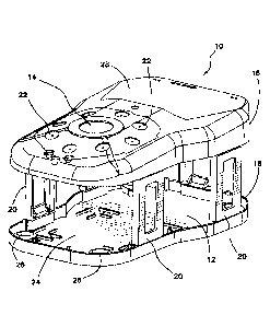
[0010] FIG. 1 illustrates a top perspective view of a surgical training
device
according to the present invention.
[0011] FIG. 2 illustrates a top perspective view of a model according to
the
present invention.
- 5 -
[0012] FIG. 3 illustrates a top perspective view of a model
according to the
present invention.
[0013] FIGs. 4A-4D illustrate various eyelets according to the
present
invention.
DETAILED DESCRIPTION OF THE INVENTION
[0014] A surgical training device 10 that is configured to mimic the
torso of a
patient such as the abdominal region is shown in FIG. 1. The surgical training
device
provides a body cavity 12 substantially obscured from the user for receiving
simulated or live tissue or model organs or training models of the like
described in this
invention. The body cavity 12 is accessed via a tissue simulation region 14
that is
penetrated by the user employing devices to practice surgical techniques on
the tissue
or practice model found located in the body cavity 12. Although the body
cavity 12 is
shown to be accessible through a tissue simulation region, a hand-assisted
access
device or single-site port device may be alternatively employed to access the
body
cavity 12. An exemplary surgical training device is described in U.S. Patent
Application Serial No. 13/248,449 entitled "Portable Laparoscopic Trainer"
filed on
September 29, 2011. The surgical training device 10 is particularly well
suited for
practicing laparoscopic or other minimally invasive surgical procedures.
[0015] Still referencing FIG. 1, the surgical training device 10
includes a top
cover 16 connected to and spaced apart from a base 18 by at least one leg 20.
FIG. 1
shows a plurality of legs 20. The surgical training device 10 is configured to
mimic the
torso of a patient such as the abdominal region. The top cover 16 is
representative of
the anterior surface of the patient and the space 12 between the top cover 16
and the
base 18 is representative of an interior of the patient or body cavity where
organs
reside. The surgical trainer 10 is a useful tool for teaching, practicing and
demonstrating various surgical procedures and their related instruments in
simulation
of a patient undergoing a surgical procedure. Surgical instruments are
inserted into
the cavity 12 through the tissue simulation region 14 as well as through pre-
established
apertures 22 in the top cover 16. Various tools and techniques may be used to
CA 2885302 2019-10-24
CA 02885302 2015-03-18
WO 2014/052612 PCT/US2013/061949
- 6 -
penetrate the top cover 16 to perform mock procedures on simulated organs or
practice
models placed between the top cover 16 and the base 18. The base 18 includes a
model-receiving area 24 or tray for staging or holding a simulated tissue
model or live
tissue. The model-receiving area 24 of the base 18 includes frame-like
elements for
holding the model (not shown) in place. To help retain a simulated tissue
model or live
organs on the base 18, a clip attached to a retractable wire is provided at
locations 26.
The retractable wire is extended and then clipped to hold the tissue model in
position
substantially beneath the tissue simulation region 14. Other means for
retaining the
tissue model include a patch of hook-and-loop type fastening material (VELCRO
)
affixed to the base 18 in the model receiving area 24 such that it is
removably
connectable to a complementary piece of hook-and-loop type fastening material
(VELCRO()) affixed to the model.
[0016] A video display monitor 28 that is hinged to the top cover 16 is
shown
in a closed orientation in FIG. 1. The video monitor 62 is connectable to a
variety of
visual systems for delivering an image to the monitor. For example, a
laparoscope
inserted through one of the pre-established apertures 22 or a webcam located
in the
cavity and used to observe the simulated procedure can be connected to the
video
monitor 28 and/or a mobile computing device to provide an image to the user.
Also,
audio recording or delivery means may also be provided and integrated with the
trainer
to provide audio and visual capabilities. Means for connecting a portable
memory
storage device such as a flash drive, smart phone, digital audio or video
player, or other
digital mobile device is also provided, to record training procedures and/or
play back
pre-recorded videos on the monitor for demonstration purposes. Of course,
connection
means for providing an audio visual output to a screen larger than the monitor
is
provided. In another variation, the top cover 10 does not include a video
display but
includes means for connecting with a laptop computer, a mobile digital device
or tablet
such as an IPADO and connecting it by wire or wirelessly to the trainer.
[0017] When assembled, the top cover 16 is positioned directly above the
base 18 with the legs 20 located substantially around the periphery and
interconnected
between the top cover 16 and base 18. The top cover 16 and base 18 are
substantially
the same shape and size and have substantially the same peripheral outline.
The
CA 02885302 2015-03-18
WO 2014/052612 PCT/US2013/061949
- 7 -
internal cavity is partially or entirely obscured from view. In the variation
shown in FIG.
1, the legs include openings to allow ambient light to illuminate the internal
cavity as
much as possible and also to advantageously provide as much weight reduction
as
possible for convenient portability. The top cover 16 is removable from the
legs 20
which in turn are removable or collapsible via hinges or the like with respect
to the base
18. Therefore, the unassembled trainer 10 has a reduced height that makes for
easier
portability. In essence, the surgical trainer 10 provides a simulated body
cavity 12 that
is obscured from the user. The body cavity 12 is configured to receive at
least one
surgical model accessible via at least one tissue simulation region 14 and/or
apertures
22 in the top cover 16 through which the user may access the models to
practice
laparoscopic or endoscopic minimally invasive surgical techniques.
[0018] A model 30 for the practice of passing sutures in laparoscopic
procedures according to the present invention is shown in FIG. 2. The model 30
is
configured to be placed inside the surgical training device 10 described above
or other
similar surgical trainer. The model 30 includes a base 32, and a plurality of
eyelets 34
connected to the surface of the base 32.
[0019] The base 32 of the model 30 is a platform that serves as a bottom
support for the rest of the model 30 and it is sized and configured such that
the model
does not tip over. The platform is made of any material such as metal or
plastic. The
base 32 is of sufficient heft to maintain the stability of the model 30 in the
upright
position while being manipulated by a user. The model 30 is sized and
configured to be
placed into the body cavity 12 of the surgical trainer 10 in the location of
the model
receiving area 24. The underside of the base 32 is provided with means to
affix the
model 30 inside the surgical trainer 10. Such means to affix the model 30
inside the
trainer 10 include but are not limited to adhesive, suction cup, magnet, snap-
fit, and a
hook-and-loop type fastener material attached to the bottom surface of the
base 32 and
configured to connect with a complementary hook-and-loop type fastener
material or
adhesive attached to the base 18 of the surgical trainer 30.
[0020] The base 32 of the model 30 includes an outer surface 36 which may
be flat or contoured in various ways. For example, the outer surface can be
convex as
shown in FIG. 2. The outer surface 36 may be concave, curved, sloped,
undulating or
CA 02885302 2015-03-18
WO 2014/052612 PCT/US2013/061949
- 8 -
otherwise have any configuration or geography including an upward hill, a
downward
hill, valleys and peaks including smaller surface additions such bumps or
divots that
compliment the larger features. The geography of the outer surface 36 creates
a
varying surface or numerous planes to permit the user to practice depth
perception in
laparoscopic surgery. In one variation, the base 32 is not rigid and solid but
is pliable,
resilient and flexible, and deflectable when manipulated with surgical
instruments that
would be used in laparoscopic surgery. As such, the base 32 is made of
pliable,
resilient material such as rubber or silicone. Another example of the
geography of the
outer surface 36 of the base 32 is shown in FIG. 3. The model 30 in FIGs. 2
and 3 is
shown positioned with the operative outer surface 36 facing upwardly. However,
the
model 30 may be positioned on its side in the trainer 10 to provide another
variation and
representation of internal bodily structures for practicing laparoscopic
procedures. In
this alternative orientation, the side surface of the model 30 is provided
with eyelets 34.
[0021] The model 30 includes a plurality of eyelets or apertures 34
connected
to the base 32 such that the eyelets 34 are configured to reside above the
outer surface
36 or side surface of the model 30 as shown in FIGs. 2 and 3. An exemplary
eyelet 34
is shown in FIG. 4A. In general, the eyelet 34 is configured to provide an
opening
through which a clinician can practice passing a needle and suture. The eyelet
34
includes a neck portion 38 and a head portion 40. The head portion 40 includes
at least
one aperture 42 defining an aperture plane in which it lies. Although the
aperture 42 is
shown to have a circular shape, the invention is not so limited and the
aperture 42 can
have any shape such as a polygon or closed curve. While FIG. 4A depicts a
closed
aperture 42, an open aperture 44 is within the scope of the present invention
as shown
in FIG. 4B. An open or hook-like aperture 44 is an aperture that is open and
only
partially enclosed by surrounding material of the head portion 40 leaving an
opening or
entry into the aperture 40 that is anywhere from approximately 1/8 to 1/4 of
the aperture
perimeter in size. In one variation, the aperture 42 of the eyelet 34 is
covered with a
layer of silicone or other penetrable material that may include a mesh or
fabric
reinforcement such that passing a needle and suture through the aperture 42
requires
piercing the covering of the aperture 42 with the needle and suture. The
covering
mimics real tissue and thus contributes to the realism of the exercise.
CA 02885302 2015-03-18
WO 2014/052612
PCT/US2013/061949
- 9 -
[0022] In one
variation, the eyelet 34 is rigid. In another variation, the neck
portion 38 of the eyelet 34 is flexible while the head portion 40 is rigid and
in another
variation both the neck portion 38 and head portion 40 are flexible or capable
of being
deflected. A deflectable or flexible eyelet 34 increases the difficult of
performing suture
passing. In another variation, the eyelet 34 is pre-bent or angled. The plane
defined by
the aperture intersects with the longitudinal axis of the neck portion 38 as
shown in
FIGs. 40 and 4D. In general, the eyelet 34 provides an aperture 42 for the
surgeon to
practice passing a needle and suture through. The neck 38 of the eyelet 34 is
configured to space the aperture 42 from the outer surface 36 of the base 32.
Other
means for spacing the aperture 42 from the outer surface 36 of the base 32 are
within
the scope of the present invention. Also, the neck 38 is configured to connect
to the
base 32 and as such, the neck 38 may include threads, adhesive or other means
for
connection to the base. Also, the eyelet 34 may be mounted to the base 32 such
that
the entire eyelet 34 rotates or is rotatable with respect to the base 32 and,
in another
variation, the eyelet 34 is configured such that the head 40 of the eyelet 34
rotates with
respect to the neck portion 38 in a free-spinning eyelet configuration. Such
resulting
rotatability of the aperture 42 with respect to the base 32 increases the
difficulty of
passing sutures.
[0023] A
plurality of eyelets 34 are connected to the outer surface 36 of the
base 32 as shown in FIGs. 2 and 3. In another variation, one or more eyelets
34 is
retractable with respect to the outer surface 36 such that the retractable
eyelet 34 has a
first position in which the aperture 42 of the eyelet 34 is at a first
distance relative to the
outer surface 36 and a second position in which the aperture 42 is at a second
distance
relative to the outer surface 36 wherein the second distance is greater above
the outer
surface 36 than the first distance. In one variation, the eyelet 34 is biased
towards the
first position such that the eyelet 34 has a tendency to spring back toward
the first
position. Furthermore, at least one eyelet 34 is connected to the base 32 such
that at
least a portion of the eyelet 34, such as at least a portion of the aperture
42 of the eyelet
36, is beneath the upper surface 36 so that the eyelet 34 is visible to user
but, in order
to pass a suture through the eyelet 34, the eyelet 34 laying partially beneath
the surface
is pulled-up or extracted by the user and held with one instrument in the
extracted
CA 02885302 2015-03-18
WO 2014/052612 PCT/US2013/061949
- 10 -
position so that the suture needle and suture may be passed through the
aperture 42 of
the eyelet 34 with another instrument held in the opposite hand. When released
from
the extracted position, the eyelet 34 would retract back to its at least
partial sub-surface
position. The retractable eyelet 34 is embedded in an elastic base different
from the
upper surface 36 or spring biased with respect to the upper surface 36. Also,
the
retractable eyelet 34 is biased in the retracted position such that force is
required to pull
the eyelet above surface and hold it in position above the upper surface 36
for suture
passing. When released, the eyelet 34 would be pulled back toward beneath the
surface. In another variation, the retractable eyelets 34 are not biased
inwardly but
move in and out between a first position and a second above-surface position
wherein
the first position may be at least partially beneath the surface. The eyelets
34 would be
slotted to move within a slot axially relative to the upper surface 36. Each
eyelet 34
may be the same or the plurality of eyelets 34 may include a mixture of
eyelets 34
having different features described above such as eyelets with apertures 42 of
different
sizes and shapes, flexible eyelets, rotatable eyelets, covered eyelets, open
eyelets,
deflectable eyelets, retractable eyelets, plastically deformable eyelets which
when
deflected remain deflected and deflectable eyelets that resume their previous
position
after being deflected. The plurality of eyelets 34 may include eyelets of
different colors
including colors that blend in against the background or color of the outer
surface 36 of
the base 32 for increased difficulty in visualizing the eyelet aperture 42 on
a camera
viewing monitor. Also, at least one of the eyelets 34 attached to the base 32
may also
be colored such that the eyelet 34 visually stands out or is in contrast when
viewed
against the background or outer surface 36 of the base with a laparoscope.
Furthermore, the plurality of eyelets 34 may include one or more groups of
eyelets that
have the same color, thus being color-coded so that a predetermined path along
which
a suture must be passed is defined by the color of the eyelets 34. For
example, a set of
green-colored eyelets 34 may define either a predetermined path that is
particular to a
surgical procedure or may define a relatively easy skill level defined by
eyelets 34 with
relatively large apertures 42, for example. Alternatively, the predetermined
path may be
marked not with the coloring of the eyelets 34 but with markings 46 on the
outer surface
36 of the base 32 as shown in FIG. 2. Such markings 46 on the outer surface 36
can
CA 02885302 2015-03-18
WO 2014/052612 PCT/US2013/061949
- 1 1 -
include anatomical landmarks from which the user can deduct the correct
pathway to
follow for passing sutures. Alternatively, the markings 46 are lines drawn on
the outer
surface 36 between eyelets 34 interconnecting them to define the predetermined
path.
The line 46 is contrast colored against the base 32 as in FIG. 2 and may be
color-coded
to indicate a particular predetermined pathway. Also, among the plurality of
eyelets 34
attached to the base 32, groups of eyelets 34 may be interconnected with
markings 46
such as lines drawn on the base 32 that connect the eyelets 34 within a
certain group.
The certain group of eyelets can define a predetermined pathway to follow for
testing
the skill of the user making sure that all eyelets 34 of a particular group
lying along a
particular pathway have been passed through with a suture. Hence, the
arrangement
and choice of eyelets 34 in a subset of eyelets 34 among a plurality attached
to the
base, can be used to improve the skill of passing a needle and suture through
an
aperture and as such the pathways and eyelets selected in each pathway can
vary in
difficulty from relatively easy eyelets, for example, ones having large
apertures,
standing upright, being rigid or located in relatively flat areas of the outer
surface and
being starkly contrasted against the background to more difficult eyelets, for
example
ones comprising smaller apertures, flexible eyelets, deflectable eyelets so
eyelets
colored so as to blend in with the background. The base 32 may be sold as part
of a kit
with a plurality of different types of eyelets 34 described above which the
user would
then assemble by selecting from the plurality of different eyelets and then
placing them
as desired into the base 32 to form a custom pathway for practice. The eyelets
34 and
base 32 are configured such that the eyelets 34 can be pushed through the
outer
surface 36 of the base 32 to securely attach the eyelets 34. The kit may also
include
organs or other anatomical features that can also be connected to the base to
create an
anatomy suitable for a particular practice.
[0024] A predetermined pathway for passing sutures may be predefined
based on the surgical procedure to be practiced. For example, the practice of
closing
the vaginal vault may require a generally circular pathway at a particular
angle with
eyelets having small apertures. Accordingly, such a pathway may be defined and
marked by eyelets of the same color or markings on the base for the surgeon to
follow.
Another surgical procedure such as anastomosis of a bowel may require a larger
CA 02885302 2015-03-18
WO 2014/052612 PCT/US2013/061949
- 12 -
generally circular pathway of closely spaced pairs of eyelets. Hence, the
surgical
procedure to be practiced may determine the types of eyelets used and their
arrangement and the markings indicating that particular pathway to the user.
[0025] The eyelets 34 are embedded within the base in a variety of
patterns
and configurations creating patterns and pathways. Some pathways may be aimed
at
making sure the clinician visualizes all the eyelets and successfully passes
through all
within a set without missing ones that are difficult to visualize or to pass a
suture
through. Of course, the eyelets are placed at differing heights and angles
with the
objective being for the surgeon to pass an actual suture needle or simulated
suture
needle through each eyelet and in a specific order to complete each pathway.
There
are multiple pathways with different sized eyelets for different skill levels
which allows
for skill advancement within the same platform. The practice model 30 is
placed inside
a laparoscopic trainer 10 and a laparoscope is inserted into the cavity 12 to
observe the
model 30. A suture needle and suture are passed through one of the apertures
22 or
tissue simulation region 14 into the cavity 12 and the procedure of passing
the suture
through the eyelets 34 is observed on the video display monitor 28 providing a
two-
dimensional video representation to the practitioner of the three-dimensional
model 30
inside the laparoscopic trainer 10 and obscured from direct visualization. The
model 30
and trainer 10 combination advantageously allow the user to practice
identifying a
desired surgical pathway for the suture, moving the needle and passing the
suture
through a number of eyelets 34 laparoscopically.
[0026] The model 30 may include interchangeable eyelets 34 in which the
user may personally select certain eyelets or select a predetermined set of
eyelets that
corresponds to a pathway of a surgical procedure for practicing certain
skills, difficulty
levels or procedures. The model 30 is advantageously challenging and
adjustable for
all skill levels and effective in that the user must use both hands equally to
complete the
path. The suture needle must also be manipulated to be facing the proper
direction for
each pass in order to successfully pass it through the aperture. Hence, the
model is
particularly useful for the practice of laparoscopic suture passing,
determining and
visualizing tissue planes, the practice of depth perception and visualization
of eyelets,
hand-to-hand transfer of instruments and needles, suturing and tissue
manipulation.
CA 02885302 2015-03-18
WO 2014/052612 PCT/US2013/061949
- 13 -
This model allows clinicians to keep their skills sharp or to "warm-up"
beforehand for
successful outcomes in real surgery.
[0027] While certain embodiments have been particularly shown and
described with reference to exemplary embodiments thereof, it will be
understood by
those of ordinary skill in the art that various changes in form and details
may be made
therein without departing from the spirit and scope thereof as defined by the
following
claims.