Note: Descriptions are shown in the official language in which they were submitted.
PORTAL AND METHOD FOR MANAGEMENT OF DIALYSIS THERAPY
[0001]
TECHNICAL FIELD
= (0002] The present invention relates to system and method
adapted to expedite a
determination of the required treatment based on a point-of-care measurement
of a bodily fluid
sample.
BACKGROUND
[0003] A large portion - in excess of 8 percent in 2009 - of funds
available under the
Medicare program is directed towards the treatment and medications of End
Stage Renal Disease
(ESRD) patients, which emphasizes the significance of this area of medical
treatment. The success
of the sought treatments, however, remains substantially low as evidenced by
the related statistical
data. According to 1999-2004 National Health and Nutrition Examination Survey
(NHANES), the
prevalence of chronic kidney disease (CI(D) in the US adult population was
about 16.8% of the
overall U.S. population 20 years and older, which indicated a significant
increase as compared to
the numbers determined in 1988, for example. For CKD patients with ESRD,
kidney replacement
or dialysis to preserve any residual renal function is commonly required.
Millions of people
worldwide are receiving renal replacement therapy, and this number grows at an
annual rate of
about 8%. Treatments for the ESRD account for $39.5 billion US dollars in both
public and private
spending.
[0004] The examination of bodily fluid samples provides support for
patient care to-date.
For example, hemodialysis (HD) and peritoneal dialysis (PD) are the currently
employed methods
1
CA 2886057 2017-08-17
CA 02886057 2017-02-07
to treat advanced and permanent kidney failure. The PD patients account for
about 7% of all
dialysis patients in the USA as compared to outside the US (Canada, Mexico,
Europe, Asia) where
this number is much higher (between 35% and 80%). The PD treatment is
recognized to be
significantly less expensive than the HD treatment per year per patient.
Incentives are emerging to
keep patients on PD therapy. For example, a patient may qualify immediately
for the PD coverage,
whereas the HD coverage does not begin until after a 90 day grace period.
Accordingly, the HD
may be considered inconvenient by many patients, who would find it hard to
travel to a HD center
several times per week and spend between 3 and 5 hours per visit on an HD
procedure that requires
support from a healthcare team. Nevertheless, both the HD and PD procedures re
found to be quite
useful.
[0005] Home-based therapy, which includes home hemodialysis and PD, would
provide an
advantageous alternative to the existing implementations of the HD and PD due
to lower cost and
higher patient satisfaction. Barriers to home-based implementation of PD are
defined, in part, by
the risk of recurring peritonitis or inflammation of the peritoneum that
diminishes the filtering
properties of the peritoneal membrane and potentially reduces the time-window
available for kidney
transplant. Peritonitis is clinically defined as the occurrence of a turbid
effluent in the dialysate
containing more than 100 white blood cells (WBCs) per microliter, of which
more than 50% are
neutrophils The PD patients exchange the PD fluid 2-5 times a day. When the PD
procedures are
implemented as home-based procedures, patients are expected to observe the
cloudiness/turbidity of
their dialysate at every exchange and initiate a call to their caregivers if
they observe cloudiness in
the fluid. However, interpretations based on cloudiness of the dialysate do
not provide the accurate
means to predict peritonitis
[0006] Accordingly, there exists a need in a practical modality overcoming
the above-
described deficiencies.
SUMMARY
[0007] Embodiments of the present invention provide a system for at least
one of
identifying and counting target cells in a bodily fluid sample, the system
including
an element with a network of microfluidic channels, a light source, an optical
detector positioned
adjacently to the element without an optical component therebetween such that
the optical detector
is adapted to receive light through the element, and a non-transitory computer
readable medium
2
CA 02886057 2017-02-07
having computer readable program code for counting target cells in a fluid
dialysis sample. The
dialysis sample may include but not limited to a hemodialysis or peritoneal
dialysis sample. The
program code includes a series of computer readable program steps to effect
(i) acquiring data
representing a gray-scale image (or color image, or multi- or hyperspectral
image) of said network
formed on a surface of the optical detector in light that has traversed the
network of microfluidic
channels; and (ii) processing the acquired data to obtain a visually-enhanced
image using state-of-
the-art techniques of image enhancement to standardize the input image, as
well as effectuating a
feature detection to identify structural features of the carrier in which the
dialysis sample is contains,
and using shape detection and morphology identification algorithm to perform
cell identification
and count in the dialysis sample. In one example, the processing of data may
include (iia)
converting the acquired data to data representing a gray-scale image of the
network of channels;
(iib) at least one of filtering in a frequency domain and filtering in a
spatial domain of so converted
data; and (iic) when a channel of the network contains a fluidic s. ample with
identified biological
cells, counting the cells in relation to the channel parameters and a
predefined threshold value to
determine a count value.
[0008] Embodiments of the invention also provide a system for at least one
of identifying
and counting target cells in a bodily fluid sample, which system includes a
plurality of microfluidic
channels; a light source positioned to transmit light through this plurality
of channels; an optical
detector positioned adjacent to the plurality of channels to receive light
from the light source after
passing through the plurality of channels such as to form an irradiance
distribution representing the
sample contained in at least one of the plurality of channels at the optical
detector; and one or more
processors having thereon operational computer code configured to perform one
or more steps of a
method for at least one of identifying and counting target cells in the
sample. Such method includes
(i) acquiring data representing an initial image of the plurality of channels
formed on a surface of
the optical detector in light that has traversed the channels; (ii) converting
the acquired data to
monochrome image data representing the plurality of microfluidic channels; and
(iii) processing the
monochrome image data by filtering the data in at least one of a frequency
domain and a spatial
domain. The method may further include a step of identifying target cells from
the processed
monochrome image data and, optionally, counting the identified target cells.
[0009] Embodiments of the invention additionally provide an article of
manufacture
comprising a microprocessor and a computer readable medium that includes
computer readable
3
CA 02886057 2017-02-07
program code for counting target cells in a fluid peritoneal dialysis sample
contained in a channel of
a microfluidic system. The system includes a microfluidic chip having one or
more of channels; an
optical detector adjacent to the microfluidic chip; and a light source adapted
to transmit light
through the channel onto the optical detector such as to form an image in the
fluid peritoneal
dialysis sample at the detector in absence on an imaging optical component.
The computer readable
program code includes a series of program steps to enable counting of the
target cells based at least
in part on conversion of data representing the formed image from color scale
to gray scale.
Additionally or alternatively, the computer readable program code further
comprises steps to enable
the counting of cells based on a probability of the target cells to be located
near a surface of the
channels and a likelihood of bond formation between so located cells and
receptors.
[0010] Embodiments of the invention additionally provide a system for at
least one of
identifying and counting target cells in a bodily fluid sample. The system
includes one or more
computer processors and a computer-readable medium comprising computer code
which is
configured to perform, when used to operate one or more computer processors,
one or more steps of
a method for at least one of identifying and counting target cells in a bodily
fluid sample. The
system includes a microfluidic chip having one or more microfluidic channels;
an optical detector
adjacent to the microfluidic chip; and a light source adapted to transmit
light through a microfluidic
channels onto the optical detector such as to form an irradiance distribution
representing the bodily
fluid sample at the optical detector in absence of an optical component
forming an optical conjugate
of the fluid sample at the detector. In such a system, the step(s) of the
method are effectuated based
at least in part on conversion of data representing the formed irradiance
distribution to a
monochrome scale.
[0011] Embodiments additionally provide a method for identifying cells
contained in a fluid
peritoneal dialysis sample. Such method includes (i) receiving data
representing an image of
neutrophils formed in light, that has traversed a microfluidic channel
containing said fluid sample,
without the use of an optical imaging component; and (ii) processing the
received data to determine
a count of the neutrophils based on probability of the cells to be located
near a surface of the
microfluidic channel and likelihood of bond formation between so located cells
and receptors. The
method may further include determining a probability of a steady-state
adhesion of the neutrophils
to the surface of the microfluidic channel. Alternatively or in addition, the
method may include
4
CA 02886057 2017-02-07
generating visually-perceivable triggering indicator when the determined count
exceeds a threshold
value adjustable based on a user input.
[0012] Embodiments additionally provide a method for at least one of
identifying and
counting target cells in a bodily fluid sample, which method includes (i)
providing a system
comprising a plurality of microfluidic channels, a light source configured to
transmit light through
such plurality of channels, and an optical detector configured to receive
light from the light source
after passing through the plurality of channels such as to form an irradiance
distribution representing
the fluid sample at the detector; (ii) arranging the bodily fluid sample in at
least one of the plurality
of channels; (iii) illuminating the plurality of channels with the light
source; (iv) acquiring data
representing an initial image of the plurality of channels at the detector in
transmission of light
through at least one channel; (iv) converting the acquired data to monochrome
image data; (v)
processing the monochrome image data by filtering these data in at least one
of a frequency domain
and a spatial domain; (vi) identifying target cells from the processed
monochrome image data; and
optionally counting the identified target cells.
[0013] Embodiments additionally provide a non-transitory tangible computer
readable
medium having stored thereon computer code operational on one or more
processors of a computer
system to perform a method for at least one of identifying and for counting
target cells contained in
a bodily fluid sample, which method comprises acquiring data representing an
initial image of at
least one of a plurality of microfluidic samples on a surface of an optical
detector in light from a
light source that has passed through the at least one of the plurality of
channels. The method
additionally includes converting the acquired data to monochrome image data
and processing the
monochrome image data by filtering these data in at least one of a frequency
domain and a spatial
domain. The method further includes identifying target cells from the
processed monochrome
image data and, optionally, counting the identified target cells.
BRIEF DESCRIPTION OF THE DRAWINGS
[0014] The invention will be more fully understood by referring to the
following Detailed
Description in conjunction with the Drawings, of which:
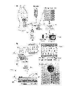
CA 02886057 2017-02-07
Fig. 1 provides diagrams A through E illustrating the infection monitoring in
peritoneal
dialysis (PD) patient with the use of a point-of-care embodiment of a
microchip of the invention.
Fig. 2 presents images A through 1 illustrating operation of a computer
program product and
a method of the invention. The diagram J shows a plot of neutrophil capture
specificity with the use
of an embodiment of the chip over a wide neutrophil concentration range.
Fig. 3 shows, in perspective view, a detector and a chip adapted for imaging
according to an
embodiment of the invention.
Fig. 4 includes images A and B of a PD microfluidic chip according to one
embodiment.
Fig. 5A is a diagram illustrating an embodiment of the image-restoration
algorithm of the
invention.
Fig. 5B is a diagram illustrating the transformation of a high-frequency-
boosted filtered
image to a frequency domain.
Fig. 5C provides a contour plot and a surface plot describing filtering
characteristics of a
band-pass filter used with an embodiment of Fig. 3A.
Fig. 6 provides binary versions of an image of a disk (on the left) and of a
high-frequency
boosted image of circular cells used with the process of the embodiment of
Fig. 5A.
Figs. 7A and 7B are, respectively, a gray scale image and a black-and-white
image
representing outputs of the frequency-domain filtering the spatial-domain
filtering, according to an
embodiment of the invention.
Fig. 8 is a flow-chart illustrating an embodiment of the image data processing
algorithm.
Fig. 9 present diagrams A through E illustrating the results of modeling of
CD66b+
neutrophil capture according to an embodiment of the invention.
Fig. 10 is a schematic diagram showing bounding of a neutrophil cell to a
surface of a chip
while stained with FITC-CD66b antibody.
Fig. 11 shows images A and B representing, respectively, a channel of a chip
following the
injection of a turbid PD sample and the same channel following the PBS
flushing procedure.
Figs. 12A through 12F are FCS plots representing examples of analyses of PD
samples.
Figs. 13A through 13F are plots showing the results of validation of
operationability of an
embodiment of the invention.
6
CA 02886057 2017-02-07
DETAILED DESCRIPTION
[0015] Hemodialysis (also referred to as haemodialysis, or HD) is a method
that is used to
achieve the extracorporeal removal of waste products such as creatinine and
urea and free water
from the blood when the kidneys are in a state of renal failure. Peritoneal
dialysis is used as an
alternative to HD, though it is far less commonly used. While the PD, as
compared to traditional
HD, provides higher quality of care, both procedures are available and provide
ease of access to the
patients. It is understood that, while in the disclosure below, the reference
is made to PD, such
reference is made only to make the disclosure concise and that the use of HD
fluid or any other
bodily fluid as a sample for analysis discussed below was considered to be
within the scope of the
invention.
[0016] A recognized clinical barrier in peritoneal dialysis is the risk of
peritonitis, i.e.
inflammation of peritoneum. Peritonitis is clinically defined as the
occurrence of a turbid dialysate
containing more than 100 white blood cells (WBCs) per microliter of which more
than 50% are
neutrophils. As infection progresses, a substantial increase in WBCs results
in higher degree of
opaqueness of the peritoneal dialysate. However, at the early stages of
infection the change in the
turbidity is not visually detectable.
[0017] The decision-making on whether the infection has occurred is
currently based on
assessment, by the patient himself, of a degree of opaqueness and visually
perceived cloudiness of
the bodily fluid such as the dialysate, which is understandably subjective
and, at least for that
reason, is not quite adequate to monitor peritonitis. Clinically, the WBCs and
neutrophil counts are
used to assess the occurrence of peritonitis. On the other hand, since a cell
counting platform is not
accessible at the point-of-care (POC), when the bodily fluid or dialysate
appears turbid, it is
considered to be an indication of a potential infection and patients are
expected to initiate a call to
their caregivers followed by an immediate visit to the doctor's office or
emergency room. In
reference to Table 1, the observations performed by patients can, overall, be
misleading causing
unnecessary emergency room visits, hospitalizations, and subsequent doctor
office visits, which
could have been avoided with reliable bedside testing.
7
CA 02886057 2017-02-07
Table 1: Results of comparison of the use of an embodiment with current
clinical practice
Current standard
Current standard practice
PD microchip
practice at home at clinic
Visual turbidity
Clinical practice/method Microchip Flow
cytometer (FACS)
measurement
Reproducibility Quantitative Not quantitative Quantitative
Accuracy High Low High
1-2 days for results to come
Depends on how long it
Time <1 hour from the clinic (30
min.
takes the patient to decide
analysis time)
Cost < 1 USD per test N/A 50 USD per test
Trained personnel No No Yes
Differential cell count Yes No quantitative data Yes
Actionable outcome Yes Yes, if the patient Yes
[00181 The above-
described situation is exacerbated by the facts that (i) the bodily fluid or
dialysate's cloudiness (turbidness) can be caused by other reasons (for
example, drugs such as
manidipine hydrochloride, a dihydropyridine-type calcium channel blocker) that
do not necessarily
indicate peritonitis, and (ii) the lack of appropriate quantitative monitoring
technology complicates
the monitoring of the health status of a HD/PD patient. Overall, there exists
a not addressed yet,
unmet clinical need for rapid and quantitative monitoring of a dialysate to
determine the risk of
infection.
[0019] To address this clinical need, embodiments of the present invention
provide a
disposable bodily fluid microchip (for example, an HD/PD microchip) and a
corresponding method
for operation of such microchip to enable rapid quantification of neutrophils
in a dialysate to
monitor the health status of an HD/ PD patient. In a pilot study, neutrophil
microchip counts in
dialysates of 20 HD/PD patients were obtained (ranging from 16 + 2 to 842 29
neutrophils per
100 microliters) over a time period of up to 190 days. An HD/PD microchip was
operable to
successfully determine the status of the patients. The proposed embodiments
are broadly applicable
for rapid quantitative analysis of body fluids at the bedside / point of care
location, covering a broad
range of diseases that require continuous or repetitive monitoring. References
throughout this
specification to "one embodiment," "an embodiment," "a related embodiment," or
similar language
mean that a particular feature, structure, or characteristic described in
connection with the referred
to "embodiment" is included in at least one embodiment of the present
invention. Thus,
appearances of the phrases "in one embodiment," "in an embodiment," and
similar language
8
CA 02886057 2017-02-07
throughout this specification may, but do not necessarily, all refer to the
same embodiment. It is to
be understood that no portion of disclosure, taken on its own and in possible
connection with a
figure, is intended to provide a complete description of all features of the
invention.
10020] In addition, the following disclosure may describe features of the
invention with
reference to corresponding drawings, in which like numbers represent the same
or similar elements
wherever possible. In the drawings, the depicted structural elements are
generally not to scale, and
certain components are enlarged relative to the other components for purposes
of emphasis and
understanding. It is to be understood that no single drawing is intended to
support a complete
description of all features of the invention. In other words, a given drawing
is generally descriptive
of only some, and generally not all, features of the invention. A given
drawing and an associated
portion of the disclosure containing a description referencing such drawing do
not, generally,
contain all elements of a particular view or all features that can be
presented is this view, for
purposes of simplifying the given drawing and discussion, and to direct the
discussion to particular
elements that are featured in this drawing. A skilled artisan will recognize
that the invention may
possibly be practiced without one or more of the specific features, elements,
components, structures,
details, or characteristics, or with the use of other methods, components,
materials, and so forth.
Therefore, although a particular detail of an embodiment of the invention may
not be necessarily
shown in each and every drawing describing such embodiment, the presence of
this detail in the
drawing may be implied unless the context of the description requires
otherwise. In other instances,
well known structures, details, materials, or operations may be not shown in a
given drawing or
described in detail to avoid obscuring aspects of an embodiment of the
invention that are being
discussed. Furthermore, the described single features, structures, or
characteristics of the invention
may be combined in any suitable manner in one or more further embodiments.
[0021] Moreover, if the schematic flow chart diagram is included, it is
generally set forth as
a logical flow-chart diagram. As such, the depicted order and labeled steps of
the logical flow are
indicative of one embodiment of the presented method. Other steps and methods
may be conceived
that are equivalent in function, logic, or effect to one or more steps, or
portions thereof, of the
illustrated method. Additionally, the format and symbols employed are provided
to explain the
logical steps of the method and are understood not to limit the scope of the
method. Although
various arrow types and line types may be employed in the flow-chart diagrams,
they are
understood not to limit the scope of the corresponding method. Indeed, some
arrows or other
9
CA 02886057 2017-02-07
connectors may be used to indicate only the logical flow of the method. For
instance, an arrow may
indicate a waiting or monitoring period of unspecified duration between
enumerated steps of the
depicted method. Without loss of generality, the order in which processing
steps or particular
methods occur may or may not strictly adhere to the order of the corresponding
steps shown.
[0022] The invention as recited in claims appended to this disclosure is
intended to be
assessed in light of the disclosure as a whole, including features disclosed
in prior art to which reference
is made.
[0023] As was already alluded to above, while in the examples discussed
below, the
references are made only to PD, such references are made only for the sake of
keeping the
description concise and simplified, and use of HD procedure and HD fluid
sample or any other
bodily fluid as a sample for analysis intended to be within the scope of the
invention.
[0024] In reference to Fig. 1, diagrams A through E are used to illustrate
the principle of
monitoring the infection in PD patients using a point-of-care embodiment. A PD
microchip 100
uses a small volume of waste dialysate 114 from the patient effluent to
capture CD66b neutrophils
on a surface of a channel of the chip 100 with high specificity and
efficiency. An image 118 of the
microchip is taken and the captured neutrophils are quantified with automated
software, 120. As
shown at 124, the determined dialysate neutrophil counts are optionally sent
to storage such as
electronic records, for example, which can then be assessed by the caregivers
to monitor the risk of
infection during PD treatment. The diagram B illustrates the injection of PD
patient dialysate
samples into the channels 100a of the microchip 100 to selectively capture the
neutrophils at the
channel surface. The diagram C provides a schematic representation of a
functionalized channel
surface 130 with CD66 antibody and captured neutrophils from a pool of white
blood cells in
dialysate. While the CD66+ neutrophils are selectively captured on the channel
100a, other cells
flow and exit the channel without being captured, substantially unabated. The
diagram D illustrates,
schematically, a means for imaging, detection and quantification of the
neutrophils captured in the
channels 100a of the chip 100. The means 140 includes a light source 144
adapted to illuminate the
chip 100 and the captured cells 146 such that the shadows 148 of the captured
cells 146 are
projected onto the surface of a charge coupled device (CCD) sensor 150. The
means 140 is
configured such that the CCD 150 is enabled to detect the light intensity
distribution corresponding
to the shadows 148 of the captured cells 146 without the need for an objective
lens and with the use
of an automated cell recognition and quantification computer program product
loaded on a
CA 02886057 2017-02-07
computer system. The diagram E presents, in the top view, an image of an
embodiment of the PD
microfluidic chip 100.
[0025] In the following portion of the present disclosure, in reference to
Figs. 2 through 8,
non-limiting examples embodiments of a system and method for acquisition and
processing of an
image of neutrophils are discussed in more detail.
[0026] The diagram A of Fig. 2 and Fig. 3 illustrate the positioning of
the specific
embodiment 100 on top of the CCD 150 without an optical component
therebetween. The diagram
B of Fig. 2 shows an image 200 of the embodiment 100 produced with the CCD
150. The diagram
C of Fig. 2 provides a shadow image 210 of cells 146 captured on a surface of
a channel 100a,
which are later marked by an embodiment of the computer program product of the
invention to
produce the overall cell count. The marked with green color cells 246 are
shown in the diagram D
of Fig. 2. Following this procedure, images of individual cells are identified
by modifying contrast
of such images, an example of which is shown in an image of the cell 256 of
the diagram E of Fig.
2. The diagrams illustrating noise reduction associated with the processing of
imaging data and
frequency domain transformation procedures, as well as data filtering with the
use of a 3D passband
filter are illustrated in the diagrams F and G of Fig. 2, respectively.
Embodiments of the Algorithm.
[0027] To complement an algorithm of the invention (referred to herein as
the HVC
method), an embodiment of the computer program product of the invention
includes program code
for processing image data, received by the electronic data-processing computer
circuitry (which
maybe referred to as a processor, for short) after an initial phase of image
restoration, with the use
of frequency domain (FD) operations. After the FD-processing, the data are re-
processed using
spatial domain (SD) operations to produce a final result based on an
intersection of the two outputs.
The computer program product may include program codes to effectuate the
following steps:
i. Automatic and/or manual selection of channels on a chip
ii. Image restoration to improve image contrast
iii. Frequency domain band-pass filtering
iv. Round object detection using matched filtering
11
CA 02886057 2017-02-07
v. Fine tuning the matches found by (iii) and/or (iv), and
vi. Incorporating user feedback to update the threshold specifying which
objects should be
counted as cells.
[00281 The above-mentioned steps of an embodiment of the image-processing
algorithm of
the invention are described below.
[0029] Automatic and/or manual selection of channels on a chip. It may be
required to
count the cells in each of the channels 100a of the chip 100 of Fig. 1
separately. In doing so, not all
of the area within the borders of a channel should be considered due to
optical "smearing" of images
of the cells near the boundary of a channel, which is an imaging drawback
unique to each CCD
image. Embodiment of the invention addresses this problem by providing the
user with the
following options.
[0030] Fully automatic mode. This mode is built-in for the mass processing
of images
taken on a particular chip. In further reference to Figs. I. and 2, several
pre-determined identifying
markers (such as square markers 260 in the image of the diagram of Fig. 2, for
example) can be
engraved/imprinted on each corner of the chip 100 for localization and
orientation of the channels
100a using the morphological data processing algorithm (such as, for example,
the hit-miss
transform in Matlab's image processing toolbox), with a structural element
equal to the binary mask
of the marker. For each chip carrying such markers, the user is enabled to
generate a 'template'
identifying the markers 260 (squares as shown) and the areas inside the
channels 100a in which the
count should be performed. Fig. 4 shows images A and B with the insides of the
four squares 260
marked with a magenta asterisk. The areas inside the three channels 100a (of
the image A), where
the cell counting should be performed are highlighted with red (410R), green
(410G) and blue
(410B). Small variations in the placement of the chip 100 under the CCD 150
(such as a degree of
parallelism between the chip and the surface of the CCD, for example) can be
tolerated. For chips
without the identifying markers, this option works as long as the chip is
placed under the CCD in a
fashion consistent with the template. Once the computer system identifies or
recognizes the
presence and location/orientation of the channels of the chip, the
morphological data processing
algorithm automatically performs the cell count without waiting for further
user input.
12
CA 02886057 2017-02-07
[0031] Semi-automatic mode. In this mode, and in reference to image A of
Fig. 4, the user
generates one 'template' per chip, and the channels in each new image are
automatically colored
(for example, as discussed above, highlighted with red, green and blue
shades). The program code
idles and waits for the user input representing that the identification, fine-
tuning of the borders of
the channels and the channel selection is complete.
[0032] Fully manual mode. In this mode, the computer program product
enables the
processor to allow the user to re-sizably and movably mark any region in an
image (whether
rectangular or square in shape, such as the area 430 of image B of Fig. 4, for
example) which may
be smaller than the length of a channel, and then to perform the cell count
only in the marked
isolated area 430.
[0033] Image Restoration. If an acquired image has low contrast, a
normalization and/or
standardization procedure is performed to ensure that an image of a cell
stands out against the
background. In reference to an image 510 of Fig. 5A, for example, some of the
acquired images
include color images. It is appreciated that the color information does not
add practical value to or
modify the cell-counting task. The first step in one specific implementation
of the image restoration
procedure, according to an embodiment of the invention, is to convert the
image 510 to a gray scale
image 512. The gray scale image 512 may still be characterized by low
contrast; in this case, the
image histogram is stretched out (for example, with the use of Matlab's
adapthisteg function), as
shown at 514. Following this, using high-boost filtering, the high-frequency
components in the
image are enhanced, while the low-frequency components are maintained, at 516
, to form a high-
frequency-boosted image 518. This image is the input image to frequency domain
filtering.
[0034] Frequency domain hand-pass filtering. As a result of the boosting of
high-frequency
characteristics of the image 514, the 2D Fourier Transform 524 of the high-
frequency-boosted
filtered image 518 may exhibit a "halo" 528 shown in Fig. 5B. The halo 528 can
be seen
surrounding the high-energy area at low frequencies (at center of the
frequency-domain image).
The presence of this halo is due to the presence and nature of cells. Program
code applying a
custom-designed band-pass filter 530 to the Fourier Transform 524 of the image
518 reverts the
transform 524 back to the spatial domain (not shown) and produces the effect
of visually
emphasizing the cells in the spatial image by removing spatial frequency
components that do not
13
CA 02886057 2017-02-07
contribute to the halo. Frequency characteristic of an embodiment of this
custom filter are shown,
in top and perspective views, in Fig. 5C.
[0035] While the frequency domain band-pass filtering emphasizes the cells
and attenuates
other unwanted elements of the image, cells are not the only contributors to
the -halo" and some
amount of noise remains. The noise-producing elements can include, for
example, air-bubbles,
fibers, and fine structures from the sample that are not cells. Instead of
trying to handle each
contribution to image noise, an embodiment of the HCC method of the invention
takes advantage of
the facts that (i) the cells of interest are almost always roundish and have
substantially the same
diameter; (ii) the imaging is carried out with the same system; (iii) the chip
is always positioned at
the substantially the same distance from the CCD. This makes it possible to
use morphology of the
cells of interest and identify round objects of a certain diameter, as
discussed further below.
[0036] Detection of round objects using matched filtering. At this stage,
the imaging data
representing the high-boost filtered image 518 are used as input again. The
image 518 is first
converted into a binary image and then convolved with a template of a "disk".
Fig. 6 illustrates a
binary version 618 of the image 518 of Fig. 5, as well as the template 620 as
a binary version of a
roundish cell (the square shape satisfactorily approximates a disk in a noisy
image) with which
program code of the embodiment of the computer program product thereafter
convolves the image
618.
[0037] Fine tuning the matches. Figs. 7A and 7B show a gray scale image
710 and a black-
and-white image 720 representing outputs of the frequency-domain filtering the
spatial-domain
filtering. The final count of cells is computed by finding the intersection of
the results of
thresholded black and white image 720 from frequency domain filtering and
black and white image
output from the matched filtering. In this binary intersection image, hits
within three pixels of each
other are collapsed into one. This is to ensure there are no duplicates and
the cell count is not over-
estimated.
[0038] Incorporating user feedback to update the threshold specifying
which objects should
be counted as cells. The final result depends on the value of the threshold
that was specified. Once
the counting of cells is accomplished, each valid cell is marked on the
original image and displayed
to the user for visual confirmation. At this point, the user has the ability
to adjust the value of the
threshold and, therefore, the results. The number of cells is displayed on the
figure window and is
14
CA 02886057 2017-02-07
dynamically updated. The higher the threshold, the fewer the number of points
identified as cells but
these have higher likelihood of actually being cells. The lower the threshold,
more points are
identified as cells but it the number of false positives generally increase.
[0039] The overall image processing algorithm described above is
summarized in the
flowchart of Fig. 8.
[0040] In the following portion of the disclosure, in reference to Figs. 9
and in further
reference to Fig. 1, embodiments related to the structure of microchannels of
the proposed chip to
optimize the cell-capture (such as, for example. CD66b+ cells) are discussed
in more detail. The
flow dynamics and migration of CD66b+ neutrophils to the microchip surface are
characterized by
multibody interactions of white blood cells (CD66b+ neutrophils) and
platelets, as illustrated in the
diagram A of Fig. 9. At low shear rates, the increasingly blunted velocity
profiles and enhanced
CD66b+ neutrophil margination have been observed in microvessels. For the
purposes of this
disclosure, the microchannels 100a with rectangular cross-sections were
produced with two
different heights (or depths), h = SO and 80 pan. Since the volume flow rates
were set equal for
both cases to Q = 2 p1/min, the corresponding Reynolds numbers, Re = puh = PQ
are
equal for both cases and equal to Re 0.01.
Here, p is the density of the fluid, Q is the volume
flow rate, 11 15 the viscosity of the fluid, and w is the width of the
channel. It is observed that the
Reynolds number for the microchannels 100a is low, and the inertial forces are
small compared to
the viscous forces, and thus the flow may be characterized as a creeping flow
for which Re 1.
The creeping flow on its own cannot resolve the underlying mechanisms for
migration of
neutrophils to the periphery of microchannels due to the well-known time
reversibility of low Re
flows. Time reversibility does not foster stiff CD661f neutrophils to
marginate to the microchip
wall. Small curvature changes on the surface of CD66b neutrophils can in
theory cause to anti-
symmetries, and thus transport of these cells by crossing the streamlines.
However, this would be
expected to slowly bring the CD66b+ neutrophils to the microchannel center,
rather than towards the
wall.
[0041] To design microchannels 100a of an embodiment 100 of Fig. 1 for
effective capture
of CD66b+ neutrophils from patient dialysate samples, the present theoretical
model, illustrated
schematically in the diagrams B through E of Fig. 9, assumes that affinity-
based cell-capture
process includes two probabilistic periods.
CA 02886057 2017-02-07
[0042] These probabilities include (1) the probability that the cells in
questions are located
near the surface of the microchannel 100a and margination (if any) of cells
towards equilibrium
zones, which can potentially feed the cell population near the channel
surface, as shown in the graph
of the diagram B; and (2) the likelihood of bond formation between the cells
locates near the
channel surface and the receptors, as shown in the graph of the diagram C of
Fig. 9. To determine
the latter, the probabilistic kinetic formulation can be used. To assess the
importance of
margination, i.e. the migration of CD66b+ neutrophils towards equilibrium
zones, we first calculate
margination velocities (shown in the graph of the diagram D of Fig. 9):
pdywBc = 0.17u _2 h 0 (12.84 cywBc (1 ywgc
(1)
clt E0 h h YWBC*
where ywBc is the vertical distance of CD66b neutrophils, dywBc/dt is the
margination velocity,
ywBc* is the equilibrium vertical distance, a is the radius of CD66b+
neutrophils, Po and E0 are fluid
density and viscosity. At the next step, the average margination velocity and
average flow
velocities are calculated as urn = [fo (dywBc/dt)dyl/h, and uf = [To
u(y)dy]/h, respectively.
Following this calculating, time scales are compared by t* = (h/um)/(L/uf).
[0043] As time scale ratio of margination and flow is >> 1 (C-100), initial
distribution of
neutrophils will be the determining factor. Here, we simply assume that cells
will be normally
distributed across the flow as:
P ,exp (y-11)2) (2)
0 v211- 2o z /
where j is the mean location and o-2 is the variance of cell distribution. In
further reference to the
diagram B of Fig. 9, the probability for cells to be located in a thin zone
between the channel
surface and a threshold, ho, (maximum distance) at which ligand-receptor bonds
can be formed:
ri
a-hoeXp (¨=(-311 dy
(3)
20-2
[0044] The neutrophils were adhered to the bottom surface of the
microchannel. The
effective fluidic forces that can potentially dislodge neutrophils are given
by F 67raliuSFs and
M = 6n-a3 STs where 1 is the separation distance to the wall, id is the shear
stress at the wall, Fs
and Ts are shape-dependent coefficients. The shear stress on microchannel
surface, r,õ can be
calculated as:
Tw = 6/1(2/(Wh2) (4)
16
CA 02886057 2017-02-07
Here, shear stresses are 0.0078 Pa for It = 80 pm and 0.02 Pa for h = 50 [urn.
Accordingly, and in
further reference to the diagram C of Fig. 9, the steady-state adhesion
probability is approximated as
A a2 a s
Pa = ffro2exp ¨ [6 (ay + 8,4)Fs + 8 --ro Ts X 702 Tnr} (inr Ili./ Ka ) (5)
kbT
where mr and m1 are the receptor and ligand densities, respectively; Ka is
the association constant
at zero load of the ligand-receptor pair; A, is the contact area; A is the
characteristic length of the
ligand-receptor bonds; kb is the Boltzmann constant; Fat, is the dislodging
force acting per unit
ligand-receptor pair; y is the cell aspect ratio; and T is the temperature.
[00451 Finally, and in reference to the diagram E of Fig. 9, the combined
probability of
neutrophils captured by the surface is assessed as Pt = PsPa. The increase in
total cell counts for
both flow rates as neutrophil concentration increases can be easily attributed
to the enhanced
diffusion and collision dynamics of these cells towards the microchannel wall.
For the same
neutrophil concentration, a decrease in thickness of the microchannel leads to
higher shear rates and
an increase in total number of captured cells. Either packing larger number of
cells in the same
domain, or decreasing the size of the domain while keeping the number of cells
constant leads to
enhanced margination and increase in capture of neutrophils. Lastly, higher
shear rates lead to
larger detaching forces for the captured neutrophils, as well as longer
exposure time for freely
flowing neutrophils. If the process was dominated by the former phenomenon or
its effect was
amplified, it would cause to (faster) detachment of neutrophils and lower cell
count.
Examples of Embodiments of Microfluidic Chip.
[0046] Referring again to Fig. 1, in one implementation, the embodiment 100
was
manufactured with the use of Poly(methyl methacrylate) (PMMA) (by McMaster
Carr, Atlanta,
GA) as the backing of the chip attached to an approximately 50 [tm to 80 [tm
thick double-sided
adhesive film (iTapstore, Scotch Plains, NJ) to provide the channel height.
Both components were
cut to about 24 x 40 mm. Six pores of equal width were cut into the PMMA, with
three pores at one
end representing the inlets and the three pores at the opposite end
representing the outlets. The
17
CA 02886057 2017-02-07
channels, each 4.3 mm x 25 mm long, were cut into the DSA; during assembly
these channels were
aligned with each set of inlet and outlet pores and fixed onto the PMMA
surface. Once the plasma-
treated glass slide is centered and fixed on the remaining side of the DSA,
the microfluidic chip is
formed and ready for silanization. A length of a channel 100a was chosen to be
about 30 mm as
such length provided sufficient CD66b interaction for neutrophil
immobilization. A channel width
of about 4 mm provides an adequate amount of surface area for cell capture of
approximately 100
jtL PD sample. Glass slides (Corning, Lowell, MA) were plasma treated with
oxygen plasma (at
about 100 mW and 1% oxygen) for about 1 minute in the PX-250 chamber (March
Instruments,
Concord, MA) and used as a cap for the chip 100 to provide a tight seal to the
channels 100a.
Examples of Functionalization of Microfluidic Channels.
[0047J Materials for Channel Functionalization. 200 proofs of ethanol
(Et0H), for dilutions
and washing of channels, and dimethyl sulfoxide (DMSO), the solvent for GMBS
stock solution,
were purchased from Sigma-Aldrich Chemical Company (St. Louis, MO). 3-
mercaptopropyl
trimethoxysilane (3-MPS), a silanization agent, was also purchased from Sigma-
Aldrich Chemical
Company (St. Louis, MO). N-y-maleirnidobutyryloxy succinimide ester (GMBS), a
coupling agent,
was purchased from Pierce Biotechnology (Rockford, IL). 1X phosphate buffered
saline (PBS)
solution, for dilution of aqueous reagents and washing, was purchased from
Gibco (Grand Island,
NY). NeutrAvidin, a functional protein for biotin binding, was purchased from
Fisher Scientific
(Fair Lawn, NJ). Lyophilized albumin from bovine serum (BSA), for blocking
nonspecific
bindings/interactions, was purchased from Sigma-Aldrich Chemical Company (St
Louis, MO).
Biotinylated anti-human carcinoembryonic antigen-related cell adhesion
molecule 8 (CEACAM-8)
antibody, used for the capture of neutrophils in clinical PD samples, was
purchased from R&D
Systems (Minneapolis, MN).
[0048] Materials for Neutrophil Capture and Analysis. The syringe pump,
used for passing
the PD sample through the microfluidic chip, was purchased from
SyringePump.com (Farmingdale,
NY). The 1 mL Luer Lock syringes, used to inject the PD fluid through the
microfluidic chip, were
purchased from BD Biosciences (Franklin Lakes, NJ). flie 0.01" inner diameter
nylon tubing, used
to connect the syringes to the microfluidic chip, was purchased from Cole-
Parmer (Vernon Hills,
IL). The microfluidic chip was cleaned with Zeiss Lens Cleaner, purchased from
Carl Zeiss Optical
18
CA 02886057 2017-02-07
Inc. (Cleveland, OH). The charge-couples device (CCD), used for imaging of the
chip to quantify
neutrophil capture, was purchased from Imperx Inc. (Boca Raton, FL) and
incorporated into the
'black box' with an LED light source with 86.9 Id) resistance and 2.3V power
source.
[0049] Solution
Preparation. Once the microfluidic chip was assembled, the procedure
required to functionalize the microfluidic channels was performed, as outlined
in Tables 2 and 3.
[0050] First,
silanization solution was pipetted through each channel and incubated for 30
minutes. The silanization solution is a dilution of 200 mM 3-MPS in Et0H. GMBS
Solution is a
dilution of 2 mM GMBS solution in Et0H. NeutrAvidin solution is a 100 lug/mL
solution of
NeutrAvidin in PBS. 1% BSA solution is 10 m,g/mL solution of BSA in PBS.
Antibody solution is a
200 ng/mL solution of CEACAM-8 antibody in PBS (1000x dilution from stock).
[0051] Any unbound 3-MPS was flushed from the channel with Et0H. Next, GMBS
solution was pipetted through each channel and incubated for 30 minutes.
Unreacted GMBS was
flushed from the channel with Et0H. The channels were then washed with PBS to
remove the
organic solvent. The NeutrAvidin solution was pipetted through each channel
and incubated in
darkness (due to light sensitivity) for 60 minutes for immobilization onto
GMBS. Unbound
NeutrAvidin was washed from the channel with PBS. Next, 1% BSA Solution was
pipetted through
each channel and incubated for 30 minutes for surface passivation. Unbound BSA
was washed from
the channels with PBS. Antibody solution was then pipetted through each
channel, incubated for 30
minutes, pipetted through each channel again, and incubated for 30 more
minutes.
Table 2. M Example of Operating Procedure for microfluidic chip fabrication
and initial functionalization (all steps are
carried out at Room Temperature)
Step Description Requirements Duration Specifications
0 Glass Slide Cleaning Wipe with Ethanol ¨20s per side
Dry with N2 gas
Glass Slide Plasma Air Plasma 90s
Treatment
2 Assembly Glass Slide and PMMA Ensure
plasma treated face of
connected with DSA slide is
inside channels
3 3-MPS Treatment 100ttL pipetted through each 30 min
Silanization solution in Et0H
channel incubation used.
Wrap pores in parafilm to
prevent evaporation
4 Ethanol Rinse 100ttL pipetted through each 200
proofs Et0H is used
channel
GMBS treatment I 00tiL pipetted through each 30 min GMBS
Solution in Et0H used.
19
02-07
channel incubation Wrap pores in Parafilm
to
prevent evaporation
6 Ethanol Rinse 100uL picpAettc0d2t8h8ro615h7 17-
each 200 proofs Et0H is used
channel
7 PBS Rinse 300 uL pipetted through each Filtered PBS is used.
channel
8 NeutrAvidin treatment 15 ftL pipetted through each 60
min NeutrAvidin solution in PBS
channel incubation used. Wrap pores in
parafilm to
prevent evaporation.
9 PBS Rinse 100 1._. pipetted through each Filtered PBS is used.
channel
BSA application 15 p.1_, pipetted through each 30 min 1% BSA solution
in PBS used.
channel incubation Wrap pores with
parafilm to
prevent evaporation.
11 PBS Rinse 30 fit pipetted through each Filtered PBS is used.
channel
12 Storage Do not proceed to Table S2 Chip is
in chemically stable state.
until PD sample is obtained
PBS: Phosphate Buffered Saline
Et0H: 100% Ethanol
Table 3. An Example of Operating Procedure for application of surface
chemistry within microfluidic channels.
Step Description Requirements Duration Specifications
0 Initiate Application of This process requires
approximately one hour to complete. For optimal
Surface Chemistry results, the clinical sample should be ready immediately
following second
antibody incubation.
1 Antibody application 15 tit pipetted through each 30
min Antibody solution in
channel incubation PBS is used. Wrap
pores
with parafilm to prevent
evaporation.
2 PBS Wash 30 fit pipetted through each Filtered PBS is used.
channel
3 Antibody application 154, pipetted through each 30
min Antibody solution in
channel incubation _ PBS is used. Wrap pores
CA 02886057 2017-02-07
with parafilm to prevent
evaporation.
Examples of Capture of Neutrophils Within Microfhtidic Channels.
[0052] In reference to Table 4, the syringes and the syringe pump were set
up. Each syringe
was filled with 200 [1.L clinical PD sample (three syringes per sample). 30-
gauge, Luer Lock, blunt
needles were attached to the syringes; nylon tubing was fitted to the pointed
end of each needle.
The syringes were then loaded into the syringe pump. The pump was run until
all of the plungers
were at the same level and fluid was flowing the nylon tubing. Then, the free
end of each piece of
nylon tubing was epoxied into each respective inlet port in the microfluidic
chip. A 2001,1 pipette
tip was placed in each outlet port for depleted sample collection. The syringe
pump was run until
100 ill', passed through each microfluidic channel. Then the tubing and
pipette tips were removed
from the microfluidic chip inlets, the surfaces of the microfluidic chip were
cleaned using the Zeiss
Lens Cleaner. Finally, the microfluidic chip was placed on the CCD image
sensor, and an image
was saved on the computer for quantification, as discussed above.
Table 4. An Example of Operating Procedure for testing clinical peritoneal
dialysis (PD) samples with functionalized
surface chemistry.
Step Description Requirements Duration Specifications
0 Preparations Set up Syringe Pump to Fill three syringes per
sample
flow sample through chip with >100 uL PD sample
each;
connect syringes to microfluidic
chips; load syringe pump
1 PD Sample Injection Syringe Pump is run 50 min 2
uLiminute, 1004 sample
FpialstesereddthprBosugihs used
2 PBS Wash 100 pl pipetted through
each channel
3 Surface Cleaning Wipe the outsides of the 70% Et0H is used
microfluidic chips
thoroughly
4 CCD Imaging Clean image sensor; place Image sensor is cleaned
directly
21
CA 02886057 2017-02-07
microfluidic chip directly with lens paper and lens
image sensor; save images cleaning solution
to hard drive
Automatic Cell Images are analyzed using Adjust
parameters to ensure
Counting the MATLAB function accurate counting numbers
6 Manual Cell Counting Images are analyzed and a 30
minutes A manual cell counter, in
(if necessary) manual cell count is
conjunction with the magnified
performed digital image, is used.
Demonstration of Robust Performance of an Embodiment of Microfhtidic Chip in
Analysis of
Turbid PD Dialysate Samples.
[0053] The proposed embodiments of the PD microtluidic chip possess the
ability to
provide accurate results even while a sample procured from the PD patient
samples contains
additional particles such as fibers or RBCs. In particular, and in reference
to Fig. 10, one
implementation of the microchip includes an immunoassay based chip in which
any bound
neutrophils remain on the surface while other particles are removed by a wash
buffer.
[0054] The effect of degree of opaqueness of a fluid sample on the quality
of optical
imaging was also addressed. It was determined that the PD fluid with high
particle concentrations
did not have an effect on the CCD image when following the flush with wash
buffer. If the PD chip
was imaged before samples were flushed however, the noise in the CCD image
would be
significant. As illustrated in Figs. 11A and 11B, for example, a PD sample
with high degree of
opaqueness (turbidity) was injected in the embodiment 100 of the PD chip and
imaged immediately
after that to produce the image of Fig. 11A. The same sample was additionally
imaged following
the PBS flushing step to produce an image of Fig. 11B. The turbidity increase
was a result of fibers
accumulating in the PD dialysate. The comparison between the images of Figs.
11A and 11B
shows the significant decrease in noise that occurs when a wash buffer is
added. Despite the high
turbidity of the fluid, the CCD 150 was still capable of providing a clear
image of the overall
capture on the surface.
[0055] FACS results were also affected by various particles in the PD
fluid. Dead cells
often cause noise in the FACS images making it difficult to gate the target
cells such as neutrophils.
With the increase in the overall particle number there is also an increase in
time needed for
determination of cell concentration values. Figs. 12A and 12B provide FACS
plots characterizing a
typical sample that includes lymphocytes, monocytes and neutrophils. The
neutrophils stained with
22
CA 02886057 2017-02-07
FITC-CD66b are gated and displayed in the corresponding FSC vs. SSC region.
Fig. 12C and 12D
provide FACS plots representing results of the characterization of a turbid PD
sample procured
from the patient. Dead cells as well as additional particles cause noise
levels to increase in FSC vs.
SSC, thereby complicating the gating of the cells (as can be seen from a plot
region to the left of the
gated R2 region of Fig. 12C, for example). At such noise levels it becomes
difficult to determine
specific cell counts and accurately determine cell concentrations. Fig. 12E
and 12F show the FACS
plots characterizing a sample obtained from a patient who uses Icodextrin as
his dialysate sugar (as
opposed to the traditional dextrose dialysate). Icodextrin, being a high molar
mass sugar, is
recognized on the flow cytometer as an event. As a result, more than 100X the
events is required to
obtain a considerable WBC concentration.
[0056] Fig. 13 shows plots A through F illustrating validation of
operationability of
embodiments of the invention. Fig. 13A is a plot demonstrating the results of
statistical comparison
of automated counting the neutrophils in the PD sample according to
embodiments of the invention
and manual cell counting. A high correlation (r=0.96, p<0.01) between manual
and software cell
counts was observed. Fig. 13B is a plot showing the effect of channel
dimensions and flow rate on
neutrophil capture efficiency and demonstrating higher capture efficiency
attained with 50 Jam
channels as compared to 80 mn channels discussed above. Further, 50 !.im high
design provided a
higher correlation (1-0.90, p<0.01) to flow cytometer neutrophil counts,
compared to 80 pm high
channel design (r=0.74, p<0.01). Fig. 13C is a plot representing neutrophil
counts performed on PD
patient samples. A significant correlation (r=0.90, p<0.01) between sample
neutrophil
concentration and PD chip cell count was observed. Fig. 13D is a plot
representing dependence of
capture efficiency of an embodiment of the PD chip as a function of neutrophil
concentration in
samples described in Table 2. The microchip counts with PD patient samples
displayed statistically
significant correlation (r=0.83, p<0.05) with FACS counts. Measurement of the
clinical samples,
the results of which are shown in Figs. 13E and 13F, indicated significant
correlation (Pearson
correlation: 0.90, p<0.01) between the flow cytometry results (gold standard)
and the microchip for
neutrophil counts in the range of 0 to 300 cells/ 1. Such range covers the
clinically relevant
detection range.
[0057] In accordance with examples of embodiments, a microfluidic system
and a method
for an early detection of infection and documenting of PD hygiene compliance
rates are provided.
A home healthcare portal for improved management of peritoneal dialysis
therapy, discussed in this
23
CA 02886057 2017-02-07
disclosure, is adapted to aid the clinician in monitoring healthcare of the
patient at a point-of-care.
The inventors are not aware of any POC rapid technologies existing to monitor
PD fluid at the
bedside of the patient. The envisioned embodiments utilize the discard PD
fluid and to enable count
of the WBC and neutrophils in the fluid. Based on such measurement of a PD,
the system provides
an indicator (such as a color indicator, red or green LED, for example) in
response to which the
communication between the clinician and the patient is initiated leading to a
clinical decision
regarding a potential infection, thereby reducing or eliminating a time-delay
of treatment. The
recorded data will be sent to an electronic record where patient, the
caregivers can access. The
diagnostic decision will be made still by the doctor and the nurse interacting
with the patient. The
proposed embodiments replace unreliable cloudiness or turbidity based
measurement methodologies
for the benefit of the patient avoiding acute cases, time delays and providing
a more patient-friendly
PD characterization method than hemodialysis.
[0058] While specific values chosen for these embodiments are recited, it
is to be
understood that, within the scope of the invention, the values of all of
parameters may vary over
wide ranges to suit different applications.
[0059] Implementation of a method of the invention and/or enablement of
the operation of a
system of the invention described above may be effectuated with a use of a
processor specifically
and particularly programmed to perform the steps of the required algorithm.
Such processor can be
controlled by instructions stored in a tangible, computer-readable memory.
Those skilled in the art
should readily appreciate that instructions or programs defining the functions
of the present
invention may be delivered to a processor in many forms, including, but not
limited to, information
permanently stored on non-writable storage mediaõ information alterably stored
on writable storage
media, or information conveyed to a computer through communication media,
including wired or
wireless computer networks. In addition, while the invention may be embodied
in software, the
functions necessary to implement the invention may optionally or alternatively
be embodied in part
or in whole using appropriate firmware and/or hardware components (such as,
for example,
combinatorial logic, Application Specific Integrated Circuits, and Field-
Programmable Gate
Arrays).
[0060] While the invention is described through the above-described
exemplary
embodiments, it will be understood by those of ordinary skill in the art that
modifications to, and
variations of, the illustrated embodiments may be made without departing from
the disclosed
24
CA 02886057 2017-02-07
inventive concepts. Accordingly, the invention should not be viewed as being
limited to the
disclosed embodiment(s).