Note: Descriptions are shown in the official language in which they were submitted.
81786971
MICROFLUIDIC SYSTEM FOR REPRODUCING FUNCTIONAL UNITS OF
TISSUES AND ORGANS IN VITRO
Cross-Reference to Related applications
This application is related to and claims priority from co-pending US
Provisional Application No. 61/707,907 to Neumann et al. filed 9/29/2012 and
entitled
"Microfluidic System for Reproducing Functional Units of Tissues and Organs In
Vitro"; and further claims priority from co-pending US Provisional Application
No. 61/721,002 to Neumann et al. filed 10/31/2012.
Field of the Invention
The present invention relates to methods for reproducing functional units of
tissues and organs in vitro, and, more particularly, to systems including
tissue-
engineered microenvironments on a chip.
Backciround of the Invention
While the investment in pharmaceutical research and development has been
growing exponentially, the number of new drugs approved by FDA has remained
unchanged in the past 60 years. Nearly 95% of new drug candidates fail between
pre-clinical and clinical phases of development, mainly due to drug-associated
toxicity. Clearly, better pre-clinical drug-screening assays are needed.
Currently
pharmacokinetic and toxicological evaluation of drug candidates relies largely
on
animal test systems, which evidently show only very limited predictive value
for
clinical efficacy and toxicity. In addition, maintaining animal models drives
up
dramatically the cost of drug development. There are also a number of high-
throughput two dimensional (2D) cell line models commonly used in drug
development. Their predictive value is very limited, which is attributed to
the loss of
physiological context. More sophisticated humanized cell-based assays from 3D
cell
cultures to organs-on-chips are being developed that can address the
1
Date Recue/Date Received 2021-10-15
CA 02886247 2015-03-25
WO 2014/052835
PCT/1JS2013/062307
limitations of 20 cell culture and minimize or potentially even completely
replace animal models. In 3D cell-culture models cells are grown within 3D
microenvironment that mimics structural, biochemical and mechanical
aspects found in vivo. Such cultures are known to restore specific
biochemical and morphological features characteristic of corresponding
tissues in vivo. The examples of conventional static 3D cultures include
hydrogel-incapsulated cells, "sandwich" cultures, multicellular spheroids,
cells grown on microcarriers and microstructured support. While powerful
these models still lack the complexity required for pharmacokinetic studies
and have a number of other shortcomings: 1) limited nutrient supply and
accumulation of metabolic waste products that can confound cell responses
to drugs, 2) inability to mimic spatiotemporal biochemical gradients existing
in vivo, 3) lack of mechanical cues such as flow, perfusion, pressure,
mechanical stress, 4) difficult to probe, 5) problematic real-time imaging,
and
6) biochemical analysis cannot be performed in live cells due to reaction-
diffusion phenomena. Furthermore, it has not yet been possible to engineer
microsystems that integrate multiple organ/tissue mimetics with active
vascular conduits and barrier tissues.
As a result, there is an established, yet unmet need for less
expensive, more sophisticated, more controlled systems for drug studies,
vaccine development and other types of medical research. The present
invention provides new and novel approaches for such controlled systems,
including a system for integrating vascular cells and organ cells to reproduce
functional units of tissues and organs in vitro. These and other important new
teachings are evident from the specification and claims hereinbelow.
Medical research for the development of vaccines against parasitic
diseases requires not only availability and access to the parasitic cells in
vitro, but also the ability to model a host organism of the parasite. The
human malaria parasite infects mosquitoes, which in turn act as vectors in
transmitting the disease to humans. To develop and identify effective
vaccines against malaria, investigators require a model system in which the
2
CA 02886247 2015-03-25
WO 2014/052835
PCT/1JS2013/062307
malaria parasite can be studied in the context of the mosquito midgut, where
it naturally lives. However, there are currently no systems available to
culture
the developmental insect stages of the human malaria parasite Plasmodium
falciparum in vitro. Furthermore, research on the infectious stages of the
malaria parasites, the sporozoites, depends on inaccurate and difficult to
control methods that involve live, infected mosquitoes. Such approaches
introduce many undesirable variables into the analysis. For example, live
mosquitoes are not subject to controlled blood intake parameters and
analysis of drug effectiveness must be performed by a relatively subjective
manual examination of dissected mosquito intestines.
As a result, there is an established, yet unmet need for less expensive
and more controlled systems for both an in vitro culture system to produce all
parasite insect stages, including, for example, parasite mosquito stages, and
a testing platform for drug and vaccine studies, vaccine development and
other types of medical research related to diseases such as malaria. The
present invention provides new and novel approaches for such a controlled
system, including a model for reproducing functional units of the mosquito
midgut system and the culture of the malaria parasite stages therein. These
and other important new teachings are evident from the specification and
claims here in below.
Summary of the Disclosure
This summary is provided to introduce a selection of concepts in a
simplified form that are further described below in the Detailed Description.
This summary is not intended to identify key features of the claimed subject
matter, nor is it intended to be used as an aid in determining the scope of
the
claimed subject matter.
A microfluidic system for generating compartmentalized
microenvironments of tissues and organs in vitro and for independently
perfusing the compartments is herein disclosed. A microfluidic device
includes at least a first perfusion path and a second separate perfusion path.
The microfluidic device also has a chamber containing a matrix, where the
matrix surrounds at least one void whose lumen is in fluidic connection
3
81786971
exclusively with the first perfusion path, where the at least one void can be
populated
with at least one cell type in such way that the cells are in direct contact
with the
matrix and the matrix is in fluidic connection exclusively with the second
separate
perfusion path.
In another aspect, a method for reproducing a functional unit of an
invertebrate
in vitro, as a tissue-engineered microenvironment for the culture of parasites
is
disclosed including providing a microfluidic device having at least a first
perfusion
path and a second separate perfusion path, the microfluidic device also having
a
chamber. The chamber is filled with a matrix, where the matrix surrounds at
least one
void whose lumen is in fluidic connection exclusively with the first perfusion
path,
where the at least one void is populated with at least one cell type in such
way that
the cells are in direct contact with the matrix, and where the matrix is in
fluidic
connection exclusively with the second separate perfusion path. The at least
one void
is seeded with invertebrate cells and the invertebrate cells are perfused to
proliferate
and generate an Invertebrate organ or tissue. Parasite stages are cultivated
in the
microenvironment to provide a testing microenvironment.
The present invention as claimed relates to:
-
a microfluidic system for generating compartmentalized microenvironments of
tissues and organs in vitro and for independently perfusing the compartments
comprising: a microfluidic device having at least a first perfusion path and a
second
separate perfusion path; the microfluidic device also having a chamber
containing a
perfusable matrix, where the matrix surrounds at least one void whose lumen is
in
fluidic connection exclusively with the first perfusion path, where the at
least one void
can be populated with at least one cell type to provide for tubular cell
structures, and
in such way that the cells are in direct contact with the matrix; and where
the matrix is
in fluidic connection exclusively with the second separate perfusion path;
-
a microfluidic system for reproducing functional units of tissues
and organs in vitro comprising: a plurality of microfluidic devices having at
least
a first perfusion path and a second separate perfusion
path;
4
Date Re9ue/Date Received 2020-11-23
81786971
the plurality of microfluidic devices each also having a chamber containing a
perfusable matrix, where the matrix surrounds at least one void whose lumen is
in
fluidic connection exclusively with the first perfusion path, where the at
least one void
is populated with at least one cell type to provide for tubular cell
structures, and in
such way that the cells are in direct contact with the matrix; where the
matrix is in
fluidic connection exclusively with the second separate perfusion path;
wherein the
plurality of microfluidic devices are integrated onto a platform; and wherein
each of
the plurality of microfluidic devices mimics at least a partial organ module;
and
a system for reproducing a functional unit of an invertebrate tissue in vitro,
as
a tissue-engineered microenvironment comprising: a microfluidic device having
at
least a first perfusion path and a second separate perfusion path; the
microfluidic
device also having a chamber containing a perfusable matrix, where the matrix
surrounds at least one void whose lumen is in fluidic connection exclusively
with the
first perfusion path, where the at least one void can be populated with at
least one
invertebrate cell type to provide for tubular cell structures, and in such way
that the
cells are in direct contact with the matrix; and where the matrix is in
fluidic connection
exclusively with the second separate perfusion path.
Brief Description of the Drawings
While the novel features of the invention are set forth with particularity in
the
appended claims, the invention, both as to organization and content, will be
better
understood and appreciated, along with other objects and features thereof,
from the
following detailed description taken in conjunction with the drawings, in
which:
FIG. 1A and FIG. 1D show examples of the two-compartment and
three-compartment (single-cell tube and dual-cell tube) TEM-chips,
respectively.
FIG. 1B and FIG. lE Illustrate the technical design of two TEM-chip types.
FIG. 1C and FIG. 1F schematically illustrate single and dual fluidic conduits
respectively for lumenal fluid flow.
4a
Date Re9ue/Date Received 2020-11-23
CA 02886247 2015-03-25
WO 2014/052835
PCT/US2013/062307
FIG. 2A shows an example of cells embedded in the matrix
surrounding a microvascular-like cell tube including human brain astrocytes
and pericytes embedded in the proximity of the microvascular tube
consisting of Human Umbilical Vein Endothelial Cells (HUVECs).
FIG. 2B shows an example of cells embedded in the matrix
surrounding a microvascular-like cell tube wherein pericytes and astrocytes
are recruited to the walls of the microvascular tube.
FIG. 20 and FIG. 2D illustrate stimulated outgrowth of HUVECs.
FIG. 3A, FIG. 3B and FIG. 3C show an example of a three-
compartment model showing a kidney module with a HEK293-tube and a
corresponding vascular-cell tube created from HUVECs over a four-day
period.
FIG. 4A - FIG. 4C show an example of a three-compartment setup
including an intestine module with a cell tube generated from the H129-cell
line and a corresponding vascular-cell tube created from HUVECs.
FIG. 5A shows an example of a three-compartment model showing a
liver module with a liver-cell tube generated from Hep-G2 cells and a
vascular-cell tube generated with HUVECs.
FIG. 5B shows hepatocytes embedded into the matrix surrounding the
blood vessel.
FIG. 6A ¨ FIG. 6F show an example of a blood-brain-barrier model, in
particular illustrating paracellular permeability across the wall of a
vascular-
cell tube (engineered from primary human microvascular endothelial cells) in
a two-compartment device.
FIG. 7A and FIG. 7B show a reorganization of cells in the BBB model
consisting of hOMEC/03 (human brain microvascular cell line) and ECM-
embedded pericytes and astrocytes (primary human brain cells).
FIG. 8A - FIG.8F jointly show an example of a two-compartment
model of tumor-endothelium interactions over a time period of 7 days. FIG.
8D, FIG 8E, and FIG 8F show different focal planes of the same specimen to
illustrate multiple sprouts growing toward the cancer cell cluster.
5
CA 02886247 2015-03-25
WO 2014/052835 PCT/1JS2013/062307
FIG. 9A - FIG. 9D jointly show an example of a three-compartment
model of tumor-endothelium interactions.
FIG. 10A and FIG. 10B jointly show an example of cancer cell
extravasation.
FIG. 11 illustrates an example of four connected TEM-chips forming a
complex system with each chip representing a different organ.
FIG. 12 illustrates an example of an alternative architecture for
connecting four TEM-chips, each representing a different organ, to a
complex system.
FIG. 13 illustrates an example of an alternative architecture employing
a plurality of many more physiological modules integrated into one circuit.
FIG. 14A-FIG. 140 show an example of a two-compartment mosquito
midgut chip showing a cell tube with a mosquito 4A-3A-cell-coated tubule
developing over a time period of 5 days.
FIG. 15A-FIG. 15F show examples of early stage oocysts in very
preliminary culture environments.
FIG. 16 schematically shows an example of a midgut chip.
FIGs. 17A-17D show an example of enriched GFP-expressing
Plasmodium faloparum ookinete 48hrs post-fertilization in suspension of
RBC's.
FIGs. 17E-17F are examples including a cell tube of 4A-3B cells with
injected GFP-expressing parasites in stages of zygotes and developing and
matured ookinetes.
In the drawings, identical reference numbers identify similar elements
or components. The sizes and relative positions of elements in the drawings
are not necessarily drawn to scale. For example, the shapes of various
elements and angles are not drawn to scale, and some of these elements
are arbitrarily enlarged and positioned to improve drawing legibility.
Further,
the particular shapes of the elements as drawn, are not intended to convey
6
CA 02886247 2015-03-25
WO 2014/052835
PCT/US2013/062307
any information regarding the actual shape of the particular elements, and
have been solely selected for ease of recognition in the drawings.
Detailed Description of the Preferred Embodiments
The examples presented herein are for the purpose of furthering an
understanding of the invention. The examples are illustrative and the
invention is not limited to the example embodiments.
Unless the context requires otherwise, throughout the specification
and claims which follow, the word ''comprise" and variations thereof, such as,
"comprises" and "comprising" are to be construed in an open, inclusive
sense that is as "including, but not limited to."
Reference throughout this specification to "one example" or "an
example embodiment," "one embodiment," "an embodiment" or combinations
and/or variations of these terms means that a particular feature, structure or
characteristic described in connection with the embodiment is included in at
least one embodiment of the present disclosure. Thus, the appearances of
the phrases "in one embodiment" or "in an embodiment" in various places
throughout this specification are not necessarily all referring to the same
embodiment. Furthermore, the particular features, structures, or
characteristics may be combined in any suitable manner in one or more
embodiments.
Definitions
Generally, as used herein, the following terms have the following
meanings unless the context suggests otherwise:
As used herein, "BBB" is understood to mean blood-brain barrier,
formed by brain specific vascular endothelium.
As used herein, "ELISA" has its generally accepted meaning and is
understood to mean enzyme-linked immunosorbent assay.
As used herein, "HUVEC" has its generally accepted meaning and is
understood to mean human umbilical vein endothelial cells.
As used herein, "PDMS" has its generally accepted meaning and is
understood to mean polydimethylsiloxane.
7
CA 02886247 2015-03-25
WO 2014/052835
PCT/1JS2013/062307
As used herein, "plurality" is understood to mean more than one. For
example, a plurality refers to at least 3, 4, 5, 70, 1,000, 10,000 or more.
As used herein, "TEM" is understood to mean tissue-engineered
m icroenvironments.
As used herein, "tissue" is defined as an ensemble of one or several
similar types of cells from the same origin, together with extracellular
matrix
secretions, that is specialized to carry out one or more specific functions.
As used herein, "organ" means a higher level of organizational
structure consisting of multiple tissues, where an organ function is only
possible by the interaction of multiple tissues.
Example Embodiments
Microfluidic devices for the generation of tissue-engineered
microenvironments (TEM) have been developed by the inventors hereof.
These devices contain a chamber filled with a three-dimensional matrix. The
matrix contains tubular voids that can be populated with various cell types,
resulting in tubular cell structures. These cell tubes are lumenally connected
to fluidic channels of the devices and, thus, can be perfused with nutrient
solutions, test substances, cell solutions or other fluids. Lumenal perfusion,
and perfusion or diffusion through the matrix, allow for tight control of the
micro-environmental conditions within the devices. Fluid pressure and shear
stress are known to affect cell shape, proliferation, differentiation, and
protein
expression.
The fluidic devices are designed as small chips made of
polydimethylsiloxane (PDMS) sandwiched between a glass plate and a
polycarbonate plate. These tissue-engineered rnicroenvironment chips
(TEM-chips) are designed for generating in vitro models that reproduce the
micro-architectural and functional parameters of various tissues and organs.
Because the setup leads to tubular cell structures that are completely
surrounded by matrix (for example gelled collagen I, fibrin, or combinations
of collagen I, IV, and/or hyaluronan), direct contact of the cells with non-
8
CA 02886247 2015-03-25
WO 2014/052835
PCT/1JS2013/062307
biological materials is prevented. Contact with tissue-derived proteins has
been shown to support physiological behavior in vitro. On the other hand,
contact with non-biological materials can adversely affect cellular responses.
The architecture of the TEM-chips allows for the generation of two or
more tissue compartments that can be independently perfused and may be
separated from one another by, for example, cellular barriers or other
barriers. For example, a single tubular cell structure within a collagen
matrix
presents a two-compartment system, consisting of a lumenal compartment
within the cell tube and an extralumenal compartment comprised by the
surrounding matrix. Both compartments are separated by a layer of cells that
form a barrier between "inside" and "outside". This compartmentalized setup
mimics the micro-architecture of many tissues and organs, for example
microvasculature, renal tubules, and seminiferous cell tubules. Importantly,
this setup allows cells to polarize, which is especially important for tissues
with barrier functions.
III. TEM-chip Design
The TEM-chips used and contemplated in the examples herein are
optically clear and constructed in such way that enables compatibility with
fluorescent imaging, confocal, brightfield, and phase-contrast microscope
imaging. Fluid samples, collected from any of the input or output fluidic
ports,
can be analyzed using offline techniques such as liquid chromatography,
mass spectrometry, ELSA, or gel electrophoresis. In multi-compartment
TEM-chips, cell tubes can be perfused independently with media of choice
for cell seeding, nutrition and culture maintenance. These media can be
supplemented with bioactive agents (for example antibodies, drugs, toxins,
or vaccines). For certain studies, the perfusate might be blood, blood
components, or blood surrogates. The lumenal fluid path might also serve for
administration of microparticles, nanoparticles, single cells, or cell
aggregates (for example blood cells, cancer cells, cell spheroids), or
microorganism (viruses, bacteria, or parasites). All perfusates can be
9
CA 02886247 2015-03-25
WO 2014/052835
PCT/1JS2013/062307
collected using ports for fluid sampling for further analysis. Additionally,
cells
can be extracted from the devices to assess gene or protein expression.
Referring now jointly to FIG. 1A - FIG. 1F, there shown are examples
of the two-compartment and three-compartment (single-cell tube and dual-
cell tube) TEM-chips, respectively. FIG. 1A - FIG. 1C display a two-
compartment chip, and FIG. 1D - FIG. 1F display a three-compartment chip.
Specifically referring now to FIG. 1B and FIG. lE the technical design
of two TEM-chip types is shown where: L1-L2 represents fluidic connections
to perfuse the organ cell tube lumenally. L3-L4 represents connections for
perfusion of the vascular cell tube. Ti represents the cell tube formed by
organ cells; T2 represents the cell tube formed by vascular cells. B1-B4
represent bubble traps. N1-N4 represent areas where a septum can be
located, allowing a non-coring septum needle to be inserted for fluid
injection
or sampling. Ni and N4 also specifically represent the cell injection port,
where cells are injected to flow into the void in the biological matrix; thus
forming a cell tube or solid cell mass (Ti, T2). M1-M2 represent the fluid
connections to the extracellular biological matrix, where fluid flow or
diffusion
of injected compounds takes place. In the devices shown, the preferred
method for formation of voids in the biological matrix is using a mandrel,
which is inserted into the device at L2 until it reaches Ni (or L4 to N4)
prior
to injection of the biological matrix via M1 or M2. After the matrix is
gelled,
this mandrel is removed, leaving a void in the matrix which is fluidically
connected to L1-L2 or L3-L4.
Within a single chip as shown in FIG. 1C, there are two separate,
independently perfusable compartments: one lumenal compartment, and one
matrix compartment. The compartments are separated by the cellular barrier
formed by the cell tube. FIG. 1F shows a three-compartment chip schematic,
where each of the two cell tubes has separated, independent fluidic
connections, in addition to the matrix compartment.
As used in certain applications, multi-compartment TEM-chips can be
used to create combinations of structural and/or functional units of organs or
CA 02886247 2015-03-25
WO 2014/052835
PCT/US2013/062307
tissues in vitro. For example, three-compartment TEM-chips can integrate a
tube made from vascular cells together with a tube made from tissue/organ-
specific cells. By including a lumenally perfused vascular structure into the
system, nutrients can be provided to the tissue/organ-specific cells and
metabolic products can be removed, mimicking vascular function in vivo.
Other combinations of tubes from various cell sources, with and without
blood vessels, are possible.
The matrix compartment mimics the intercellular space in vivo, which
plays a significant and complex role on the cellular, tissue, and systemic
level. In addition to the cells seeded within the tubular voids, the matrix
compartment can be populated with cell types, adding additional flexibility to
the design of the microenvironment architecture, for example astrocytes,
pericytes, smooth muscle cells, fibroblasts, hepatic cells can be chosen for
integration into the extracellular matrix. Many other cell types from various
sources, either alone or in combination, are potential candidates to be
embedded in the extracellular matrix. Cells can be evenly dispersed
throughout the matrix or deposited in specific locations with the matrix. They
can be grouped in specific arrangements, combined with other cell types, or
embedded as pre-formed structures (such as spheroids). As shown in the
preliminary studies using TEM-chips, adding specific cell types to the
extracellular matrix compartment influences cellular responses from cells
comprising the cell tubes.
Referring now to FIG. 2A, FIG. 2B, FIG. 2C and FIG. 2D there shown
are examples of embedment of cells in a matrix surrounding a vascular-cell
tube created of HUVECs. FIG. 2A shows human brain astrocytes and
pericytes that are embedded in the proximity of the vascular-cell tube. FIG.
2B shows pericytes and astrocytes that get recruited to the walls of the
vascular-cell tube and stimulate HUVEC sprouting (as best shown in FIG. 20
and FIG. 2D). Considering the figures together, it can be seen how cellular
responses are affected by the presence of other cell types. Further. non-
11
CA 02886247 2015-03-25
WO 2014/052835
PCT/1JS2013/062307
cellular components (such as micro and nanoparticles, meshes, or slow-
release materials) can be added to the matrix as well.
IV. Examples of tissue/organ-specific Models
The emphasis of the TEM-chip system is to use it with human cells
(primary or cultured), in order to study human physiology, pathology, or the
response to bioactive compounds such as pharmaceuticals, vaccines,
cosmetics or toxic compounds. However, TEM-chips are also applicable to
the use of animal cells, such as for the study of animal physiology and
pathology, for comparing drug response with data obtained from laboratory
animals, and for the study of diseases that are transmitted from animals to
humans.
In order to establish a proof of principle, a number of TEM-chip
systems were developed. These included:
- kidney, intestine, and liver 3D tissue micro-environments as in vitro
vascularized organ mimics of "single-organ" functional subunits;
- a blood-brain barrier model to demonstrate functionality of a multi-cell
barrier type system;
- a vascularized tumor model demonstrating the suitability of the assay
in tumor biology and for studies on tumor-endothelium interactions;
and
- an extravasation model for studying the ability of circulating tumor
cells to migrate through blood vessels to form metastases.
Systems for studies on example models of functional organ subunits
may be built on either two- or three-compartment TEM-chips, with one of the
cell tubes representing a blood vessel. In the three-compartment TEM-chips,
the distance between the vascular cell tube and the organ-cell tube is kept at
< 0.5 mm to facilitate diffusion of compounds from the vascular-cell tube to
the tissue/organ-like cell tube or vice versa, and for the development of
direct
cell-to-cell contact between vascular sprouts and organ cells, if that is
desired. However, this distance can easily be adjusted as needed.
CA 02886247 2015-03-25
WO 2014/052835
PCT/1JS2013/062307
Kidney Model
Referring now to FIG. 3A, FIG. 3B and FIG. 3C, an example of a
three-compartment model showing a kidney model with a HEK293-tube and
a corresponding vascular-cell tube created from HUVECs over a four day
period is shown. For the illustrations shown the scale bars = 150 pm. For the
prediction of renal clearance of drugs and other substances, in vitro models
that capture the multicellular complexity and 3D-architecture of the human
kidney are highly desirable. The kidney TEM-chip was created by seeding
Human Embryonic Kidney cells (HEK-293) into one of the two tubular voids
within collagen I. Primary human umbilical vein endothelial cells (HUVECs)
were then seeded in the second void and cultured under continuous
perfusion with cell culture medium.
Still referring to FIG. 3A, FIG. 3B and FIG. 3C, in the experiment
shown the kidney structure was purposely not perfused; exchange of
nutrients and metabolic end products was provided solely by the vascular-
cell tube. Diffusion of nutrients to and from the vascular-cell tube was
sufficient to sustain the culture of kidney cells for at least one week. For
functional assessment, lumenal perfusion can be used to examine apical
absorption into the cells from the lumen and excretion out of the cells into
the
lumen, while matrix perfusion can be used to assess basolateral transporter
function.
Intestine Model
Referring now to FIG. 4A - FIG. 4C, there shown is an example of a
three-compartment setup including an intestine-model with a cell tube
generated from HT29-cell line and a corresponding vascular-cell tube
created from HUVECs. For the illustrations shown the scale bars = 150 pm.
Together with the liver, the intestine is involved in first-pass removal of
drugs or toxins and is an important barrier tissue that regulates the
adsorption of orally administered drugs. The intestinal barrier consists of an
epithelial monolayer of cells bound to each other by tight junctions.
Substances primarily cross this barrier by membrane diffusion. Predicting the
13
CA 02886247 2015-03-25
WO 2014/052835
PCT/US2013/062307
transfer of compounds administered to the digestive tract from intestine to
the circulatory system is crucial for the evaluation of drug candidates.
However, none of the available in vitro models of the intestinal barrier
comprises a vascular component. The intestine TEM-chip include a
functional vascular component in parallel with gut epithelium for studies on
drug and toxin adsorption.
Referring now more specifically to FIG. 4A and FIG. 4B, human colon
carcinoma-derived HT-29 and Caco-2 cells were utilized to form a cell tube
generating an intestine-like TEM. Similar to the kidney model, intestinal
cells
were seeded into one of the tubular voids and allowed to spread. HUVEC
cells were seeded into the second void and cultured under constant flow of
culture medium. The cell tube with the intestinal cells was not perfused and
was maintained by the diffusion of metabolites to and from the vascular-cell
tube (as seen also in FIG. 4C).
Liver Model
Referring now to FIG. 5A an example of a three-compartment model
showing a liver model with a liver-cell tube generated from Hep-G2 cells and
a vascular-cell tube generated with HUVECs; both separated by the third
compartment is shown. FIG. 5B shows hepatocytes embedded into the
matrix surrounding the blood vessel. For the illustrations shown the scale
bars= 150 m.
The liver regulates key processes such as blood glucose
homeostasis, plasma protein synthesis, detoxification, bile production and
transport. Because of the complexity of the liver, in vitro models such as sub-
cellular homogenates of the liver, as well as primary hepatocyte cultures that
are commonly used to evaluate the biotransformation of drugs, fail to
maintain hepatocyte-specific functions in vitro. There is a critical need to
develop in vitro models of liver physiology that mimic the 3D
microenvironment, including hepatocyte polarity and interactions with other,
non-parenchymal liver cells. Furthermore, there is a special interest in
systems that allow for consolidation of liver models with other organ models,
14
CA 02886247 2015-03-25
WO 2014/052835
PCT/1JS2013/062307
in particular with a gastrointestinal barrier model and/or a model of the
kidney. Together with the liver these organs eliminate drugs and other
compounds.
In the liver TEM-chip, human hepatocellular carcinoma cells (Hep-
G2), HUVEC cells and collagen-I matrix were used as main components. To
mimic the hepatic sinusoid HUVECs were seeded into one of the collagen
voids. Hep-G2 cells were seeded into the other of the collagen voids and
allowed to proliferate and expand (as shown in FIG. 5A). In an effort to
generate structures that resemble hepatocyte plates in vivo, hepatocytes
were also embedded into the matrix surrounding the cell tube (as shown in
FIG. 5B). The culture was maintained by perfusion of the vascular-cell tube.
Such a model can be adapted for the study of the pre-erythrocytic stages of
malaria. After initial infection, the malaria parasite travels to the liver
where it
develops and undergoes a first stage of replication. This stage of parasitic
development is of extreme interest to investigators as it represents the most
promising target for malaria vaccine development. In the adapted liver chip,
the void can be seeded with primary human hepatocytes or established
hepatocellular carcinoma cell lines such as HepG2 and HC-04. After
seeding, these cells are allowed to proliferate and expand to form cell tubes.
In order to generate structures closer to in vivo-like sinusoid liver tissue
hepatocytes can also be embedded into the matrix surrounding the cell tube.
The cell tube itself can be complemented with other liver sinusoid cells such
as Kupffer cells derived from established hepatocyte co-culture cell lines.
Parasites can then be injected into the established liver tissue chips, invade
hepatocytes to form liver stages, and develop into mature and merozoite-
producing liver stages (schizonts) while being maintained by perfusion of the
liver cell tube or the surrounding matrix.
In one example using liver cells the microenvironment may be used to
culture pre-erythrocytic stages of the malaria parasite Plasmodium
falciparum, Plasmodium vivax, Plasmodium berghei, Plasmodium
81786971
falciparum, Plasmodium ovale curtisi, Plasmodium ovale waffikeri, Plasmodium
malariae, Plasmodium knowlesi and/or Plasmodium yoelii.
Blood-brain barrier Model
Referring now jointly to FIG. 6A - FIG. 6F show an example of a blood-brain-
barrier model in particular illustrating paracellular permeability across the
wall of
vascular-cell tube (engineered from primary human microvascular endothelial
cells) in
a two-compartment device.
FIG. 6A shows an oblique illumination microscopic image. FIG. 6B-FIG. 6D
are wide-field fluorescence images of a vascular-cell tube after 5 minutes of
perfusion. FIG. 6B shows perfusion with Oregon Green, MW 368. FIG. 6C shows
perfusion with Alexa Fluor 488-dextran, MW 4 KDa. FIG. 6D shows perfusion with
Alexa Fluor 594-dextran, MW 10 KDa. In this example, 14 out of 28 tested
vascular-
cell tubes were found to be impermeable to BSA, while average permeability
through
the vascular-cell tube wall was calculated at 1 x 10⁻⁶ cm/s (N=28), which is
comparable to that of isolated mammalian venules (-2 x 10⁻⁶ cm/s; Yuan, W. et
al,
Microvascular Research 77 (2009) 166-173). Permeability of vascular-cell tubes
to
Oregon Green (2.5 x 10⁻⁵ cm/s, N=22) is comparable to the values reported for
rat
brain endothelial cells-astrocytes co-cultures (1.1 x 10⁻⁵ cm/s; Blasig, I. et
al,
Microvasc Res. 2001 Sep; 62(2):114-27). Permeability to 10K dextran was found
to
be similar to in vivo, at 2.7 x 10-i cm/s (N=6). Complete coverage of the
vascular-cell
tubes wall is demonstrated by the expression of endothelial markers VE-
cadherin (as
shown in FIG. 6E) and PECAM (as shown in FIG. 6F).
The blood-brain barrier model is designed as a two-compartment TEM-chip.
A blood-brain barrier model was created from the cell types that comprise the
human
brain neurovascular unit including microvascular endothelial cells, pericytes
and
astrocytes. This tissue-like environment contains human brain pericytes and
astrocytes embedded in 3D extracellular matrix (ECM) that support the vascular-
cell
tube, thus mimicking the in vivo architecture and allowing physical contact
between
the different cell types.
16
Date Recue/Date Received 2021-10-15
CA 02886247 2015-03-25
WO 2014/052835
PCT/1JS2013/062307
This vascular-cell tube is exposed to lumenal flow. Test drugs can be added
to the fluid path that runs through the vessel. Drug penetration through the
vessel can be measured by analyzing the fluid collected outside the vessel
(ECM washout), or by visualizing the drug with fluorescent tracers.
Referring now to FIG. 7A and FIG. 7B, a re-organization of cells in the
BBB model consisting of hCMEC/D3 (human brain microvascular cell line)
and ECM-embedded pericytes and astrocytes (primary human brain cells) is
shown. Specifically FIG. 7A shows astrocytes and pericytes, embedded in
the matrix leads to close association of these cell types with the ECs,
causing a gradual decrease in vessel diameter as seen in FIG. 7B.
The results demonstrate that the vascular-cell tubes display
morphological and functional characteristics of microvascular endothelium in
vivo. The cells within the vascular-cell tubes possess endothelial morphology
and show typical pericellular localization of endothelial markers (as best
shown in FIG. 6A-FIG. 6F). Cells form a tightly packed layer with contact-
inhibited morphology. Both, matrix-embedded astrocytes and pericytes are
recruited to the vascular-cell tubes and exert a profound influence on its
morphology (as best shown in FIG. 7A and FIG. 7B). The barrier functions
obtained with the BBB model are similar or superior to published data on
other in vitro BBB models.
Cancer Model
The cancer TEM-chip was developed to allow for studies on
interactions of cancer cells and cells of the microvascular endothelium, such
as homing signals during intra- and extravasation, tumor angiogenesis, and
markers expressed by the neo-vasculature. Importantly, the model allows
for the screening of anti-cancer drugs and evaluation other therapies, such
as the effect of radiation on cancer cells and on tumor vasculature. In the
three-compartment chips one of the cell tubes can be populated with cancer
cells in the form of a cell tube or cell cylinder (as shown above with
reference
to FIG. 3A, FIG. 3B and FIG. 30) while the other cell tube can be seeded
17
CA 02886247 2015-03-25
WO 2014/052835
PCT/1JS2013/062307
with endothelial cells in order to generate a vascular-cell tube with the
ability
to sprout toward the cancer-cell tube (see FIG. 8A-FIG. 8F and FIG. 9A -
FIG. 9D discussed in more detail below).
Referring now to FIG. 8A-FIG. 8F, an in vitro image of an example of
a two-compartment model of tumor-endothelium interactions over a period of
7 days. Specifically referring to FIG. 8A, there shown are cancer cell cluster
of BT-474 cells (breast cancer cell line) that were embedded in collagen in
the proximity of a mandrel that is used to create a tubular void. HUVECs
were then seeded into the void as shown in FIG. 8B. FIG. 80- FIG. 8F are
close-up views of the sprouts that grew from a "parent" HUVEC-tube toward
the cancer cells. For the illustrations shown the scale bars = 150 pm.
Referring now to FIG. 9A - FIG. 9D jointly show an example of a
three-compartment model of tumor-endothelium interactions over a 16 day
period. FIG. 9A shows Caco-2 (human colorectal adenocarcinoma) cells that
were deposited in one collagen void (bottom tube) after the HUVEC tube
was formed (top tube). FIG. 9B shows sprouts that formed from a parent
HUVEC vessel four days after seeding. FIG. 9C and FIG. 9D show human
liver carcinoma cells (Hep-02 cell line) that were deposited in the bottom
collagen channel and HUVECs were seeded in the top channel. For the
illustrations shown the scale bars = 150 pm.
For the experiments human breast cancer cells (BT-474), colorectal
adenocarcinoma cells (Caco-2), and hepatocarcinoma cells (Hep-32) were
used. Cancer cells were seeded into one of two tubular voids and HUVECs
in the other. The cultures were maintained by perfusion through the vascular-
cell tube (HUVEC tube) only; the cell tube populated with cancer cells was
not perfused. As shown, for example, in FIG. 80 and FIG. 9D, the vascular-
cell tubes developed sprouts that were directed toward the cancer-cell
structures.
Cancer-cell Extravasation Model
18
CA 02886247 2015-03-25
WO 2014/052835
PCT/1JS2013/062307
Referring now jointly to FIG. 10A and FIG. 10B, an example of a
cancer cell extravasation is shown. Specifically referring to FIG. 10A,
fluorescently-tagged prostate cancer (P03) cells are lumenally administered
to a HUVEC tube where they adhere to the inner wall of the endothelial
sprouts as indicated by arrows 10.
Now referring to FIG. 10B, the progression of extravasation can be
monitored continuously. 20 hours after seeding, P03 cells have migrated
through the endothelium into the surrounding ECM right image as indicated
by arrows 10.
Extravasation is the ability of circulating tumor cells to migrate through
blood vessels to form metastases. The mechanisms by which tumor cells
penetrate the endothelial cell junctions remain one of the least understood in
cancer progression, in part due to the lack for appropriate models. The study
of factors that influence mechanisms by which tumor cells penetrate
endothelial cell layers is expected to translate into new cancer therapeutics.
Only one type of in vitro model is currently commercially available for the
study of extravasation: the Boyden-Chamber/Transwell-Invasion-Assay,
developed for studies on chemotaxis by Boyden in the 1960s. While
inexpensive and easy to perform, this assay does not allow real-time
observations of tumor cells and endothelium. In addition, this assay
addresses tumor cell migration under static conditions, despite the important
role of shear stress on interactions between endothelium and circulating
tumor cells as well as tumor cell deformation. The TEM-chips allow for the
real-time study of tumor-cell extravasation using sprouting microvasculature
within a tissue-like matrix in the presence of lumenal flow. Furthermore, the
model allows to add and to vary key elements, such as additional cells,
extracellular matrix, growth factors, as well as perfusion parameters and
other physical conditions. For example, the matrix can be populated with
different stroma cells (normal, reactive, or senescent), various cancer-cell
types, or patient-specific cells (for personalized drug testing).
19
CA 02886247 2015-03-25
WO 2014/052835
PCT/1JS2013/062307
The cancer-cell extravasation TEM-chip was designed using both the
two- and the three-compartment setups. In the two-compartment devices,
single "parent" vascular-cell tubes are created, which are subsequently
induced to angiogenic sprouting. To test their extravasation potential in the
system, suspensions of fluorescently-tagged (i.e. with CellTracker dyes)
highly metastatic P0-3 prostate carcinoma cells (as best seen in FIG. 10A)
were added to the lumenal fluid flow and deposited into the vessel sprouts.
The extravasation potential is measured by determining the fraction of
cancer cells that have migrated through the endothelial sprouts into the
matrix within a certain time frame versus the fraction of cancer cells that
remain trapped within the sprouts.
In the three-compartment devices two "parent" vascular-cell tubes are
created whose sprouts subsequently anastomose and form capillary
networks. Fluid flow can be routed from one "parent" cell tube via the
capillary network into the second "parent" cell tube¨resembling a vascular
bed with an arterial and a venous end. Cancer cells can be circulated
through this vascular bed for evaluating their metastatic potential. Their
progression through the endothelial tubule wall can then be monitored
continuously or in time intervals.
Integration of different Tissue/Organ Models
The TEM-chip design allows using individual chips as single modules
that can be integrated with others into a larger platform, thereby creating
multi-organs setups that have physiological and pathological significance,
such as a combination of intestine, liver and kidney modules. Platforms with
two, three and up to 10 TEM-chips, each representing the same or different
tissue/organ types are proposed for development.
These integrated multi-organ platforms will present novel ways to
investigate toxicological effects of drug candidates and other substances, not
only on individual organ cultures but also on complex organ systems of
multiple organ models in corresponding sequences (e.g. intestine, liver and
CA 02886247 2015-03-25
WO 2014/052835
PCT/1JS2013/062307
kidney). Such setups can include combinations of structural/functional
subunits of the same organ (e.g. proximal with distal kidney tubule) or
different organs (e.g. intestinal barrier with liver and blood-brain-barrier).
Circulatory and unidirectional flow systems have been designed. FIG. 11 ¨
FIG. 13 demonstrate fluidic setups integrating four to 10 TEM-chips as
discussed hereinbelow.
Referring now to FIG. 11, an example of four connected TEM-chips
forming a complex system with each chip representing a different organ is
illustrated. A central, two-compartment liver TEM-chip, connected to kidney,
intestine and BBB TEM-chips. Other organ-type TEMs can be added as
desired by the investigator. All modules share a common fluidic path which
represents vascular ("blood") flow. Oxygen may be diffused into the flow to
take the place of physiological systems not present, such as the lung. 11
represents a port for injection of nutrients to be absorbed by the intestine
cell
tube and passed to the vascular cell tube. El represents a port for extraction
of fluid for analysis, such as glucose monitoring. Note that sensors can be
directly inserted at these points for measurement (examples: oxygen, pH). 12
represents a port for injection of compounds to be buffered/absorbed by the
liver, E2 represents a port for extraction of the fluid filtered by the liver-
chip;
studying the change in concentration of said compound and its kinetics
indicates a preliminary liver functionality. I3-E3 represent ports for
extraction
of bile from the liver module. 14 represents a
port for injection of a
compound for blood-brain barrier testing, where ports E4 and E5 are
sampled for measurement of barrier function. 15 represents the port for
injection, for example, of nitrogenous substances into the kidney-chip. Port
E7 is sampled from the proximal tubule of this kidney module, and analyzed
for nitrogenous substances. Other kidney function can be demonstrated by
injection of glucose solution at port 16, leaving port E6 open to atmosphere
and checking if glucose solution collects in the matrix compartment.
Referring now to FIG. 12, an example of an alternative architecture for
connecting four TEM-chips, each representing a different organ, to a
21
CA 02886247 2015-03-25
WO 2014/052835
PCT/1JS2013/062307
complex system is illustrated. A central, three-compartment liver-chip, and is
connected to the kidney, intestine and BBB TEM-chips.
Referring now to FIG. 13, an example of an alternative architecture
illustrates an example of an alternative architecture employing a plurality of
many more physiological modules integrated into one circuit. One of three
shutoff valve pairs 50A, 50B and 50C is active at any given time. A (not-
shown) recirculating pump for shutoff sections may be required.
Up to this point a microfluidic system for generating multiple
compartmentalized microenvironments of tissues and organs in vitro has
been described. The system disclosed above allows independent perfusion
of the separate compartments. The system is designed for generating in vitro
models of tissues and organs that mimic in vivo functionality.
To briefly recap, a microfluidic device contains a chamber that has
been filled with a matrix that surrounds at least one void. The fluidic
channels
of the device are connected to the chamber in such a way that fluid flowing
through the void has no connection with the matrix and fluid flowing through
the matrix has no connection with the void. Multiple cell types can be
seeded into the void, where the cells can form functional tissue or organ
units. The cells within the void are separated from the matrix by a cellular
membrane that forms a barrier. Thus, no artificial materials are required for
cell attachment or scaffolding. Key features of this system include: the
compartmentalized setup, lack of artificial materials, and ability for
independent perfusion.
Together, these features allow the system to closely mimic the in vivo
environment and allow the user the flexibility to study multiple aspects of
tissue biology. In particular, the ability to independently perfuse
compartments separated by a cell membrane allows one to carry out
previously unfeasible experiments. These include experiments related to cell
barriers such as investigating the barrier capabilities of specific cells in
response to different stimuli and investigating the transport of different
compounds across a cellular barrier. Further, investigators can
CA 02886247 2015-03-25
WO 2014/052835
PCT/1JS2013/062307
independently sample multiple compartments to isolate different cellular
outputs, like cytokines or drug metabolites. Gradients can be created from
one void, and cellular impact studied in separate tissues or cells populating
a
second void. Flnally, the system allows the user to study interactions
between multiple tissues. This is particularly important when connecting
multiple microfluidic devices to understand how different tissues and stimuli
interact. Using the system, the modules will share a common fluidic path that
represents vascular ("blood") flow, allowing investigators the ability to
accurately predict how compounds will be metabolized and tissue function
impacted in response to a variety of stimuli.
DESCRIPTION OF APPLICATIONS FOR REPRODUCING FUNCTIONAL
UNITS OF INVERTEBRATE TISSUES AND ORGANS AS CULTURING
ENVIRONMENTS FOR PARASITES
Having described the basic microfluidic devices hereinabove, more
specific applications for these devices will now be addressed, specifically
with respect to vaccine research applications. While examples herein
address mosquito midgut chips and cells, the invention is not so limited. It
will be understood by those skilled in the art having the benefit of this
disclosure that cells of other invertebrates may be employed for various
other applications. For example, it is contemplated that tick cells may be
used in a tick cell chip for purposes of analyzing potential drugs related to
tick borne diseases such as, for example, Lyme disease and other related
conditions. Similarly, cells from fruit flies may be employed to make testing
chips for parasitic diseases, including malaria and others. While the
examples herein address the recapitulation of a mosquito midgut
environment, it can be used also for generating other invertebrate tissues,
for
example a mosquito salivary gland microenvironment for the culture of
Plasmodium sporozoites.
Referring again concurrently to FIG. 1A-FIG. 1C, there shown is an
example of a TEM-Chip design with FIG. 1A showing a photograph of a
readily assembled TEM-Chip and FIG. 1B displaying a schematic of the two-
compartment system in detail as built by Nortis, Inc. of Seattle, WA. Within a
23
CA 02886247 2015-03-25
WO 2014/052835
PCT/1JS2013/062307
single chip as shown in FIG. 10, there are two separate, independently
perfusable compartments, one lumenal compartment, and one matrix
compartment. The compartments are separated by the cellular barrier
formed by the cell tube. In the Nortis chips, the matrix compartment
comprises the extracellular matrix, which naturally surrounds tissues and
blood vessels in the form of connective tissue or interstitium. Together, both
compartments result in the unique architecture of the culturing device, and
yield a substantial benefit that is novel and unique to the system: if
necessary, the cell tube constituting the midgut tissue can be provided with
nutrition through media flow from the side perfusion ports and around the cell
tube instead of being applied through the engineered cell tube. This spatial
separation of flow from the tissue-specific cells and the cell tube lumen
protects both from damage and disturbance due to shear stress from
medium flow while it at the same time allows optimal nutritional support by
diffusion.
In addition to the cells seeded within the tubular voids, the
extracellular matrix compartment can be populated with cells as desired for
individual experimental designs. This compartment can be perfused
independently from the cell tube and samples can be taken for cellular or
biochemical analysis. Primary mosquito midgut cells or cells from
established mosquito cell lines can be chosen to be integrated and
embedded into the extracellular matrix. Other cell types from various sources
(e.g. D. melanogaster), either alone or in combination, are potential
candidates to be embedded in the extracellular matrix as well.
The option for populating the matrix-compartment with additional cells
and cell types allows for additional variables of the experimental conditions,
e.g. stimulation of conditioning of the cell culturing medium and environment,
resulting in manipulation of cell proliferation, growth and organization. As
shown in preliminary studies with other tissue microenvironments, adding
specific cell types to the extracellular matrix compartment (typically done by
mixing cells into the biological matrix) influences cellular responses from
cells comprising the cell tubes.
24
CA 02886247 2015-03-25
WO 2014/052835
PCT/US2013/062307
EXAMPLE I: Mosquito midgut microenvironment
The emphasis of the herein-disclosed TEM-Chip system is for use
with mosquito midgut cells (primary or cultured) in order to generate a
mosquito midgut-like physiology and to create a microenvironment that
allows for the successful culture of Plasmodium falciparum insect stages.
However, the TEM-Chip described here can be used to culture other
Plasmodium species as well, such as Plasmodium vivo( or the murine
parasite species of Plasmodium berghei, Plasmodium falciparum, and
Plasmodium yoelii. This system will provide an optimized platform for testing
of potential malaria vaccine candidates, transmission blocking vaccine
candidates or other antimalarial compounds and allow a substantial
improvement on in vitro malaria parasite cultures and of the current "gold
standard" of classic membrane feeding assays.
In order to establish a proof of principle for the suitability of the TEM-
Chips as a culture environment for mosquito cells, a preliminary system was
developed in which cells were seeded from an established mosquito cell line
into the TEM-Chip and cultured to confluence. This system is built on a two-
compartment chip with the cell tube representing a mosquito midgut-like
structure.
The extracellular matrix compartment was composed of Collagen I
and the inner, surface of the void was coated with poly-L Lysine prior to cell
seeding. The coating of the Collagen I surface was accomplished by lumenal
perfusion of the collagen void with a 10 tg/m1 poly-L Lysine solution for 1
hour at room temperature and at a flow rate of 0.25-5 p,l/min.
The cell tube was formed by cultured, immortalized mosquito cells
(4A-3B cells) which were derived from a cell preparation of mosquito larvae
and published and deposited to ATCC/MR4 previously (George K.
Christophodes, Imperial College, London, 2002). The cells were harvested
from cell culture vessels after mild trypsinization and injected through the
Ni
septum at a concentration of -1rnio cells per ml and circulated for -15
CA 02886247 2015-03-25
WO 2014/052835
PCT/US2013/062307
minutes at room temperature and at a flow rate of 5m1/min. The culture of
4A-3B cells was maintained with Schneiders insect culture medium
supplemented with 10% inactivated Fetal Bovine Serum in both, the original
culture vessel and inside the TEM-Chip. After seeding, the flow rate for fresh
medium was maintained overnight and cells were left to adhere to the lumen
walls of the collagen void. Subsequently, the cells were cultured within the
chip for up to 5 additional days with constant flow of fresh medium at 0.25-5
ul/min to ensure viability, lasting cell adhesion and cell maintenance within
the chip.
As a result, the tissue generated from those mosquito cells forms as a
circular, single-cell monolayer coating the internal surface of the cell tube
lumen, thus forming the desired cell tube just as anticipated. The cells were
provided with nutrition by perfusion through the cell tube lumen.
With preliminary experiments using immortalized embryonic mosquito
cells already showing great promise, it is believed that other mosquito-
derived cell types will be equally functional in the chips. Thus, using
primary
or established cells directly isolated from freshly dissected mosquito midguts
is planned for use for future experiments to ensure closest similarity of the
engineered mosquito midgut environment to the native tissue.
Referring now concurrently to FIG. 14A-FIG. 14C, an example of a
two-compartment mosquito midgut chip showing a cell tube with a mosquito
4A-3B-cell coated tubule developing over a time period of 5 days is shown.
The images show the mosquito cell chip seeded with mosquito cells and the
formed mosquito cell tube within the TEM-Chip. FIG. 14A shows a collagen
void with seeded cells on day 1. FIG. 14B shows the same cell tube of FIG.
14A two (2) days later with mosquito cells attached and spreading. FIG. 14C
shows the same cell tube on day 5 with cells still attached and grown to
confluence.
EXAMPLE II: Plasmodium falciparum culture environment
26
CA 02886247 2015-03-25
WO 2014/052835
PCT/1JS2013/062307
In one useful embodiment a chip-model for the creation of a culture
environment for Plasmodium insect stages within the mosquito midgut chip
described above was designed. The targeted end point stages are the
sporozoite-producing oocysts, a late stage in the parasite life cycle which
requires the completion of a number of viable earlier stages and thus needs
to take place within an optimal culture environment. Plasmodium parasites
undergo repeated replication in an asexual life cycle that occurs in red blood
cells (RBCs) within the host blood stream.
Over time, some of the developing parasites develop into sexual
stages and rest, instead of developing into further replicating asexual
stages.
Once transferred into a mosquito midgut after a blood meal, the mature
sexual stages (gametocytes) leave the RBCs, fertilize each other and
transform into motile ookinetes. These cells actively leave the midgut
environment by passage through the midgut epithelium and settle at the
outer interface between epithelium and surrounding basal lamina, which in
turn is surrounded by the mosquito hemolymph. There, the ookinetes will
transform into oocysts and begin to grow, then produce and eventually
release the infectious stages of sporozoites which then are transferred by the
mosquito to the next host, perpetuating the cycle. Thus, in order to create a
culture environment that supports the development of oocysts, an
environment must be provided that allows for fertilization and ookinete
formation. Previous research that has been adapted and optimized by
another laboratory during previous projects is available and has led to the
identification of those conditions and allows us to produce early stage
oocysts in very preliminary culture environments (see FIG. 15A-FIG. 15F
described below).
Referring now concurrently to FIG. 15A-FIG. 15F examples of early
stage oocysts in very preliminary culture environments are shown
Referring now specifically to FIG. 15A there shown is an example
image of a GFP expressing oocyst generated with the setup shown in FIG. 1.
Referring now specifically to FIG. 15B there shown is an example
image of the in-vitro generated oocysts, showing that they are identical in
27
CA 02886247 2015-03-25
WO 2014/052835
PCT/1JS2013/062307
size and shape to oocysts generated in vivo with the membrane feeding
assay.
Referring now specifically to FIG. 150-FIG. 15F there shown is an
example of in-vitro generated oocysts expressing circumsporozoite protein
(CSP), which is an indicator for proper development. (FIG. 150) phase
contrast, (FIG. 15D) DAPI, (FIG. 15F) CSP label, (FIG. 15E) overlay; where
the scaling bar =10 microns as shown in FIG. 150.
To promote further understanding of the method and system of the
invention, hereinbelow a far more sophisticated approach by applying
previously determined culture conditions to the of mosquito midgut chips is
disclosed for the first time.
Preliminary research has indicated a beneficial effect of mosquito
cells co-cultured with the parasites and the use of culture medium enriched
by several factors, amongst them extracts from red blood cells and mosquito
pupae. By using the Nortis TEM chips substantially all of those needs may
be accommodated and a culture environment can be provided that is
furthermore optimized by another, critical feature. In contrast to all other
approaches previously published, the co-cultured cells in the mosquito
midgut TEM-Chips have the ability to polarize their cell architecture to an
"inside" and an "outside" cell surface assembly, therefore offering a
repertoire of cell surface receptors to the migrating ookinetes that is much
closer to the native environment than achieved in any previous work.
To be able to easily visualize cultured parasites transfected parasites
will be used that constitutively express luciferase and green fluorescent
protein (GFP) and which were produced previously in a different laboratory.
A suspension of red blood cells is injected with an enriched, high
concentration of mature parasite gametocytes or enriched parasite ookinetes
(See FIG. 17A-17F) into the lumen of the mosquito midgut chip until a state
of densely packed RBCs completely filling the lumen of the generated midgut
is reached. The gametocytes will be produced using previously developed
protocols. 16-day cultures of sexually determined parasites will be cultured
to
maturity at 37 C. Subsequently and prior to injection, the parasites will be
28
CA 02886247 2015-03-25
WO 2014/052835
PCT/1JS2013/062307
enriched to allow for higher exflagellation rates, fertilization efficiency,
and
ookinete yields. Enrichment will be achieved by concentrating the parasites
magnetically over MACS columns (Miltenyi). This convenient approach is
possible due to the parasites' content of iron-hemozoin. This technique
yields a ratio of up to 50% of parasite-containing red blood cells. A drop in
temperature to 26 C will induce parasite exflagellation which will result in
fertilization and ookinete formation within the RBC-packed midgut lumen
when the conditions are optimal.
During the following first 24 hours post-inoculation the culture will be
maintained by perfusion with an "ookinete medium" developed based on
published protocols. Perfusion with culture medium will be maintained either
through the cell tube or through the side perfusion ports. After 24 hours at
24-26 C, ookinetes should be fully developed, motile and leaving the midgut
lumen; a process that we anticipate to be able to monitor microscopically and
in real time without interrupting the culture due to the clear material of the
TEM-Chip.
After completion of ookinete development, the medium will be
replaced by "oocyst medium", previously developed by the group, and
perfused through the side ports and the tube-surrounding matrix. As before,
during this process the developing culture within the cell tube will be
continuously monitored. Once sessile on the ablumenal side of the midgut
wall the parasites are anticipated to transform into oocysts; however, culture
conditions and co-culture with mosquito cells might have to be adjusted to
achieve optimal results, e.g. by seeding additional mosquito cells inside the
tube-surrounding matrix to further condition the culturing medium. After 10-
12 days of continuous culture, the number of developing oocysts per
mosquito midgut chip can be counted manually or in an automated manner
since the GFP-expressing parasites will be emitting strong fluorescence that
can easily be detected by automated microscope and camera software. For
a schematic image displaying a projection of how the system is expected to
develop at day 12 see FIG. 16 described below.
29
CA 02886247 2015-03-25
WO 2014/052835
PCT/1JS2013/062307
Referring now specifically to FIG. 16 there schematically shown is an
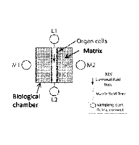
example of a midgut chip. The lumen 160 of a cell tube created from
mosquito midgut epithelial cells 162 is loaded with a suspension of malaria-
infected red blood cells. The parasites 164 undergo sexual reproduction and
migrate through the midgut epithelium into the surrounding matrix 170 where
they transform into oocysts (0C) 172. OCs appear as brightly fluorescent
spheres (-20 micron diameter). Assay readout is the number of OCs on the
ablumenal side of the midgut: the smaller the CC count the higher the
transmission-blocking activity of the test compound. The microenvironment is
maintained by perfusion with growth medium.
Once feasibility is robustly established, the midgut chip and culture
conditions may be optimized to increase the yields of oocysts per midgut
microenvironment and thus to increase statistical relevance of experiments
possible per chip. Besides optimizing the culture conditions, the
characteristics of the Nortis TEM chip and manner of its fabrication allows
creating longer midgut tubes or arrays of multiple midgut tubes within one
chip. This can increase the overall culture volume and surface for oocysts to
settle and develop. Thus, several hundred oocysts per chip could be
achievable.
With an oocyst culture protocol established the system can be applied
to studies on potential compounds for malaria vaccines or transmission
blocking vaccines. However, with oocysts developing to a state of maturity,
the system will provide the first option ever described to produce large
numbers of Plasmodium falciparum sporozoites in vitro - a vital step to
produce a much needed malaria vaccine.
Referring now to FIGs. 17A-17D, an example of enriched GFP-
expressing Plasmodium falciparum ookinete 48hrs post-fertilization in
suspension of RBC's is shown. FIG. 17A and FIG. 17C were taken under
GFP fluorescence. FIG. 17B and FIG. 17D were taken using transmitted
light.
Referring now to FIGs. 17E-17F, there shown are examples including
a cell tube of 4A-3B cells with injected GFP-expressing parasites in stages of
CA 02886247 2015-03-25
WO 2014/052835
PCT/1JS2013/062307
zygotes and developing and matured ookinetes. FIG. 17E was taken under
GFP fluorescence. FIG. 17F was taken using transmitted light.
The invention has been described herein in considerable detail in
order to comply with the Patent Statutes and to provide those skilled in the
art with the information needed to apply the novel principles of the present
invention, and to construct and use such exemplary and specialized
components as are required. However, it is to be understood that the
invention may be carried out by specifically different equipment, and devices
and reconstruction algorithms, and that various modifications, both as to the
equipment details and operating procedures, may be accomplished without
departing from the true spirit and scope of the present invention.
31