Note: Descriptions are shown in the official language in which they were submitted.
CA 2887062 2017-03-30
- 1 -
TITLE: IRIS SHIELD
BACKGROUND OF THE INVENTION
This invention relates to ophthalmic surgeries performed on the eye. The
invention relates specifically to such surgeries where an increased pressure
is
developed inside the eye enclosure. Such surgeries include, for example and
without
limitation, cataract surgery, vitrectomy, glaucoma procedures, and other
procedures
undertaken behind the iris.
During such surgeries, one or more surgical openings are created in the
anterior
portion of the eye for the purpose of inserting, into the eye, various
surgical instruments,
as well as fluids and other assisting items which are used in the surgical
procedure or
which are temporarily inserted into the eye during the procedure, or which are
inserted
for the purpose of leaving such item in the eye as part of the surgical
procedure. Such
surgical openings are commonly created adjacent, and anterior of, the iris,
generally
adjacent the outer perimeter of the iris, optionally in the sclera.
During such surgical procedures, it is common to add one or more fluids to the
anterior chamber of the eye, and it is common that at least some of such added
fluid is
positioned behind the iris. The addition of such fluid can cause an increase
in the fluid
pressure inside the eye_
For example, during cataract surgery, one or more surgical openings may be
made adjacent the outer perimeter of, and in front of, the iris. One or more
instruments
are inserted through the surgical openings, in front of the iris, and
manipulated inside the
anterior chamber, along with administration of suitable fluids inside the
anterior
chamber, in removing the original natural lens, and inserting a replacement
intraocular
lens in its place.
In the alternative, during the surgery, the patient may tense the eyelids,
which
also raises the pressure inside the eye.
Elements of the iris tissue are relatively thin, and are loosely connected to
each
other. The iris, as a whole is quite mobile. Accordingly, the iris responds to
any such
increase in pressure by moving away from the area of relatively greater
pressure toward
an area of relatively lower pressure.
In addition, the strength of the iris tissue can vary from patient to patient,
depending on a number of health-related factors, and/or life style factors,
including any
drugs, such as sympathetic blockers, which the patient may be using.
CA 02887062 2015-04-01
WO 2014/055284- 2 - PCT/US2013/061296
Thus, as the pressure inside the anterior chamber increases during the
surgical
procedure, there is a tendency for the iris is to move away from the increased
pressure
toward an area of relatively lower pressure. The ambient atmosphere outside
the eye is
such an area of lower pressure. The surgical opening(s) are made through
relatively soft
and extensible tissue. Any area of the surgical opening which is not fully
occupied by an
instrument or other article, or to the extent the surgical opening can be
enlarged slightly by
the internal pressure inside the anterior chamber, such as by stretching the
tissue
surrounding the surgical opening, provides a path for the iris tissue to move
toward that area
of lower pressure outside the eye. Any such movement of the iris tissue
outside its normal
zone of movement creates abnormal stresses on the iris tissue, and can be
damaging to the
iris tissue.
The result of such abnormal movement of the iris tissue is the protrusion of
iris
tissue, commonly referred to as prolapse, through the surgical opening. Such
prolapse of
iris tissue through the surgical opening creates abnormal stresses in the iris
tissue and can,
in some cases, result in tearing of the iris tissue.
FIGURE 1 shows an example of a generally healthy human eye 2 before a surgical
procedure has been initiated. There are no openings in the outer tissue
adjacent the
anterior chamber. There is no path for prolapse of the iris through the eye
enclosure.
FIGURE 2 shows the same eye as in FIGURE 1 but illustrating such prolapse.
FIGURE 2 illustrates a surgical opening 3 created through the cornea 4 or
sclera 5 adjacent
the outer edge of the iris 6. As a result of increased pressure inside the
anterior chamber,
for example during such surgical procedure, the iris has begun to prolapse
through the
surgical opening and thus a prolapsed portion 8 of the iris extends outside
the eye.
It is desirable to prevent such prolapse, which can damage and/or tear the
iris tissue
and, even where the prolapsed tissue can be drawn back inside the eye
enclosure through
the surgical opening, such trauma to the eye can result in the patient
experiencing pain
during the surgery and the patient may experience excessive glare post-
surgery.
Thus, the problem to be solved by the invention is to address such prolapse,
either
by preventing the occurrence of such prolapse, or by providing remedial
measures to
reverse such prolapse after the prolapse occurs such that the iris tissue
moves back inside
the eye enclosure.
A conventional treatment for iris prolapse is to watch for prolapse, and to
react to
such prolapse when prolapse is observed during the surgical procedure. The
conventional
reaction to prolapse, once the prolapse is observed, is to maintain the
increased pressure in
the anterior chamber behind the iris, and to increase the pressure inside the
anterior
chamber in front of the iris. Such increase in pressure in front of the iris
apparently urges the
iris tissue which remains inside the eye envelope to move reanivardly inside
the eye
CA 02887062 2015-04-01
WO 2014/055284 PCT/US2013/061296
- 3 -
enclosure, thus drawing the prolapsed tissue back through the surgical opening
and inside
the eye.
Another conventional treatment is to depress the iris and the intraoptical
lens inside
the eye when the iris prolapses_
Still another conventional treatment is to place a viscoelastic plug in any
surgical
opening which is not needed immediately, for the period when the pressure is
to be
increased in the anterior chamber, and then to remove the viscoeiastic plug as
part of
completing the surgical procedure.
Any tissue which may have been torn in the process of a prolapse may be
permanently lost. In addition, any iris tissue which has been exposed to
ambient
atmosphere in the meantime, has also been exposed to any contaminants in the
atmosphere, including any pathogenic bacteria, viruses, and the like with
which the
prolapsed tissue may have come into contact in the atmosphere. So allowing the
prolapse
to occur, and then responding to such occurrence, entails additional risk of
contamination
and/or tissue tearing, including the risk of corresponding complications
developing as a
result of the surgery_
Thus it would be desirable to provide a proactive method for preventing the
occurrence of iris prolapse,
It would further be desirable to prevent the iris tissue from becoming exposed
to the
ambient environment outside the eye enclosure,
It would be further desirable to, as much as possible, prevent the stress and
potential
for tearing of iris tissue as a result of increased pressure inside the eye
enclosure while
performing ophthalmic surgery.
It would be desirable that the device would catch the pupil (inner) edge of
the iris and
maintain dilation of the pupil allowing for the duration of surgery, thereby
assisting
visualization of the deeper contents (lens) during surgery.
It would further be desirable to avoid inserting small temporary plugs into
the surgical
opening; lest such small items become fragmented; or lest such small items be
inadvertently
left inside the anterior chamber at the end of the surgery.
These and other needs are alleviated, or at least attenuated, by the novel
products,
systems, and methods, of the invention.
CA 02887062 2015-04-01
WO 2014/055284- -
PCT/US2013/061296
4
SUMMARY OF THE INVENTION
This invention provides apparatus and methods for preventing prolapse of iris
tissue
or any other eye tissue through a surgical opening in the cornea or sclera,
out of the eye
during an ophthalmic surgical procedure At an early stage of the surgical
procedure, a
suitable flexible biocompatible polymeric shield is inserted into the anterior
chamber of the
eye and placed in a position overlying the iris adjacent each of the surgical
openings into the
anterior chamber and is thus between the iris tissue and any surgical
openings. If/when the
pressure inside the anterior chamber increases during the surgical procedure,
any anterior
movement of the iris toward the cornea or sclera, namely toward any such
surgical opening
in response to such pressure, also lifts the shield anteriorly, such that the
shield remains
between the iris and the respective surgical openings. Thus, the shield blocks
the surgical
opening and prevents movement of eye material to and through the surgical
opening.
Namely, the shield closes off access to the surgical opening from inside the
eye. The
surgical opening can, of course, still be accessed by surgical tools and
materials from
outside the eye by inserting such articles through the surgical opening and
pushing such
articles past the flexible shield.
In a first family of embodiments, the invention comprehends an iris shield
configured
to temporarily overlie an iris of an eye adjacent a surgical opening which has
been made
through an anterior portion of such eye during an ophthalmic surgical
procedure, whereby
the shield prevents prolapse of iris tissue past the iris shield and through
such surgical
opening during such surgical procedure, the iris shield to be removed prior to
completion of
such surgical procedure, such iris having a generally annular configuration,
including an
outer edge and an inner edge, the iris shield comprising a flexible
biocompatible polymeric
sheet which can be folded lengthwise on itself, the sheet having an anterior
side and a
posterior side, an inner edge extremity and an outer edge extremity, and a
sheet width of
about 1 mm to about 5 mm between the inner edge extremity and the outer edge
extremity,
the sheet having a first end and a second end, and a length therebetween, the
outer edge
extremity of the flexible biocompatible polymeric sheet extending along a
configuration which
can overlie the outer edge of a such iris for which the sheet has been
configured, such that
the polymeric sheet is between the iris and the surgical opening and extends
inwardly
toward the inner edge of the iris, thus to shield the iris against prolapse
through the surgical
opening during the surgical procedure.
In some embodiments, the polymeric sheet has an anterior surface and a
posterior
surface, and a sheet height is defined between the anterior surface and the
posterior
CA 02887062 2015-04-01
WO 2014/055284- 5 - PCT/US2013/061296
surface, the posterior surface being generally free from appendages which
might engage the
inner edge of such iris.
in some embodiments, the sheet has an anterior surface and a posterior
surface,
further comprising at least first and second retention flanges, spaced from
each other and
extending from the posterior surface adjacent the inner edge extremity of the
polymeric
sheet at one or more angles of from 30 degrees acute to, and projectable onto,
the posterior
surface of said sheet to 135 degrees obtuse to, and not projectabie onto, the
posterior
surface of the sheet.
In some embodiments, a given retention flange has a length extending from a
flange
base at the polymeric sheet to a flange distal end remote from the polymeric
sheet, the
length of the retention flange being less than the width of the sheet adjacent
the given
retention flange.
In some embodiments, a sheet height is defined between the anterior surface
and
the posterior surface, and at least one of the retention flanges has a
thickness less than the
height of the biocompatible polymeric sheet.
In some embodiments, the width of the sheet, between the inner and outer edge
extremities, is greater than about 2 mm to about 3 mm,
In some embodiments, the sheet has a shore A durometer hardness of 20 to 75.
In some embodiments, the sheet height is generally uniform across the width of
the
sheet between the inner edge extremity and the outer edge extremity, and
optionally has a
sheet height of about 100 microns to about 500 microns.
In some embodiments, the sheet extends radially at least 30 degrees,
optionally up
to about 350 degrees, such as about an axis of the sheet.
In some embodiments, the inner edge extremity of the sheet generally overlies
the
inner edge of the iris of an eye for which the shield is configured, when the
iris is dilated,
such that insertion of the shield into the eye, and engaging the shield with
the inner edge of
the iris, results in minimal, if any, retraction of the iris, and optionally
results in stabilizing the
iris against constriction during the surgical procedure.
In some embodiments, the sheet extends along a path selected from annular
paths,
'V"-shaped paths, and paths configured in shapes of polygons.
In some embodiments, the shield further comprises at least one control element
which is used to assist in controlling positioning of the shield relative to
the iris at a relatively
earlier stage of the surgical procedure and/or to assist in controlling
removing the shield from
the eye at a relatively later stage of the surgical procedure.
In some embodiments, the at least one control element is an aperture extending
through the sheet from the top of the sheet to the bottom of the sheet,
CA 02887062 2015-04-01
WO 2014/055284- 6 - PCT/US2013/061296
In some embodiments, the aperture is proximate one of the first and second
ends of
the sheet.
in some embodiments, the sheet, when at rest on an underlying flat supporting
surface, has a generally flat top surface which extends from the inner edge
extremity to the
outer edge extremity.
In a second family of embodiments, the invention comprehends an iris shield
configured to temporarily overlie an iris of an eye adjacent a surgical
opening which has
been made through an anterior portion of the eye during an ophthalmic surgical
procedure,
thereby to prevent prolapse of iris tissue past the iris shield and through
the surgical opening
during the surgical procedure, the iris shield to be removed prior to
completion of the surgical
procedure, the iris having a generally annular configuration, including an
outer edge and an
inner edge, the iris shield comprising an annular flexible biocompatible
polymeric sheet
which can be folded on itself, the sheet having an inner edge extremity and an
outer edge
extremity, and a sheet width of about 1 mm to about 5 mm between the inner
edge extremity
and the outer edge extremity, the outer edge extremity of the flexible
biocompatible
polymeric sheet extending along a configuration which can overlie the outer
edge of a such
iris for which the sheet has been configured, such that the polymeric sheet is
between the
iris and the surgical opening and extends inwardly toward the inner edge of
the iris, thus to
shield the iris against prolapse through the surgical opening during the
surgical procedure.
In some embodiments, the iris shield extends 360 degrees as a closed annulus
about
an axis of the sheet.
In a third family of embodiments, the invention comprehends a method of
treating a
living eye during an ophthalmic surgery, the eye having an anterior chamber,
and an iris in
the anterior chamber, the iris having an outer edge and an inner edge, and a
width between
the outer edge and the inner edge, the method comprising creating a surgical
opening into
the anterior chamber of the eye: inserting an iris shield into the anterior
chamber through the
surgical opening in the anterior chamber; positioning the iris shield over the
iris adjacent the
surgical opening such that the iris shield overlies a substantial portion of
the iris adjacent the
surgical opening, the iris shield thus being positioned between the iris and
the surgical
opening so as to prevent prolapse of iris tissue through the surgical opening
during the
ophthalmic surgery; performing one or more additional surgical procedures
while the iris
shield is in such overlying relationship with respect to the iris adjacent the
surgical opening:
and as part of completing the ophthalmic surgery, displacing the iris shield
from over the iris
and removing the iris shield from the eye,
CA 02887062 2015-04-01
WO 2014/055284- 7 - PCT/US2013/061296
In some embodiments, the positioning of the iris shield in overlying
relationship over
the iris includes positioning the iris shield such that the iris shield is
overlying the outer edge
of the iris adjacent the surgical opening.
In some embodiments, the removing of the iris shield from the eye comprises
removing the iris shield through the surgical opening.
In some embodiments, the ophthalmic surgery comprises creating more than one
surgical opening, accessible by the iris, into the anterior chamber of the
eye, the method
including selecting one or more suitable iris shields, and positioning the one
or more iris
shields such that iris shield material is disposed between the iris and each
such surgical
opening,
in some embodiments, the one or more additional surgical procedures includes a
procedure which raises the pressure inside the anterior chamber of the eye,
the iris shield
being effective, when such pressure is so raised, to shield the iris from the
effect of such
increased pressure, sufficient to prevent prolapse of iris tissue out of the
eye through a such
respective surgical opening in the anterior chamber of the eye.
In some embodiments, the iris shield comprises a sheet having an anterior and
a
posterior, an inner edge extremity and an outer edge extremity, and first and
second
retention flanges spaced from each other, each such retention flange extending
from a
posterior surface of the sheet at the inner edge extremity, at an angle of
from 30 degrees
acute to, and projectable onto, the posterior surface of the sheet to 135
degrees obtuse to,
and not projectable onto, the posterior surface of the sheet, the method
further comprising
engaging the retention flanges with the inner edge of the iris, the iris
shield thus engaging
the inner edge of the ins.
In some embodiments, the iris shield prevents the iris from constricting
during the
surgical procedure, optionally stabilizing the iris against constriction
during the ophthalmic
surgery.
In some embodiments, the engaging of the retention flanges with the inner edge
of
the iris does not result in substantial retraction of the iris.
In some embodiments, the iris shield extends about greater than 180 degrees of
the
circumference of the iris, and may comprise an annular biocompatible polymeric
sheet.
In some embodiments, the iris shield overlies at least 75 percent of the width
of the
iris between the inner edge and the outer edge, including substantially
overlying the outer
edge of the iris,
In some embodiments, the iris shield is positioned over the iris, and between
the
surgical opening and enough of that portion of the iris which is proximate the
surgical
opening to prevent prolapse of iris tissue through the surgical opening,
CA 02887062 2015-04-01
WO 2014/055284- -
PCT/US2013/061296
8
BRIEF DESCRIPTION OF THE DRAWINGS
FIGURE 1 shows a relatively healthy human eye prior to any performance of
ophthalmic surgery.
FIGURE 2 shows the eye of FIGURE 1 with a prolapsed iris after a surgical
opening
has been created in the eye enclosure and pressure has been increased in the
anterior
chamber behind the iris.
FIGURE 3 is a pictorial representation of a simple iris shield of the
invention.
FIGURE 4 is a view from the front of an eye, with the iris shield of FIGURE 3
installed
adjacent a surgical opening in the sclera, the iris shield being between the
iris and the outer
layers of the eye enclosure.
FIGURE 6 is a pictorial view of a second embodiment of iris shields of the
invention.
FIGURE 6 is an edge view of the iris shield of FIGURE 5.
FIGURE 7 is another pictorial view of the embodiment of iris shields
illustrated in
FIGURES 5 and 6.
FIGURE 8 is a pictorial view of a third embodiment of iris shields of the
invention.
FIGURE 9 is a pictorial view of an embodiment like that of FIGURE 8, except
that the
retention flanges are at obtuse angles with respect to the shield sheet.
FIGURE 10 shows a front view of the eye, with the iris fully dilated, showing
the iris
shield of FIGURE 8 in place inside the anterior chamber, in its shielding
position over the iris,
between the iris and the eye enclosure tissue.
FIGURE 11 is a front view of an eye as in FIGURE 10, wherein the eye is no
longer
dilated, showing the ins shield being removed from the anterior chamber of the
eye through
the surgical opening.
FIGURE 12 shows a cross-section of an eye before any surgery-induced increase
in
pressure inside the anterior chamber, with an iris shield of FIGURES 5-7 in
place overlying
the iris and between the iris and the anterior tissue which forms part of the
eye enclosure.
FIGURE 13 shows a cross-section of the eye of FIGURE 12 after the pressure
inside
the anterior chamber has been increased, showing the iris shield performing
its shielding
function between the iris and the surgical opening.
FIGURE 14 is a view from the front, of an eye as in FIGURE 4, with an iris
shield
installed, overlying the iris, wherein the iris shield extends about the full
circumference of the
iris, and is thus a full-circle shield having four retention flanges engaged
with the inner edge
of the iris.
FIGURE 15 shows a top view of a fourth iris shield of the invention, in a "V"-
shape.
FIGURE 16 shows a top view of a fifth iris shield of the invention, in a
"polygon"-
shape.
CA 02887062 2015-04-01
WO 2014/055284- -
PCT/US2013/061296
9
The invention is not limited in its application to the details of
construction, or to the
arrangement of the components set forth in the following description or
illustrated in the
drawings. The invention is capable of other embodiments or of being practiced
or carried
out in various other ways. Also, it is to be understood that the terminology
and phraseology
employed herein is for purpose of description and illustration and should not
be regarded as
limiting. Like reference numerals are used to indicate like components,
CA 02887062 2015-04-01
WO 2014/055284- 10 - PCT/US2013/061296
DETAILED DESCRIPTION OF THE ILLUSTRATED EMBODIMENTS
in the invention, a sheet-shaped shield of siightly stiff, biocompatible
polymeric
material is inserted into the anterior chamber of the eye, through a
peripheral corneal or
sclera surgical opening, and placed on the iris inside the eye to prevent
prolapse of iris
tissue outside the eye by adding blocking support to the iris tissue and
optionally engaging
the inner edge of the iris to, as an additional benefit, prevent constriction
of the iris over the
Pupil.
Referring to the drawings, FIGURES 3 and 4 show a rather simplistic embodiment
of
the invention. FIGURE 3 shows a curved silicone shield 10 which extends 90
degrees about
the circumference of an imaginary circle. FIGURE 4 shows the shield 10 in use
over a
portion of the iris during a surgical procedure.
Shield 10 is made of a flexible surgical-grade polymer. The outline of the
shield
shown in FIGURES 3 and 4 generally conforms, along its length, to the outline
of the iris of
the human eye, including the outer edge extremity 12 of the shield, the inner
edge extremity
14 of the shield, and the width "W" between the inner and outer edge
extremities. The first
and second ends 16A, 16B of the shield are also curved as the ends connect the
inner and
outer edge extremities to each other. Apertures 18A, 186 function as control
elements for
controlling and manipulating the shield and are located inwardly of ends 16A,
166, generally
equidistant from edge extremities 12, 14 and the respective ends 16A, 16B,
Such control
elements can take a number of forms, including protrusions, or indentations,
as well as the
apertures illustrated.
FIGURE 4 shows a human eye 2 in isolation, with no surrounding tissue being
shown. Of course, in a real surgical procedure, the eye is disposed in an eye
socket in the
face of the patient. However, none of such surrounding tissue is shown in
FIGURE 4. Thus,
the elements of the eye, as visible to casual observation from the front, are
the pupil 22, the
iris 6, and the white portion of the sclera 5. During surgery, a small
surgical opening 3 is
created in the front portion of the eye, typically adjacent the outer edge of
the iris. Such
surgical opening is typically about 2-3 mm in length.
Various preliminary steps may be performed in the surgical procedure of e.g. a
cataract removal and replacement, prior to any injection, into the eye, of any
material which
would increase the internal pressure inside the eye. During the surgical
procedure, shield 10
is inserted through the surgical opening, and positioned over the iris
adjacent the surgical
opening prior to the application of any significant increase in pressure
inside the eye
enclosure. Thus, shield 10 is inserted before any material is injected into
the eye to e.g.
fracture a crystallized natural lens which is to be removed and replaced.
CA 02887062 2015-04-01
WO 2014/055284- 11 - PCT/US2013/061296
Typically, the shield will be folded on itself lengthwise, e.g. along a
longitudinal axis,
and placed in an injector instrument, such as those used to inject artificial
e.g, intraocular
lenses, in order to more easily fit through the surgical opening. Using a
suitable such
insertion tool, the shield is inserted through the surgical opening, and into
the eye. Once the
shield has passed completely through the surgical opening, the shield is
released from the
tool and allowed to unfold.
A suitable manipulation tool is then engaged with the shield at one or both of
apertures 18A, 18B and used to complete the unfolding of the shield if needed
and to
position the unfolded shield over the iris as shown, the shield generally
extending in
opposing directions from the surgical opening and about the circumference of
the iris. As
the shield is being positioned in the eye, the length of shield 10 is
generally centered on the
surgical opening if only one surgical opening has been created. The shield, in
its Use
position as illustrated in FIGURE 4, thus provides a protective covering over
the iris at and
adjacent the surgical opening. Where more than one surgical opening is created
in the eye,
a shield is selected for use whose length is great enough that the length of
the shield can
extend past all such surgical openings.
Any increase in pressure inside the eye is commonly transferred to the iris as
an
outwardly-directed, anteriorly-directed force, thus urging the iris to move
outwardly of,
anteriorly of, the eye,
Iris tissue is typically quite soft. Where the iris material is sufficiently
soft, the iris
tissue can thus flow toward any lower pressure at the surgical opening unless
such
movement is impeded/blocked. With the shield positioned in overlying
relationship over the
ins as illustrated in FIGURE 4, such upwardly, and outwardly, anteriorly-
directed force urges
the iris against the underside/postenor surface of the shield. Shield 10
receives that force
and spreads the force along the length and width of the shield. Since the
shield has
generally-fixed length, width, and height dimensions, and is not liquidous,
thus cannot flow,
such upwardly, outwardly, anteriorly-directed force moves the shield, from its
rest position
overlying the iris, anteriorly against the outer tissues of the eye, such as
against the cornea
and optionally against a portion of the sclera
Thus, while shield 10 is quite flexible, and with a generally central portion
of the
shield adjacent surgical opening 3, the generally limited extensibility of the
shield, in the
length and width dimensions, does not allow the shield to change shape enough
to be forced
out the surgical opening. And since the shield is between the iris and the
surgical opening
and is wide enough to prevent the iris material from circumventing the shield
and flowing out
the surgical opening, the shield serves as an effective barrier, protecting
the iris such that iris
material does not flow beyond the shield toward the surgical opening, and thus
does not
prolapse out the surgical opening.
CA 02887062 2015-04-01
WO 2014/055284- 12 - PCT/US2013/061296
With the shield in place as shown in FIGURE 4, the surgeon then continues the
surgery according to conventionally-accepted surgical procedures, with
instrument elements,
supplies, and/or implant elements being passed into and out of the eye
enclosure through
the one or more surgical openings, and wherein the shield is between the iris
and the
instrument elements, supplies, and/or other implant elements. Thus, the shield
not only
serves to protect the iris from the effects of increases ................. in
pressure, the shield also serves as a
buffer/shield to prevent, or reduce in extent or severity, direct contact of
the instruments,
supplies, implant elements, and the like, with the rather delicate iris
tissue.
Where more than one surgical opening is made into the eye during the surgical
procedure, a second shield may, optionally, be placed adjacent the second
surgical opening.
In the alternative, a longer such shield may be selected as the sole shield
used, whereby the
single shield extends along and well past all of the surgical openings
providing prolapse
protection to the iris at all of the surgical openings.
Once the pressure inside the anterior chamber of the eye returns to more
normal
pressures, and where the pressure is expected to not again increase to a high
enough level
to facilitate prolapse of iris tissue, shield 10 is removed through the
surgical opening, again
using a suitable instrument to manipulate the shield by interaction with the
edges of
apertures 18A, 186.
Shield 10 can be inserted into the eye any time after a surgical opening is
created,
with commonly used micro forceps, or with a commonly used injector system
wherein the
shield may be folded lengthwise on itself, e.g, about its longitudinal axis.
After the shield has
been inserted through the surgical opening and into the anterior chamber,
forceps are
engaged in apertures 18A and/or 188 and thus used to manipulate the shield
into place over
the iris. Once the remaining steps in the surgical procedure have been
completed, the
shield is then removed using a small hook commonly known as a Connor Wand, a
Sinskey
Hook, or the like,
Once shield 10 has been removed from the eye, through the surgical opening,
the
surgery again proceeds and/or concludes according to conventionally-accepted
surgical
procedures.
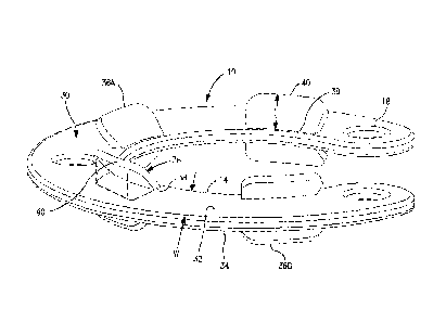
FIGURES 5, 6, and 7 illustrate a second embodiment of iris shields of the
invention.
The shield of FIGURES 5-7 differs in at least 3 substantial ways from the
shield of FIGURES
3-4.
First, while both shields are generally circular in shape, the shield of
FIGURES 3-4
extends about 90 degrees about a circumference of a such circle while the
shield of
FIGURES 5-7 extends about 270 degrees about the circumference of the circle,
e.g, about
central axis 'A" of the shield. Thus, the shield of FIGURES 5-7, in use,
nominally covers
about 3 times as much surface area of the iris as is covered by the shield of
FIGURES 3-4.
CA 02887062 2015-04-01
WO 2014/055284- -
PCT/US2013/061296
13
Second, the shield 10 of FIGURES 3-4 is a flat, smooth sheet 30 on both its
top
surface 32 and its bottom surface 34. By contrast, the shield of FIGURES 5-7
has a series
of retention flanges 36. A given retention flange 36 extends, from a flange
base 38 adjacent
the inner edge extremity 14 of the sheet 30 at the top or bottom surface of
sheet, along a
length "L" of the flange, to a distal end 40 of the flange remote from the
flange base. In the
embodiments of FIGURES 5-7, the lengths "L of the retention flanges are less
than the
Width of the shield.
In the embodiments of FIGURES 5-7, four retention flanges 36A are spaced from
each other along the length of the top surface of the sheet, and another four
retention
flanges 366 are spaced from each other along the length of the bottom surface
of the sheet.
Flanges can extend at any angle to the respective surface of the sheet, from
about 30
degrees acute angle, including a perpendicular angle, to an obtuse angle of
about 135
degrees, e.g. in increments of about 1 degree, with respect to the respective
surface of the
sheet. In FIGURES 5-7, each such flange 36 extends at an acute angle of about
35 degrees
to the respective top or bottom surface of the sheet.
In typical embodiments, the entirety of each acute angle flange can be
projected onto
the sheet as illustrated with one of the flanges in dashed outline on the left
side of FIGURE
7, Typically, the acute angle flanges which overlie the sheet, or underlie the
sheet, are
shorter in length "L" than the width "W" of the sheet, whereby the outline of
a given acute-
angle flange can be projected onto the sheet, at a perpendicular angle to the
sheet. By
contrast, obtuse angle flanges typically cannot be projected onto the sheet
unless the base
38 of the respective flange is located away from inner edge extremity 14 of
the sheet, toward
outer edge extremity 12 of the sheet.
The purpose of retention flanges 36 is to engage and hold the inner edge 39 of
the
iris (FIGURE 10), thus to control, prevent movement of the inner edge of the
iris. With
suitable stiffness in the material of sheet 30, the shield holds its shape
over the iris, thus
generally holding the iris immobile during certain portions of the surgical
procedure,
preventing the iris from constricting such that the iris cannot constrict over
the pupil of the
eye and thus impede visualization of rearwardly-disposed portions of the
anterior chamber,
where the lens is located, by the attending surgeon. Typically, and as shown
in FIGURE 5-
7, but not necessarily, all of the retention flanges on a given shield will
bear the same
angularity with respect to sheet 30.
In use inside the anterior chamber of an eye, flanges 36 extend posteriorly
from the
bottomiposterior surface of the sheet in order to engage the inner edge of the
iris. Thus, any
flanges which extend anteriorly from sheet 30 are extraneous to a given
surgical procedure.
However, by having flanges extending from both the anterior surface and the
posterior surface of the sheet inside the eye, the shield is insensitive to
top-or-bottom
CA 02887062 2015-04-01
WO 2014/055284- -
PCT/US2013/061296
14
orientation of the shield when the shield is inserted into the eye. Such lack
of sensitivity of
the shield to orientation allows the surgeon to insert the shield without
concern with
potentially needing to invert the shield after the shield has been inserted.
Thus, the shields
represented by FIGURES 5-7, having retention flanges extending from both the
top surface
of the sheet and the bottom surface of the sheet, present suitable retention
flange orientation
irrespective of which surface of the shield presents itself to the iris when
the shield emerges
from the insertion tool inside the anterior chamber.
Height "H" of sheet 30, between the top surface and the bottom surface, is
about 100
microns to about 600 microns: optionally about 200 microns to about 400
microns yet further
optionally about 350 microns. The thicknesses of the retention flanges is
about 100 microns
to about 300 microns, optionally about 200 microns to about 300 microns, with
the
thicknesses of the flanges being optionally less than the height of the sheet
30 to which such
flanges are attached.
The height of sheet 30 is driven by a number of factors including, without
limitation,
the hardness of the sheet material, the width of the sheet, the flexibility of
the sheet,
foldability of the sheet, rigidity of the sheet: strength of the sheet, and
the like. The thickness
of a retention flange 36 is driven by flexibility of the flange, strength of
the flange, the ability
of the flange to conform to the surface of the iris or the surface of the
overlying cornea or
sclera, and the like under pressure which typically exists in the eye during
eye surgical
procedures. In general, in fulfilling their functions relative to the iris,
inside the anterior
chamber, flanges 36 have a greater requirement to be foldable, flexible, than
does sheet 30.
Retention flanges 36 may be shaped as desired, such as rectangular, scalloped,
trapezoids, ovals, or the like, and are spaced from each other.
The third way the shield of FIGURES 5-7 differs from the shield of FIGURES 3-4
is
that, while the shield 10 of FIGURES 3-4 has two apertures 18, the shield of
FIGURES 5-7
has 3 apertures 18. The third aperture in the longer sheet provides an
additional location for
engaging the shield and manipulating the shield into position over the iris,
and for engaging
the shield and removing the shield from the eye through the surgical opening.
The number
of apertures can be selected and specified by the designer of a such shield as
greater than 3
apertures, or less than two apertures, in accord with specific needs
contemplated for the
particular shield.
A given shield 10 of the invention, both sheet 30 and any flanges 36, is
typically
made of a single material, and sheet 30 and flanges 36 are typically
fabricated
simultaneously as a single piece. A typical fabrication process is injection
or other type of
molding of a fluid polymeric composition, followed by cooling, solidification
to fix the shield
material in the desired configuration,
CA 02887062 2015-04-01
WO 2014/055284- -
PCT/US2013/061296
15
Shields 10 can be made from a variety of polymeric materials, such as various
ones
of the silicones, acrylics, and collamers. Specific compositions, and
combinations of
compositions; can be selected by those skilled in the art based on known
physical
properties, and biocompatibilities of materials of interest. Conventional
biocompatible
additive packages can be used as desired.
Especially height of sheet 30 is at least in part driven by strength and
stiffness of the
material once fabricated into the sheet form. Two non-limiting examples of
suitable such
material for use in shields of the invention are NuSil Med-4950 and NuSil Med
4970
silicones, having 50 Shore A and 70 Shore A hardnesses; respectively, both
available from
NuSil Technology, Carpinteria, California. Other conventionally available
materials may be
selected for other hardness specifications.
Typical hardness of the sheet 30, after fabrication, is about 20-75 Shore A,
optionally
about 20-40 Shore A.
A typical shield of the invention; as that shown in FIGURES 5-7, has an outer
diameter of about 12 mm to about 13 mm. A shield having such outer diameter
can be
comfortably fitted into, and will generally extend across substantially the
entirety of, the
anterior chamber of an adult human eye. Thus, where the shield extends at
least half way
around the anterior chamber, e.g. at least 180 degrees about the anterior
chamber,
opposing sides of the shield are generally positioned proximate or against the
outer
perimeter of the anterior chamber. Given an appropriate stiffness of the
shield; such that the
shield is not easily compressed inwardly toward its own central axis inside
the anterior
chamber by ambient pressures exerted inside the anterior chamber during the
surgical
procedure, opposing sides of the shield tend to center the shield about the
central axis of the
anterior chamber as defined between the front of the eye and the back of the
eye, Thus,
such shield, having a length corresponding to at least 180 degree progression
about the
central axis of the shield, assists the surgeon in his steps of positioning
the shield uniformly
about the iris such that the shield is positioned to cover as much of the iris
as possible along
the circumferential length of the shield; with specific attention to covering
as much as
possible of the iris tissue which is adjacent the surgical opening. In
general, the surgeon
positions the shield such that, to the extent reasonably possible, the outer
edge extremity of
the shield overlies that portion of the outer edge of the iris which is
adjacent a surgical
opening, and in addition; as much as possible for a given shield, such that
the shield overlies
that portion of the iris which extends radially from the surgical opening.
Where more than one surgical opening has been created in the eye, the surgeon
positions the shield so as to so protect the iris adjacent all, or as many as
possible, of the
surgical openings. Specifically, where more than one surgical opening has been
created;
CA 02887062 2015-04-01
WO 2014/055284- -
PCT/US2013/061296
16
the surgeon selects a shield which has a length great enough to extend past
each of the
surgical openings.
Thus, for a surgical procedure where only one surgical opening will be
created, a
simple shield such as the one shown in FIGURES 3-4, extending only 90 degrees
about the
circumference of the anterior chamber, may be sufficient, with or without
retention flanges
36.
Where two or more surgical openings will be created, typically opposite each
other
about the edge of the cornea or sclera, a shield is selected having a length
which extends at
least past both surgical openings. Thus, a shield extending about 270 degrees
of the
circumference of the anterior chamber may be selected. In some cases, such as
where
there may be unknown steps required during the surgical procedure, which may
suggest an
unknown number of surgical openings to perform such steps, a full-circle
shield may be
selected, namely a shield which extends about the full 360 degree
circumference of the
anterior chamber; or a substantially full circle shield having two ends but
extending up to less
than 360 degrees, e.g. about 350 degrees, may be selected.
Prior to beginning a surgical procedure, the surgeon will already know
measurements
of the patient's eye and will have secured a suitable supply of shields of the
size or sizes
expected to be needed for the specific patient and/or surgical procedure. Such
size may be
greater than 12-13 mm, or may be less than 12-13 mm, depending on the
measurements of
the respective patient.
Thus, a manufacturer of such shields for use in human eyes may typically
fabricate
such shields in at least three outer diameter sizes, for example, the average
12-13 mm size
outer diameter, one slightly larger than 12-13 mm, such as 14-15 mm, and one
slightly
smaller than 12-13 mm, such as 10-11 rem,
The width of the shield, between the outer edge extremity and the inner
edge
extremity, should be great enough to cover, and provide a shielding effect, to
enough of the
iris that any portion of the width of the iris which is not overlain by the
shield is not
susceptible to moving to and/or through the surgical opening. Typical width
'W" of an
effective such shield is about 1 mm to about 3 mm, optionally about 2 mm to
about 3 mm,
optionally about 2,2 mm to about 2.6 mm, optionally about 2.4 mm.
Given the above discussions of the outer diameter of the shield and the width
of the
shield, the inner diameter of the shield can be calculated to be about 6 mm to
about 9 mm,
optionally 6 mm to about 7 mm, optionally about 6.2 mm.
A shield of the invention is used only during the surgical procedure. The
shield is
removed as one of the latter steps in the surgery. The shield is not left in
the eye after the
surgical procedure has been completed.
CA 02887062 2015-04-01
WO 2014/055284- 17 - PCT/US2013/061296
Returning now to the drawings. FIGURE 8 shows a pictorial view of a shield
which
extends 220 degrees about its central axis "A". Because this embodiment of the
shield
extends more than 180 degrees about the central axis, sufficiently stiff
shields represented in
FIGURE 8 have the above tendency to assist the surgeon in positioning the
shield in the
anterior chamber. Still referring to the embodiment shown in FIGURE 8,
retention flanges 36
extend perpendicularly upwardly from sheet 30. Given that the shield is
designed to overlie
the iris, given that flanges 36 must extend toward the interior of the eye to
so engage the
inner edge of the iris, the surface seen in FIGURE 8 is bottom surface 34 of
the shield
As a review, FIGURES 5-7 show retention flanges 36 at acute angles relative to
sheet 30; and FIGURE 8 shows retention flanges 36 perpendicular to sheet 30.
FIGURE 9 illustrates an embodiment of shield 30 where retention flanges 36 are
obtuse to sheet 30, extending away from both the outer edge extremity and the
inner edge
extremity, of the sheet, and toward central axis "A' of the shield. Measured
from the top
surface of sheet 30, flanges 36 extend at obtuse angles a of about 135 degrees
to sheet 30.
FIGURE 10 shows a photographic representation of a fully dilated eye with the
shield
10 of FIGURE 8 in place over the dilated iris. The width V" of the shield
generally
corresponds with the dilated width of the iris. The width of the shield
adjacent surgical
opening 3 generally overlies the entirety of the width of the iris, thus
protecting substantially
all of the width of the iris adjacent opening 3 from prolapse through opening
3.
FIGURE 11 shows a photograph representation of the eye as in FIGURE 10, but
not
dilated, where the width of the shield is substantially less than the width of
the iris. The
surgeon is in the process of removing the shield from the anterior chamber,
optionally
inserting the shield into the anterior chamber unfolded, through surgical
opening 3. For the
removal process, forceps, not shown, are used to pull and otherwise manipulate
the Shield
through the opening and out of the eye.
In the illustrations shown in FIGURES 10 and 11, shield 10 has been inverted
from
the image shown in FIGURE 8 such that retention flanges 36 extend away from
the viewer
on the non-visible bottom surface of the shield, and thus are not seen in
FIGURES 10 and
11.
FIGURES 12 and 13 show cross-sections of an eye 2 where a surgical opening 3
has been created, and where shield 10 has been inserted into anterior chamber
7 and
positioned in a location overlying iris 6 In FIGURE 12, the pressure inside
the anterior
chamber is generally equalized between the front portion 44 of the anterior
chamber, in front
of the iris, and the rear portion 46 of the anterior chamber to the rear of
the iris, such that the
iris is positioned in its normal, undisturbed, location adjacent lens 48.
As shown in FIGURE 12, shield 10 has retention flanges 36 extending from both
the
top surface 32 of sheet 30, and the bottom surface 34. As seen in FIGURES 12
and 13, the
CA 02887062 2015-04-01
WO 2014/055284- 18 - PCT/US2013/061296
shield has been manipulated about the inner edge of the iris such that the
retention flanges
on the bottom surface of the shield have been worked under the inner edge of
the iris, thus
to retain the shield firmly in its shielding position overlying the iris. As a
further benefit of the
use of retention flanges, the flanges prevent the iris from constricting while
the shield is so in
place over the iris.
In FIGURE 13, the pressure inside the rear portion 40 of the anterior chamber
is
greater than the pressure in the front portion 44 of the anterior chamber,
such that the iris
has been pushed upwardly against the cornea 4, including against surgical
opening 3. As
seen in FIGURE 13, shield 10 is between the iris and opening 3, thus
temporarily closing off
opening 3, temporarily obstructing opening 3, and preventing any prolapse of
the iris, or any
other material, through the opening,
FIGURES 12 and 13 illustrate the flexibility of flanges 36, such that the
flanges on the
bottom of the shield generally deflect against the iris, between the lens and
the iris, when the
pressure is generally equalized between the front and rear portions of the
anterior chamber.
Similarly, when the pressure gradient occurs between the front and rear
portions of the
anterior chamber, and the iris moves against the shield, and toward the
cornea, the flanges
on the top surface of the shield deflect against the top surface of the
shield, between the
shield and the cornea, thus avoiding having open space between the top surface
of the
shield and the flanges, through which iris tissue, or other material in the
eye, might escape to
and through the surgical opening.
FIGURE 14 shows a front view of the eye of FIGURES 12 and 13, with the shield
in
place overlying the iris. The flanges underlying sheet 30 are shown in dashed
outline, as is
the inner edge 39 of the ins. The flanges on the top surface of the sheet have
been omitted
so the underlying flanges can be seen.
FIGURES 15 and 16 show shield sheets 30 which illustrate that sheet 30 need
not
have a circular configuration. FIGURE 15 shows a sheet 30 having a "V"-shaped
configuration,
FIGURE 16 shows a sheet 30 having a straight-sided "U"-shaped
configuration, which can also be referred to as an open-ended polygonal
configuration.
FIGURES 15 and 16 thus illustrate that the sheet can have a wide variety of
configurations,
though the dimensions of such sheets still have the requirement that the
sheet, in use in the
eye, must provide the opportunity for the surgeon to position the shield such
that the shield
presents sufficient shielding material adjacent any surgical opening,
sufficient to prevent
prolapse of iris tissue. Any such shields may be fabricated, and used, with or
without
retention flanges.
While the invention has been described supra with respect to use in a human
eye,
the iris shields disclosed herein can as well be used in animal eyes. For such
uses, the
CA 02887062 2015-04-01
WO 2014/055284- 19 - PCT/US2013/061296
inner diameter, the outer diameter, and the width of the shield will be
specified and
fabricated according to the sizes of the eyes to be treated.
Although the invention has been described with respect to various embodiments,
it
should be realized this invention is also capable of a wide variety of further
and other
embodiments within the spirit and scope of the appended claims.
Those skilled in the art will now see that certain modifications can be made
to the
apparatus and methods herein disclosed with respect to the illustrated
embodiments, without
departing from the spirit of the instant invention. And while the invention
has been described
above with respect to the preferred embodiments, it will be understood that
the invention is
adapted to numerous rearrangements, modifications, and alterations, and all
such
arrangements, modifications, and alterations are intended to be within the
scope of the
appended claims.
To the extent the following claims use means plus function language, it is not
meant
to include there, or in the instant specification, anything not structurally
equivalent to what is
shown in the embodiments disclosed in the specification.