Note: Descriptions are shown in the official language in which they were submitted.
CA 02887933 2015-04-10
WO 2014/059442
PCT/US2013/065104
ANTIBODY BASED REAGENTS THAT SPECIFICALLY RECOGNIZE
TOXIC OLIGOMERIC FORMS OF TAU
RELATED APPLICATION
This application claims priority under 35 U.S.0 119(e) to provisional U.S.
Serial Number 61/713,441 filed October 12, 2012, which application is
incorporated hereby
by reference.
BACKGROUND OF THE INVENTION
Numerous studies have implicated small soluble oligomeric aggregates of A13 as
toxic
species in Alzheimer's disease (AD), and increasing evidence also implicates
oligomeric
forms of tau as having a direct role in disease pathogenesis of AD and other
tauopathies such
as Frontotemporal Dementia (FTD). As the focus of A13 studies has slowly
shifted toward
soluble Ar3 species and mechanisms, new reagents were needed that could
specifically
identify the variety of different aggregate species present. Indeed, many
contradictory studies
on the role of A13 aggregation in AD were reported and progress impeded
because suitably
selective reagents were not available to characterize the aggregate species
present. Increasing
evidence from cell and animal models indicate that oligomeric rather than
fibrillar forms of
tau are toxic and correlate with neuronal degeneration, therefore well
characterized reagents
that can specifically recognize the diversity of tau morphologies present in
the human brain
are critically needed to facilitate studies to identify the most promising tau
species for use as
biomarkers of disease and to study toxic mechanisms.
The microtubule associating protein tau is a major component of the
neurofibrillary
tangles associated with AD and tauopathies that are characterized by
hyperphosphorylation
and aggregation of tau. Tau plays an important role in assembly and
stabilization of
microtubules. Tau is a natively unfolded protein, and similar to a number of
other natively
unfolded proteins, it can aberrantly fold into various aggregate morphologies
including [3-
sheet rich fibrillar forms. The different types of post-translational
modifications of tau in AD
include phosphorylation, glycosylation, glycation, prolyl-isomerization,
cleavage or
truncation, nitration, polyamination, ubiquitination, sumoylation, oxidation
and aggregation.
Tau has 85 putative phosphorylation sites, and excess phosphorylation can
interfere with
microtubule assembly. Tau can be modified by phosphorylation or by reactive
nitrogen and
oxygen species among others. Elevated total tau concentration in CSF has been
correlated
with AD, as has the presence of various phosphorylated tau forms, and the
ratio of tau to
1
CA 02887933 2015-04-10
WO 2014/059442
PCT/US2013/065104
A1342. Reactive nitrogen and oxygen can modify tau facilitating formation of
aggregate
forms including oligomeric species. Levels of oligomeric tau have also been
implicated as a
potential early diagnostic for AD. Therefore, determination of total tau,
phosphorylated tau
and oligomeric tau concentrations all have potential value as diagnostics for
neurodegenerative diseases including tauopathies and AD.
Tau is an intrinsically unstructured protein due to its very low hydrophobic
content
containing a projection domain, a basic proline-rich region, and an assembly
domain.
Hexapeptide motifs in repeat regions of tau give the protein a propensity to
form n-sheet
structures which facilitate interaction with tubulin to form microtubules as
well as self-
interaction to form pathological aggregates such as paired helical filaments
(PHF).
Hyperphosphorylation of tau, particularly in the assembly domain, decreases
the affinity of
tau to the microtubules and impairs its ability to regulate microtubule
dynamics and axonal
transport. In addition, parts of the basic proline-rich domain and the pseudo-
repeat also
stabilize microtubules by interacting with its negatively charged surface.
Alternative splicing
of the second, third and tenth exons of tau results in six tau isoforms of
varying length in the
CNS. The assembly domain in the carboxyl-terminal portion of the protein
contains either
three or four repeats (3R or 4R) of a conserved tubulin-binding motif
depending on
alternative splicing of exon 10. Tau 4R isoforms have greater microtubule
binding and
stabilizing ability than the 3R isoforms. Human adult brains have similar
levels of 3R and 4R
isoforms, whereas only 3R tau is expressed at the fetal stage. In tauopathies,
mutations
altering the splicing of tau transcript and the ratio of 3R to 4R tau isoforms
are sufficient to
cause neurodegenerative disease. Therefore tau in human brain tissue can exist
in a variety of
different lengths and morphologies and with multiple post-translational
modifications.
Tau plays a critical role in the pathogenesis of AD and studies show that
reduction of
tau levels in AD animal models reverses disease phenotypes and that tau is
necessary for the
development of cognitive deficits in AD models caused by over-expression of
Ar3. While
NFTs have been implicated in mediating neurodegeneration in AD and
tauopathies, animal
models of tauopathy have shown that memory impairment and neuron loss do not
associate
well with accumulation of NFT. Animal studies showed improvement in memory and
reduction in neuron loss despite the accumulation of NFTs, a regional
dissociation of neuron
loss and NFT pathology, and hippocampal synapse loss and dysfunction and
microglial
activation months before the accumulation of filamentous tau inclusions. The
pathological
structures of tau most closely associated with AD progression are tau
oligomers. All these
studies suggest that tau tangles are not acutely neurotoxic, but rather that
pretangle
2
CA 02887933 2015-04-10
WO 2014/059442
PCT/US2013/065104
oligomeric tau species are responsible for the neurodegenerative phenotype,
similar to toxic
role of oligomeric Af3 species.
Numerous studies suggest that extracellular tau species contribute to
neurotoxicity
through an "infectious" model of disease progression. For example, tau
pathology spreads
contiguously throughout the brain from early to late stage disease,
extracellular tau
aggregates can propagate tau misfolding from the outside to the inside of a
cell, brain extract
from a transgenic mouse with aggregated mutant human tau transmits tau
pathology
throughout the brain in mice expressing normal human tau, induction of pro-
aggregation
human tau induces formation of tau aggregates and tangles composed of both
human and
normal murine tau (co-aggregation), and levels of tau rise in CSF in AD,
whereas Ar3 levels
decrease. A receptor-mediated mechanism for the spread of tau pathology by
extracellular tau
has been described.
Collectively, these studies all indicate that aggregated oligomeric species of
tau, both
intracellular and extracellular are vitally important in AD and other
tauopathies. In order to
more clearly define the role of individual tau forms in disease, there is a
critical need to
develop a series of well-defined reagents that selectively recognize
individual target
morphologies, and to use these reagents to identify which tau forms are the
best biomarkers
for AD, which forms are involved in toxicity both intra- and extracellularly,
and which forms
in brain tissue and CSF samples can distinguish between healthy and AD
patients.
Therefore, reagents that can specifically target tau oligomers would be
valuable tools
for diagnostic and therapeutic applications for AD, frontotemporal dementia,
other
tauopathies and neurodegeneration following traumatic brain injury.
Accordingly, there exists the need for new therapies and reagents for the
treatment of
Alzheimer's disease, frontotemporal dementia, other tauopathies and
neurodegeneration
following traumatic brain injury, in particular, therapies and reagents
capable of effecting a
therapeutic and diagnostic benefit at physiologic (e.g., non-toxic) doses.
SUMMARY OF THE INVENTION
The present invention discloses an antibody or antibody fragment that
specifically
recognizes oligomeric tau but does not bind monomeric tau, fibrillar tau or
non-disease
associated forms of tau. As used herein, the phrase "specifically recognizes
oligomeric tau"
indicates that it does not bind to or recognize non-specific proteins. As used
herein, the term
"antibody" includes scFv (also called a "nanobody"), humanized, fully human or
chimeric
antibodies, single-chain antibodies, diabodies, and antigen-binding fragments
of antibodies
3
CA 02887933 2015-04-10
WO 2014/059442
PCT/US2013/065104
(e.g., Fab fragments). As used herein, the term "oligomer" refers to a dimer,
trimer, tetramer,
pentamer, hexamer, heptamer, octamer, nonamer, decamer, undecamer or
dodecamer.
Accordingly, in certain embodiments, the oligomeric tau is dimeric tau,
trimeric tau,
tetrameric tau, pentameric tau, hexameric tau, heptameric tau, octameric tau,
nonameric tau,
decameric tau, undecameric tau or dodecameric tau. In certain embodiments, the
oligomeric
tau is dimeric tau or trimeric tau. In certain embodiments, the oligomeric tau
is trimeric tau.
In certain embodiments, the oligomer is soluble.
In certain embodiments, the antibody fragment does not contain the constant
domain
region of an antibody.
In certain embodiments, the antibody fragment is less than 500 amino acids in
length,
such as between 200-450 amino acids in length, or less than 400 amino acids in
length.
Certain embodiments of the invention provide an antibody fragment comprising
amino acid sequence SEQ ID NO:1, SEQ ID NO:9, SEQ ID NO:11, SEQ ID NO:13, SEQ
ID
NO:15, SEQ ID NO:17, or SEQ ID NO:19. In certain embodiments, the antibody
fragment
comprises amino acid sequence SEQ ID NO:1, SEQ ID NO:9 or SEQ ID NO:11.
Certain embodiments of the invention provide a binding molecule that binds to
oligomeric tau and does not bind monomeric tau, fibrillar tau or non-disease
associated forms
of tau, wherein the binding molecule comprises the sequence of SEQ ID NO:1,
SEQ ID
NO:9, SEQ ID NO:11, SEQ ID NO:13, SEQ ID NO:15, SEQ ID NO:17, or SEQ ID NO:19.
In certain embodiments, the binding molecule comprises the sequence of SEQ ID
NO:1, SEQ
ID NO:9, or SEQ ID NO:11.
Certain embodiments of the invention provide an antibody or antibody fragment
as
described herein, wherein said antibody fragment is isolated according to a
method
comprising the steps of:
a. a negative panning of a scFV phage library wherein said negative panning
eliminates phage that bind to non-desired antigens wherein said negative
panning comprises serially contacting phage with:
(i) a generic protein; and
(ii) mononeric forms of tau;
and monitoring the binding of said phage to the generic protein and monomeric
forms
of tau using Atomic Force Microscope (AFM) Imaging and repeating steps (i) and
(ii)
until no phage is observed binding to antigen by said AFM imaging to produce
an
aliquot of phage that does not bind to monomeric tau, fibrillar tau, or non-
disease
associated forms of tau;
4
CA 02887933 2015-04-10
WO 2014/059442
PCT/US2013/065104
b. contacting the aliquot of phage that does not bind to monomeric tau,
fibrillar
tau, or non-disease associated forms of tau with tau oligomers and incubating
for time sufficient to allow binding of phage to said oligomers; and
c. eluting the bound phage particles from step (b).
Certain embodiments of the invention provide an antibody or antibody fragment
isolated according to a method comprising the steps of:
(a) negative panning a scFV phage library comprising serially contacting
phage
with:
(i) a generic protein; and
(ii) mononeric forms of tau;
and until less than 5% of the phage is observed binding to antigen, which
produces an aliquot of phage that does not bind to monomeric tau, fibrillar
tau
or non-disease associated forms of tau;
(b) positive panning of the aliquot from step (a) comprising contacting the
aliquot
of phage from step (a) with tau oligomers, and incubating for time sufficient
to allow binding of phage to said brain derived tau oligomers; and
(c) eluting the bound phage particles from step (b).
In certain embodiments, the tau oligomer used in the positive panning is
trimeric tau
4N1R.
In certain embodiments, the generic protein is bovine serum albumin (BSA).
In certain embodiments, the negative panning further comprises serially
contacting
phage with brain derived control samples that do not contain oligomeric tau.
In certain embodiments, the observing of the binding of the phage to the
antigen is by
using Atomic Force Microscope (AFM) Imaging. In certain embodiments, the
negative
panning is repeated until less than 0-10% phage was observed by AFM imaging as
binding to
antigen in step (a).
Certain embodiments of the invention provide a method of inhibiting the
aggregation
of tau comprising contacting a composition that comprises tau monomers with an
antibody,
antibody fragment or binding molecule as described herein. In certain
embodiments, the
aggregation of tau is in a cell. In certain embodiments, the aggregation of
tau is in brain
tissue. In certain embodiments, the contacting with an antibody, antibody
fragment or
binding molecule decreases the rate of formation of tau aggregates as compared
to said rate in
the absence of composition or binding molecule.
5
CA 02887933 2015-04-10
WO 2014/059442
PCT/US2013/065104
Certain embodiments of the invention provide a method of detecting the
presence of
tau in a physiological sample comprising contacting a sample with an antibody,
antibody
fragment or a binding molecule as described herein and determining the binding
of said
composition with said tissue sample wherein binding of said composition to
said tissue
sample is indicative of the presence of tau oligomers in said tissue sample
wherein said
presence of said tau oligomers is indicative of early stage AD, frontotemporal
dementia, other
tauopathies or neurodegeneration following traumatic brain injury. In certain
embodiments,
the physiological sample is brain tissue, serum, cerebrospinal fluid (CSF),
blood, urine or
saliva.
Certain embodiments of the invention provide a method of preventing or
inhibiting
the accumulation of tau in the brain of a mammal comprising administering to
said mammal a
composition comprising an antibody fragment or a binding molecule as described
herein.
BRIEF DESCRIPTION OF THE DRAWINGS
Figure 1. Height distribution analysis of various tau samples obtained from
AFM
images. AD Tau #1 and AD Tau #3 represent tau samples obtained purified from
AD brain
tissue. Tau412M and Tau441M represent monomeric samples of 3R (tau 412) and 4R
(tau
412) tau samples. Tau 4410 represents and oligomeric sample of tau441.
Differences in
height and oligomeric state between samples are readily detected.
Figures 2A-2B. Lactate dehydrogenase (LDH) test of various tau species on SHSY-
5Y human neuroblastoma cells. (A) Toxicity of monomeric, dimeric and trimeric
tau 1N4R
(aka. tau 412) towards SHSY-5Y cells. (B) Toxicity of monomeric, dimeric and
trimeric tau
2N4R (aka. tau 441) towards SHSY-5Y cells. In both (A) and (B), for each group
from left
to right, the first bar is 3 h, the second bar is 18 h, the third bar is 24 h
and the fourth bar is 48
h.
Figures 3A-3C. (A) F9T scFv amino acid sequence. (B) Comparison of the DNA
sequence for F9 scFv (before repair) and F9T-7 scFv (after repair). (C) DNA
sequences for
F9T, F9, F9T-7L and F9T-7F-RC.
Figures 4A-4F. DNA sequences from six scFv clones specific for trimeric tau
(top)
and corresponding amino acid sequences (bottom), including (A) F9T; (B) D1 IC;
(C) D4G;
(D) G12C; (E) H2A; and (F) H7T.
Figures 5A-5B. (A) DNA sequences for C6T, F9T, D11C, D4G, G12C, H2, H2A
and H7T. (B) Comparison of the DNA sequences for C6T, F9T, D11 C, D4G, G12C,
H2,
H2A and H7T.
6
CA 02887933 2015-04-10
WO 2014/059442
PCT/US2013/065104
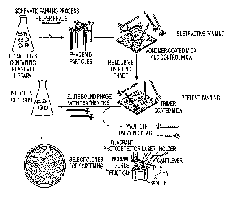
Figures 6A-6C. The novel biopanning process combines subtractive panning and
positive panning from phagemid scFv library and the single cloning screening
using
AFM. (A) Schematic panning process, in which the mica carrier can be replaced
by
immunotubes for bulk amount of non-desired antigen such as BSA for rapid
removal of
irrelevant phage particles, especially during subtractive panning. (B)
Subtractive panning
against BSA performed to eliminate non-specific phage. Left, middle and right
images are the
phage pool affinity check after BSA tube #1, #3 and #5 respectively. The
absence of phage
binding in the right-handed side image denotes the accomplishment of
subtractive panning
against BSA. The scale bar of 1 gm applies to all three images. (C) Positive
panning against
tau trimer was performed and imaged with AFM. A duplicate of positive panning
compared
with pure desired antigen proves that the antigen is free of phage and the
phage pool depleted
of non-desired antigen binders still contains phage that specifically binds to
desired antigen.
Left image is the purified trimeric tau 1N4R immobilized on mica; Right image
is the same
piece of mica on which the remaining phage pool of subtractive panning was
deposited and
non-binding particles were washed off. The scale bar of 1 lam applies to both
images.
Figure 7. Particle size analysis of oligomeric tau captured by single clone
scFv-
displayed phage from rhTau 2N4R mixed aggregates. Size of clone phage targets
compared with those of purified rhTau monomer, dimer and trimer. Individual
particle
capturing a phage was measured the size by section function in Nanoscope
Analysis. The
mean value of each clone phage target falls in between 2.5nm and 3.0nm, in
accordance with
rhTau 2N4R trimer size range. (Error bar:+/- standard deviation)
Figure 8. Three clones, F9T, D11C and H2A in scFv form recognize and retain
from 9-month 3xTG-AD mice hippocampus abnormal phosphorylated tau species that
are immunoreactive with AT8. Negative control is PBS as the target analyte and
set as 1.0
to be used as normalization standards. Signals lower than 2.0 are recorded as
negative while
signals above 2.0 are recorded as positive. (Error ban+/- standard deviation)
Figure 9. The comparison of secondary antibody affinity to 9-month 3xTG-AD
mice brain extracts captured by three types of primary antibody scFv. The mean
comparison was performed within each group of the same primary antibody.
(Error bar:+/-
standard deviation)
Figure 10. Oligomeric tau targeted scFv clones (F9T, D11C and H2A) affinity to
different 3xTG-AD mice brain extracts detected by F9T scFv-phage. Two mice for
each
age were tested in triplicates. The data were grouped by the mice ages. The
mean comparison
was performed within each group of the same mice age. (Error bar:+/- standard
deviation)
7
CA 02887933 2015-04-10
WO 2014/059442
PCT/US2013/065104
Figure 11A-11C. Densitometric analysis of dot blot reactivity of F9T scFv with
brain homogenates from age-matched human middle temporal gyrus (MTG).
Densitometric value of dots signal is based on a scale of 0 to 1, with 0
equals the background
signal and 1 equals the positive signal of anti-pLB scFv dots. Statistical
analysis is performed
in one-way ANOVA comparing means of two groups. (A) compares patients grouped
by
antemortem diagnosis as non-demented (ND) and Alzheimer's (AD). That AD group
means
is different from ND group mean (p<0.05) signifies F9T scFv can detect AD from
ND. (B)
compares patients grouped by postmortem examination results defined by Braak
stages and
neuritic plaque frequencies directly implying the AD progression. Braak stages
I-II (early
stage) were both diagnosed as non-demented but half of the cases bear slight
plaque compare
with the other half without plaques. Braak stages III-IV (AD middle stages)
display moderate
plaques while Braak stages V-VI (AD late stages) display severe plaques. F9T
scFv affinity
to these MTG extracts directly correlates with their AD progression defined by
Braak stages
and plaque frequency. (C) Sample dot blot affinity test of purified F9T scFv
on homogenized
MTG tissue from non-demented and Alzheimer's patients.
Figure 12. DNA sequences of the starting region and the first heavy chain
framework region (HCFR1) of selected scFvs from Sheets' library and standard
scFvs
from the generic library from which Sheets' was developed. F9, H7, D4, D1 1
and G12 are
five scFvs selected targeting rhTau 1N4R trimer, the rest are standard scFvs.
Except for one
missing base pair for each clone causing frame shift, all these scFvs from
Sheets' library
contain the similar FR regions as those from the generic library. All of these
missing base
pairs (highlighted in dark background) lie either at the beginning of HCFR1 or
the connection
of HCFR1 and the methionine start codon unaffecting the restriction site NcoI,
scFv
expression initiation or any complementarity-determining regions(CDRs).
Inserting the
missing base pair retaining the amino acid sequences of selected clones
sequences in generic
library enables these clones to express soluble scFv without interfering their
epitope-binding
sites, thus maintain their specificities.
Figure 13. Designed primers for clone sequence revision. Forward primers
contain
NcoI (5'-CCATGG-3' in italic) upstream of scFv sequence and the missing base
pair(underlined). The reverse primer includes Not! (5'-GCGGCCGC-3' in italic)
downstream
of scFv sequence. By performing a polymerase chain reaction using the paired
primers and
corresponding clone DNA template, revised clone scFv DNA fragments can be
produced up
to 23 copies for subcloning into E.coli and producing scFv and phage.
8
CA 02887933 2015-04-10
WO 2014/059442
PCT/US2013/065104
Figure 14: Tau protein structural features in linear diagram. A full-length
tau protein
with 441 amino acids (tau441 or tau 2N4R) is shown. Alternative splicing
showed in yellow
rectangles results in a total of six isoforms, denoted by either their total
number of amino
acids or the number of N'-terminal exons (Ns) and microtubule-associated
repeats (Rs).
Figure 15: Schematic of nonreactive monomer, reactive monomer, and reactive
oligomer. Reactivity implies the ability to form an intermolecular disulfide
linkage.
Intramolecular disulfide linkage causes formation of nonreactive tau monomer.
The free
thiols in a reactive monomer allow formation of an intermolecular or
intramolecular disulfide
linkage. Reactive oligomer has one or more free thiols readily forming
disulfide linkage with
reactive monomeric tau for the oligomer extension purpose.
Figures 16A-16B: Plots of height distribution of monomeric, dimeric, and
trimeric
fractions of rhTau 1N4R (a) and tau 2N4R (b).The height value of each particle
was
measured using Gwyddion. The numbers of particles falling in continuous size
ranges were
calculated and normalized into count percentages. The peak values give an
approximate value
for each tau species particle size. As expected, high-degree oligomers are
larger than low-
degree oligomers within the same isoform, and corresponding oligomeric
aggregates from the
longer isoform are larger than aggregates from the shorter isoform.
Figures 17A-17B: Neurotoxicity of extracellular 15.5nM monomeric, dimeric, and
trimeric forms of 1N4R and 2N4R tau variants toward (a) nondifferentiated
human
neuroblastoma cells (SH-SY5Y) and (b) Retinoic-acid-differentiated SH-SY5Y
cells was
measured after 48-hour incubation using an LDH assay. For both four-repeat tau
isoforms,
trimeric form is more neurotoxic than monomeric and dimeric forms (P < 0.001)
on either
neuron type. Full-length trimeric rhTau is more neurotoxic than 1N4R trimeric
rhTau. (P <
0.05).
Figures 18A-18D: Time and concentration dependence of neurotoxicity induced by
trimeric rhTau (1N4R and 2N4R) toward neuroblastoma cells measured by LDH
assay.
Nondifferentiated SH-SY5Y cells incubated with (a) 1N4R tau and (b) 2N4R tau;
retinoic-
acid-differentiated SHSY5Y cells incubated with (c) 1N4R tau and (d) 2N4R tau.
Figures 19A-19B: Comparison of rhTau induced neurotoxicity toward
nondifferentiated SH-SY5Y cells and retinoic-acid- (RA-) differentiated SHSY5Y
cells. The
data combine toxicity results of 15.5nM monomeric, dimeric, and trimeric forms
of both
1N4R and 2N4R tau variants. (a) After 3 hours-incubation, RA-differentiated SH-
SY5Y cells
are more vulnerable to extracellular trimeric rhTau toxicity than
nondifferentiated SHSY-5Y
cells are (P <0.05). (b) After 48-hours incubation, nondifferentiated SH-SY5Y
cells are more
9
CA 02887933 2015-04-10
WO 2014/059442
PCT/US2013/065104
vulnerable to extracellular trimeric rhTau toxicity than RA-differentiated SH-
SY5Y cells (P
<0.05).
DETAILED DESCRIPTION OF THE INVENTION
Tau is a protein involved in microtubule function in the brain. Aggregation of
tau can
lead to neuronal damage and dementia and traumatic brain injury. Increasing
evidence
suggests that small soluble oligomeric aggregate forms of tau may be the toxic
species rather
than the large fibrillar aggregates found during autopsies. Developing
reagents against these
species represents a potential therapeutic option. In the present invention,
using a bio-panning
protocol to identify single chain antibody fragments (scFv, also called
nanobodies) against
low (pico-molar) quantities of tau oligomers, the inventors identified binding
reagents with
therapeutic and diagnostic properties. Specifically, the inventors have
generated single chain
antibody fragments (scFvs or nanobodies) that selectively recognize oligomeric
forms of the
protein tau. These isolated scFvs that have potential value as diagnostics,
therapeutics and
imaging agents for neurodegeneration. As diagnostics, these antibody fragments
can be used
to detect the presence of oligomeric tau in serum, CSF or other fluid samples
as a
presymptomatic indication of neurodegeneration. Oligomeric tau may be an early
indicator
of Alzheimer's disease, frontotemporal dementia, other tauopathies and of
neurodegeneration
following traumatic brain injury. The antibody fragments can also be used as
therapeutics to
selectively target the toxic oligomeric tau aggregates protecting neurons from
damage.
Finally, the reagents can also be used as imaging agents to detect the
presence of tau
aggregates and neurodegeneration in vivo. The antibody fragments can be
readily labeled for
PET scans or other imaging techniques.
The biopanning studies were performed to isolate single chain variable
fragments
(nanobodies) against the different tau species. The biopanning protocol that
was used
combines the imaging capabilities of AFM with the binding diversity of phage-
displayed
antibody technology. To isolate nanobodies against specific oligomeric
morphologies of a
target protein, the protocol was modified to include negative panning steps to
remove clones
that bind to non-desired protein forms. To isolate nanobodies against
oligomeric tau two
negative panning steps were incorporated. In the first negative panning step,
all non-specific
"sticky" clones were removed by panning against a generic protein, bovine
serum albumin
(BSA). In the second negative panning step, all clones that bind to the non-
desired
monomeric form of tau were removed. A sample of pure monomeric tau was
obtained for the
CA 02887933 2015-04-10
WO 2014/059442
PCT/US2013/065104
negative panning to remove phage clones binding monomeric tau, and then
aliquots of the
remaining phage were used to screen for dimeric and trimeric specific clones
respectively.
Since it was found that the trimeric tau species was much more toxic to human
neuronal cell
lines than monomeric or dimeric, the inventors focused efforts on isolating
phage clones that
were selective for trimeric tau 4N1R. After negative panning against BSA and
monomeric
tau, ¨ 100 clones were obtained from the positive selection against trimeric
tau 4N1R. Each
phage clone was screened by AFM for binding to the different tau species. Each
phage
sample was coincubated with monomeric, dimeric and trimeric tau samples which
had been
previously fixed to a mica substrate. Unbound phage was removed by excess
stringent rinsing
and remaining bound phage were imaged by AFM. After screening all 100 clones
in this
manner, clones that selectively bound either dimeric or trimeric tau, but not
monomeric tau,
were identified. After screening all 100 phage clones, 6 clones were selected
for further
study based on highest specificity for trimeric tau.
The DNA sequence of each of the six clones was validated to ensure that a full
length
scFv was encoded. In each of the six cases a single base pair was missing at
the beginning of
the coding sequence. In order to produce soluble scFv for further
characterization, it was
necessary to correct the frame shift to enable efficient expression of the
scFv. DNA and
amino acid sequences of the clones are shown in Figures 3-5. Specifically, the
amino acid
sequences of the 6 selected cloned scFvs are: F9T (SEQ ID NO:1), F9T (SEQ ID
NO:9),
D11C (SEQ ID NO:11), D4G (SEQ ID NO:13), G12C (SEQ ID NO:15), H2A (SEQ ID
NO:17), or H7T (SEQ ID NO:19). DNA sequences are also included in Figures 3-5.
The corrected F9 clone, F9T, expressed at very high levels, purified readily
and
maintained high specificity for oligomer tau over monomeric tau and fibril tau
in the phage
form viewed by AFM, so this clone was selected for further study. The Dll
clone was also
identified as selectively binding to trimeric but not monomeric tau. Both
clones also
selectively recognize tau aggregates in post-mortem human brain tissue
containing tau
tangles but not in age matched normal tissue, although with slightly different
reactivity
profiles. Therefore both F9T and D11C nanobodies have promise as therapeutics
to block
neuronal toxicity induced by naturally occurring aggregates of tau following
TB!.
In a broad sense the scFv compositions of the present invention (e.g., the F9T
and
D11 C) may be described as compounds that are tau binding compounds. These
compounds
may therefore be used in diagnostic as well therapeutic applications and may
be either
administered to patients or used on patient tissue samples. In some
embodiments, the
compositions of the present invention may be used for in vivo imaging of tau,
and distinguish
11
CA 02887933 2015-04-10
WO 2014/059442
PCT/US2013/065104
between neurological tissue with toxic tau forms and normal neurological
tissue. As such the
nanobody compositions of the invention may be used to detect and quantitate
tau oligomers
in diseases including, for example, Alzheimer's Disease, frontotemporal
dementia, other
tauopathies and of neurodegeneration following traumatic brain injury. In
another
embodiment, the compounds may be used in the treatment or prophylaxis of
neurodegenerative disorders. Also provided herein are methods of allowing the
compound to
distribute into the brain tissue, and imaging the brain tissue, wherein an
increase in binding of
the compound to the brain tissue compared to a normal control level of binding
indicates that
the mammal is suffering from or is at risk of developing a neurodegenerative
disease, such as
Alzheimer's Disease, frontotemporal dementia, other tauopathies or
neurodegeneration
following traumatic brain injury.
The methods of the present invention are conducted to provide early stage
diagnosis
of Alzheimer's Disease, frontotemporal dementia, other tauopathies or
neurodegeneration
following traumatic brain injury. As explained herein the nanobodies of the
invention (e.g.,
F9T or D11C) are ones that specifically recognize tau oligomers (e.g.,
trimeric tau). Thus,
compositions comprising these antibodies and antibody fragments may be used to
identify the
presence of tau oligomers in a biological sample from a patient to be tested
for a tauopathy,
such as Alzheimer's disease, wherein the presence of tau oligomers in the
sample is
indicative that the patient has or is likely to develop the tauopathy (e.g.,
Alzheimer's disease).
In certain embodiments, the assay format that is used may be any assay format
that typically
employs antibody compositions. Thus, for example, the biological sample may be
examined
using immunohistology techniques, ELISA, Western Blotting, and the like.
For purposes of the diagnostic methods of the invention, the compositions of
the
invention (e.g., F9T or D11C) may be conjugated to a detecting reagent that
facilitates
detection of the scFv. For example, example, the detecting reagent may be a
direct label or
an indirect label. The labels can be directly attached to or incorporated into
the detection
reagent by chemical or recombinant methods.
In one embodiment, a label is coupled to the scFv through a chemical linker.
Linker
domains are typically polypeptide sequences, such as poly gly sequences of
between about 5
and 200 amino acids. In some embodiments, proline residues are incorporated
into the linker
to prevent the formation of significant secondary structural elements by the
linker. In certain
embodiments, linkers are flexible amino acid subsequences that are synthesized
as part of a
recombinant fusion protein comprising the RNA recognition domain. In one
embodiment, the
flexible linker is an amino acid subsequence that includes a proline, such as
Gly(x)-Pro-
12
CA 02887933 2015-04-10
WO 2014/059442
PCT/US2013/065104
Gly(x) where x is a number between about 3 and about 100. In other
embodiments, a
chemical linker is used to connect synthetically or recombinantly produced
recognition and
labeling domain subsequences. Such flexible linkers are known to persons of
skill in the art.
For example, poly(ethylene glycol) linkers are available from Shearwater
Polymers, Inc.
Huntsville, Ala. These linkers optionally have amide linkages, sulfhydryl
linkages, or
heterofunctional linkages.
The detectable labels can be used in the assays of the present invention to
diagnose a
neurodegenerative disease, such as Alzheimer's Disease, these labels are
attached to the
scFvs of the invention, can be primary labels (where the label comprises an
element that is
detected directly or that produces a directly detectable element) or secondary
labels (where
the detected label binds to a primary label, e.g., as is common in
immunological labeling). An
introduction to labels, labeling procedures and detection of labels is found
in Polak and Van
Noorden (1997) Introduction to Immunocytochemistry, 2nd ed., Springer Verlag,
N.Y. and in
Haugland (1996) Handbook of Fluorescent Probes and Research Chemicals, a
combined
handbook and catalogue Published by Molecular Probes, Inc., Eugene, Oreg.
Patents that
described the use of such labels include U.S. Pat. Nos. 3,817,837; 3,850,752;
3,939,350;
3,996,345; 4,277,437; 4,275,149; and 4,366,241.
Primary and secondary labels can include undetected elements as well as
detected
elements. Useful primary and secondary labels in the present invention can
include spectral
labels such as green fluorescent protein, fluorescent dyes (e.g., fluorescein
and derivatives
such as fluorescein isothiocyanate (FITC) and Oregon GreenTM, rhodamine and
derivatives
(e.g., Texas red, tetrarhodimine isothiocynate (TRITC), etc.), digoxigenin,
biotin,
phycoerythrin, AMCA, CyDyes.TM., and the like), radiolabels (e.g., 3H, 1251,
35s, 14C, 32p,
33P, etc.), enzymes (e.g., horse radish peroxidase, alkaline phosphatase
etc.), spectral
calorimetric labels such as colloidal gold or colored glass or plastic (e.g.
polystyrene,
polypropylene, latex, etc.) beads. The label can be coupled directly or
indirectly to a
component of the detection assay (e.g., the detection reagent) according to
methods well
known in the art. As indicated above, a wide variety of labels may be used,
with the choice of
label depending on sensitivity required, ease of conjugation with the
compound, stability
requirements, available instrumentation, and disposal provisions.
Exemplary labels that can be used include those that use: 1) chemiluminescence
(using horseradish peroxidase and/or alkaline phosphatase with substrates that
produce
photons as breakdown products as described above) with kits being available,
e.g., from
Molecular Probes, Amersham, Boehringer-Mannheim, and Life Technologies/Gibco
BRL; 2)
13
CA 02887933 2015-04-10
WO 2014/059442
PCT/US2013/065104
color production (using both horseradish peroxidase and/or alkaline
phosphatase with
substrates that produce a colored precipitate (kits available from Life
Technologies/Gibco
BRL, and Boehringer-Mannheim)); 3) fluorescence using, e.g., an enzyme such as
alkaline
phosphatase, together with the substrate AttoPhos (Amersham) or other
substrates that
produce fluorescent products, 4) fluorescence (e.g., using Cy-5 (Amersham),
fluorescein, and
other fluorescent tags); 5) radioactivity. Other methods for labeling and
detection will be
readily apparent to one skilled in the art.
Where the scFv-based compositions of the invention (e.g., F9T and D11C) are
contemplated to be used in a clinical setting, the labels are preferably non-
radioactive and
readily detected without the necessity of sophisticated instrumentation. In
certain
embodiments, detection of the labels will yield a visible signal that is
immediately
discernable upon visual inspection. One example of detectable secondary
labeling strategies
uses an antibody that recognizes tau oligomers in which the antibody is linked
to an enzyme
(typically by recombinant or covalent chemical bonding). The antibody is
detected when the
enzyme reacts with its substrate, producing a detectable product. In certain
embodiments,
enzymes that can be conjugated to detection reagents of the invention include,
e.g., (3-
galactosidase, luciferase, horse radish peroxidase, and alkaline phosphatase.
The
chemiluminescent substrate for luciferase is luciferin. One embodiment of a
fluorescent
substrate for P-galactosidase is 4-methylumbe11ifery1-O-D-galactoside.
Embodiments of
alkaline phosphatase substrates include p-nitrophenyl phosphate (pNPP), which
is detected
with a spectrophotometer; 5-bromo-4-chloro-3-indoly1 phosphate/nitro blue
tetrazolium
(BCIP/NBT) and fast red/napthol AS-TR phosphate, which are detected visually;
and 4-
methoxy-4-(3-phosphonophenyl) spiro[1,2-dioxetane-3,2'-adamantane], which is
detected
with a luminometer. Embodiments of horse radish peroxidase substrates include
2,2'azino-
bis(3-ethylbenzthiazoline-6 sulfonic acid) (ABTS), 5-aminosalicylic acid
(5AS), o-
dianisidine, and o-phenylenediamine (OPD), which are detected with a
spectrophotometer,
and 3,3,5,5'-tetramethylbenzidine (TMB), 3,3' diaminobenzidine (DAB), 3-amino-
9-
ethylcarbazole (AEC), and 4-chloro-1 -naphthol (4C1N), which are detected
visually. Other
suitable substrates are known to those skilled in the art. The enzyme-
substrate reaction and
product detection are performed according to standard procedures known to
those skilled in
the art and kits for performing enzyme immunoassays are available as described
above.
The presence of a label can be detected by inspection, or a detector which
monitors a
particular probe or probe combination is used to detect the detection reagent
label. Typical
detectors include spectrophotometers, phototubes and photodiodes, microscopes,
scintillation
14
CA 02887933 2015-04-10
WO 2014/059442
PCT/US2013/065104
counters, cameras, film and the like, as well as combinations thereof.
Examples of suitable
detectors are widely available from a variety of commercial sources known to
persons of
skill. Commonly, an optical image of a substrate comprising bound labeling
moieties is
digitized for subsequent computer analysis.
As noted herein throughout the scFvs of the invention (e.g., F9T and D11C) are
targeted specifically to tau oligomers that are characteristic of Alzheimer's
Disease,
frontotemporal dementia, other tauopathies or neurodegeneration following
traumatic brain
injury. As such, the scFvs of the invention also may be used to specifically
target therapeutic
compositions to the sites of tau aggregation. In this embodiment, any
therapeutic agent
typically used for the treatment of these tauopathies, such as Alzheimer's
disease, may be
conjugated to scFvs in order to achieve a targeted delivery of that
therapeutic agent. Various
drugs for the treatment of AD are currently available as well as under study
and regulatory
consideration. The drugs generally fit into the broad categories of
cholinesterase inhibitors,
muscarinic agonists, anti-oxidants or anti-inflammatories. Galantamine
(Reminyl), tacrine
(Cognex), selegiline, physostigmine, revistigmin, donepezil, (Aricept),
rivastigmine (Exelon),
metrifonate, milameline, xanomeline, saeluzole, acetyl-L-carnitine, idebenone,
ENA-713,
memric, quetiapine, neurestrol and neuromidal are just some of the drugs
proposed as
therapeutic agents for AD that can be conjugated to the scFv compositions of
the invention
and targeted for therapeutic intervention of AD.
The scFv compositions of the invention can be used in any diagnostic assay
format to
determine the presence of tau oligomers. A variety of immunodetection methods
are
contemplated for this embodiment. Such immunodetection methods include enzyme
linked
immunosorbent assay (ELISA), radioimmunoassay (RIA), immunoradiometric assay,
fluoroimmunoassay, chemiluminescent assay, bioluminescent assay, and Western
blot,
though several others are well known to those of ordinary skill. The steps of
various useful
immunodetection methods have been described in the scientific literature.
In general, the immunobinding methods include obtaining a sample suspected of
containing a protein, polypeptide and/or peptide (in this case the tau
oligomers), and
contacting the sample with a first antibody, monoclonal or polyclonal (in this
case a scFv of
the invention, such as F9T or D11C), in accordance with the present invention,
as the case
may be, under conditions effective to allow the formation of immunocomplexes.
The immunobinding methods include methods for detecting and quantifying the
amount of the tau oligomer component in a sample and the detection and
quantification of
any immune complexes formed during the binding process. Here, one would obtain
a sample
CA 02887933 2015-04-10
WO 2014/059442
PCT/US2013/065104
suspected of containing tau oligomers, and contact the sample with an antibody
fragment of
the invention, such as F9T or D11 C, and then detect and quantify the amount
of immune
complexes formed under the specific conditions.
Contacting the chosen biological sample with the antibody under effective
conditions
and for a period of time sufficient to allow the formation of immune complexes
(primary
immune complexes) is generally a matter of simply adding the antibody
composition to the
sample and incubating the mixture for a period of time long enough for the
antibodies to form
immune complexes with, i.e., to bind to, any antigens present. After this
time, the sample-
antibody composition, such as a tissue section, ELISA plate, dot blot or
western blot, will
generally be washed to remove any non-specifically bound antibody species,
allowing only
those scFv molecules specifically bound within the primary immune complexes to
be
detected.
In general, the detection of immunocomplex formation is well known in the art
and
may be achieved through the application of numerous approaches. These methods
are
generally based upon the detection of a label or marker, such as any of those
radioactive,
fluorescent, biological and enzymatic tags. U.S. patents concerning the use of
such labels
include U.S. Pat. Nos. 3,817,837; 3,850,752; 3,939,350; 3,996,345; 4,277,437;
4,275,149 and
4,366,241, each incorporated herein by reference. Of course, one may find
additional
advantages through the use of a secondary binding ligand such as a second
antibody and/or a
biotin/avidin ligand binding arrangement, as is known in the art.
As noted above, an scFv of the invention may itself be linked to a detectable
label,
wherein one would then simply detect this label, thereby allowing the amount
of the primary
immune complexes in the composition to be determined. Alternatively, the first
antibody that
becomes bound within the primary immune complexes may be detected by means of
a second
binding ligand that has binding affinity for the antibody. In these cases, the
second binding
ligand may be linked to a detectable label. The second binding ligand is
itself often an
antibody, which may thus be termed a "secondary" antibody. The primary immune
complexes are contacted with the labeled, secondary binding ligand, or
antibody, under
effective conditions and for a period of time sufficient to allow the
formation of secondary
immune complexes. The secondary immune complexes are then generally washed to
remove
any non-specifically bound labeled secondary antibodies or ligands, and the
remaining label
in the secondary immune complexes is then detected.
Further methods include the detection of primary immune complexes by a two
step
approach. A second binding ligand, such as an antibody, that has binding
affinity for the
16
CA 02887933 2015-04-10
WO 2014/059442
PCT/US2013/065104
scFV (e.g., F9T or D11C) is used to form secondary immune complexes, as
described above.
After washing, the secondary immune complexes are contacted with a third
binding ligand or
antibody that has binding affinity for the second antibody, again under
effective conditions
and for a period of time sufficient to allow the formation of immune complexes
(tertiary
immune complexes). The third ligand or antibody is linked to a detectable
label, allowing
detection of the tertiary immune complexes thus formed. This system may
provide for signal
amplification if this is desired.
One method of immunodetection designed by Charles Cantor uses two different
antibodies. A first step biotinylated, monoclonal or polyclonal antibody (in
the present
example a scFv of the invention, such as F9T or D11 C) is used to detect the
target antigen(s),
and a second step antibody is then used to detect the biotin attached to the
complexed
nanobody. In this method the sample to be tested is first incubated in a
solution containing
the first step nanobody. If the target antigen is present, some of the
nanobody binds to the
antigen to form a biotinylated nanobody/antigen complex. The nanobody/antigen
complex is
then amplified by incubation in successive solutions of streptavidin (or
avidin), biotinylated
DNA, and/or complementary biotinylated DNA, with each step adding additional
biotin sites
to the nanobody/antigen complex. The amplification steps are repeated until a
suitable level
of amplification is achieved, at which point the sample is incubated in a
solution containing
the second step antibody against biotin. This second step antibody is labeled,
as for example
with an enzyme that can be used to detect the presence of the antibody/antigen
complex by
histoenzymology using a chromogen substrate. With suitable amplification, a
conjugate can
be produced which is macroscopically visible.
Another known method of immunodetection takes advantage of the immuno-PCR
(Polymerase Chain Reaction) methodology. The PCR method is similar to the
Cantor method
up to the incubation with biotinylated DNA, however, instead of using multiple
rounds of
streptavidin and biotinylated DNA incubation, the
DNA/biotin/streptavidin/antibody complex
is washed out with a low pH or high salt buffer that releases the antibody.
The resulting wash
solution is then used to carry out a PCR reaction with suitable primers with
appropriate
controls. At least in theory, the enormous amplification capability and
specificity of PCR can
be utilized to detect a single antigen molecule.
As detailed above, immunoassays, in their most simple and/or direct sense, are
binding assays. Certain preferred immunoassays are the various types of enzyme
linked
immunosorbent assays (ELISAs) and/or radioimmunoassays (RIA) known in the art.
Immunohistochemical detection using tissue sections is also particularly
useful. However, it
17
CA 02887933 2015-04-10
WO 2014/059442
PCT/US2013/065104
will be readily appreciated that detection is not limited to such techniques,
and/or western
blotting, dot blotting, FACS analyses, and/or the like may also be used.
The diagnostic assay format that may be used in the present invention could
take any
conventional format such as ELISA or other platforms such as luminex or
biosensors. The
present invention shows the sequence of the F9T (SEQ ID NO:1), F9T (SEQ ID
NO:9),
D11C (SEQ ID NO:11), D4G (SEQ ID NO:13), G12C (SEQ ID NO:15), H2A (SEQ ID
NO:17), or H7T (SEQ ID NO:19) scFvs. These sequences can readily be modified
to
facilitate diagnostic assays, for example a tag (such as GFP) can be added to
these scFvs to
increase sensitivity. In one exemplary ELISA, antibodies (in the present case
the scFvs of the
invention, such as F9T or D11 C) are immobilized onto a selected surface
exhibiting protein
affinity, such as a well in a polystyrene microtiter plate. Then, a test
composition suspected of
containing tau oligomers, such as a clinical sample (e.g., a biological sample
obtained from
the subject), is added to the wells. After binding and/or washing to remove
non-specifically
bound immune complexes, the bound antigen may be detected. Detection is
generally
achieved by the addition of another antibody that is linked to a detectable
label. This type of
ELISA is a simple "sandwich ELISA." Detection may also be achieved by the
addition of a
second antibody, followed by the addition of a third antibody that has binding
affinity for the
second antibody, with the third antibody being linked to a detectable label.
In another exemplary ELISA, the samples suspected of containing the antigen
are
immobilized onto the well surface and/or then contacted with binding agents
(e.g., scFvs of
the invention, such as F9T or D11C). After binding and/or washing to remove
non-
specifically bound immune complexes, the bound anti-binding agents are
detected. Where the
initial binding agents are linked to a detectable label, the immune complexes
may be detected
directly. Again, the immune complexes may be detected using a second antibody
that has
binding affinity for the first binding agents, with the second antibody being
linked to a
detectable label.
Another ELISA in which the antigens are immobilized, involves the use of
antibody
competition in the detection. In this ELISA, labeled antibodies (or
nanobodies) against an
antigen are added to the wells, allowed to bind, and/or detected by means of
their label. The
amount of an antigen in an unknown sample is then determined by mixing the
sample with
the labeled antibodies against the antigen during incubation with coated
wells. The presence
of an antigen in the sample acts to reduce the amount of antibody against the
antigen
available for binding to the well and thus reduces the ultimate signal. This
is also appropriate
for detecting antibodies against an antigen in an unknown sample, where the
unlabeled
18
CA 02887933 2015-04-10
WO 2014/059442
PCT/US2013/065104
antibodies bind to the antigen-coated wells and also reduces the amount of
antigen available
to bind the labeled antibodies.
Irrespective of the format employed, ELISAs have certain features in common,
such
as coating, incubating and binding, washing to remove non-specifically bound
species, and
detecting the bound immune complexes.
In coating a plate with either tau oligomers or an scFv of the invention
(e.g., F9T or
D11 C), one will generally incubate the wells of the plate with a solution of
the antigen or
scFvs, either overnight or for a specified period of hours. The wells of the
plate will then be
washed to remove incompletely adsorbed material. Any remaining available
surfaces of the
wells are then "coated" with a nonspecific protein that is antigenically
neutral with regard to
the test antisera. These include bovine serum albumin (BSA), casein or
solutions of milk
powder. The coating allows for blocking of nonspecific adsorption sites on the
immobilizing
surface and thus reduces the background caused by nonspecific binding of
antisera onto the
surface.
In ELISAs, it is probably more customary to use a secondary or tertiary
detection
means rather than a direct procedure. Thus, after binding of a protein or
antibody to the well,
coating with a non-reactive material to reduce background, and washing to
remove unbound
material, the immobilizing surface is contacted with the biological sample to
be tested under
conditions effective to allow immune complex (antigen/antibody) formation.
Detection of the
immune complex then requires a labeled secondary binding ligand or antibody,
and a
secondary binding ligand or antibody in conjunction with a labeled tertiary
antibody or a third
binding ligand.
"Under conditions effective to allow immune complex (antigen/antibody)
formation"
means that the conditions preferably include diluting the tau oligomers and/or
scFv
composition with solutions such as BSA, bovine gamma globulin (BGG) or
phosphate
buffered saline (PBS)/Tween. These added agents also tend to assist in the
reduction of
nonspecific background.
The "suitable" conditions also mean that the incubation is at a temperature or
for a
period of time sufficient to allow effective binding. Incubation steps are
typically from about
1 to 2 to 4 hours or so, at temperatures preferably on the order of 25 C to
27 C., or may be
overnight at about 4 C or so.
Following all incubation steps in an ELISA, the contacted surface is washed so
as to
remove non-complexed material. An example of a washing procedure includes
washing with
a solution such as PBS/Tween, or borate buffer. Following the formation of
specific immune
19
CA 02887933 2015-04-10
WO 2014/059442
PCT/US2013/065104
complexes between the test sample and the originally bound material, and
subsequent
washing, the occurrence of even minute amounts of immune complexes may be
determined.
To provide a detecting means, the second or third antibody will have an
associated
label to allow detection. This may be an enzyme that will generate color
development upon
incubating with an appropriate chromogenic substrate. Thus, for example, one
will desire to
contact or incubate the first and second immune complex with a urease, glucose
oxidase,
alkaline phosphatase or hydrogen peroxidase-conjugated antibody for a period
of time and
under conditions that favor the development of further immune complex
formation (e.g.,
incubation for 2 hours at room temperature in a PBS-containing solution such
as PBS-
Tween).
After incubation with the labeled antibody, and subsequent to washing to
remove
unbound material, the amount of label is quantified, e.g., by incubation with
a chromogenic
substrate such as urea, or bromocresol purple, or 2,2'-azino-di-(3-ethyl-
benzthiazoline-6-
sulfonic acid (ABTS), or 11202, in the case of peroxidase as the enzyme label.
Quantification
is then achieved by measuring the degree of color generated, e.g., using a
visible spectra
spectrophotometer.
In various aspects of the invention, it will be desirable to further subject
patients to
more traditional diagnostic approaches for tauopathies, such as AD. Such
general approaches
for diagnosis are set out below.
The diagnosis of both early (mild) cognitive impairment and AD are based
primarily
on clinical judgment. However, a variety of neuropsychological tests aid the
clinician in
reaching a diagnosis. Early detection of only memory deficits may be helpful
in suggesting
early signs of AD, since other dementias may present with memory deficits and
other signs.
Cognitive performance tests that assess early global cognitive dysfunction are
useful, as well
as measures of working memory, episodic memory, semantic memory, perceptual
speed and
visuospatial ability. These tests can be administered clinically, alone or in
combination.
Examples of cognitive tests according to cognitive domain are shown as
examples, and
include "Digits Backward" and "Symbol Digit" (Attention), "Word List Recall"
and "Word
List Recognition" (Memory), "Boston Naming" and "Category Fluency" (Language),
"MMSE 1-10" (Orientation), and "Line Orientation" (Visuospatial). Thus,
neuropsychological tests and education-adjusted ratings are assessed in
combination with
data on effort, education, occupation, and motor and sensory deficits. Since
there are no
consensus criteria to clinically diagnose mild cognitive impairment, various
combinations of
the above plus the clinical examination by an experienced neuropsychologist or
neurologist
CA 02887933 2015-04-10
WO 2014/059442
PCT/US2013/065104
are key to proper diagnosis. As the disease becomes more manifest (L e.,
becomes a dementia
rather than mild cognitive impairment), the clinician may use the criteria for
dementia and
AD set out by the joint working group of the National Institute of Neurologic
and
Communicative Disorders and Stroke/AD and Related Disorders Association
(NINCDS/ADRDA). On occasion, a clinician may request a head computed
tomography
(CT) or a head magnetic resonance imaging (MRI) to assess degree of lobar
atrophy,
although this is not a requirement for the clinical diagnosis.
As noted above, there are various drugs that are presently in use or under
development for the treatment of Alzheimer's Disease, frontotemporal dementia,
other
tauopathies or neurodegeneration following traumatic brain injury. The present
invention
contemplates the use of scFvs of the invention, such as F9T or D11 C, based
"diagnostic"
methods to further assess the efficacy of treatments. Given the role of tau in
these diseases,
the ability of a particular therapy to reduce the amount of oligomeric tau
will be indicative of
an effective treatment, as these forms have been shown to be toxic.
The present invention may involve the use of pharmaceutical compositions which
comprise an agent conjugated to a scFv of the invention, such as F9T or D11C,
for delivery
into a subject having Alzheimer's disease, frontotemporal dementia, other
tauopathies or
neurodegeneration following traumatic brain injury. Such an agent will ideally
be formulated
into a pharmaceutically acceptable carrier. As used herein, "pharmaceutically
acceptable
carrier" includes any and all solvents, dispersion media, coatings,
surfactants, antioxidants,
preservatives (e.g., antibacterial agents, antifungal agents), isotonic
agents, absorption
delaying agents, salts, preservatives, drugs, drug stabilizers, gels, binders,
excipients,
disintegration agents, lubricants, sweetening agents, flavoring agents, dyes,
such like
materials and combinations thereof, as would be known to one of ordinary skill
in the art.
Except insofar as any conventional carrier is incompatible with the active
ingredient, its use
in the therapeutic or pharmaceutical compositions is contemplated.
A "variant" of an amino acid sequence of an antibody or antibody fragment
described
herein or a nucleic acid sequence encoding such an amino acid sequence, is a
sequence that
is substantially similar to SEQ ID NO:1, SEQ ID NO:2, SEQ ID NO:3, SEQ ID
NO:4, SEQ
ID NO:5, SEQ ID NO:6, SEQ ID NO:7, SEQ ID NO:8, SEQ ID NO:9, SEQ ID NO:10,
SEQ ID NO:11, SEQ ID NO:12, SEQ ID NO:13, SEQ ID NO:14, SEQ ID NO:15, SEQ
ID NO:16, SEQ ID NO:17, SEQ ID NO:18, SEQ ID NO:19, SEQ ID NO:20 or SEQ ID
NO:21. Variant amino acid and nucleic acid sequences include synthetically
derived amino
acid and nucleic acid sequences, or recombinantly derived amino acid or
nucleic acid
21
CA 02887933 2015-04-10
WO 2014/059442
PCT/US2013/065104
sequences. Generally, amino acid or nucleic acid sequence variants of the
invention will have
at least 40, 50, 60, to 70%, e.g., 71%, 72%, 73%, 74%, 75%, 76%, 77%, 78%, to
79%,
generally at least 80%, e.g., 81%-84%, at least 85%, e.g., 86%, 87%, 88%, 89%,
90%, 91%,
92%, 93%, 94%, 95%, 96%, 97%, to 98%, sequence identity to SEQ ID NO:1, SEQ ID
NO:2, SEQ ID NO:3, SEQ ID NO:4, SEQ ID NO:5, SEQ ID NO:6, SEQ ID NO:7, SEQ
ID NO:8, SEQ ID NO:9, SEQ ID NO:10, SEQ ID NO:11, SEQ ID NO:12, SEQ ID
NO:13, SEQ ID NO:14, SEQ ID NO:15, SEQ ID NO:16, SEQ ID NO:17, SEQ ID NO:18,
SEQ ID NO:19, SEQ ID NO:20 or SEQ ID NO:21.
The present invention includes variants of the amino acid sequences of the
antibodies
and antibody fragments described herein, as well as variants of the nucleic
acid sequences
encoding such amino acid sequences (L e., SEQ ID NO:1, SEQ ID NO:2, SEQ ID
NO:3,
SEQ ID NO:4, SEQ ID NO:5, SEQ ID NO:6, SEQ ID NO:7, SEQ ID NO:8, SEQ ID
NO:9, SEQ ID NO:10, SEQ ID NO:11, SEQ ID NO:12, SEQ ID NO:13, SEQ ID NO:14,
SEQ ID NO:15, SEQ ID NO:16, SEQ ID NO:17, SEQ ID NO:18, SEQ ID NO:19, SEQ
ID NO:20 or SEQ ID NO:21). "Variants" are intended to include sequences
derived by
deletion (so-called truncation) or addition of one or more amino acids to the
N-terminal
and/or C-terminal end, and/or addition of one or more bases to the 5' or 3'
end of the nucleic
acid sequence; deletion or addition of one or more amino acids/nucleic acids
at one or more
sites in the sequence; or substitution of one or more amino acids/nucleic
acids at one or more
sites in the sequence. The antibodies and antibody fragments described herein
may be altered
in various ways including amino acid substitutions, deletions, truncations,
and insertions.
Methods for such manipulations are generally known in the art. For example,
amino acid
sequence variants of the enzyme can be prepared by mutations in the DNA.
Methods for
mutagenesis and nucleotide sequence alterations are well known in the art. The
substitution
may be a conserved substitution. A "conserved substitution" is a substitution
of an amino
acid with another amino acid having a similar side chain. A conserved
substitution would be
a substitution with an amino acid that makes the smallest change possible in
the charge of the
amino acid or size of the side chain of the amino acid (alternatively, in the
size, charge or
kind of chemical group within the side chain) such that the overall enzyme
retains its spatial
conformation but has altered biological activity. For example, common
conserved changes
might be Asp to Glu, Asn or Gin; His to Lys, Arg or Phe; Asn to Gln, Asp or
Glu and Ser to
Cys, Thr or Gly. Alanine is commonly used to substitute for other amino acids.
The 20
essential amino acids can be grouped as follows: alanine, valine, leucine,
isoleucine, proline,
phenylalanine, tryptophan and methionine having nonpolar side chains; glycine,
serine,
22
CA 02887933 2015-04-10
WO 2014/059442
PCT/US2013/065104
threonine, cystine, tyrosine, asparagine and glutamine having uncharged polar
side chains;
aspartate and glutamate having acidic side chains; and lysine, arginine, and
histidine having
basic side chains.
As used herein, "sequence identity" or "identity" in the context of two
nucleic acid or
polypeptide sequences makes reference to a specified percentage of residues in
the two
sequences that are the same when aligned for maximum correspondence over a
specified
comparison window, as measured by sequence comparison algorithms or by visual
inspection. When percentage of sequence identity is used in reference to
proteins it is
recognized that residue positions which are not identical often differ by
conservative amino
acid substitutions, where amino acid residues are substituted for other amino
acid residues
with similar chemical properties (e.g., charge or hydrophobicity) and
therefore do not change
the functional properties of the molecule. When sequences differ in
conservative
substitutions, the percent sequence identity may be adjusted upwards to
correct for the
conservative nature of the substitution. Sequences that differ by such
conservative
substitutions are said to have "sequence similarity" or "similarity." Means
for making this
adjustment are well known to those of skill in the art. Typically this
involves scoring a
conservative substitution as a partial rather than a full mismatch, thereby
increasing the
percentage sequence identity. Thus, for example, where an identical amino acid
is given a
score of 1 and a non-conservative substitution is given a score of zero, a
conservative
substitution is given a score between zero and 1. The scoring of conservative
substitutions is
calculated, e.g., as implemented in the program PC/GENE (Intelligenetics,
Mountain View,
California).
EXAMPLES
Example 1.
A vast number of studies have correlated protein aggregation with
neurodegenerative
diseases including AD, Parkinson's and Dementia with Lewy Bodies. Numerous
recent
studies suggest that specific oligomeric forms of these proteins are involved
in neuronal
toxicity and can interfere with important functions including long term
potentiation. Various
soluble oligomeric species of A13 and a-syn have been shown to occur early
during the course
of AD and PD, and increasing evidence implicates oligomeric forms of tau in AD
and other
tauopathies.
Assays are being developed to study tau oligomer content in CSF, and initial
results
suggest increased levels of tau oligomers in AD CSF compared to non-AD
specimens. We
23
CA 02887933 2015-04-10
WO 2014/059442
PCT/US2013/065104
developed novel methods to purify recombinant human tau isoforms and to
stabilize their
oligomeric structures formed by disulfide linkages. Additionally, we purified
tau from human
AD brain that retains its hyperphosphorylation. These preparations have been
used in mice to
show that extracellular tau oligomers, but not monomer, inhibited long term
potentiation of
hippocampal synapses and the formation of associative fear memory. The
oligomeric
preparation of AD tau produced a similar effect indicating that
hyperphosphorylation of tau
did not affect inhibition of memory. Taken together tau oligomers were the
forms of tau
necessary to produce the disease-related effects and validate these structures
as a target for
drug discovery (Moe, J., et al. Validation of extracellular tau oligomer
target for drug
discovery in a novel animal model. in Society for Neuroscience. 2010. San
Diego, CA).
We also developed a novel biopanning technology that combines the imaging
capability
of Atomic Force Microscopy (AFM) with the diversity of antibody libraries.
This unique
combination of antibody diversity and imaging capability has enabled us to
isolate single
chain antibody variable domain fragment (scFv or nanobody) reagents to an
array of
morphologies of key proteins involved in neurodegenerative diseases including
A13 and
alpha-synuclein (a-syn). We isolated nanobodies that specifically recognize
monomeric
(Emadi, S., et al., Inhibiting Aggregation of alpha-Synuclein with Human
Single Chain
Antibody Fragments. Biochemistry, 2004. 43: p. 2871-2878), fibrillar
(Barkhordarian, H., et
al., Isolating recombinant antibodies against specific protein morphologies
using atomic force
microscopy and phage display technologies. Protein Eng Des Sel, 2006. 19: p.
497-502), and
two different oligomeric a-syn morphologies (Emadi, S., et al., Isolation of a
human single
chain antibody fragment against oligomeric alpha-synuclein that inhibits
aggregation and
prevents alpha-synuclein-induced toxicity. J Mol Biol, 2007. 368: p. 1132-44;
Emadi, S., et
al., Detecting morphologically distinct oligomeric forms of alpha-synuclein. J
Biol Chem,
2009. 284: p. 11048-58). The anti-oligomeric a-syn nanobodies do not cross
react with
oligomeric Af3, and specifically label PD brain tissue but not AD or healthy
tissue (Emadi, S.,
et al., Detecting morphologically distinct oligomeric forms of alpha-
synuclein. J Biol Chem,
2009. 284: p. 11048-58). In addition, we isolated nanobodies to different
regions of full
length AP (Liu, R., et al., Single chain variable fragments against beta-
amyloid (Abeta) can
inhibit Abeta aggregation and prevent abeta-induced neurotoxicity.
Biochemistry, 2004. 43:
p. 6959-67) and to three distinct naturally occurring oligomeric AP
morphologies (Zameer,
A., et al., Anti-oligomeric Abeta single-chain variable domain antibody blocks
Abeta-induced
toxicity against human neuroblastoma cells. J Mol Biol, 2008. 384: p. 917-28).
One, A4,
specifically recognizes a larger oligomeric AP species, inhibits aggregation
and extracellular
24
CA 02887933 2015-04-10
WO 2014/059442
PCT/US2013/065104
toxicity of A13, does not cross react with oligomeric a-syn, and specifically
labels A13
aggregates in human AD brain samples, but not PD or healthy brain tissue
(Zameer, A., et
al., Anti-oligomeric Abeta single-chain variable domain antibody blocks Abeta-
induced
toxicity against human neuroblastoma cells. J Mol Biol, 2008. 384: p. 917-28).
A second
nanobody, El, recognizes a smaller trimeric or tetrameric A13 species, and
similar to A4
inhibits aggregation and extracellular toxicity of A13, does not cross react
with oligomeric a-
syn, and labels Afil aggregates in human AD but not healthy brain tissue.
Utilizing an AD
brain derived oligomeric Ar3 preparation obtained from Dr. Selkoe (Walsh,
D.M., et al.,
Naturally secreted oligomers of amyloid beta protein potently inhibit
hippocampal long-term
potentiation in vivo. Nature, 2002. 416: p. 535-9; Walsh, D.M. and D.J.
Selkoe, Abeta
Oligomers - a decade of discovery. J Neurochem, 2007), we isolated a third
nanobody, C6,
that specifically recognizes oligomeric Al3 species derived from human AD
brain tissue, but
does not recognize A13 aggregates generated in vitro. The different
specificities of each
nanobody can be readily observed when each nanobody is expressed on the
surface of a
filamentous bacteriophage and antibody/antigen complexes are imaged by AFM
(Kasturirangan, S., et al., Nanobody specific for oligomeric beta-amyloid
stabilizes non-toxic
form. Neurobiol Aging, 2010.). Therefore, the combination of antibody
libraries and AFM
imaging technologies enables us to isolate and carefully characterize reagents
that recognize
specific protein variants including four different naturally occurring
aggregated forms of a-
syn and four different naturally occurring aggregated forms of A13.
Another powerful advantage of our AFM panning protocol is that not only can we
isolate and characterize reagents to specific protein morphologies, but we can
do so using
only picograms or less of material. In addition the sample does not need to be
purified, and
the protein does not need to be chemically modified in any way. We can
actually isolate
nanobodies against a single molecule of the target antigen (Shlyalchtenko,
L.S., et al., Single-
molecule selection and recovery of structure-specific antibodies using atomic
force
microscopy. Nanomedicine, 2007. 3: p. 192-7). This unique combination of
capabilities to
isolate different tau isoforms and to generate and characterize reagents that
specifically
recognize individual protein variants provides us with the means to generate
reagents that
specifically recognize an array of different tau variants present in human AD
brain.
While several reagents already exist that can recognize monomeric and
phosphorylated tau, these reagents cannot distinguish between different
aggregated states of
tau. Reagents that can detect specific forms of tau can provide very powerful
tools to
facilitate diagnosis of AD and other tauopathies and to follow progression of
these diseases or
CA 02887933 2015-04-10
WO 2014/059442
PCT/US2013/065104
to evaluate therapeutic strategies. While many neurodegenerative diseases have
overlapping
clinical symptoms and cellular and biochemical mechanisms such as an increase
in
inflammatory markers, and aggregation of similar proteins, the reagents we
propose to
develop here will have well defined specificities and selectivities for
selected tau forms and
should facilitate specific diagnoses of AD and other tauopathies. In
combination with other
protein and morphology specific reagents against A13 and a-syn species, these
reagents can be
used to detect the presence of biomarkers which can readily detect and
distinguish many
related neurodegenerative diseases including AD, PD, FTD and LBD.
Isolation of different size oligomeric tau species from human AD brain tissue.
An array of different tau isoforms and aggregated assemblies has been shown to
be
critically important in AD and other tauopathies. Reagents that can
specifically label
different tau species are needed to clarify the roles of the different forms.
Soluble tau
oligomers are purified using immunoaffinity and size fractionation to isolate
oligomers with
different numbers of subunits and isoform composition and multiple post-
translational
modifications associated with AD such as hyperphosphorylation, truncation,
nitrosylation,
ubiquitination and glycation. The oligomers in certain embodiments are
heterogeneous
containing known tau associating proteins such as beta amyloid, ApoE and alpha-
synuclein.
Aberrant binding of tau with other proteins associated with AD is also
anticipated to facilitate
generating disease-specific morphologies for nanobody selection. Further,
nanobodies are
generated against the different tau variants and the nanobodies are used to
identify which
forms of tau best distinguish AD from healthy tissue in brain and CSF samples.
Methods. Preparation of brain derived oligomeric tau. Tau oligomers are
purified
from normal and AD brain specimens with tau pathology acquired from the New
York Brain
Bank (The Taub Institute, Columbia University). Ten grams of tissue will be
used for each
preparation using the method developed by Ivanovova et al. (Ivanovova, N., et
al., High-yield
purification of fetal tau preserving its structure and phosphorylation
pattern. J Immunol
Methods, 2008. 339: p. 17-22) with modifications to isolate different size
species of tau
oligomers. The advantages of this method include the preservation of tau
phosphorylation,
simplicity, and high purity of product. We have already used a modified
version of this
method to isolate tau from an AD brain specimen and have shown that
phosphorylation state
is preserved. Immunoblot analysis of recombinant tau441 and Tau purified from
AD brain
showed specific interaction of tau phospho-epitope-specific antibodies against
tyrosine 231
and 217 with AD tau but no reactivity with recombinant tau, whereas phospho-
independent
monoclonal antibody HT7 against total tau interacts with both recombinant and
AD tau.
26
CA 02887933 2015-04-10
WO 2014/059442
PCT/US2013/065104
Brain tissue is homogenized in cold 1% perchloric acid, incubated on ice 20
min., and
centrifuged at 15,000x g for 20 min. The cleared supernatant is concentrated
and buffer
exchanged into buffer (20 mM Tris-HC1 pH 7.4, 150 mM NaC1, 0.1% Tween 20)
using
Amicon centrifugal devices (Millipore). Gel and Immunoblot analysis of cleared
supernatant
indicated the presence of multiple size aggregates of tau with multiple types
of modifications
causing the appearance of a smear with some predominant bands consistent in
size with
dimer and trimer. These species were purified by non-denaturing methods
described below.
Treatment of the preparation with reductant results in a lowered amount of
higher molecular
weight species indicating that at least some of the tau oligomers were
stabilized by disulfide
bonds. The incubation time can be varied to increase or decrease the content
of higher order
oligomeric tau.
Purification and characterization of oligomeric tau. Affinity chromatography
to
purify oligomeric tau from other protein species is performed using the
monoclonal antibody
HT-7 (Thermo Scientific). We have observed that the tau epitope recognized by
HT-7 is
available for binding in tau oligomers and because it binds tau independently
of
phosphorylation status. The antibody (6 mg) will be coupled to CNBr activated
Sepharose
according manufacturer's protocol and packed into a Poly-Prep column C10/10
(GE
Healthcare). The brain extract is filtered at a flow rate of 0.2 ml/min, and
unbound protein are
washed off with buffer. Tau is eluted with 0.1 M glycine pH 2.6 in 0.5 ml
fractions which is
neutralized with 50 .11 M Tris-HC1 pH 9 prior to analysis by non-reducing SDS-
PAGE.
Tau oligomers will be characterized and fractionated using high-pressure
liquid
chromatography (Beckman Coulter System Gold 32Karat HPLC). A high-resolution
gel
filtration column (Biosep-SEC-3000, Phenomenex) is used to resolve tau
oligomer species
ranging in size from monomer (45.9 Daltons) to dodecamer (552 KD). The Biosep-
SEC-3000
has an exclusion range of 5 to 700 kDa under native conditions. The column is
run using lx
PBS buffer using isocratic conditions at RT, and a run time of 40 minutes. BSA
(68 kDa)
migrates with a retention time of 24.8 minutes under these conditions. It is
expected that a
range of oligomers (dimer, trimer, tetramer...dodecamer) are resolved using
the column
although depending on the species present some overlap is expected. Non-
reducing SDS-
PAGE and irnmunoblots are used to analyze the tau content in the fractions.
AFM is used to
determine the size distribution of the tau oligomers in the fractions as shown
(see Fig. 1).
Generation and characterization of nanobodies against specific brain derived
oligomeric tau forms isolated from AD brain tissue.
27
CA 02887933 2015-04-10
WO 2014/059442
PCT/US2013/065104
We have developed protocols that enable us to readily isolate individual
clones from
phage display libraries that recognize specific protein morphologies. We have
continued to
refine our panning protocols to facilitate isolation of reagents against
targets that are available
in limited amounts, that cannot be purified or that are unstable. Isolation of
nanobodies
against monomeric, fibrillar and two different oligomeric forms of a-syn, and
monomeric,
fibrillar and two different oligomeric r3-amyloid species have previously been
performed.
Also, isolation of a nanobody against a third distinct oligomeric beta-amyloid
morphology
has been performed. We have now included additional negative panning steps to
remove
nonspecific and undesired binding activities, so virtually all clones isolated
after only a single
round of panning specifically recognize the target antigen. Using this
technique, we can
isolate various morphology specific ligands using only nanograms of target. We
have also
developed AFM based protocols to characterize ligand binding (Wang, M.S., et
al.,
Characterizing Antibody Specificity to Different Protein Morphologies by AFM.
Langmuir,
2008), so we can not only isolate morphology specific ligands with only
minimal material,
but we can characterize binding specificity with limited material as well.
This unique
capability is ideally suited to isolating ligands against specific protein
morphologies as we
have demonstrated.
We have performed similar panning protocols to isolate nanobodies against
different
morphologies of tau generated by synthetically cross-linking tau monomers. In
studies we
have isolated nanobodies that specifically recognize synthetic dimeric, but
not monomeric or
synthetic trimeric tau.
Methods. AFM panning against tau aggregates. Nanobodies to the brain derived
tau
aggregate morphologies are isolated as described previously (Barkhordarian,
H., et al.,
Isolating recombinant antibodies against specific protein morphologies using
atomic force
microscopy and phage display technologies. Protein Eng Des Se!, 2006. 19: p.
497-502;
Emadi, S., et al., Isolation of a human single chain antibody fragment against
oligomeric
alpha-synuclein that inhibits aggregation and prevents alpha-synuclein-induced
toxicity. J
Mol Biol, 2007. 368: p. 1132-44; Emadi, S., et al., Detecting morphologically
distinct
oligomeric forms of alpha-synuclein. J Biol Chem, 2009. 284: p. 11048-58;
Zarneer, A., et
al., Anti-oligomeric Abeta single-chain variable domain antibody blocks Abeta-
induced
toxicity against human neuroblastoma cells. J Mol Biol, 2008. 384: p. 917-28).
Only
nanograms of material are required for the present panning protocols. To
ensure that the
nanobodies isolated from the panning protocol recognize oligomeric tau, a
series of negative
28
CA 02887933 2015-04-10
WO 2014/059442
PCT/US2013/065104
selections is performed prior to the positive selection on the brain derived
oligomeric tau
samples. First a negative panning step is performed on the control protein BSA
to remove all
non-specific sticky nanobodies. Next a negative panning step is performed
using brain
derived monomeric tau to remove all nanobodies binding monomeric tau. Then a
negative
panning step is performed using a control non-diseased brain sample that was
prepared
similarly to the AD brain sample to remove nanobodies binding to non-disease
associated
forms of tau (primarily different monomeric forms) and any brain proteins that
may purify
with tau. Verification is performed after each negative panning step that all
phage binding to
the non-target samples have been removed by AFM. An aliquot of the remaining
phage is
added to mica containing a fresh aliquot of the non-target sample and unbound
phage are
removed. If any phage are observed still binding to the off target samples, a
second round of
negative panning is performed. The process is repeated until no remaining
phage bind the off
target sample. After the negative panning steps, the remaining phage are added
to the
positive brain derived tau oligomer sample and positive clones recovered as
described
(Barkhordarian, H., et al., Isolating recombinant antibodies against specific
protein
morphologies using atomic force microscopy and phage display technologies.
Protein Eng
Des Sel, 2006. 19: p. 497-502; Emadi, S., et al., Isolation of a human single
chain antibody
fragment against oligomeric alpha-synuclein that inhibits aggregation and
prevents alpha-
synuclein-induced toxicity. J Mol Biol, 2007. 368: p. 1132-44; Emadi, S., et
al., Detecting
morphologically distinct oligomeric forms of alpha-synuclein. J Biol Chem,
2009. 284: p.
11048-58; Zameer, A., et al., Anti-oligomeric Abeta single-chain variable
domain antibody
blocks Abeta-induced toxicity against human neuroblastoma cells. J Mol Biol,
2008. 384: p.
917-28).
Nanobody Characterization. There are numerous techniques that can be used to
determine binding specificity of each of the nanobodies isolated against the
different target
tau morphologies depending on the availability and stability of the target
antigen.
Specificity using Biacore, ELISA, Western Blot or Dot Blot. For those tau
morphologies that can be obtained in reasonable quantity, accurate binding
kinetics can be
determined by surface plasmon resonance using a BIAcore X biosensor. Since
chemical
immobilization may affect various aggregated protein morphologies, binding
specificity can
be determined by ELISA, western or dot blot, depending on how easy it is to
purify the target
aggregate morphology. The protocols for each of these assays have been
published (Emadi,
S., et al., Isolation of a human single chain antibody fragment against
oligomeric alpha-
synuclein that inhibits aggregation and prevents alpha-synuclein-induced
toxicity. J Mol Biol,
29
CA 02887933 2015-04-10
WO 2014/059442
PCT/US2013/065104
2007. 368: p. 1132-44; Emadi, S., et al., Inhibiting Aggregation of alpha-
Synuclein with
Human Single Chain Antibody Fragments. Biochemistry, 2004. 43: p. 2871-2878;
Zameer,
A., et al., Single Chain Fv Antibodies against the 25-35 Abeta Fragment
Inhibit Aggregation
and Toxicity of Abeta42. Biochemistry, 2006. 45: p. 11532-9; Liu, R., et al.,
Single chain
variable fragments against beta-amyloid (Abeta) can inhibit Abeta aggregation
and prevent
abeta-induced neurotoxicity. Biochemistry, 2004. 43: p. 6959-67; Zhou, C., et
al., A human
single-chain Fv intrabody blocks aberrant cellular effects of overexpressed
alpha-synuclein.
Mol Ther, 2004. 10: p. 1023-31; Liu, R., et al., Residues 17-20 and 30-35 of
beta-amyloid
play critical roles in aggregation. J Neurosci Res, 2004. 75: p. 162-71; Liu,
R., et al.,
Proteolytic antibody light chains alter beta-amyloid aggregation and prevent
cytotoxicity.
Biochemistry, 2004. 43: p. 9999-10007).
Specificity using AFM If a oligomeric tau sample is not be able to determined
for
nanobody specificity by conventional means such as western blot as described
above,
different AFM based methods can be used to determine antibody specificity for
antigen
targets that are not suitable for analysis as described above, or that are
available in only
limited amounts. Nanobody specificity can be determined by height distribution
analysis as
described (Wang, M.S., et al., Characterizing Antibody Specificity to
Different Protein
Morphologies by AFM. Langmuir, 2008), by recognition imaging (Marcus, W.D., et
al.,
Isolation of an scFv targeting BRG1 using phage display with characterization
by AFM.
Biochem Biophys Res Commun, 2006. 342: p. 1123-9), or by using phage displayed
nanobodies (see Fig. 1).
Identification of oligomeric tau specific nanobodies that distinguish between
CSF and
brain tissue from healthy and AD patients.
Individual nanobodies are screened against normal and AD brain tissue
specimens
using ELISA, dot blot and immunohistochemistry to identify those nanobody
reagents that
have the most potential to distinguish between healthy and AD cases.
Methods: Western- and Dot- blot assays: All assays are performed essentially
as
described (Emadi, S., et al., Isolation of a human single chain antibody
fragment against
oligomeric alpha-synuclein that inhibits aggregation and prevents alpha-
synuclein-induced
toxicity. J Mol Biol, 2007. 368: p. 1132-44; Emadi, S., et al., Detecting
morphologically
distinct oligomeric forms of alpha-synuclein. J Biol Chem, 2009. 284: p. 11048-
58).
Immunohistochemistry of brain tissue. Brain tissue is pre-treated with 0.1%
triton X-100
for 30 minutes. Nanobody is then added (0.2 mg/ml) to the brain sections and
incubated for 1
hour at room temperature. Primary antibodies (mouse anti-c-Myc and anti-
Synaptophysin
CA 02887933 2015-04-10
WO 2014/059442
PCT/US2013/065104
(Santa Cruz) 1/500 dilution in BSA 3%) are then applied and incubated for 1
hour at room
temperature. The brain sections are washed 3 times with PBS and incubated with
1/1000
dilution of secondary antibody in BSA 3% (goat anti-mouse IgG Alexa Fluor 488
and goat
anti-rabbit Alexa Fluor 594, invitrogen) for 45 min at room temperature.
Images are taken
with a confocal microscope at 60 X magnification.
Example 2.
Alzheimer's disease (AD) is a prevalent neurodegenerative disease in which the
progressive neuronal loss and cognitive dysfunction are observed. About 5
million Americans
are living with AD and this number is believed to triple by 2050.
Unfortunately, no cure has
been found as of yet. The main pharmacological approaches are symptom
treatment such as
acetylcholine inhibitor. AD has two neuropathological features: extracellular
deposit of
amyloid beta fibrillar or diffuse and intracellular neurofibrillary tangle
(NFT) of tau.
Increasing evidence suggests that protein misfolding, aggregation and fibril
formation both
features are closely associated with the pathogenesis of AD. Normal tau plays
important role
in assembling neuron microtubule and stabilizing its structure and
physiological function
while abnormally hyperphosphorylated tau and its oligomeric and aggregated
forms are
considered correlated with synapse loss. Reagents targeting oligomeric tau
over monomeric
or aggregated tau or any nonspecific protein that potentially interfere with
the aggregation
process without disturbing the normal tau function are needed. Besides,
detecting tau
oligomer not aggregated tau with such reagents is a promising diagnostic
approach in early
stage of AD cases.
As described herein, single chain variable fragment (scFv) against tau
oligomers is
such a reagent. scFv is a fusion of one pair of heavy chain and light chain
variable domains of
immunoglobulin G (IgG) to make one antigen-binding site which is specific to
only one
epitope on the antigen. scFv specificity can be increased by affinity mature.
scFv has smaller
molecular weight (291W) potentially penetrating blood-brain-barrier before
it's compromised
in the later stage of AD. Due to lack of constant domains, scFv is unlikely to
induce
inflammation in clinical test. To fulfill scFv screening, recovering and
reproducing, we used
Sheets phagemid library which is a human phage-displayed scFv library of up to
6.7x10⁹
variety. An individual phage-display scFv clone is a filamentous bacteriophage
with a
molecule of scFv expressed on its surface and linked with a g3p. It is easy to
be identified
with atomic force microscopy (AFM) and infectious to common E. coli strains to
facilitate
genetic modification.
31
CA 02887933 2015-04-10
WO 2014/059442
PCT/US2013/065104
We have successfully performed a novel biopanning combining phage-displayed
library and AFM to obtain scFv clones specific to trimeric tau which shows in
Lactate
Dehydrogenase (LDH) test as the most toxic of all available tau species in our
lab. After
DNA sequence modification, F9T keeps specificity to oligomeric tau and
displays efficient
soluble scFv expression and purification. F9T also demonstrated potentials of
discriminate
AD from ND on human middle temporal gyrus (MTG) tissue and human cerebrospinal
fluid,
both of which are enriched with abnormal tau in AD.
Results.
I. Select scFv against different tau oligomeric forms using purified
tau dimer, trimer,
mixed oligomer and monomer samples using AFM biopanning protocols
A. Select the most toxic morphology of tau through LDH test on
SH-SYSY
neuroblastoma cell line and its cholinergic differentiated form.
In order to identify the most promising oligomeric tau species to target with
our
antibody fragment library, we first tested which tau species were toxic toward
a neuronal cell
line. We performed cell viability assays using undifferentiated and
cholinergic like SH-SY5Y
human neuroblastoma cells treated with monomeric, dimeric or trimeric tau. We
tested two
different isoforms of tau, 4N1R and 4N2R and measured toxicity using an LDH
assay as
described. As shown in Fig. 2 (A) and (B), trimeric tau 4N1R and 4N2R were
toxic to
undifferentiated SHSY-5Y cells while monomeric and dimeric tau were not.
Essentially
identical results were obtained when the SHSY-5Y cells were first
differentiated to a
cholinergic-like phenotype (data not shown) before treatment with monomeric,
dimeric or
trimeric tau. Toxicity induced by trimeric tau showed concentration
dependence. These
results suggest that trimeric tau may be a critically important species in the
progression of
AD.
B. Biopanning against purified synthetic trimeric tau 4N1R using the Sheets
phagemid library.
We performed biopanning studies to isolate single chain variable fragments
(nanobodies) against the different tau species. We utilize a novel biopanning
protocol that
combines the imaging capabilities of AFM with the binding diversity of phage-
displayed
antibody technology. To isolate nanobodies against specific oligomeric
morphologies of a
target protein, we have modified the protocol to include negative panning
steps to remove
clones that bind to non-desired protein forms. To isolate nanobodies against
oligomeric tau
we incorporated two negative panning steps. In the first negative panning
step, we remove all
non-specific "sticky" clones by panning against a generic protein, bovine
serum albumin
32
CA 02887933 2015-04-10
WO 2014/059442
PCT/US2013/065104
(BSA). In the second negative panning step, we remove all clones that bind to
the non-
desired monomeric form of tau. During our negative panning, we removed as many
non-
desired clones as possible. The purity of monomeric tau was critical since we
did not want to
lose oligomeric tau clones during this step. We obtained a sample of pure
monomeric tau for
the negative panning to remove phage clones binding monomeric tau, and then
used aliquots
of the remaining phage to screen for dimeric and trimeric specific clones
respectively. Since
we found that the trimeric tau species was much more toxic to human neuronal
cell lines than
monomeric or dimeric, we focused our efforts on isolating phage clones that
were selective
for trimeric tau 4N1R.
C. Screen monoclonal phage specific to trimeric tau 4N1R by AFM
After negative panning against BSA and monomeric tau, we recovered around 100
clones from the positive selection against trimeric tau 4N1R. Since the
amounts of tau
samples provided to us by Oligomerix was limiting, we were not able to screen
each of the
phage by conventional ELISA to determine which of the 100 clones had high
specificities for
trimeric over monomeric tau. Instead we screened each individual phage clone
by AFM for
binding to the different tau species. We coincubated each phage sample with
monomeric,
dimeric and trimeric tau samples which had been previously fixed to a mica
substrate.
Unbound phage was removed by excess stringent rinsing and remaining bound
phage were
imaged by AFM. After screening all 100 clones in this manner, we identified
clones that
selectively bound either dimeric or trimeric tau, but not monomeric tau. The
AFM panning
protocol allows us to screen all 100 clones for binding specificity using only
nanogram
amounts of antigen, although the assay is quite time-consuming. After
screening all 100
phage clones, we selected 6 clones for further study based on highest
specificity for trimeric
tau.
D. DNA Sequencing and frame-shifi correction of isolated clones.
We validated the DNA sequence of each of the six clones to ensure that a full
length
scFv was encoded (Figure 4). In each of the six cases a single base pair was
missing at the
beginning of the coding sequence. In order to produce soluble scFv for further
characterization, we needed to correct the frame shift to enable efficient
expression of the
scFv. We designed forward and reverse primers which enabled us to modify each
scFv
sequence by polymerase chain reaction (PCR) and correct the frame shift (see,
e.g., Figure 3).
We then cloned the corrected scFv sequence into an expression plasmid that
also contained a
c-myc tag for identification and a poly-histidine tag for purification. The
corrected F9 clone,
F9T, expressed at very high levels, purified readily and maintained high
specificity for
33
CA 02887933 2015-04-10
WO 2014/059442
PCT/US2013/065104
oligomer tau over monomeric tau and fibril tau in the phage form viewed by
AFM, so this
clone was selected for further study.
IL Select fragments that show disease specificity in AD brain
sections
A. Preliminary specificity and affinity test of selected nanobodies on age
matched
Alzheimer's and non-demented brain sections
While monomeric tau plays a crucial role in microtubule assembly and
stability,
oligomeric tau is toxic to cells. Oligomeric tau may be the result of tau
hyperphosphorylation
and other posttraslational modifications. Oligomeric tau dettaches from
microtubules and
may then aggregate further to form the neurofibillary tangles which are a
hallmark feature of
Alzheimer's. It is likely that misfolding and aggregation of tau is intimately
linked with
misfolding and aggregation of amyloid-beta (AP), so detection of the different
oligomeric
forms of these proteins has promise in diagnosis and treatment of Alzheimer's.
We analyzed
the reactivity of the F9T nanobody against trimeric tau with homogenized post-
mortem brain
tissue, which was obtained from the middle temporal gyrus of different Braak
stage
Alzheimer's brain defined by the extraneuronal plaque frequency. All brain
samples are of
age matched and were generously provided by Thomas G. Beach from Banner Sun
Health
Research Institute. We analyzed six samples obtained from non-demented sources
(ND1 to
ND6). The ND samples, patients who demonstrated no obvious symptons of
dementia, were
broken into two categories based on presence of A13 plaques: ND1, ND2 and ND3
had no A13
plaque (Braak stage Ito II) while ND4, ND5 and ND6 all had slight plaque
frequency (Braak
stage Ito II). The six Alzheimer's brain tissue samples (AD1 to AD6) were from
patients
diagnosed with Alzheimer's disease. The samples were divided by plaque
frequency where
AD1, AD2 and AD3 brain samples show moderate frequent plaque (Braak stage III
to IV)
while AD4, AD5 and AD6 brain samples show the most severe plaque frequency
(Braak
stage III to IV). Both F9T preparations show similar reactivity where the
strongest signals are
obtained from AD2 and AD3, although strong signals are obtained with 5 of 6 AD
samples.
Interestingly, there is almost no reactivity with any of the cognitively
normal tissue samples
that did not contain any Af3 plaques. The three cognitively normal samples
that did contain
plaques did show reactivity suggesting an interaction between AP aggregation
and tau
aggregation as noted above. Another interesting trend is that the AD1-3
samples have higher
reactivity on average than the AD4-6 samples. The high plaque frequency of th
AD4-6
samples may indicate the presence of more neurofibrillar tau and less
oligomeric tau.
B. Preliminary specificity test of selected nanobody displayed phage F9T on
human cerebrospinal fluid (CSF) as a potential diagnostic technique
34
CA 02887933 2015-04-10
WO 2014/059442
PCT/US2013/065104
Total tau (T-tau) and phosphorylated tau (P-tau) levels in CSF are important
biomarkers for Alzheimer's for several reasons. That T-tau level increase in
AD CSF can
discriminate sporadic AD from non-demented age-matched controls with high
sensitivity. T-
tau level also reflects neuronal and axonal degeneration which enables broader
use to other
dementias than AD such as Creutzfeldt-Jakob disease (CJD) and Lewy body
disease (LBD).
Compared with normal level of P-tau in other common dementias and normal
aging, P-tau
level increase markedly in AD. Combining this feature with decreased A1342 in
CSF, a
promising diagnostic approach can be obtained. F9T's ability of recognizing
tau over other
forms of amyloid can be a crucial part of such an approach. F9T nanobody
displayed phage's
interaction with human CSF proteins was imaged by atomic force microscopy. The
preliminary result demonstrates the presence of phage binding in AD versus the
absence of
phage in ND, which is in accordance with the fact that AD CSF contains an
increased level of
total tau. While parameters may be adjusted, this test needs is potentially a
more sensitive
way of detecting CSF tau level and a promising diagnostic technique for AD and
other
tauopathies.
Example 3.
Traumatic Brain Injury (TBI) affects over 1.7 million people each year, and
over
230,000 soldiers have suffered TBI on the battlefield
(http://www.dvbic.org/traumatic-brain-
injury-tbi-awareness-and-prevention). Around 10-20 % of soldiers serving in
Iraq and
Afghanistan have suffered TBI from different sources. It is well established
that traumatic
brain injury (TBI) can disrupt cognitive functioning. The brain is very
sensitive to stress and
injury and responds by expressing a variety of neuromorphological and
neurochemical
changes. TBI induces axonal injury and damage to protein transport mechanisms,
so
neurofilament proteins, such as tau, which accumulate in axons following TBI,
play a critical
role in TBI. Following TBI, increased levels of tau in brain fluid, CSF and
serum samples are
all predictive of adverse long-term clinical outcomes. Neurofibrillary tau
aggregates have
been identified in soldiers suffering from TBI as well as in many athletes
such as football
players that suffer repeated head trauma, suggesting a similar mechanism
behind these
injuries. Aggregates of tau are also the major component of the
neurofibrillary tangles that
are a hallmark feature in the brains of Alzheimer's disease (AD) patients, and
TBI is a risk
factor for AD. Therefore tau clearly plays a critical role in brain function,
particularly
cognitive functions, and the ability of tau to support cognitive function is
impaired following
TBI.
CA 02887933 2015-04-10
WO 2014/059442
PCT/US2013/065104
Mild traumatic brain injury (mTBI) frequently leads to
chronic traumatic encephalopathy (CTE) and other neurodegenerative disorders
including
AD, Parkinson's disease (PD), and amyotrophic lateral sclerosis. While the
mechanism of
progression and risk factors for mTBI to convert to CTE and other
neurodegenerative
diseases are not well known, accumulation of tau aggregates in neurofibrillary
tangles (NFTs)
and glial tangles in various regions of the brain are a common feature
indicating that tau is a
viable therapeutic target to prevent neurodegeneration following TBI. Tau is a
natively
unfolded protein that can aberrantly fold into various aggregate morphologies
including 13-
sheet containing fibrillar forms found in the NFTs and in different oligomeric
species
(Garcia-Sierra, F., et al., Conformational changes and truncation of tau
protein during tangle
evolution in Alzheimer's disease. J Alzheimers Dis, 2003. 5: p. 65-77;
Ghoshal, N., et al., Tau
conformational changes correspond to impairments of episodic memory in mild
cognitive
impairment and Alzheimer's disease. Exp Neurol, 2002. 177: p. 475-93; Grundke-
Iqbal, I., et
al., Abnormal phosphorylation of the microtubule-associated protein tau (tau)
in Alzheimer
cytoskeletal pathology. Proc Nat! Acad Sci U S A, 1986. 83: p. 4913-7;
Schweers, 0., et al.,
Structural studies of tau protein and Alzheimer paired helical filaments show
no evidence for
beta-structure. J Biol Chem, 1994. 269: p. 24290-7). While NFTs have been
implicated in
mediating neurodegeneration in AD and tauopathies, animal models of tauopathy
have shown
that memory impairment and neuron loss do not associate well with accumulation
of NFT,
but do associate well with oligomeric forms of tau (Santacruz, K., et al., Tau
suppression in a
neurodegenerative mouse model improves memory function. Science, 2005. 309: p.
476-81)
and a regional dissociation of neuron loss and NFT pathology. The pathological
structures of
tau most closely associated with AD progression were shown to be tau oligomers
(Berger, Z.,
et al., Accumulation of pathological tau species and memory loss in a
conditional model of
tauopathy. J Neurosci, 2007. 27: p. 3650-62; Maeda, S., et al., Increased
levels of granular
tau oligomers: an early sign of brain aging and Alzheimer's disease. Neurosci
Res, 2006. 54:
p. 197-201; Sahara, N., S. Maeda, and A. Takashima, Tau oligomerization: a
role for tau
aggregation intermediates linked to neurodegeneration. Curr Alzheimer Res,
2008. 5: p. 591-
8). Similar to the many studies that implicate oligomeric rather than
fibrillar forms of A13 in
neuronal dysfunction, these studies all indicate that oligomeric tau
aggregates, rather than tau
tangles, are acutely neurotoxic and are responsible for the neurodegenerative
phenotype.
Therefore toxic oligomeric tau species are likely play a critical role in
neurodegeneration
following TB!. Oligomeric tau species have been shown to contribute to
neurotoxicity
through an "infectious" model of disease progression. Extracellular tau
aggregates can initiate
36
CA 02887933 2015-04-10
WO 2014/059442
PCT/US2013/065104
tau misfolding intracellularly (Frost, B., R.L. Jacks, and M.I. Diamond,
Propagation of tau
misfolding from the outside to the inside of a cell. J Biol Chem, 2009. 284:
P. 12845-52), tau
pathology spreads contiguously throughout the brain from early to late stage
disease
(Schonheit, B., R. Zarski, and T.G. Ohm, Spatial and temporal relationships
between plaques
and tangles in Alzheimer-pathology. Neurobiol Aging, 2004. 25: p. 697-711),
and brain
extract from a transgenic mouse with aggregated mutant human tau transmits tau
pathology
when introduced into the brains of mice expressing normal human tau
(Clavaguera, F., et al.,
Transmission and spreading of tauopathy in transgenic mouse brain. Nat Cell
Biol, 2009. 11:
p. 909-13). A receptor-mediated mechanism for the spread of tau pathology by
extracellular
tau has been described (Gomez-Ramos, A., et al., Characteristics and
consequences of
muscarinic receptor activation by tau protein. Eur Neuropsychopharmacol, 2009.
19: p. 708-
17). These studies further support oligomeric tau as a particularly promising
therapeutic
target for TBI-neurodegeneration.
The present inventors have developed unique technology that enables us to
isolate
reagents that bind specific morphologies of a target protein. The inventors
have combined
the imaging capabilities of atomic force microscopy (AFM) with the binding
diversity of
phage display antibody technology to allow us to identify the presence of
specific protein
morphologies and then isolate reagents that bind a target morphology
(Barkhordarian, H., et
al., Isolating recombinant antibodies against specific protein morphologies
using atomic
force microscopy and phage display technologies. Protein Eng Des Sel, 2006.
19: p. 497-
502). The inventors have developed a series of morphology specific single
chain antibody
fragments (nanobodies) that have great promise for distinguishing between
different
neurodegenerative diseases and for targeting specific toxic aggregate species
and have
recently isolated several nanobodies that selectively bind toxic oligomeric
tau species. The
nanobodies distinguish between AD and healthy post-mortem human tissue and can
detect
oligomeric tau in post-mortem AD CSF samples. Here the inventors utilize the
oligomeric
tau specific nanobodies as therapeutics to block toxicity of tau following TBI
in mouse
models. The pool of morphology-specific nanobodies are also used to analyze
brain tissue of
the mice for the presence of different aggregated protein species.
Neurobehavioral, biochemical and neuropathological characterization of mouse
models of neurodegeneration (Abdullah, L., et al., Proteomic CNS profile of
delayed
cognitive impairment in mice exposed to Gulf War agents. Neuromolecular Med.
13: p. 275-
88; Abdullah, L., et al., Lipidomic Profiling of Phosphocholine Containing
Brain Lipids in
Mice with Sensorimotor Deficits and Anxiety-Like Features After Exposure to
Gulf War
37
CA 02887933 2015-04-10
WO 2014/059442
PCT/US2013/065104
Agents. Neuromolecular Med; Todd Roach, J., et al., Behavioral effects of CD4O-
CD4OL
pathway disruption in aged PSAPP mice. Brain Res, 2004. 1015: p. 161-8), in
particular
different TBI models (Crawford, F., et al., Identification of plasma
biomarkers of TBI
outcome using proteomic approaches in an APOE mouse model. J Neurotrauma. 29:
p. 246-
60; Crawford, F., et al., Apolipoprotein E-genotype dependent hippocampal and
cortical
responses to traumatic brain injury. Neuroscience, 2009. 159: p. 1349-62;
Crawford, F.C., et
al., Genomic analysis of response to traumatic brain injury in a mouse model
of Alzheimer's
disease (APPsw). Brain Res, 2007. 1185: p. 45-58; Ferguson, S., et al.,
Apolipoprotein E
genotype and oxidative stress response to traumatic brain injury.
Neuroscience. 168: p. 811-
9) including a recently developed mild TBI (mTBI) model of single (s-mTBI) and
repetitive
(r-mTBI, 5 injuries given at 48h intervals) injury (Mouzon, B.C., et al.,
Repetitive mild
traumatic brain injury in a mouse model produces learning and memory deficits
accompanied by histological changes. J Neurotrauma) have been previously
published. In
wild type C57BL/6 mice this injury demonstrates acute motor and cognitive
deficits in both
paradigms, but more significant deficits following r-mTBI, and
neuropathological analyses
show axonal injury and reactive gliosis, more evident in the r-mTBI mice.
Ongoing studies
reveal progressive neuropathological changes and persistence of
neurobehavioral deficits in
the r-mTBI model at 6, 12 and 18 months, whereas s-mTBI recover the level of
performance
of anesthesia control groups. We have also administered this mTBI paradigm to
the hTau
transgenic mouse which expresses all isoforms of non-mutant human tau on a
null murine tau
background. In young hTau mice r-mTBI appears to precipitate
hyperphosphorylation of Tau
while in aged hTau mice (18 months) r-mTBI exacerbates the existing burden of
Tau
pathology and glial activation. This r-mTBI model in hTau mice is thus an
ideal platform in
which to evaluate potential TBI therapeutics targeting tau pathogenicity. Tau
induced
toxicity and memory deficits following TBI can be safely reduced by
selectively targeting
toxic oligomeric tau species using recombinant antibody fragments that do not
initiate an
inflammatory response.
Example 4
Isolation and Characterization of Single Chain Variable Fragments Selective
for
a Neurotoxic Oligomeric Tau species
Alzheimer's disease (AD) is a devastating progressive neurodegenerative
disease that
causes brain atrophy, memory deterioration and cogrlitve loss in affected
individuals. It is the
sixth leading cause of death in the United States, currently affecting over
5.4 million
Americans with annual costs of over $200 billion in medical care. Although AD
was first
38
CA 02887933 2015-04-10
WO 2014/059442
PCT/US2013/065104
discovered over a hundred years ago, and substantial progress has been made in
understanding the etiology of the disease, there are still no effective
therapeutic or definitive
diagnostic approaches available. AD is characterized by the presence of two
hallmark
pathologies: extracellular neuritic plaques containing insoluble fibrillar
aggregates of
amyloid-beta (Ar3) and intracellular neurofibrillary tangles (NFTs) containing
fibrillar
aggregates of tau. Although these insoluble aggregated species have long been
considered as
the primary toxic elements of AD, increasing evidence indicates that small
soluble oligomeric
forms of both A13 and tau play more critical roles in the onset and
progression of AD than the
fibrillar aggregates. The role of A13 aggregation in AD in particular has been
extensively
studied, however despite very promising results in animal models, various
therapeutic routes
of targeting A13 aggregation have had only very limited success in clinical
trials. In part due
to the rather disappointing results obtained from therapeutic trials targeting
A13, the role of tau
in the progression of AD is gaining more attention, including studies to
elucidate the roles of
different variants and aggregate forms of tau.
Tau is a microtubule-associated protein, generally located in the axons of
neurons,
where it is involved in the assembly and stabilization of microtubules from
tubulin. Although
human tau is encoded by a single gene on chromosome 17q21, six major tau
isoforms can be
formed by alternative splicing of exons 2, 3 and 10. Tau can also be post-
translationally
modified by phosphorylation, glycosylation, ubiquitinylation, or glycation
among others
resulting in a wide variety of different tau species that can exist in vivo.
Since
hyperphosphorylated tau species are predominantly found in the hallmark NFTs,
phosphorylation of tau has been extensively studied and inhibiting kinases
involved in tau
phosphorylation has been pursued as a potential therapeutic approach. Levels
of
hyperphosphorylated tau have also been studied as biomarkers for AD, and
ratios of different
tau isoforms particularly phosphorylated variants correlate well with
tauopathies including
FTD and AD. Hyperphosphorylation of the microtubule-binding domain (MBD) of
tau
results in a conformational change that promotes misfolding and loss of
physiological
function. However, phosphorylation of tau may also be required for some
cellular functions
including adult neurogenesis, as new adult-born granule neurons contain a
significant amount
of a hyperphosphorylated three repeat tau variants. Therapeutic strategies
aimed at regulating
kinase activity bear the risk of interrupting normal phosphorylation dependent
functions of
tau. Given the complexity of the many different potential isoforms of tau that
can occur in
vivo and the uncertainty as to the physiological effects of tau
hyperphosphorylation and
aggregation, the roles of different hyperphosphorylated and aggregated tau
variants in AD
39
CA 02887933 2015-04-10
WO 2014/059442
PCT/US2013/065104
remain controversial and the most promising diagnostic or therapeutic targets
are still not
known.
Similar to the neurotoxic effects observed with soluble oligomeric aggregates
of Ar3,
numerous studies indicate that soluble aggregates of tau play an important
role in the
pathology of AD. Both brain derived and recombinant oligomeric tau aggregate
species
disrupt intracellular calcium levels and are toxic to cultured human neuronal
cells when
added extracellularly. In animal models expressing human tau,
neurodegeneration-related
phenotypes including behavioral impairments, neuronal loss, and synapse
lesions correlate
better with the presence of soluble tau oligomeric and prefilament species
than with fibrillar
NFT levels. Neuronal loss also precedes NFTs formation suggesting involvement
of other
species such as oligomeric tau variants. In postmortem human brains, high
oligomeric tau
levels were detected in the frontal lobe cortex at early stages of AD before
the presence of
NFTs. Oligomeric tau may also be responsible for transmission of pathology
with a prion-like
mechanism as NFT tau pathology spreads from brain regions seeded with
oligomeric tau into
other regions resulting in aggregation of endogenous tau. We have previously
shown using
non-phosphorylated recombinant human tau (NPrhTau) that trimeric, but not
monomeric or
dimeric aggregates are toxic to human neuronal cells.
Here we describe isolation of antibody based reagents that selectively
recognize the
toxic NPrhTau trimeric species. We used a single chain variable domain
antibody fragment
(scFv) library (Sheets, M.D., et al., Efficient construction of a large
nonimmune phage
antibody library: the production of high-affinity human single-chain
antibodies to protein
antigens. Proc Natl Acad Sci U S A, 1998. 95(11): p. 6157-62) as source of
binding diversity
and an atomic force microscopy (AFM) based biopanning protocol (Barkhordarian,
H., et al.,
Isolating recombinant antibodies against specific protein morphologies using
atomic force
microscopy and phage display technologies. Protein Eng Des Sel, 2006. 19(11):
p. 497-502;
Emadi, S., et al., Isolation of a human single chain antibody fragment against
oligomeric
alpha-synuclein that inhibits aggregation and prevents alpha-synuclein-induced
toxicity. J
Mol Biol, 2007. 368(4): p. 1132-44; Emadi, S., et al., Detecting
morphologically distinct
oligomeric forms of alpha-synuclein. J Biol Chem, 2009. 284(17): p. 11048-58;
Kasturirangan, S., et al., Nanobody specific for oligomeric beta-amyloid
stabilizes nontoxic
form. Neurobiol Aging, 2012. 33(7): p. 1320-8; Kasturirangan, S., et al.,
Isolation and
characterization of antibody fragments selective for specific protein
morphologies from
nanogram antigen samples. Biotechnol Prog, 2013. 29(2): p. 463-71; Zameer, A.,
et al.,
Single chain Fv antibodies against the 25-35 Abeta fragment inhibit
aggregation and toxicity
CA 02887933 2015-04-10
WO 2014/059442
PCT/US2013/065104
of Abeta42. Biochemistry, 2006. 45(38): p. 11532-9) as a selection tool to
isolate scFvs that
selectively bound the trimeric tau species. We utilized several subtractive
panning steps in the
selection protocol to ensure the removal of all scFvs cross-reactive with
monomeric tau and
other off-target proteins. We identified three different scFvs that bound
trimeric but not
monomeric or fibrillar tau. The three different scFvs all bound distinct
epitopes on the
trimeric tau aggregate. The scFvs reacted with naturally occurring oligomeric
tau in brain
tissue from a transgenic AD mouse model that overexpresses both AO and tau and
showed
that significant levels of oligomeric tau are present in brain tissue from
this mouse model
long before NFTs are detected. The scFvs also reacted with oligomeric tau
naturally present
in post-mortem human AD brain tissue. Levels of oligomeric tau in the post-
mortem human
brain tissue correlated with progression of AD as oligomeric tau levels
increase with Braak
stage.
Materials and Methods
scFv Phage Display Library- The Sheets phage display scFv library (Sheets,
M.D., et
al., Efficient construction of a large nonimmune phage antibody library: the
production of
high-affinity human single-chain antibodies to protein antigens. Proc Natl
Acad Sci U S A,
1998. 95(11): p. 6157-62) with an estimated diversity of 6.7x10⁹ was
generously provided by
Dr. Yu Zhou (Department of Anesthesia, University of San Francisco) and used
for
biopanning. Phage was produced and purified as previously described (Marks,
J.D., et al., By-
passing immunization. Human antibodies from V-gene libraries displayed on
phage. J Mol
Biol, 1991. 222(3): p. 581-97). A final titre of 1013-1014 pfu/mL was used for
biopanning.
Aggregated Tau species- Two isoforms (1N4R and 2N4R) of non-phosphorylated
recombinant human tau (NPrhTau) species were used for the panning protocols.
Monomeric
and oligomeric forms of tau were generated as described above. A fibrillar
2N4R aggregate
stock was prepared following a heparin fibrillation protocol by mixing rhTau
2N4R monomer
(final molarity of 4 p.M) and low molecular weight heparin (final molarity of
4 M) in final
20 mM tris-HC1 pH 7.4 and final 5 mM DL-Dithiothreitol (DTT) in deionized
water (DI
water). The mixture was incubated at 37 C for 2 weeks with occasional
stirring.
Biopanning against NPrhTau 1N4R trimer- The biopanning process was performed
essentially as previously described (Kasturirangan, S., et al., Isolation and
characterization of
antibody fragments selective for specific protein morphologies from nanogram
antigen
samples. Biotechnol Prog, 2013. 29(2): p. 463-71) with the following
modifications. The
biopanning process is divided into "subtractive panning" and "positive
panning" steps
(Figure 6). The subtractive panning steps are designed to remove all scFv-
displayed phage
41
CA 02887933 2015-04-10
WO 2014/059442
PCT/US2013/065104
from the library pool which bind to non-desired antigens including a control
protein bovine
serum albumin (BSA) used to remove non-specific binding phage and monomeric
tau used to
remove all phage binding non-aggregated linear epitopes of tau. The positive
panning step
then recovers any scFv-displayed phage from the remaining phage pool that
selectively bind
trimeric tau. Each step is monitored by AFM to ensure that essentially all
phage binding BSA
and monomeric tau are removed and phage binding to trimeric tau are recovered.
Subtractive panning step- A set of high affinity immunotubes were coated with
2
mL/tube of 1 mg/mL BSA in carbonate/bicarbonate coating buffer pH 9.6 and
another set
with 2 mL of 12 g/mL tau 1N4R monomer in the same coating buffer and incubated
overnight at 4 C. After antigen immobilization, immunotubes were washed
extensively with
phosphate buffered saline (PBS) and sealed to keep moist. A total volume of
0.5 mL of the
phage display library was added to the first tube coated with BSA. The tube
was then sealed
and incubated at room temperature for 30 minutes with gentle agitation
ensuring that the
phage solution did not contact uncoated regions in the immunotube. After
incubation, the
phage solution was removed and additional unbound phage rinsed off with 100 1
PBS. The
phage and rinse solutions were combined and added to a second tube containing
BSA and
then to sequential tubes following the same procedure for each tube. The final
recovered
phage solution volume was approximately 1 mL. A 10 L aliquot of phage
solution
recovered after incubation with each tube was added to mica containing BSA and
imaged by
AFM to determine whether there were any phage remaining in the phage pool that
could still
bind BSA. If no bound phage were observed, the subtractive panning step
successfully
removed essentially all phage binding to the target antigen, in this case BSA.
A second
subtractive panning round was performed using immunotubes coated with
monomeric 1N4R
rhTau to remove all phage binding monomeric tau. The process was performed and
monitored as described above. After the second round of subtractive panning,
the final
remaining phage solution was stored in 100 L aliquots at -80 C.
Positive panning- A 10 I aliquot of 60 g/mL of positive target antigen,
trimeric
rhTau 1N4R, was deposited on a piece of freshly cleaved mica, incubated at
room
temperature for 10 minutes, and then extensively washed with DI water and
dried. A 200 I
aliquot of the remaining phage pool obtained after subtractive panning was
added to the mica,
incubated at room temperature for 10 minutes, and then washed with 2 mL 0.1%
tween/DI
water and at least 10 mL DI water to remove all unbound phage. The positive
panning step
was performed in duplicate for analysis by AFM to verify the presence of phage
binding
trimeric tau. Bound phage were eluted with 1.4% triethylarnine (TEA) and
neutralized after 5
42
CA 02887933 2015-04-10
WO 2014/059442
PCT/US2013/065104
minutes with an equal volume of 1M Tris-HC1 pH 7.4 buffer. The eluted phage
stock were
recovered as described (Emadi, S., et al., Detecting morphologically distinct
oligomeric forms
of alpha-synuclein. J Biol Chem, 2009. 284(17): P. 11048-5). Single colonies
were collected,
individually grown and stored at -80 C.
Atomic force microscope (AFM) imaging- AFM imaging and analysis were performed
as described previously (Wang, M.S., et al., Characterizing antibody
specificity to different
protein morphologies by AFM Langmuir, 2009. 25(2): p. 912-8). Aliquots were
deposited
and incubated for 10 min on freshly cleaved mica at room temperature before
the mica
surface was washed extensively with DI water and dried with compressed
nitrogen flow. To
image phage binding specificity for the different tau isoforms, an additional
stringent wash
with 0.1% tween/DI water was performed to remove non-specific binding phage.
The coated
mica samples were imaged in air using a MultiMode AFM Nanoscope IIIA system
(Veeco/Digital instruments, Santa Barbara, CA) operating in tapping mode using
silicon
AFM probes (VISTAprobes, nanoscience instruments).
Single clone screening with AFM- Following positive panning, a phage
preparation
from each individual recovered clone was analyzed for target binding
specificity by AFM.
Aliquots of each phage were added to mica coated with either BSA, monomeric or
trimeric
tau. Samples showing the highest binding levels toward trimeric tau, but no
reactivity toward
BSA or monomeric tau were selected for further characterization.
DNA sequence correction- DNA sequences of recovered clones were obtained and
compared with other known scFv sequences (Marks, J.D., et al., By-passing
immunization.
Human antibodies from V-gene libraries displayed on phage. J Mol Biol, 1991.
222(3): p.
581-97). All recovered clones from the positive panning step contained a
missing base pair
near the amino terminal of the scFv sequence resulting in a shift in the
reading frame (Figure
12). The reading frame shift was corrected in selected clones using polymerase
chain reaction
(PCR) with customized primers (Figure 13). The forward primers encompass the
NcoI site
(5'-CCATGG-3') upstream of scFv sequence and include the missing base, while
the reverse
primer encompasses the NotI site (5'-GCGGCCGC-3') downstream of scFv sequence.
The
corrected scFv gene sequences were ligated into the pGEMT plasmid vector for
sequencing
to confirm the desired DNA sequence, and then ligated into the pIT2 plasmid
vector which
contains a hexahistidine tag and c-myc tag for protein expression. The pIT2
plasmids were
transformed into either E. coli strain HB2151 for scFv expression or TG1 for
phage
expression.
43
CA 02887933 2015-04-10
WO 2014/059442
PCT/US2013/065104
Phage binding specificity assay- Binding specificities of the sequence
corrected phage
clones were verified by AFM. Purified phage were deposited and incubated on
mica coated
with the different tau species and imaged to confirm binding specificity as
described above.
Soluble scFv production and purification- Production and purification of the
sequence
corrected scFv proteins were performed as described previously (Barkhordarian,
H., et al.,
Isolating recombinant antibodies against specific protein morphologies using
atomic force
microscopy and phage display technologies. Protein Eng Des Sel, 2006. 19(11):
p. 497-502).
Concentrated supernatant, periplasm and cell lysate fractions were prepared
separately and
tested for presence of scFv. Most of the scFv was located in the periplasmic
fraction, with
lower amounts excreted to the supernatant as expected (Kipriyanov, S.M., G.
Moldenhauer,
and M. Little, High level production of soluble single chain antibodies in
small-scale
Escherichia coli cultures. J Irnmunol Methods, 1997. 200(1-2): p. 69-77). All
fractions
containing scFv were pooled and purified using a Ni-NTA agarose beads column
(Qiagen,
5mL beads for 1L expression culture) and imidazole elution essentially as
described
(Kasturirangan, S., S. Boddapati, and M.R. Sierks, Engineered proteolytic
nanobodies reduce
Abeta burden and ameliorate Abeta-induced cytotoxicity. Biochemistry, 2010.
49(21): p.
4501-8).
Dot blot assay with human brain tissue- Postmortem human brain samples from
the
middle temporal gyrus (MTG) of Alzheimer's disease (AD) and cognitively normal
non-
demented (ND) cases were generously provided by Dr. Thomas Beach (Director of
Banner/Sun Health Research Institute Brain Bank). Brain extracts from the MTG
of age-
matched ND and AD patients were homogenized in Tris-HC1/EDTA buffer with
protease
inhibitor. The homogenate was spun down to remove solids and the supernatant
containing
all the soluble protein was collected and adjusted to a total protein
concentration of 3 mg/mL.
Aliquots of 2 pit 3 mg/mL brain tissue were dotted on gridded nitrocellulose
membrane and
probed with anti-oligomeric tau scFv essentially as described (Zameer, A., et
al., Anti-
oligomeric Abeta single-chain variable domain antibody blocks Abeta-induced
toxicity
against human neuroblastoma cells. J Mol Biol, 2008. 384(4): p. 917-28).
Samples were
analyzed in triplicates using purified scFv. Reactivity of scFv with brain
tissue samples was
analyzed using ImageJ and recorded in the form of densitometric value
(Kasturirangan, S., et
al., Isolation and characterization of antibody fragments selective for
specific protein
morphologies from nanogram antigen samples. Biotechnol Prog, 2013. 29(2): p.
463-71).
Each value was calibrated on a scale of 0 to 1 in which 0 denotes the
background and 1
denotes the positive control of anti-phosphorylas b (plb) scFv.
44
CA 02887933 2015-04-10
WO 2014/059442
PCT/US2013/065104
Mouse brain tissue- Brains from 5, 8, 11 months old wild-type mice and 5, 9
and 13
months old 3xtransgenic Alzheimer's (3x TG-AD) mice overexpressing human tau
P301L
(Oddo, S., et al., Amyloid deposition precedes tangle formation in a triple
transgenic model
of Alzheimer's disease. Neurobiol Aging, 2003. 24(8): p. 1063-70) were
generously provided
by Dr. Travis Dunckley (Translational Genomics, Phoenix, AZ). Mouse
hippocampus was
homogenized as described above for human brain samples.
Phage biotinylation- Phage were biotinylated for enhanced signal detection in
ELISA
using the EZ-Link Pentylamine-biotin kit (Thermo Scientific). A 1011 pfu/mL
aliquot of
phage stock (0.729mg/m1) was incubated with Pentylamine-Biotin (4.86mM final
concentration) and 1-Ethy1-343-dimethylaminopropyl] carbodiimide hydrochloride
(EDC) of
0.1M final concentration at room temperature for 2 hours with stirring. Excess
Pentylamine-
Biotin and EDC were removed with desalting columns.
Capture ELISA- High affinity polystyrene microtiter 96-well plates were coated
with
100 ill/well of 0.3 mg/ml purified single clone scFv (capture antibody) and
incubated at 37 C
for two hours. After the unbound scFvs were removed, the plates were washed
three times
with 0.1% tween/PBS. The plates were then blocked with 2% non-fat milk/PBS at
37 C for
one hour. After a tween/PBS wash, an aliquot of 100 .1/well of 0.2 mg/ml mouse
brain
homogenate (target analytes) was added, incubated at 37 C for two hours and
then washed
with tween/PBS. PBS was used as a negative control. A 100 pt/well aliquot of
107 pfu/ml
biotinylated phage (detection antibody) was incubated for coating at 37 C for
two hours. The
wells were washed with tween/PBS, and then a 100 uL/well aliquot of 0.5
lig/mlavidin-HRP
was added and incubated at 37 C for one hour. The plates were washed again
with
tween/PBS and binding monitored using a chemiluminescent ELISA kit
(SuperSigmal ELISA
Femto Maximum Sensitivity Substrate (Thermo Scientific)). The chemiluminescent
signal
was read 1 minute after addition to the mixture. The immunoreactivity signals
were
normalized by dividing the absolute chemiluminescent readings of the samples
by that of
PBS control. Within each independent experiment, the mean signal obtained from
all the
wild-type mice samples was used as a baseline to normalize the transgenic mice
signals.
To verify that the isolated scFvs were binding oligomeric tau in the mouse
brain tissue
samples we used a sandwich ELISA where the scFvs were used as a capture
antibody and a
commercially available monoclonal antibody against hyperphosphorylated tau
aggregates,
AT8 (Thermo Scientific) was used as a detection antibody. AT8 binds tau forms
with
phosphorylated Ser202/Thr205 found in PHF-tau. An anti-mouse antibody
conjugated with
horse radish peroxide(HRP) (Thermo Scientific) was used to detect bound AT8.
CA 02887933 2015-04-10
WO 2014/059442
PCT/US2013/065104
Size analysis of individual phage target- To determine the size of the target
antigen
bound by individual phage particles, a 10 I aliquot of the fibrillar tau
aggregate mixture
(19.5 g/mL, 10 xdilution of the original prepared stock) was deposited on
mica, and a 10 Al
aliquot of 1012 pfu/ml phage was added, incubated and rinsed as described
above. The
aggregated mixture of rhTau 2N4R contained monomeric, oligomeric and fibrillar
aggregates. AFM images (5 m2) were obtained and processed using Nanoscope
Analysis.
The diameter of each target antigen particle bound at the tip of the phage was
calculated by
taking the difference between the maximum height of the particle and the
adjusted baseline.
At least 6 different antigen particles for each individual clone were measured
and averaged to
determine the particle height of the target antigen.
Statistical analysis- Samples were analyzed by one-way ANOVA with p<0.05
standard and LSD post hoc significant differences test. All analyses were
performed with
SPSS 21.0 (IBM Corp., Armonk, NY)
Results and Discussion
Isolation of scFvs selectively binding oligomeric tau
We utilized an AFM based panning protocol that incorporates sequential
subtractive
and positive panning steps (Figure 6A) to isolate scFvs that selectively bind
a toxic trimeric
tau species. We first eliminated from the scFv library pool all those phage
containing scFvs
reactive with the control protein (BSA) (Figure 6B) and then monomeric tau.
After the
subtractive panning steps, we isolated phage that selectively bound trimeric
rhTau 1N4R tau
using a single positive panning step. We recovered 96 phage clones from the
positive
panning step against trimeric tau (Figure 6C). Phage from each of the 96
clones were
prepared separately and used to verify binding specificity for trimeric tau,
monomeric tau and
BSA by AFM. We selected twenty clones that selectively recognized trimeric tau
for further
study. The DNA sequence of the twenty clones were obtained to verify the
presence of full
length scFv, and six distinct full-length scFvs were selected for further
analysis (H2, F9, Dl 1,
G12, H7, D4 and G12). Although all six clones contained complete scFv
sequences, they all
each lacked a DNA base pair shortly downstream of the N-terminal NcoI site and
the
methionine start codon (Figure 12) resulting in a reading frame shift. The
pelB leader
sequence contains multiple methionine start codons in different reading frames
that may
facilitate the expression of full length scFvs despite the altered reading
frame resulting from
the missing base pair at the N-terminal. To enhance soluble scFvs expression
efficiency, we
corrected the reading frame shift using PCR. We then verified that the each
sequence
corrected scFv maintained the same binding specificity of the original clone
by AFM. Three
46
CA 02887933 2015-04-10
WO 2014/059442
PCT/US2013/065104
sequence corrected clones F9T, D11C and H2A were then selected for further
studies based
on binding specificity and distinctive CDR sequences.
Verification of binding specificity to tau trimer
To verify that the F9T, D11C and H2A scFvs were selectively binding trimeric
tau,
we incubated a phage displayed version of each scFv with a sample of
aggregated tau and
used AFM to determine the average height of the particles bound by each phage
particle. We
then compared the height of the bound particles to the known height values of
different tau
aggregate species. The average height of at least 6 different bound antigens
for each scFv was
determined and compared to the size of known oligomeric tau aggregates. The
target antigen
size for all three scFvs correspond to the size of a rhTau 2N4R trimer (from
2.5 nm to 3.0
nm) providing further evidence that the scFvs selectively target trimeric tau
(Figure 7).
Characterization of binding epitopes
Purified soluble scFv protein for each corrected scFv sequence had the
expected
29kDa, the full length size for an scFv. Since the scFvs were isolated against
a synthetic
oligomeric tau variant, we then tested whether the purified F9T, D11C and H2A
scFvs could
recognize naturally occurring tau aggregates in brain tissue of an AD mouse
model and
whether they cross-react with random proteins in brain. All three scFv clones
bound
oligomeric tau aggregates preferentially present in hippocampus tissue
homogenates from 9-
month old 3xTG AD mouse model compared to wild-type mice (Figure 8). The bound
aggregates were detected using the anti-tau antibody AT8 to verify that the
scFvs were
selectively binding tau aggregates. Detection with AT8 also indicates that the
tau aggregates
in the brain tissue samples targeted by F9T, D11C and H2A can also be
phosphorylated even
though the initial antigen targets were not phosphorylated.
Since the three selected clones (i.e., F9T, D11C and H2A) contain distinctive
CDR
sequences, we determined whether they bind similar or different epitopes on
trimeric tau
using a capture ELISA protocol where purified scFv was used as a capture
antibody and the
phage displayed version of each scFv was used as a detection antibody. We
tested different
combinations of capture and detection scFvs using the 3 x TG-AD mouse brain
homogenates
as antigen. When F9T-displayed phage was used as the detection antibody,
strong signals
were obtained when all three scFvs used used as capture antibodies. In
contrast, when
D 11C-displayed phage was used as a detection antibody, lower signals were
obtained with
D11C and H2A as capture antibodies, and no signal with F9T; and when H2A-
displayed
phage was used as a detection antibody, a lower signal was obtained with D11C
as the
capture antibody and no signal with either F9T or H2A as capture antibodies
(Figure 9).
47
CA 02887933 2015-04-10
WO 2014/059442
PCT/US2013/065104
Since F9T-displayed phage gives a strong signal even when F9T scFv is used as
the capture
antibody, F9T recognizes a trimer specific epitope that occurs in multiple
locations on the tau
aggregate. The antigen recognized by D11C may also have multiple epitopes
since D11C
phage showed reactivity to tau aggregates captured by D11C scFv. However the
antigen
recognized by H2A may have only a single epitope since no signal was obtained
with tau
aggregates captured by the H2A scFv. Since F9T phage produced the strongest
immunoreactivity with brain extracts retained by all three scFvs as capture
antibody, we used
F9T phage as the detection antibody in all further capture ELISAs.
Time dependent presence of oligomeric tau in AD mouse brain tissue
We analyzed how oligomeric tau concentration varied with time in the 3x TG-AD
mice using the three scFvs against oligomeric tau. In the 3xTG mouse model,
insoluble tau
tangles are typically detected around 12-15 months of age, however we find
that oligomeric
tau levels are already high at 5-months of age, peak at 9-months and decline
by 13-months
(Figure 10). As expected we see similar trends with all three scFvs since each
scFv
recognizes different epitopes of the same oligomeric tau species. Samples from
age-matched
wild-type mice did not show the presence of any oligomeric tau reactive with
these scFvs.
The results from this mouse model indicate that the concentration of
oligomeric tau species
increases at early time points (5-9 months) before insoluble tau tangles begin
to form, and
then decreases after neurofibrillary tangles begin to form (12-15 months)
suggesting that the
oligomeric tau species may be incorporated into the NFTs. Since oligomeric tau
aggregates
are already present at 5-months well before presence of NFTs they have promise
as an early
diagnostic for AD.
Analysis of post-mortem human brain tissue
Since the scFvs effectively detected oligomeric tau species present in brain
tissue
from an AD mouse model, we next probed post-mortem human middle temporal gyrus
(MTG) extracts from different Braak stages for the presence of oligomeric tau
using the F9T
scFv (Figure 11). Oligomeric tau was readily detected in the human brain
samples using
F9T. Interestingly the concentration of oligomeric tau in the sampes increases
with
increasing Braak stage as there is only minimal oligomeric tau in the ND Braak
stage I-II
samples, higher values in the ND Braak stage I-II samples with slight plaques,
higher values
again in AD samples with moderate plaques (Braak stage III-IV) and the highest
signals in
AD samples with heavy plaques (Braak stage V-VI). There is a significant
difference
between the levels of oligomeric tau in the AD samples compared to both the ND
samples
without plaques and the ND samples with slight plaques. These results indicate
that
48
CA 02887933 2015-04-10
WO 2014/059442
PCT/US2013/065104
morphology specific reagents such as F9T can be used to detect the presence of
oligomeric
tau species in human samples and have promise not only as early diagnostics
for AD but also
to help stage progression of the disease.
Summary
Aggregates of A13 and tau are the primary protein constituents of the hallmark
senile
plaques and neurofibrillary tangles of AD. While many studies have focused on
accumulation and aggregation of A13 as an initiating factor in AD pathogenesis
and neuronal
death with tau dysfunction considered to be a downstream event following Afil
aggregation,
other studies suggest that tau interacts with AO to accelerate the progression
of AD, and that
reducing aggregated tau levels are also important to ameliorate AD symptoms.
Ar3 and tau
aggregation may be linked by separate mechanisms driven by a common upstream
cause.
Numerous studies have implicated the role of soluble oligomeric AP species in
mediating
toxicity in AD, and evidence now suggests that oligomeric tau may also play
toxic roles in
AD. Recent studies indicate that soluble tau species including oligomeric,
prefibrillar and
immature prefilament forms play more crucial roles in AD than the hallmark
NFTs which
instead may rather play an adaptive and protective role. Oligomeric tau has
been shown to
have prion-like self-propagating features and can be endocytosed into neurons
where they
can induce endogenous tau pathology in vivo. Therefore the roles of oligomeric
and fibrillar
tau species in AD progression is getting increasing attention and oligomeric
tau is a
promising therapeutic target for AD. Because of the diversity of tau species
that may be
present in the human brain due to the alternative post-transcriptional
splicing and post-
translational modifications that may occur, there is a critical need to
develop reagents that can
selectively identify individual tau aggregate variants to probe the roles of
the various forms in
disease progression and to assess their value as diagnostic and therapeutic
targets.
We previously reported that a trimeric, but not monomeric or dimeric tau
species was
neurotoxic at low nanomolar levels. Here we isolated three different scFvs
(F9T, D11C and
H2A) that selectively recognize this toxic trimeric tau species. All three
scFvs have unique
CDR sequences, bind to different epitopes on the tau aggregate and detect
oligomeric tau
species in an AD mouse model by 5-months of age. While NFTs are a hallmark
feature of
AD, oligomeric tau species may play an intermediate role in tau aggregation
and have been
shown to play an important role in neuronal toxicity, so identification and
quantification of
oligomeric tau variants has great promise as an biomarker for early diagnosis
of AD. Here we
show that the oligomeric tau levels can be used to differentiate post-mortem
human AD brain
tissue samples from age-matched cognitively normal cases. Quantification of
oligomeric tau
49
CA 02887933 2015-04-10
WO 2014/059442
PCT/US2013/065104
also distinguishes the post-mortem human samples according to Braak stage
which is based
on Al3 plaque and abnormal tau immunohistochemical staining.
Example 5
Trimeric Tau Is Toxic to Human Neuronal Cells at Low Nanomolar
Concentrations
Alzheimer's disease (AD) is the most common form of dementia, characterized by
progressive cognitive impairment, cerebral atrophy, and neuronal loss, with
death generally
occurring four to eight years after diagnosis. Two pathological hallmarks of
AD, extracellular
neuritic plaques primarily composed of amyloid beta (Afl) and intracellular
neurofibrillary
tangles (NFTs) primarily composed of tau protein, were originally identified
in 1907 by Dr.
Alzheimer. While great strides have been made in understanding the mechanisms
that
promote aggregation of Al3 and tau into the hallmark plaques and tangles,
comparatively little
progress has been achieved in halting or curing the disease. Analysis of
familial AD cases
implicated production of Afl as a primary factor in progression of AD, leading
to the rise of
the amyloid cascade hypothesis which states that Afl misfolding and
aggregation initiates AD
pathogenesis and triggers other effects such as tau phosphorylation,
aggregation, and tangle
formation. The amyloid hypothesis had dominated the field for more than a
decade and has
driven numerous clinical studies for therapeutic interventions including
several immunization
studies targeting NI?. However failure of several clinical trials targeting
Af3 has cast doubt on
its relevance as a therapeutic target. Increasing evidence indicates that tau
also plays an
important role in the progression of AD. Tau misfolding and aggregation can
take place
independently of amyloid formation, and in many cases the presence of tau
lesions is
associated with AD without presence of Ai& aggregates. Clearance of Af3
plaques without
reducing soluble tau levels is insufficient to ameliorate cognitive decline in
double transgenic
mice overexpressing Afi' and tau P301L. These results among many others
indicate that
oligomeric tau may be an important therapeutic target for AD.
Tau in its monomeric form is a microtubule associated protein crucial for
microtubule
assembly and stabilization. Six major tau isoforms can be generated by
alternative
posttranscriptional splicing of exon 2 and exon 3 on the N-terminal projection
domain and of
exon 10 (Repeat 2) on the assembly domain (Figure 14). Tau contains three or
four similar
repeats in the microtubule binding domain (MBD) that binds to and helps
promote
microtubule stability and function. For example, Repeat 2 and Repeat 3 contain
hexapeptide
motifs of PHF6* and PHF6, respectively (Figure 14). These motifs increase the
tendency to
CA 02887933 2015-04-10
WO 2014/059442
PCT/US2013/065104
form fl-sheet structures that can interact with tubulins to form microtubules
and also facilitate
self-assembly to generate oligomeric and higher-order aggregates. Tau isoforms
with or
without the second microtubule-binding repeat can aggregate, but only the
isoforms with the
second repeat can form extended oligomeric forms mediated by disulfide
linkages due to the
additional cysteines in the second repeat (Figures 14 and 15). Therefore, in
this study we
utilized tau isoforms containing the second repeat unit to study the role of
tau aggregation in
neurotoxicity.
Hyperphosphorylation of tau is required for the release of tau from
microtubules and
its mislocalization to the somatodendritic compartment enabling tau to self-
associate into
oligomers and higher-order aggregates. However, the hyperphosphorylation of
tau is not
directly related to its toxicity but rather a mechanism to regulate its
interaction with tubufin to
stabilize microtubules and to regulate transport along microtubules.
Expression of exogenous
tau in mature hippocampal neurons leads to blockage of transport along
microtubules and
degeneration of synapses that can be rescued by phosphorylation of tau by
kinase MARK2 to
unblock the microtubule tracks. Significantly, tau in the extracellular space
is reported to be
less phosphorylated than intracellular tau and more toxic in its
dephosphorylated state.
Extracellular oligomers of recombinant full-length human tau protein were
shown to be
neurotoxic in mice and impair memory consolidation, and similar work at other
labs has
shown similar effects with recombinant tau oligomers and tau oligomers
composed of
hyperphosphorylated tau from AD brain. Thus, the hyperphosphorylation of tau
associated
with disease may be a causal factor in tau self-association into oligomers,
but the
hyperphosphorylation of tau in and of itself may not be the basis for the
toxicity of
extracellular tau oligomers.
Neurofibrillary tangles (NFTs) have traditionally been correlated with
neuronal loss
and considered to be key intracellular indicators of AD. Approaches for
targeting tau
aggregation have focused on inhibiting hyperphosphorylation and fibril
formation, reducing
total tau levels, or stabilizing microtubules. However, accumulating evidence
suggests that
soluble oligomeric rather than insoluble fibrillar tau species are neurotoxic
and play an
important role in the onset and progression of AD. Although NFTs are a
hallmark feature of
AD, they can exist in AD neurons for up to 20 to 30 years before postmortem
confirmation
and therefore are less likely to induce immediate toxicity in AD brain. In
animal models of
tauopathy, the presence of NFTs does not correlate well with neuronal loss and
memory
deficits. Reduction in neuronal loss and improvement in memory performance are
observed
despite an increase in NFTs. In addition, the presence of NFT pathology does
not localize
51
CA 02887933 2015-04-10
WO 2014/059442
PCT/US2013/065104
well with areas of neuronal loss, synapse loss or dysfunction in the
hippocampus along with
microglial activation occurs well before the presence of NFTs. In contrast,
oligomeric tau
was implicated in numerous studies as playing a key role in AD progression and
to be a
primary initiator of neurotoxicity and neurodegeneration. Oligomeric tau has
been identified
in early stages of neuronal cytopathology in AD and closely correlates with
hyperphosphorylation on microtubule-binding sites. Tau oligomers can propagate
endogenous tau pathology throughout the brain similarly to prions,
demonstrating their
neuronal toxicity. The presence and concentrations of two tau oligomers (140
kDa and 170
kDa) correlate with memory loss in various age rTg4510 mice. Oligomeric tau
also induces
synaptic and mitochondrial dysfunction. Although tau is predominantly
intracellular, the role
of extracellular tau is gaining attention as extracellular oligomeric tau can
have acute effects
on long-term potentiation in hippocampal slices and can transmit pathology to
healthy
neurons. Detection of oligomeric tau levels in human CSF and blood is also a
promising AD
diagnostic biomarkers along with total and hyperphosphorylated tau levels.
Because of the
important role of oligomeric tau in AD and the recognition of the importance
of extracellular
tau in disease, it is critical to identify the key toxic tau species in
disease etiology. Here we
show our studies of the extracellular neurotoxicity of monomeric, dimeric, and
trimeric forms
of two four-repeat recombinant human tau variants to help identify the key tau
species
involved in the onset and progression of AD.
2. Materials and Methods
2.1. Recombinant Human Tau (rhTau) Preparation and Purification. rhTau was
purified as monomers from bacterial (BL21 DE3) clones with tau constructs in
the pET21B
and pET29a vectors. Standard methods were used to grow and induce the protein
with 1mM
IPTG. Pelleted cells were lysed with CelLytic B lysis buffer, lysozyme,
benzonase, and
protease inhibitors according to the manufacturer's protocol (Sigma Aldrich,
St. Louis, MO).
Cation exchange (GEHealthcare Life Sciences) was used for the first step of
purification with
SPSepharose resin for both tau constructs, and 300mM NaC1 in 25mM Tris-HC1 pH
7.4 was
used to elute tau protein. Amicon Ultra Centrifugal Devices (Millipore) were
used to buffer-
exchange the protein preparations into 50mM Tris-HC1 pH 7.4. Protein
concentration was
determined using a BCA assay (Thermo Fisher Scientific). Tau oligomers were
generated by
incubating tau monomers at a concentration of 5 iM in 50mM Tris buffer pH 7.4
with
100mM NaCl at 37 C overnight. The monomeric and oligomeric species were
resolved by
6%PAGE, eluted, and buffer-exchanged into 50mM Tris-HC1. Fractions were
analyzed by
nonreducing SDS-PAGE to minimize degradation of oligomeric proteins and silver
staining
52
CA 02887933 2015-04-10
WO 2014/059442
PCT/US2013/065104
to enhance the signal and to verify the purity of tau variants. Protein
concentration was
determined using the BCA assay.
2.2. Height Distribution Analysis. AFM sample preparation and imaging were
performed as described previously (H. Barkhordarian, S. Emadi, P. Schulz, and
M. R. Sierks,
"Isolating recombinant antibodies against specific proteinmorphologies using
atomic force
microscopy and phage display technologies," Protein Engineering, Design and
Selection, vol.
19, no. 11, pp. 497-502, 2006; A. Zameer, P. Schulz, M. S.Wang, and M.R.
Sierks, "Single
chain Fv antibodies against the 25-35A/3 fragment inhibit aggregation and
toxicity of A/342,"
Biochemistry, vol. 45, no. 38, pp. 11532-11539, 2006; S. Emadi, H.
Barkhordarian, M. S.
Wang, P. Schulz, and M. R. Sierks, "Isolation of a human single chain antibody
fragment
against oligomeric a-synuclein that inhibits aggregation and prevents a-
synuclein-induced
toxicity," Journal of Molecular Biology, vol. 368, no. 4, pp. 1132-1144, 2007;
A. Zameer, S.
Kasturirangan, S. Emadi, S. V. Nimmagadda, and M. R. Sierks, "Anti-oligomeric
Af3 single-
chain variable domain antibody blocks An-induced toxicity against human
neuroblastoma
cells," Journal of Molecular Biology, vol. 384, no. 4, pp. 917-928, 2008; S.
Emadi, S.
Kasturirangan, M. S. Wang, P. Schulz, and M. R. Sierks,
"Detectingmorphologically distinct
oligomeric forms of a-synuclein,"The Journal of Biological Chemistry, vol.
284, no. 17, pp.
11048-11058, 2009; M. S.Wang, A. Zameer, S. Emadi, andM. R. Sierks,
"Characterizing
antibody specificity to different protein morphologies by AFM," Langmuir, vol.
25, no. 2, pp.
912-918, 2009.) Aliquots of 10 tiL 0.5004 purified tau variants in 50mM Tris-
HC1 buffer
were deposited on separate mica pieces for imaging using MultiMode AFM
Nanoscope IIIA
system (Veeco/Digital instruments, Santa Barbara, CA) which was set in tapping
mode and
equipped with silicon AFM probes (VISTA probes, Nanoscience Instruments).
Height
distribution analysis of the different tau samples was fit to a normal
distribution probability
model using Gwyddion 2.20. All detectable protein molecules were assumed to be
spherical
and the height values approximate their diameters.
2.3. Cell Culture and Treatments. SH-SY5Y human neuroblastoma cell lines
(American Tissue Culture Collection) were cultivated in tissue culture flask
(Falcon by
Becton Dickinson Labware). Cells were grown in a medium containing 44% v/v
Ham's F-12
(Irvine Scientific), 44% v/v MEM Earle's salts (Irvine Scientific), 10% v/v
denatured fetal
bovine serum (FBS) (Sigma Aldrich), 1% v/v MEM nonessential amino acids
(Invitrogen),
and 1% v/v antibiotic/antimycotic (Invitrogen). Media were renewed once every
two to three
days. The cells were passaged to a new flask when they were confluent in the
flask. For
53
CA 02887933 2015-04-10
WO 2014/059442
PCT/US2013/065104
toxicity studies, the SH-SY5Y cells were seeded in a 48-well cell culture
cluster plate (Costar
by Corning Incorporated) with 5x10⁴ cells/well in 300 pt fresh medium. Each
experiment
was conducted in triplicate. Cell density was estimated by reading a fixed
volume on a
hemocytometer. After growth in a 37 C incubator for 24 hours, the tissue
culture media were
replaced with fresh serum-free media for the neurotoxicity test on
nondifferentiated cells. To
investigate tau toxicity on cholinergic neurons, a duplicate set of the
cultured cells was
induced into cholinergic-like phenotype by incubation with retinoic acid at a
final
concentration of 10 M for 3 to 5 days (S. Emadi, S. Kasturirangan, M. S.
Wang, P. Schulz,
and M. R. Sierks, "Detectingmorphologically distinct oligomeric forms of a-
synuclein,"The
Journal of Biological Chemistry, vol. 284, no. 17, pp. 11048-11058, 2009; S.
Pahlman, J. C.
Hoehner, E. Nanberg et al., "Differentiation and survival influences of growth
factors in
human neuroblastoma," European Journal of Cancer A, vol. 31, no. 4, pp. 453-
458, 1995;
M. Encinas, M. Iglesias, Y. Liu et al., "Sequential treatment of SH-SY5Y cells
with retinoic
acid and brain-derived neurotrophic factor gives rise to fully differentiated,
neurotrophic
factor-dependent, human neuron-like cells," Journal of Neurochemistry, vol.
75, no. 3, pp.
991-1003, 2000;] S. P. Presgraves, T. Ahmed, S. Borwege, and J. N. Joyce,
"Terminally
differentiated SH-SY5Y cells provide a model system for studying
neuroprotective effects of
dopamine agonists," Neurotoxicity Research, vol. 5, no. 8, pp. 579-598,
2003.). The
cultivated nondifferentiated and cholinergic-like neurons were treated with
monomeric,
dimeric, and trimeric variants of 1N4R and 2N4R at final concentrations of
2.26 nM, 4.50
nM, 11.15 nM, and 15.50 nM. A PBS negative control was used as a standard for
subsequent
LDH assay analysis. Cultures were incubated with tau species at 37 C and
sampled at 3, 18,
24, and 48 hour time points by harvesting 30 jiL /well aliquots 5 of culture
supernatant.
2.4. LDH Assay. The LDH protocol is adapted from a commercial kit (Sigma
Aldrich)
based on the generic protocol of Decker and Lohmann-Matthes. The LDH assay was
performed as described previously (A. Zameer, P. Schulz, M. S.Wang, and M.R.
Sierks,
"Single chain Fv antibodies against the 25-350 fragment inhibit aggregation
and toxicity of
A/342," Biochemistry, vol. 45, no. 38, pp. 11532-11539, 2006.). Absorbance was
measured at
490nm (reference wavelength 690 nm). Relative absorbance values were
calculated by
subtracting the reference values from the values obtained at 490 nm. LDH%
values greater
than 150 are considered toxic.
2.5. Statistical Analysis. The relative absorbance values of all samples were
normalized to those of controls which were set as 100% for each independent
experiment.
54
CA 02887933 2015-04-10
WO 2014/059442
PCT/US2013/065104
Group mean values were analyzed by one-way ANOVA with P <0.05 standard and LSD
post
hoc significant differences test. All analyses were performed with SPSS 21.0
(IBM Corp.,
Armonk, NY).
3. Results
3.1. rhTau Aggregate Analysis. We expressed recombinant human tau in a
bacterial
host system to eliminate any posttranslational phosphorylation of tau and
therefore remove
any potential effects that phosphorylation may have on tau aggregation or loss
of function.
The resulting nonphosphorylated human recombinant tau (NPrhTau) monomers
contain
reactive cysteine groups with free thiols, facilitating the formation of
intramolecular disulfide
bonds to make stable nonreactive monomers and the formation of intermolecular
disulfide
bonds to produce tau oligomers and higher degree aggregates (Figure 15). The
polymerization reaction is controlled by incubation time and protein
concentration. The
nonreactive monomeric, dimeric, and trimeric forms of both the 2N4R and 1N4R
splice
variants generate stable aggregate morphologies with defined size profiles
dependent on the
degree of oligomerization and length of the splice variant as evidenced by SDS-
PAGE and
AFM height distribution analysis (Figure 16). The oligomer heights increment
for each
additional monomeric tau unit is fixed within a certain isoform, which is
0.5nm for 1N4R
variants and 1.0 nm for the 2N4R variants (Figure 16). The size of each
respective 2N4R
species is also larger than the corresponding 1N4R species (Figure 16) as
expected given that
tau 2N4R contains the extra N-terminal insert compared with the 1N4R variants.
3.2. Extracellular rhTau Induced Neurotoxicity Test. While neither the
monomeric or
dimeric forms of tau from either the 1N or 2N splice variants displayed
detectable toxicity,
the trimeric form of both variants exerted marked toxicity toward
nondifferentiated (Figure
17(a)) and retinoic acid induced cholinergic-like neurons (Figure 17(b)) with
LDH values
well above the toxic threshold of 150 at low nanomolar concentrations (11.15
nM, and 15.50
nM).The fulllength 2N4R trimeric tau form displayed significantly higher
toxicity than the
1N4R trimeric form toward nondifferentiated neurons (Figure 17(a)), although
the effect is
diminished in the cholinergic-like neurons (Figure 17(b)). When trimeric tau
was added to
nondifferentiated SH-SY5Y cells, an increase in toxicity was observed with
time at the
highest concentrations for both the 1N4R (Figure 18(a)), and 2N4R (Figure
18(b)) trimeric
variants. However, when trimeric tau was added to the cholinergic-like
neurons, the toxicity
of the 1N (Figure 18(c)) and 2N (Figure 18(d)) variants was relatively
consistent over the
first 24 hours, but increased after 48 hours. Both variants of trimeric tau
showed increased
toxicity toward the cholinergic-like neurons compared to the nondifferentiated
neurons at
CA 02887933 2015-04-10
WO 2014/059442
PCT/US2013/065104
short incubation times (Figure 19(a)) but the reverse was observed at longer
incubation times
(Figure 19(b)).
4. Discussion
While the amyloid cascade hypothesis has dominated studies into the etiology
of AD
over the last decade or more, the importance of tau in the onset and
progression of AD is
steadily becoming more apparent. Tau pathology has been observed in the
absence of Aig
deposits in children and young adult cases, and tau aggregates in the
entorhinal-hippocampal
regions precede the onset of Ng pathology. Numerous studies have shown that
various
oligomeric forms of Aiq are toxic to neurons and can impair cognitive
performance, thus
implicating their potential role as valuable biomarkers for diagnosing AD.
Similar to the
important role of various soluble oligomeric Al.? species in AD, different
soluble oligomeric
forms of tau may also play a critical role in AD, also causing neuronal loss
and cognitive
dysfunction. Therefore to facilitate diagnoses and therapeutic treatments for
AD, it is
important to identify the key tau species involved in the onset and
progression of the disease.
Given that tau has multiple splice variants and posttranslational modification
sites, we
attempted to simplify the complex diversity of tau forms by focusing on two
nonphosphorylated human recombinant tau isoforms, 1N4R and 2N4R. These two
four-
repeat (4R) isoforms of tau both have all four repeats of the microtubule-
associated domains
and are more prone to form the aggregates readily phosphorylated by brain
protein kinases
than those with only three repeats (3R) due to the presence of Repeat 2 with a
microtubule-
affinity enhancing hexapeptide motifs and an additional cysteine that forms
disulfide linkages
to stabilize the aggregates.
The most disease-relevant tau material to use to study toxicity of
extracellular tau
forms would be well characterized tau oligomers purified from AD cerebrospinal
fluid (CSF)
using methods to preserve their posttranslational modifications, including
phosphorylation,
glycation, ubiquitination, aggregation, and truncation. Preparations from
several non-AD and
AD cases would be necessary to understand the significance of the results.
Here we
performed an initial study focused specifically on unmodified tau protein
oligomers and
control monomer to specifically understand the relevance of oligomer structure
to
extracellular toxicity.
We determined the toxicity of the different tau variants using both
nondifferentiated
and cholinergic-like neuroblastoma cell lines to determine how aggregate size
and cell
phenotype affected toxicity. Cholinergic cells are particularly vulnerable in
AD with
56
CA 02887933 2015-04-10
WO 2014/059442
PCT/US2013/065104
significant neuronal loss in the nucleus basalis of Meynert (NBM), that is,
the hippocampus
and the cortex. NBM is enriched in cholinergic cells and undergoes
degeneration and a
significant decrease of acetylcholine production in AD. Decreased levels of
acetylcholine and
a number of other cortical cholinergic markers lead to clinical dementia and
impairment in
cognitive function, indicating that cholinergic cells are particularly
vulnerable in AD. Here
we show that trimeric, but not monomeric or dimeric, tau is toxic to neuronal
cells at low
nanomolar concentrations and that the full-length 2N tau variant is more toxic
than the
shorter 1Nvariant to nondifferentiated neurons (Figure 17). Both trimeric tau
variants cause
toxicity to both nondifferentiated SH-SY5Y cells and retinoic acid induced
cholinergic-like
neurons when tau was applied extracellularly at nanomolar levels (Figure 18).
However, the
cultured cholinergic-like neurons show increased susceptibility to trimeric
tau induced
toxicity at short incubation times compared with similar nondifferentiated
neurons (Figure
19(a)), perhaps partially accounting for the increased vulnerability of
cholinergic-like
neurons in AD. Since the nondifferentiated cells were equally susceptible to
trimeric tau
induced toxicity at longer incubation times (Figure 19(b)), these results
suggest that toxicity
of extracellular trimeric tau is not dependent on receptors or proteins
specifically associated
with cholinergic cells but that toxicity might be facilitated by them. Our
results are consistent
with a recent study showing that low molecular weight (LMW) misfolded tau
species
exclusive of monomeric tau can be endocytosed by neurons and transported both
anterogradely and retrogradely to induce endogenous tau pathology in vivo
while fibrillar tau
and brain-derived filamentous tau cannot be endocytosed. This suggests that
tau toxicity
may be spread through cells in certain brain regions by endocytosis of
trimeric and larger
oligomeric forms of tau and that this uptake is facilitated in cholinergic
neurons. Neuronal
toxicity of oligomeric tau may share similar properties to that of oligomeric
Afi' where the
critical feature involved in neuronal toxicity is the aggregation state of the
protein more than
posttranslational modifications.
While there are a wide variety of tau variants that occur in vivo including
different
posttranslational modifications, splice variants, and aggregated species, this
study begins to
more systematically probe the role of selected tau variants in AD. Further
studies are needed
to determine the contribution of splice variants and AD-specific
posttranslational
modifications found in extracellular tau to the toxicity of the tau variants
and to how these tau
variants affect other neuronal models including primary neurons or induced
pluripotent stem
cells. Well characterized reagents that can selectively identify specific tau
variants and
morphologies will be useful for these further studies.
57
CA 02887933 2015-04-10
WO 2014/059442
PCT/US2013/065104
All publications, patents and patent applications are incorporated herein by
reference.
While in the foregoing specification this invention has been described in
relation to certain
embodiments thereof, and many details have been set forth for purposes of
illustration, it will
be apparent to those skilled in the art that the invention is susceptible to
additional
embodiments and that certain of the details described herein may be varied
considerably
without departing from the basic principles of the invention.
The use of the terms "a" and "an" and "the" and similar referents in the
context of
describing the invention are to be construed to cover both the singular and
the plural, unless
otherwise indicated herein or clearly contradicted by context. The terms
"comprising,"
"having," "including," and "containing" are to be construed as open-ended
terms (L e.,
meaning "including, but not limited to") unless otherwise noted. Recitation of
ranges of
values herein are merely intended to serve as a shorthand method of referring
individually to
each separate value falling within the range, unless otherwise indicated
herein, and each
separate value is incorporated into the specification as if it were
individually recited herein.
All methods described herein can be performed in any suitable order unless
otherwise
indicated herein or otherwise clearly contradicted by context. The use of any
and all
examples, or exemplary language (e.g., "such as") provided herein, is intended
merely to
better illuminate the invention and does not pose a limitation on the scope of
the invention
unless otherwise claimed. No language in the specification should be construed
as indicating
any non-claimed element as essential to the practice of the invention.
Embodiments of this invention are described herein, including the best mode
known
to the inventors for carrying out the invention. Variations of those
embodiments may become
apparent to those of ordinary skill in the art upon reading the foregoing
description. The
inventors expect skilled artisans to employ such variations as appropriate,
and the inventors
intend for the invention to be practiced otherwise than as specifically
described herein.
Accordingly, this invention includes all modifications and equivalents of the
subject matter
recited in the claims appended hereto as permitted by applicable law.
Moreover, any
combination of the above-described elements in all possible variations thereof
is
encompassed by the invention unless otherwise indicated herein or otherwise
clearly
contradicted by context.
58